Embed Size (px)
Citation preview
Fluoride Vol. 34 No. 1 43-50 2001 Research Report 43
———————————————aFor correspondence: Dr. Aggarwal Shashi, Department of Zoology, Punjabi University,Patiala - 147 002, Punjab, India. E-mail: [email protected]; bDepartment of Anatomy,Dayanand Medical College and Hospital, Ludhiana, India.
HISTOPATHOLOGY OF MYOCARDIAL DAMAGEIN EXPERIMENTAL FLUOROSIS IN RABBITS
A Shashi,a SP Thaparb
Patiala, India
Summary: Young albino rabbits were administered 5, 10, 20, and 50 mg of so-dium fluoride/kg body weight/day subcutaneously for 3.5 months. The controlanimals were given 1 mL of double distilled water/kg body weight/day. In thefluoridated rabbits, the myocardium showed cloudy swellings, sarcoplasmicvacuolization, and small hemorrhages followed by fibrous necrosis. The de-generative changes were most pronounced in animals treated with 50 mg ofsodium fluoride/kg body weight/day. The myocardium exhibited fibrous ne-crosis, dissolution of nuclei, fibrillolysis, extensive vacuole formation and in-terstitial cells in the connective tissue. The degree of myocardial damageseemed to be directly proportional to the dosage of fluoride administered. Inthe control animals, the myocardium showed normal structure without any ofthe changes mentioned above.Keywords: Acute fluoride intoxication, Albino rabbits, Heart histopathology, Myocardial
damage, Sodium fluoride.
INTRODUCTIONAlthough acute toxic effects of fluoride on the heart are fairly well known,
information about the exact nature of these effects as well as chronic effectsis still very limited. In experimental animals given massive doses of fluo-ride, cardiac irregularities and low blood pressure have been reported.1,2
Changes in the electrocardiogram and heart enlargement in children werelinked to fluoride in the drinking water,3 but there is less certainty about thenature of these effects.
Previously, we found that experimental fluorosis in rabbits produced path-ological lesions in the trachea,4 pulmonary damage,5 hypertrophy and hy-perplasia in skeletal muscle,6 functional sterility,7,8 and structural alterationsin the lens.9 The present study was undertaken to examine myocardial dam-age in albino rabbits resulting from acute and chronic fluoride intoxication.
MATERIALS AND METHODSA total of 60 albino rabbits of both sexes weighing 400-650 g were used
in these experiments. They had free access to food (rabbit pellet chow) andnormal low-fluoride tap water ad libitum.Experimental design: The animals were divided into five groups of 12 ani-mals each. Four groups were administered 1-mL subcutaneous injections ofsodium fluoride (NaF) in doses of 5, 10, 20, and 50 mg/kg body weight/dayfor 3.5 months. The control rabbits were administered 1 mL of double dis-tilled water/kg body wt/day. All animals were weighed weekly. At the end
44 Shashi, Thapar
Fluoride 34 (1) 2001
of the experiment, they were sacrificed under ether anaesthesia, and theirhearts were removed for histopathological study.Histopathology: Small samples of cardiac muscle were fixed in alcoholicBouin's fixative and Carnoy’s fluid, washed in 70% alcohol, dehydrated intert-butyl alcohol, cleared in amyl acetate, infiltrated, and embedded in par-affin. They were serially sectioned at 7 µm and stained with iron haema-toxylin and eosin. Photomicrographs were taken with an Olympus cameraattached to a trinocular Olympus microscope.
RESULTSHistopathological examination of the heart sections showed clear differ-
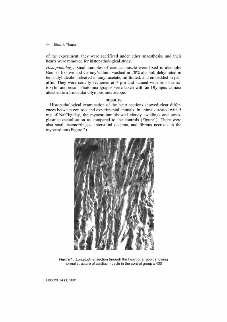
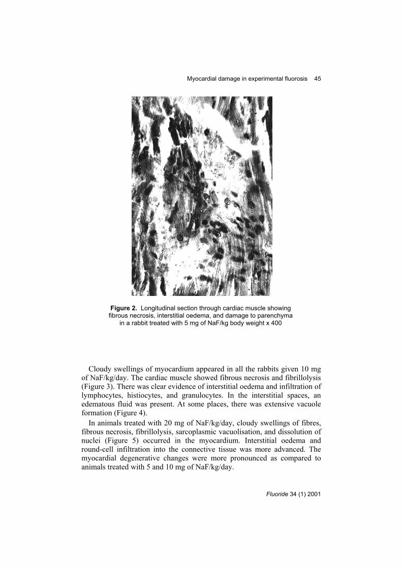
ences between controls and experimental animals. In animals treated with 5mg of NaF/kg/day, the myocardium showed cloudy swellings and sarco-plasmic vacuolisation as compared to the controls (Figure1). There werealso small haemorrhages, interstitial oedema, and fibrous necrosis in themyocardium (Figure 2).
Figure 1. Longitudinal section through the heart of a rabbit showingnormal structure of cardiac muscle in the control group x 400
Myocardial damage in experimental fluorosis 45
Fluoride 34 (1) 2001
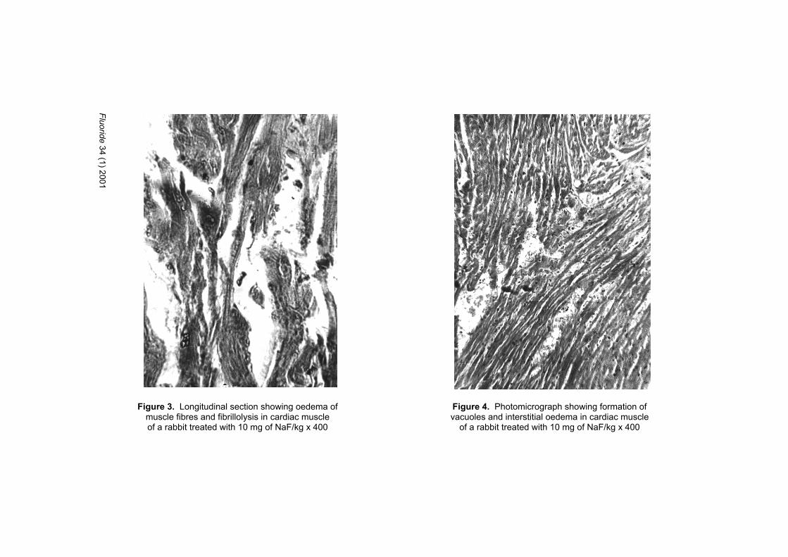
Cloudy swellings of myocardium appeared in all the rabbits given 10 mgof NaF/kg/day. The cardiac muscle showed fibrous necrosis and fibrillolysis(Figure 3). There was clear evidence of interstitial oedema and infiltration oflymphocytes, histiocytes, and granulocytes. In the interstitial spaces, anedematous fluid was present. At some places, there was extensive vacuoleformation (Figure 4).
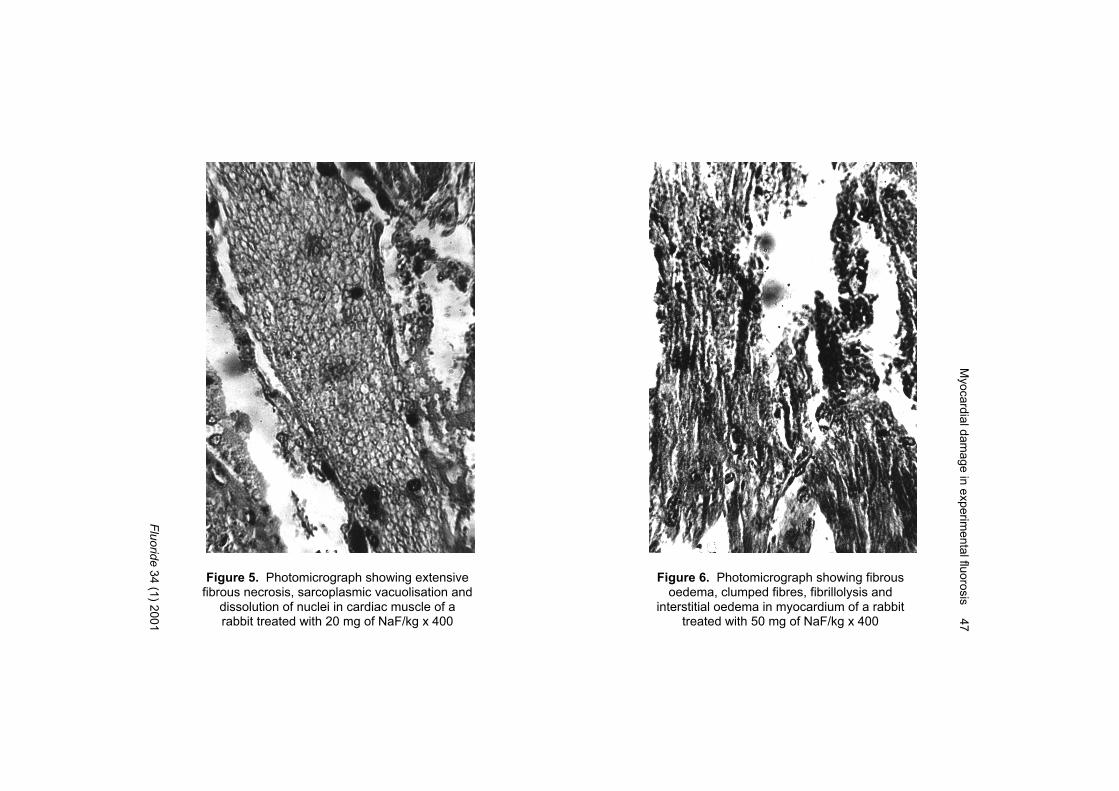
In animals treated with 20 mg of NaF/kg/day, cloudy swellings of fibres,fibrous necrosis, fibrillolysis, sarcoplasmic vacuolisation, and dissolution ofnuclei (Figure 5) occurred in the myocardium. Interstitial oedema andround-cell infiltration into the connective tissue was more advanced. Themyocardial degenerative changes were more pronounced as compared toanimals treated with 5 and 10 mg of NaF/kg/day.
Figure 2. Longitudinal section through cardiac muscle showingfibrous necrosis, interstitial oedema, and damage to parenchyma
in a rabbit treated with 5 mg of NaF/kg body weight x 400
Fluoride 34 (1) 2001
Figure 3. Longitudinal section showing oedema ofmuscle fibres and fibrillolysis in cardiac muscleof a rabbit treated with 10 mg of NaF/kg x 400
Figure 4. Photomicrograph showing formation ofvacuoles and interstitial oedema in cardiac muscle
of a rabbit treated with 10 mg of NaF/kg x 400
Myocardial dam
age in experimental fluorosis 47
Fluoride 34 (1) 2001
Figure 5. Photomicrograph showing extensivefibrous necrosis, sarcoplasmic vacuolisation and
dissolution of nuclei in cardiac muscle of arabbit treated with 20 mg of NaF/kg x 400
Figure 6. Photomicrograph showing fibrousoedema, clumped fibres, fibrillolysis and
interstitial oedema in myocardium of a rabbittreated with 50 mg of NaF/kg x 400
48 Shashi, Thapar
Fluoride 34 (1) 2001
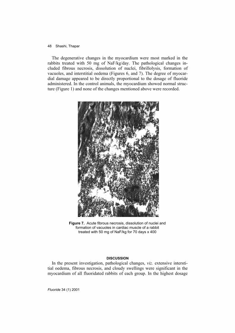
The degenerative changes in the myocardium were most marked in therabbits treated with 50 mg of NaF/kg/day. The pathological changes in-cluded fibrous necrosis, dissolution of nuclei, fibrillolysis, formation ofvacuoles, and interstitial oedema (Figures 6, and 7). The degree of myocar-dial damage appeared to be directly proportional to the dosage of fluorideadministered. In the control animals, the myocardium showed normal struc-ture (Figure 1) and none of the changes mentioned above were recorded.
DISCUSSIONIn the present investigation, pathological changes, viz. extensive intersti-
tial oedema, fibrous necrosis, and cloudy swellings were significant in themyocardium of all fluoridated rabbits of each group. In the highest dosage
Figure 7. Acute fibrous necrosis, dissolution of nuclei andformation of vacuoles in cardiac muscle of a rabbit
treated with 50 mg of NaF/kg for 70 days x 400
Myocardial damage in experimental fluorosis 49
Fluoride 34 (1) 2001
group (50 mg of NaF/kg/day), the disintegration of myocardial fibres, roundcell infiltration, and acute hemorrhages were more intensely marked.
Okushi10 reported cloudy swellings, infiltration with round cells, thicken-ing of adventitia, diffuse hemorrhages, and vacuolar and colloid degenera-tion of myocardium in rabbits fed 10-100 mg of NaF for 132 days. Thechanges were more prominent in the papillary muscles and on the insidewall of the myocardium than at the exterior cardiac wall. In rats treated with0.71 to 31.03 mg/kg of fluoride for one month, Takamori1 reported the pres-ence of cloudy swellings, vacuolar degeneration, round-cell infiltration, andhemorrhages and, in rabbits, regressive degeneration, cellular infiltration,hyperaemia, hemorrhages, and thickening of the vessel walls in the heartmuscle. Almost the same changes were observed in the cardiac muscle offluoride-intoxicated rabbits during the present investigation.
In humans, Okushi3 found a higher incidence of myocardial damage elec-trocardiographically and cardiac dilatation roentgenocardiacally in inhabi-tants of a high-fluoride zone, where the drinking water contained 6-13 ppmfluoride. In residents of a Japanese village, where the fluoride levels rangedbetween 0.5 and 6.2 ppm, Takamori et al11 reported myocardial damage anddilatation of the cardiac muscle and established a direct relationship betweenincreased myocardial damage and mottled enamel by means of electrocar-diograms. In acute fluorosilicate poisoning, Muller and Bock12 observed oe-dema of the myocardium with diapedesis of erythrocytes and leukocytes,along with acute right dilatation of the heart, in addition to a general venoushyperaemia. Degenerative changes consisting mainly of fragmentation ofmuscle fibres in the heart of a fluorotic patient were reported by Fasske,13
who noted the replacement of delicate sarcolemma by fibrous structure.During our experiments, fragmentation and degeneration of cardiac mus-
cle fibres were observed. However, the replacement of sarcolemma by fi-brous tissue did not occur. Pribilla14 noticed fibrous necrosis, dissolution ofnuclei, fibrillolysis, interstitial oedema, minute hemorrhages and infiltrationof histiocytes, lymphocytes, and granulocytes in the myocardium of patientswith acute silicofluoride intoxification. We also saw these same types of de-generative changes in cardiac muscle.
Since there are degenerative changes in myocardial fibres in acute poi-soning due to massive doses of fluoride, the effect of persistent minute dosesof fluoride intake over prolonged periods cannot be ignored. Variations inthe occurrence of abnormalities in the fluoridated myocardium may be ex-plained on the basis of differences in the dose and period of administration.The present investigation suggests that fluoride interferes with myocardialmetabolism, a condition observed by Iwase,14 who demonstrated histo-chemically that fluoride caused degenerative changes in the myocardium of

50 Shashi, Thapar
Fluoride 34 (1) 2001
rabbits administered 10-30 mg of NaF/kg/day orally for 15-169 days, fol-lowed by changes in the localisation of glycogen.
ACKNOWLEDGEMENTThe first author is grateful to the Indian Council of Medical Research,
New Delhi, India, for financial assistance.REFERENCES
1 Takamori T. The heart changes of growing albino rats fed on varied contentsof fluorine In: Gordonoff T, editor. The toxicology of fluorine symposium;1962 Oct 15-17; Bern, Switzerland. Basel/Stuttgart: Schabe; 1964. p. 125-9.
2 Leone NC, Geever EF, Moran NC. Acute and subacute toxicity studies of so-dium fluoride in animals. Public Health Rep 1956;71:459-67.
3 Okushi I. Changes of the heart muscle due to chronic fluorosis Part I. Electro-cardiogram and cardiac X-rays in inhabitants of high fluoride zone. ShikokuActa Med 1954;5:159-65.
4 Shashi; Singh JP, Thapar SP. Fluoride-induced pathological changes in trachea- an experimental study in rabbits. J Anatomical Soc India 1987;36:179-83.
5 Shashi, Thapar SP, Singh JP. Pulmonary damage caused by fluoride in rabbitsduring experimental fluorosis. Acta Pathol Microbiol Immunol Scand 1988;96:333-6
6 Shashi. Fluoride toxicity and muscular manifestations: Histopathological ef-fects in rabbit. Fluoride 1989;22:72-7.
7 Shashi. Histopathological changes in rabbit testes during experimental fluoro-sis. Folia Morphologica (Praha) 1990;38:63-5.
8 Shashi. Histopathological changes in rabbit ovary during experimental fluoro-sis. Indian J Pathol Microbiol 1990;33:113-7.
9 Shashi. Fluoride induced changes in rabbit lens during experimental fluorosis.J Environ Biol 1990;11:Suppl. 2:247-51.
10 Okushi I. Experimental studies on the effects of sodium fluoride upon theheart muscle of rabbits. Shikoku Acta Med 1954;5:238-45.
11 Takamori T, Miyanaga S, Kawahara H, Okushi I, Hirao M, Wakatsuki H.Electrocardiographic studies of the inhabitants in high fluoride districts. Toku-shima J Experimental Medicine 1956;3:50-3.
12 Müller W, Bock KD. Acute poisoning with hydrosilicofluoric acid. Med Klin1958;53:502-3.
13 Fasske E. Acute fluoride poisoning. Gordonoff T, editor. In: The toxicology offluorine symposium; 1962 Oct 15-17; Bern, Switzerland. Basel/Stuttgart:Schabe; 1964. p. 130-5.
14 Pribilla O. Four cases of acute silicofluoride intoxication, Clinical and patho-logical findings. Fluoride 1968;1:102-9.
15 Iwase T. Studies on the glycogen and phosphorylase variations in myocar-dium, skeletal muscle, liver in experimental fluorosis. Part I Influence of fluo-rine on glycogen. Shikoku Acta Med 1958;12:616-23.
——————————————————————Published by the International Society for Fluoride Research
Editorial Office: 727 Brighton Road, Ocean View, Dunedin 9051, New Zealand