Embed Size (px)
Citation preview

BASIC RESEARCH www.jasn.org
Histones from Dying Renal Cells Aggravate KidneyInjury via TLR2 and TLR4
Ramanjaneyulu Allam,*† Christina Rebecca Scherbaum,* Murthy Narayana Darisipudi,*Shrikant R. Mulay,* Holger Hägele,* Julia Lichtnekert,* Jan Henrik Hagemann,*Khader Valli Rupanagudi,* Mi Ryu,* Claudia Schwarzenberger,‡ Bernd Hohenstein,‡
Christian Hugo,‡ Bernd Uhl,§ Christoph A. Reichel,§ Fritz Krombach,§ Marc Monestier,|
Helen Liapis,¶ Kristin Moreth,** Liliana Schaefer,** and Hans-Joachim Anders*
*Medizinische Klinik und Poliklinik IV, Klinikum der Universität München, Munich, Germany; †Department ofBiochemistry, University of Lausanne, Epalinges, Switzerland; ‡Division of Nephrology, University of Dresden,Dresden, Germany; §Walter Brendel Centre of Experimental Medicine, University of Munich, Munich, Germany;|Temple Autoimmunity Center, Department of Microbiology and Immunology, Temple University School of Medicine,Philadelphia, Pennsylvania; ¶Department of Pathology and Immunology, Washington University School of Medicine,St. Louis, Missouri; and **Institut für Allgemeine Pharmakologie und Toxikologie, Klinikum der Goethe-Universität,Frankfurt am Main, Germany
ABSTRACTIn AKI, dying renal cells release intracellular molecules that stimulate immune cells to secrete proin-flammatory cytokines, which trigger leukocyte recruitment and renal inflammation.Whether the release ofhistones, specifically, from dying cells contributes to the inflammation of AKI is unknown. In this study, wefound that dying tubular epithelial cells released histones into the extracellular space, which directlyinteractedwith Toll-like receptor (TLR)-2 (TLR2) and TLR4 to induceMyD88, NF-kB, andmitogen activatedprotein kinase signaling. Extracellular histones also had directly toxic effects on renal endothelial cells andtubular epithelial cells in vitro. In addition, direct injection of histones into the renal arteries of micedemonstrated that histones induce leukocyte recruitment, microvascular vascular leakage, renal inflam-mation, and structural features of AKI in a TLR2/TLR4-dependent manner. Antihistone IgG, which neutral-izes the immunostimulatory effects of histones, suppressed intrarenal inflammation, neutrophil infiltration,and tubular cell necrosis and improved excretory renal function. In summary, the release of histones fromdying cells aggravates AKI via both its direct toxicity to renal cells and its proinflammatory effects. Be-cause the induction of proinflammatory cytokines in dendritic cells requires TLR2 and TLR4, these resultssupport the concept that renal damage triggers an innate immune response, which contributes to thepathogenesis of AKI.
J Am Soc Nephrol 23: 1375–1388, 2012. doi: 10.1681/ASN.2011111077
AKI involves a significant sterile inflammatoryresponse that contributes to the extent of tubularnecrosis and renal dysfunction.1,2 In this regard,postischemic AKI resembles ischemic injuries inother organs (e.g., in the heart during myocardialinfarction, in the brain during ischemic stroke, orin skeletalmuscles during limb ischemia).3 In all theseconditions, reperfusion of ischemic tissue is associ-ated with the production of reactive oxygen speciesthat activate tissue cells to secrete proinflammatorycytokines and chemokines. This process recruits neu-trophils and activated macrophages that exaggerate
Received November 15, 2011. Accepted May 2, 2012.
R.A., C.R.S., and M.N.D. contributed equally to this work.
Published online ahead of print. Publication date available atwww.jasn.org.
Correspondence: Dr. Hans-Joachim Anders, Medizinische Klinikund Poliklinik IV, Klinikum der Universität München, Pettenkoferstr.8a, D-80336Munich, Germany. Email: [email protected]
Copyright © 2012 by the American Society of Nephrology
J Am Soc Nephrol 23: 1375–1388, 2012 ISSN : 1046-6673/2308-1375 1375

organ inflammation, tissue injury, and malfunction.1 Duringthe past decade it has become evident that the initiation of thissterile inflammatory response is largely based on the activationof Toll-like receptors (TLRs). TLRs are germline encoded pat-tern-recognition receptors that have important roles in innateimmunity against all sorts of pathogens by recognizing variouspathogen-associated molecular patterns.
In a major breakthrough in the understanding of nonin-fectious types of inflammation, TLRs were found to also rec-ognize endogenous damage-associated molecular patterns(DAMPs), which have identical properties to activate innateimmunity and tissue inflammation as pathogens.3 This con-cept was first established by showing that necrotic cells triggercytokine production and neutrophil recruitment viaTLRs andits dominant signaling adaptor MyD88.4,5 Subsequent workhas identified several endogenous intracellular molecules thathave the potential to activate TLR signaling and cytokine secre-tion, such as high-mobility group protein (HMG) B1 (TLR2,TLR4, and the receptor for advanced glycation end products[RAGE]), as well as hypomethylated CpG-DNA (TLR9).4 Insupport of this concept, mice deficient for TLR2, TLR4, orMyD88 or mice treated with HMGB1-blocking antibodiesare protected from postischemic intrarenal inflammation,which largely prevents tubular cell necrosis and acute renal fail-ure. 6–9 The same evidence is available for ischemia-reperfusioninjuries in other organs, such as the liver,10 the heart,11,12 and thebrain.13 Our work reported here is based on the assumption thatadditional intracellular molecules that can act as DAMPs andsense renal tissue damage to the immune system remain to bediscovered.14
Histones are a group of nuclear proteins that form hetero-octamers to wind up the double-stranded DNA to formchromatin as well as chromosomes. Histones are released fromdying neutrophils during bacterial infections for host defense,the so-called neutrophil extracellular traps (NETs). 15,16 Thebactericidal effect of extracellular histones also damages selftissues.17 For example, histone release directly contributes tofatal outcomes in murine endotoxinemia by activating andkilling vascular endothelial cells.18 We therefore speculatedthat chromatin release from dying renal cells would shuttlehistones in the extracellular space, where they act as DAMPs byactivating one or more pattern recognition receptors. In ad-dition, we speculated that this process would contribute tosterile inflammation during postischemic kidney injury aswell as to septic AKI.
Because of the essential role of histones for chromatin as-sembly, histone-deficient mice could not be generated to testour concept experimentally. In fact, it was necessary to neutralizehistones specifically in the extracellular compartment, whichbecame possible by using the same histone-specific IgG andcontrol IgG that have been used by others for similar in vitroand in vivo studies.18Herewe report that dying tubular epithelialcells release histones into the extracellular space, thereby con-tributing to postischemic and septic kidney inflammation andinjury. Furthermore, we report that extracellular histones
directly activate and potentially kill renal endothelial and tubu-lar cells. In addition, both TLR2 and TLR4 are required to trans-late histone recognition into MyD88 signaling and the secretionof proinflammatory mediators.
RESULTS
Histones Are Released from Necrotic Tubular EpithelialCellsOur concept of histones being an endogenous danger signal toactivate renal inflammation is based on the assumption thatdying renal cells release histones into extracellular compart-ments. In fact, immunoblotting for histones revealed a strongpositivity for histones in cell culture supernatants of hydrogenperoxide–treated tubular epithelial cells similar to cells killedby repetitive freeze-thawing after 24 hours (Figure 1A).
Extracellular Histones Kill Renal Endothelial Cells andTubular Epithelial CellsExtracellular histones have been reported to promote fatalsepsis bydirectlydamaging thepulmonarymicrovasculature.18
To test a putative toxic role of histones on renal cells, we in-cubated murine renal endothelial cells or tubular epithelialcells with a total histone preparation. Endothelial and tubularcell viability was reduced in a dose-dependent manner within24 hours, whereas agonists for TLR2, -4, and -9 had no effect(Figure 1B). Next we analyzed the supernatants of the endo-thelial cell experiments by flow cytometry for nonadherentannexin V/propidium iodine–double-positive (apoptotic)cells and annexin V–negative/ propidium iodine–positive (ne-crotic) cells. Histone exposure increased both types of cells in adose-dependent manner within 24 hours (Figure 1D). Thus,extracellular histones negatively affect the viability of renalendothelial and tubular cells.
Extracellular Histones Increase Leukocyte Adhesionand Microvascular PermeabilityWe used in vivo microscopy on mouse cremaster muscles tostudy whether histone exposure induces leukocyte recruit-ment and enhances microvascular permeability. After 6 hoursof local histone application, there was a significant elevation inleukocyte intravascular adherence and transendothelial migra-tion compared with controls (Figure 2, A–D). Immunostainingof cremaster muscles identified 90.0%62.6% of transmigratedCD45+ cells (total leukocytes) as Ly-6G+ cells (neutrophils)and the rest as F4/80+ (monocytes/macrophages). In addition,histone exposure significantly increased leakage of FITC dex-tran into the interstitial compartment (Figure 2, E and F).When histones were preincubated with activated protein C,an enzyme that digests histones (Figure S1A), the histone-related effects were abrogated (Figure 2, B, C, and E). Leuko-cyte recruitment and microvascular permeability significantlyincrease when the microvasculature is exposed to extracellularhistones.
1376 Journal of the American Society of Nephrology J Am Soc Nephrol 23: 1375–1388, 2012
BASIC RESEARCH www.jasn.org
Histone Injection into the Renal Artery Induces RenalCell NecrosisNext we sought to study the effects of extracellular histones onthe kidney. Because intravenous histone injection kills miceimmediately via severe alterations of the pulmonary micro-vasculature,18 we injected histones directly into the left renalartery of anesthetized mice (Figure 3A). Unilateral histone in-jection led to widespread necrosis of the renal cortex and outermedulla and massive neutrophil infiltrates after 24 hours,whereas the contralateral kidney remained unaffected (Figure3B). Electron microscopy displayed that LPS injection alonehad already led to dilation of peritubular capillaries and in-terstitial edema but that histone injection aggravated vasculardilation and induced condensation of microvascular endothelialcell chromatin, implying apoptosis (Figure 3C).Histone-inducedrenal cell necrosis was associatedwith an increase in renalmRNAexpression ofmultiple proinflammatory mediators, such as IL-6,
TNF-a, and inducible nitric oxide synthase(Figure 3D). When the histones were pre-digested with activated protein C (Supple-mental Figure 1A) or when the identicalexperiment was conducted in Tlr2/4-deficient mice, renal necrosis, neutrophilrecruitment, and cytokine expression weresignificantly reduced compared with resultsseen when histone was injected into wild-type mice (Figure 3, B and C and Supple-mental Figure 1B). This histone effect wasabsent without a systemic intravenous in-jection of a nontoxic dose of endotoxin (1.0mg/kg body weight) 12 hours before his-tone injection (Supplemental Figure 2),which was necessary to induce renal TLR2and TLR4 expression (Figure 3E). Previousstudies had already excluded any effect dueto the injection procedure itself.19 Thus,extracellular histones massively aggravaterenal inflammation and renal cell necrosis.
Histone Neutralization ReducesEndotoxin-Induced AKIDo extracellular histones also contribute toseptic AKI? To address this issue, we used aneutralizing antibody to histones that waspreviously used to establish the functionalcontribution of extracellular histones in le-thal endotoxemia in mice.18 Mice were in-jectedwith 20mg/kg antihistone IgG or with20 mg/kg control IgG 2 hours before the in-traperitoneal injection of LPS (10 mg/kg).Antihistone IgG administration significantlyreduced serum creatinine levels at 12 hoursafter LPS injection (Figure 4). This reductionwas associated with a significant decrease inseptic tubular injury, as assessed by semi-
quantitativemorphometry (Figure 4). In addition, renal neutro-phil counts were significantly reduced with histone blockade(Figure 4). We conclude that extracellular histones contributeto AKI in murine endotoxemia.
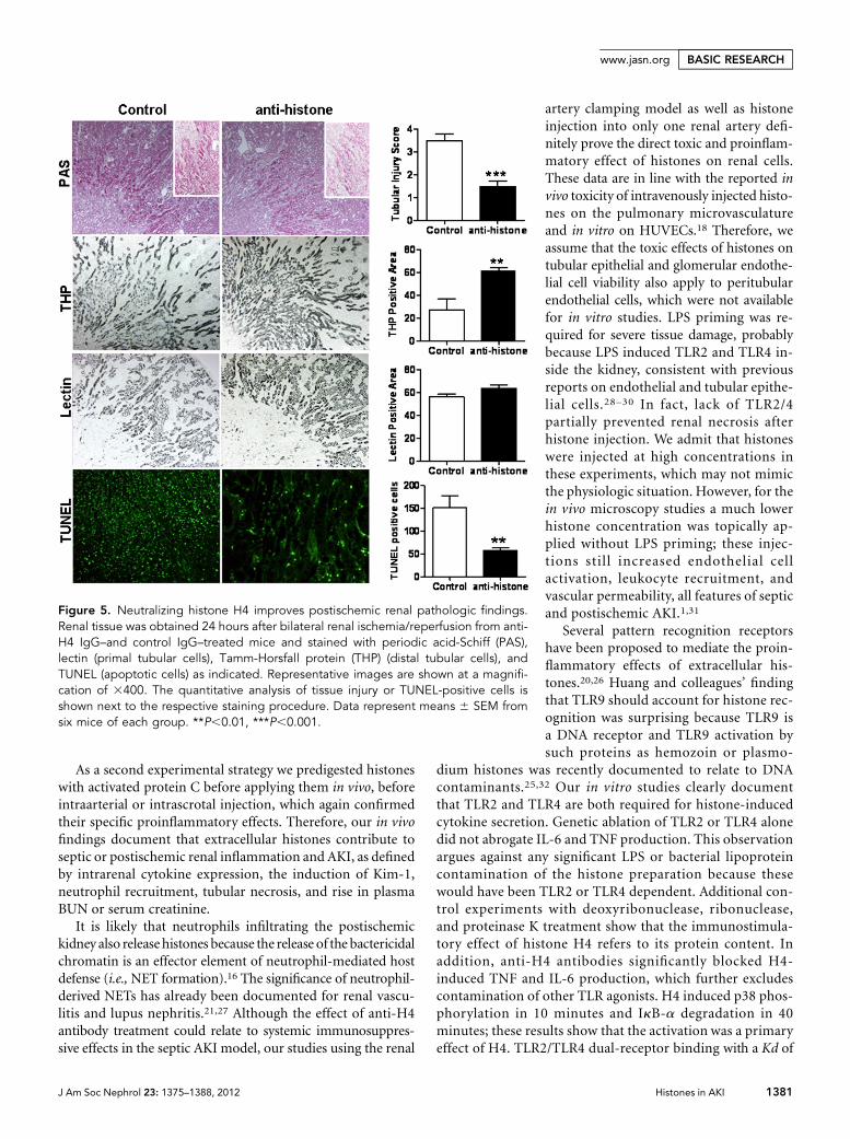
Histone H4 Neutralization Reduces Renal Ischemia-Reperfusion InjuryDo extracellular histones also contribute to postischemic AKI?Injection of antihistone IgG immediately after bilateral renal-artery clamping prevented postischemic tubular damage 24hours after surgery compared with treatment with control IgG(Figure 5). This finding was quantified by semiquantitativescoring using a composite score of brush border loss, tubularcell flattening, tubular cell necrosis, and granular cast forma-tion. Distal tubules were protected by antihistone IgG, as de-termined by staining for Tamm-Horsfall protein, whereasproximal tubular cells, identified with tetragonolobus lectin
Figure 1. Dying renal cells release histones into extracellular compartments. (A) Ne-crosis was induced in primary tubular epithelial cells as described in the ConciseMethods section. Histone H4 was detected in necrotic supernatants by using anti-H4antibody. Recombinant histone H4 was loaded as a positive control. (B) Renal endo-thelial cell proliferation was determined in a period of 24 hours by bioluminescenceassay, as described in the Concise Methods. Data represent mean OD 6 SEM of threeexperiments measured at a wavelength of 492 nm. (C and D) Renal endothelial cellswere stimulated with CpG, 6 mg/ml; camptothecin (CPT), 10 mM; or histones. Dosesare given in mg/ml. Results show flow cytometry from floating cells in culture super-natants. Data represent the mean of total positive cell numbers of positive cells ofthree independent experiments. PI, propidium iodine.
J Am Soc Nephrol 23: 1375–1388, 2012 Histones in AKI 1377
www.jasn.org BASIC RESEARCH

staining, were not significantly preserved (Figure 5). Thenumbers of terminal deoxynucleotidyl transferase–mediateddigoxigenin-deoxyuridine nick-end labeling (TUNEL)–positiverenal cells were significantly reduced in antihistone-treatedmice (Figure 5), indicating less tubular cell apoptosis in thepostischemic kidney. This improvement in structural damagewas associated with significantly reduced blood urea nitrogenlevels 24 hours after surgery (117625 versus 5862; P=0.02),suggesting that antihistone IgG prevented loss of renal excre-tory function during postischemic AKI. Extracellular chroma-tin has been reported to trigger tissue inflammation;18,20,21
hence, we assessed the intrarenal mRNA ex-pression of proinflammatory cytokines inboth treatment groups. Antihistone IgGsignificantly reduced the renal mRNA levelsof the tubular injury marker KIM-1, whichwas associated with lower mRNA levels ofthe proinflammatory cytokines IL-6 and IL-12, the intercellular adhesionmolecule, andthe neutrophil-attracting chemokineCXCL2 (Figure 6A). This finding was con-sistent with the lower numbers of neutro-phils that infiltrated the renal interstitiumin antihistone-treated compared with con-trol IgG–treatedmice, especially in the areasaround necrotic tubuli (Figure 6B). To-gether, neutralizing histones reduce theinduction of cytokines and chemokines,neutrophil recruitment, tubular injury,and renal failure in the postischemic kidney.
Histones Activate the Secretion ofProinflammatory CytokinesHaving shown that damaged tubular cells re-lease histones and that histone neutralizationreduces postischemic sterile inflammationand kidney damage, we speculated thatextracellular histones directly induce theexpression of proinflammatory cytokines.In fact, a total histone preparation inducedthe secretion of TNF and IL-6 in bonemarrow–derived dendritic cells (BMDCs)(Figure 7A). Histones are protein comple-xes formed by 1H, H2A, H2B, H3, and H4proteins. Hence, we tested the individualproteins to induce inflammatory cytokinesin BMDCs. All histone proteins inducedTNF and IL-6 production (Figure 7B).However, there were differences in termsof induction in cytokine production witheach protein. To further study the signalingmechanism, we used histone H4 as proto-type. H4-induced NF-kB and mitogen ac-tivated protein (MAP) kinase activation, asshown by IkB-a degradation and p38 phos-
phorylation as readout for NF-kB and MAP kinase activation(Figure 7C). Dose- and time-dependent studies revealed thatH4-induced cytokine production was most prominent at aconcentration of 10 mg for IL-6 and TNF and appeared as earlyas 3 hours after H4 stimulation (Supplemental Figure 2, A andB). LPS contamination did not account for this response becausepreincubation with polymyxin B did not affect H4-inducedcytokine induction but completely blocked LPS-induced cyto-kine induction (Figure 7D). The immunostimulatory effect ofH4 was also unaltered by preincubation with deoxyribonucle-ase or ribonuclease, excluding immunostimulatory nucleic
Figure 2. In vivo microscopy of cremaster muscles. In vivo microscopy was performedon cremaster muscle postcapillary venules as described in the Concise Methodssection. Six mice in each group were treated with intrascrotal injections of vehicle ortotal histones as indicated. Leukocyte rolling (A), firm adhesion (B), and trans-endothelial migration (C) were determined 6 hours after injection. Histone+APCmeans that histone had been pre-incubated with activated protein C (APC) beforeinjection. Data are means 6 SEM. #P,0.05 versus control, *P,0.05 versus histones.(D) Representative images illustrate the increase in leukocyte adhesion and trans-endothelial migration after histone challenge (right) versus control (left). (E) Micro-vascular FITC-dextran leakage was determined 6 hours after injection. Data aremeans 6 SEM. #P,0.05 versus control, *P,0.05 versus histones. (F) Representativeimages illustrate the increase in vascular dextran permeability after histone challenge(right) versus control (left).
1378 Journal of the American Society of Nephrology J Am Soc Nephrol 23: 1375–1388, 2012
BASIC RESEARCH www.jasn.org

acid contaminations (Figure 7E). Only H4 digestion with pro-teinase K (Figure 7E) or activated protein C (SupplementalFigure 3) prevented cytokine induction, indicating that thepeptide content of H4 was required for its immunostimulatory
effect. Supporting these results, antihistoneantibodies significantly reducedH4-inducedTNF and IL-6 production (Figure 7F). Thus,extracellular histones are potent inducers ofproinflammatory cytokine secretion.
Both TLR2 and TLR4 Are Required forthe Recognition of Histone-H4Which pattern recognition receptors me-diate these biologic effects of extracellularH4? We hypothesized that a TLR might beresponsible for H4 recognition and signal-ing. All TLRs use MyD88 (myeloid differ-entiation primary response gene 88) as anadaptor molecule to induce signaling,except for TLR3, which signals throughToll/IL-1 receptor domain–containingadapter-inducing IFNb).22 To test our hy-pothesis, we investigated H4 activation inMyd88- and Trif-deficient BMDCs. H4-induced TNF and IL-6 production com-pletely abrogated in Myd88-deficientBMDCs (Figure 8, A and B). To test whichMyD88-dependent TLR is responsible forH4 recognition, we exposed H4 to BMDCsfrom respective knockout mice. Deficiencyof both TLR2 and TLR4 completely pre-vented the activation of TNF and IL-6(Figure 5, C–E), whereas individual gene de-letions of Tlr1, Tlr2, Tlr4, Tlr6, and Cd14 didnot prevent cytokine production (Figure 8,C–E). The observation that both TLR2 andTLR4 can signal for H4-induced cytokineproduction is similar to what has been re-ported for other DAMPs, such as high-mobility group protein B19 or biglycan.23
HMGB1 signals throughRAGE,24 but we ob-servedH4 signaling to beRAGE-independent(Figure 8F). We did not investigate TLR7and TLR9 because endosomal acidifi-cation with chloroquine significantly ab-rogated CpG-induced TNF productionbut not H4 (Supplemental Figure 4). Inaddition to H4, other histone proteinsalso showed TLR2/TLR4 dependency(Supplemental Figures 5 and 6). Thus, ex-tracellular histones trigger proinflamma-tory cytokines via both TLR2 and TLR4,which activates the MyD88 signalingpathway.
Histone-H4 Directly Interacts with TLR2 and TLR4To checkwhetherH4physically interactswith TLR2 andTLR4,we performed microscale thermophoresis binding assay. Thebinding of NT-647 fluorescence-labeled H4 to recombinant
Figure 3. Histone injection into the renal artery. (A) Twelve hours after intraperitoneal LPSinjection (1 mg/kg body weight), the abdominal aorta and the left renal artery was pre-pared and a microcannula was placed into the left renal artery (left) for histone injection.The puncture site was mounted with glue before closure of the wound (right). (B) Rep-resentative images of periodic acid-Schiff (PAS) stainings and for neutrophils are shown ata magnification of 350. The quantitative analysis of tissue injury and neutrophil numbersper high-power field are shown on the right. (C) Dilated peritubular capillaries and in-terstitial edema are illustrated by transmission electron microscopy. Endothelial cells withcondensed nuclear chromatin seem to undergo apoptosis. Original magnification37500.(D) Total kidney mRNA levels of TNF-a, IL-6, and inducible nitric oxide synthetase (iNOS)were determined in LPS-primed and histone-injected left kidneys. Histone preincubation(before injection) with recombinant activated protein C (APC) reduced intrarenal cytokineexpression. (E) Renal mRNA levels of TLR2 and TLR4 with and without LPS priming. 18srRNA levels were used as internal control. Data represent means 6 SEM from nine mice.†P,0.05, **P,0.01, ***P,0.001 versus LPS (B and C) or saline control (D).
J Am Soc Nephrol 23: 1375–1388, 2012 Histones in AKI 1379
www.jasn.org BASIC RESEARCH

human TLR2 (Figure 9A) and TLR4/MD2 complex (Figure9C)was analyzed bymicroscale thermophoresis. To determinethe affinity of the binding reaction, a titration series of TLR2(Figure 9A) and TLR4/MD2 (Figure 9C) proteins were per-formed, while fluorescence-labeled H4 was kept at a constantconcentration of 5 nM. The change in the thermophoreticsignal of H4 suggested a Kd of 4.261.7 nM (n=3) for TLR2(Figure 9A) and 6.063.7 nM (n=3) for TLR4/MD2 (Figure9C). In contrast, thermophoresis of NT-647 fluorescence-labeled albumin (5 nM, negative control) tested for its bindingto TLR2 (Figure 9B) and TLR4/MD2 (Figure 9D) in the same
experimental setting, showed no depen-dence on TLR2 or TLR4/MD2 concentra-tion. So these results show that H4 inducesproinflammatory cytokines by directly in-teracting with TLR2 and TLR4. To furthervalidate our results in vivo, we injected H4protein, 20mg/kg, intravenously into wild-type and TLR2/TLR4 double-knockoutmice and measured cytokine productionin the plasma after 6 hours. H4-inducedIL-6 and TNF production observed inwild-type mice was abrogated with TLR2/TLR4 deficiency (Figure 9E). None of thesemice displayed a renal phenotype in termsof elevated serum creatinine levels, tubulardamage, or renal neutrophil infiltration(data not shown). These results show thathistone H4 activates TLR2 and TLR4 to in-duce cytokine production in mice.
DISCUSSION
Histones wind up DNA and regulate genetranscription inside the nucleus, but in theextracellular space histones elicit toxic andproinflammatory effects.17 Hence, we hadhypothesized that histone release fromdamaged tubular epithelial cells would pro-mote renal inflammation in a DAMP-likefashion, implicating an interaction withdistinct pattern recognition receptors.Our studies confirm this concept and iden-tify TLR2 and TLR4 to translate histonerecognition into cytokine secretion in aMyD88-dependent manner.
Acute tubular necrosis implies the re-lease of intracellular molecules into theextracellular space. This has also been docu-mented for nuclear particles, such as thenucleoprotein HMGB1,14,25 and our in vitrostudies demonstrate histone release fromdying tubular cells, (such as occurs with ex-posure to oxidative stress). Elegant studies
have found that HMGB1 acts as a DAMP in sterile renal in-flammation by inducing renal ischemia-reperfusion injuryin mice treated with Hmgb1-blocking antibodies.9 We usedthe same experimental strategy by applying specific histone-neutralizing antibodies that block histones in extracellularcompartments in vivo as well as in vitro. Antihistone anti-bodies have already been used to effectively deplete extracellu-lar histones in several in vivo studies, which, for example,protected from fatal endotoxinemia or toxic liver injury.18,26
The effects of this antibody could relate to its specific bindingproperties to histones compared with the effects of control IgG.
Figure 4. Neutralizing histone H4 protects from septic AKI. (A) Serum creatinine wasdetermined 12 hours after intraperitoneal LPS injection. Two hours before, groups ofmice had received anti-histone H4 or control IgG. Data represent means 6 SEM.***P,0.001. (B) Renal tissue from mice of both groups was stained with periodic acid-Schiff (PAS) or for neutrophils. Representative images are shown at magnifications asindicated. Data represent means 6 SEM from six mice of each group. hpf, high-powerfield.
1380 Journal of the American Society of Nephrology J Am Soc Nephrol 23: 1375–1388, 2012
BASIC RESEARCH www.jasn.org
As a second experimental strategy we predigested histoneswith activated protein C before applying them in vivo, beforeintraarterial or intrascrotal injection, which again confirmedtheir specific proinflammatory effects. Therefore, our in vivofindings document that extracellular histones contribute toseptic or postischemic renal inflammation and AKI, as definedby intrarenal cytokine expression, the induction of Kim-1,neutrophil recruitment, tubular necrosis, and rise in plasmaBUN or serum creatinine.
It is likely that neutrophils infiltrating the postischemickidney also release histones because the release of the bactericidalchromatin is an effector element of neutrophil-mediated hostdefense (i.e., NET formation).16 The significance of neutrophil-derived NETs has already been documented for renal vascu-litis and lupus nephritis.21,27 Although the effect of anti-H4antibody treatment could relate to systemic immunosuppres-sive effects in the septic AKI model, our studies using the renal
artery clamping model as well as histoneinjection into only one renal artery defi-nitely prove the direct toxic and proinflam-matory effect of histones on renal cells.These data are in line with the reported invivo toxicity of intravenously injected histo-nes on the pulmonary microvasculatureand in vitro on HUVECs.18 Therefore, weassume that the toxic effects of histones ontubular epithelial and glomerular endothe-lial cell viability also apply to peritubularendothelial cells, which were not availablefor in vitro studies. LPS priming was re-quired for severe tissue damage, probablybecause LPS induced TLR2 and TLR4 in-side the kidney, consistent with previousreports on endothelial and tubular epithe-lial cells.28–30 In fact, lack of TLR2/4partially prevented renal necrosis afterhistone injection. We admit that histoneswere injected at high concentrations inthese experiments, which may not mimicthe physiologic situation. However, for thein vivo microscopy studies a much lowerhistone concentration was topically ap-plied without LPS priming; these injec-tions still increased endothelial cellactivation, leukocyte recruitment, andvascular permeability, all features of septicand postischemic AKI.1,31
Several pattern recognition receptorshave been proposed to mediate the proin-flammatory effects of extracellular his-tones.20,26 Huang and colleagues’ findingthat TLR9 should account for histone rec-ognition was surprising because TLR9 isa DNA receptor and TLR9 activation bysuch proteins as hemozoin or plasmo-
dium histones was recently documented to relate to DNAcontaminants.25,32 Our in vitro studies clearly documentthat TLR2 and TLR4 are both required for histone-inducedcytokine secretion. Genetic ablation of TLR2 or TLR4 alonedid not abrogate IL-6 and TNF production. This observationargues against any significant LPS or bacterial lipoproteincontamination of the histone preparation because thesewould have been TLR2 or TLR4 dependent. Additional con-trol experiments with deoxyribonuclease, ribonuclease,and proteinase K treatment show that the immunostimula-tory effect of histone H4 refers to its protein content. Inaddition, anti-H4 antibodies significantly blocked H4-induced TNF and IL-6 production, which further excludescontamination of other TLR agonists. H4 induced p38 phos-phorylation in 10 minutes and IkB-a degradation in 40minutes; these results show that the activation was a primaryeffect of H4. TLR2/TLR4 dual-receptor binding with a Kd of
Figure 5. Neutralizing histone H4 improves postischemic renal pathologic findings.Renal tissue was obtained 24 hours after bilateral renal ischemia/reperfusion from anti-H4 IgG–and control IgG–treated mice and stained with periodic acid-Schiff (PAS),lectin (primal tubular cells), Tamm-Horsfall protein (THP) (distal tubular cells), andTUNEL (apoptotic cells) as indicated. Representative images are shown at a magnifi-cation of 3400. The quantitative analysis of tissue injury or TUNEL-positive cells isshown next to the respective staining procedure. Data represent means 6 SEM fromsix mice of each group. **P,0.01, ***P,0.001.
J Am Soc Nephrol 23: 1375–1388, 2012 Histones in AKI 1381
www.jasn.org BASIC RESEARCH

4.261.7 nM and 6.063.7 nM is remarkable but consistentwith data for other endogenous DAMPs, such as HMGB1or biglycan.32,33 Obviously, TLR2-TLR4 cooperation sup-ports the recognition of these endogenous proteins andactivates the MyD88 signaling pathway. MyD88 signalingfinally activates NF-kB–dependent cytokines such as IL-6or TNF. Theoretically, it could be possible that other TLRsor RAGE also contribute to histone recognition in a similarmanner, which could not be detected in single knockoutcells.
Myd88-deficient mice lack postischemic renal sterile in-flammation,6–8 a phenomenon that can now be explained bylack of immune recognition of histones or HMGB1,9 and po-tentially other DAMPs that remain to be identified. Otherstudies have already documented that deletion of TLR2 orTLR4 is sufficient to significantly reduce postischemic kidneyinjury. 6–8 These data suggest that histone recognition is notthe predominant element of intrarenal danger signaling in
the postischemic kidney. TLR2 andTLR4 areboth expressed on intrarenal immune cellsaswell as on renal parenchymal cells. 6–8,28,29
Studies with TLR2/TLR4 bone marrowchimeric mice revealed that postischemicdanger signaling via TLR2/TLR4 domi-nates in renal parenchymal cells.6,8Thefact that histone neutralization protecteddistal but not proximal tubular epithelialcells in the postischemic kidney shouldrelate to the fact that only distal tubularcells upregulate TLR2 and TLR4 in thepostischemic kidney.30 Furthermore, wehave recently shown that postischemiccytokine induction in intrarenal dendriticcells is suppressed by the constitutively ex-pressed single immunoglobulin IL-1–relatedreceptor as well as the induction of IFN-related factor-4, two inhibitors of TLRsignaling.34–36
This study identifies extracellular histo-nes as mediators of postischemic and septicAKI. Histones are released from dyingtubular epithelial cells and act as DAMPs,which require TLR2 and TLR4 for theinduction of proinflammatory cytokines.Thus, renal cell injury triggers renal inflam-mation because the innate immune systemtranslates the recognition of dead cell re-leases, such as histones, into inflammationvia the same receptors that recognize bac-terial factors during infection. Histoneneutralization might be a novel option tosuppress immunopathology after tissuedamage.
CONCISE METHODS
Animal StudiesC57BL/6J mice, 6–12 weeks old, genetically deficient (.F6) in Tlr1,37
Tlr2,22 Tlr4,22 Cd14,38 Tlr6,37 Rage,39 Myd88,40 or Trif41 have been
described. Tlr2- and Tlr4-deficient mice were crossed to produce
Tlr2/4 double-deficient mice. The respective genotype was assured
by PCR from tail-tip DNA. Histones were injected (10 mg/kg body
weight) into the left renal artery 12 hours after a single injection of
LPS, 1 mg/kg (Sigma-Aldrich, Steinheim, Germany), at a total vol-
ume of 200 ml, as described elsewhere.19 In one experiment, the
histones were predigested with 500 nM activated protein C (Sigma-
Aldrich). Both kidneys were harvested 24 hours later. Septic kidney
injury was induced by injection with antihistone IgG, 20 mg/kg, or
control IgG 2 hours before LPS injection (10 mg/kg intraperitone-
ally). Renal ischemia-reperfusion injury was induced under general
anesthesia as described elsewhere.36 In brief, both renal pedicles were
clamped for 30 minutes with microaneurysm clamps (Medicon,
Figure 6. Assessment of postischemic renal inflammation. (A) Total RNA was ex-tracted from kidneys from anti-H4 IgG– and control IgG–treated mice. KIM-1 andcytokine mRNA expression levels were determined by real-time PCR and expressedas mean of the ratio 18S rRNA6 SEM; ‡P,0.05 versus control. (B) Renal sections werestained for neutrophils, and representative images are shown at a magnification of3400. The quantitative analysis of interstitial neutrophils is shown next to the re-spective staining procedure. Data represent means 6 SEM from six mice of eachgroup. **P,0.01.
1382 Journal of the American Society of Nephrology J Am Soc Nephrol 23: 1375–1388, 2012
BASIC RESEARCH www.jasn.org

Tuttlingen, Germany) via 1-cm flank incisions. Body temperature
was continuously measured with a rectal probe and maintained at
36–37°C throughout the procedure by placing the mice on a heating
pad. After clamp removal, the kidney was inspected for restoration of
blood flow before the wound was closed with standard sutures. Im-
mediately after surgery or 2 hours after LPS injection, mice were
intraperitoneally injected with anti-histone antibody BWA3, 20 mg/
kg,18 or control IgG (Abcam, Cambridge, United Kingdom). Mice
were euthanized 24 hours after reperfusion or
12 hours after LPS injection, and pieces from
kidneys were harvested for further processing.
All experiments were performed according to
German animal protection laws and had been
approved by the local government authorities.
Assessment of Kidney Inflammationand InjuryKidneys were embedded in paraffin, and 2-mm
sections were used for periodic acid-Schiff
stains and immunostaining as described else-
where.42 Postischemic tubular injury was scored
by assessing the percentage of tubules in the cor-
ticomedullary junction that displayed tubular
cell flattening, cell necrosis, loss of brush border,
and luminal cast formation, as follows: 0, none;
1, #10%; 2, 11%–25%; 3, 26%–45%; 4, 46%–
75%; and 5, .76%. Septic tubular injury was
scored in a same way, but ballooning and vacuo-
lization of tubular cells were also considered as
tubular damage markers.43 For histochemistry
we used biotinylated Lotus tetragonolobus lec-
tin stain (Vector Labs, CA), Tamm-Horsfall pro-
tein stain (Santa Cruz Biotechnology, Inc., Santa
Cruz, CA), rat antimouse neutrophils (Serotec,
Oxford, United Kingdom), and the TUNEL kit
(Roche, Mannheim, Germany) to quantify ap-
optotic cells. To count interstitial cells, 10 corti-
cal high-power fields (4003) were analyzed.44
BUN and creatinine were measured using urea
or creatinine FS kits (DiaSys Diagnostic Sys-
tems, Holzheim, Germany) according to the
manufacturer’s protocols.
In vitro StudiesMouse renal endothelial cells were generated as
recently described elsewhere.45 Their prolifera-
tion was determined after 24 hours using Cell-
Titer 96 Cell Proliferation Assay (Promega,
Madison, WI) reading absorbance at 492 nm.
BMDCswere generated by established protocols
as described elsewhere.46 Necrotic cell superna-
tants were prepared frommouse tubular cells by
repeated freezing and thawing or hydrogen
peroxide (H2O2; 1mM) treatment for 24 hours.
RPMI 1640 GlutaMAX-I medium (Invitrogen,
Carlsbad, CA) was supplemented with 10% (vol/vol) FBS (Biochrom
AG, Berlin, Germany), 1% of penicillin and streptomycin (PAA
Laboratories GmbH, Pasching, Austria). OptiMEM reduced-serum
medium was from Invitrogen. We purchased ATP; ultrapure LPS
(from Escherichia coli strain K12); pI:C RNA; Pam3Cys; CpG
(InvivoGen, San Diego, CA); poly(dA-dT)cpoly(dT-dA) sodium
salt; crude LPS (E. coli serotype 0111:B4); N-acetyl-L-cysteine (Sigma-
Aldrich, St. Louis,MO); cytochalasinD; latrunculin B (Enzo Lifesciences
Figure 7. Histones activate TNF and IL-6 cytokine production. (A and B) TNF and IL-6ELISA of supernatants from mouse BMDCs stimulated for 6 hours with total histones(30 mg/ml) and individual histones (30 mg/ml) or LPS (1 mg/ml). (C) BMDCs were stimu-lated with H4 at the indicated time points. Cell lysates were immunoblotted and probedfor the indicated proteins. (D) BMDCs were stimulated for 6 hours with H4 and LPS in thepresence or absence of polymyxin B treatment. Supernatants were analyzed for TNF andIL-6 by ELISA. (E) BMDCs were stimulated for 6 hours with H4 or pretreated H4 withdeoxyribonuclease, ribonuclease, and proteinase K. Supernatants were analyzed for TNFand IL-6 by ELISA. (F) BMDCs were stimulated for 6 hours with H4 in the presenceor absence of anti-H4 antibodies (H4 Ab) or control IgG antibodies. Supernatants wereanalyzed for TNF and IL-6 by ELISA. In A, B, and D–F, the data represent the mean 6 SDof three independent experiments. ¶P,0.05 by t test. Data shown in part C were repeatedtwo times. Related data are presented in Supplemental Figure 1. ND, not detected.
J Am Soc Nephrol 23: 1375–1388, 2012 Histones in AKI 1383
www.jasn.org BASIC RESEARCH

Figure 8. Histone H4 activates TLR2/TLR4-MyD88 to induce cytokines. ELISA for TNF and IL-6 in supernatants from wild-type (WT) anddifferent gene-deficient BMDCs stimulated with H4 (30 mg/ml), LPS (1 mg/ml), Pam3Cys (1 mg/ml), and Poly I:C RNA (5 mg/ml) for 6hours as indicated. A–F illustrate dendritic cell stimulation with histones and various other TLR agonists in wild-type or gene-deficientmice as indicated. The data represent the mean 6 SD of three independent experiments. #Not detected. See also SupplementalFigures 2 and 3. ND, not detected.
1384 Journal of the American Society of Nephrology J Am Soc Nephrol 23: 1375–1388, 2012
BASIC RESEARCH www.jasn.org

GmbH, Lörrach, Germany); ammonium pyrrolidine dithiocarbamate
(Alexis, Lörrach, Germany); chloroquine; camptothecin (Sigma-
Aldrich); CA-074-Me (Calbiochem, Darmstadt, Germany); calf
thymus–derived total histones (1H and H3; Roche Diagnostics
GmbH, Mannheim, Germany); human recombinant H4, H2A, and
H2B (Millipore, Billerica, MA); and control mouse IgG (Abcam PLC,
Cambridge, United Kingdom). We generated mouse antibodies to H4
(BWA3) from autoimmune mice as described elsewhere.47 All cells
were stimulated in serum free RPMI 1640 me-
dium (Invitrogen) at a density of 13106 cells/
ml. Cells were stimulated for 6 hours with total
histones (50 mg/ml); 1H, H2A, H2B, H3, and
H4(30mg/m); LPS (1mg/ml); pI:CRNA(5mg/ml);
Pam3Cys (1 mg/ml); and poly (dA:dT) (5 mg/ml)
transfected with Lipofectamine 2000 according
to the manufacturer’s protocol (Invitrogen).
Latrunculin B (3 mM), cytochalasin D (5 mM),
chloroquine (5 mg/ml), cathepsin B inhibitor
CA-074-Me (10 mM), and N-acetyl-L-cysteine
(50mM)were added before 30minutes of stim-
ulation. Cell culture supernatants were ana-
lyzed for IL-6 and TNF cytokine secretion by
ELISA according to the manufacturer’s instruc-
tions (BD Pharmingen, San Diego, CA).
Flow CytometryFlow cytometric analyses of endothelial cells
were performed on an FACS Calibur flow cyto-
meter (BD Biosciences) as described elsewhere.48
Cells were stimulated with CpG, camptothecin,
or histones for 3, 6, or 24 hours, respectively.
Supernatants were used for analysis because
dead cells dissolved from the plate membrane
and moved into fluid media. Every supernatant
was counted for their cell amounts, then washed
with PBS and incubated with binding buffer
containing FITC–anti-annexin V (BD, Franklin
Lakes, NJ) or propidium iodide (BD Biosciences)
for 15 minutes at room temperature.
RNA Preparation and Real-TimeRT-PCRReverse transcription and real-time PCR from
total renal RNA was prepared as described
elsewhere.36 SYBR Green Dye detection system
was used for quantitative real-time PCR on
Light Cycler 480 (Roche, Mannheim, Ger-
many). Gene-specific primers (300 nM; Meta-
bion, Martinsried, Germany) were used as listed
in Table 1. Controls consisting of ddH2O were
negative for target and housekeeper genes.
Western BlottingPrecipitated media supernatants or cell extracts
were analyzed by standard immunoblot tech-
nique as described elsewhere.46 Anti–IkB-a, anti–phospho p38, anti–
total p38, and anti–histone H4 antibodies were from Cell Signaling
Technology (Danvers, MA).
Histone 4-TLR2/TLR4 Binding Assay Using MicroscaleThermophoresisProtein-protein interactions of histone H4 with recombinant human
TLR2 or humanTLR4/MD2 complex (both fromR&DSystems) were
Figure 9. Histone H4 directly interacts with TLR2 and TLR4/MD2. (A and C) Binding ofNT-647 fluorescence-labeled H4 to recombinant human TLR2 (A) and TLR4/MD2complex (C). To determine the affinity of binding, a titration series of TLR2 (1000–1.95nM) and TLR4/MD2 proteins (350–0.68 nM) was performed while fluorescence-labeledH4 was kept at a constant concentration of 5 nM. The change in the thermophoreticsignal of H4 suggested a Kd of 4.261.7 nM for TLR2 and 6.063.7 nM for TLR4/MD2.Kd was calculated from three independent thermophoresis measurements usingNanoTemper software. The fluorescence was measured before laser heating (F initial)and after 30 seconds of laser on time (F hot). The normalized fluorescence F norm = Fhot/F initial reflects the concentration ratio of labeled molecules. F norm is plotteddirectly and multiplied by a factor of 10, yielding the relative change in fluorescenceper mill (FNorm [‰]). (B and D) NT-647 fluorescence-labeled albumin (5 nM) tested forits binding to TLR2 (B) and TLR4/MD2 (D) was used as negative control. (E) Mice wereinjected intravenously with histone H4 (20 mg/kg). After 6 hours, IL-6 and TNF cyto-kines were measured in plasma by ELISA. The data represent mean 6 SD from fourmice in each group. **P,0.01 by t test. #Not detected. KO, knockout; WT, wild-type.
J Am Soc Nephrol 23: 1375–1388, 2012 Histones in AKI 1385
www.jasn.org BASIC RESEARCH

determined by changes in the thermophoretic movement of NT-
647 fluorescence-labeled (Monolith NTT Protein Labeling Kit,
NanoTemper Technologies GmbH, Munich, Germany) histone
H4 using the microscale thermophoresis binding assay (Nano-
Temper Technologies).49 A titration series of TLR2 (1000–1.95 nM)
and TLR4/MD2 proteins (350–0.68 nM), each diluted 1:1 with
PBS containing 0.0025% Tween 20 and 0.5% BSA, was performed.
The concentration of NT-647 fluorescence-labeled H4 was kept
constant (5 nM). The TLRs were incubated with H4 for 30 minutes
in the dark to enable binding. The reaction was then aspirated into
glass capillaries and sealed with wax, and the thermophoretic
movement of labeled H4 was monitored with a laser on for 30
seconds and off for 5 seconds at a laser voltage of 60%. To dem-
onstrate that the changed thermophoresis of H4 was actually due
to its interaction with TLR2 or TLR4/MD2, NT-647 fluorescence-
labeled albumin (5 nM, Thermo Fisher Scientific) was tested for its
binding to TLR2 and TLR4 in the same experimental setting as
negative controls. Fluorescence was measured before laser heating
(F initial) and after 30 seconds of laser on time (F hot). The nor-
malized fluorescence F norm = F hot/F initial reflected the con-
centration ratio of labeled molecules. F norm was plotted directly
and multiplied by a factor of 10, yielding a relative change in
fluorescence per milliliter. Kd was calculated from three indepen-
dent thermophoresis measurements using NanoTemper Software
(NanoTemper Technologies).
In Vivo Microscopy on Mouse Cremaster MusclesThe surgical procedure and the technical setup for in vivomicroscopy
and dextran permeability of the cremaster muscle have been de-
scribed elsewhere.50 After 6 hours of intrascrotal stimulation with
histones (500 mg), in vivo microscopy was performed. For the
quantitative analysis of the leukocyte migration measures, CapImage
software (Dr. Zeintl, Heidelberg, Germany) was used. Firmly ad-
herent cells were determined as those resting in the associated
blood flow for more than 30 seconds and related to the luminal
surface per 100-mm vessel length. Transmigrated cells were coun-
ted in regions of interest covering 75 mm on both sides of a vessel
over 100 mm of vessel length. For measurement of centerline blood
flow velocity, green fluorescent microspheres (2-mm diameter;
Molecular Probes, Leiden, the Netherlands) were injected via an
arterial catheter, and their passage through the vessels of interest
was recorded using the FITC filter cube under appropriate strobo-
scopic illumination (exposure 1 millisecond, cycle time 10 milli-
second, l = 488 nm). From measured vessel diameters and
centerline blood flow velocity, apparent wall shear stress was cal-
culated, assuming a parabolic flow velocity profile over the vessel
cross section.51 As a measure of microvascular permeability, five
postcapillary vessel segments as well as the surrounding perivas-
cular tissue were excited at 488 nm, and emission .515 nm was
recorded by a CCD camera (Sensicam, PCO, Kelheim, Germany)
30 minutes after injection of FITC-dextran (Sigma Aldrich) using
an appropriate emission filter (LP 515). Mean gray values of fluo-
rescence intensity were measured by digital image analysis (TILL-
visION 4.0, TILL Photonics) in six randomly selected regions of
interest (50350 mm2), localized approximately 50 mm distant
from the venule under investigation. Phenotyping of transmigra-
ted leukocytes was performed on paraffin-embedded tissue sec-
tions immunostained with rat-anti-mouse CD45, Ly6G, or F4/80
mAb (Serotec, Oxford, United Kingdom) and counterstained with
Mayer’s hemalaun.52
Statistical AnalysesData were expressed as mean 6 SD. Comparison between two
groups was performed by two-tailed t test. A P value , 0.05 was
considered to represent a statistically significant difference. All
statistical analyses were calculated using Graph Pad Prism (GraphPad
Software, Inc., La Jolla, CA).
ACKNOWLEDGMENTS
We thank Shizuo Akira for Myd88 knockout mice; Bruce Butler for
Trif mutant mice; M. Sperandio for RAGE knockout mice; and
Jürgen Heesemann for TLR1, TLR6, and CD14 knockout mice.
The expert technical assistance of Dan Draganovic, Janina Man-
delbaum, Sandy Walther, and Claudia Schwarzenberg is gratefully
acknowledged.
This work was supported by SFB 815, Project A5, and SCHA 1082/
2-1 to L.S. This work was funded by grants from the Deutsche
Forschungsgemeinschaft (GRK1202, AN372/9-2, andAN372/14-1) to
H.J.A. and SFB 914 Project B3 to C.A.R. and F.K.
DISCLOSURESNone.
Table 1. Primers used for real-time RT-PCR
Target Primer Sequence
CXCL2 Forward primer 59-CGGTCAAAAAGTTTGCCTTG-39Reverse primer 59-TCCAGGTCAGTTAGCCTTGC-39
CXCL10 Forward primer 59-ATGGATGGACAGCAGAGAGC-39Reverse primer 59-GGCTGGTCACCTTTCAGAAG-39
CCL5 Forward primer 59-GTGCCCACGTCAAGGAGTAT-39Reverse primer 59-CCACTTCTTCTCTGGGTTGG-39
ICAM-1 Forward primer 59-AACAGTTCACCTGCACGGAC-39Reverse primer 59-GTCACCGTTGTGATCCCTG-39
IL-6 Forward primer 59-TGATGCACTTGCAGAAAACA-39Reverse primer 59-ACCAGAGGAAATTTTCAATAGGC-39
Il 12 Forward primer 59-AGTCCCTTTGGTCCAGTGTG -39Reverse primer 59-AGCAGTAGCAGTTCCCCTGA-39
KIM-1 Forward primer 59-TGGTTGCCTTCCGTGTCTCT-39Reverse primer 59-TCAGCTCGGGAATGCACAA-3
Nos2 Forward primer 59-TGAAGAAAACCCCTTGTGCT-39Reverse primer 59-TTCTGTGCTGTCCCAGTGAG-3
TNF-a Forward primer 59-CCACCACGCTCTTCTGTCTAC-39Reverse primer 59-AGGGTCTGGGCCATAGAACT-39
18s RNA Forward primer 59-GCAATTATTCCCCATGAACG-39Reverse primer 59-AGGGCCTCACTAAACCATCC-39
ICAM, intercellular adhesion molecule.
1386 Journal of the American Society of Nephrology J Am Soc Nephrol 23: 1375–1388, 2012
BASIC RESEARCH www.jasn.org

REFERENCES
1. Bonventre JV, Zuk A: Ischemic acute renal failure: an inflammatorydisease? Kidney Int 66: 480–485, 2004
2. Swaminathan S,GriffinMD: First responders: understandingmonocyte-lineage traffic in the acutely injured kidney. Kidney Int 74: 1509–1511,2008
3. Rock KL, Latz E, Ontiveros F, Kono H: The sterile inflammatory re-sponse. Annu Rev Immunol 28: 321–342, 2010
4. Kono H, Rock KL: How dying cells alert the immune system to danger.Nat Rev Immunol 8: 279–289, 2008
5. Lichtnekert J, Vielhauer V, Zecher D, Kulkarni OP, Clauss S, Segerer S,Hornung V, Mayadas TN, Beutler B, Akira S, Anders HJ: Trif is not re-quired for immune complex glomerulonephritis: Dying cells activatemesangial cells via Tlr2/Myd88 rather than Tlr3/Trif. Am J Physiol Renal
Physiol 296: F867–F874, 20096. Leemans JC, Stokman G, Claessen N, Rouschop KM, Teske GJ,
Kirschning CJ, Akira S, van der Poll T, Weening JJ, Florquin S: Renal-associated TLR2 mediates ischemia/reperfusion injury in the kidney.J Clin Invest 115: 2894–2903, 2005
7. Shigeoka AA, Holscher TD, King AJ, Hall FW, Kiosses WB, Tobias PS,Mackman N, McKay DB: TLR2 is constitutively expressed within thekidney and participates in ischemic renal injury through both MyD88-dependent and -independent pathways. J Immunol 178: 6252–6258,2007
8. Wu H, Chen G, Wyburn KR, Yin J, Bertolino P, Eris JM, Alexander SI,Sharland AF, Chadban SJ: TLR4 activation mediates kidney ischemia/reperfusion injury. J Clin Invest 117: 2847–2859, 2007
9. Wu H, Ma J, Wang P, Corpuz TM, Panchapakesan U, Wyburn KR,Chadban SJ: HMGB1 contributes to kidney ischemia reperfusion injury.J Am Soc Nephrol 21: 1878–1890, 2010
10. Zhai Y, Shen XD,O’Connell R, Gao F, LassmanC, Busuttil RW, ChengG,Kupiec-Weglinski JW: Cutting edge: TLR4 activation mediates liverischemia/reperfusion inflammatory response via IFN regulatory factor3-dependent MyD88-independent pathway. J Immunol 173: 7115–7119, 2004
11. Oyama J, Blais C Jr, Liu X, PuM, Kobzik L, Kelly RA, Bourcier T: Reducedmyocardial ischemia-reperfusion injury in toll-like receptor 4-deficientmice. Circulation 109: 784–789, 2004
12. Shishido T, Nozaki N, Yamaguchi S, Shibata Y, Nitobe J, Miyamoto T,Takahashi H, Arimoto T, Maeda K, Yamakawa M, Takeuchi O, Akira S,Takeishi Y, Kubota I: Toll-like receptor-2 modulates ventricular remodel-ing after myocardial infarction. Circulation 108: 2905–2910, 2003
13. Tang SC, ArumugamTV, Xu X, Cheng A,Mughal MR, Jo DG, Lathia JD,Siler DA, Chigurupati S, Ouyang X, Magnus T, Camandola S, MattsonMP: Pivotal role for neuronal Toll-like receptors in ischemic brain injuryand functional deficits. Proc Natl Acad Sci U S A 104: 13798–13803,2007
14. Anders HJ: Toll-like receptors and danger signaling in kidney injury.J Am Soc Nephrol 21: 1270–1274, 2010
15. Kawasaki H, Iwamuro S: Potential roles of histones in host defense asantimicrobial agents. Infect Disord Drug Targets 8: 195–205, 2008
16. Papayannopoulos V, Zychlinsky A: NETs: A new strategy for using oldweapons. Trends Immunol 30: 513–521, 2009
17. Chaput C, Zychlinsky A: Sepsis: The dark side of histones.Nat Med 15:1245–1246, 2009
18. Xu J, Zhang X, Pelayo R,Monestier M, Ammollo CT, Semeraro F, TaylorFB, Esmon NL, Lupu F, Esmon CT: Extracellular histones are majormediators of death in sepsis. Nat Med 15: 1318–1321, 2009
19. Hohenstein B, KuoMC, AddabboF, YasudaK, Ratliff B, SchwarzenbergerC, Eckardt KU, Hugo CP, Goligorsky MS: Enhanced progenitor cell re-cruitment and endothelial repair after selective endothelial injury of themouse kidney. Am J Physiol Renal Physiol 298: F1504–F1514, 2010
20. Huang H, Evankovich J, YanW, Nace G, Zhang L, Ross M, Liao X, BilliarT, Xu J, Esmon CT, Tsung A: Endogenous histones function as alarmins
in sterile inflammatory liver injury through Toll-like receptor 9 in mice.Hepatology 54: 999–1008, 2011
21. Kessenbrock K, Krumbholz M, Schönermarck U, Back W, Gross WL,Werb Z, Gröne HJ, Brinkmann V, Jenne DE: Netting neutrophils inautoimmune small-vessel vasculitis. Nat Med 15: 623–625, 2009
22. Takeuchi O, Hoshino K, Kawai T, Sanjo H, Takada H, Ogawa T, TakedaK, Akira S: Differential roles of TLR2 and TLR4 in recognition of gram-negative and gram-positive bacterial cell wall components. Immunity
11: 443–451, 199923. Schaefer L, Babelova A, Kiss E, Hausser HJ, Baliova M, Krzyzankova M,
Marsche G, Young MF, Mihalik D, Götte M, Malle E, Schaefer RM,Gröne HJ: The matrix component biglycan is proinflammatory andsignals through Toll-like receptors 4 and 2 inmacrophages. JClin Invest115: 2223–2233, 2005
24. Scaffidi P, Misteli T, Bianchi ME: Release of chromatin protein HMGB1by necrotic cells triggers inflammation. Nature 418: 191–195, 2002
25. Gowda NM, Wu X, Gowda DC: The nucleosome (histone-DNA com-plex) is the TLR9-specific immunostimulatory component of Plasmo-dium falciparum that activates DCs. PLoS ONE 6: e20398, 2011
26. Xu J, Zhang X, Monestier M, Esmon NL, Esmon CT: Extracellular his-tones are mediators of death through TLR2 and TLR4 in mouse fatalliver injury. J Immunol 187: 2626–2631, 2011
27. Hakkim A, Fürnrohr BG, Amann K, Laube B, Abed UA, Brinkmann V,Herrmann M, Voll RE, Zychlinsky A: Impairment of neutrophil extra-cellular trap degradation is associated with lupus nephritis. Proc Natl
Acad Sci U S A 107: 9813–9818, 201028. Pawar RD, Castrezana-Lopez L, Allam R, Kulkarni OP, Segerer S,
Radomska E, Meyer TN, Schwesinger CM, Akis N, Gröne HJ, AndersHJ: Bacterial lipopeptide triggers massive albuminuria in murine lupusnephritis by activating Toll-like receptor 2 at the glomerular filtrationbarrier. Immunology 128[Suppl]: e206–e221, 2009
29. Tsuboi N, Yoshikai Y, Matsuo S, Kikuchi T, Iwami K, Nagai Y, Takeuchi O,Akira S, Matsuguchi T: Roles of toll-like receptors in C-C chemokine pro-duction by renal tubular epithelial cells. J Immunol 169: 2026–2033, 2002
30. Wolfs TG, Buurman WA, van Schadewijk A, de Vries B, Daemen MA,Hiemstra PS, van ’t Veer C: In vivo expression of Toll-like receptor 2 and4 by renal epithelial cells: IFN-gamma and TNF-alpha mediated up-regulation during inflammation. J Immunol 168: 1286–1293, 2002
31. Zarjou A, Agarwal A: Sepsis and acute kidney injury. J Am Soc Nephrol
22: 999–1006, 201132. Parroche P, Lauw FN, Goutagny N, Latz E, Monks BG, Visintin A,
Halmen KA, Lamphier M, Olivier M, Bartholomeu DC, Gazzinelli RT,Golenbock DT: Malaria hemozoin is immunologically inert but radicallyenhances innate responses by presenting malaria DNA to Toll-like re-ceptor 9. Proc Natl Acad Sci U S A 104: 1919–1924, 2007
33. Park JS, Svetkauskaite D, He Q, Kim JY, Strassheim D, Ishizaka A,Abraham E: Involvement of toll-like receptors 2 and 4 in cellular acti-vation by high mobility group box 1 protein. J Biol Chem 279: 7370–7377, 2004
34. Gong J, Wei T, Stark RW, Jamitzky F, Heckl WM, Anders HJ, Lech M,Rössle SC: Inhibition of Toll-like receptors TLR4 and 7 signaling path-ways by SIGIRR: A computational approach. J Struct Biol 169: 323–330,2010
35. Lassen S, Lech M, Römmele C, Mittruecker HW, Mak TW, Anders HJ:Ischemia reperfusion induces IFN regulatory factor 4 in renal dendriticcells, which suppresses postischemic inflammation and prevents acuterenal failure. J Immunol 185: 1976–1983, 2010
36. Lech M, Kulkarni OP, Pfeiffer S, Savarese E, Krug A, Garlanda C,Mantovani A, Anders HJ: Tir8/Sigirr prevents murine lupus by sup-pressing the immunostimulatory effects of lupus autoantigens. J Exp
Med 205: 1879–1888, 200837. Netea MG, van de Veerdonk F, Verschueren I, van der Meer JW,
Kullberg BJ: Role of TLR1 and TLR6 in the host defense against dis-seminated candidiasis. FEMS Immunol Med Microbiol 52: 118–123,2008
J Am Soc Nephrol 23: 1375–1388, 2012 Histones in AKI 1387
www.jasn.org BASIC RESEARCH

38. Perera PY, Vogel SN, DetoreGR, Haziot A, Goyert SM: CD14-dependentand CD14-independent signaling pathways in murine macrophagesfrom normal and CD14 knockout mice stimulated with lipopolysaccharideor taxol. J Immunol 158: 4422–4429, 1997
39. Liliensiek B, Weigand MA, Bierhaus A, Nicklas W, Kasper M, Hofer S,Plachky J, Gröne HJ, Kurschus FC, Schmidt AM, Yan SD, Martin E,Schleicher E, Stern DM, Hämmerling G G, Nawroth PP, Arnold B: Re-ceptor for advanced glycation end products (RAGE) regulates sepsisbut not the adaptive immune response. J Clin Invest 113: 1641–1650,2004
40. Kawai T, Adachi O, Ogawa T, Takeda K, Akira S: Unresponsiveness ofMyD88-deficient mice to endotoxin. Immunity 11: 115–122, 1999
41. Hoebe K, Du X, Georgel P, Janssen E, Tabeta K, Kim SO, Goode J, LinP, Mann N, Mudd S, Crozat K, Sovath S, Han J, Beutler B: Identificationof Lps2 as a key transducer of MyD88-independent TIR signalling.Nature 424: 743–748, 2003
42. Kulkarni O, Pawar RD, Purschke W, Eulberg D, Selve N, Buchner K,Ninichuk V, Segerer S, Vielhauer V, Klussmann S, Anders HJ: Spie-gelmer inhibition of CCL2/MCP-1 ameliorates lupus nephritis in MRL-(Fas)lpr mice. J Am Soc Nephrol 18: 2350–2358, 2007
43. Wang Y, HuangWC,Wang CY, Tsai CC, Chen CL, Chang YT, Kai JI, LinCF: Inhibiting glycogen synthase kinase-3 reduces endotoxaemicacute renal failure by down-regulating inflammation and renal cellapoptosis. Br J Pharmacol 157: 1004–1013, 2009
44. Ninichuk V, Khandoga AG, Segerer S, Loetscher P, Schlapbach A,Revesz L, Feifel R, Khandoga A, Krombach F, Nelson PJ, Schlöndorff D,Anders HJ: The role of interstitial macrophages in nephropathy of type2 diabetic db/db mice. Am J Pathol 170: 1267–1276, 2007
45. Akis N, Madaio MP: Isolation, culture, and characterization of endo-thelial cells from mouse glomeruli. Kidney Int 65: 2223–2227, 2004
46. Allam R, Darisipudi MN, Rupanagudi KV, Lichtnekert J, Tschopp J,Anders HJ: Cutting edge: Cyclic polypeptide and aminoglycoside
antibiotics trigger IL-1b secretion by activating the NLRP3 in-flammasome. J Immunol 186: 2714–2718, 2011
47. Monestier M, Fasy TM, Losman MJ, Novick KE, Muller S: Structure andbinding properties of monoclonal antibodies to core histones fromautoimmune mice. Mol Immunol 30: 1069–1075, 1993
48. Allam R, Sayyed SG, Kulkarni OP, Lichtnekert J, Anders HJ: Mdm2promotes systemic lupus erythematosus and lupus nephritis. J Am SocNephrol 22: 2016–2027, 2011
49. Wienken CJ, Baaske P, Rothbauer U, Braun D, Duhr S: Protein-bindingassays in biological liquids using microscale thermophoresis. NatCommun 1: 100, 2010
50. Khandoga AG, Khandoga A, Anders HJ, Krombach F: Postischemicvascular permeability requires both TLR-2 and TLR-4, but only TLR-2mediates the transendothelial migration of leukocytes. Shock 31: 592–598, 2009
51. Hägele H, Allam R, Pawar RD, Reichel CA, Krombach F, Anders HJ:Double-stranded DNA activates glomerular endothelial cells andenhances albumin permeability via a toll-like receptor-independentcytosolic DNA recognition pathway. Am J Pathol 175: 1896–1904,2009
52. Reichel CA, Khandoga A, Anders HJ, Schlöndorff D, Luckow B,Krombach F: Chemokine receptors Ccr1, Ccr2, and Ccr5 mediateneutrophil migration to postischemic tissue. J Leukoc Biol 79: 114–122,2006
See related editorial, “Dying Cells and Extracellular Histones in AKI: Beyond aNET Effect?,” on pages 1275–1277.
This article contains supplemental material online at http://jasn.asnjournals.org/lookup/suppl/doi:10.1681/ASN.2011111077/-/DCSupplemental.
1388 Journal of the American Society of Nephrology J Am Soc Nephrol 23: 1375–1388, 2012
BASIC RESEARCH www.jasn.org