Embed Size (px)
Citation preview

In the name of God
HISTOLOGY OF THE CIRCULATORY HISTOLOGY OF THE CIRCULATORY SYSTEM
Dr. Zahiri
Dr. Maria Zahiri
The cardiovascular system is subdivided into two functional parts
Bl d l t1. Blood vascular system
1. Lymph vascular system
Dr. Maria Zahiri
Blood vascular system
• Distributes nutrients, gases, hormones to all parts of thed• Body
• collects wastes produced during cellular metabolism
• Consists of blood vessels (arteries, arterioles, capillaries, venules, veins) and a muscular pump (heart)

Dr. Maria Zahiri
Lymph vascular system
• Collects tissue fluid from tissues and returns it to the Collects tissue fluid from tissues and returns it to the blood vascular system.
• Consists of blind-ended capillaries (lymphatic capillaries) p ( y p p )connected to venous vessel (lymphatic vessels) and various lymphoid organs (e.g., lymph nodes).

Dr. Maria Zahiri
Structure of vessel walls
• Except for the smallest vessels, blood and lymphatic vessel walls can also be viewed as three-layered structures.y
• a. Inner layer = tunica intima• b Middle layer = tunica media• b. Middle layer = tunica media• c. Outer layer = tunica adventitia

Dr. Maria Zahiri
Tunica intima• Thin
• (1) Endothelium• (1) Endothelium
• (2) A subendothelial layer (L.C.T. + muscular cell )
• Internal elastic lamina • (in arterries- have fenestrae)
Dr. Maria Zahiri
Endothelium
• Angiotensin I AngiotensinII (effect on B.P)• Lipolysis of Lipoprotein• Lipolysis of Lipoprotein• Produce Vasoactive factores(endothelin, NO)• Produce VEGF (Vascular Endothelium Growth Factor)
• Prevent the adhesion of the blood cells to the wall of the vessel and prevent the thrombosis and further vessel and prevent the thrombosis and further complications of thrombosis.

Dr. Maria Zahiri
Tunica media• thick
(1) Ci l th l ( 0 l )• (1) Circular smooth muscle( 5 - 40 layers)• (2) Small amount of CT with collagen fibers , elastic and
reticular fibers, Gp, PG(3) Thickness decreases as diameter of vessel decreases• (3) Thickness decreases as diameter of vessel decreases
• External elastic lamina (May be indistinct in smaller muscular arteries)arteries)
Dr. Maria Zahiri
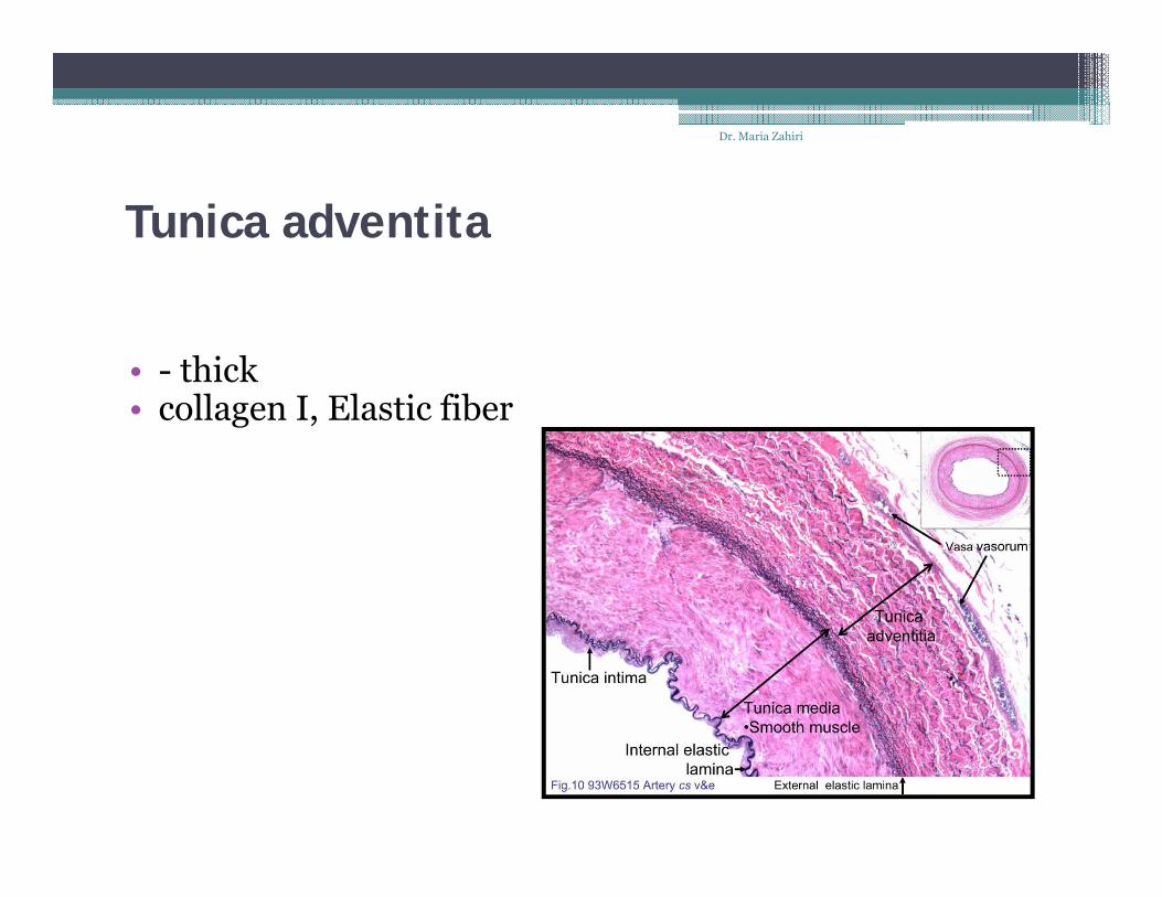
Tunica adventita
• - thick• collagen I, Elastic fiber
Dr. Maria Zahiri
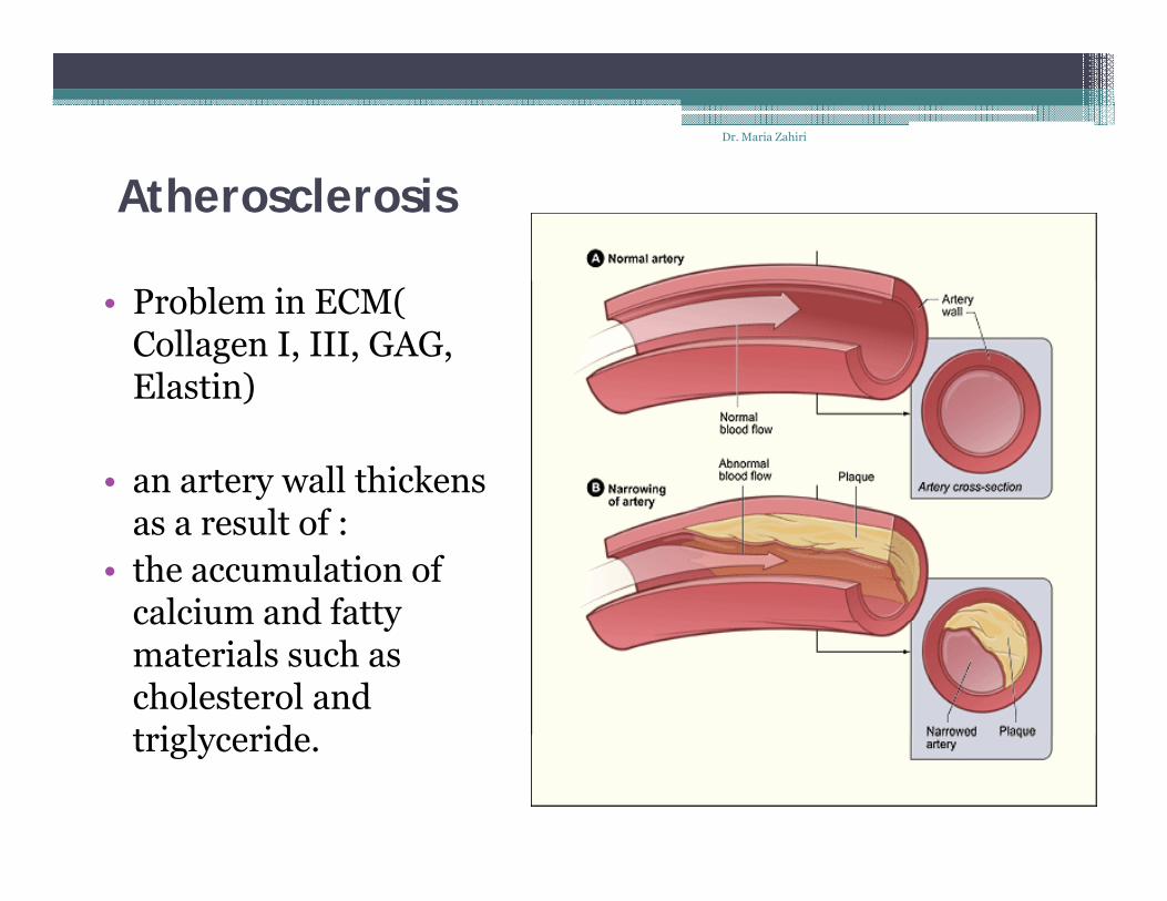
Atherosclerosis
• Problem in ECM( Collagen I, III, GAG, Elastin)Elastin)
• an artery wall thickens as a result of :
• the accumulation of calcium and fatty calcium and fatty materials such as cholesterol and triglyceride triglyceride.
Dr. Maria Zahiri
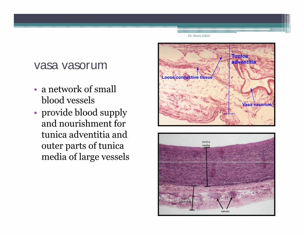
vasa vasorum
• a network of small blood vessels blood vessels
• provide blood supply and nourishment for t i d titi d tunica adventitia and outer parts of tunica media of large vessels

Dr. Maria Zahiri
elastic arterieslarge large veinvein elastic arteries
muscular arteries
large large veinvein
mediummedium--sized sized veinvein
arteriolesvenulesvenules
capillaries

A Comparison of a Typical Artery and a Typical VeinDr. Maria Zahiri
A Comparison of a Typical Artery and a Typical Vein
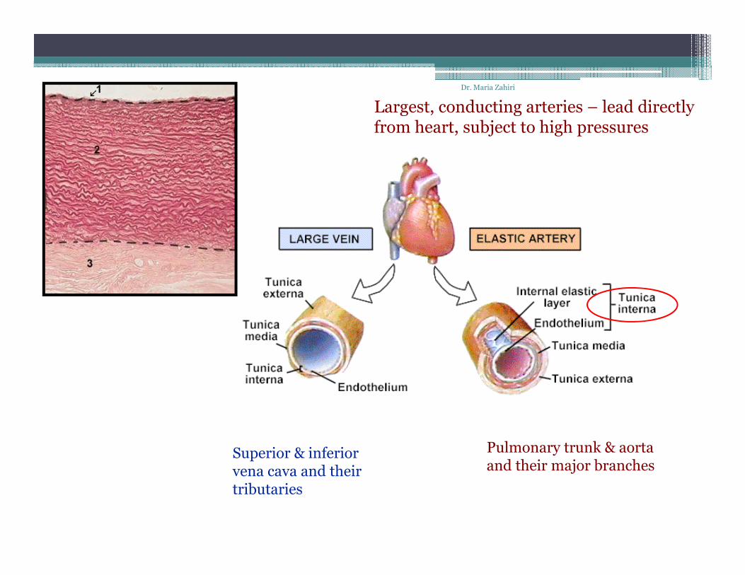
Largest conducting arteries lead directly Dr. Maria Zahiri
Largest, conducting arteries – lead directly from heart, subject to high pressures
Pulmonary trunk & aortaand their major branches
Superior & inferiorvena cava and their and their major branchesvena cava and theirtributaries

Dr. Maria Zahiri
aneurysm• Greek: "dilation“
is a locali ed blood filled balloon like b lge in the all of a blood • is a localized, blood-filled balloon-like bulge in the wall of a blood vessel
• can occur in any blood vessel A l ithi th h t it lf• Aneurysms can also occur within the heart itself.
A ruptured aneurysm can lead to bleeding and subsequent hypovolemic shock, leading to death.
• Aneurysms are a result of a weakened blood vessel wall, and can be a result of a hereditary condition or an acquired disease.
Dr. Maria Zahiri
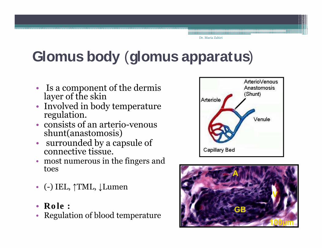
Glomus body (glomus apparatus)
• Is a component of the dermis layer of the skin
• Involved in body temperature Involved in body temperature regulation.
• consists of an arterio-venous shunt(anastomosis)
d d b l f • surrounded by a capsule of connective tissue.
• most numerous in the fingers and toestoes
• (-) IEL, ↑TML, ↓Lumen
l• Role :• Regulation of blood temperature
Dr. Maria Zahiri
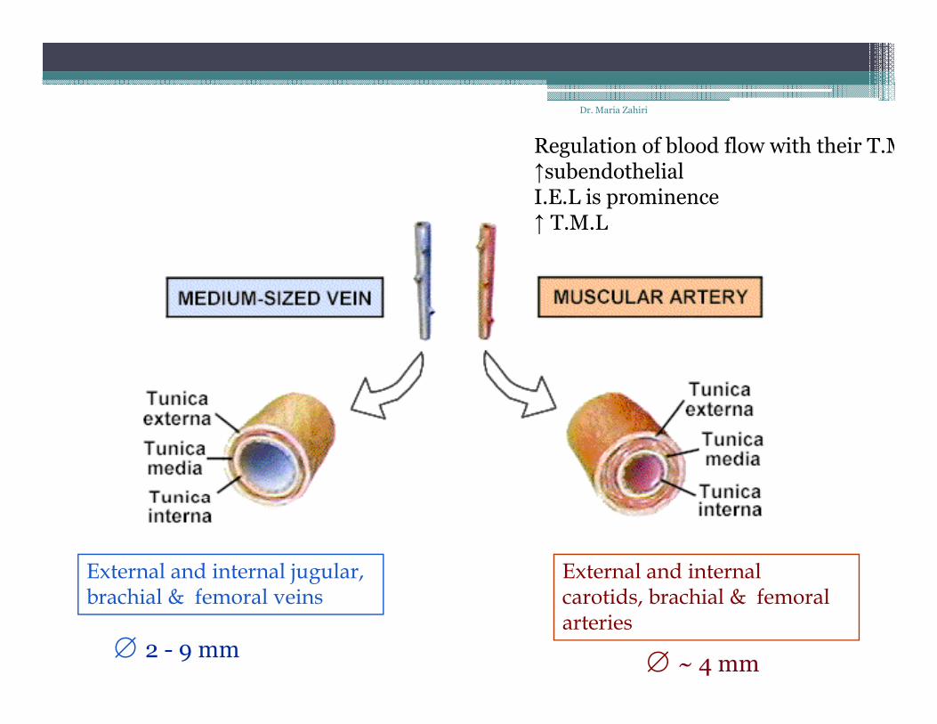
Regulation of blood flow with their T.M↑subendothelialI.E.L is prominence↑ T M L↑ T.M.L
External and internal id b hi l & f l
External and internal jugular, b hi l & f l i carotids, brachial & femoral
arteriesbrachial & femoral veins
2 - 9 mm ~ 4 mm

Dr. Maria Zahiri
↓subendothelial(-) I.E.L
↓subendothelialpericyte
↓ T.M.Lpericyte↓ T.M.L
~ 10-50 µm ~ 30 µm

Dr. Maria Zahiri
Capillariesp• smallest of a body's blood vessels(microcirculation)
• endothelial linings are only one cell layer thick(5-10 μm) connect arterioles and venules
• enable the exchange of water, oxygen, carbon dioxide, and many other nutrients and waste chemical substances between blood and the tissuesbetween blood and the tissues

Dr. Maria Zahiri
Types
• There are three main types of capillaries:• Continuous• Continuous• Fenestrated• Sinusoidal

Dr. Maria Zahiri
Continuous (Somatic)
• Endothelial cells provide an • Endothelial cells provide an uninterrupted lining, and they only allow small molecules, like water and ions to diffuse through tight junctions
• Tight junctions can be further divided into two subtypes:
• with numerous transport vesicles p(skeletal muscles, connective tissue, gonads, Exocrine glands)
with few vesicles (central nervous • with few vesicles (central nervous system)
• These capillaries are a constituent of the pblood-brain-barrier.

Fenestrated (Visceral)Dr. Maria Zahiri
Fenestrated (Visceral)
• "fenestra," Latin for "window"• have pores in the endothelial cells (60-
80 nm in diameter)
• Have a diaphragm
• In the renal glomerulus there are cells with no diaphragms called podocyte foot with no diaphragms called podocyte foot processes or "pedicels,“
d i l d i t ti • endocrine glands, intestines, pancreas, and glomeruli of kidney

Dr. Maria Zahiri
• Both of these types of blood vessels have continuous basal lamina

Dr. Maria Zahiri
Sinusoidal capillaries• are a special type of fenestrated capillariesare a special type of fenestrated capillaries• have larger openings in the endothelium
• allow red and white blood cells (7 5μm - 25μm diameter) • allow red and white blood cells (7.5μm - 25μm diameter) and various serum proteins to pass using a process that is aided by a discontinuous basal lamina
• These capillaries lack pinocytotic vesicles
• Sinusoid blood vessels are primarily located in the bone • Sinusoid blood vessels are primarily located in the bone marrow, lymph nodes, and adrenal gland.
Dr. Maria Zahiri
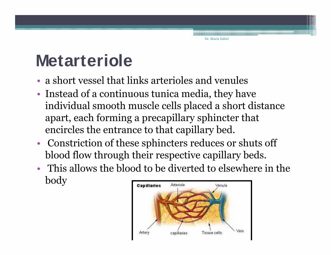
Metarteriolea short vessel that links arterioles and venules• a short vessel that links arterioles and venules
• Instead of a continuous tunica media, they have individual smooth muscle cells placed a short distance apart, each forming a precapillary sphincter that encircles the entrance to that capillary bed.
• Constriction of these sphincters reduces or shuts off • Constriction of these sphincters reduces or shuts off blood flow through their respective capillary beds.
• This allows the blood to be diverted to elsewhere in the body
Microanatomy of Lymphatic Vessels
• A. Lymph capillaries• 1 Structure - blind-ended tubules; consist only of endothelium (which 1. Structure blind ended tubules; consist only of endothelium (which
lacks cell junctions); similar to post capillary venules of blood vascular system
• 2 Function to collect excess tissue fluid• 2. Function - to collect excess tissue fluid
• B. Small to medium lymphatic vessels • 1. Structure• (similar to venous blood vessels of the next smaller size)
a Smaller lymphatic vessels consist of endothelium surrounded • a. Smaller lymphatic vessels consist of endothelium surrounded by collagen and elastic fibers and a few smooth muscle cells

Medium-sized lymphatic vessels
• b. • (1) Tunica intima - thin; endothelium surrounded by few
ll d l ti fib b f ld d t f lcollagen and elastic fibers; may be folded to form valves
• (2) Tunica media - thin; helically arranged smooth muscle, elastic fibers
• (3) Tunica adventita - thicker; collagen and elastic fibers, few smooth muscle cells
• 2. Function - to collect lymph from lymph capillaries

Large lymphatic vesselsLarge lymphatic vessels
• C. include the thoracic duct and right lymphatic duct.• 1 Structure• 1. Structure• a. Tunica intima - thin• (1) Endothelium• (2) Subendothelial layer of collagen and elastic fibers, some
l d l h ly g
longitudinal smooth muscle• b. Tunica media - thickest; longitudinal and circular smooth
muscle bundles, loose FECT (similar to a medium blood vein)
• c. Tunica adventita - not well developed; coarse collagen fibers, few longitudinal smooth muscle
2 F ti t ll t l h f di i d l h ti l • 2. Function - to collect lymph from medium sized lymphatic vessels and return it to largeveins
• D. Lymphatic vessels of any size may appear empty, may contain y p y y pp p y, yfaint pink material (proteins),or may contain lymphocytes.

• 1. The heart wall can be viewed as a three-layered structure.
• a. Inner layer = endocardium• b. Middle Layer = myocardium• c Outer layer = epicardium (also called the • c. Outer layer = epicardium (also called the
pericardium)

1. endocardium - in a sense, simply a continuation of tunica intima, p y
a. consists of 3 parts
* endothelium resting on a basal lamina and associated thin layer of collangenous fibers
* Beneath the endothelium is the sub-endothelial layer of connective tissue containing elastic fibers and a few smooth muscle cells (some texts call this loose C.T. and others dense C.T.)muscle cells (some texts call this loose C.T. and others dense C.T.)
* Beneath that lies the subendocardial layer of loose connective Beneath that lies the subendocardial layer of loose connective tissue that contains small blood vessels and nerves. In the ventricle, the Purkinje fibers are associated with the subendocardial zone.
2. Myocardium - consists of 2 components
a. fascicles of cardiac muscle cells that connect to the fibrous connective tissue skeleton of the heart
b. Noncontactile, modified muscle cells that form the impulse (action potential) generatingand conducting system of the heart
c. cardiac muscle

3. Epicardium - epithelium and connective tissue covering of heart. Also called the visceral pericardiumthe visceral pericardium.
a. squamous to cuboidal epithelial external lining - a continuation of the mesothelium that lines the pericardial cavity.
b. below this is a layer of connective tissue with high concentration of elastic fibers - elastic layer
http://www.finchcms.edu/anatomy/histology/organology/circulatory/o_c_15.html

Artery
White blood cells
Artery
White blood cells
Platelets
Red blood cells
• Deliver O2• Remove metabolic wastes• Maintain temperature pH and fluid volume• Maintain temperature, pH, and fluid volume• Protection from blood loss- platelets• Prevent infection- antibodies and WBC• Transport hormones• Transport hormones

Plasma-55%
Buffy coat-<1%
Formed Formed elements-45%
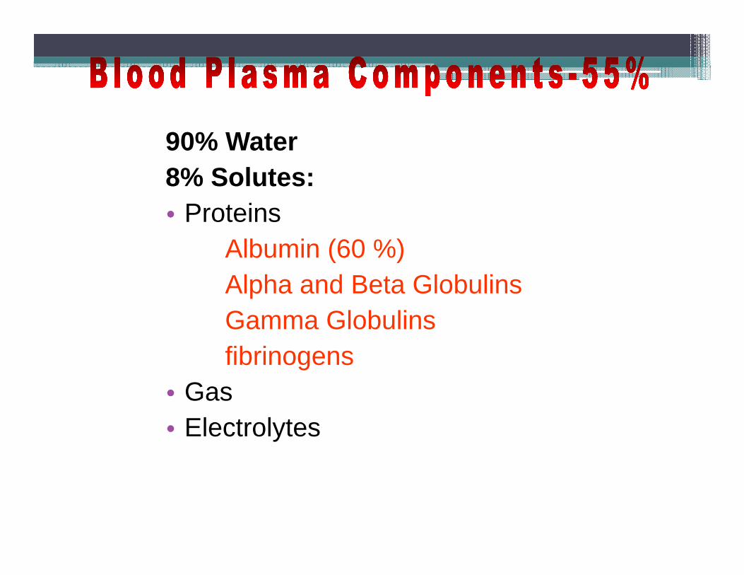
90% Water8% Solutes:8% Solutes:• Proteins
Albumin (60 %)Albumin (60 %)Alpha and Beta GlobulinsGamma GlobulinsGamma Globulinsfibrinogens
• GasGas• Electrolytes

• Leukocytes
• PlateletsPlatelets
• Erythrocytes (red blood cells)• Leukocytes (white blood cells)• Platelets (thrombocytes)Platelets (thrombocytes)

E th tErythrocytes

Erythrocyte7.5m in diay y 7 5 Anucleate- so can't reproduce Hematopoiesis- production of RBC
F i i Function- transport respiratory gases Hemoglobin- 2 chains and 2 chains Lack mitochondria Why? Lack mitochondria. Why? Men- 5 million cells/mm35 / Women- 4.5 million cells/mm3
Life span 100-120 days and then destroyed in spleen (RBC graveyard)

Hematopoiesisp• Hematopoiesis (hemopoiesis): blood cell
formationformation ▫ Occurs in red bone marrow of axial skeleton, girdles
and proximal epiphyses of humerus and femur

Hematopoiesisp• Hemocytoblasts (hematopoietic stem cells)▫ Give rise to all formed elements▫ Give rise to all formed elements▫ Hormones and growth factors push the cell toward
a specific pathway of blood cell developmentp p y p• New blood cells enter blood sinusoids

ErythropoiesisErythropoiesis
• Erythropoiesis: red blood cell production▫ A hemocytoblast is transformed into a
proerythroblast▫ Proerythroblasts develop into early y p y
erythroblasts
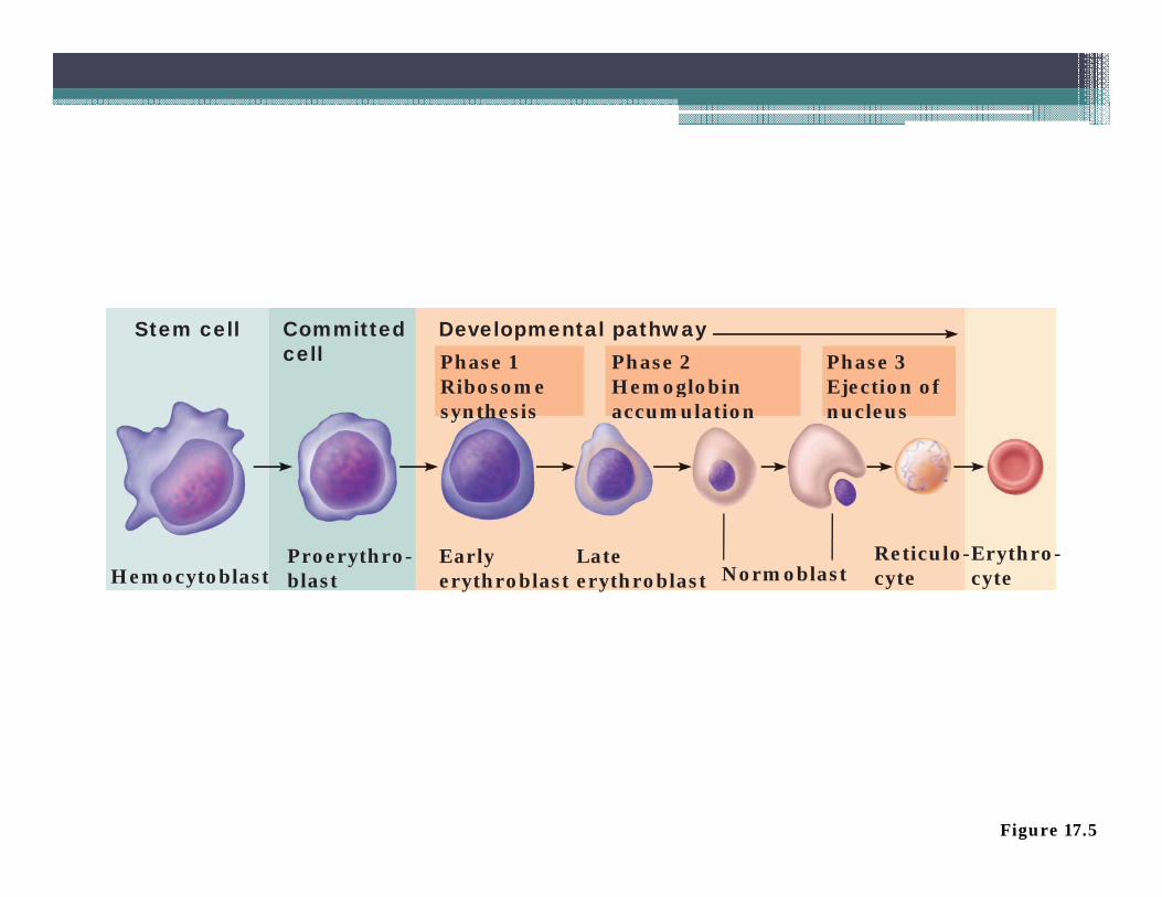
Stem cellPhase 1Ribosome
Phase 2Hemoglobin
Phase 3Ejection of
Committedcell
Developmental pathway
Ribosomesynthesis
Hemoglobinaccumulation
Ejection ofnucleus
HemocytoblastProerythro-blast
Earlyerythroblast
Lateerythroblast Normoblast
Reticulo-cyte
Erythro-cyte
Figure 17.5

R l ti f E th i iRegulation of Erythropoiesis
• Too few RBCs leads to tissue hypoxia• Too many RBCs increases blood viscosity• Too many RBCs increases blood viscosity• Balance between RBC production and
destruction depends on▫ Hormonal controls ▫ Adequate supplies of iron, amino acids, and B
vitaminsvitamins

Hormonal Control of Erythropoiesis
•Erythropoietin (EPO)y p ( )▫Direct stimulus for erythropoiesis ▫Released by the kidneys in▫Released by the kidneys in response to hypoxia

Hormonal Control of Erythropoiesisy p• Causes of hypoxia▫ Hemorrhage or increased RBC destruction▫ Hemorrhage or increased RBC destruction
reduces RBC numbers▫ Insufficient hemoglobin (e.g., iron deficiency)g ( g , y)▫ Reduced availability of O2 (e.g., high altitudes)

Hormonal Control of Erythropoiesisy p• Effects of EPO▫ More rapid maturation of committed bone marrow▫ More rapid maturation of committed bone marrow
cells▫ Increased circulating reticulocyte count in 1–g y
2 days• Testosterone also enhances EPO production,
resulting in higher RBC counts in males
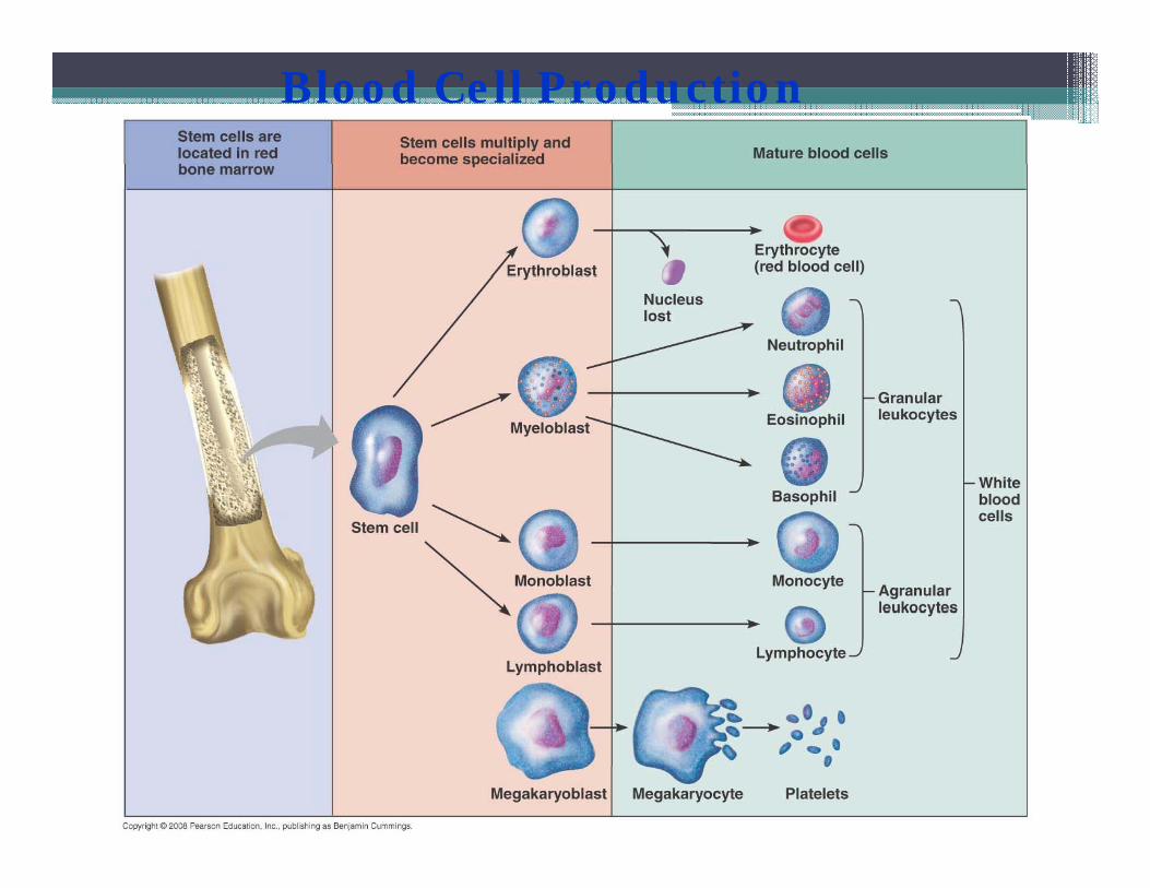
Blood Cell Production
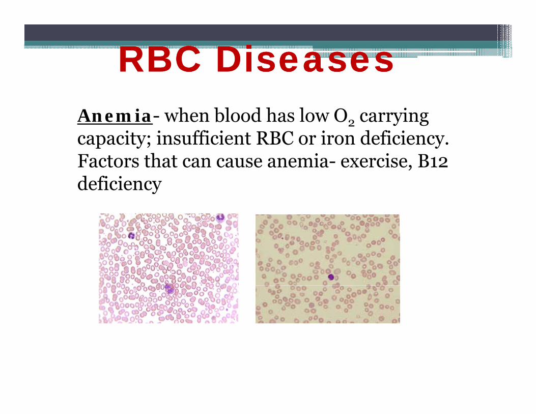
RBC DiseasesRBC DiseasesAnemia- when blood has low O2 carrying capacity; insufficient RBC or iron deficiency.Factors that can cause anemia- exercise, B12 deficiencydeficiency

Si kl ll iRBC DiseasesRBC Diseases
Sickle-cell anemia-
•HbS results from a change in just one of the 287 amino acids in the chain in the globin molecule.
•Found in 1 out of 400 African Americans.ou d out o 400 ca e ca s.
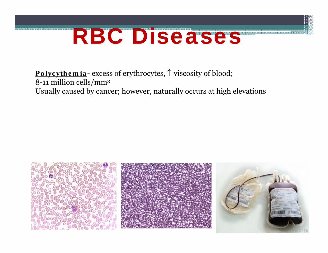
RBC DiseasesRBC DiseasesPolycythemia- excess of erythrocytes, viscosity of blood;8-11 million cells/mm3
Usually caused by cancer; however, naturally occurs at high elevations
2. White Blood Cells (Leukocytes). ( y )
M bil i f b d ’ d f • Mobile units of body’s defense system:
• “Seek and Destroy” Functions:y1. Destroy invading microorganisms2. Destroy abnormal cells (ie: cancer )
• Clean up cellular debris (phagocytosis)
A i t i i j i3. Assist in injury repair

5 - Types of WBC’s
GranulocytesAgranulocytes yAgranulocytes
Each WBC has a specific function Each WBC has a specific function

Blood Cell Origin and Production
Bone Marrow
Circulation
Figure 11-8

Types of WBC’s
l h l lPolymorphonuclear Granulocytes
hil1.Neutrophils2 Eosinophils2.Eosinophils3.Basophils3 p

1. NEUTROPHILS
* 50-70% of all leukocytes5 7 y(most abundant of WBC’s)
* Important in inflammatory responses
* Phagocytes that engulf
responses
g y gbacteria and Debris

2. EOSINOPHILS
* 1-4% of the WBC's
* Attack parasitic worms
* Important in allergic reactions

3. BASOPHILS3
* 0.5% of the WBC's
* Release histamine
0.5% of the WBC s
* Release histamineand heparin
* Important in Allergic R tiReactions
Types of WBC’s
Mononuclear Agranulocytes
4. Monocytes5. Lymphocytes(B d T ll )(B and T cells)

4. MONOCYTES4. MONOCYTES
* 2-6 % of the WBC's
* Exit blood (diapedesis) to become macrophages
* Phagocytic = defend against i d b t iviruses and bacteria

5. LYMPHOCYTES5. LYMPHOCYTES
* 25 33 % of the WBC's
* B lymphocytes:
* 25-33 % of the WBC s
* B-lymphocytes: Produce Antibodies
* T-lymphocytes: Di tl d t iDirectly destroy virus-invaded cells and cancer
llcells
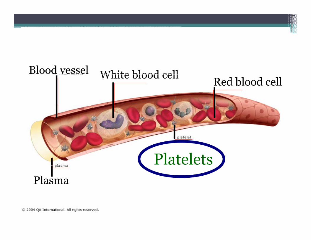
Blood vessel White blood cellRed blood cell
White blood cell
PlateletsPlasma

3. Platelets (Thrombocytes)
* Cell fragments bound to megakaryocytes* “Bud Off” and are released into the blood

Never let monkeys eat bananas
4,000-11,000 cells/mm 3
GranulocytesNeutrophils 40 70%
y
Neutrophils- 40-70%Eosinophils- 1-4%Basophils <1%Basophils- <1%
AgranulocytesMonocytes 4 8%Monocytes- 4-8%Lymphocytes- 20-45%
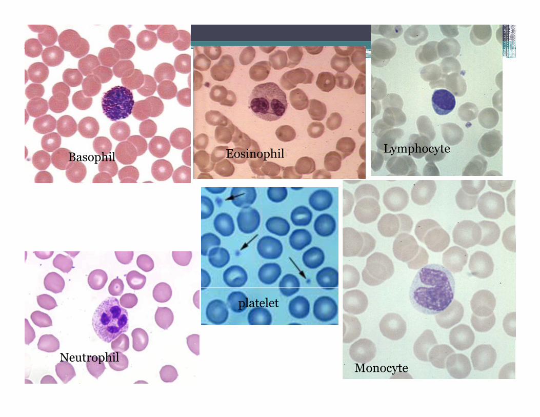
Basophil Eosinophil Lymphocyte
platelet
NeutrophilMonocyte

• Leukopenia• Abnormally low WBC count—drug induced
• Leukemias• Cancerous conditions involving WBCs
• Named according to the abnormal WBC clone involved

Platelets• Small fragments of megakaryocytes• Formation is regulated by thrombopoietin• Formation is regulated by thrombopoietin• Blue-staining outer region, purple granules• Granules contain serotonin, Ca2+, enzymes, y
ADP, and platelet-derived growth factor (PDGF)

Stem cell Developmental pathway
Hemocyto-blast Megakaryoblast
PromegakaryocyteMegakaryocyte Platelets
Figure 17.12

Hemostasis- stoppage of bleedingPlatelets: 250,000-500,000 cells/mm3
Tissue Damage
Platelet Pl gPlatelet Plug
Clotting Factors
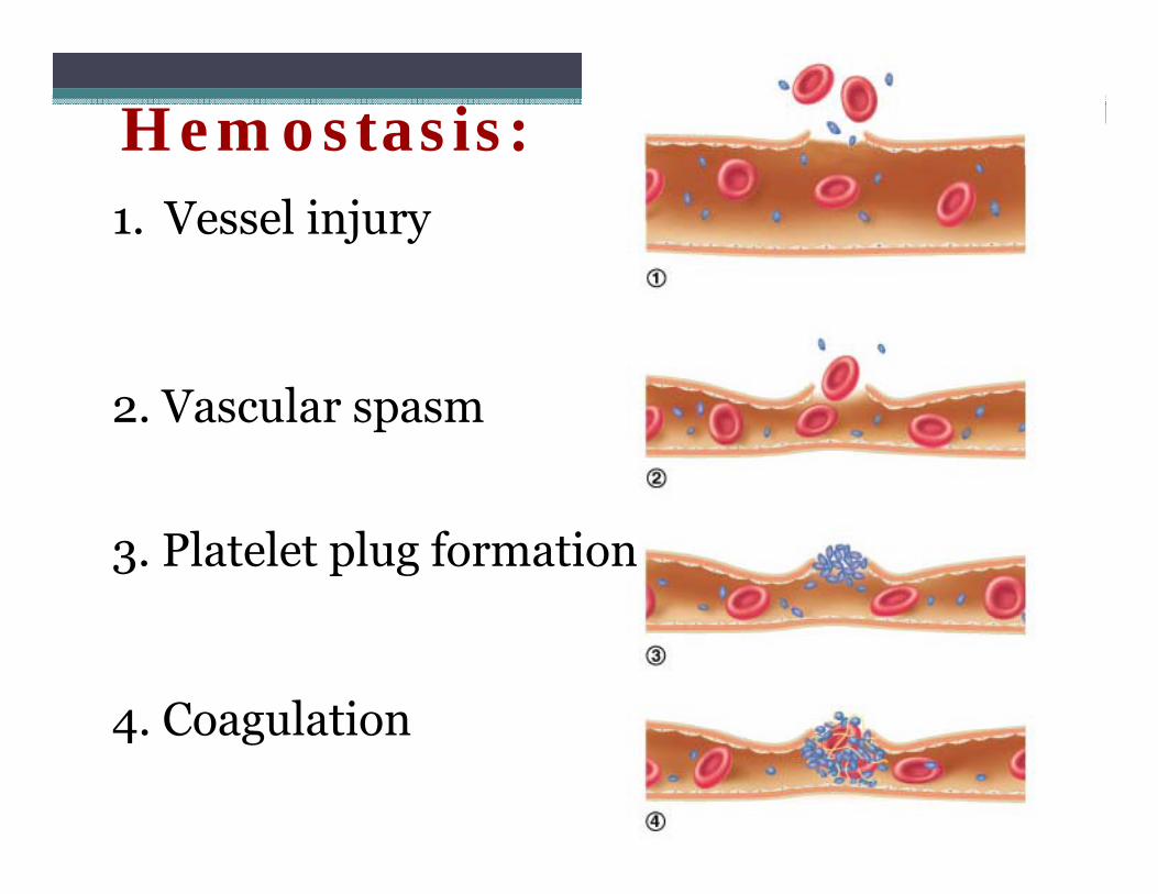
Hemostasis:1. Vessel injury
V l 2. Vascular spasm
3. Platelet plug formation
4 Coagulation4. Coagulation
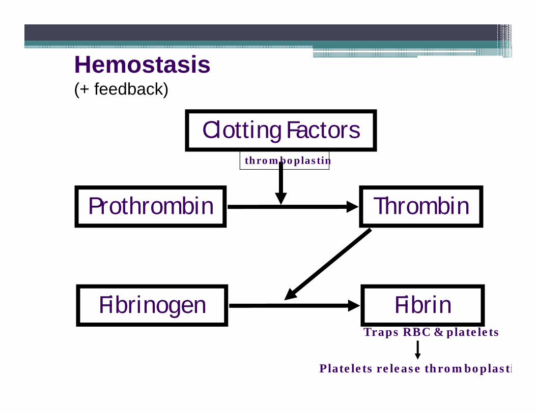
Hemostasis(+ feedback)
Clotting FactorsClotting Factorsthromboplastin
Prothrombin Thrombin
Fibrinogen FibrinTraps RBC & plateletsp p
Platelets release thromboplasti

Blood ClotBlood Clot
RBC
Fibrin thread
Platelet
Fibrin thread
Disorders of Hemostasis• Thromboembolytic disorders: undesirable clot
formationformation• Bleeding disorders: abnormalities that prevent
normal clot formationnormal clot formation

Thromboembolytic Conditionsy• Thrombus: clot that develops and persists in an
unbroken blood vesselunbroken blood vessel▫ May block circulation, leading to tissue death
• Embolus: a thrombus freely floating in the bloodEmbolus: a thrombus freely floating in the blood stream▫ Pulmonary emboli impair the ability of the body to
obtain oxygen▫ Cerebral emboli can cause strokes

Thromboembolytic Conditionsy• Prevented by
A i i▫ Aspirin Antiprostaglandin that inhibits thromboxane
A2A2▫ Heparin Anticoagulant used clinically for pre- and g y p
postoperative cardiac care▫ Warfarin Used for those prone to atrial fibrillation
Dr. Maria Zahiri
باشيد•• باشيدموفق موفق باشيدموفق باشيدموفق