Embed Size (px)
Citation preview
Histology & Its Methods of Study
Department Of General Histology

Recommended Reading:Benefits of General and Clinical
Pathology DepartmentStudent WorkbookRoss – “Histology”Holes “Human Anatomy and
Physiology”Any book on the physiology and
histology with a note: For Medical Students.
Introduction Histology is the study of the tissues of the
body and how these tissues are arranged to constitute organs. The Greek root histo can be translated as either "tissue" or "web" and both translations are appropriate because most tissues are webs of interwoven filaments and fibers, both cellular and noncellular, with membranous linings. Histology involves all aspects of tissue biology, with the focus on how cells' structure and arrangement optimize functions specific to each organ.
Tissues are made of two interacting components: cells and extracellular matrix. The extracellular matrix consists of many kinds of molecules, most of which are highly organized and form complex structures, such as collagen fibrils and basement membranes. The main functions once attributed to the extracellular matrix were to furnish mechanical support for the cells, to transport nutrients to the cells, and to carry away catabolites and secretory products. We now know that, although the cells produce the extracellular matrix, they are also influenced and sometimes controlled by molecules of the matrix. There is, thus, an intense interaction between cells and matrix, with many components of the matrix recognized by and attaching to receptors present on cell surfaces. Most of these receptors are molecules that cross the cell membranes and connect to structural components of the intracellular cytoplasm. Thus, cells and extracellular matrix form a continuum that functions together and reacts to stimuli and inhibitors together.
Each of the fundamental tissues is formed by several types of cells and typically by specific associations of cells and extracellular matrix. These characteristic associations facilitate the recognition of the many subtypes of tissues by students. Most organs are formed by an orderly combination of several tissues, except the central nervous system, which is formed almost solely by nervous tissue. The precise combination of these tissues allows the functioning of each organ and of the organism as a whole.
The small size of cells and matrix components makes histology dependent on the use of microscopes. Advances in chemistry, molecular biology, physiology, immunology, and pathology—and the interactions among these fields—are essential for a better knowledge of tissue biology. Familiarity with the tools and methods of any branch of science is essential for a proper understanding of the subject. This chapter reviews several of the more common methods used to study cells and tissues and the principles involved in these methods.
Overview of methods used HistologyThe objective of a histology
course is to lead the student to understand the microanatomy of cells, tissues and organs and correlate structure with function.
Auxillary techniques include^Histochemistry and biochemistryAutoradiographyOrgan and tissue culture
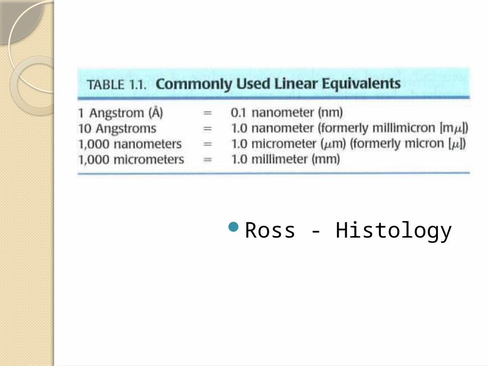
Ross - Histology
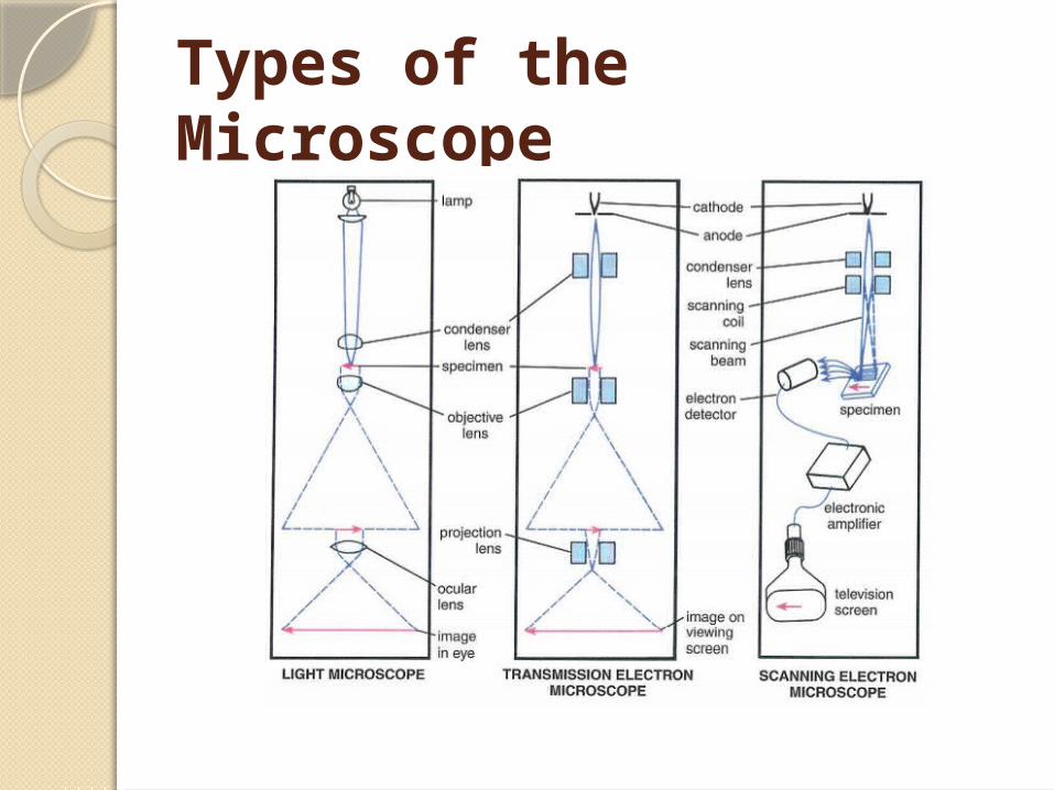
Types of the Microscope
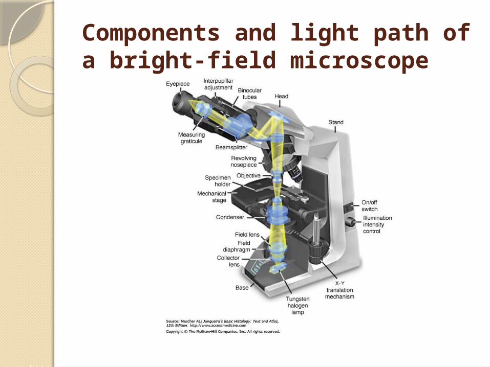
Components and light path of a bright-field microscope
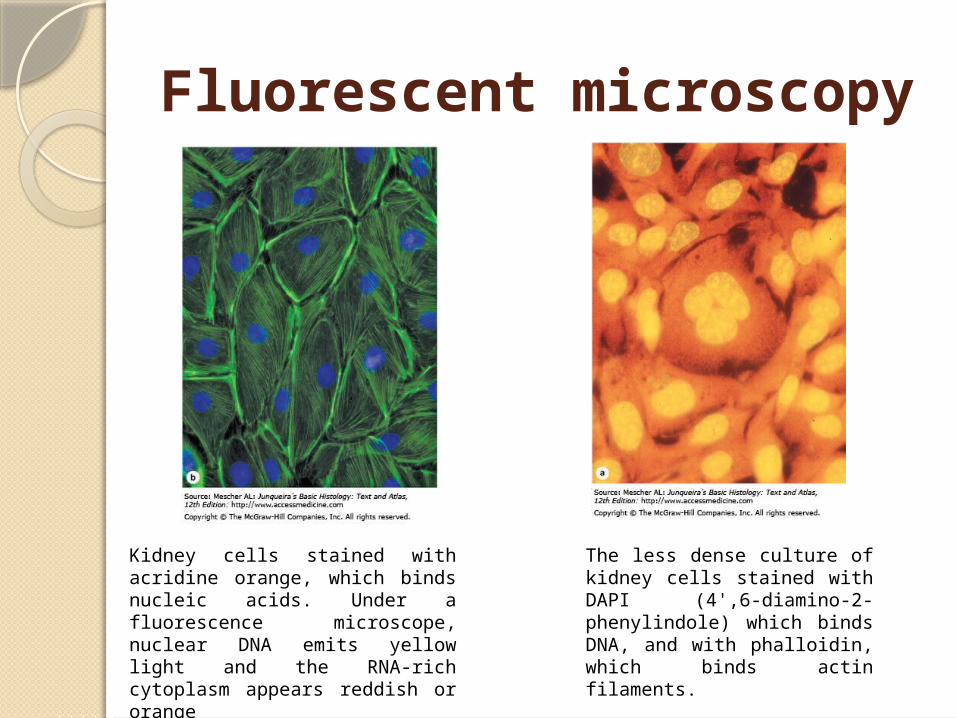
Fluorescent microscopy
Kidney cells stained with acridine orange, which binds nucleic acids. Under a fluorescence microscope, nuclear DNA emits yellow light and the RNA-rich cytoplasm appears reddish or orange
The less dense culture of kidney cells stained with DAPI (4',6-diamino-2-phenylindole) which binds DNA, and with phalloidin, which binds actin filaments.
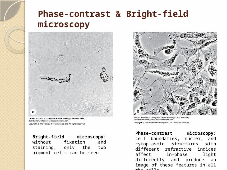
Phase-contrast & Bright-field microscopy
Bright-field microscopy: without fixation and staining, only the two pigment cells can be seen.
Phase-contrast microscopy: cell boundaries, nuclei, and cytoplasmic structures with different refractive indices affect in-phase light differently and produce an image of these features in all the cells.
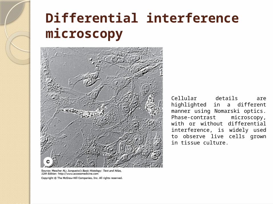
Differential interference microscopy
Cellular details are highlighted in a different manner using Nomarski optics. Phase-contrast microscopy, with or without differential interference, is widely used to observe live cells grown in tissue culture.
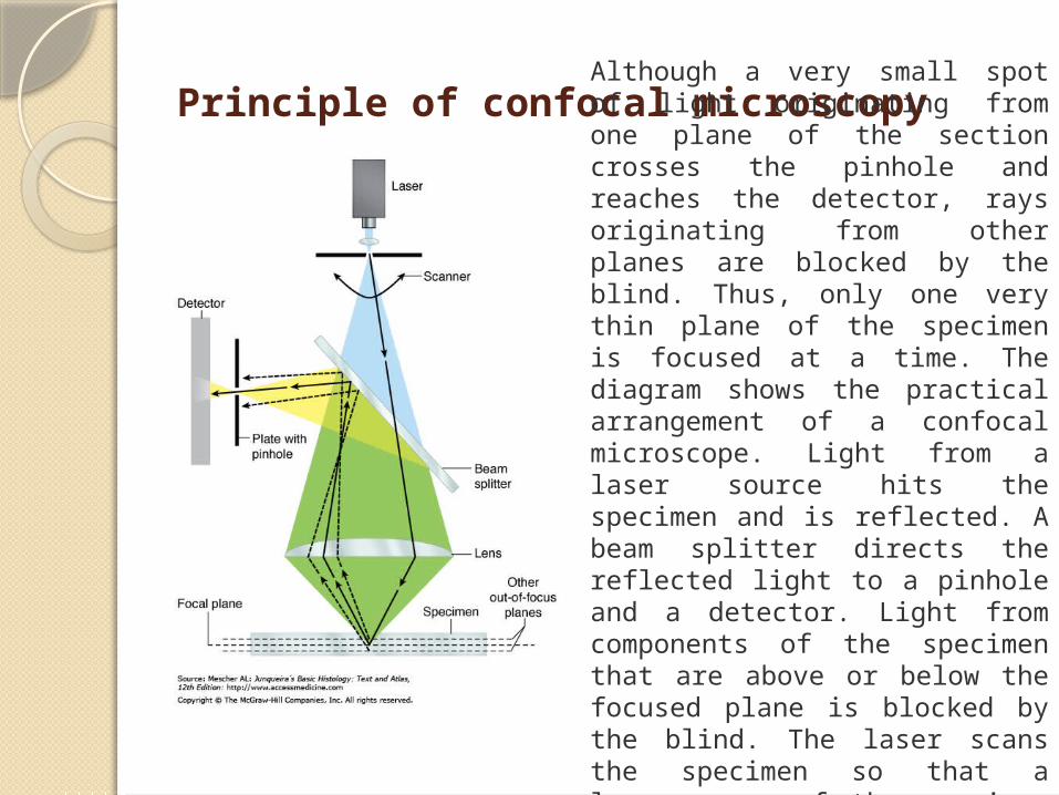
Principle of confocal microscopyAlthough a very small spot of light originating from one plane of the section crosses the pinhole and reaches the detector, rays originating from other planes are blocked by the blind. Thus, only one very thin plane of the specimen is focused at a time. The diagram shows the practical arrangement of a confocal microscope. Light from a laser source hits the specimen and is reflected. A beam splitter directs the reflected light to a pinhole and a detector. Light from components of the specimen that are above or below the focused plane is blocked by the blind. The laser scans the specimen so that a larger area of the specimen can be observed.
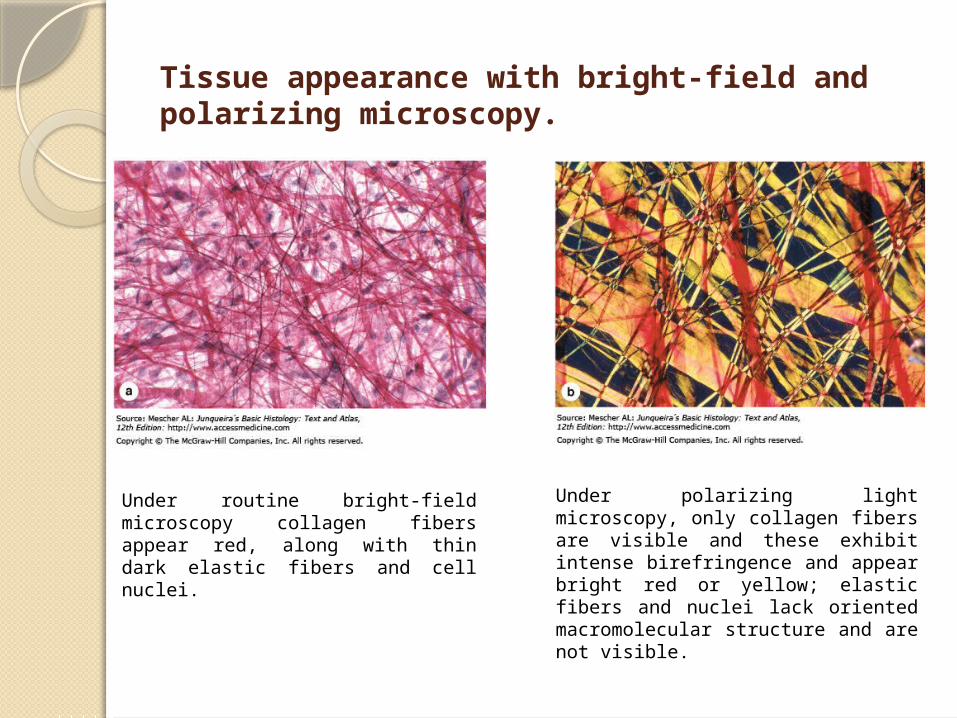
Tissue appearance with bright-field and polarizing microscopy.
Under routine bright-field microscopy collagen fibers appear red, along with thin dark elastic fibers and cell nuclei.
Under polarizing light microscopy, only collagen fibers are visible and these exhibit intense birefringence and appear bright red or yellow; elastic fibers and nuclei lack oriented macromolecular structure and are not visible.
Transmission Electron Microscopy
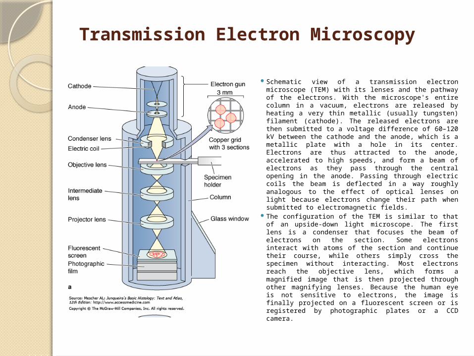
Schematic view of a transmission electron microscope (TEM) with its lenses and the pathway of the electrons. With the microscope's entire column in a vacuum, electrons are released by heating a very thin metallic (usually tungsten) filament (cathode). The released electrons are then submitted to a voltage difference of 60–120 kV between the cathode and the anode, which is a metallic plate with a hole in its center. Electrons are thus attracted to the anode, accelerated to high speeds, and form a beam of electrons as they pass through the central opening in the anode. Passing through electric coils the beam is deflected in a way roughly analogous to the effect of optical lenses on light because electrons change their path when submitted to electromagnetic fields.
The configuration of the TEM is similar to that of an upside-down light microscope. The first lens is a condenser that focuses the beam of electrons on the section. Some electrons interact with atoms of the section and continue their course, while others simply cross the specimen without interacting. Most electrons reach the objective lens, which forms a magnified image that is then projected through other magnifying lenses. Because the human eye is not sensitive to electrons, the image is finally projected on a fluorescent screen or is registered by photographic plates or a CCD camera.
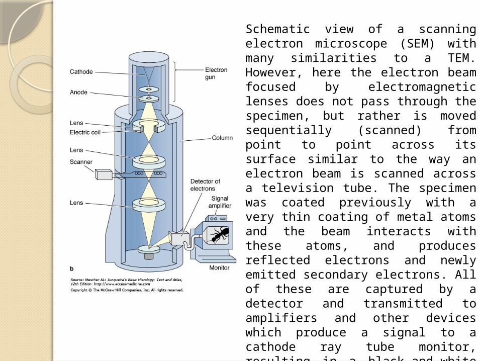
Schematic view of a scanning electron microscope (SEM) with many similarities to a TEM. However, here the electron beam focused by electromagnetic lenses does not pass through the specimen, but rather is moved sequentially (scanned) from point to point across its surface similar to the way an electron beam is scanned across a television tube. The specimen was coated previously with a very thin coating of metal atoms and the beam interacts with these atoms, and produces reflected electrons and newly emitted secondary electrons. All of these are captured by a detector and transmitted to amplifiers and other devices which produce a signal to a cathode ray tube monitor, resulting in a black-and-white image. The SEM shows only surface views of the coated specimen but with a striking three-dimensional quality. The inside of organs or cells can be analyzed by sectioning them to expose their internal surfaces.

Preparation of Tissues for Study
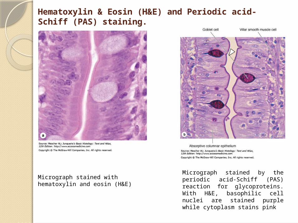
Hematoxylin & Eosin (H&E) and Periodic acid-Schiff (PAS) staining.
Micrograph stained with hematoxylin and eosin (H&E)
Micrograph stained by the periodic acid-Schiff (PAS) reaction for glycoproteins. With H&E, basophilic cell nuclei are stained purple while cytoplasm stains pink
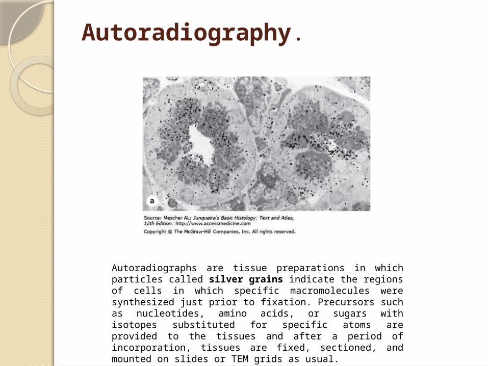
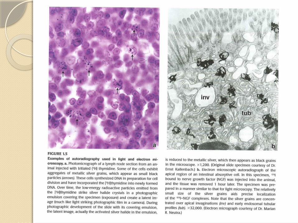
Autoradiography.
Autoradiographs are tissue preparations in which particles called silver grains indicate the regions of cells in which specific macromolecules were synthesized just prior to fixation. Precursors such as nucleotides, amino acids, or sugars with isotopes substituted for specific atoms are provided to the tissues and after a period of incorporation, tissues are fixed, sectioned, and mounted on slides or TEM grids as usual.
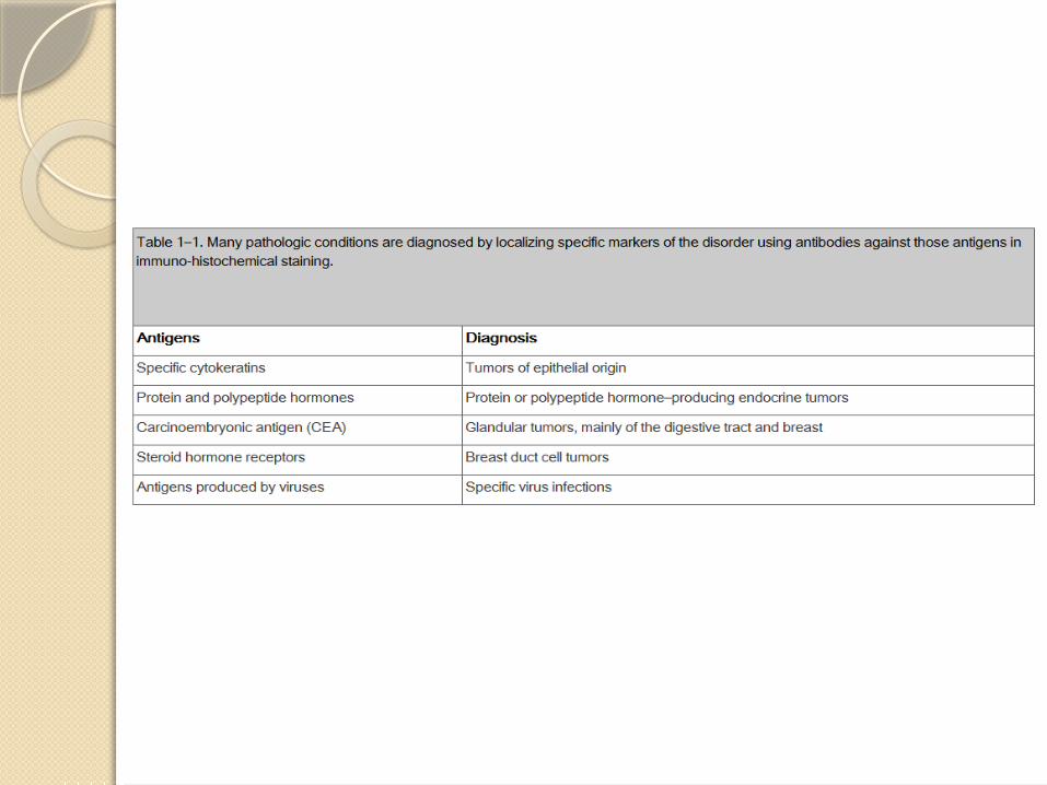
Detection Methods Using Specific Interactions between Molecules
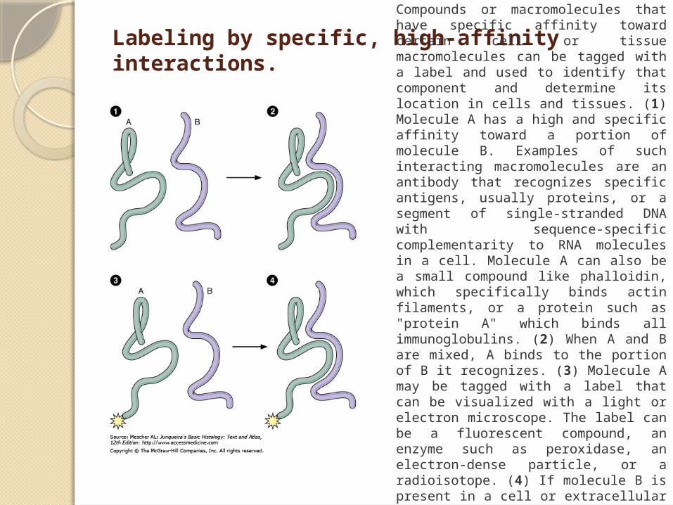
Labeling by specific, high-affinity interactions. Compounds or macromolecules that
have specific affinity toward certain cell or tissue macromolecules can be tagged with a label and used to identify that component and determine its location in cells and tissues. (1) Molecule A has a high and specific affinity toward a portion of molecule B. Examples of such interacting macromolecules are an antibody that recognizes specific antigens, usually proteins, or a segment of single-stranded DNA with sequence-specific complementarity to RNA molecules in a cell. Molecule A can also be a small compound like phalloidin, which specifically binds actin filaments, or a protein such as "protein A" which binds all immunoglobulins. (2) When A and B are mixed, A binds to the portion of B it recognizes. (3) Molecule A may be tagged with a label that can be visualized with a light or electron microscope. The label can be a fluorescent compound, an enzyme such as peroxidase, an electron-dense particle, or a radioisotope. (4) If molecule B is present in a cell or extracellular matrix that is incubated with labeled molecule A, molecule B can be detected and localized by visualizing the labeled molecule A bound to it.
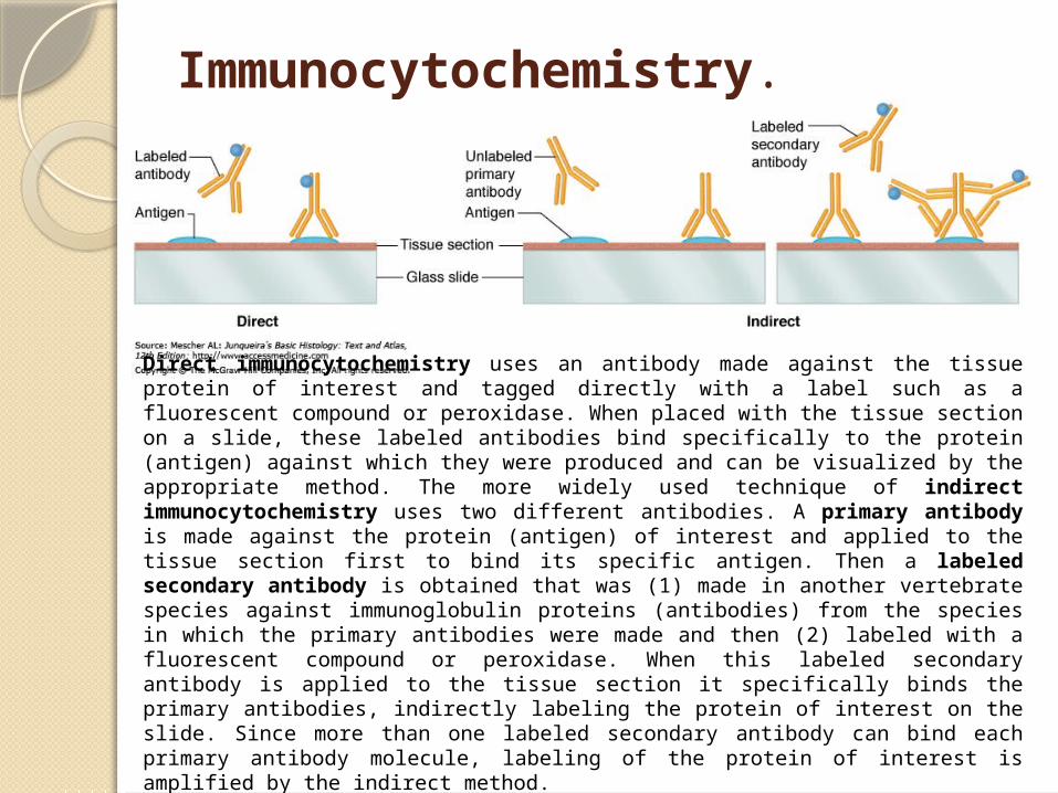
Immunocytochemistry.
Direct immunocytochemistry uses an antibody made against the tissue protein of interest and tagged directly with a label such as a fluorescent compound or peroxidase. When placed with the tissue section on a slide, these labeled antibodies bind specifically to the protein (antigen) against which they were produced and can be visualized by the appropriate method. The more widely used technique of indirect immunocytochemistry uses two different antibodies. A primary antibody is made against the protein (antigen) of interest and applied to the tissue section first to bind its specific antigen. Then a labeled secondary antibody is obtained that was (1) made in another vertebrate species against immunoglobulin proteins (antibodies) from the species in which the primary antibodies were made and then (2) labeled with a fluorescent compound or peroxidase. When this labeled secondary antibody is applied to the tissue section it specifically binds the primary antibodies, indirectly labeling the protein of interest on the slide. Since more than one labeled secondary antibody can bind each primary antibody molecule, labeling of the protein of interest is amplified by the indirect method.
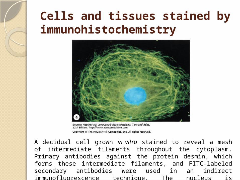
Cells and tissues stained by immunohistochemistry
A decidual cell grown in vitro stained to reveal a mesh of intermediate filaments throughout the cytoplasm. Primary antibodies against the protein desmin, which forms these intermediate filaments, and FITC-labeled secondary antibodies were used in an indirect immunofluorescence technique. The nucleus is counterstained light blue with DAPI.
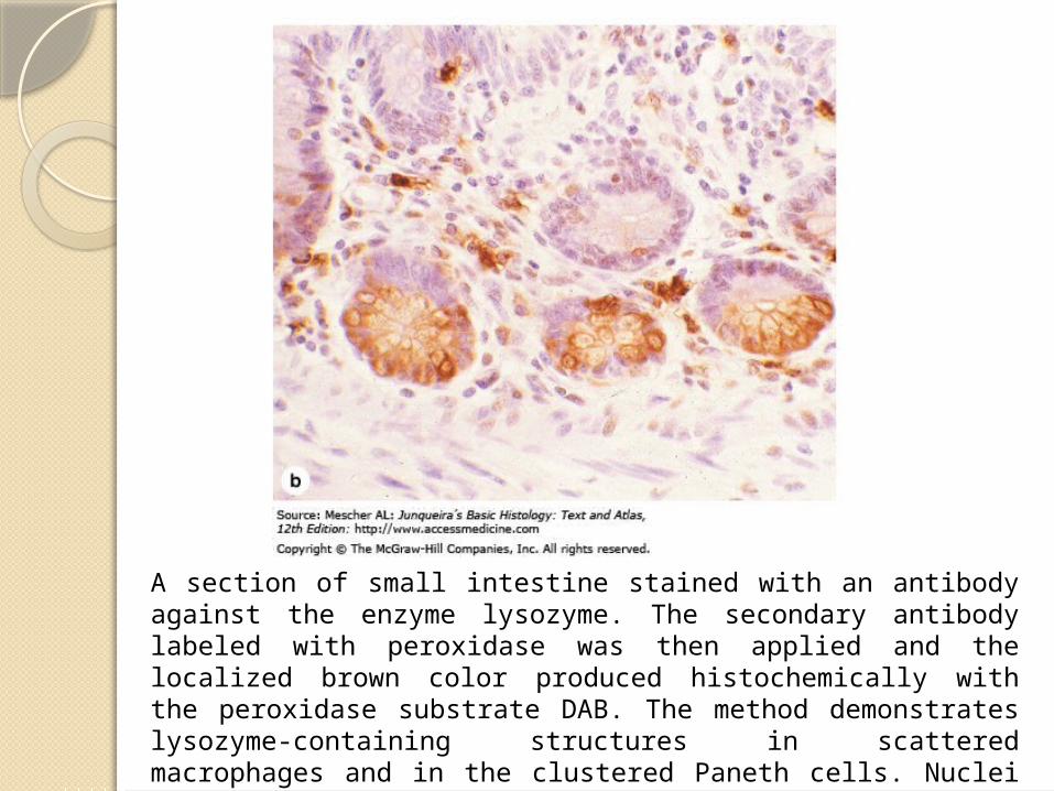
A section of small intestine stained with an antibody against the enzyme lysozyme. The secondary antibody labeled with peroxidase was then applied and the localized brown color produced histochemically with the peroxidase substrate DAB. The method demonstrates lysozyme-containing structures in scattered macrophages and in the clustered Paneth cells. Nuclei were counterstained with hematoxylin.
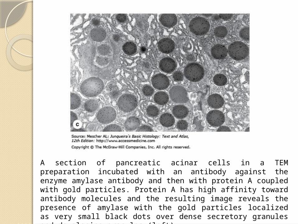
A section of pancreatic acinar cells in a TEM preparation incubated with an antibody against the enzyme amylase antibody and then with protein A coupled with gold particles. Protein A has high affinity toward antibody molecules and the resulting image reveals the presence of amylase with the gold particles localized as very small black dots over dense secretory granules and developing granules (left).
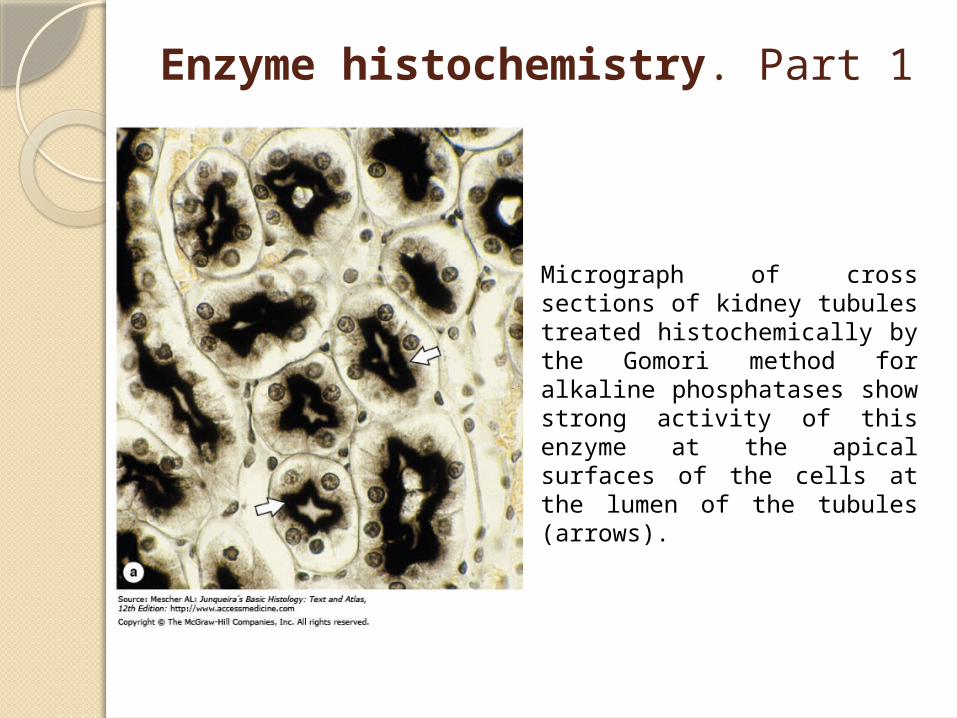
Enzyme histochemistry. Part 1
Micrograph of cross sections of kidney tubules treated histochemically by the Gomori method for alkaline phosphatases show strong activity of this enzyme at the apical surfaces of the cells at the lumen of the tubules (arrows).
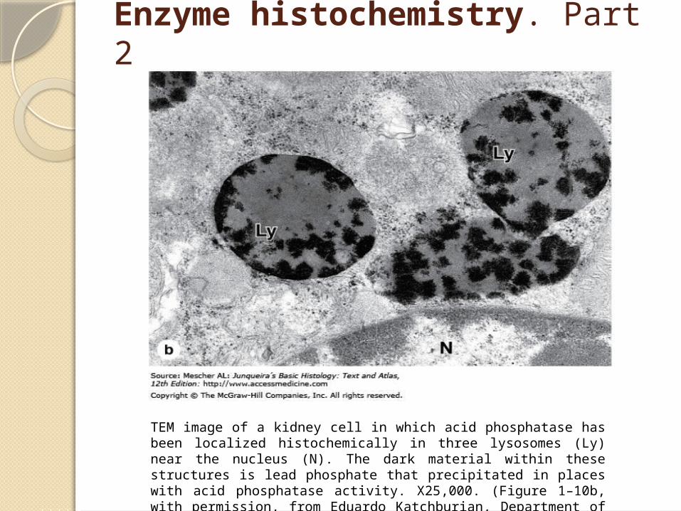
Enzyme histochemistry. Part 2
TEM image of a kidney cell in which acid phosphatase has been localized histochemically in three lysosomes (Ly) near the nucleus (N). The dark material within these structures is lead phosphate that precipitated in places with acid phosphatase activity. X25,000. (Figure 1–10b, with permission, from Eduardo Katchburian, Department of Morphology, Federal University of Sao Paulo, Brazil.)
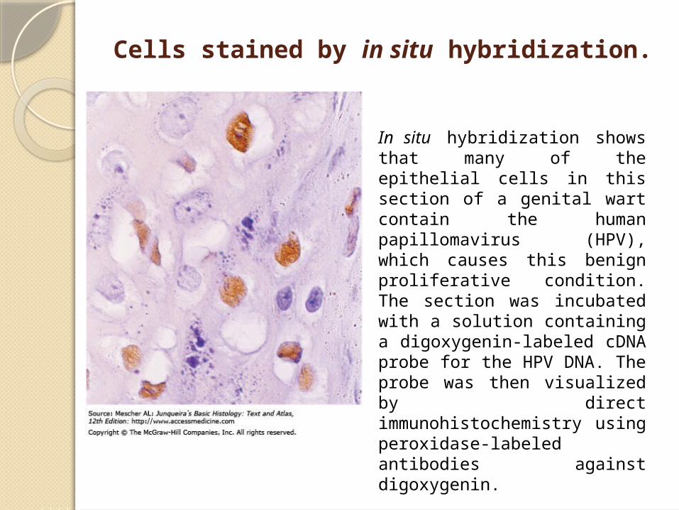
Cells stained by in situ hybridization.
In situ hybridization shows that many of the epithelial cells in this section of a genital wart contain the human papillomavirus (HPV), which causes this benign proliferative condition. The section was incubated with a solution containing a digoxygenin-labeled cDNA probe for the HPV DNA. The probe was then visualized by direct immunohistochemistry using peroxidase-labeled antibodies against digoxygenin.
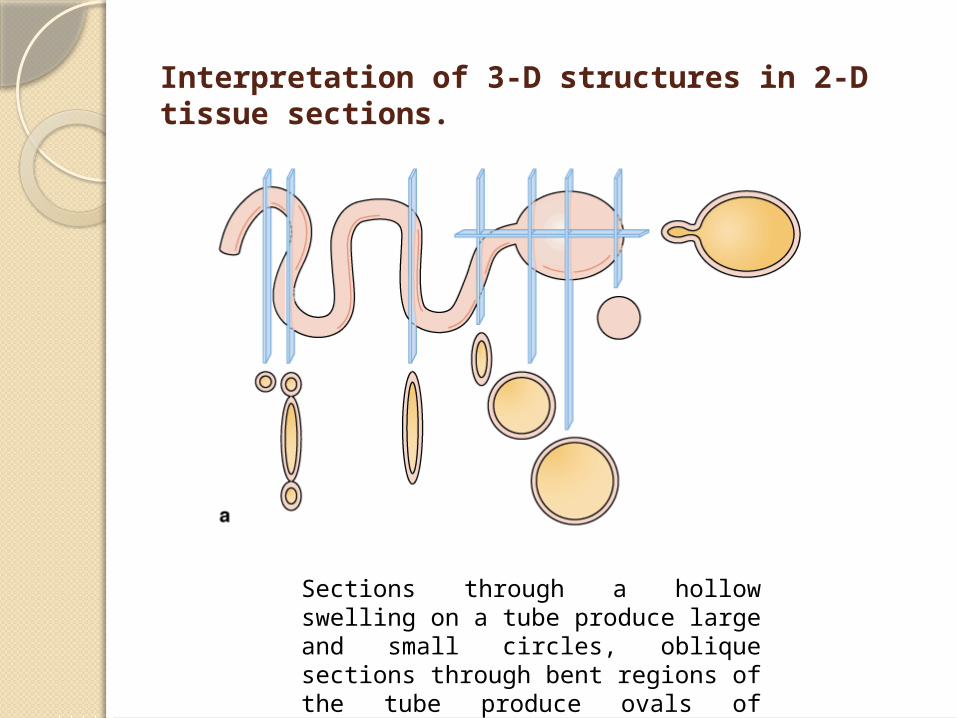
Interpretation of 3-D structures in 2-D tissue sections.
Sections through a hollow swelling on a tube produce large and small circles, oblique sections through bent regions of the tube produce ovals of various dimensions.
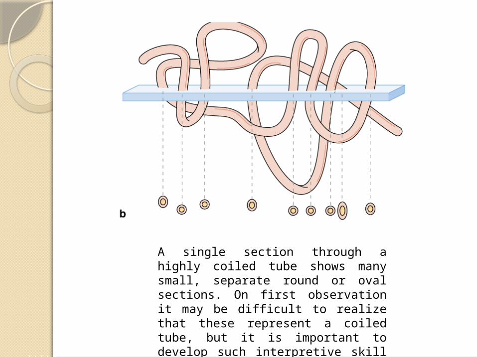
A single section through a highly coiled tube shows many small, separate round or oval sections. On first observation it may be difficult to realize that these represent a coiled tube, but it is important to develop such interpretive skill in understanding histological preparations.
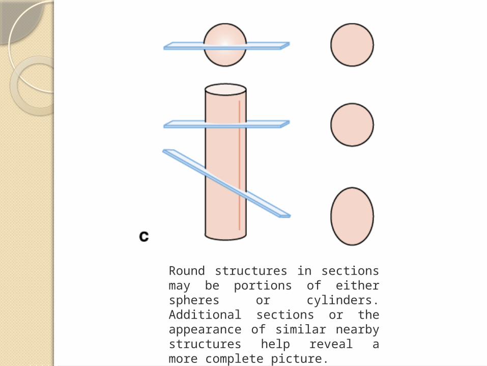
Round structures in sections may be portions of either spheres or cylinders. Additional sections or the appearance of similar nearby structures help reveal a more complete picture.
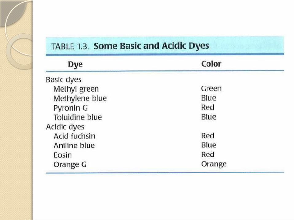
Light Microscopy Stains
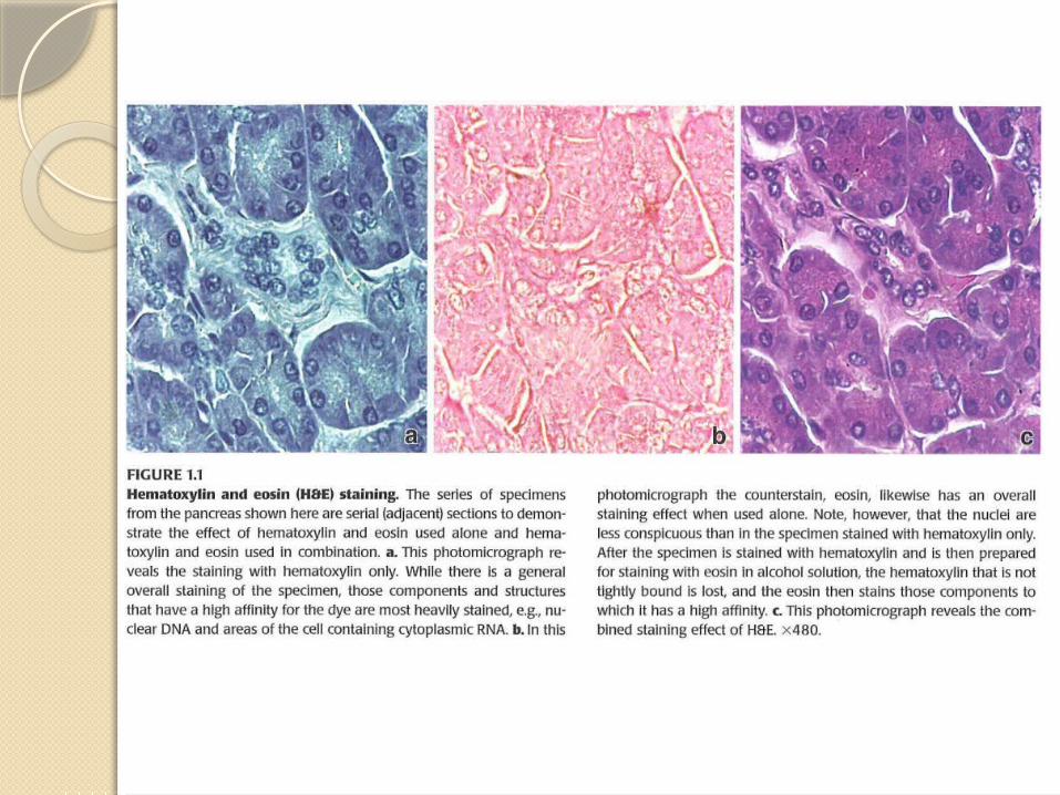
Hematoxylin and Eosin (H&E)
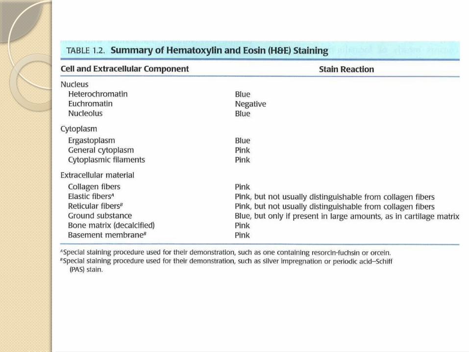
Hematoxylin stains cellular regions rich in basophilic macromolecules (DNA or RNA) a purplish blue or blue-black color. It is the most common stain for demonstrating cell nuclei and cytoplasm rich in rough ER. Usually used as the contrasting "counterstain" with hematoxylin, eosin is an acidic stain that binds to basic macromolecules such as collagen and most cytoplasmic proteins, especially those of mitochondria. Eosin stains regions rich in such structures a pinkish red color. Tissue sections showing only structures with shades of purple and pink are stained with H&E.
Pararosaniline-Toluidine Blue (PT)
This dye combination stains chromatin shades of purple and cytoplasm and collagen a lighter violet. These stains penetrate plastic sections more readily than H&E and are used here primarily with acrylic resin-embedded sections to provide better detail of cell and tissue structures. Toluidine blue is also commonly used for differential staining of cellular components, particularly cytoplasmic granules.
Mallory Trichrome
This procedure employs a combination of stains applied in series which results in nuclei staining purple; cytoplasm, keratin, and erythrocytes staining bright red or orange; and collagen bright or light blue. Mallory trichrome is particularly useful in demonstrating cells and small blood vessels of connective tissue. Similar stains, such as Masson trichrome and Gomori trichrome, yield comparable results except that collagen stains blue-green or green.
Picro-Sirius-Hematoxylin (PSH)
The dye Sirius red in a solution of picric acid stains collagen red and cytoplasm a lighter violet or pink, with nuclei purple if first stained with hematoxylin. Under the polarizing microscope, collagen stained with picro-sirius red is birefringent and can be detected specifically.
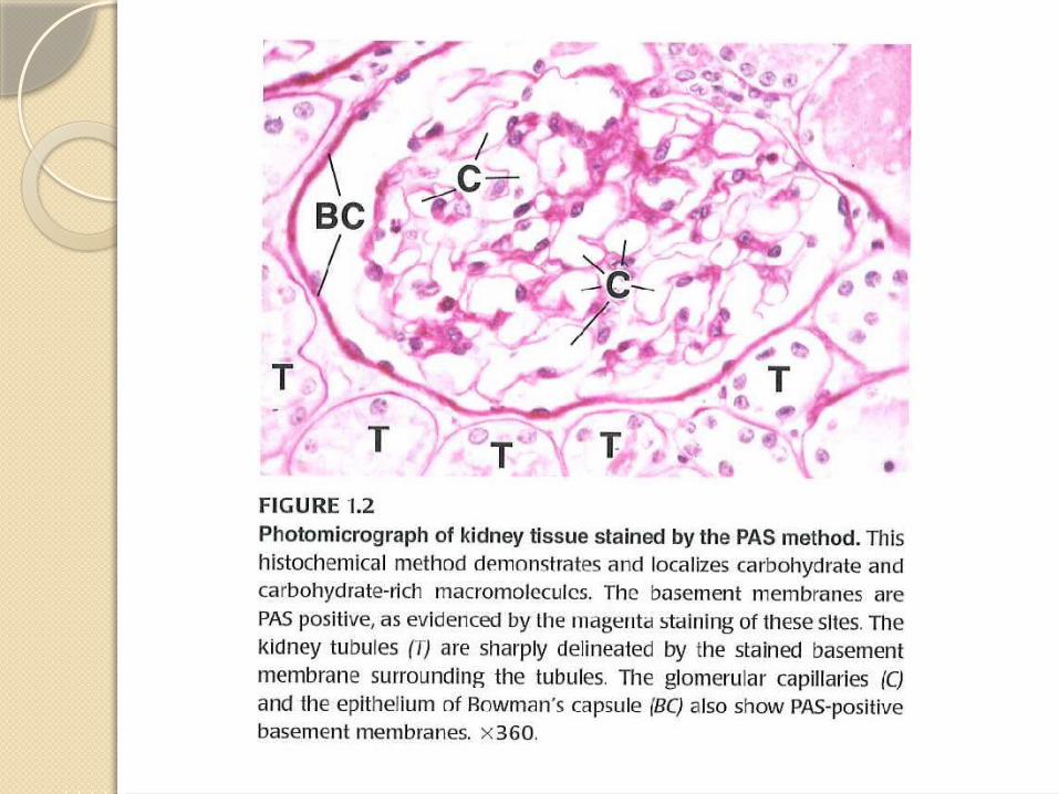
Periodic Acid–Schiff Reaction (PAS)
This histochemical procedure stains complex carbohydrate-containing cell components, which become magenta (shades of purplish pink). PAS is commonly used to demonstrate cells filled with mucin granules, glycogen deposits, or the glycocalyx.

Wright-Giemsa Stain
These are two similar combinations of stains that are widely used on fixed cells of blood or bone marrow smears to demonstrate types of blood cells. Granules in leukocytes are seen to have differential affinity for the stain components. Nuclei stain purple and erythrocytes stain uniformly pink or pinkish orange.

Silver or Gold Stains
Various procedures employing solutions of silver or gold salts have been developed to demonstrate filamentous structures in neurons and fibers of reticulin (type III collagen). By these "metal impregnation" techniques these filaments stain dark brown or black. Such stains have been largely replaced now by immunohistochemical procedures.

Stains for Elastin
Several staining methods have been developed to distinguish elastic structures from collagen, most of which stain the elastin-rich structures brown or shades of purple. Examples of such stains are Weigert's resorcin fuchsin, aldehyde fuchsin, and orcein Van Gieson stains.

Stains for Lipid
When special preparation techniques are used to retain lipids of cells, such as in frozen sections, lipophilic dyes are used to demonstrate lipid droplets and myelin. Oil red O and Sudan black stain lipid-rich structures as their names suggest. Osmium tetroxide (osmic acid), which is used as a fixative for TEM, is reduced to a black substance by unsaturated fatty acids and is also used to demonstrate lipids.

Other Common Stains
Many basic aniline dyes, including azures, cresyl violet, brilliant cresyl blue, luxol fast blue, and light green, are used because of the permanence and brightness of the colors they impart to cellular and extracellular structures in paraffin sections. Many such stains were initially developed for use in the textile industry.

Thank you for attention!


![Histology Photo Atlas[1]](https://img.pdfslide.us/doc/110x75/5448e6a2b1af9f57618b4d19/histology-photo-atlas1.jpg)


