Embed Size (px)
Citation preview

HISTOLOGY OF BORON DEFICIENCY IN PLANTS
BY
MARIE P. LOHNLS
JHededeellngen van de Landbouwhoogeàchool Deel 44 .—• Verhandeling 3
H. VEENMAN & ZONEN — WAGENINGEN — 1940
l049°^3

3 3
HISTOLOGY OF BORON DEFICIENCY IN PLANTS
by Marie P. Löhnis
CONTENTS page
1. Introduction 3 a. Scope of the investigation 3 b. Anatomy of boron deficient plant parts 4 c. Methods 5
2. Boron deficiency in Cereals 6 a. Introduction 6 6. Triticum vulgare 7 c. Avena sativa 8 d. Secale cereale •. 10 e. Hordeum vulgare 11 ƒ. Discussion 13
3. Boron deficiency in Swedish Turnips. (Brassica napus v. napo-brassica) 13 a. General pathology 13 b. The structure of sound roots 17 c. Comparison of sound and affected tissue 18 d. Discussion 23
4. Aspects of the pathology of boron deficiency 26 a. Etiology of injury in Swedes 26 b. Etiology of the injury in Cereals 29 c. Conclusions SO
5. Summary 31
1. INTRODUCTION
a. Scope of the investigation.
In „Plant development in the absence of boron" (1937) the writer has published the results obtained with various cereals when cultivated in a medium deprived of boron. In these conditions Hordeum vulgare did not shoot its ears. Secale developed ears, the upper part of which bore withered glumes, but the ear failed to flower. Triticum vulgare produced atrophied anthers and no grain developed. Avena sativa produced fullgrown anthers, the pollen grains however were chiefly

empty ; the grains were inhibited in development. I n none of these cultures any difference could be observed in the vegetative development of roots and shoots when compared with p lants supplied with boron.
As these symptoms of boron deficiency were considered to be due to earlier disturbances occurring in the plants, an a t t empt was made to trace these initial stages of injury in the various cereals. I n order to accomplish this a microscopical investigation was carried out.
The histological disturbances in the cereals were compared with those of the thickened roots of Brassica napus v. napobrassica (Swedish turnip) suffering from a lack of boron.
b . Anatomy of boron deficient plant parts.
W A R I N GTON (1923) notes a swelling of the cambium cells in the vascular system. Nuclei are distinct. Thin-walled young xylem elements are absent. The lumen of tracheides is small and blocked. SOMMER and SOROKIN (1928) describe the tissue of the root t ips of Pisum sativum. A distinct hyperplasia occurs in the plerome. Regularity in the arrangement of the cells is lost ; they are irregular in shape and size. I n the periblem cells are larger t han normal with smaller nuclei. HAAS and KLOTZ (1931) s tate t h a t in Citrus boron appears to be essential for cell division in the meristematic tissue of growing points, but is likewise essential for cambial activity. When boron is deficient the cambium and portions of the phloem disintegrate. The xylem tissue disintegrates to a much smaller degree, if a t all.
MARTIN (1934) s tates t h a t in sugar cane in the early stage of boron deficiency certain of the lower cells in the chlorophyll bearing bundle sheaths have become greatly enlarged.
JAMALAINEN (1935) in a detailed s tudy of „brown hear t " in Swedes notes an elongation of parenchymatous cells in the interior of the root. I n rare cases an elongation of cambium cells is apparent . R O W E (1936) describes anatomical details in sugar beets. The cells of the vascular system in process of differentiation appear very susceptible. Frequently the cells become thick-walled but apparently not rigid. Many of the cells appear to have undergone division and the two daughter cells are separated by an extremely thin wall. I n later stages the cambium cells have become irregularly swollen and divided. Sieve tubes of healthy plants may contain plugs of densily staining material, which presumably is callose, either developed as a solid rod or disposed as a peripheral layer. These phenomena occur much more frequently in the phloem of diseased plants where they are very typical.
VAN SCHREVEN (1934) notes in tobacco an enormous enlargement of t he phloem, numerous radial divisions taking place. I n the main the

stelar s tructure is affected bu t individual cells of the ground parenchyma may be affected as well. Brown discoloration of cell walls is presumably the initial stage which often begins a t the points of junction of the cells. Frequently cells are found having only one or two joint brown walls. Generally, the cells of diseased xylem tissue are smaller and have thinner walls as compared with those of heal thy tissue. The same phenomena are described in tomato and in po ta to (1935a and b and 1939).
c. Methods.
In order to obtain suitable material showing the effect of boron deficiency, plants had to be cultivated in a medium deprived of boron. The method described in the earlier paper (1937) was followed.
The nutrient solution of van der Crone, modified according to BBYAN (1921) was used. Traces of elements according to the formula of SOMMEE and SOBOKIN (1928) were added. The formula used is:
K N 0 3 1 g; KCL 0,75; CaS04 .2H40 0,2 g; MgS02 .7H20 0,2 g; Ca3(P04)2 0,2 g; F eP0 4 0,2 g; H 3 B0 3 0,5 mg; MnS04 1,5 mg; A12(S04)3 0,5 mg ; CuS04 0,125 mg; KJO,25 mg; 1 litre water. Chemically pure salts were used without any further recrystallisation. Plants were grown in commercial jam jars of 300 cc. After use the jars were cleaned in boiling very dilute HCl and rinsed with water. Plantlets were put up one in each of three holes of the paraffined metal lids. The jars were wrapped up first in black and then in white paper. The cultures were kept in a small unheated greenhouse. Harmful insolation could be kept off by muslin curtains. Germination was effected in moist sand, and, as soon as the roots had a t tained the length of a few cm the plantlets were pu t up in the culture vessels. The water lost in transpiration was replaced daily. According to the rate of growth, the nutrient solution was renewed ; in periods of rapid growth this was done every two weeks.
For the preparation of the boron-free solution distilled water was used. For the cultures supplied with boron, tapwater was made use of, as former investigations had shown clearly t h a t the development in distilled water did not differ in any way from t ha t in tapwater as long as all the oligopleronts had been supplied.
The t e rm Oligopleront, suggested by Dr K. T. W IERINGA and N E L L Y
TONCKENS in Chronica Botanica (in press), (nlrjQooi = to satiate) is used for application to those chemical elements of which only minute traces are essential for normal development. As oligodynamic has been reserved for toxic action, oligopleront may be acceptable for beneficial action.
The fixative used consisted of: Acetic acid (glacial) 2,5 cc, alcohol (50%) 100 ce, formol 6 cc. Material may be kept indefinitely in this solution. I n some cases a comparative s tudy was made of material preserved in a 4 % formaline solution, which proved to be a suitable fixative for the anthers.

A permanent staining of nuclei, cell walls and starch grains was aimed at. The method adopted consisted of a staining with Heiden-hain's haematoxylin. When the iron alum mordant was applied 30 minutes and the stain 30 minutes, a short period of destaining generally sufficed. As a counterstain, staining cell walls and starch grains the inverted method of Nemec in the modification without antimonyl tartrate as described in Strasburger-Koernicke's Botanisches Praktikum was applied. The slides were kept half an hour in a solution of 2 % tannin in water, washed and stained about 20 minutes in a saturated aqueous solution of gentian violet. After rinsing in water and a short passage through 50 %, 96 % and absolute alcohol and xylol, canada balsam was applied. All drawings and photographs have been made from sections thus treated.
2 . BORON DEFICIENCY IN CEREALS
a. Introduction.
As the stages of injury had to be detected in the young ears still enclosed by the leaf sheaths, and, as along with this, the stages of development of the shoot corresponding with a definite stage of development of the ear had to be determined, it has taken several years before a clear microscopal picture of initial injury could be presented.
As a rule earlier observations could be corroborated in the culture experiments. In 1938 however the temperature in May was abnormally low. The roots were very slow to develop in the culture solution and this entailed a weak development of all vegetative parts. The plants bloomed when the amount of vegetative substance of the plants was lower than in former years. I t appeared that all symptoms of boron deficiency occurred this year in a later stage of development than had been noted thus far. This disturbance of the normal sequence complicated the fixation of stages of the material showing effects of boron deficiency. The induction of injury in stages in which it had not occurred up till then, however, furnished material of comparison among the various cereals otherwise unavailable. Further details will be found in the chapters on the various cereals.
When not otherwise stated the experimental sets consist of 8 culture vessels.
For the microscopical investigation sections were cut 5 or 7 [i thick. For the investigation of ripe pollen grains a thickness of 10 p was adopted.

b. Triticum vulgaris Var. van Hoek spring wheat.
Macroscopical phenomena. Boron deficiency in wheat is recognised by an atrophy of the anthers which reach about half of their normal length and contain no pollen. In 1938 when deficiency symptoms in all cereals studied occurred at a later stage of development, the atrophy in part of the anthers of wheat plants deprived of boron was less severe. Generally a difference could be observed between the flowers of one spikelet. The anthers of the older flowers (fl. 1) were, although smaller than normal ones, less atrophied than those of the younger flower (fl. 2), the size of which agreed with anthers in boron free cultures as observed jn former years. In the middle part of the ear, which is the first to develop, some anthers were observed, containing deaf pollen grains.
Microscopical phenomena. In order to study the initial injury causing this atrophied condition, ears were fixed in various stages of development and sections from this material were studied. The earliest symptoms of injury were detected in ears attaining a length of 1,3-1,5 cm. At this stage the shoots had developed five leaves and the culm had attained a length of about 20 cm. In earlier stages of development all tissues seemed quite normal.
In Fig. 1 the initial injury occurring in an anther, having a length of 0,22 mm, is represented. The endothecial layer has not yet been formed; thus, the wall of the theca consists of three layers. In these layers no disturbance whatever occurs. The sporogenous tissue, however, is severely affected. Nuclei are disturbed and the protoplasm is unevenly dispersed. Fig. 2 gives the picture of an anther measuring 0,35 mm. The wall consists of four layers and again its cells are sound. The pollen-mother-cells, however, are much disturbed and the cells vary much in size. Several cell divisions appear to have been inhibited and these large cells contain clumps of chromatic material. Apparently a further stage of the injury as represented in Fig. 1 is met with here. Fig. 3 gives a still later stage of the disturbance. The length of the anther is 0,66 mm. The anther was obtained from a 3 cm long ear of a plant with six leaves and a culm 25 cm long. Only remains of disturbed sporogenous tissue occur ; no cells can be recognised. The wall of the anthers however is quite normal. Apparently the wall of such an anther will keep on growing and ultimately an atrophied anther will be produced.
Fig. 4 represents an anther measuring 0,4 mm taken out of a 2 cm ear grown on a boron supplied plant. Fig. 5 shows part of an anther 0,42 mm in length; the length of the ear, which had developed on a plant deprived of boron, was 1,5 cm. Both plants had developed simultaneously in 1938. The form and size of the pollen-mother-cells
in Fig. 5 are fairly regular. When, however, the nuclei are compared with those of Fig. 4 a great irregularity will be noted.
Apparently the initial injury occurs here in a later stage of development than the one shown in Fig. 1. This is in agreement with the fact t h a t the anthers produced in 1938 were atrophied in a lesser degree than in former years, whilst some of them bore deaf pollen grains. The initial injury of such an anther may easily have commenced a t a later stage.
c. Avena sativa. Variety Mansholt Haver III.
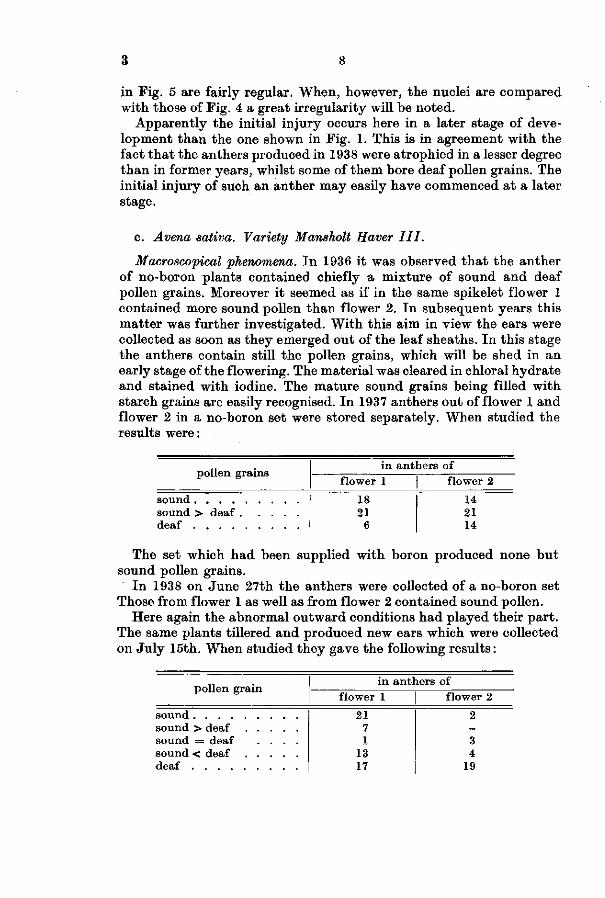
Macroscopical phenomena. I n 1936 it was observed t ha t the anther of no-boron plants contained chiefly a mixture of sound and deaf pollen grains. Moreover it seemed as if in the same spikelet flower 1 contained more sound pollen than flower 2. In subsequent years this mat ter was further investigated. With this aim in view the ears were collected as soon as they emerged out of the leaf sheaths. In this stage the anthers contain still the pollen grains, which will be shed in an early stage of the flowering. The material was cleared in chloral hydrate and stained with iodine. The mature sound grains being filled with starch grains are easily recognised. In 1937 anthers out of flower 1 and flower 2 in a no-boron set were stored separately. When studied the results were:
pollen grains
sound sound > deaf . . . deaf
in anthers of flower 1 flower 2
18 21
6
14 21 14
The set which had been supplied with boron produced none bu t sound pollen grains.
I n 1938 on June 27th the anthers were collected of a no-boron set Those from flower 1 as well as from flower 2 contained sound pollen.
Here again the abnormal outward conditions had played their par t . The same plants tillered and produced new ears which were collected on Ju ly 15th. When studied they gave the following results:
pollen grain
sound > deaf sound = deaf . . . .
in anthers of flower 1 | flower 2
21 7 1
13 17
2 -3 4
19
9 3
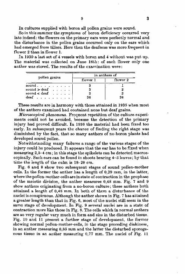
In cultures supplied with boron all pollen grains were sound. So in this summer the symptoms of boron deficiency occurred very
late indeed : the flowers on the pr imary ears were perfectly normal and the disturbance in the pollen grains occurred only on the ears which had emerged from tillers. Here then the deafness was more frequent in flower 2 t han in flower 1.
I n 1939 a last set of 4 vessels with boron and 4 without was pu t up . The material was collected on June 16th: of each flower only one anther was stored. The results of the examination were :
pollen grains
sound > deaf
in flower 1
3 3 2
36
anthers of | flower 2
1 2 2
38
These results are in harmony with those a t ta ined in 1935 when most of the anthers examined had contained none bu t deaf grains.
Microscopical phenomena. F requent repetition of the culture experiments could not be avoided, because the detection of the pr imary injury had proved difficult. I n 1936 the material had been fixed too early. I n subsequent years the chance of finding the r ight stage was diminished by the fact, t ha t so many anthers of no-boron plants had developed sound pollen.
Notwithstanding many failures a range of the various stages of the injury could be produced. I t appears t h a t the ear has to be fixed when measuring 2,5-4 cm ; in this stage the spikelets can be detected macros-copically. Such ears can be found in shoots bearing 4-5 leaves ; by t h a t t ime the length of the culm is 18-20 cm.
Fig. 6 and 8 show two subsequent stages of sound pollen-mother cells. I n the former the anther has a length of 0,39 mm, in the latter, where the pollen-mother-cells are in s tate of contraction in the prophase of the meiotic division, the anther measures 0,48 mm. Fig. 7 and 9 show anthers originating from a no-boron culture ; these anthers both a t tained a length of 0,48 mm. In both of them a disturbance of the nuclei is conspicuous. Although the anther shown in Fig. 7 has a t ta ined a greater length than t ha t in Fig. 6, most of the nuclei still seem in the same stage of development. I n Fig. 9 several nuclei are in a s tate of contraction more like those in Fig. 8. The cells which in normal anthers are so very regular vary much in form and size in the disturbed tissue. Fig. 10 and 11 present a further stage of development, the former showing normal pollen-mother-cells, in the stage preceding diakinese, in an anther measuring 0,85 mm and the lat ter the disturbed sporoge-nous tissue in an anther measuring 0,77 mm. The nuclei of Fig. 11

3 10
seem to be in various stages of the prophase of the meiotic division. In some cells which contain two nuclei the formation of the cell wall has apparently been inhibited. The next stage in development is presented in Fig. 12 and 13; here the telephase of a normal meiotic division is shown. Fig. 14 and 15 represent an anther in a similar stage of development with free lying pollen-mother-cells very definitely deformed. The length of this anther is 0,74 mm. Again it is the damage of the nucleus which is most conspicuous. These abnormal cells however will still be able to produce the normal exine of a pollen grain. Fig. 16 shows the next s tage: pollen grains with very disturbed irregular nuclei ; one of the nuclei of the pollen grain depicted a t the base of the illustration is apparently in a s tate of division. Normal young pollen grains before and during the formation of generative nuclei are shown in Fig. 17. The last stage, the deaf pollen grain is pictured in Fig. 18. The wall of a pollen grain with porus can be clearly recognised; no t race however of any protoplasmatic content can be noted. The sound young pollen grains of Fig. 19 contain the three nuclei and small starch grains the number of which will increase during maturat ion.
I n no instance could any disturbance in the wall of the thec'a be detected; the damage remains strictly confined to the sporogenous tissue.
d. Secale cereale. Variety Petkus spring rye.
Macroscopical phenomena. Experiments in 1934 and 1935 had shown t ha t in the absence of boron rye p lants developed ears the tops of which bore withered glumes. Over the whole of the ear no flowering occurred.
I n 1936 cultures of rye without any additional supply of boron were pu t up in order to detect a presumed disturbance in the vegetative tissue a t the apex of the ear. Budding ears fixed a t a length of 1,2 cm appeared, however, to have been too immature.
I n 1938 a no-B set of p lants was pu t up . These p lants suffered severely from cold weather conditions. Growing poorly the roots were slow in reaching the unsolved ferriphosphate a t the bot tom of the culture vessels. Although the iron deficiency was corrected by adding ferricitrate, the development remained poor. Ears were collected as soon as they emerged from the leaf sheaths. Contrary to t he results of former years this year only few withered tops occurred : merely 8 tops were disturbed whilst 17 were healthy. Whilst in former years no flowering could be noted, now, notwithstanding the absence of boron many stamens pushed through the glumes. Among these were anthers which only bore deaf pollen. They did not dehisce and therefore t h e pollen was not shed. Atrophied anthers without any pollen occurred

11 3
under the glumes. In a general way the production of sound pollen decreased from the base of the ear upward ; in cases where the pollen at the base of the ear was deaf, the anthers at the apex were atrophied and, whenever all the pollen at the base was sound, both sound and deaf pollen occurred at the apex. Here again in the spikelets anthers of flower 1 were better developed than of flower 2. I t shows very clearly that the plant will put to use any available trace of boron and that the sequel of development of the plant parts determines the degree of the injury.
All pistils developed quite normally and an examination on July 12th showed that grain had developed although very small when compared with the plants supplied with boron.
No correlation existed between the disturbance in the apex of the ear and in the anthers. Thus among the 8 ears with a more or less shrivelled apex 4 produced sound pollen at their base; among 17 ears with sound apices 8 produced sound pollen at their base, one even up to its apex, whilst two bore atrophied anthers over the whole of their length.
Microscopical symptoms. As the occurrence of shrivelled tops was rare in this year, the attempts to find early stages of this disturbance did not meet with success. Disturbance of the anthers was frequent. Pig. 20 and 21 show an anther of 1,52 mm in length where the initial damage appears to have occurred at a late stage of development. The pollen-mother-cells are very irregular in form, the nuclei vary much in size and the occurrence of three nuclei in one cell points to an inhibition of the formation of a cell wall. This disturbance has much analogy with the stage of Avena in Fig. 11 ; it is considered to be the initial stage of the deaf pollen grains, such as occurred in 1938 in the rye plants deprived of boron.
e. Hordeum vulgare. Variety Mansholt tweerijige gerst.
Macroscopical phenomena. Experiments in 1933 and 1936 had shown that barley grown in a medium deprived of boron did not shoot its ears; young ears however were present in the leaf sheaths. In 1936 vegetative injury was noted microscopically. In 1937, however, only a few young ears could be fixed and the plants were not grown until they were mature. In 1938 further experiments were carried out.
In this year like in Avena sativa the deficiency symptoms occurred much later than had been noted up till then. All ears emerged out of the leaves. Fifteen ears, collected as soon as they had emerged, were examined. In two ears the tips, which had remained wholly meriste-matic, bordered directly on a normally developed region, and, in two cases a bare somewhat curved mature top zone could be clearly

3 12
distinguished from, the lower axis. These were the only vegetative disturbances noted.
I n the basal pa r t of all ears sound pollen occurred ; one produced sound pollen over the whole length of the axis. Six of them produced deaf pollen grains in the apical region and in 9 the anthers a t the apex were atrophied. Again the pistils in all of the flowers were sound.
Microscopical phenomena. I n 1936 five ears were fixed, when these had a t tained a length of 0,8-2,5 cm. In all of them a disturbance of the vegetative par t s of the apices was conspicuous. Fig. 22 represents such a disturbance in a budding ear of 0,3 cm; i t should be compared with a sound one of the same length depicted in Fig. 23. Fig. 24 shows an initial stage of this injury in an ear 0,5 cm in length. The parenchymatous cells adjacent to the procambium s t rand are somewhat extended, their walls are swollen and they contain large nuclei. The cells of the procambium are undisturbed. The disturbance of the vascular s t rand in a further stage is shown in Fig. 25. I t s cells are crushed by pressure of the extended cells in the centre.
I n one ear of 1 cm in length a disturbance in the vascular s t rand could be followed downward in the rachis (Photo 26). The spikelets are a t rophied; this a trophy, however, is secondary and the result of the damage of the vascular s t rands supplying the spikelets.
I n 1937 6 ears of a no-boron set were fixed, when these had a t tained a length of 0,4-5 cm. At t h a t stage the p lants had developed 4-5 leaves and the length of the culm was 16 cm. In one ear of 0,4 cm a damage of the apex was detected and in all of them a disturbance of the sporoge-nous tissue of the anthers. An initial stage occurring in an anther of 0,53 mm (length of the ear 1,5 cm) is represented in fig. 27. The division of t he nuclei, and, in i ts sequence the division of the cells is quite disturbed. An initial injury occurring a t this stage of development is considered to result ul t imately in an atrophied anther with remains of disturbed sporogenous tissue, such as is shown in photo 28 taken from 1938 material.
I n 1938 9 budding ears out of a no-boron set have been fixed varying in length from 0,2-5,5 cm. In 3 measuring 0,3 cm, vegetative injury of the t ip was conspicuous, 3 of the same length were vegetatively sound as well as 3 larger ones of 4,5-5,5 cm. Individually fixed spikelets of these larger ears showed t ha t the initial injury to the sporogenous tissue occurs a t a later stage t h an t ha t shown in Fig. 27. Fig. 29 represents an anther measuring 0,8 mm. Apparently several large cells with giant nuclei are quite disturbed, bu t along with these small cells occur, which have continued their development. I n Fig. 31 (length of the anther 1 mm) such smaller cells of various shape form the majority. The same an ther is represented in photo 30 and may be compared with a sound one of similar length (0,99 mm) which contains pollen-mother-

13 3
cells all in a same stage of development (Photo 32). The damaged tissue in Fig. 31 is much like t h a t noted in rye (Fig. 20). The writer considers these disturbances as the initial stages in the development of the deaf pollen grains.
f. Discussion.
When the disturbance in the anthers in the various cereals is compared, it appears in all cases to be of the same nature . The injury is always strictly limited to the sporogenous tissue.
The initial disturbance occurs in the nuclei. A coalescence of several nuclei may give rise to giant nuclei. Neighbouring cells may contain quite different stages in the prophase of the meiotic division all however in disturbed condition.
If the detremental effect of the boron deficiency has manifested itself in the early stages of the developing sporogenous tissue, the formation of the cells is inhibited. When the disturbance sets in a t a later stage, cell walls are formed. The resulting sporogenous tissue consists of cells which vary greatly in size and shape; the cell walls, however, always appear quite normal. Even the wall of a mature pollen grain in which the content has collapsed, cannot be distinguished from the wall of a sound one.
The four cell layers which make up the wall of the theca, the t ape tum included, are always undisturbed.
As far as the embryosac-mother-cell and its surrounding tissue could be studied in sections of the material grown without boron, i t was always quite normal.
The walls of the anthers have a high content of s tarch grains when the pollen-mother-cells are in meiotic division. I t could be noted t ha t in the anthers of Avena where the pollen-mother-cells were disturbed (Fig. 14) the starch grains occurred as abundantly as in sound anthers. The deafness of the pollen grains may not be ascribed to a lack in the supply of carbohydrates.
2 . BORON DEFICIENCY IN SWEDISH TURNIPS (BRASSICA NAPA V. NAPO-
BRASSICA)
a. General pathology.
I n the roots of Swedish turnip (Brassica napus v. napo-brassica (L) Peterm.) material was found, which was very suitable for a detailed s tudy of the anatomical s tructure of vegetative tissue affected by boron s tarvation.
I n various par ts of the world a disease of swedes known as ,,brown hea r f 'occurs , which has been recognised as caused by boron deficiency.

3 14
I n the Netherlands i t occurs in a district along the river Waal, where, near the village of Brakel, swedes are grown every year after a crop of early potatoes, in a heavy clay soul with a high content of calcium carbonate. Moreover i t has been noted on poor sandy soils, where poor results have resulted in the abandoning of the cultivation of swedes.
Independently of each other the disease has been studied by various au thors : W H I T E H E A D (1935) in North Wales, JAMALAINEN (1935) in Finland and O ' B R I E N and D E N N I S (1935) in Scotland.
The foliage of the p lants affected by boron deficiency is quite healthy ; the production of sound young leaves goes on whilst roots are already affected. Externally no disturbance in the roots can be observed. When however the roots are cut open very marked injury can be noted. I n the description of the internal injury D E N N I S and O ' B R I E N
(1937) is ci ted:
The diseased tissue is seen to occur in patches arranged in zones which occupy the outer portion of the swollen xylem of the root. No case has been observed where the discoloration occurred outside the cambium ring in phloem or cortical tissue. I n very severe cases the mottling may involve the whole centre of the root ; more often a more or less complete ring occurs with healthy tissue in the centre. I n a longitudinal section the brown patches appear elongated, parallel to the surface and converge towards the base of the organ. Only in the most severe cases do they extend upward beyond the shoulder towards the neck. The colour of affected tissue varies. In some cases it presents a greyish watersoaked appearance, more often it is definitely of a yellowish-brown hue.
Culture in nutrient solution. I t has been tried by various authors to induce the deficiency symptoms in water or sand cultures. JAMA-
LAINEN, working with sand cultures, observed symptoms of deficiency in cultures which had received 0, 0,1 or 1 mg H^BO3. P lants supplied with 10 mg H 3 B 0 3 produced healthy roots. From 1 mg upward, however, the foliage was injured by excess of boron.
H I L L and GRANT (1935) carried out experiments with sand cultures to determine the need of boron of turnips. When no boron was supplied merely a thin root developed. 10,08 mg H 3 B 0 3 sufficed for the production of well sized roots, which however proved hollowhearted. 20, 16 and 30, 21 mg sufficed for the production of roots affected only in a very small degree. Very definite symptoms in the foliage occurred when the lower doses were supplied. This trouble of „hollow-heart" has not been compared with the „brown hear t " known in field practice. Presumably a bacterial rot has in the former case invaded the region primarily affected by boron deficiency.
D E N N I S and O ' B R I E N have carried out a number of water culture experiments. I n these however deficiency symptoms in the foliage were so severe t h a t practically no thickening of the roots occurred. The dose of boron might have been too low. In sand cultures in the set
15 3
not supplied with boron the roots were much smaller than in the set provided with 0,0125 mg borax ; only one out of five in the former set had brown spots inside the cambium ring. These authors conclude : On the whole, whilst the results of the culture experiments confirm and extend those of JAMALAINEN and HILL and GRANT, they indicate that it is a difficult matter to reproduce exactly symptoms of brown-heart under artificial conditions.
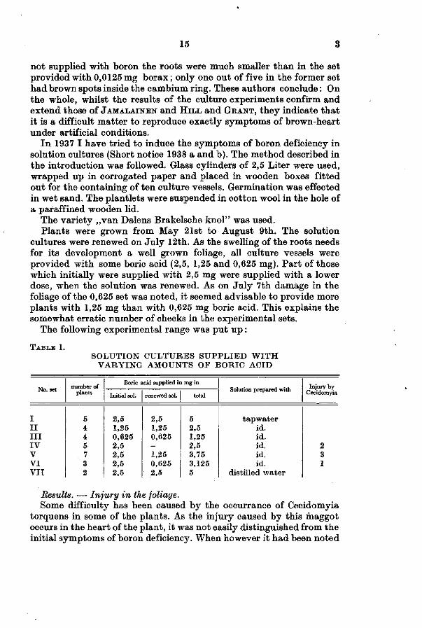
In 1937 I have tried to induce the symptoms of boron deficiency in solution cultures (Short notice 1938 a and b). The method described in the introduction was followed. Glass cylinders of 2,5 Liter were used, wrapped up in corrogated paper and placed in wooden boxes fitted out for the containing of ten culture vessels. Germination was effected in wet sand. The plantlets were suspended in cotton wool in the hole of a paraffined wooden lid.
The variety ,,van Dalens Brakeische knol" was used. Plants were grown from May 21st to August 9th. The solution
cultures were renewed on July 12th. As the swelling of the roots needs for its development a well grown foliage, all culture vessels were provided with some boric acid (2,5, 1,25 and 0,625 mg). Part of those which initially were supplied with 2,5 mg were supplied with a lower dose, when the solution was renewed. As on July 7th damage in the foliage of the 0,625 set was noted, it seemed advisable to provide more plants with 1,25 mg than with 0,625 mg boric acid. This explains the somewhat erratic number of checks in the experimental sets.
The following experimental range was put up :
TABLE 1. SOLUTION CULTURES SUPPLIED W I T H
VARYING AMOUNTS OF BORIC ACID
No. set number of
plants
Boric acid supplied in mg in
Initial sol. renewed sol. total Solution prepared with Injury by
Cecidomyia
I I I I I I IV V VI VII
5 4 4 5 7 3 2
2,5 1,25 0,625 2,5 2,5 2,5 2,5
2,5 1,25 0,625 -1,25 0,625 2,5
5 2,5 1,25 2,5 3,75 3,125 5
tapwater id. id. id. id. id.
distilled water
2 3 1
Results. — Injury in the foliage. Some difficulty has been caused by the occurrance of Cecidomyia
torquens in some of the plants. As the injury caused by this maggot occurs in the heart of the plant, it was not easily distinguished from the initial symptoms of boron deficiency. When however it had been noted

3 16
t ha t it always caused a suberation of the leaf base, i t could be recognised in an early stage. A few plants had to be discarded for this reason; they are mentioned in Tab. 1.
The symptoms caused by boron deficiency consist of a curling of the youngest leaves, often accompanied by a marginal browning. Somewhat older leaves show besides curling a bending to the lower surface of the leaf t ips and a chlorotic colour. The mature outer leaves take on a pinkish violet hue along the margins and the lower surface, whilst the individual leaf t ips and the petioles bend downward. The horizontal inclination of the foliage is very conspicuous. (Fig. 33 on the r ight). These symptoms are in agreement with figures presented by H I L L and GRANT.
The initial symptoms in the heart of the plants could be noted in set I I I on Ju ly 7th, in set I I on Ju ly 23rd. I n set I the foliage was healthy up till the end. I n set IV the initial symptoms were produced on July 23rd, in V on August 2nd and in VI on Ju ly 28th.
Thus the amount of boron supplied to the cultures determines the stage of development in which the initial symptoms of boron deficiency occur in the foliage.
Injury in the roots. Since all roots were cut between August 2nd and 9th this experiment could not furnish da ta about the onset of the injury.
When examined the turnips had a t tained a size varying from 4 ,5-8 cm in height and 4,5-7 cm in width. No correlation between supply of boron and the size could be noted, except in set I I I which developed smaller turnips.
The roots of all plants, supplied with less t han 5 mg boric acid were affected. The disturbance consisted of an enlarged cambium zone, very conspicuous by its water-soaked appearance, and of brownish water-soaked patches in the interior of the root. Photo 34 represents a heal thy root taken out of set I , a diseased root of set IV which had merely been supplied with boron in the earlier period, and one of set I I I which had been supplied with a weak boron solution during the whole of its development. Four roots supplied with 5 mg boric acid in tapwater and two in distilled water had remained perfectly healthy. I n only one root from the tapwater set, although the foliage of the plant was undisturbed, an enlarged cambium zone and a slight discoloration of the lower inner par ts could be noted. This p lant is represented in the middle of Fig. 33 ; on its left figures a heal thy root and on its r ight a p lant of set VI with a diseased top and root.
When the results of this experiment are compared with the injury as it is known in field practice, they only par t ly agree. Never has any injury of the foliage been noted in the field; even when the roots were very badly affected the top would still produce healthy young leaves.

Fig. 35 represents a severely diseased root dug up in a field. The brown water-soaked region in its interior is very conspicuous. No injury of the cambium zone along the cortex, however, occurs. In fact the writer noted merely in one very badly affected turnip injury of the cambium zone, in the shoulder and upper part of the root. This root is represented in Fig. 36. JAMALAINEN mentions as of rare occurrence radially extended cells in the cambium.
The fact, that the symptoms induced artificially do not wholly agree with the symptoms occurring in the field, has probably to be ascribed to the fact that in the former case the symptoms set in at an earlier stage. The one plant of set I, where injury in the roots occurred whilst the foliage was still undamaged, is nearest to the reproduction of the field symptoms. (Photo 33 central plant). This means that cultures should be continued during a longer period with a good supply of boron, in order that deficiency symptoms may manifest themselves at a later stage. This however is hard to accomplish, as culture solutions with large mature plants are apt to get spoiled. Such plants will easily shed part of the mature thin roots and this material will nourish anaerobic bacteria; on hot days toxic sulphides may easily spoil the solution! The best way to prevent these mishaps is to make use of sand cultures. An attempt at this however did not meet with success, as the glass sand used apparently still contained an appreciable amount of boron and no deficiency symptoms occurred.
The microscopical structure of the roots grown in the culture solution showed however a marked similarity to the structure of diseased turnips in the field. In this way proof is furnished that boron deficiency is the primary cause of the disease of „brown heart".
b. The structure of sound roots.
The thickened root of a swede is a very favorable object for the study of histological changes induced by boron deficiency. In a vegetation point, the common initial place of occurrence in many plant species, the symptoms will soon be masked by a disintegration of the tissue, as soon as the nutritional supply by the vascular strands is inhibited. In the swedes however the initial symptoms occur in the roots and the disturbed tissue does not collapse, but remains in a living condition. The injured region increases and during a long period initial and subsequent symptoms may both be studied. In this way the swede furnishes very satisfactory material.
For the microscopical study of the roots the staining technique described in the introduction has been adopted.
The cellulose is coloured blue (represented by close dotting in the further figures) and the middle lamella in the healthy tissue is very
3 18
darkly stained. Matter which stains darkly occurs much more frequently in the diseased tissue. Ruthenium red can stain this substance and i t takes on an orange red colour when t reated with saffranine. The latter characteristics point to the presence of pectic substance. In the sketches this substance is indicated by black shading. The walls of the wood vessels either did not stain (indicated by a sparse dotting) or gave the blue stain of cellulose. The nuclei and protoplasm were stained by the haematoxylin. When diseased tissue was stained by Sudan I I I the cell walls did not t ake up the stain. Sections were cut 7 fi or 10 [i thick according to the na ture of the material investigated.
JAMALAINEN refers to a s tudy of SÖDING (1924) on the anatomy of Brassica napus v. napobrassica. The lat ter paper has been of great help in understanding the complicated anatomical s tructure of the thickened root of a swede.
The s tructure and secondary growth are in several respects abnormal. The cells of the cambium are, in longitudinal sections, fusiform, those of the cambium, parenchymatous. The fusiform cells produced by the cambium form transverse walls, and in the interior of the root all parenchymatous cells are of similar shape although of different origin. I n the region bordering on the cambium zone xylem s t rands occur in radial rows. Scattered over the interior of the tuber is an anas-tomising net of vascular bundles, which have originated from the parenchymatous cells. When sections of the internal s tructure are %
studied all stages of development of these secondary vascular bundles may be seen. The s tructure of the mature vascular bundle varies. Usually there is a central phloem which is surrounded by a sheath of very regular fusiform cells in radial rows (Reihenkambium of SÖDING) ,
producing parenchyma in the periphery. I n the outer region of these vascular bundles xylem strands may occur (Fig. 54, 55). Xylem rarely occurs centrally. Groups of xylem vessels occur scattered in the parenchyma. I n well developed roots the main cambium zone is still in existence ; the secondary increase in thickness, however, is chiefly due to the production of cells by the sheaths round the secondary vascular bundle.
c. Comparision of sound and affected tissue.
The cambium. The disturbance of the cambium will be described in the first place, as an injury of meristematic tissue is of fundamental importance. I n Photo 37 in a cross section through a sound root cambium and r ay cambium are represented. The cambium zone consists of a few cell layers. I n Photo 38 the disturbance of the cambium is represented such as i t has been induced by cultivation in a medium, low in boron content (henceforth referred to as „artificial" induction). The
19 3
normal narrow zone of cambium cells has developed into a wide region (on Photo 38 only 1/i of its actual width is represented) of thin-walled cells of irregular shape. Fig. 39 represents the injured ray cambium. The nuclei appear undisturbed and no intercellular spaces occur. Fig. 40 shows the cambium adjacent to the phloem. Part of the cells are of the nature of those occurring in Fig. 39; besides these, however, many cells with thickened darkly stained walls occur. These presumably are cells which under normal conditions would have developed into phloem cells.
Photo 41 represents a cross-section through the shoulder part of the root which is represented in Photo 36; its cambium was disturbed. This naturally produced disturbance appears to be of the same nature as the artificially induced symptoms.
The longitudinal sections through the cambium of sound roots, represented in Fig. 42, show the parenchymatous cells of the ray cambium and in Photo 43 the cambium with its elongated fusiform cells can be discerned. Photo 44 and Fig. 45 represent radial sections through roots in which boron deficiency symptoms have been artificially induced. The change in structure of the cambium is very conspicuous : the normally elongated thin-walled cells have developed into isodia-metric cells of various shape. The border zone of cambium and phloem is represented under higher magnification in Photo 46. On the right in the thin-walled tissue some elongated cells with thicker walls occur and a swollen sieve tube is conspicuous. The impression is given that the ultimate shape of cells in the injured tissue depends on the stage of development attained at the moment the boron starvation in the individual cell sets in. Entirely meristematic cells will change into the thin-walled cells of irregular shape. Those which had already become differentiated into bastlike cells may still be recognised by thicker walls and a more elongated shape.
The injury caused by boron deficiency in meristematic tissue consists in a disturbance of the normal development of the cells. They keep on dividing, but the normal differentiating and maturating process is inhibited. The ultimate shape of the cells seems to be determined by their local position among other cells. No abnormal cell content, as is usually met with in diseased tissue, was noted in any of the disturbed cambium cells. In the disturbed tissue no starch grains occur.
The periderm. A similar disturbance in the normal development could be noted in the periderm of artificially and naturally affected roots.
Fig. 47 is a cross section of the periderm of a normal root: the phel-lem consists of a few cell layers and no phelloderm occurs. In Fig. 48 a longitudinal section is represented where a lenticel-like structure occurs in the phellem. In Fig. 49 in a cross section of a naturally affected

3 20
root the development of an extensive tissue bordering on the inner side of the phellogen is conspicuous. In Fig. 50 the longitudinal section of an artificially affected root is shown: instead of being slightly elongated the cells of the periderm are radially extended.
The disturbance of the tissue originated in the phellogen is of exactly the same nature as the disturbance in the tissue originated in the ray cambium. Again the normal sequel of differentiation and maturation appears to be inhibited.
Vascular bundles. In the interior of the normal roots secondary vascular bundles occur in all stages of development. The main direction is vertical but much anastomising occurs and in transverse sections of the root vascular bundles may be cut longitudinally.
Initial stages of the development of the central phloem are represented in Fig. 51 and 52. The swollen cell walls shown in the centre of Fig. 52 stain like cellulose. Between the swollen walls some pectic substance occurs. In Fig. 53 the central phloem of a more mature vascular bundle is represented. The swelling of the walls has increased and the lumen of the cells is narrowed. Here and there strands of pectic substance can be noted in the walls. I t is not clear whether the thick walls consist of one swollen wall or of the walls of a number of compressed cells. For a thorough understanding of the symptoms of the diseased tissue, the elucidation of this point is of importance, but notwithstanding the fact that a large number of preparations were carefully studied, no definite conclusion could be drawn.
Photo 54 shows a mature vascular bundle and Fig. 55 represents a part of it under higher magnification. The central primary phloem, the younger phloem parts, the cambium sheath of fusiform cells and the xylem in the outer region can be clearly seen. The phloem bundles alternate with radially extended rows of fusiform cells. In Fig. 55 the thickened darkly stained walls in the central phloem are conspicuous.
The longitudical structure of a normal vascular bundle is shown in Fig. 56. Fig. 57 represents the central primary phloem under high magnification. The sieve tubes and companion cells can be discerned. The cell walls, and, what seems to be a swollen middle lamella, are darkly stained. Again it cannot be definitely ascertained whether the swollen cell walls are composed of one or several cells. The phloem bordering on the sheath cells is shown in Fig. 58.
In Fig. 60 the phloem of a vascular bundle occurring naturally in affected tissue is represented. I t should be compared with Fig. 59, where part of a sound vascular bundle is drawn under the same magnification. The general structure of sound and affected vascular bundles are of the same nature; the individual cells in the former, however, are less clearly defined in shape, the cell walls are thinner and the cells appear somewhat swollen. The most conspicuous symptom is the

21 3
occurrence of material occupying the cell lumen. For the sake of convenience this material will be further referred to as „plugging mat ter" .
The colour of this substance is darker t han the cellulose bu t lighter t han the pectic substance. The thickened walls of the central phloem have taken on a lighter shade ; the pectic substance seems to have disappeared and the walls give the cellulose stain.
Fig. 61 and 62 give a more detailed impression of the disturbance in the central phloem in cross section. This stage although rarely met in the microscopic preparations studied may elucidate the happenings in the affected cells. Again the thickened walls have taken on the blue stain of cellulose. Some of the cell walls appear to have disintegrated into separate narrow strands. I n some others darker stained s trands are embedded in the wall substance. Plugging mat ter in- the lumen of the cells is very frequent and conspicuous. When this same preparation after a destaining was restained with chlor zinc iodide the plugging material took on the violet shade of cellulose.
The nature of the disturbance in the phloem which is represented in Fig. 61 and 62 is perhaps comprehensible, if i t is assumed t ha t the cell walls have been subjected to a dissolving process. P a r t of the sab-stance of the cell walls might have formed the so-called „plugging ma t t e r " which stains darker than cellulose bu t lighter than pectic substance.
Fig. 63 shows another stage in the pathological anatomy of boron deficient cells. All cut cells show what seems a thin bu t uneven layer of cellulose. When, however, succeeding sections were studied the mat ter was recognised as a foamy mass filling the whole cell. Although this substance takes on a similar stain as cellulose, it can, however, be recognised in the preparations of the diseased tissue by its finely reticulate texture .
I n order to s tudy this substance further some preparations were destained in weak acid after the balsam had been solved. The blue stain could not be made to fade out wholly and a slight blue coloration remained. After t rea tment with chlor zinc iodide the foamy mat ter could be clearly distinguished from cellulose : it took on very distinctly a blueish green colour. As the matter before t rea tment with chlor zinc iodide was blue, the shade induced by this stain is assumed to be yellow. Protoplasm stained yellow and cellulose the ordinary violet. A t rea tment with Sudan I I I , however, gave a more positive result : t he foamy mat ter took on very definitely the orange red stain, whilst the surrounding cell walls remained unstained. Therefore this substance probably contains fat groups. I n the drawings an a t t empt has been made t o represent this substance in such a way to suggest i ts foamy character. The substance plugging one cell in Fig. 60 has the

3 22
properties of the „foamy" matter. Thus it appears of another nature than the plugging substance in Fig. 61 and 62.
In Fig. 64 another aspect of the downbreak of vascular strands is shown. Greatly extended fusiform cells with distinct pits and thin strands of cellulose occur in the cell lumen.
Apparently a downbreak of cell walls occurs along various lines. I t is to be noted however that in all these cases the nuclei appear quite normal. The initial disturbance in mature cells occurs in the walls.
Thus far only the downbreak of mature cells has been described and though the symptoms of disintegration have been carefully studied a clear concept of the nature of the diseased tissue has not yet been arrived at. I t was expected that vascular strands would occur as frequently in diseased tissue as in sound ; after the study of diseased tissue, however, it appeared that vascular bundles were here of much rarer occurrence. This fact could hardly be explained by non-formation ab origine; in fact the initial disturbance caused by boron starvation occurs in a mature stage, when the anatomical structure of the thickened root is completely developed. On the other hand a tissue of isodiametric cells with somewhat enlarged darkly stained intercellular spaces was frequently observed in diseased tissue (Fig. 69 and 70).
This discrepancy between sound and diseased tissue could be elucidated as soon as an inverse process in the etiology of boron deficiency, e.g. the formation of young cells, had been observed. Preparations which show this phenomenon satisfactorily were rare, however, because of the extreme thinness of the cell walls. Fig. 65 to 67 deal with this phase of the process.
On the left in Fig. 65 a normal sieve tube with its companion cells occurs. On the right the initial forms of a sieve tube and its companion cells, although extended, may still be discerned. Here and there the walls of the sieve tube are thin and young cells, with very delicate walls, have been formed in the lumen of the initial phloem cells. Apparently mature phloem changes back into a meristematic condition In Fig. 66 a further stage of this metamorphosis is given. The original phloem cell, probably a greatly swollen sieve tube, is changing back into thin-walled tissue which fills its original lumen. In the upper part it seems as if the original companion cell is still present. What is considered to be a still later phase in this process is represented in Fig. 67. On the still existing thickened wall borders a tissue of isodiametric cells with large intercellular spaces mostly filled with the foamy substance described above. In the tissue represented in Fig. 68 the intercellular spaces have still increased in size and in some cases adjoining cells project into the spaces. The formation of a new tissue is very evident. Ultimately the cells become thickwalled (Fig. 69 and 70) and stain a dark colour. Such a tissue frequently occurs in diseased regions.

23 3
If the process of regression is once understood, it becomes clear that in Fig. 61 this phenomenon may explain the co-existence of swollen cell walls and of very thin ones. Fig. 71 shows a longitudinal section of a vascular strand which has suffered a partial regression. Conspicuous are the isodiametric cells of secondary origin which surround the fusiform cells of the vascular strand filled with the foamy substance. Fig. 72 to 74 represent various histological changes in vascular strands. In Fig. 72 many young cells have developed in the more central part, where the thick walls have partly lost their pectic substance. In Fig. 73 the fusiform cells alternating with the phloem show enlarged intercellular spaces filled with pectic substance. Finally Fig. 74 shows a sheath of extended cells surrounding the vascular bundle. Such elongated cells occur frequently in diseased tissue, 'generally in rows accompanied by the isodiametric tissue (Photo 75.) The original vascular bundle is quite changed into these tissues.
The various histological anomalies may be explained, when the stage of development of the individual cells, at the time that the effects of the boron deficiency sets in, is considered. The differentiation into fusiform cells of Fig. 73 is considered to have taken place before the deficiency occurred, whilst the elongated cells are thought to have originated from dividing sheath cells before their maturation into parenchymatous cells was complete.
In meristematic cells suffering from boron starvation the maturation process is inhibited, as has already been described for cambium cells. Photo 76 clearly shows such a stage occurring in the sheath cells of a vascular strand, which has given rise to a tissue quite similar to the tissue arising from a boron deficient cambium.
Summarising the phenomena observed in diseased regions of the inner root, several processes were noted to occur side by side : a. a disintegration of mature elements. b. the origin of young cells in these débris and their further maturation.
Attention has to be drawn to the fact, that in the cortex of sound roots, primary phloem may be met in a state of disintegration which resembles the stages shown in Fig. 61-63. Some of the cells may be plugged with substance which is only slightly stained with Sudan I I I . The primary phloem is here probably subject to a similar process of disintegration as the phloem in secondary vascular bundles deficient in boron. In the sound cortex this process is due to a normal stage in the development, namely blocking of older parts of the phloem. This may mean, that the disintegration of the cell walls in the phloem of the vascular bundles is only indirectly connected with the boron deficiency : the normal course of the histological development might be disturbed by a disturbance of the regulating mechanism of the plant as a whole.
I t is worth while to compare all these phenomena occurring in natu-

3 24
rally injured tissue with phenomena accompanying an artificially induced boron deficiency.
Photo 77 shows the injury in the fusiform cells of a vascular s trand, and quite agrees with photo 76.
Fig. 78 and 79 show the symptoms occurring in a sieve tube of a vascular s t rand where the s tarvation had been artificially induced. Again the walls of the sieve tube vary in thickness and young cells with large nuclei and very thin walls develop in the original lumen.
No room is left for doubt t ha t the microscopic symptoms occurring in field material may be perfectly reproduced under controlled conditions in nutr ient solutions.
Xylem. I t has been mentioned on page 18 t ha t xylem occurs less frequently than phloem in the thickened roots. I n the changes in the tissue due to lack of boron they play a minor par t .
I n young uninjured xylem vessels the walls containing cellulose take on a blue stain. I n older vessels the walls remain unstained. When res-tained with aniline sulphate, the walls t ake on the yellow stain of lignin, (represented in Fig. 80 to 84 by sparse dotting). I n Fig. 80 which represents injured xylem, „plugging mat te r" stained as cellulose, pectic substance in the lumen of the vessels, and a darkly stained layer bordering the vessels a t various points are conspicuous. Fig. 81 and 82 drawn under a magnification of 1250 X give more minute details. They show a thick layer of pectic substance occurring between the lignified par t of the wall and a tertiary cell wall containing cellulose. The bordered pits and their elongated and widened canals take on the stain of pectic material. I n Fig. 83 where is represented a longitudinal section, a layer which is detached from the lignified pa r t of the wall, and which represents the „plugging mat ter" , is very conspicuous. When restained with chlor zinc iodide this layer stains violet, t hus indicating cellulose and pectic substance.
The disintegration of the xylem par ts may be followed in Fig. 80 to 83. Attention is drawn to the nuclei occurring in some vessels which indicate a living condition. I n Fig. 84 a formation of young cells in the disintegrated vessel is shown which occurred very rarely in the material studied. Here a phenomenon occurs which is analogous to the development of secondary tissue in the phloem parts . Generally the breakdown of the xylem, however, is not as thorough as in the phloem, and xylem vessels are frequently found in the stage pictured in Fig. 80.
Even in sound roots in the region bordering the cambium, where xylem strands occur in radial rows, vessels may be noted in a similar stage of disintegration. Although they occur much more frequently in t h e injured tissue, their occurrence may not be considered as symptomatic of boron deficiency.

25 3
Macroscopic aspect of injured tissue. When t he affected tissue is examined in unstained condition under the microscope, merely a slight discoloration can be noted in the cell walls and in the matter filling the intercellular spaces. A light yellow shade may occur, but many cells in the affected tissue are colourless. Here and there some brownish granulated mat ter occurs in the cell lumen. The yellowish brown macroscopical aspect had lead to the expectation of a more definite discoloration of the microscopic material. Most likely the brown aspect of the tissue is due to the brownish cell walls and cell content scattered throughout the more or less t ranslucent thin-walled eels.
I n sound roots small starch grains are scattered in all the parenchymatous cells. A general property of the affected tissue is the absence of starch grains. Initially this absence was considered to be the cause of the water-soaked appearance of diseased tissue. I n 1939, however, sound roots were examined which had survived an early very severe frost period. Although when cut open, they showed the usual milkwhite appearance, starch grains were absent. Hence the water-soaked aspect must be due to to some other cause. JAMALAINEN
ascribes it to the absence of intercellular spaces.
d. Discussion. I t is important to compare the observed boron deficiency symptoms
with those described by other authors. Cambium. WARINGTON describes a disturbance in the cambium of
Vicia faba where large radially elongated cells occur with prominent nuclei. JAMALAINEN s tates t h a t in very rare cases an extension of the cambium cells occurs in swedes with brown heart, and presents a very convincing photo. H O W E notes irregularly swollen cambium in roots of beets.
Enlarged cells. WARINGTON describes large cells occupying the place of a number of smaller cells in the roots of Vicia faba. MARTIN notes greatly enlarged cells in the vascular bundle sheaths in the leaves of sugar cane. VAN SCHREVEN describes an enormous enlargement of phloem in the roots of tobacco, numerous radial divisions taking place.
Swelling of middle lamella and intercellular spaces. O ' B R I E N and D E N N I S observe these symptoms in diseased tissue of swedes and VAN
SCHREVEN notes a brown discoloration beginning a t the point of junction of the cell walls of the parenchyma.
Plugging of the cell lumen. WARINGTON s tates t ha t the lumen of the tracheides in the growing point of Vicia faba is frequently, more or less completely blocked by some unknown substance and R O W E notes in sieve tubes of beet roots plugs of densely stained material, presumably callose. This may be developed as a solid rod through the cell or disposed as a peripheral layer.

3 26
Thin cell walls. ROWE describes phenomena in beet roots which appear very similar to those which occur in swedes: besides thick-walled cells of irregular shape she notes, that many cells of the ground tissue and a few in the diseased vascular strands appear to have undergone division, and that two daughter cells are separated by delicate cell walls.
Apparently the symptoms of boron starvation occurring in Swedish turnip and in the rachis of the ear of barley agree closely with the symptoms shown by other plants.
The conclusions drawn from the observations presented in this paper may therefore be valid for vegetative tissue in general.
The initial disturbance of vegetative tissue occurs in the cell walls. As the staining of the „plugging matter" agrees with the staining of the walls, it is probable that the „plugging matter", occurring in the lumen of phloem cells and of xylem vessels, is a result of a disintegration or dissolving of cell wall layers. I t was observed that the plugging never occurred where young secondary cells had developed. I t may be conceived that this „plugging matter" is a stage in the process commencing with the disintegration of the mature cells and culminating in the development of the secondary tissue which takes its place. This is corroborated by the fact that in xylem vessels the plugging is frequent and formation of secondary cells could very rarely be noted.
The foamy substance, which presumably contains fat groups, described on page 21, has been found in mature cells which may be in a primary stage of disintegration (Fig. 63), and in cells of the secondary tissue which are in the act of developing. x)
4 . ASPECTS OF THE PATHOLOGY OF BOEON DEFICIENCY
a. Etiology of the injury in Swedes.
Initial symptoms of boron starvation usually occur in the vegetation point. The well known type of this disease is the „heart rot" in sugar beets and mangolds where the initial disturbance in the foliage is followed by the injury in the roots. No disturbance of the foliage of swedes suffering from brown heart has, however, thus far been noted in the field. Although deficiency symptoms in the leaves of swedes cultivated in culture solutions had been frequently induced by the writer, nothing abnormal could be noted in the aerial parts of field plants suffering from brown heart. All plants examined were still in the act of producing sound young leaves.
x) After this paper was completed a, paper of MABY MC ARTHUR, Histology of some physiological disorders of the apple fruit, was published in Canadian Journal of Research Vol. 18, 1, p . 26, 1940. Her description of the disorder in apples agrees in many" details with the disorder in swedes.

27 3
With the determination of their boron content in view samples of 5 roots each were collected a in a field which had been recently turned from meadow into agricultural land, where practically all roots were diseased and 6 in a field where all roots examined were sound. In the roots with brown heart the injured inner part was separated from the healthy outer region. In the same way the outer tissue of the sound roots was separated from the inner tissue.
Dr LEHR had the kindness to make the estimations; following the revised method of Schulek and Vastagh (Hudig and Lehr 1939). Estimations were made in samples of 5 g.
The results are presented in Table 2.
TABLE 2. CONTENT OF BORON IN HEALTHY AND DISEASED SWEDES
Materia,! Par ts per million B
in dry weight in ash
Foliage : Sound plants Diseased plants
Sound roots: Inner region Outer region .
Diseased roots: Inner region Outer region .
25 23
12 13
8 13
173 155
185 217
90 231
In sound plants the boron content of the foliage is twice as high as that of the inner roots. In the healthy roots the difference in the content of the inner and outer region is negligible. The injured parts of the roots, however, is markedly lower in content than the undamaged outer region or the corresponding parts of sound roots. These figures suggest that a lack of boron in the diseased region is the cause of the injury. The boron content of the foliage is much higher than that of the roots in both sound and diseased plants.
Whilst this paper was being prepared, Dr LEHR X) kindly supplied the writer with further data he had compiled from the investigation of material from several experimental fields. In agreement with the results previously obtained it was found that sound plants had a higher boron content in the foliage than in the roots; but, contrary to previous results, the boron content of the foliage of diseased plants was in some cases much lower than that of the roots, and some diseased
*) After this paper was completed a paper of J . J . L B H B . De betekenis van borium voor de plant. (English Summary. Thé significance of boron for the p lant) . Thesis Utrecht, 1940 has been published.

3 28
roots had a higher boron content t han sound roots of the same field. I n 1938apaperof BRANDENBURG on the use of boron in agricultural
practice was published. He collected his material of Swedish turnips from soils where brown heart was apt to occur and his data cover a wide range of investigations. From these da ta it may be concluded t ha t the boron content of the foliage is always much higher t han t h a t of the roots and t ha t diseased foliage and roots are materially lower in content t han foliage and roots of sound plants.
The da ta from the various sources have only one point in common, namely, t h a t in sound plants the foliage has a higher content of boron than the roots. Hence an explanation of the sensitiveness to boron deficiency of the roots on the basis of a higher content of boron under normal conditions t han the foliage is out of the question.
So the initial disturbance in the roots still remains to be explained. As the foliage is supplied with boron by means of the roots, we would expect the roots to retain the boron they need. I t is probable however t ha t the boron is an essential constituent of some organic compound synthesised in the leaves, and, as this compound presumably is of paramount importance for the normal development of the cells, i t only seems na tura l t h a t the local needs are in first place met with, before the roots are supplied. I n this way the inner economics of the plant would be responsible for a deficiency in the roots.
BRANDENBURG (1938) has succeeded in growing heal thy plants in a medium free of boron by supplying boron through the leaves. W I L L I
MAYER (1939 a and b) succeeded in growing tomato plants up till fruiting by supplying all boron through the leaves either by sprinkling of a solution or by application of a boron containing lanolin paste. The fact t h a t boron may reach other p lant par ts by means of the leaves may corroborate the hypothesis t ha t boron must necessarily be subjected to some synthetic process in the leaves before i t can be made use of by the roots.
I n the roots of beets the injury is known to follow the damage in the foliage. As R O W E has shown, an individual leaf t race can be followed downward into the root where it is connected with an individual arc of a vascular ring. Damage to the leaf is followed by damage in the corresponding par t of the vascular ring. The anatomical s tudy made by R O W E , who describes histological disturbances characteristic for boron deficiency in the roots, furnishes clear data t h a t the damage in the root is caused by a lack of boron and not or not wholly by a lack in the supply of storage material from the foliage. The etiology can be understood on the surmise t ha t as a result of the disintegration of the individual leaf trace the boron containing substance synthesised by the leaves were lacking. If the boron as it is supplied by the soil could be made use of directly by the roots, the existence of a correlation be-

29 3
tween the injured leaf traces and definite regions in the root would not be possible.
One is t empted to consider such a boron'containing substance as a hormone.
Weight is added to this t rend of thought by the na ture of the disturbance. The affected tissue, instead of being killed is changing in character : a disturbance in the maturat ion of the meristematic cells inhibits differentiation into xylem and phloem elements and on the other hand existing elements are subject to a progress of disintegration and regression.
This process of disintegration of the vascular bundles is not necessarily characteristic for a deficiency of boron in the plant. As has been s tated page 23 in the cortex of sound roots disturbed cell walls and plugged cell lumen, which closely resemble the conditions shown in Fig. 61 to 63, may occur in the pr imary phloem strands. I n roots sufficiently supplied with boron such a blocking was never noted in the phloem strands of the inner part . I t may be conceived t h a t when boron is lacking the regular course of development of these s trands is disturbed and the symptoms of disease occur. Here again it may be surmised t ha t a lacking of a hormone induces this disturbance in the development.
Etiology of the injury in the cereals. I t is no easy mat ter to comprehend the etiology of the injury in the anthers of the cereals. The most outstanding characteristic of ' the affected anthers is t h a t the locality of the injury is so very restricted. The sporogenous tissue may be in a s tate of disturbance, whilst the walls of the anther are quite normal.
I n the paper on „Plant development in the absence of boron" the author has published some estimations on the content of boron in the pollen and in the glumes of wheat. I t was found t ha t the content of pollen (10,4 and 2,2 p.p.m.) was materially higher t h an the content of glumes (1,7 and 5,5 p.p.m.). As the material had been very scanty it was tried to get further data .
I n 1938 and 1939 pollen was collected. The method followed was described in the earlier paper. The samples of wheat pollen were t oo small to allow of an est imation of the boron content. Of rye, however, a sufficent quan t i ty could be collected in both these years. The younger green leaves, glumes, empty anthers and pollen were collected separately of blooming rye. The anthers were examined under a binocular microscope to make sure t h a t all the pollen had been shed.
Again Dr L E H E had the kindness of making t he est imations. The results are given in Table 3.
3 30
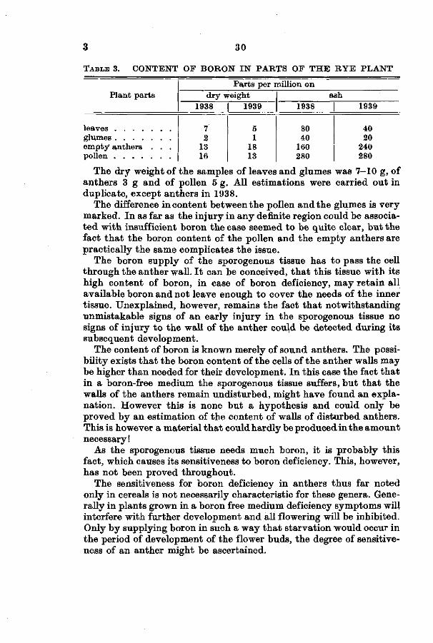
TABLE 3. CONTENT OF BORON IN PARTS OF T H E RYE PLANT
Plant parts
empty anthers . . .
Par ts per million on dry weight
1938 | 1939
7 2
13 16
5 1
18 13
ash 1938 | 1939
80 40
160 280
40 20
240 280
The dry weight of the samples of leaves and glumes was 7-10 g, of anthers 3 g and of pollen 5 g. All estimations were carried out in duplicate, except anthers in 1938.
The difference in content between the pollen and the glumes is very marked. In as far as the injury in any definite region could be associated with insufficient boron the case seemed to be quite clear, but the fact that the boron content of the pollen and the empty anthers are practically the same complicates the issue.
The boron supply of the sporogenous tissue has to pass the cell through the anther wall. I t can be conceived, that this tissue with its high content of boron, in case of boron deficiency, may retain all available boron and not leave enough to cover the needs of the inner tissue. Unexplained, however, remains the fact that notwithstanding unmistakable signs of an early injury in the sporogenous tissue no signs of injury to the wall of the anther could be detected during its subsequent development.
The content of boron is known merely of sound anthers. The possibility exists that the boron content of the cells of the anther walls may be higher than needed for their development. In this case the fact that in a boron-free medium the sporogenous tissue suffers, but that the walls of the anthers remain undisturbed, might have found an explanation. However this is none but a hypothesis and could only be proved by an estimation of the content of walls of disturbed anthers. This is however a material that could hardly be produced in the amount necessary!
As the sporogenous tissue needs much boron, it is probably this fact, which causes its sensitiveness to boron deficiency. This, however, has not been proved throughout.
The sensitiveness for boron deficiency in anthers thus far noted only in cereals is not necessarily characteristic for these genera. Generally in plants grown in a boron free medium deficiency symptoms will interfere with further development and all flowering will be inhibited. Only by supplying boron in such a way that starvation would occur in the period of development of the flower buds, the degree of sensitiveness of an anther might be ascertained.

31
c. Conclusions.
The microscopical examination of injury caused by boron deficiency has furnished further evidence of the fundamental pa r t boron takes in cell processes. I n vegetative tissue lack of boron influences the condition of the cell walls, in the sporogenous tissue the nuclei are primarily injured. I n meristematic tissue differentiation and maturat ion are inhibited. I t may be t h a t biochemistry which in recent years has lead us so much further in the conception of hormones, may throw further light on the action of boron.
Additional note.
R E H M (1937) had published an investigation with Impatiens from which he draws the conclusion t ha t boron deficiency symptoms are secondary phenomena caused by a higher intake of anions than kations. SCHMIDT (1937) ascribes symptoms of boron deficiency to a disturbance caused by a flooding of the plant with ni trates. I t seems to the present writer t h a t BRANDENBURG (1939) and MAIER (1938 and 1939) have adequately shown the improbability of both these assumptions.
SUMMARY
A microscopical s tudy has been made of deficiency symptoms occurring in the anthers of wheat, oats, rye and barley, grown in a medium deprived of boron.
The injury is strictly limited to the sporogenous tissue. I n all cereals it is of a same nature . The original injury occurs in the nuclei. When this injury occurs a t an early stage of development, cell division is inhibited ; a t a later stage cells are formed which are irregular in shape and size. The cell walls are normal.
The stage of development of the sporogenous cells, when the deficiency sets in, determines the ul t imate symptoms in the anthers.
The cells in the wall of the anther and the embryosac mother cell are always undisturbed.
I n barley the t ip of the rachis of a budding ear may be injured. I n this vegetative tissue the pr imary disturbance is in the cell walls; nuclei are normal.
I n healthy rye the pollen has a higher content of boron t han the glumes and leaves. Emp ty anthers have the same boron content as the pollen.
I t is probable t h a t the high content of boron of the pollen grains of cereals may explain t he sensitiveness t o boron deficiency of the sporogenous tissue.
Swedish turnips. Deficiency symptoms in roots and foliage can be

3 32
induced in Swedish turnips grown in a medium with a low boron concentration. I n the roots the cambium and the inner tissue are disturbed. The disturbance in the inner par t of the root agrees macroscopically with symptoms of „brown hea r t " noted in the field.
A microscopical s tudy has been made of sound and diseased roots, grown in the field and in culture solution.
Boron deficiency induces the production of a wide zone of thin-walled cells by the cambium and the phellogen.
I n thé secondary vascular bundles in the interior of the root, the injury occurs in the cell walls. Various modes of disintegration of mature elements were noted. Young cells originate in the débris of disintegrated mature cells. Ultimately a tissue of thick-walled cells, different from any tissue occurring in sound roots, is formed.
The ul t imate destiny of a cell is determined by its stage of development a t the onset of boron starvation.
Histological symptoms of artificially induced boron s tarvation agree closely with the symptoms of „brown hear t" in field material.
Sound roots are lower in boron content t han sound foliage. Therefore the normal content does not explain the sensitiveness of the roots to boron deficiency.
The etiology of boron s tarvation is discussed and it is suggested t ha t boron is a consti tuant of a substance synthesised in the leaves.
Acknowledgments. Thanks are due to Professor J . HONING for providing the opportu
nity of carrying out the microscopical investigation in the Laborator ium voor Erfelijkheidsleer.
To Professor J . Hudig for the opportunity of growing the solution cultures in his private laboratory.
To Miss SXTSARAH TRÜTER M.Sc. for her helpfulness in improving the English.
The photographs have, been made by Mr. CHR. VAN DE P E P P E L .
Wageningen, November 1939.

33 3
LITERATURE CITED
BRANDENBURG, E., 1939. Ueber die Grundlagen der Boranwendung in der Landwirtschaft. Phytop. Ztschr. 12, p . 1.
BBYAN, O. C , 1921. Effects of different reactions on the growth and nodule formation of Soybean. Soil Sc. 13, p. 271.
DENNIS , R. W. and O ' B R I E N , D. G., 1937. Boron in Agriculture. West of Scotland Agric. coll. Res. Bull. 5.
HAAS , A. R. C. and KLOTZ, L. J . , 1931. Some anatomical and physiological changes in Citrus produced by boron deficiency. Hilgardia 5, p . 175.
H I L L , H. and GRANT, E. P., 1935. The growth of turnips in artificial culture. .Scient. Agric. 15, p . 654.
HUDIG , J . and LEHR, J. J., 1938. Einige Bemerkungen über Chili Salpeter in Beziehung zur Borfrage. Zeitschr. Bodenk. u. Pflanzenern. 9/10 (54/55), p . 552.
JAMALAINEN, E. A., 1935. Tutkimuksia lantun ruskaotaudista. Ref. Untersuchungen über „Ruskotai t i" Krankheit der Kohlrübe. Vartion maatalous-koetoiminnan julkaisuja 72.
LÖHNIS, M. P., 1937. Plant development in the absence of boron. Meded. Land-bouwhoogeschool 41, verh. 3.
LÖHNIS, M. P., 1938a. Het „Bruin" van koolrapen in waterculturen. Land-bouwk. Tijdschr. 50 No 608, p . 151.
LÖHNIS , M. P., 19386. Brown heart in Swedes. Chron. Bot. 4, p . 9. MAIER, W., 1938. Die Notwendigkeit des Bors in der Pflanze. Ber. D. Bot. Ges.
56, p. (84). MAIER, W., 19396. Die Wirkung des Bors auf die Pflanze bei direkter Zufuhr
von Borsäure zum Sprosse. Chron. Bot. 5, p . 334. MARTIN, J . P., 1934. Boron deficiency symptoms in Sugarcane. Hawaian
Planters Rec. 38, p . 95. O ' B R I E N , D. G. and D E N N I S , R. W. G., 1935. Raan or boron deficiency in
Swedes. Scott. Journ. of Agric. 18, p . 326. R O W E , E. A., 1936. A s tudy of heart-rot of younger sugar-beet plants grown in
culture solutions. Ann. of Bot. 50, p. 735. SCHREVEN, D. A. VAN, 1934. Uitwendige en inwendige symptomen van boriumgebrek bij t abak (Summ. External and Internal symptoms of boron
deficiency in tobacco) Tijdschr. over Plantenziekten 40, p. 98. SCHREVEN, D. A. VAN, 1935a. Uitwendige en inwendige symptomen van
boriumgebrek bij tomaat (Summ. External and Internal symptoms of boron deficiency in Tomato) Tijdschr. o. Plantenziekten 41, p . 1.
SCHREVEN, D. A. VAN, 19356. Physiologische proeven met de aardappelplant. Landbouwk. Tijdschr. 47 No. 579, p . 621.
SCHREVEN, D. A. VAN, 1939. De gezondheidstoestand van de aardappelplant onder de invloed van twaalf elementen. Thesis, Wageningen.
SÖDING, H., 1924. Anatomie der Wurzel-Stengel- und Rübenbildung vom Oelraps and Steckrübe (Brassica napus L. var. oleifera und var. napo-brassica). Botan. Arch. 7, p . 41.
SOMMER, A. L. and SOROKIN, H., 1928. Effects of the absence of boron and of some other essential elements on the cell and tissue structure of the root t ips of Pisum sativum. Plant Phys. 3, p . 237.
WARINGTON, K., 1923. The effect of boric acid and borax on the Broad Bean. Ann. of Bot. 47, p. 429.
WHITEHEAD, T., 1935. Note on „Brown hear t" , a new disease of Swedes and its control. Welsh Journ. of Agric. 11, p. 235.

EXPLANATION OF PLATES
All microscopic preparations have been stained as described on page 6. The drawings have been reproduced on 6/10 of the original size, with the
exception of fig. 53, 61 and 62 which are taken up in actual size. Oil immersion has been used for magnification 640 and 1250 X (in reproduc
tion 384 and 750 X ).
ANTHEBS OF WHEAT
Fig. 1. Injured, length 0,22 m m . 384 X . 2. „ „ 0,35 m m . 384 X. 3. „ „ 0,66 m m . 384 X. 4. Sound „ 0,40 mm. 384 X.
„ 5. Injured, „ 0,42 mm. 384 X.
ANTHEBS OP OATS
Fig. 6. 7. 8. 9.
10. 11. 12. 13. 14. 15. 16. 17. 18. 19.
Sound, length 0,39 mm. Injured, ,, 0,48 mm. Sound, „ 0,48 mm. Injured, ,, 0,45 mm. Sound, ,, 0,85 mm. Injured, ,, 0,77 mm. Sound, ,, 1,02 mm. Same as Fig. 12. Injured, length 0,74 mm. Same as Fig. 14. Injured immature pollen. Sound ripening pollen. Deaf pollen. Sound ripe pollen.
384 384 384 384 384 384 261 384 261 384 384 384 384 384
X X X X X X X X X X X X X
X
ANTHEBS OF BYE
Fig. 20. Injured, length 1,52 mm. 117 „ 21. Same as Fig. 20. 384 X .
BABLEY
Rachis of young ears
Fig. 22. Injured t ip, length of ear 3 mm. 117 X. „ 23. Sound t ip, „ „ „ 3 mm. 117 X. „ 24. Injured t ip, „ „ „ 5 mm. 384 X. ,, 25. Injury in rachis, ,, ,, ,, 10 mm. 384 X. ,, 26. Injury in rachis, ,, ,, ,, 10 mm. 54 X.

35 . 3
Anthers
Fig. 27. Injured, length 0,53 mm. 384 X . „ 28. ,, „ 1,4 mm. 117 X. „ 29. „ „ 0,8 mm. 384 X. ,, 30. ,, „ 1,0 mm. 117 x . „ 31. Same as Fig. 30. 384 x . „ 32. Sound, length 0,99 mm. 117 X.
sp.t. = sporogenous tissue, p.m.c. = pollen-mother-cells, t. = t apetum. st.gr. = starch grains, n = nucellus. pro ca. = procambium.
SWEDES
Plants grown in solution cultures. 1 and 2: 5 mg ; 3 : 1,25 mg H3B03 . Roots grown in solution cultures. 1: 5 mg ; 2: 2,5 mg and 3: 1,25 mg H3B03 . Injured root. Field material. Injury in the cambium. Field material. Cross section sound cambium. 117 X.
,, ,, injured cambium. Grown in solution. 117 X. ,, ,, injured ray cambium. Grown in solution. 174 X. ,, ,, injured cambium. Grown in solution. 174 X . ,, ,, injured cambium. Field material. 117 X.
Radial section sound ray cambium. 174 x . ,, ,, sound cambium. 117 X. ,, ,, injured cambium. Grown in solution. 54 X . ,, ,, injured ray cambium. Grown in solution. 174 X. ,, ,, injured cambium. Grown in solution. 117 X.
Cross section sound periderm. Field material. 174 X . Radial section sound periderm. Field material. 174 X. Cross section injured periderm. Field material. 174 X . Radial section injured periderm. Grown in solution. 174 X .
Sound vascular strands. Field material.
Fig. 51. Cross section initial stage. 384 X. 52. ,, ,, young stage. 384 X. 53. ,, ,, central phloem. 640 X. 54. ,, ,, mature stage. 117 x . 55. Same as Fig. 54. 174 X. 56. Longitudinal section mature stage. 174 x . 57. Same as Fig. 56. 384 X. 58. Longitudinal section phloem bordering on sheath cells. 384 X . 59. Cross section phloem. 384 X .
Injured vascular strands. Field material.
Fig. 60. Cross section phloem. 384 X. ,, 61. Section central part . 640 X . „ 62. Section central part . 640 X . ,, 63. Cross section. 384 X. „ 64. Longitudinal section. 384 X.
Fig Ï ?
11
»» » Î
,, ,, 5!»
f9 19
? »
,, » J
9 »
,, Ï Ï
»»
33 34
35 36 37 38 39 40 41 42 43 44 45 46 47 48 49 50

3 36
Fig. 65. Longitudinal section. 384 X . 66. Longitudinal section. 384 X . 67. Newly formed tissue. 384 X . 68. Id. later stage. 384 X . 69. Mature isodiametric cells, 117 X . 70. Id. 384 X. 71. Longitudinal section vascular strand. 174 x . 72. Cross section phloem. 384 X . 73. Cross section fusiform cells and phloem. 384 X . 74. Cross section elongated sheath cells. 174 x . 75. Elongated and isodiametric cells. 49 x . 76. Injured tissue in sheath of vascular strand. 117 x .
Injured vascular strands. Grown in solution.
Fig. 77. Injured tissue in sheath of vascular strand. 117 x . ,, 78. Longitudinal section phloem. 384 x . „ 79. Id. 384 X.
Injury in xylem. Field, material.
Fig. 80. Cross section. 384 X . ,, 81. Cross section. 750 X . ,, 82. Cross section. 750 X . ,, 83. Longitudinal section. 384 x . ,, 84. Cross section with newly formed cells. 384 X .
co. = cortex, phi. = phloem, ca. = cambium, ph.l. — phellem. ph.g. = phello-gen. le. = lenticel. pr.phl. = pr imary phloem, s.t. = sieve tube. c.c. = companion cells, fu.c. = fusiform cells, sh.c. = sheath cells, pe.s. = pectic substance. pl.m. = plugging matter, foa.m. = foamy matter, sep. str. = separate s trands. l.g. = lignin. y.c. = young cells.


p. m. c.
Fig. 11 Fig. 12
Fig. 14

f ig. 16 Fig. 17
Fig. 20 Fig. 21

Fig. 22
Fig. 23

Fig. 24
Fig. 25
Fig. 26
Fig. 28

Fig. 31
Fig. 30 Fig. 32

Fig. 33
2 Fig. 34
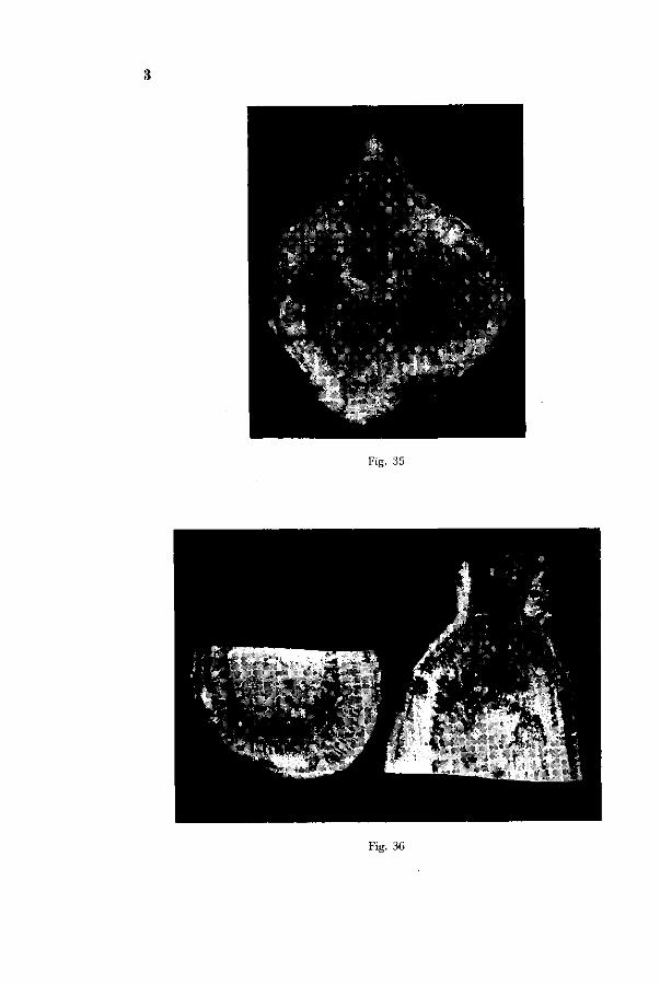
Fig. 35
Fig. 36
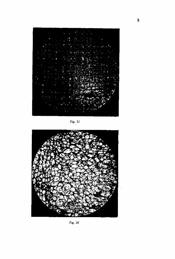
Fig. 37
Fig. 38

Fig. 41
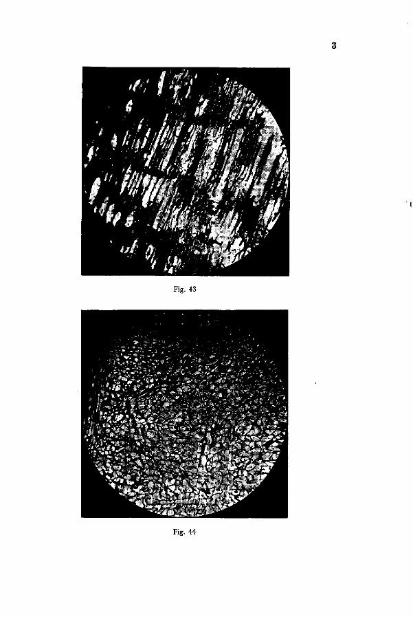
Fig. 43
Fig. 44

Fig. 46

Fig. 53

Fig. 54
Fig. 55 Fig. 56

rig. 62 Fig. 63

Fi*. 64 Fig. 65
Fig. 67 Fig. 68

Fig. 69
F l g . 70

Fig. 74

Fig. 75
Fig. 76

Fig. 77
Fig. 79
Fig. 78

Fig. 84