Embed Size (px)
Citation preview

Histology image retrieval in optimised multi-feature spaces.Zhang, Q; Izquierdo, E
© 2012 IEEE
“The final publication is available at
http://ieeexplore.ieee.org/xpls/abs_all.jsp?arnumber=6353218&tag=1”
For additional information about this publication click this link.
http://qmro.qmul.ac.uk/xmlui/handle/123456789/13885
Information about this research object was correct at the time of download; we occasionally
make corrections to records, please therefore check the published record when citing. For
more information contact [email protected]
brought to you by COREView metadata, citation and similar papers at core.ac.uk
provided by Queen Mary Research Online
1
Histology Image Retrieval in Optimised
Multi-Feature SpacesQianni Zhang and Ebroul Izquierdo, Senior Member, IEEE,
Abstract—Content based histology image retrievalsystems have shown great potential in supporting deci-sion making in clinical activities, teaching, and biolog-ical research. In content based image retrieval, featurecombination plays a key role. It aims at enhancingthe descriptive power of visual features correspondingto semantically meaningful queries. It is particularlyvaluable in histology image analysis where intelligentmechanisms are needed for interpreting varying tissuecomposition and architecture into histological concepts.This paper presents an approach to automatically com-bine heterogeneous visual features for histology imageretrieval. The aim is to obtain the most representativefusion model for a particular keyword that is associatedto multiple query images. The core of this approach isa multi-objective learning method, which aims to un-derstand an optimal visual-semantic matching functionby jointly considering the different preferences of thegroup of query images. The task is posed as an opti-misation problem, and a multi-objective optimisationstrategy is employed in order to handle potential con-tradictions in the query images associated to the samekeyword. Experiments were performed on two differentcollections of histology images. The results show thatit is possible to improve a system for content basedhistology image retrieval by using an appropriatelydefined multi-feature fusion model, which takes carefulconsideration of the structure and distribution of visualfeatures.
Index Terms—Content based image retrieval, his-tology image retrieval, feature fusion, multi-objectiveoptimisation
I. Introduction
In biology and medicine, histology is a fundamental toolthat provides information on structure and compositionof tissues at microscopic level. Nowadays, images of tissueslides are often digitized to document procedures and tosupport findings. These collections are often huge in sizeand thus hide a latent source of information that canbe greatly exploited if suitable mechanisms are availablefor accessing the data [28]. Thus, a technology that canretrieve histological images according to given queries canpotentially be a very useful tool for data archiving andanalysis, teaching and training, assisting decision makingin diagnosis and so on. When more complex systems arebeing considered, for instance, a system which provides
Q. Zhang and E. Izquierdo are with School of ElectronicEngineering and Computer Science, Queen Mary Universityof London, Mile End Road, London E1 4NS, UK. e-mail: {qianni.zhang, ebroul.izquierdo}@elec.qmul.ac.uk(see http://www.elec.qmul.ac.uk/mmv/people/qianniz.htm;http://www.elec.qmul.ac.uk/people/ebroul/index.htm).
suggestions on diagnosis based on existing histopathologydatabases, histological image retrieval can be employed asan essential component for initial search and indexing ofcontent using carefully selected keywords, such as a basictissue type.Conventional medical image retrieval systems often rely
on tags associated to images in the databases. How-ever, text-based approaches are often limited in practicesince tags may be both expensive and ambiguous. Thisis because generating manual annotation on images isextremely time-consuming, highly subjective and requiresa good level of domain-related knowledge. Another kindof approaches exploit knowledge databases like UnifiedMedical Language Systems [22], but they rely on theavailability of knowledge databases and their relativity tothe application domain.Alternatively, content-based image retrieval (CBIR) sys-
tems apply computer vision techniques to the image re-trieval problem by analysing the actual contents of theimage rather than semantic features such as keywords ortags. Recent research in CBIR indicates that such systemsare capable of retrieving medical images according to itsdomain-specific image features [14]. Thus such systemsform an important alternative and complement to tra-ditional text-based retrieval systems. In image retrievalsystems, given a query image, or a keyword query thatis associated to a group of representative images, thegoal is to retrieve from a reference library, the similarimages whose semantic meaning is as close to the queryas possible, regardless of its visual appearance. Althoughextraction algorithms for low-level image features are well-understood and able to capture subtle differences betweencolours, statistic and deterministic textures, global colourlayouts, dominant colour distributions, etc., the link be-tween such low-level primitives and high-level semanticconcepts remains an open problem [31],[41]. This problemis referred to as ‘the semantic gap’.To alleviate the semantic gap problem, there have
emerged many systems aiming at applying content-basedapproaches in more sophisticate ways in medical imageretrieval [1], [2],[17], [19], [20], [37], [39]. Many diagnosticimaging modalities, such as X-ray, computed tomography(CT), magnetic resonance imaging (MRI) are currentlyavailable and routinely used to support clinical decisionmaking, diagnosing, state of an illness-tracking, medicaleducation and research, etc. Applications of content-basedapproaches in medical image retrieval have shown benefitsin all these procedures. In addition to general image
2
retrieval systems applied to medical images, there arealso many specialised image retrieval systems developedto enable retrieval of various specific kinds of medicalimages, such as breast cancer biopsy slides [40], positronemission tomographic functional images [5], ultrasoundimages [9], different types of pathology images [42], radio-graphic images [17], and histology images [8], [37]. Moreinterestingly, the ImageCLEF medical image retrieval tasktargets modality classification of medical images and haveattracted wide attention from the area in recent years [27].Among these application domains, histology image re-
trieval has been an active research topic for modellingvisual similarity measures and retrieving tissue slides insome semantic categories. In previously reported workson histology image classification and retrieval, accuracy isoften limited due to the unreliable outputs from the featuremetrics [14],[23]. This is because the systems often rely onsingle low-level features, which are not always capable ofinterpreting visual objects into varying and complicatedsemantic meanings. To tackle this problem, the combi-nation of low-level features for semantic image retrievalhas been widely considered in the literature. In [42], asystem was proposed for retrieving histology images fromprostate, liver and heart tissues, based on four differentvisual characteristics. The work in [38] described a systemto index histology images of gastro-intestinal tract, bycategorising image blocks into semantic classes based onlocal visual patterns. The approach proposed in [29] usesa boosting algorithm based on multiple distance measurescomputed on a fixed set of features to retrieve and classifybreast histology slides. These works all intended achievingbetter performance in histology image understanding byemploying heterogeneous visual features. However, theproblems in feature combination are likely underestimated,when feature combination is done by simple concatenationof feature vectors.Such approaches are called early fusion, and they share
two main common problems. First, methods on concatena-tion of feature vectors can easily result in high dimensionalfeature space and thus suffer from “the curse of dimen-sionality” [10], [35]. Second but most importantly, differentimage features often have their own structures, distribu-tions and metric spaces. Direct concatenation of featurevectors could result in meaningless similarity measures.For instance, feature histograms are usually comparedusing a similarity measure for probability distributionswhile feature vectors should be matched using Euclideanmetrics. In addition, even if two different features arebeing compared with the same metric, their scale, domainand distribution may be completely different due to theintrinsic descriptor nature [16].In order to avoid these problems, in this paper a late-
fusion strategy is followed to combine low-level features forhistology image retrieval. Similar approaches have beenproposed in the literature, such as in [6], [15]. However,such approaches have one common disadvantage that theyassume equal importance of features in fusion. In this waythe performance of ‘good’ and ‘bad’ features is averaged
and thus could not provide great improvements. Someother approaches assign weights to different features. Forinstance, genetic algorithm is used for finding the optimi-sation weights for features in image retrieval in [13]. Thisapproach considers feature fusion independent of query,while it is not possible to expect the obtained fusionmodel to be optimal to all possible queries. In [7], multi-feature fusion was achieved using an optimal combinationof multiple kernel functions. Kernel functions were fusedusing a weighted linear combination, whose weights werefound by an optimization process that maximizes thecorrelation between low-level features, represented by ker-nel functions, and high-level semantic concepts. Anotherimportant direction is to rely on human interactions toobtain query specific weights [18]. Such approaches mightimprove the effectiveness of weighting factors, but theystill require heavy workload and specific expertise fromthe user.In this paper, we propose a histology image retrieval
method aiming at retrieving images with relevant semanticmeanings based on visual content. In each query process,the user first has in mind a semantic term that can be asso-ciated to a specific keyword, and then uses multiple queryimages that represent the semantic term for retrievingmore images that are relevant to the query on the semanticlevel. In the proposed method, we use multiple images,namely, a representative query group, in the CBIR processfor representing each semantic term. This is because, fora particular keyword, it is usually tricky to find a queryimage as an ideal visual representation. In [1], a multitieredCBIR system was proposed targeting microscopic images,enabling both multi-image query and slide-level imageretrieval. In this work, the focus was to adopt multipleimage queries in one retrieval process using the weightingterms. In comparison, we also consider using multipleimage queries to represent each keyword, but the mainfocus of the proposed approach is to derive a fusion modelfor heterogeneous visual features.In addition, for each keyword, a different representa-
tive query group is used and a unique feature fusionmodel is derived. This is because each semantic term isconsidered to have its own characteristic visual patternand the feature fusion models should be keyword-specific.The proposed approach is able to automatically learn therelative importance of each feature space correspondingto the keyword from its associating representative querygroup. The learning of a suitable feature fusion modelis posed as an optimisation problem. The optimisationis carried out using a Multi-Objective Learning (MOL)method, which involves a multi-objective optimisation(MOO) strategy [36]. The main advantage in the MOLmethod is that it is able to find a multi-feature modelthat can simultaneously encapsulate different aspects ofthe most representative visual patterns for each concept,without however assigning fixed relevance factors to eachvisual feature.Without losing generality, several different texture fea-
tures are considered in our experiments. Although, the
3
proposed method can be applied to more histologicalterms, the four fundamental tissue types in biology - con-nective, epithelial, muscular and nervous tissues - are con-sidered in our experiments as the keywords for retrieval.It is demonstrated that, using the proposed approach,it is possible to retrieve histological images according totheir semantic relevance by using properly combined visualfeatures. The proposed approach has been tested in twocollections of histology images. The first set contains over20000 images, among which 2,828 images have manuallabels of the four fundamental tissue types. The secondsmaller set contains around 442 histology images, and 130of them have manual labels on those four tissue types.The remainder of this paper is organised as follows:
Section 2 presents the visual analysis steps in histologydatabases, including feature extraction, distance calcula-tion and normalisation. Section 3 introduces the MOLmethod for feature fusion towards a multi-feature basedretrieval in histology image databases. The evaluation pro-cedure and experimental results are presented in Section4 and Section 5 discusses some concluding remarks.
II. Visual feature extraction and analysis
The design and evaluation of an image retrieval systemrely on properly defined visual features with suitable sim-ilarity matching metrics as well as correct normalisationfunctions. Without normalisation of feature spaces, com-parison between different features becomes misleading andtheir combination is meaningless. Therefore in this section,we present our work on extracting several commonly usedvisual features, analysing their characteristics and derivingtheir normalisation functions.
A. Feature extraction
The main goal of the proposed research is to developan approach for automatically combining low-level visualfeatures for retrieval of histology images according to theirfundamental tissue types. Therefore, the extraction andanalysis of useful visual features in histology images is anessential step.In the literature many well designed low-level visual fea-
tures have been proposed, describing visual content fromdifferent perspectives including colour [26], texture [32],shape [34], local feature points [25], etc. It is worth notingthat, the focus of this paper is on discussing how to obtainsuitable fusion models of features. The proposed multi-feature combination approach is independent of selectedfeatures or their distance metrics. Evaluation of differentfeatures is out of the scope of this research.Due to the nature of this specific dataset of histology
images, texture features are suitable for analysing theirvisual patterns. In this research we selected several com-monly used texture features together with architecturalfeatures due to their prominent characteristics for histol-ogy image analysis [4], [24]. Without losing generality, theeight features selected here to describe histology imagecontents are listed below. Unless specified otherwise, these
features are extracted from 3x3 blocks of an image andthen concatenated into one feature vector for that image.(1) Gabor Textures (GT): Gabor filters possess out-
standing ability of filtering in the spatial and frequencydomain. The Gabor transform is a set of directional filters,thus it is shift invariant. To calculate a GT feature, a con-volution with a Gaussian harmonic function is used, and7 different frequencies, freq = [1, 2, ..., 7], are consideredto compute 7 descriptor values per block. As a result, theGabor descriptor is composed of 63 descriptors.(2) Tamura Textures (TT): Tamura proposed six tex-
ture features corresponding to human visual perception:coarseness, contrast, directionality, line-likeness, regular-ity, and roughness. From experiments testing the signifi-cance of these features with respect to human perception,it was concluded that the first three features are veryimportant. In our experiments, two statistics are calcu-lated for contrast, directionality and coarseness, providing6 descriptors in each block. As a result, the Tamuradescriptor has 54 descriptors.(3) Zernike Moments (ZM): Zernike moments have many
desirable properties, such as rotation invariance, robust-ness to noise, expression efficiency, fast computation andmulti-level representation for describing the shapes of pat-terns. In this paper, the absolute values of the coefficientsof the Zernike polynomial approximation are computedper block, providing 72 descriptors in each region. Then,the Zernike descriptor has 648 bins.(4) SIFT-based dictionary (SIFT): SIFT feature is
known for its ability in handling intensity, rotation, scaleand affine variations. A histogram of SIFT converts eachpatch to 128-dimensional vector. Thus in this paper,each block in the process is represented by the rotation-invariant feature descriptor, using a histogram of 128 bins.(5) DCT dictionary (DCT): DCT histograms are invari-
ant to translation and rotation. In this paper, each blockis represented by the coefficients of the Discrete CosineTransform, applied to each channel of the RGB colourspace. The 21 most significant coefficients per channel arepreserved. In this way, the dictionary of patterns will havecolour information as well.(6) Gray-Level Co-Occurrence Matrix (GLCM): GLCM
textures are obtained by a tabulation of how often differentcombinations of pixel brightness values occur in an image.When the co-occurrence matrix is formed using a set ofoffsets sweeping through 180 degrees (i.e. 0, 45, 90, and135 degrees) at the same distance, it is able to achievea degree of rotational invariance. A number of statisticscan be derived from the co-occurrence matrix, which arecalculated in the adjacency of one pixel in each of thefour directions (horizontal, vertical, left and right diago-nal). Four of them are considered - contrast, correlation,energy and homogeneity - to form a feature vector of fourdimensions.(7) MPEG-7 Edge Histogram (EH): EH describes the
local edge distribution of an image. The descriptor is scaleinvariant and supports rotation invariant and rotation sen-sitive matching operations. It is obtained by first dividing
4
an image into 4x4 sub-images and then calculating thelocal-edge histogram bins. Edges in the 16 sub-images arecategorised into five types - vertical, horizontal, diagonal45 degrees, diagonal 135 degrees and non-directional -forming a histogram of 80 bins.(8) MPEG-7 Homogeneous Textures (HT): A HT de-
scriptor provides a quantitative representation using 62numbers, including the image intensity average, standarddeviation of the image pixels, energies of the 30 partitionedfrequency channels based on the human visual system, andenergy deviations of these 30 channels. The 30 partitionedchannels ensure a scale and rotation-invariant descriptionand matching of texture. To extract these numbers from animage, the image is first filtered with a bank of orientationand scale tuned filters using Gabor filters. The first andthe second moments of the energy in the frequency domain, i.e., energies and energy deviations, in the correspondingchannels are then used as the components of the texturedescriptor.
B. Distance calculation and normalisation
In the next step, feature distances are calculated using aspecifically defined distance metric of each feature space.The details of distance metrics used in this research aregiven in Section IV.Then all the obtained distances are normalised. The goal
of normalisation is to guarantee the appropriateness ofcomparing different measurements that differ in scale anddomain, while preserving the underlying characteristicsof the data. In this research, distance metrics computedfrom different image features are normalised based on thefitted probability density functions for their correspondingfeature spaces.Let d′ be the distance value computed from a particular
image feature. The statistical normalisation is computedas:
d =(d′ − µ)
σ, (1)
where d is the normalised distance value and µ and σ
are the mean and standard deviation of the underlyingdistance distribution.The critical problem in deriving appropriate normalisa-
tion function is to precisely estimate the distance distribu-tion of the feature space. The distribution of feature dis-tances is highly dependent on the structure of the featureand image content. One can assume a normal distributionof the distances to estimate µ and σ without any furtheranalysis. However, the true distribution of distances mightbe more precisely approximated using other ProbabilityDistribution Functions (PDFs). Therefore in this research,six types of PDFs are considered, including Normal,Gamma, Laplace, Log-norm, Rayleigh and Exponential.These PDFs are denoted as Pk, k = 1, 2, ..., 6. Based on dis-tance samples derived from the database, the parametersΘk of each possible Pk are estimated to determine whetherthe data is being drawn from the associated PDF or not.
TABLE IProbability Distribution Functions in the set of possible
approximations for each distance distribution.
Distri-bution
Θ = PDF Mean Standarddeviation
Normal (µ, σ)1√2πσ2
exp
(
− (x− µ)2
2σ2
)
µ σ2
Gamma (k, θ) xk−1exp(−x/θ)
Γ(k)θkkθ kθ2
Laplace (µ, b)1
2bexp
(
−|x− µ|b
)
µ 2b2
Log-norm
(µ, σ)1
x√2πσ2
exp
(
− (ln x− µ)2
2σ2
)
eµ(
eσ2 − 1
)
e2µ+σ2
Rayleigh (σ)x
σ2exp
(−x2
2σ2
)
σ
√
π
2
4− π
2σ2
Expo-nential
(λ) λexp (−λx)1
λ
1
λ2
Finally, to select the best distribution approximation forthe underlying data, the Kullback-Leibler (KL) divergenceis evaluated between the histogram of actual distances andthe estimated PDF. For two distributions P and Q, the KLdivergence between them is estimated by:
DKL(P ||Q) =
∫ ∞
−∞
p(x)logp(x)
q(x)dx, (2)
where p and q denote the densities of P and Q. Theprocedure for distance normalisation can be summarisedas the following steps:
1) For each Pk, k = 1, 2, ..., 6, estimate the parametersΘk using the data samples in distance matrix D.
2) Build a histogram H of distances with m bins usingthe data samples in D.
3) For each bin in the histogram H, calculate the valueof Pk in Pk to approximate the shape of H.
4) Calculate the KL-divergence between the histogramH and the approximation Pk.
5) Select Pk with the minimum value of KL divergenceas the best fit PDF.
Table I shows the six PDFs considered in this work forapproximation of distance distributions. Notice that foreach PDF, the normalisation parameters µ and σ are cal-culated in different ways using the estimated parametersΘ, which are computed from the sample according to thecorresponding rules.
For the remaining of this paper, when distance valuesare mentioned, they have been normalised following thesteps described in Section II.
III. Multi-feature based histology imageretrieval
The proposed approach to multi-feature based histologyimage retrieval relies on the MOL method, that is able toautomatically learn a suitable multi-feature model from arepresentative group containing multiple query images asa visual representation for the keyword.
5
A. Representative query images
To define a suitable combination model for each keywordrelated to the histology database, multiple query imagesare employed in a query process. Due to the complexand varying visual appearance in different keywords, itis unrealistic to assume there exist a single image querygiving an optimal representation of a semantic keyword.Therefore, in this paper, we use a group of query images,referred to as the representative query group, to approxi-mate a suitable representation for one keyword. For a givenkeyword, let us use R ⊂ G to denote a representativegroup, where G is the complete image set. To improvethe discriminative power of the low-level features, twokinds of representative query samples are considered. R+
contains the most relevant samples for the correspondingkeyword, referred to as positive group; R− representsnegative group in which the samples are irrelevant to thekeyword of concern but may look similar to the positivequery samples; R = R+ ∪ R−. If new histology keywordsare added or the database is populated with new imagesin new concepts, new representative query groups for theincoming concepts need to be generated.
Assume a total number of n feature spaces are con-sidered. Having the representative group R ready, wecan calculate the centroid vj of representative group R
in a given feature space Fj , j = 1, 2, ..., n. Fj could beany feature space described in Section II, or any othersuitable visual features in the literature. vj in featurespace Fj is calculated by finding the sample in positivegroup R+ with the minimal sum of distances to all otherpositive samples in R+. Let vi,j and vk,j be the featurevectors extracted from positive samples ri and rk in featurespace Fj , i, k ∈ [1, |R+|], and d(vi,j , vk,j) be the featuremetric estimating the distance between these two imagesin feature space Fj . We note that vj actually equals tothe feature vector in Fj extracted from one of the positivesamples in R+. The centroid of representative group R inFj can be defined as:
vj = argmin{vi,i∈[1,|R+|]}{∑
k∈[1,|R+|]
d(vi,j , vk,j)}. (3)
Taking vj as an anchor, for a given image gi in G, wherei ∈ [1, |G|], the distance from gi to the centroid vj of featurespace Fj can be calculated as:
di,j = d(vj , vi,j). (4)
For the set of n feature spaces {Fj |j = 1, 2, ..., n}, all thecentroids across different feature spaces form a particularset of vectors V = {v1, v2, ..., vn}, in which each vj isthe centroid vector of feature space Fj . In general, V isreferred to as the generalised centroid of representativegroup R, since it does not necessarily attach to a positivequery sample in R. Note that V is always calculatedconsidering only positive samples. This is because thenegative samples can be randomly scattered in the metric
space and calculating the generalised centroid by takingthem into account would be meaningless.For a representative query group R with m image
samples for a particular keyword, a distance matrix M
of size m× n can thus be constructed.
M =
d1,1 d1,2 · · · d1,nd2,1 d2,2 d2,n...
. . ....
dm,1 dm,2 · · · dm,n
(5)
Each element di,j (in row i and column j) in M isthe distance from representive sample ri to centroid vj infeature space Fj . In this way, the keyword is representedby a distance matrix covering multiple feature spaces.
B. Optimisation of multi-feature model and histology im-age retrieval
The aim of MOL method is to define a suitable multi-feature model for the visual representation of a specifichistological keyword. The core of this method is a learn-ing process towards an optimal combination model byassigning each involved low-level feature space Fj a properweight αj . This can be achieved by optimising an objectivefunction or a set of objective functions for variable α.Since several representative samples are used for a goodvisual representation of a keyword, the interest of eachsingle query sample may conflict with others. Thus weconstruct an objective function for each query sample in R,and use a multi-objective optimisation strategy to find asolution that can achieve a common optimum for all thesefunctions.Based on the distance matrix M given in (5), a set of ob-
jective functions can be constructed for the optimisation ofmulti-feature model. Each objective function is formed asweighted linear combinations of feature-specific distances.Considering M , a total number of m objective functionscan be constructed:
D(A) =
D1 =∑n
j=1 αj d1,j ,
D2 =∑n
j=1 αj d2,j ,...,
Dm =∑n
j=1 αj dn,j .
(6)
The MOL method seeks to learn from the representativegroup, a suitable set of weights A = {α1, α2, ..., αn},subject to the constraint:
∑nj=1 αj = 1. The problem of
learning a multi-feature fusion model is now transformedto finding a solution that optimises each of these objectivefunctions in (6).Generally speaking, an optimum is usually defined as
the maximum or minimum of some objective(s). Theoptimal solution A should lead to the maximal or minimalvalue of the objective function(s) in D(A) among all possi-ble scalar combination of A that satisfy the constraints. Inthe proposed MOL method, the optimum is regarded asthe minimum of objective functions for positive samplesin R+ and maximum of objective functions for negative
6
samples in R−. The problem of learning a multi-featurefusion model is now posed as to find a solution thatoptimises each of these, in some cases contradicting, objec-tive functions. Observe that different representative querysamples may display different visual characters but thesedifferences need to be harmonised via the simultaneousoptimisation of multiple objectives corresponding to differ-ent representative samples. This optimisation process forfusion model learning is achieved using the MOO strategy.The MOO strategy is able to find a general optimum acrosspotentially conflicting objectives by taking the interest ofeach single objective into account. Thus it is widely usedin real-life optimisation problems [36].While various algorithms have been developed using
MOO strategy, in this paper Pareto Archived EvolutionStrategy (PAES) is adopted as the MOO algorithm tooptimise the combination models for the following reasons.Studies have been carried out to compare PAES with othertwo well-known and respected multi-objective genetic al-gorithms - the Niched Pareto Generic Algorithm and theNondominated Sorting Genetic Algorithm in [21]. Theirresults provided strong evidence that PAES performedconsistently well on a range of multi-objective optimisa-tion tasks. It was also shown that PAES required fewercomparison processes to perform selection and acceptance,and this claim was empirically evidenced by the timingof experiments. This algorithm usually generates a set ofpotential Pareto optimal solutions Φ = {A1, A2, A3, ....}.Thus, a second step is required to decide which one of thesesolutions is the most suitable or feasible. The meaning ofoptimum in our specific task can be described as to findthe ‘optimal’ multi-feature distance in which all the pointsrepresenting the positive samples in the target multi-feature space are closely gathered around the generalisedcentroid while the points for the negative samples arerandomly scattered around the generalised centroid. Thus,a sensible selection criterion can be defined as to find theA that satisfies:
A = argminA∈Φ
∑
j∈[1,|R+|] Dj(A)∑
i∈[1,|R−|] Di(A). (7)
For a particular tissue type, an optimal multi-featuredistance fusion model of an image gi ∈ G can be obtained:
D(gi) =
n∑
j=1
αj di,j , (8)
where αj ∈ A. According to these multi-feature dis-tances, histology images can be ranked and retrieved basedon their multi-feature distances with respect to the querykeyword.
C. Why multi-objective optimisation?
Visual descriptors are different in nature and may haveconflicting interest when they are jointly used to representa semantic term. In order to take into consideration the
preferences of all different features, in this paper we con-sider estimating a set of weighting factors for the featurespaces according to their relevance to the representativequery group. The core of the proposed method for derivinga concept-specific fusion model is the MOO strategy.There are similar works in the literature using various
optimisation algorithms for obtaining multi-feature fusionmodels, such as in [11], the downhill simplex method isused to optimise a single objective function. A voting kNNrule is used to derive an objective function for optimisa-tion. However, in this case the objective function obtainedis the result of joining the interests of different samplesinto a single one. The optimised model only reflects thepreference of the overall objective, without considering tosatisfy the preference of any individual sample.Similarly in our case, to obtain suitable weighting fac-
tors for the fusion model, one might consider constructingan overall objective function out of (6) and simply optimiseit. One possible approach can be to sum up all thefunctions in (6):
D′(A) =
m∑
i=1
Di =
n∑
j=1
αj(
m∑
i=1
di,j). (9)
The task now is to find the set of weighting factors thatminimize the objective function (9):
A = argminA={α1,α2,...,αn}
n∑
j=1
αj(
m∑
i=1
di,j)
. (10)
For the particular representative group, the values ofthe term
∑mi=1 di,j , j ∈ {1, 2, ..., n} are determined. Let
Fk, k ∈ {1, 2, ..., n} be the feature space in which thesum of distances from each representative sample to thecentroid vk is smaller than those in other feature spaces:∑m
i=1 di,k ≤∑m
i=1 di,j , where k ∈ {1, 2, ..., n} , j ∈{1, 2, ..., n} \ {k}. The minimal value of (9) can then beachieved by a particular A, in which each α has thefollowing values:
αx =
{
1, x = k
0, x 6= kx ∈ {1, 2, ..., n} (11)
In this case, the resulting weight factors A indicatethat all the credit is given to one of the feature spaceFk and all other features are not taken into accountat all. In the end the task becomes a selection of one‘best’ feature rather than fusion of multiple features. Forone particular representative group and the representedkeyword, it might be possible to say Fk is the ‘best’.However, it may not be the case when different testingimages are present. In most cases, properly defined featurefusion model may produce better retrieval performancecompared to using any of the single features.In comparison to the single objective based optimisation
schemes, the advantage of employing the proposed MOLmethod is that, each representative sample and its corre-sponding objective function are treated separately in an
7
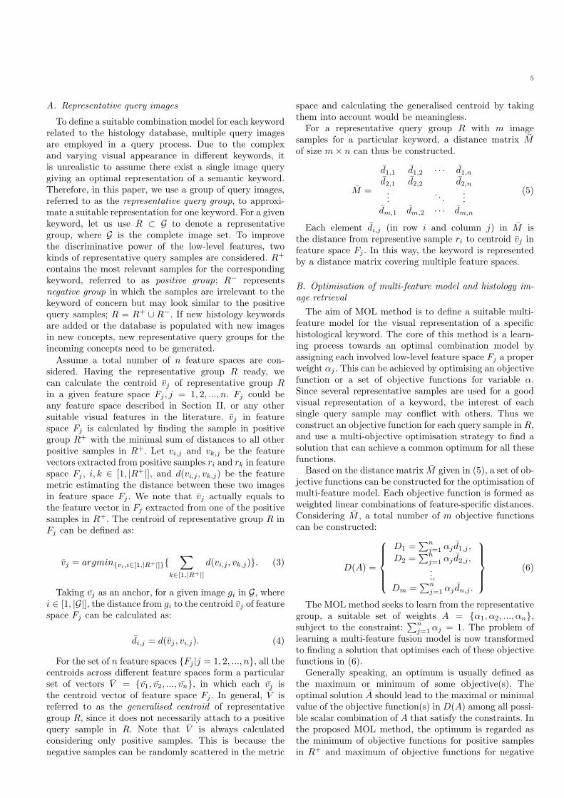
Fig. 1. Image samples of tissue types.
optimisation process. The interest of each representativesample is taken into account while the overall interest isbeing satisfied. MOO is used to find the solution thatcan achieve a balanced local optimum for each objective,without compromising the other objectives. The obtainedresults are regarded as the ‘general optimum’ for all ob-jectives.
IV. Experiments
The first histology image set used for experiments inthis research was the ‘BiMed’ database [3]. The experimentcarried out on this dataset is referred to as ‘experiment I’in the following text. This dataset contains about 20,000histology image samples. The aim of this database wasto support academic and research activities in biologyby allowing medical students, doctors and researchers inbiomedical field to access a wide variety of microscopyimages in the four fundamental tissue types of livingbeings: connective, epithelial,muscular and nervous. Thesefour tissue types are considered as keywords and in a queryprocess, each of them is represented by a group of queryimages. A few examples of each tissue type in the databaseare shown in Figure 1, illustrating different biologicalstructures and visual characters of the four types. Imagesin this collection have been acquired from tissue slidestaken from different mice organs including brain, liver,heart, lung, kidney and skin among others. All sampleswere drawn from healthy specimens. Tissue slides wereprepared using different staining methods including Hema-toxylin and Eosin and Immunohisto chemical procedures.In addition, different zoom factors were used to acquiredigital images according to the structure of interest. Mostsamples in this dataset were un-annotated, making thesesamples inaccessible using textual-based search methods.However, a portion of these images were annotated byexpert biologists. This annotation contains information onsome particular structures, organ, system and fundamen-tal tissue. Thus, although experiments were performed onthe whole dataset, we selected a subset of 2,828 imagesamples with full annotation on the four fundamental tis-sues, for evaluation of the proposed method. Five texturefeatures were extracted from the images in the BiMeddatabase and employed in experiment I. These featuresare GT, TT, ZM, SIFT and DCT.The second dataset used for testing and validation
purposes was the ‘Blue Histology’ images [33]. This set
contains 442 histology images which were fully annotated,among which, 130 were labelled with the keywords of fourfundamental tissue types: connective, epithelial, muscularand nervous. The experiment performed on this secondset is called ‘experiment II’ in the following of the paper.Experiments were performed on all 442 images in thisdataset, but the evaluation was conducted only againstthose 130 images with labels on the four tissue types. Asexplained before, the proposed approach is supposed to beindependent of employed features. In order to prove this, inexperiment II, a different feature bank was used, includingGT, TT, GLCM, EH and HT. The aim of performing asecond experiment with this dataset is to demonstrate thevalidity of the tested approach when database is differentor a different feature bank is used.
A. Experiment I
1) Feature distance estimation: For experiment I, atotal number of five texture features were extracted fromthe images in BiMed database. These features are GT, TT,ZM, SIFT and DCT.Among the five features in experiment I, GT, TT and
ZM are feature vectors. Each of these three feature vectorswere computed per block in a 3 × 3 grid, leading toan image analysis in 9 different regions. Each featurevector was constructed by concatenating together thevalues computed in each block and preserving the spatialarrangement of the processed regions [30].These feature vectors are evaluated using the Euclidean
distance in the subsequent stages, which is computed as:
d2(x,y) =
√
√
√
√
n∑
i=0
(xi − yi)2. (12)
where x are y are two feature vectors in the one featurespace, such as GT, TT or ZM, while xi and yi representthe feature numbers in the ith dimension of that featurespace.The two histogram features, SIFT and DCT, were
constructed using a bag-of-features approach, that maybe considered as a texture analysis [12]. This strategyallows to estimate the presence of local patterns in images.First, a set of local patches or blocks are extracted fromimages and a local descriptor is computed for each ofthem. Then, a dictionary of patterns is constructed usinga vector quantization algorithm to merge together patcheswith similar visual appearance. In our implementation, thek-means algorithm was used to cluster similar patches andto set cluster centroids as dictionary elements. Finally,a histogram is computed for each image, counting theoccurrence of each element in the dictionary among theblocks extracted from the image. The most importantparameters of this image representation are the selectionof the local descriptor and the size of the dictionary. Twodifferent strategies have been followed in this work, bothusing a dictionary size equal to 500 elements.These feature histograms are evaluated using the His-
togram Intersection measure:
8
TABLE IINormalisation parameters
PDF Parameter I Parameter II
DCT Gamma k = 0.087 σ = 3124.743
Gabor Textures Log-norm µ = −0.799 σ = 0.601
SIFT Normal µ = 556.950 σ = 181.939
Tamura Textures Gamma k = 1.241 σ = 1712.942
Zernike Moments Log-norm µ = −2.531 σ = 0.131
d∩(x,y) =
n∑
i=0
min{xi, yi}, (13)
where x and y are histograms and xi and yi are theircorresponding i-th bins. This is a similarity measure in-stead of being a distance measure, i.e. the more similartwo images are, the larger the score is.2) Distance normalisation: As explained in Section
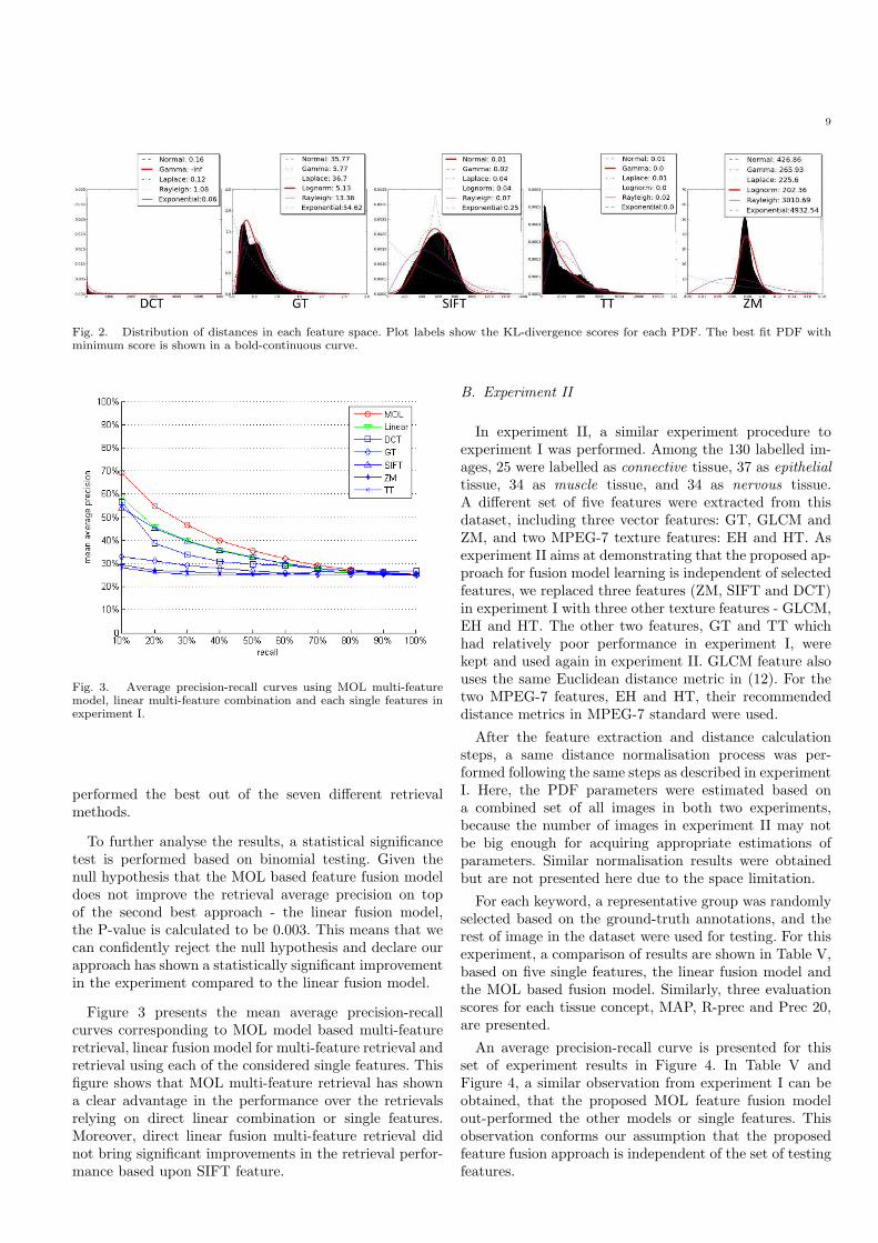
II, an important step following calculating a distance isthe normalisation of it. Original distances are estimatedin each feature space using distance functions describedabove. Normalisation of features is a key step to guaranteethe correctness of the derived multi-feature model. Sincethe distance distribution depends on the feature structureand image contents, the normalisation parameters areunknown a priori. The distribution of distances mightbe approximated using any PDF, whose parameters areestimated in different ways. To overcome this problem, weconsider a set of six possible PDFs. Using the distancesamples in the computed matrix, the parameters of eachpossible PDF are estimated in an attempt to match thetrue distribution of the distances. An approximation of thedistribution is then calculated for each bin of the empiricalhistogram using the estimated parameters. To identify thebest approximation to the distribution of distances, thePDF that minimises the KL-divergence score is selected.Figure 2 shows the distribution of distances in each of
the considered feature spaces, as well as the approximationwith each of the PDFs considered in Table I. Using eachPDF, a lowest possible KL-divergence score is calculatedbetween the PDF approximation and the empirical his-togram, as shown in Figure 2. For each feature space, thebest fit PDF is the one with the lowest score among allconsidered PDFs. The best fit PDFs are indicated witha bold continuous curve, while the other PDFs are indashed curves. The estimated parameters of the best fitPDF for each feature distribution are recorded for furthernormalisation purposes. For example, table II shows theestimated parameters for the best fit PDF for the featuredistributions in experiment I.3) Histology image retrieval: As mentioned before, in
experiment I evaluation of retrieval performance was con-ducted against a subset containing 2,828 histology imageswith manual labels for the four tissue types. The sub-set contains 484 samples for connective tissue, 804 forepithelial tissue, 514 for muscular tissue and 1026 fornervous tissue. For evaluation, a 5-fold cross-validationscheme is used, in which the whole dataset is randomlydivided into five equally sized groups. In each test, one
TABLE IIIExperiment I: retrieval evaluation of four tissue typesusing MOL approach across 5 folds, mean and standard
deviation (SD) values reported.
Tissue types AP R-prec Prec 20
Mean SD Mean SD Mean SD
Connective 0.378 0.024 0.354 0.014 0.830 0.249
Epithelial 0.467 0.018 0.409 0.020 0.840 0.188
Muscular 0.338 0.024 0.303 0.045 0.760 0.167
Nervous 0.584 0.032 0.525 0.020 0.930 0.027
TABLE IVExperiment I: retrieval evaluation of the proposed resultscompared to single features and linear fusion model across
5 folds, mean values reported.
Feature mean
AP
R-prec Prec 20
GT 0.296 0.290 0.383
TT 0.272 0.255 0.313
ZM 0.273 0.267 0.343
SIFT 0.352 0.357 0.658
DCT 0.328 0.325 0.675
All features linear comb. 0.402 0.356 0.748
MOL feature comb. 0.442 0.398 0.840
of the five groups is used as the training set and theother four are used for testing. The positive representativegroup of each type of tissue contains 10 relevant samplesthat are randomly selected from the training set based onthe ground-truth annotations. Using the retrieval resultsof each positive representative group, the correspondingnegative representative group is selected as the first 10retrieved non-relevant samples in the same training set.The performance measures presented include mean andstandard deviation values across five folds in AveragePrecision (AP); R-Precision (R-prec), which is obtainedat the point where precision and recall get the same value;and precision after the first 20 retrieved samples (Prec 20),as shown in Table III.As presented in Table III, among the four different tissue
types, some results are better compared to the others.For instance, the muscular tissue results are relativelyless accurate than the other three. There are probablytwo reasons for that. First, the number of muscular tissueimages is less than the others. There are 484 connective,804 epithelial, 514 muscular and 1026 nervous tissue sam-ples in the evaluation dataset of experiment I. The taskof retrieving images of a less popular query concept isusually more difficult than popular ones. Second, eachdifferent tissue type has its unique visual characteristicsand patterns. Some of them may be trickier to recogniseand differentiate from the others.Table IV shows mean retrieval performance across four
concepts, and using each of the five single features, and twodifferent feature fusion models. All features linear comb. isthe direct linear combination model of all the five featureswith the same importance weights for each feature space.This fusion model follows a direct linear combinationapproach. MOL feature comb. represents the results usingthe proposed MOL feature combination model. As it canbe observed in Table IV, the proposed MOL method
9
Fig. 2. Distribution of distances in each feature space. Plot labels show the KL-divergence scores for each PDF. The best fit PDF withminimum score is shown in a bold-continuous curve.
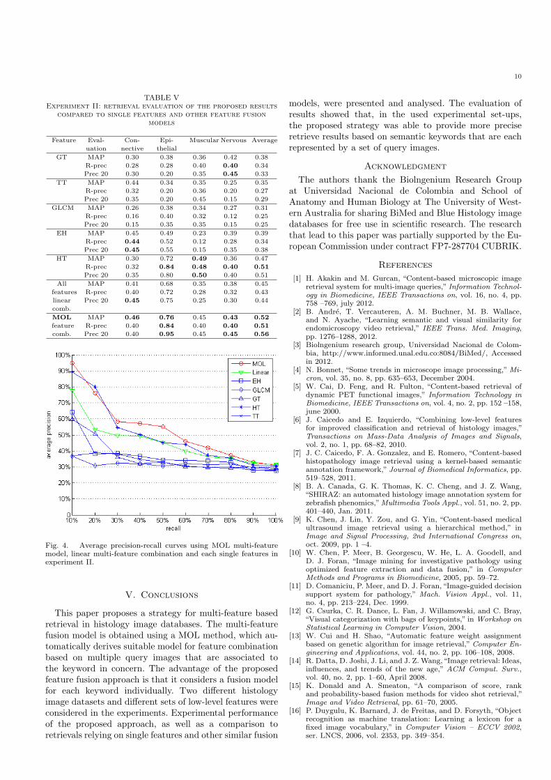
Fig. 3. Average precision-recall curves using MOL multi-featuremodel, linear multi-feature combination and each single features inexperiment I.
performed the best out of the seven different retrievalmethods.
To further analyse the results, a statistical significancetest is performed based on binomial testing. Given thenull hypothesis that the MOL based feature fusion modeldoes not improve the retrieval average precision on topof the second best approach - the linear fusion model,the P-value is calculated to be 0.003. This means that wecan confidently reject the null hypothesis and declare ourapproach has shown a statistically significant improvementin the experiment compared to the linear fusion model.
Figure 3 presents the mean average precision-recallcurves corresponding to MOL model based multi-featureretrieval, linear fusion model for multi-feature retrieval andretrieval using each of the considered single features. Thisfigure shows that MOL multi-feature retrieval has showna clear advantage in the performance over the retrievalsrelying on direct linear combination or single features.Moreover, direct linear fusion multi-feature retrieval didnot bring significant improvements in the retrieval perfor-mance based upon SIFT feature.
B. Experiment II
In experiment II, a similar experiment procedure toexperiment I was performed. Among the 130 labelled im-ages, 25 were labelled as connective tissue, 37 as epithelialtissue, 34 as muscle tissue, and 34 as nervous tissue.A different set of five features were extracted from thisdataset, including three vector features: GT, GLCM andZM, and two MPEG-7 texture features: EH and HT. Asexperiment II aims at demonstrating that the proposed ap-proach for fusion model learning is independent of selectedfeatures, we replaced three features (ZM, SIFT and DCT)in experiment I with three other texture features - GLCM,EH and HT. The other two features, GT and TT whichhad relatively poor performance in experiment I, werekept and used again in experiment II. GLCM feature alsouses the same Euclidean distance metric in (12). For thetwo MPEG-7 features, EH and HT, their recommendeddistance metrics in MPEG-7 standard were used.
After the feature extraction and distance calculationsteps, a same distance normalisation process was per-formed following the same steps as described in experimentI. Here, the PDF parameters were estimated based ona combined set of all images in both two experiments,because the number of images in experiment II may notbe big enough for acquiring appropriate estimations ofparameters. Similar normalisation results were obtainedbut are not presented here due to the space limitation.
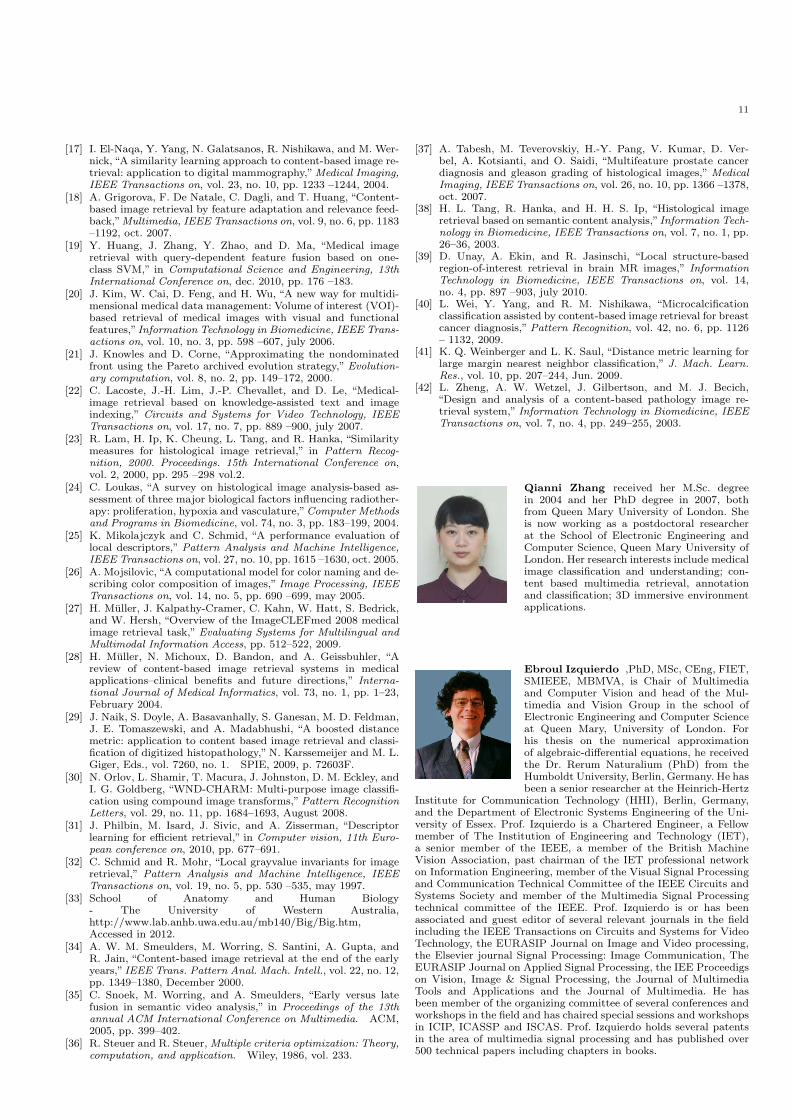
For each keyword, a representative group was randomlyselected based on the ground-truth annotations, and therest of image in the dataset were used for testing. For thisexperiment, a comparison of results are shown in Table V,based on five single features, the linear fusion model andthe MOL based fusion model. Similarly, three evaluationscores for each tissue concept, MAP, R-prec and Prec 20,are presented.
An average precision-recall curve is presented for thisset of experiment results in Figure 4. In Table V andFigure 4, a similar observation from experiment I can beobtained, that the proposed MOL feature fusion modelout-performed the other models or single features. Thisobservation conforms our assumption that the proposedfeature fusion approach is independent of the set of testingfeatures.
10
TABLE VExperiment II: retrieval evaluation of the proposed results
compared to single features and other feature fusionmodels
Feature Eval-
uation
Con-
nective
Epi-
thelial
Muscular Nervous Average
GT MAP 0.30 0.38 0.36 0.42 0.38
R-prec 0.28 0.28 0.40 0.40 0.34
Prec 20 0.30 0.20 0.35 0.45 0.33
TT MAP 0.44 0.34 0.35 0.25 0.35
R-prec 0.32 0.20 0.36 0.20 0.27
Prec 20 0.35 0.20 0.45 0.15 0.29
GLCM MAP 0.26 0.38 0.34 0.27 0.31
R-prec 0.16 0.40 0.32 0.12 0.25
Prec 20 0.15 0.35 0.35 0.15 0.25
EH MAP 0.45 0.49 0.23 0.39 0.39
R-prec 0.44 0.52 0.12 0.28 0.34
Prec 20 0.45 0.55 0.15 0.35 0.38
HT MAP 0.30 0.72 0.49 0.36 0.47
R-prec 0.32 0.84 0.48 0.40 0.51
Prec 20 0.35 0.80 0.50 0.40 0.51
All MAP 0.41 0.68 0.35 0.38 0.45
features R-prec 0.40 0.72 0.28 0.32 0.43
linear
comb.
Prec 20 0.45 0.75 0.25 0.30 0.44
MOL MAP 0.46 0.76 0.45 0.43 0.52
feature R-prec 0.40 0.84 0.40 0.40 0.51
comb. Prec 20 0.40 0.95 0.45 0.45 0.56
Fig. 4. Average precision-recall curves using MOL multi-featuremodel, linear multi-feature combination and each single features inexperiment II.
V. Conclusions
This paper proposes a strategy for multi-feature basedretrieval in histology image databases. The multi-featurefusion model is obtained using a MOL method, which au-tomatically derives suitable model for feature combinationbased on multiple query images that are associated tothe keyword in concern. The advantage of the proposedfeature fusion approach is that it considers a fusion modelfor each keyword individually. Two different histologyimage datasets and different sets of low-level features wereconsidered in the experiments. Experimental performanceof the proposed approach, as well as a comparison toretrievals relying on single features and other similar fusion
models, were presented and analysed. The evaluation ofresults showed that, in the used experimental set-ups,the proposed strategy was able to provide more preciseretrieve results based on semantic keywords that are eachrepresented by a set of query images.
Acknowledgment
The authors thank the Biolngenium Research Groupat Universidad Nacional de Colombia and School ofAnatomy and Human Biology at The University of West-ern Australia for sharing BiMed and Blue Histology imagedatabases for free use in scientific research. The researchthat lead to this paper was partially supported by the Eu-ropean Commission under contract FP7-287704 CUBRIK.
References
[1] H. Akakin and M. Gurcan, “Content-based microscopic imageretrieval system for multi-image queries,” Information Technol-ogy in Biomedicine, IEEE Transactions on, vol. 16, no. 4, pp.758 –769, july 2012.
[2] B. Andre, T. Vercauteren, A. M. Buchner, M. B. Wallace,and N. Ayache, “Learning semantic and visual similarity forendomicroscopy video retrieval,” IEEE Trans. Med. Imaging,pp. 1276–1288, 2012.
[3] Biolngenium research group, Universidad Nacional de Colom-bia, http://www.informed.unal.edu.co:8084/BiMed/, Accessedin 2012.
[4] N. Bonnet, “Some trends in microscope image processing,”Mi-cron, vol. 35, no. 8, pp. 635–653, December 2004.
[5] W. Cai, D. Feng, and R. Fulton, “Content-based retrieval ofdynamic PET functional images,” Information Technology inBiomedicine, IEEE Transactions on, vol. 4, no. 2, pp. 152 –158,june 2000.
[6] J. Caicedo and E. Izquierdo, “Combining low-level featuresfor improved classification and retrieval of histology images,”Transactions on Mass-Data Analysis of Images and Signals,vol. 2, no. 1, pp. 68–82, 2010.
[7] J. C. Caicedo, F. A. Gonzalez, and E. Romero, “Content-basedhistopathology image retrieval using a kernel-based semanticannotation framework,” Journal of Biomedical Informatics, pp.519–528, 2011.
[8] B. A. Canada, G. K. Thomas, K. C. Cheng, and J. Z. Wang,“SHIRAZ: an automated histology image annotation system forzebrafish phenomics,”Multimedia Tools Appl., vol. 51, no. 2, pp.401–440, Jan. 2011.
[9] K. Chen, J. Lin, Y. Zou, and G. Yin, “Content-based medicalultrasound image retrieval using a hierarchical method,” inImage and Signal Processing, 2nd International Congress on,oct. 2009, pp. 1 –4.
[10] W. Chen, P. Meer, B. Georgescu, W. He, L. A. Goodell, andD. J. Foran, “Image mining for investigative pathology usingoptimized feature extraction and data fusion,” in ComputerMethods and Programs in Biomedicine, 2005, pp. 59–72.
[11] D. Comaniciu, P. Meer, and D. J. Foran,“Image-guided decisionsupport system for pathology,” Mach. Vision Appl., vol. 11,no. 4, pp. 213–224, Dec. 1999.
[12] G. Csurka, C. R. Dance, L. Fan, J. Willamowski, and C. Bray,“Visual categorization with bags of keypoints,” in Workshop onStatistical Learning in Computer Vision, 2004.
[13] W. Cui and H. Shao, “Automatic feature weight assignmentbased on genetic algorithm for image retrieval,” Computer En-gineering and Applications, vol. 44, no. 2, pp. 106–108, 2008.
[14] R. Datta, D. Joshi, J. Li, and J. Z. Wang,“Image retrieval: Ideas,influences, and trends of the new age,” ACM Comput. Surv.,vol. 40, no. 2, pp. 1–60, April 2008.
[15] K. Donald and A. Smeaton, “A comparison of score, rankand probability-based fusion methods for video shot retrieval,”Image and Video Retrieval, pp. 61–70, 2005.
[16] P. Duygulu, K. Barnard, J. de Freitas, and D. Forsyth, “Objectrecognition as machine translation: Learning a lexicon for afixed image vocabulary,” in Computer Vision – ECCV 2002,ser. LNCS, 2006, vol. 2353, pp. 349–354.
11
[17] I. El-Naqa, Y. Yang, N. Galatsanos, R. Nishikawa, and M. Wer-nick, “A similarity learning approach to content-based image re-trieval: application to digital mammography,”Medical Imaging,IEEE Transactions on, vol. 23, no. 10, pp. 1233 –1244, 2004.
[18] A. Grigorova, F. De Natale, C. Dagli, and T. Huang, “Content-based image retrieval by feature adaptation and relevance feed-back,”Multimedia, IEEE Transactions on, vol. 9, no. 6, pp. 1183–1192, oct. 2007.
[19] Y. Huang, J. Zhang, Y. Zhao, and D. Ma, “Medical imageretrieval with query-dependent feature fusion based on one-class SVM,” in Computational Science and Engineering, 13thInternational Conference on, dec. 2010, pp. 176 –183.
[20] J. Kim, W. Cai, D. Feng, and H. Wu, “A new way for multidi-mensional medical data management: Volume of interest (VOI)-based retrieval of medical images with visual and functionalfeatures,” Information Technology in Biomedicine, IEEE Trans-actions on, vol. 10, no. 3, pp. 598 –607, july 2006.
[21] J. Knowles and D. Corne, “Approximating the nondominatedfront using the Pareto archived evolution strategy,” Evolution-ary computation, vol. 8, no. 2, pp. 149–172, 2000.
[22] C. Lacoste, J.-H. Lim, J.-P. Chevallet, and D. Le, “Medical-image retrieval based on knowledge-assisted text and imageindexing,” Circuits and Systems for Video Technology, IEEETransactions on, vol. 17, no. 7, pp. 889 –900, july 2007.
[23] R. Lam, H. Ip, K. Cheung, L. Tang, and R. Hanka, “Similaritymeasures for histological image retrieval,” in Pattern Recog-nition, 2000. Proceedings. 15th International Conference on,vol. 2, 2000, pp. 295 –298 vol.2.
[24] C. Loukas, “A survey on histological image analysis-based as-sessment of three major biological factors influencing radiother-apy: proliferation, hypoxia and vasculature,”Computer Methodsand Programs in Biomedicine, vol. 74, no. 3, pp. 183–199, 2004.
[25] K. Mikolajczyk and C. Schmid, “A performance evaluation oflocal descriptors,” Pattern Analysis and Machine Intelligence,IEEE Transactions on, vol. 27, no. 10, pp. 1615 –1630, oct. 2005.
[26] A. Mojsilovic, “A computational model for color naming and de-scribing color composition of images,” Image Processing, IEEETransactions on, vol. 14, no. 5, pp. 690 –699, may 2005.
[27] H. Muller, J. Kalpathy-Cramer, C. Kahn, W. Hatt, S. Bedrick,and W. Hersh, “Overview of the ImageCLEFmed 2008 medicalimage retrieval task,” Evaluating Systems for Multilingual andMultimodal Information Access, pp. 512–522, 2009.
[28] H. Muller, N. Michoux, D. Bandon, and A. Geissbuhler, “Areview of content-based image retrieval systems in medicalapplications–clinical benefits and future directions,” Interna-tional Journal of Medical Informatics, vol. 73, no. 1, pp. 1–23,February 2004.
[29] J. Naik, S. Doyle, A. Basavanhally, S. Ganesan, M. D. Feldman,J. E. Tomaszewski, and A. Madabhushi, “A boosted distancemetric: application to content based image retrieval and classi-fication of digitized histopathology,” N. Karssemeijer and M. L.Giger, Eds., vol. 7260, no. 1. SPIE, 2009, p. 72603F.
[30] N. Orlov, L. Shamir, T. Macura, J. Johnston, D. M. Eckley, andI. G. Goldberg, “WND-CHARM: Multi-purpose image classifi-cation using compound image transforms,”Pattern RecognitionLetters, vol. 29, no. 11, pp. 1684–1693, August 2008.
[31] J. Philbin, M. Isard, J. Sivic, and A. Zisserman, “Descriptorlearning for efficient retrieval,” in Computer vision, 11th Euro-pean conference on, 2010, pp. 677–691.
[32] C. Schmid and R. Mohr, “Local grayvalue invariants for imageretrieval,” Pattern Analysis and Machine Intelligence, IEEETransactions on, vol. 19, no. 5, pp. 530 –535, may 1997.
[33] School of Anatomy and Human Biology- The University of Western Australia,http://www.lab.anhb.uwa.edu.au/mb140/Big/Big.htm,Accessed in 2012.
[34] A. W. M. Smeulders, M. Worring, S. Santini, A. Gupta, andR. Jain, “Content-based image retrieval at the end of the earlyyears,” IEEE Trans. Pattern Anal. Mach. Intell., vol. 22, no. 12,pp. 1349–1380, December 2000.
[35] C. Snoek, M. Worring, and A. Smeulders, “Early versus latefusion in semantic video analysis,” in Proceedings of the 13thannual ACM International Conference on Multimedia. ACM,2005, pp. 399–402.
[36] R. Steuer and R. Steuer, Multiple criteria optimization: Theory,computation, and application. Wiley, 1986, vol. 233.
[37] A. Tabesh, M. Teverovskiy, H.-Y. Pang, V. Kumar, D. Ver-bel, A. Kotsianti, and O. Saidi, “Multifeature prostate cancerdiagnosis and gleason grading of histological images,” MedicalImaging, IEEE Transactions on, vol. 26, no. 10, pp. 1366 –1378,oct. 2007.
[38] H. L. Tang, R. Hanka, and H. H. S. Ip, “Histological imageretrieval based on semantic content analysis,”Information Tech-nology in Biomedicine, IEEE Transactions on, vol. 7, no. 1, pp.26–36, 2003.
[39] D. Unay, A. Ekin, and R. Jasinschi, “Local structure-basedregion-of-interest retrieval in brain MR images,” InformationTechnology in Biomedicine, IEEE Transactions on, vol. 14,no. 4, pp. 897 –903, july 2010.
[40] L. Wei, Y. Yang, and R. M. Nishikawa, “Microcalcificationclassification assisted by content-based image retrieval for breastcancer diagnosis,” Pattern Recognition, vol. 42, no. 6, pp. 1126– 1132, 2009.
[41] K. Q. Weinberger and L. K. Saul, “Distance metric learning forlarge margin nearest neighbor classification,” J. Mach. Learn.Res., vol. 10, pp. 207–244, Jun. 2009.
[42] L. Zheng, A. W. Wetzel, J. Gilbertson, and M. J. Becich,“Design and analysis of a content-based pathology image re-trieval system,” Information Technology in Biomedicine, IEEETransactions on, vol. 7, no. 4, pp. 249–255, 2003.
Qianni Zhang received her M.Sc. degreein 2004 and her PhD degree in 2007, bothfrom Queen Mary University of London. Sheis now working as a postdoctoral researcherat the School of Electronic Engineering andComputer Science, Queen Mary University ofLondon. Her research interests include medicalimage classification and understanding; con-tent based multimedia retrieval, annotationand classification; 3D immersive environmentapplications.
Ebroul Izquierdo ,PhD, MSc, CEng, FIET,SMIEEE, MBMVA, is Chair of Multimediaand Computer Vision and head of the Mul-timedia and Vision Group in the school ofElectronic Engineering and Computer Scienceat Queen Mary, University of London. Forhis thesis on the numerical approximationof algebraic-differential equations, he receivedthe Dr. Rerum Naturalium (PhD) from theHumboldt University, Berlin, Germany. He hasbeen a senior researcher at the Heinrich-Hertz
Institute for Communication Technology (HHI), Berlin, Germany,and the Department of Electronic Systems Engineering of the Uni-versity of Essex. Prof. Izquierdo is a Chartered Engineer, a Fellowmember of The Institution of Engineering and Technology (IET),a senior member of the IEEE, a member of the British MachineVision Association, past chairman of the IET professional networkon Information Engineering, member of the Visual Signal Processingand Communication Technical Committee of the IEEE Circuits andSystems Society and member of the Multimedia Signal Processingtechnical committee of the IEEE. Prof. Izquierdo is or has beenassociated and guest editor of several relevant journals in the fieldincluding the IEEE Transactions on Circuits and Systems for VideoTechnology, the EURASIP Journal on Image and Video processing,the Elsevier journal Signal Processing: Image Communication, TheEURASIP Journal on Applied Signal Processing, the IEE Proceedigson Vision, Image & Signal Processing, the Journal of MultimediaTools and Applications and the Journal of Multimedia. He hasbeen member of the organizing committee of several conferences andworkshops in the field and has chaired special sessions and workshopsin ICIP, ICASSP and ISCAS. Prof. Izquierdo holds several patentsin the area of multimedia signal processing and has published over500 technical papers including chapters in books.


![Stay Fit Optimised[1]](https://img.pdfslide.us/doc/110x75/577d34ea1a28ab3a6b8f27ce/stay-fit-optimised1.jpg)