Embed Size (px)
Citation preview

Title
HISTOLOGICAL STUDY OF THE LOWER LIMB INWHICH MULTIPLE MILIARY EMBOLISMS WEREPRODUCED EXPERIMENTALLY : A CONTRIBUTION TOTHE ALLERGIC ETIOLOGY OF THROMBOANGIITISOBLITERANS.
Author(s) Konishi, Seizo
Citation 日本外科宝函 (1954), 23(1): 29-37
Issue Date 1954-01-01
URL http://hdl.handle.net/2433/206062
Right
Type Departmental Bulletin Paper
Textversion publisher
Kyoto University

HISTOLOGICAL STUDY OF THE LOWER LIMB IN WHICH
MULTIPLE MILIARY EMBOLISMS ¥¥'ERE PRODUCED
EXPERIMENTALLY. A CONTRIBUTION TO THE ALLERGIC
ETIOLOGY OF THROMBOANGIITIS OBLITERANS.
From the !st Surgical Division, Kyoto U口iversityMedical School
(Director : Prof. Dr. ・Ca1sATO ARAK『)
by
$EIZO KONISHI
(Receivrd for Publication : Nov. 4. 1953)
INTRODUCTION
29
There have been manγopinions about the etiology of thromboangiitis oblite-rans. 1) Btirger stated that the cause was inflamation of blood vessels due to a certain bacterial infection. Other authors expressed other yiews: 2) The patho-‘
logical vasoconstriction of long duration results in the organic obstruction of the' blood vessels ( Schlesi.1ger).「, 3)Some poisions, s!.l ch as nicotine (Maddock & Co~ler ・ 1933) or ergot (Kaunitz & Gerlach 1933), are the cause of dysfunction of blood vessels, 4) frost-bite or coldness is an important factor (Bier and Gruber etc.), 5) the disease is concerned with a general infectious disease (Wilhelm and Rieder etc), 6) with the increase of blood viscosity ( Kaga and Maesima ), 7) with some disorder of internal secretion ( pituitrin-Holsclaw and Booth 1925, ovary-Boecke 1927, thyroid gland-Bassai e Digliotti, Kallos and Nusset 1933). And recently it has been assumed, 8) that important is the deficient chemical regulation of the general circulation in a pathologically sensitive organism (Ratschow ). 9) There are authors who try to explain the disease from the viewpoint of allergy-Sulz-berger (tobacco-extract) and Denecke (coldness, nicotine and chronic infections). Pathological changes are found in the disease not only in arteries of extremities but also arteries of nearly all organs, namely, heart, brain, mesentery, lung, kidney, spleen, adrenal gland, spermatochorda, seminal vesicle, aorta and prostate etc. Thus the allergy is regarded as a predominant etiologic facfor. In addition histo-logical changes characteristic of allergy were described in this disease by E. Jager ・and recently by Wada of our laboratory. In these studies, it was assumed that the allergic changes in th・s disease was primary, i.e. the allergy is the primary cause of the disease. But it is possible that the allergic changes may be secondary, i.e. the devitalization of tissue resulting from the disturbed cltculation may act as an allergenic agent, th us ca using secondary tissue reaction of allergy.
The present work has been carried out for the elucidation of th s problem.
METHODS OF EXPERIMENT
It is di伍cultto reproduce experimentally the progressive disturbance of arterial
30 日本外科宝働第23巻第1号
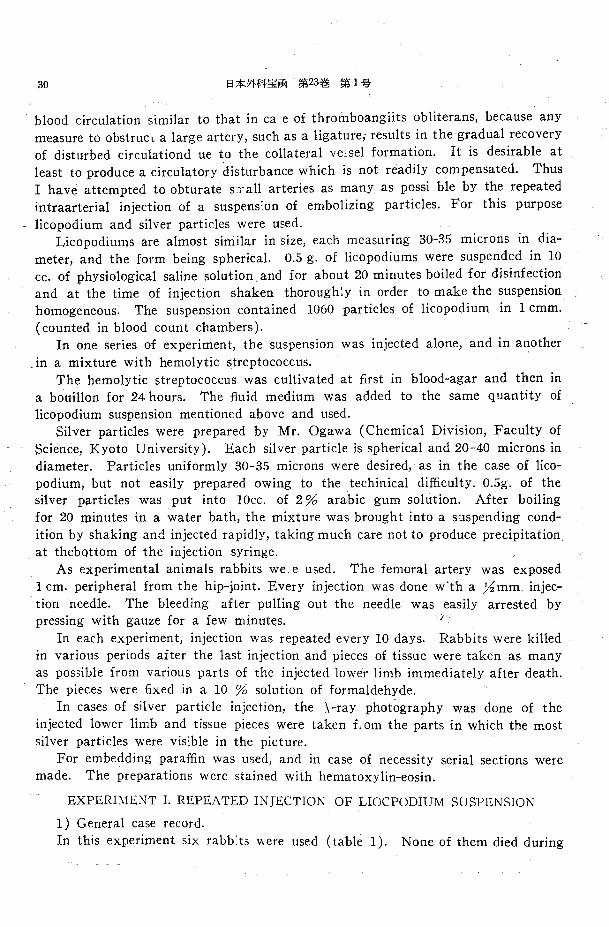
blood circulation similar to that in ca e of throinboangiits obliterans, because any measure to obstrucL a large artery, such as a ligature; results in the gradual recovery of disturbed circulationd ue to the collateral ve】selformation. It is desirable at least to produce a circulatory d凶 ubancewhich is not readily compensated. Thus I have attempted to obturate s:了allarteries as many as possi ble by the repeated intraarterial injection of a suspens:on of embolizing particles. For this purpose
licopodium and silver particles were used. Licopodiums are almost sirriilar in size, each measuring 30-35 microns in dia-
meter, and the form being spherical. 0.5 g. of licopodiums were suspended in 10 cc. of physiological saline solution and for about 20 minutes boiled for disinfection and at the time of injection shaken thoroughly in order to make the suspension homogeneous. The suspension contained 1060 particles of licopodium in 1 cmm.
(counted in blood count chambers). In one series of experiment, the suspension was injected alone, and in another
in a mixture with hemolytic streptococcus. The hemolytic streptococcus was cultivated at日rstin blood-agar and then in
a bouillon for 24 hours. The fluid medium was added to the same quantity of licopodium suspension mentioned above and used.
Silver particles were prepared by Mr. Ogawa (Chemical Division, Faculty of Science, Kyoto University). Each silver particle is spherical and 20-40 microns in diameter. Particles uniformly 30-35 microns were desired, as in the case of lico・podium, but not easily prepared owing to the techinical di伍culty.0.5g. of the silver particles was put into lOcc. of 2 % arabic gum solution. After boiling for 20 minutes in a water bath, th巴 mixturewas brought into a suspending cond-ition by shaking and injected rapidly, taking much care not to produce precipitation at thebottom of the injection syringe.
As experimental animals rabbits we. e used. The femoral artery was exposed I cm. peripheral from the hip-joint. Every injection was done w"th a Yi mm. injec-tion needle. The bleeding after pulling out the needle was easily arrested by pressing with gauze for a few minutes.
In each experiment, injection was repeated every 10 days. Rabbits were killed in var.ious periods after the last injection and pieces of tissue were taken as many as possible fτom various parts of the injected lower limb immediately after death. The pieces were日xedin a 10 % solution of formaldehyde.
In cases of silver particle injection, the ¥-ray photography was done of the injected lower limb and tissue pieces were taken £1 om the parts in which the most silver particles were visible in the picture.
For embedding para伍nwas used, and in case of necessity serial sections were made. The preparations were stained with hematoxylin-eosin.
EXPERIMENT I. REPEATED INJECTION OF LIOCPODIUM SUSPENSION
1) General case record.
In this experiment six rabb:ts were used (table 1). None of them died during
HISTOLOGICAL STUDY OF THE LOWER LIMB 31
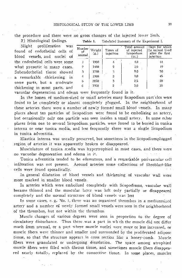
the procedure and there were no gross changes of the injected lower limb.
2) Histological findings. Table I. Tabulated Summary of the Experiment I. Slight proliferation was
found of endothelial cells of
blood vessels, and nuclei of
the endothelial cells were some
what pycnotic in many cases.
Subendothelial tissue showed
a remarkable .thickening in
some parts, but a moderate
thickening in most parts, and
Numb I I of r I Weight I Times of . al I (g.) I injection
|…。γ…ichof injected the ammal lived licopodium after the五rst
(cc.) injection
l
2
3
.1.
5
6
1900 I
1400 I
1700
1900
2050
1950
4
2
:1
3
3
2
トィ, I ,, 1 6 I 19
3.0 I 30
3.0
2.5
3.0
45
28
35
vacuolar degeneration and巴d巴mawere frequently found in it.
In the lumen of medium-sized or small’arteries many licopodium part.des were
found to be completely or almost completely plugged. In the neighborhood of
these arteries there were a number of newly formed small blood vessels. In many
places about ten particles of licopodium were found to be embolizing an artery,
but occasionally only one particle was seen insides a small artery. In some other
places from one to several licopodium. particle::. were found to be buried in tunica
interna or near tunica media, and less frequently there was a single licopodium
in tunica adventitia.
Elastica interna was usually preserved, but sometimes in the licopodiumplugged
region. of arteries it was apparently broken or disappeared.
Musculature of tunica rr:edia was hypertrophied in most cases, and there were
no vacuolar degeneration and edema in i十.
Tunica adventitia tended to be edematous, and a remarkable perivascular cell
in自ltrationwas not present. Around arteries some collections of fibroblast-like
cells were found sporadically.
In general dilatation of blood vessels and thickening of v2.scular wall were
more marked in smaller blood vessels.
In arteries which were embolized completely with licopodiums, vascular wall
became thinn巴dand the muscular lay町 wasleft only partially or disappeared completely and the normal structure of blood vessels was lost
In some cases, e. g. No. I, there was an organized‘ thrombus in a medium弓ized
artery and a number of newly formed small vessels were seen in the neighborhood
of the thrombus, but not within the thrombus.
Muscle changes of various degrees were seen in proportion to the degree of
circulatory disturbance. Thus there was a part in wh ch the muscle did not di妊er
much from normal, or a part where muscle nuclei were more or less increased, or
muscle五berswere thinner and smaller and surrounded by the proliferated adipose
tissue, so that the structure appears in cross section like a honey担 comb. Muscle
五berswere granulated or undergoing dissolution. The space among atrophied
muscle fibers were filled with fibrous tissue, and sometimes muscle五bersdisappea-
red nearly totally, replaced by the connective tissue. In some places, muscles

第 1号
became necrotic, the outline of muscle五hersbecame indistinct, staining lightly with
eosin and plenty of fragments of pycnotic muscle nuclei were seen in the inter-
fascicular space. There were many parts in which aggregates of h.eav ly degene-
rated muscle bundles were neighbored by those of nearly normal muscle bundles.
Muscle nuclei showed various changes according to the degree of degeneration
; some of them were stained normally by hematoxylin, and others stained lightly,
or transparently, or darkly etc.. The shapes of nuclei were round, oval, slender
or cylindrical etc .. Occasionally it was difficult to distinguish them from the nuclei
of connective tissue cells. Generally speaking, transparent nuclei were large in
size and dark ones were small. There were areas where small bleedings were seen
in the interstitium of a muscle. In one case muscular multinucleated giant cells
were found.
The perineurium of peripheral nerves was edematously swollen and the intima
of blood vessels in the perineurium was thickened. Proliferation of nuclei of
Schwann’s sheath wa》 oitenseeロ. The changes of nerve自berswere not CITtain
because of the lack of specific stainin .
第 3巻日本外科宝画32
EXPERI.~ENT IL REPEATED INJECTION OF A MIXED SUSPENSION OF
LICCjPODIUM AND HEMOLYTIC STR己PTOCOCCUS.
1) General case reco; d.
In this experiment six rabbits were used
before the end of an observa-
tion period. There was no case
in which gross changes of the
injected lower limb were found.
2) Histological五ndings. 、 7 I 1soo
In blood vessels prolifera- 8 I 2400
tion of the endothelium was 9 I 2800
dight. In one case a few IO I 2100
detached endothelial cells look- II i 1900
i昭 likemonocytes were seen 12 I 2000
in the lumen of a blood vessel. Nuclei of endothelial cells tended to be pycnotic.
There was not infrequently vacuolization of subendothelial cells of small blood vessels.
The changes Of blood vessels in the areas of licopodium embolism were the
same as in Experiment I. It was observed that, if licopodium particles obturated
the lumen partially, the wall became thinned on the side on which the particles
were attached and thickened on the opposite side. Perivascular collection of
五broblast-likecells was sporadically se~n and in a few cases there were remarkable perivascular infiltrations of lymphocyte-like mononuclear cells.
Changes of muscle tissue were also the same as in Experiment I. However,
the degree of changes in Experiment II seem巴dmore marked than in E}¥periment
I. Neither hemolytic streptococcus nor abscess formation was found anywhere.
died A rabbit (No. 10)
Tabulated Sllmmary of the EicperimeFt IL
’I‘otal amount I Days・ for which of the mi'.<-ed :the animal lived suspension / after the五rst
injected (cc.) I injection
3
5
0
0
5
1
qLqLqδηLqonL
4
7
7
3
4
6
(table 2).
~imes of inj~ction
qLqaqa噌
Aq4qδ
Table 2.
Nur巾 e! I d 訂 IWeight I
ani:Ual I ( g.) I

33
Fresh red blood cells were often seen in the interstitium of muscles. Among
normal muscle bundles there were degenerated ones distinctly separated from the
former. Muscular multinucleated giant cells were occasionally found near the
marginal zone of degenerated muscles.
Peripheral nerves were somewhat edematous.
HISTOLOGICAL STUDY OF THE LOWERLIMB
EXPERIMENT III. REPEATED INJECTION OF A SILVER
PARTICLE SUSPENSION.
Seven died before the
( 1) General case record.
In this experiment nine rabbits were used (table 3 ).
end of an observation period
(No. 14, 15, 16, 17, 18, 19, 20 ).
Grossly in the injected lower
limb there wer巴 depilation(No.
19, 20), small ulcer of toes (
No. 19), inflammatory swelling
(No. 16),necrosis and falling-
o妊 ofthe foot from the ankle-
joint (No. 17). These healed
after a certain period.
Histological £ndings.
(2) In arteries wher。thelumen
was £1led with more than one silver partlice, there were severe small round cell infiltrations in the wall of the
vessels and around them. Such cell ip自ltrationswere intense especially in the animals which died shortly after the injection, but not present in the long surviv-
ing animals. In arteries, of which the lumen was obliterated with one silver
particle, the normal structure of tt-.e vessel wall was lost ; intima and media
completely disappeared as if the silver particle alone exsisted in the connective tissue, any reactive change being lacking around the particle.
It seems that the silver particle was impacted tightly into a small blood vessel
by the pressure of the blood stream and the vessel wall underwent pre5sure atrophy.
Some proliferation of endothelial cells and pycnosis of nu lei were seen in
other arteries.
Thickening of intima was slight. In Case No. 18, the intima of a medium-
sized artery was thickened verrucously. In general, intima was edematous. Media
was hypertrophied, but not edemato s. In adventitia and its neighborhood at
the site of silver particle impaction, there were s~vere cell in五ltrationsas mentioned
above. In the vicinity of obturated blood ves els the new formation of abundant
small blood vessels was always seen, especially remarkable in Case I 7, where the
foot fell o任 fromthe ankle-joint.
In the muscle tissue, degeneration of various degrees was observed in the same
way as in the preceding experiments. Besides the findings described there, here
Tabulated Sumnary of the Experiment III.
Total amount I D喝ys.for whiとliof injected ithe animal lived silver sus- ¥ after t士1efirst
pet1sio 1 (cc.) I injection
6 I 10
2 ! 14
3 l 20
4 ! 37
5 I 135
2 I 5b 2 I 68
4 I 120
3 I 210
Times of 1日jection
2
1
2
2
3
1
1
2
2
3.
Numbe I _ I of rl W印刷・mal I (g.) I
3
4
5
6
7
8
9
0
1
司且
T
A
τ
A
E--A
司A
’An
4
n
L
1800
1850
1750
2500
1900
2300
2100
1950
1800
Taol'-'

34 日本外科室伯 IJ'i;23巻第1号
were scattered some muscle fibers which underwent hyaline degeneration or som巴other muscle fibers, the outlines of which were stained more intensely with hema-toxylin.
Arcund the blood vessels in the interstitium of muscle tissue, there were fresh bleedings and hemosiderin deposits.
In cases of long-surviving rabbits, muscle fibers were thinned and dispersed and in some parts disappeared, being replaced by the connective tissue. The五ndingcorresponds to that of fibrous myositis.
In all cases there were muscular mult' nucleated giant cells, seemingly plasmo-dial.
Changes of peripheral nerves were the same as in the preceding experiments.
EXPERIMNT IV. INTRAMUSCULAR INJECTION OF SILVER PARTICLES
It is questioned whether the changes observed in Experiment III are to be attributed more to the tox'.city of silver than to the obstruction of vessels.
In the present experiment, I have carried out the control experiment, in which the same suspension of silver particles as in the previous experiment is injected intramuscularly.
I) General case record.
Two rabbits were used (table 4). 0.4 cc. of the suspension was injected into
femoral muscle. Dut ing the Table 4. Tabulated Summary of Experimer,t IV. course of observation after the
injection th巴rewere no gross changes in the injected part. 10-15 days later the animals were killed and muscle pieces of the injected part were exa『
mined histologically.
2) Histological findings.
Numb出 I 1扇面云示。unfT函瓦T正面hicl1nf ~' I Weight I Times of I of injected l出ぞ animallived
ani:Oa! J ( g.) I inj出 ion j suspension j af.ter the I I i (cc.) I 1n,1ection
22 I 1900 I 1 叫十干J o.4 I 23 I i 1so I 1 I o. 4 I
10
15
Changes were localized mainly in the injected part. Muscle五bersin the nei-gh borhood of silver particles were atrophic. A number of neutrophil polymorpho-nuclear leucocytes were present, and among these there were a few eosinophil leucocytes and forign body giant cells. However the changes were not so intense as in Experiment III. In the part where silver particles did not exist, only a small grouping of neutrophil polymorphonuclear leucocytes was seen in t.he inter-stitium of muscles. There was no increase of muscle nuclei.
In short, the changes in the muscle tissue were slight a .cl localized for the most part around silver particles・ Inconsi<leri g the fact that neutrophil polymorpho-nuclear leucocytes, eosinophil leucocytfs and foreign body giant cells were seen. the cha昭 Sin this叫町imentち町m to be due mainly to the action of山一particles as foreign bodies rather th<1n to the chemical toxicity of silver. Thus it may be assumed that the changes in the previous experiment resulted from the embolism of silver particles.

HISTOLOGICAL STUDY OF THE LOWER LIMB 35
SUMMARY AND COMMENT
It has been stated that histological changes due to allergic reaction are chara-
cteristic, appearing mainly in and around blood vessels. From this view point
histological changes in my experiments will be criticized.
My results can be summarized as follows. In blood vessel there were some proliferation of endothelial cells, pycnosis of
their nuclei, (especially verrucous thickening of intima in Experiment
III), subendothelial vacuolization, hypertrophy of media, more or less obturation
of the lumenby foreign bodies, and peri、-ascular-edema. It may not be acceptable
that they are allergic changes, because histological criteria for allergic reaction, as
Klinge has shown, are fibrinoid swelling of the wall of blood vessels, fibrinous or
hyaline thrombus and perivascular in五ltrationof small round cells, including plasma
cells.
Such vascular changes were found in no case of my experiments, while they
are quite conspicuous in human thromboangiitis obliterans (Jager, Wada, Hayashi).
Therfore it may be certain that the changes of blood vessels in my experiments
are not of allergic nature, but a mere result of circulatory de自cit. Thus a tissue
damage due to the circulatory arrest does not seem to be allergenic, and therefore
it may be assumed that ,allergic changes in thromboangiitis obliterans are not
secondary to a tissue damage, but essentially primary.
In the muscle tissue, there were granular, vacuolar and rarely hyaline degene-rations, increase of adipose tissue, regressive changes of muscle日bersnuclei, ne-
crosis of muscle fiber and multinucleated giant ぽ llsof plasmodial type. These
changes are quite common in usual myositis. Ukawa found them in case of local
anaphylaxis of muscle but stat巴dthat they were not characteristic of anaphylactic
myositis. is no There good reason to believe that these changes are allergic.
In the peripheral nerves, although details were not demonstrable by hema-
toxylin・eosinstaining edema was the main change and some thickening of the
intima of blood vessels within the nerves was also seen, allergic changes being
absent. As regards gross changes of the limbs, depilation, small ulcer, and falling-o妊
of the foot from the ankle-joint were observed in Experiment III, but they were
never so incurable as those in thromboangiltis obliterans, always healing in a
comparatively short period of time. Thus it .seems that the disturbance of cir-
culation produced in my experiments caロeasilybe compensated by the formation
of collateral channels. Therefor巴 ina persisting circulatory failure as in throm-
boangiitis obliterans, there must be an ever progressing vascular obturation. This
is -the very characteristic of the disease and never to be reproduced in experiments.
Since the time when Klinge and others advanced the streptococcus allergy
theory, Criep, Mayer, Murphy and Swift etc. succeeded experimentally in the
reproduction of a series of allergic 'histological changes, e.g. rheumatic myccarditis
or rheumatic nodular giant cells. In consideration of these facts, I have attempted

36 白木/jF、|宝耐第23望号第1号
to inject intraarterially the hemolytic streptococcus together with licopodiums
(Expeirment II), but only a little severer change took place both in blood ves-
sels and .muscles as compared to the control (Experiment I), allergic changes being
likewise absent.
As to the question whether a severe acute reaction observed in the tributary
of the artery, into which silver particles were injected, may be the result of the
toxicity of silver, the answer is negative in considering the五ndingin Experiment
IV. The reaction is to be attributed to some mechanical property of silver par-
tides more五ttedfor embolism.
CONCLUSION
l) By the repeated injection of suspension of licopodium or of silver particle
into the femoral artery, definite changes took place in blood ve,ssels, muscles, and
nerves of the injected lower limb, but they were not to be regarded as allergic
changes.
2) Intensity of histological changes by the injection of silver particles was
more marked than that by the injection of licopdium.
3) Even in case of the repeated injection of hemolytic streptococcus together
with licopodium, typical allergic changes did not occur.
4) There was one case in which the foot on the inj~cted side fell o任 fromthe
ankle-joint as the result of necrosis, but the wound healed in a relatively short
time. An incurabl ulceration as seen in thromboangiitis obliterans could not be
reproduced in my experiments.
5) So far as my experiments. are concerned, it seems improbable, even if not
impossible, that the devitalized tissue in the body may be allergenic. Thus the
allergic changes in thromboangiitis obliterans may well be ass'.lmed to be primary
and not secondary to the tissue damage as a result of the circulatory failure.
REFERENCES l) Denecke, K. : Pathologisch-anatomische und klinische Untersuchungen zur Aetiologie der juvenilen Gaogran, Arch. f. kl. Chir., 177, 821. 193究. 2) J益ger,E. : Zur pathologischen Anatoc mie der Thromboa!'giitis obliterans bei juveniler Extremit証tengangran,I Mitteilung, Virch. Arch. f. path. Anat., 284, 526, 1932. U!:d II Mitteilu「g,ebenda, 284, 584, 1932. 3) Klinge, F. : Das. Ge-websbild des自eberhaftenReumatismus, Virch. Arch. f. path. Anat., 286, 344, 1932. 4) Pagel, W. : Pathologie und Histo!ogie der allergischen fa:scheinugen, Fortschritte der Allergielehre, vo:i Kallos, P. Karyger, S. Basel, New York. 1リ39.5.) Ratschow, M. : Beltrag zur Ursache der Ext-remitatengangran, Zbl. f. d. Chir., 60, 821, 1933. 6) Rossie, R. : Ueber den Formenkreis der rheu-matischen Gefassver詰nderungenmit besonderer Beriicksichtigung der rheuamtischen Gefassent-
ziindung, Virh. Arch.‘ f. path. Anat., 288, 780, 1933. 7) Sulzberger, M. B. : Recent immunologic studies in hypersensitivity to tobacco, J.A.M.A. 102, 11, 1934. 9) Eda, S. : Patho-histo!ogical stLJdie on blood ve.ssels of spontaneous gangrene, ]. Jap. Surg. Soc. 42,児7,1941. 9) Hatano, S. . A cor:tribution to the pathology of nuclei of muscle, Tokyo-Igakukai-Zassi, 39, 309, 1925, 10) Hayasi, I-I. and Wada, T. : Histo-pathological changes in spontaneous gangrene, Tr. Soc. Path. Jap. 38, Editio regionalis, 23, 1949. 11) Karattb E. : Studies on socalled _spontaneous gangrene, J. Jap. Surg. Soc.,36, 13119, 1935. 12) Okubo, S. Studies on experimental thrombosis, Kaigun-Guniikai”Zassi, 22 : 80, 1933, and ebenda 22, 550, 1933. 13) Sato, T. : Studies on spontaneous gangrene, J.Jap. Surg. Soc., 42, 577, 1941. 14) Suzue, K. and Ueno, M. : The death by sepsis following the amputation of lower limb ot

I
l~ig. 6 S. Konishi

I
唾.. :毛均咽” 三、,A
,、 企』...、.
S. Konishi

HISTOLOGICAL STUDY OF THE LOWER Li ¥Ul 37
spontaneous gangrene, Syuzyutu, 2, 30tl, 1948. JS l
Suzue, K. and Hayasi, H. : ・A new theory of
rheumatism, Nanzando, Tokyo, 1951. 16) Uka-
wa, Y. : Patho-histological studies oi1 local ana-phylaxis, II chapter, The local anaphylaxis in
muscle tissue, Kurasiki-tyuobyoin-aenpo, 2, 13司
1927. 17) Wada, H. : Concerning allergic origin
of spontaneous gangrene, Jap. Circul. J., 14, 265, Iリ51
EXPLANATION OF PLATES
Fig. 1. The lumen of an artery is obliterated
completely with a number of particles of licopo-
dium. Perivascular edema.×360
Fi;r. 2. A licopodium in adventitia x 360
Fi~. 3. Proliferation of intima in a small artery.
There are three particles of licopodium .in the
ir.tima. The lumen of the vessel obliterated
nearly completely.×80
Fig・. 4. Around obstructed arteries there are
newly formed small blood vessels in abundance.
A obstructed and organized artery B-newly formed
blood vessels. x 360
Fig・. 5. Verrucous thickening of intima of a
medium-sized art巴ry.×80
Fig・. 6. The wall of an artery, which is embo-
lized with three silver particles, is heavily in五1-
trated by leucocytes.×360
Fig. 7. A muscular multinucleated giant cell
which seems to be formed plasmodially. In the
center of the photograph.×360
Fig. 8. Fibrosed muscle tissue. Atrophied muscle
fibers are seen on both sides.×360
Fig・. 9. Increase of adipose tissue within a
muscle. x80
Fi宮.10. Remarkable di任erencein histological
changes between the two neighboring groups of
muscle bundles. 'Right side is almost normal and
left degenerated.×360
Fh. 11. Necrosis of muscle tissue. ><: 80
Fig・. 12. Peripheral nerves. There seems to be
edema beneath the perineurium.×225
実験的に多数の小栓塞を起Lfこ下肢の組織学的変化
ー一閉塞性血管炎のアレルギー性成因補遺
京都大学医学部外科学教室第一講座(荒木千里教授指導)
医学士小西誠三
特発性脱痘η組織学的研究に於て見られるアレルギ 2. 銀粒子注入例の組織変化は石松子注入例より高
一性変化に就ては,それがアレルギーに依る一次的変 度である.
化であるか,叉は血行庫害の結果起る組織崩壊産物に 3. 溶血性連鎖状球菌を石松子と共に注入したが著
依る二次的アレルギ一変化であるか明でない.この点、 明なるアレルギー性組織変化は起らない.
を明にするため,石松子,銀粒子,溶血性連鎖状球菌 4. 注入側下肢足関節以下の壊死脱落を来した例が
等を家厄股動脈にtt'入し,その下!肢に多数の小径塞を あったが,創面は比較的短時日に治癒した.
つくった後組議学的検,長を行い,次の結論を得f乙. 5. 局所以組織崩嬢産物等に依つてはアレルギー性
l. 石怯子,銀悦子討入に依り,注入下肢の血管, 組織変化はf中々起らない.則ち,特発性脱宣に見られ
筋肉,神経に高度の変性を忍めたがアレ1レギー性組織 るアレlレギー性組織変化はγ レJレギー素因に依る一次
変化とは考えられない. 的な変化であると思われろ.