Embed Size (px)
Citation preview
HISTIDINE PHOSPHORYLATION IN BACTERIAL CHEMOTAXIS
Thesis by
Cindy Maria Quezada
In Partial Fulfillment of the Requirements
for the Degree of
Doctor of Philosophy
California Institute of Technology
Pasadena, California
2003
(Defended June 4, 2003)

ii
© 2003
Cindy Maria Quezada
All Rights Reserved
iii
Acknowledgements
It has been a great privilege to be a graduate student at Caltech and to have
interacted with so many gifted people. Herein, I give thanks to all those who influenced
me personally and scientifically during this time.
When I first came to Caltech, I thought I knew who I wanted to work for. But
then I met Harry Gray and my mind changed instantly. During our first encounter, he
warned me that if I wanted to join his group, I had to like traveling. I still wonder
whether he knew exactly who stood before him when he said this to me. After that
meeting, I was sold and joined Harry’s group. It has been a pleasure to know Harry for
all these years. He is a professor who wears many hats. I will describe just three of
those. There is Harry the scientist. Six of us joined his group my year- Ivan
Dmochowski, Lila Guterman, Elizabeth Krider, Mike Machczynski, and Akif Tezcan.
Each of us worked on a completely different project, reflecting Harry’s breadth of
scientific interests and genius. Secondly, there is Harry the friend. He loves to spend
time with students. Harry is just as comfortable in a dive like the Colorado as he is at the
Ritz. He is a blast to be around, is extremely generous, always has a story to tell, and has
the ability to liven up even the stiffest party. Thirdly, there is Harry the parent, a role that
he shares with his wife, Shirley Gray. Both Harry and Shirley enjoy getting to know the
person behind the student. I thank them for going to my matriculation at Oxford, for
watching me race in the Christ Church regatta, for seeing me off to a formal event when I
was dressed in my silk sari, etc. etc. In essence, I thank them for their interest and
concern. I am grateful for their friendship.
iv
Through Harry, I met Tom Meade. We worked together on the Redox project.
The times with Tom were crazy. He taught me how to properly pack a silica column in
the middle of the night, he was conveniently available at odd hours in his office, diet
Coke in hand, and he entertained my crazy ideas. Tom had a wonderful group of people
working for him. In particular, I would like to thank Arnd Boettcher for helping me with
the heinous synthesis of the cobalt acaciden peptide complexes. Angie Louie was not
only a great scientific resource, but also became a very good friend. I thank her for her
friendship, support, shopping outings, and fun nights out on the town in incredibly
uncomfortable, but cute, shoes. Martina Hueber’s smile and pleasant demeanor made the
BI basement a nice place to work. Mary Flowers made life extremely easy by running
the lab so smoothly.
Although I was mainly working in the Beckman Institute, I also had a bench in
Noyes. I have many fond memories of the “late night crew” on the third floor of Noyes,
namely myself, the young Mr. Chang, and Kevin Hoke. We shared many fun times at
work going on late night food runs, watching Jerry Springer, and listening to Puff Daddy
with Chris Chang. There are many Gray group members that I’ve interacted with over
the years…there are too many to name, but I thank them all for making life at Caltech
more enjoyable.
My scientific interests became more biological over time, and after I returned
from an amazing year at Oxford (thank you Harry!), I was introduced to bacterial
chemotaxis, which eventually became the subject of my thesis. This work was done in
the laboratories of Mel Simon. Mel is a great scientist and I wish I could have learned
more from him. He gave me one of the most precious gifts a person can receive because
v
it is something that you can never get back. That gift would be time. He let me take all
the time I needed to be with my family and for that I shall be eternally grateful.
Mel has provided a great environment in which to do science. I know that I will
miss it when I leave. The lab is well organized and all I ever had to worry about was
doing the experiment. I would like to thank those people who made this possible. In
addition to brightening up the hall with the beautiful flowers on her desk, Josephine
Macenka ordered my supplies. I especially thank her for fulfilling my numerous last
minute rush job requests. Blanca Mariona and Maria made my life easy by providing a
constant source of clean and autoclaved glassware. They both also had a sixth sense for
when I was hungry and had no food. I thank them for their generosity. Blanca even
brought me food from her home during a really busy week so that I could just concentrate
on work. I thank her immensely for being so maternal. Santiago and previously,
Segundo, saved me a great deal of time by making buffers, media, and pouring plates. I
know that I am spoiled. Luz made me laugh, was encouraging, and was extremely
generous with food and candy as well. Joyce Kato kept the lab in shape.
I have met many great people in the Simon lab. In particular, I would like to
thank Bryan Beel, the only other chemotaxis person in the group, for his encouragement
and scientific advice. J.I. was a fun lab mate to have and kept me company during the
evenings. I thank him for all those times after 5pm when he was a mini stock room and
for being a helpful resource when I was cloning. Emil Bogenmann spent his sabbatical
on a bench next to mine. It was a pleasure getting to know him. I enjoyed our talks.
I was extremely fortunate to have met Brian Crane and Alexandrine Bilwes in
Mel’s lab. Alex is a great scientist, very neat and precise when doing experiments.
vi
Although I will never be able to grease trays as beautifully as she, I hope that some of her
experimental technique rubbed off on me. Alex and Brian taught me how to do science.
I thank them for their patience and help in crystallography. I was very sorry to see them
leave Caltech for their new positions at Cornell. But in the end, I became an honorary
member of their lab (thanks Mel for financing all those trips!). I thank them for their
hospitality during my numerous visits to Ithaca. Thank you Owen for sharing your
bathroom with me and for generously lending me papa’s truck when I needed it. Brian
became an advisor from a distance and I thank him for his input into my project and this
thesis. I would not have been able to accomplish this work without the help and guidance
of Brian and Alex. I could never thank them enough.
Members of the Crane group are responsible for all the fond memories I have of
Ithaca, even of those bitter winter days. Seong “the player” Kang is a great host and
entertainer. The brigade could learn a lot from him. Maddhavan Buddha, lord of the
rings, master of intuition, was always a source of amusing conversation. It was fun to
collaborate with Sang Park on the chemotaxis project and to hang out in Mexico. He is
such a “nice” guy. I owe many thanks to Cristian Gradinaru, mon petit-fils. Not only
was he a SHELX genius and a great help in solving the P1 structure, but he was also a
wonderful friend. Avec wem could I otherwise write beaucoup de verrueckte
multilingual emails. I enjoyed all the mischief we got into…manicures, HOT DOGS,
breaking barriers, Christmas at the airport, the beautiful and sunny uptown days at the
Marina…. I look forward to more fun in the future. Although Kartikeya Pant initially
ratted me out, he eventually made up for this act by introducing me to cycling in the
gorges Ithaca environs, by keeping my secrets, by being my talent agent, by graciously
vii
putting up with all his nicknames, by reading my stories, by having such a stately
demeanor, by having such gorges hands, by always being of enormous help at CHESS. I
thank him for helping me take over the facility and for keeping me company when he
could’ve been sleeping at home.
I also want to thank many of the Caltech staff members. Pat Anderson was a fun
person to talk to about clothes, sales, and decorating. Virginia Russell has been
incredibly kind to me and I thank her especially for all those years of cheap rent. She
never complained about all the sounds that came from my little apartment, even as they
got progressively weirder. Tess in the Registrar’s office always greeted me with a smile.
Natalie Gilmore answered all my questions about graduation. Dian Buchness has also
been very helpful, along with Alison Ross. Catherine May was an entertaining person to
talk to on the fourth floor of the BI. I would also like to thank my thesis committee: Jack
Richards, Sunney Chan, and Pamela Bjorkman for their input.
My thesis was made into a much more interesting story with the help of Professor
Rick Dahlquist and his student Damon Hamel at the University of Oregon, Eugene. I
thank Rick for letting me do the NMR experiments described in this thesis in his labs and
Damon for putting up with me for ten hard working days. Damon was a fun person to
collaborate with scientifically and also over some sangritas. I thank him and Carrey for
the fun times in Cuernavaca…sorry I made you starve!
There are some people that are not affiliated with Caltech to whom I also owe
thanks. Kevin Miller has been a good friend to me for so many years and I was happy
that we overlapped for some time in Pasadena. Through him, I met Catherine Cox, who
was an excellent and hilarious gal pal. We had many fun nights together. Enrique
viii
Ramirez introduced me to the restaurant world. I thank him for all the good food,
martinis, and for showing me a side of LA that I never knew existed. I owe many thanks
to Clarita for bringing music and dance into my life. She helped me out enormously
while I was finishing up, more than she could ever imagine. Clarita is a great mentor.
She is tough, but made the class laugh while we struggled and made us proud when we
succeeded. Besides reminding me what hips are meant for, Clarita also taught me to have
no fear, to remove the word can’t from my vocabulary, and to just go for it. Being
around such a positive person was really helpful while I was writing my thesis. I always
left her class fired up with plenty of energy to continue working into the night. I am very
thankful to her. I hope to make her proud in a tablao one of these days.
Acquaintances come and go. True friendships, those that are maintained no
matter what happens, last a lifetime. The following people have been there for me when
times got tough and I appreciate their friendship in more ways than I can ever express. I
know they will always be my real friends.
Todd Davidson has been a great influence in my life on both an intellectual and
personal level. He has introduced me to great thinkers and has been an inspiration to
read, read, read. I enjoyed our conversations about politics, philosophy, history,
language, and culture. Regardless of what topic I brought up, he always managed to
reach over to his bookshelf and pick up at least one book that he had already read on the
subject---like I said, a true inspiration. I thank him for exposing me to so many ideas. I
have become a better person by knowing him. I also thank him for being so generous and
for taking me places and showing me things majarani style.
ix
Although Sven Halstenberg (the most Salvadorean German I know) almost killed
me in the Swiss Alps while “teaching” me how to ski, I am extremely thankful for his
kindness. He has been very supportive, often making fun of himself in order to make me
laugh. I appreciate his sense of humor and friendship. I would also like to thank his
parents for including me in their family festivities.
Ari Hershowitz is one of the few people I know who can keep up with my
dancing. He is willing to dance anywhere and anytime. Maybe that is why we get along
so well. In addition to being a great dancing partner, he has also been a wonderful and
caring friend. I appreciate his concern for me. Thanks for being my cheerleader Ari,
even when you moved far away.
Kevin Hoke has been an incredible friend to me and I will never be able to repay
him for all his kindness. Kevin taught me how to synthesize cobalt acacen compounds,
but more importantly, he taught me what it means to be a good friend. Kevin has always
been there for me and has offered a helping hand whenever I needed one. I never even
had to ask. Thanks for walking me home safely, for keeping me out of trouble (or at least
trying to), for keeping me sane during candidacy, for watching updates on carrot arm
woman with me, for the fun times on Bourbon Street,..etc. I thank you particularly for
sending postcards to my mother when in Europe. That meant a lot to me and to her.
Don Low has been a great friend to talk to. I thank him for his advice and the
comfort that he has provided me. Just like a good chemist should be, Don is a great cook.
I thank him for all the Sunday night meals that eventually turned me into an X-files
addict and for treating a starving grad student to a decent dinner on his visits to Pasadena.
x
I would likely not be here if it weren’t for Kai Martell. I thank him for driving
through rain and snow to get me to Caltech, for dodging bullets with me in war zones, for
teaching me to sing Hans Albers songs as good as any fisherman from the Hamburg
Hafen, and for bringing so much insanity into my life. Most of all, I would like to thank
him for all the encouragement he provided me. He never thought an idea was too crazy
or that a goal was out of reach. I am grateful that he came into my life, because he
significantly changed it.
I read a lot about chemical bonds while writing this thesis, but I failed to run
across an important one, perhaps one of the strongest bonds around…the thesis writing
bond. This is a bond that I share with Will Wehbi. We happened to write our thesis at
the same time. It was incredibly helpful to have someone along for this roller coaster
ride. So thank you Will for being a Father, a grandma, a butterfly, an alarm clock, and an
ambulating proton to me during this time. I will fondly remember the adventures of the
“thesis writing crew”- the regiment, the all-nighters, the “15 more minutes” requests, the
tea (plain or otherwise enhanced), the ice-cream, taking a pass on Bass, those “I didn’t do
a damn thing today” days, the thirty-some countdown ceremonies, and of course the
infamous countdown pad. You certainly made the thesis writing experience more
enjoyable and words cannot express my gratitude to you. I don’t know what I did to
deserve a friend like you Will. I suppose some might say that makes me Lady Luck.
And finally, I would like to thank my family. My beautiful boys, Alec and Aaron,
never once asked me when I would finally finish. For that, I thank them. I benefited
greatly from having two older sisters, Jenny and Nina, who took their bratty little sister
everywhere they went, exposing her to many things at a young age.
xi
I was blessed with two wonderful parents. My father has been the source of my
academic inspirations. He instilled a love of books in me from a young age. He is a
renaissance man and my role model. I would consider myself a success if I were ever to
achieve half of his accomplishments. I thank him for proofreading my reports in
elementary school, for helping me build the San Juan Capistrano Mission in the 4th grade,
for helping me test the effect of various types of light on plant growth for a science fair
project, for listening to me practice my arguments for a high school debate on American
foreign policy, the list goes on and on. In essence, I thank him for spending so much
time with me as a child, even when he had a busy schedule to keep himself. I could not
have asked for a better father.
I also could not have asked for a better mother. I thank her for dedicating her life
to our family, although that surely got in the way of her own dreams and aspirations. She
was a nurturing mother, a great cook, incredibly giving, and a whole lot of fun to be
around. I thank her for dancing around the house with me, for making sure I continued to
speak Spanish, for the nutritious food, for mending my clothes, for teaching me about
manners, kindness, and generosity through her actions, for never letting me give up, for
encouraging me to see the world and giving me the funds to so. Whatever resilience I
have, I owe to her. She taught me to never give up hope, no matter how grim the outlook
might be. She also taught me that if I fall, to get back up and keep going no matter what.
I could never thank her appropriately for all that she has done for me, for shaping me into
what I have become. I dedicate this thesis to my mother. Mil gracias mami.
xii
Table of Contents
Acknowledgements iii
Table of Contents xii
Abstract xiv
Chapter 1: Introduction 1
Chapter 2: The crystal structure of the CheA histidine phosphotranfer domain from
Thermotoga maritima 43
Chapter 3: The chemical determinants of histidine reactivity in CheA 81
Chapter 4: Mutations in the conserved hydrogen bonding network of the CheA
histidine phosphotransfer domain 134
Appendix 1: 1H and 15N chemical shifts of P1 histidine residues 173 Appendix 2: Biochemical characterization of CheA domains 189
xiii
Dedicatoria
A mi madre, por ser quien fue, por haberme hecho quien soy
xiv
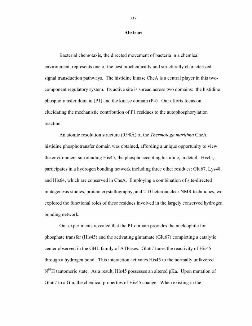
Abstract
Bacterial chemotaxis, the directed movement of bacteria in a chemical
environment, represents one of the best biochemically and structurally characterized
signal transduction pathways. The histidine kinase CheA is a central player in this two-
component regulatory system. Its active site is spread across two domains: the histidine
phosphotransfer domain (P1) and the kinase domain (P4). Our efforts focus on
elucidating the mechanistic contribution of P1 residues to the autophosphorylation
reaction.
An atomic resolution structure (0.98Å) of the Thermotoga maritima CheA
histidine phosphotransfer domain was obtained, affording a unique opportunity to view
the environment surrounding His45, the phosphoaccepting histidine, in detail. His45,
participates in a hydrogen bonding network including three other residues: Glu67, Lys48,
and His64, which are conserved in CheA. Employing a combination of site-directed
mutagenesis studies, protein crystallography, and 2-D heteronuclear NMR techniques, we
explored the functional roles of these residues involved in the largely conserved hydrogen
bonding network.
Our experiments revealed that the P1 domain provides the nucleophile for
phosphate transfer (His45) and the activating glutamate (Glu67) completing a catalytic
center observed in the GHL family of ATPases. Glu67 tunes the reactivity of His45
through a hydrogen bond. This interaction activates His45 to the normally unfavored
Nδ1H tautomeric state. As a result, His45 possesses an altered pKa. Upon mutation of
Glu67 to a Gln, the chemical properties of His45 change. When existing in the
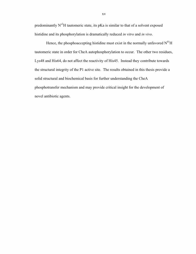
xv
predominantly Nε2H tautomeric state, its pKa is similar to that of a solvent exposed
histidine and its phosphorylation is dramatically reduced in vitro and in vivo.
Hence, the phosphoaccepting histidine must exist in the normally unfavored Nδ1H
tautomeric state in order for CheA autophosphorylation to occur. The other two residues,
Lys48 and His64, do not affect the reactivity of His45. Instead they contribute towards
the structural integrity of the P1 active site. The results obtained in this thesis provide a
solid structural and biochemical basis for further understanding the CheA
phosphotransfer mechanism and may provide critical insight for the development of
novel antibiotic agents.

1
Chapter 1
Introduction
This chapter was adapted from Bilwes A. M., Park, S. Y., Quezada, C. M., Simon, M. I., and Crane, B.R. “Structure and Function of CheA, the Histidine Kinase Central to Bacterial Chemotaxis.” in Histidine Kinases in Signal Transduction (eds. Inouye, M. & Dutta, R.) 47-72 (Academic Press, New York, 2003).
2
Bacteria are continuously exposed to environmental changes. Signal transducing
circuits, referred to as “two-component” regulatory systems or “His-Asp phosphorelays,”
process the extracellular information into a usable intracellular response [1-3]. These
pathways have also been found in lower eukaryotes, like yeast, slime mold, and plants [4-
7] and are utilized to control diverse cell responses such as bacterial chemotaxis,
sporulation, osmoregulation, pathogenesis, plant response to hormones, cell growth and
differentiation [1, 8]. These adaptable and sensitive circuits can function over a broad
time range extending from milliseconds to hours.
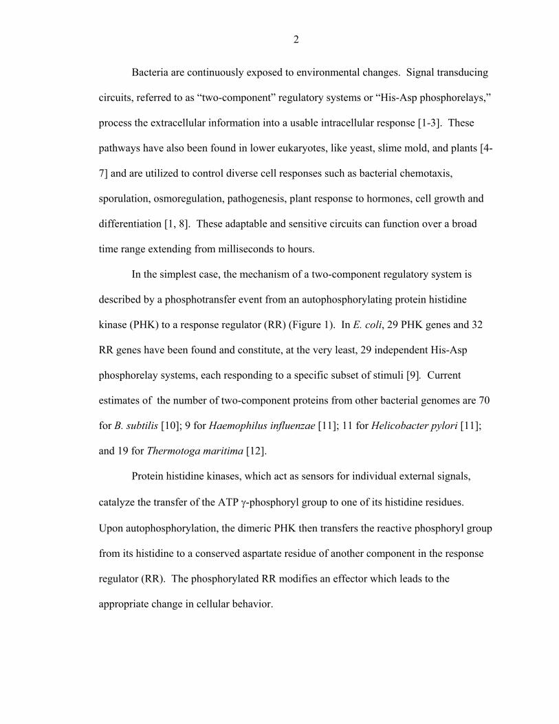
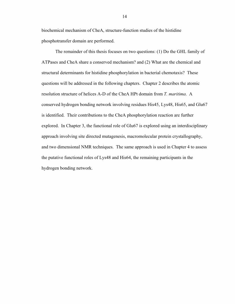
In the simplest case, the mechanism of a two-component regulatory system is
described by a phosphotransfer event from an autophosphorylating protein histidine
kinase (PHK) to a response regulator (RR) (Figure 1). In E. coli, 29 PHK genes and 32
RR genes have been found and constitute, at the very least, 29 independent His-Asp
phosphorelay systems, each responding to a specific subset of stimuli [9]. Current
estimates of the number of two-component proteins from other bacterial genomes are 70
for B. subtilis [10]; 9 for Haemophilus influenzae [11]; 11 for Helicobacter pylori [11];
and 19 for Thermotoga maritima [12].
Protein histidine kinases, which act as sensors for individual external signals,
catalyze the transfer of the ATP γ-phosphoryl group to one of its histidine residues.
Upon autophosphorylation, the dimeric PHK then transfers the reactive phosphoryl group
from its histidine to a conserved aspartate residue of another component in the response
regulator (RR). The phosphorylated RR modifies an effector which leads to the
appropriate change in cellular behavior.
3
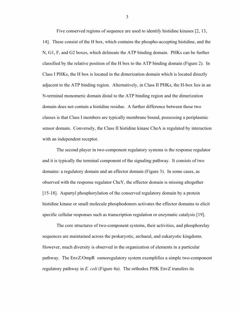
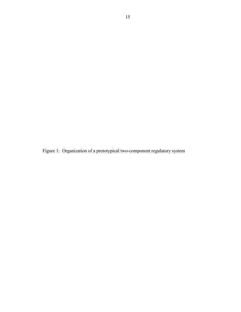
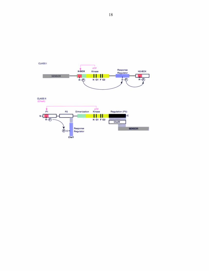
Five conserved regions of sequence are used to identify histidine kinases [2, 13,
14]. These consist of the H box, which contains the phospho-accepting histidine, and the
N, G1, F, and G2 boxes, which delineate the ATP binding domain. PHKs can be further
classified by the relative position of the H box to the ATP binding domain (Figure 2). In
Class I PHKs, the H box is located in the dimerization domain which is located directly
adjacent to the ATP binding region. Alternatively, in Class II PHKs, the H-box lies in an
N-terminal monomeric domain distal to the ATP binding region and the dimerization
domain does not contain a histidine residue. A further difference between these two
classes is that Class I members are typically membrane bound, possessing a periplasmic
sensor domain. Conversely, the Class II histidine kinase CheA is regulated by interaction
with an independent receptor.
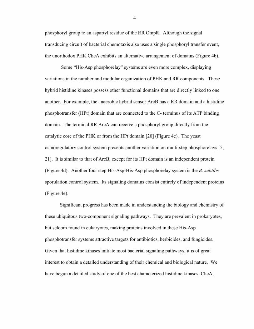
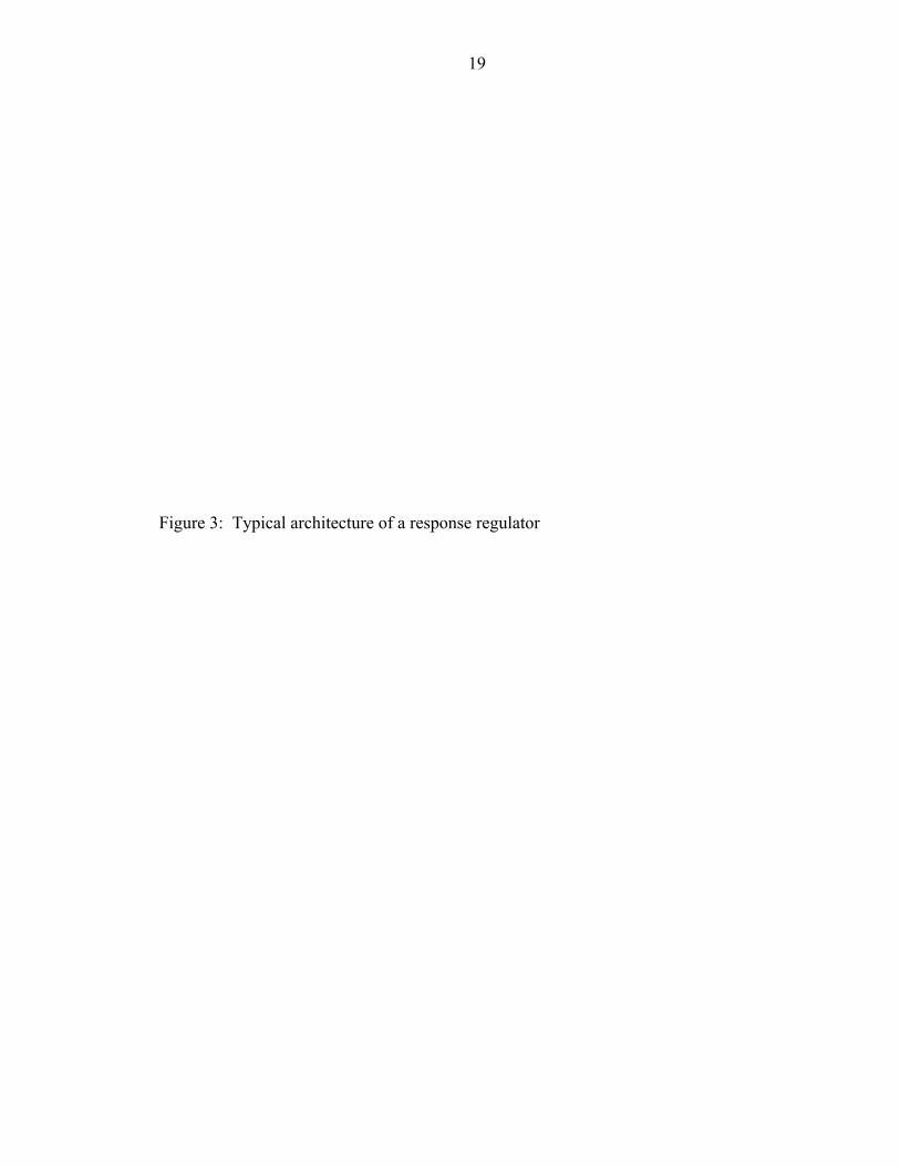
The second player in two-component regulatory systems is the response regulator
and it is typically the terminal component of the signaling pathway. It consists of two
domains: a regulatory domain and an effector domain (Figure 3). In some cases, as
observed with the response regulator CheY, the effector domain is missing altogether
[15-18]. Aspartyl phosphorylation of the conserved regulatory domain by a protein
histidine kinase or small molecule phosphodonors activates the effector domains to elicit
specific cellular responses such as transcription regulation or enzymatic catalysis [19].
The core structures of two-component systems, their activities, and phosphorelay
sequences are maintained across the prokaryotic, archaeal, and eukaryotic kingdoms.
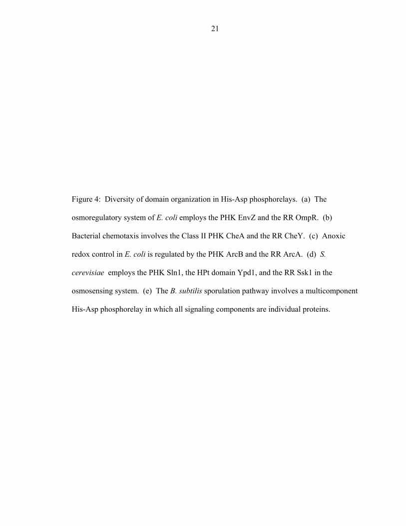
However, much diversity is observed in the organization of elements in a particular
pathway. The EnvZ/OmpR osmoregulatory system exemplifies a simple two-component
regulatory pathway in E. coli (Figure 4a). The orthodox PHK EnvZ transfers its
4
phosphoryl group to an aspartyl residue of the RR OmpR. Although the signal
transducing circuit of bacterial chemotaxis also uses a single phosphoryl transfer event,
the unorthodox PHK CheA exhibits an alternative arrangement of domains (Figure 4b).
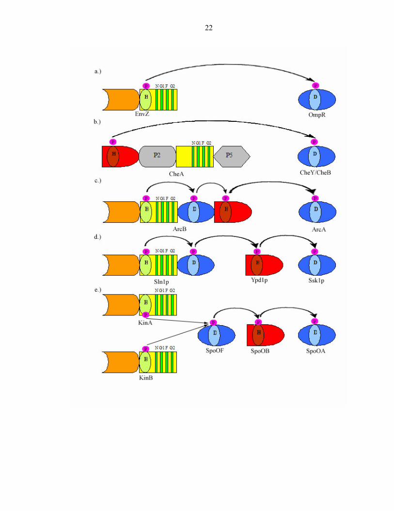
Some “His-Asp phosphorelay” systems are even more complex, displaying
variations in the number and modular organization of PHK and RR components. These
hybrid histidine kinases possess other functional domains that are directly linked to one
another. For example, the anaerobic hybrid sensor ArcB has a RR domain and a histidine
phosphotransfer (HPt) domain that are connected to the C- terminus of its ATP binding
domain. The terminal RR ArcA can receive a phosphoryl group directly from the
catalytic core of the PHK or from the HPt domain [20] (Figure 4c). The yeast
osmoregulatory control system presents another variation on multi-step phosphorelays [5,
21]. It is similar to that of ArcB, except for its HPt domain is an independent protein
(Figure 4d). Another four step His-Asp-His-Asp phosphorelay system is the B. subtilis
sporulation control system. Its signaling domains consist entirely of independent proteins
(Figure 4e).
Significant progress has been made in understanding the biology and chemistry of
these ubiquitous two-component signaling pathways. They are prevalent in prokaryotes,
but seldom found in eukaryotes, making proteins involved in these His-Asp
phosphotransfer systems attractive targets for antibiotics, herbicides, and fungicides.
Given that histidine kinases initiate most bacterial signaling pathways, it is of great
interest to obtain a detailed understanding of their chemical and biological nature. We
have begun a detailed study of one of the best characterized histidine kinases, CheA,
5
involved in one of the most extensively studied two-component signaling pathways,
bacterial chemotaxis.
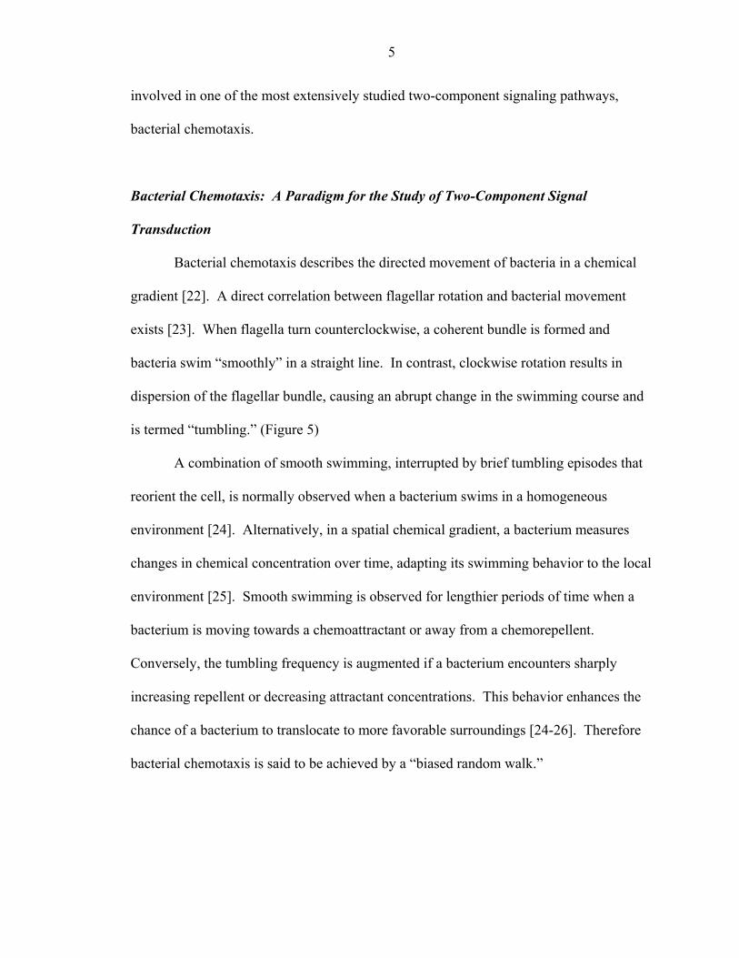
Bacterial Chemotaxis: A Paradigm for the Study of Two-Component Signal
Transduction
Bacterial chemotaxis describes the directed movement of bacteria in a chemical
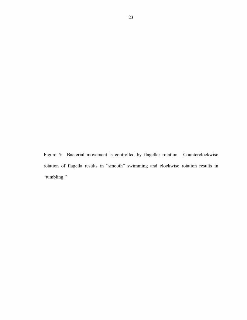
gradient [22]. A direct correlation between flagellar rotation and bacterial movement
exists [23]. When flagella turn counterclockwise, a coherent bundle is formed and
bacteria swim “smoothly” in a straight line. In contrast, clockwise rotation results in
dispersion of the flagellar bundle, causing an abrupt change in the swimming course and
is termed “tumbling.” (Figure 5)
A combination of smooth swimming, interrupted by brief tumbling episodes that
reorient the cell, is normally observed when a bacterium swims in a homogeneous
environment [24]. Alternatively, in a spatial chemical gradient, a bacterium measures
changes in chemical concentration over time, adapting its swimming behavior to the local
environment [25]. Smooth swimming is observed for lengthier periods of time when a
bacterium is moving towards a chemoattractant or away from a chemorepellent.
Conversely, the tumbling frequency is augmented if a bacterium encounters sharply
increasing repellent or decreasing attractant concentrations. This behavior enhances the
chance of a bacterium to translocate to more favorable surroundings [24-26]. Therefore
bacterial chemotaxis is said to be achieved by a “biased random walk.”
6
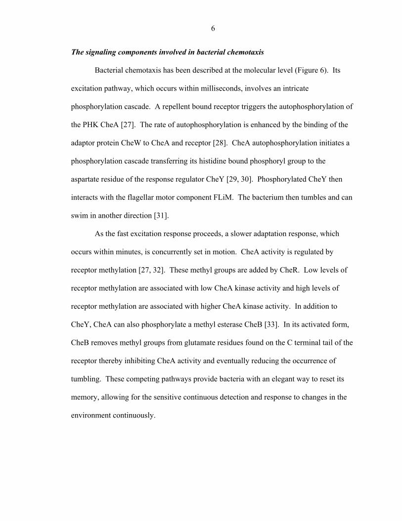
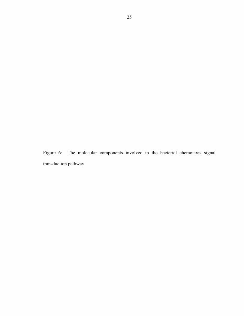
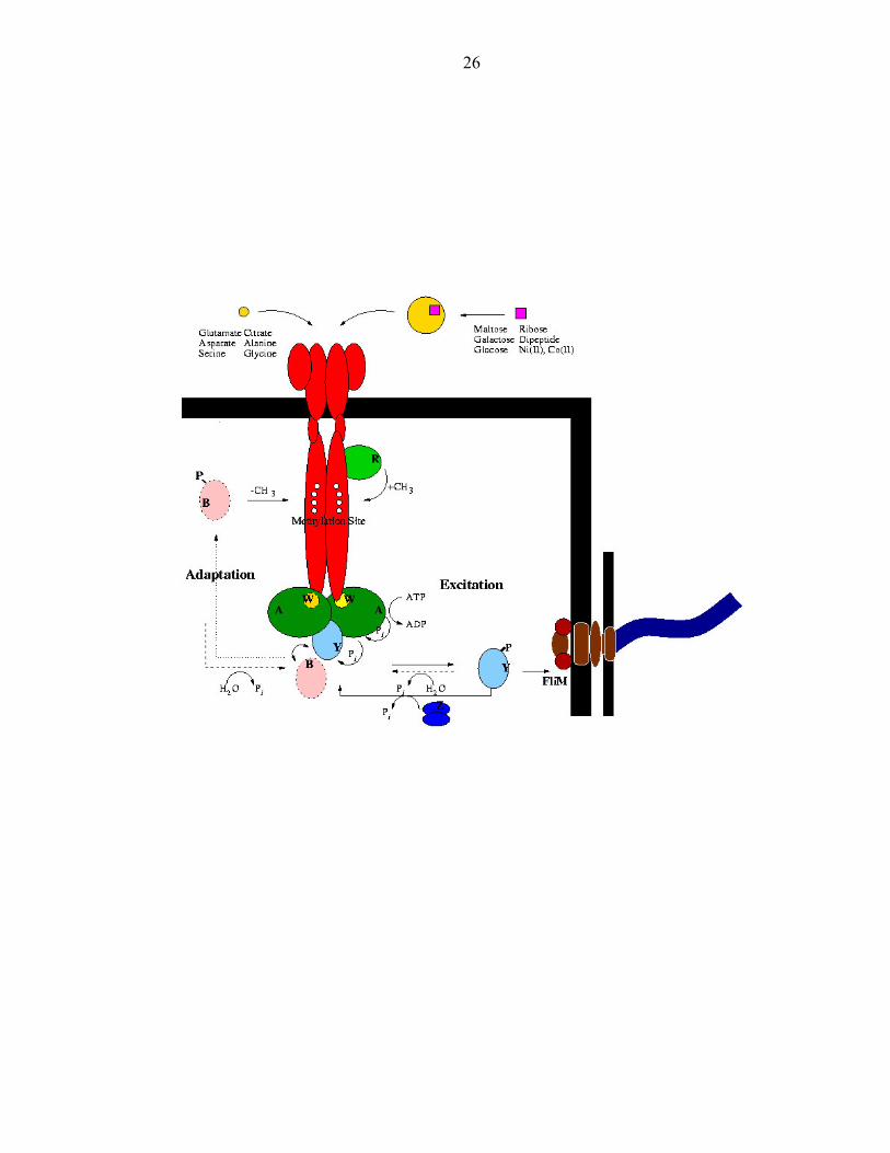
The signaling components involved in bacterial chemotaxis
Bacterial chemotaxis has been described at the molecular level (Figure 6). Its
excitation pathway, which occurs within milliseconds, involves an intricate
phosphorylation cascade. A repellent bound receptor triggers the autophosphorylation of
the PHK CheA [27]. The rate of autophosphorylation is enhanced by the binding of the
adaptor protein CheW to CheA and receptor [28]. CheA autophosphorylation initiates a
phosphorylation cascade transferring its histidine bound phosphoryl group to the
aspartate residue of the response regulator CheY [29, 30]. Phosphorylated CheY then
interacts with the flagellar motor component FLiM. The bacterium then tumbles and can
swim in another direction [31].
As the fast excitation response proceeds, a slower adaptation response, which
occurs within minutes, is concurrently set in motion. CheA activity is regulated by
receptor methylation [27, 32]. These methyl groups are added by CheR. Low levels of
receptor methylation are associated with low CheA kinase activity and high levels of
receptor methylation are associated with higher CheA kinase activity. In addition to
CheY, CheA can also phosphorylate a methyl esterase CheB [33]. In its activated form,
CheB removes methyl groups from glutamate residues found on the C terminal tail of the
receptor thereby inhibiting CheA activity and eventually reducing the occurrence of
tumbling. These competing pathways provide bacteria with an elegant way to reset its
memory, allowing for the sensitive continuous detection and response to changes in the
environment continuously.
7
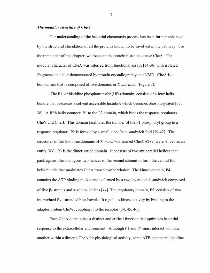
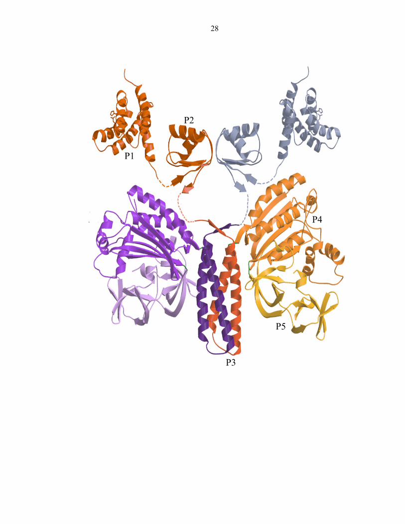
The modular structure of CheA
Our understanding of the bacterial chemotaxis process has been further enhanced
by the structural elucidation of all the proteins known to be involved in the pathway. For
the remainder of this chapter, we focus on the protein histidine kinase CheA. The
modular character of CheA was inferred from functional assays [34-36] with isolated
fragments and later demonstrated by protein crystallography and NMR. CheA is a
homodimer that is composed of five domains in T. maritima (Figure 7).
The P1, or histidine phosphotransfer (HPt) domain, consists of a four-helix
bundle that possesses a solvent accessible histidine which becomes phosphorylated [37,
38]. A fifth helix connects P1 to the P2 domain, which binds the response regulators
CheY and CheB. This domain facilitates the transfer of the P1 phosphoryl group to a
response regulator. P2 is formed by a small alpha/beta sandwich fold [39-42]. The
structures of the last three domains of T. maritima, termed CheA ∆289, were solved as an
entity [43]. P3 is the dimerization domain. It consists of two antiparallel helices that
pack against the analogous two helices of the second subunit to form the central four
helix bundle that modulates CheA transphosphorylation. The kinase domain, P4,
contains the ATP binding pocket and is formed by a two layered α-β sandwich composed
of five β- strands and seven α- helices [44]. The regulatory domain, P5, consists of two
intertwined five stranded beta barrels. It regulates kinase activity by binding to the
adaptor protein CheW, coupling it to the receptor [34, 45, 46].
Each CheA domain has a distinct and critical function that optimizes bacterial
response to the extracellular environment. Although P1 and P4 must interact with one
another within a dimeric CheA for physiological activity, some ATP-dependent histidine
8
phosphorylation can be achieved in vitro by the two separated domains [44]. Hence, all
the elements necessary for the chemistry of histidine phosphorylation are contained in the
domains P1 and P4. The features of the kinase domain, P4, are first examined, followed
by those of the histidine phosphotransfer domain, P1.
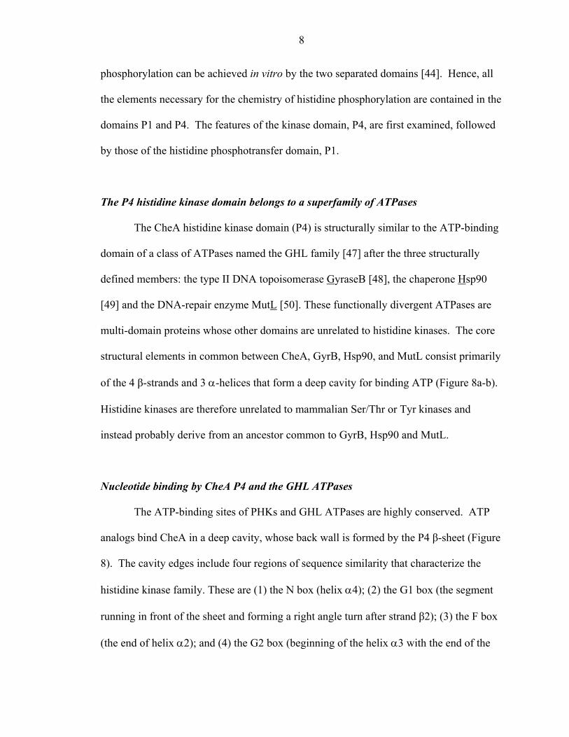
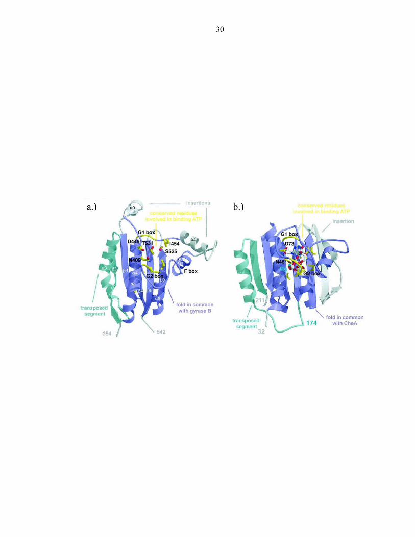
The P4 histidine kinase domain belongs to a superfamily of ATPases
The CheA histidine kinase domain (P4) is structurally similar to the ATP-binding
domain of a class of ATPases named the GHL family [47] after the three structurally
defined members: the type II DNA topoisomerase GyraseB [48], the chaperone Hsp90
[49] and the DNA-repair enzyme MutL [50]. These functionally divergent ATPases are
multi-domain proteins whose other domains are unrelated to histidine kinases. The core
structural elements in common between CheA, GyrB, Hsp90, and MutL consist primarily
of the 4 β-strands and 3 α-helices that form a deep cavity for binding ATP (Figure 8a-b).
Histidine kinases are therefore unrelated to mammalian Ser/Thr or Tyr kinases and
instead probably derive from an ancestor common to GyrB, Hsp90 and MutL.
Nucleotide binding by CheA P4 and the GHL ATPases
The ATP-binding sites of PHKs and GHL ATPases are highly conserved. ATP
analogs bind CheA in a deep cavity, whose back wall is formed by the P4 β-sheet (Figure
8). The cavity edges include four regions of sequence similarity that characterize the
histidine kinase family. These are (1) the N box (helix α4); (2) the G1 box (the segment
running in front of the sheet and forming a right angle turn after strand β2); (3) the F box
(the end of helix α2); and (4) the G2 box (beginning of the helix α3 with the end of the
9
loop preceding it). The residues pointing into the cavity from the β strands form a mainly
hydrophobic lawn on which the adenine ring hydrogen bonds with the invariant Asp (449
in T. maritima CheA). Four buried water molecules that bridge interactions between the
nucleotide base and the cavity are also found in the nucleotide complexes of Hsp90 [51,
52] and MutL [47]. A conserved Asn (409 T. maritima CheA) coordinates nucleotide -
bound Mg2+ in CheA [44], MutL [47], GyrB [48] and Hsp90 [52].
Despite striking similarities in nucleotide binding by PHKs and GHL ATPases,
there are some compelling differences. For example, an essential glutamate of the GHL
ATPases presumed to be the general base involved in water activation for ATP
hydrolysis[53] (Glu 29 for MutL) is replaced by His 405 in CheA (Figure 9). His 405
stabilizes the G2 box when Mg2+ is bound. In contrast, the general base for histidine
activation likely resides on the P1 domain (see below).
Moreover, CheA and the GHL ATPases appear to recognize the ATP phosphates
in different ways. For example, the functional analog of a CheA residue that hydrogen
bonds to the ATP β- phosphate (His 413) comes from a different loop in the ATPases.
Furthermore, interactions between nucleotide phosphates and main-chain nitrogens of the
P-loop (a glycine-rich segment found in many ATP-binding proteins that coordinates the
α- and γ- phosphates of bound ATP) are not nearly as extensive in CheA as they are in
GyrB and MutL. Perhaps P1 binding drives a more extensive interaction between
nucleotide and the CheA P-loop that resembles structures observed for the ATPases. In
fact, the G2 box (P-loop) mutation Gly 502 to Lys (E. coli residue 470) does not affect
nucleotide affinity but is inactive [54].
10
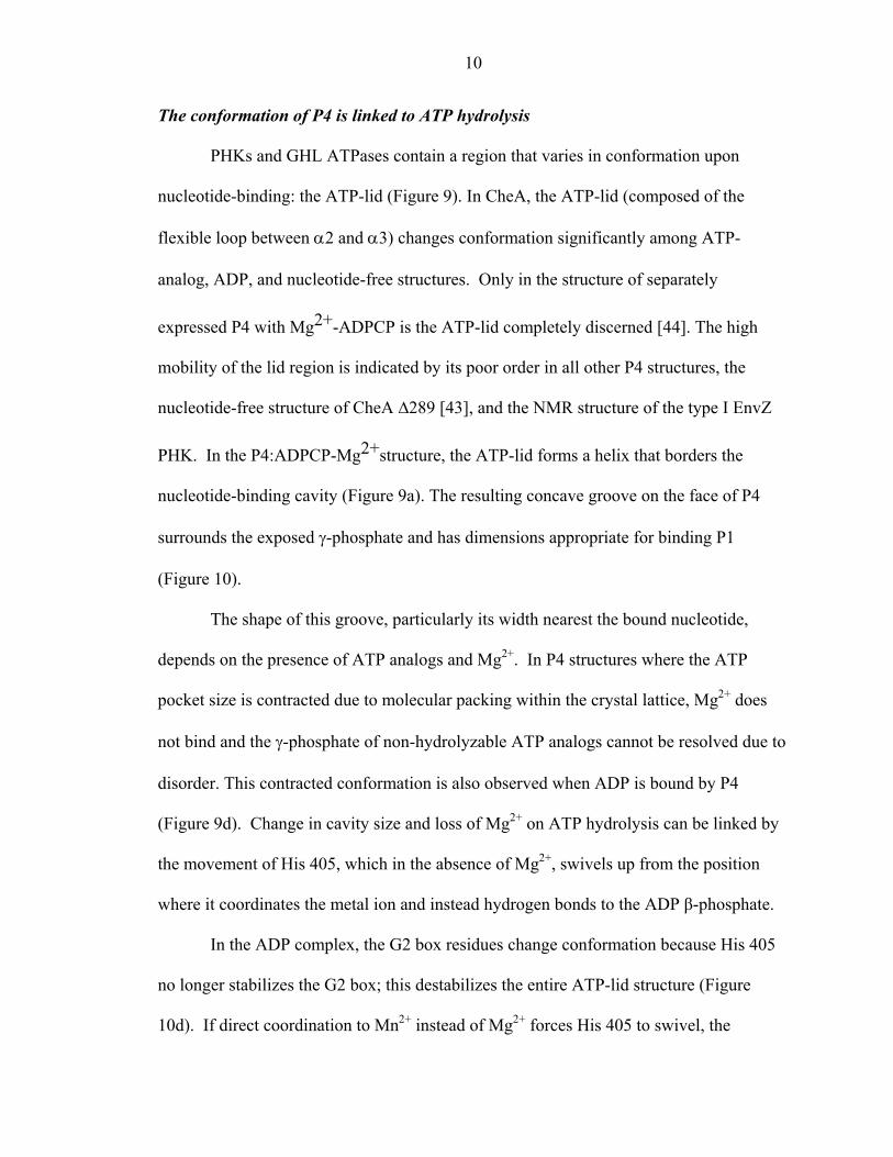
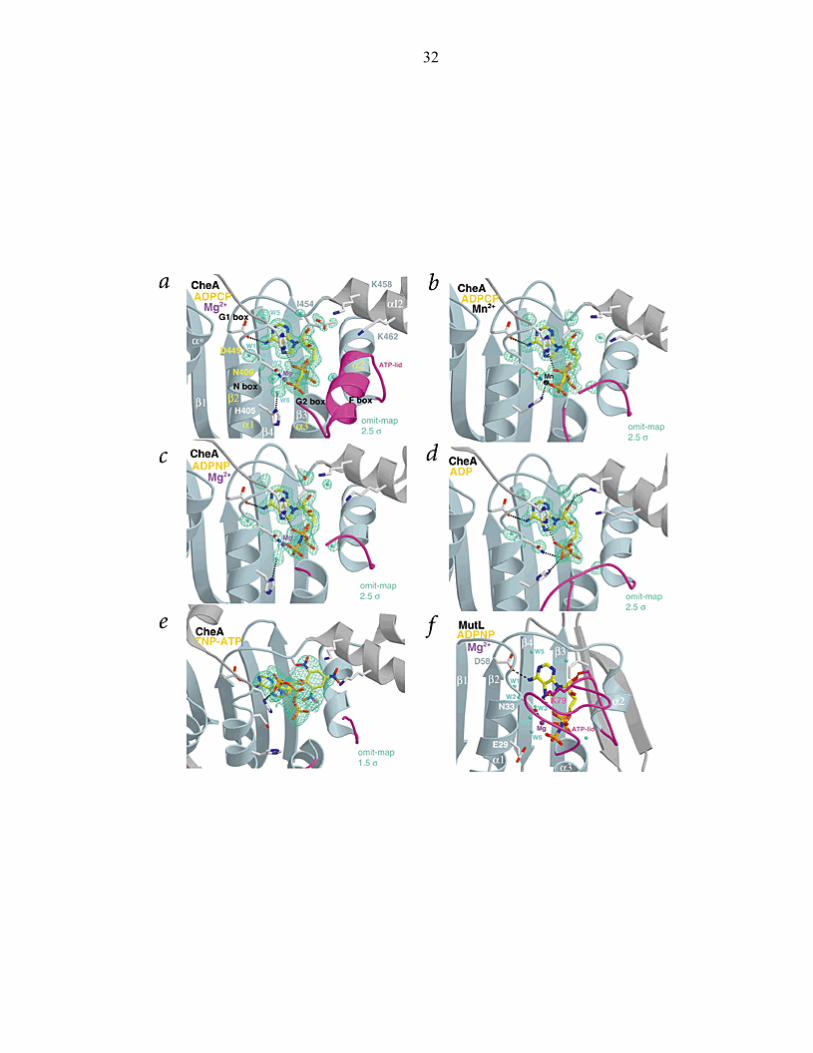
The conformation of P4 is linked to ATP hydrolysis
PHKs and GHL ATPases contain a region that varies in conformation upon
nucleotide-binding: the ATP-lid (Figure 9). In CheA, the ATP-lid (composed of the
flexible loop between α2 and α3) changes conformation significantly among ATP-
analog, ADP, and nucleotide-free structures. Only in the structure of separately
expressed P4 with Mg2+-ADPCP is the ATP-lid completely discerned [44]. The high
mobility of the lid region is indicated by its poor order in all other P4 structures, the
nucleotide-free structure of CheA ∆289 [43], and the NMR structure of the type I EnvZ
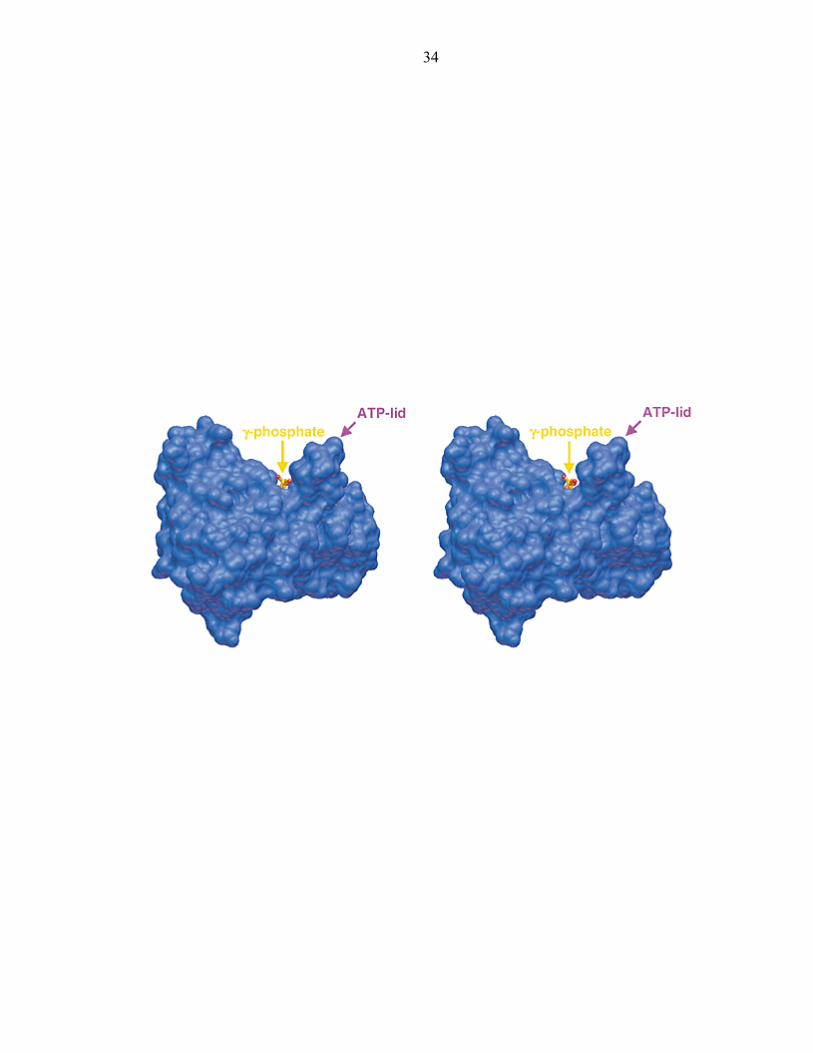
PHK. In the P4:ADPCP-Mg2+structure, the ATP-lid forms a helix that borders the
nucleotide-binding cavity (Figure 9a). The resulting concave groove on the face of P4
surrounds the exposed γ-phosphate and has dimensions appropriate for binding P1
(Figure 10).
The shape of this groove, particularly its width nearest the bound nucleotide,
depends on the presence of ATP analogs and Mg2+. In P4 structures where the ATP
pocket size is contracted due to molecular packing within the crystal lattice, Mg2+ does
not bind and the γ-phosphate of non-hydrolyzable ATP analogs cannot be resolved due to
disorder. This contracted conformation is also observed when ADP is bound by P4
(Figure 9d). Change in cavity size and loss of Mg2+ on ATP hydrolysis can be linked by
the movement of His 405, which in the absence of Mg2+, swivels up from the position
where it coordinates the metal ion and instead hydrogen bonds to the ADP β-phosphate.
In the ADP complex, the G2 box residues change conformation because His 405
no longer stabilizes the G2 box; this destabilizes the entire ATP-lid structure (Figure
10d). If direct coordination to Mn2+ instead of Mg2+ forces His 405 to swivel, the
11
conformation of the ATP-lid is similarly affected and interactions of the P-loop with the
γ-phosphate are weakened (Figure 9b). As confirmed by biochemical studies,
conformational changes in regions that likely compose the P1-binding site on P4 (the
ATP-lid) are coupled to ATP hydrolysis and Mg2+ release by movement of His 405 [55].
Biochemical investigations of other mutations at the N, G1, G2, and F boxes were
made [55]. Results suggested these conserved residues contribute to the CheA kinase
activity and ATP binding. Mutations that affect ATP binding were also found to stabilize
the transition-state complex during CheA autophosphorylation in an unknown fashion.
Alterations at a glycine residue in the G1 box are not tolerated, consistent with prior
structural analysis that mutations at this location would disrupt the structure of the ATP
binding cavity [44]. In correlating biochemical to structural studies, some of the
functional roles of conserved residues residing in the P4 domain have begun to be
assigned providing insight into the CheA mechanism.
The site of phosphorylation is located in the P1 domain
P1 contains the substrate histidine that transfers phosphate from kinase bound
ATP to the response regulators CheY and CheB [33, 56]. It is composed of a small 310
helix followed by an antiparallel four-helix bundle (helices A-D) and helix (E) that
connects to P2 via a 25-35 residue linker [37, 38]. Helix E does not contribute to helix
cluster stability nor to the phosphorylation reaction[37].
The P1 helices are amphipathic with most hydrophobic residues buried in the core
and most polar residues exposed to the surface. Inter-helix salt bridges and hydrogen
bonds are found only between helices A and D and helices B and C[38]. The five helices
12
each display very different dynamic features. Residues from helices A, C, and D show
strong protection from hydrogen exchange, indicative of local stability around the amide
hydrogen[37]. However, helix B, which contains the phospho-accepting histidine, may be
more variable in conformation as its amide protons are not strongly protected from
solvent exchange.
Sequence similarity among CheA P1 homologs is concentrated in helices B and
C, where the active site residues are located. Despite the high sequence similarity
between E. coli and T. maritima CheA P1 in this region[37], T. maritima CheA ∆289
cannot phosphorylate an E. coli P1-P2 fragment (unpublished data). The interface
between P1 and the kinase domain is therefore likely to include residues on P1 not
immediately surrounding His45.
NMR studies of protein backbone dynamics indicate that E. coli P1 forms a rigid
and compact helix bundle in both the unphosphorylated and phosphorylated states. Both
these forms of P1 have very similar backbone conformation[37]. Phosphorylation of P1
does not deprotonate His48 Nδ1H and results in only small chemical shift changes for
residues on helices B and C surrounding His48. Alternations in the local electronic
environment caused by phosphorylation are likely responsible for these changes[57]. No
interaction between P2 and phosphorylated P1 was detected by NMR[58].
The reactivity of the phospho-accepting His48, located in the middle of helix B, is
tuned by its local environment. A hydrogen bond between the His48 Nδ1H and the Glu70
carboxylate may be responsible for the high pKa (7.8) of the His48 imidazole ring, which
is the site of phosphorylation. NMR studies indicate that its Nε2 atom does not hydrogen
bond with other P1 residues, but that His48 Nδ1H is a hydrogen bond donor that remains
13
protonated at high pHs and after phosphorylation [57]. Three out of the four molecules in
the crystal structure of Salmonella P1 reveal that His48 Nδ1H hydrogen bonds to Glu70
on Helix C. Furthermore, Lys51Ala and Glu70Ala mutations in Salmonella reduce the
ATP phosphotransfer rate[38]. However, these experiments could not distinguish if the
decreased transfer rate was due to loss of binding between the P1 and the kinases or a
catalytic defect.
Investigating the mechanism of CheA phosphotransfer
In contrast to GyrB, MutL, and Hsp90, which hydrolyse ATP, PHKs must transfer
phosphate to a histidine residue. Therefore, the nucleophilic mechanism for attack on the
ATP γ-phosphate must differ between the two enzymes. In GyrB, mutagenesis studies
[53] implicate the conserved Glu 42 (Glu 29 in MutL, Glu 47 in human Hsp90) as an
essential general base for water activation. Despite high conservation of active site
residues between GyrB and histidine kinases, the latter do not contain a Glu at this
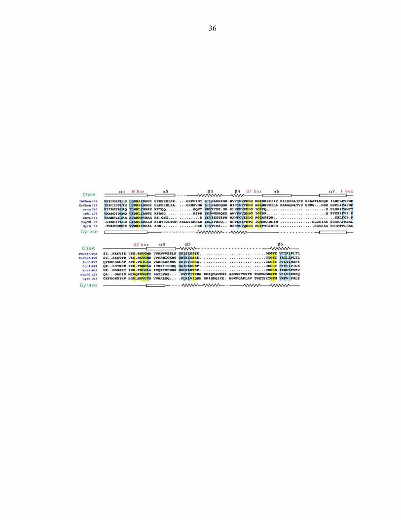
position (His 405 for CheA proteins, Asn for other histidine kinases) (Figure 11). Thus,
the CheA P1 domain may provide not only the nucleophile for phosphate transfer (His45)
but also the activating glutamate (Glu70), thereby completing the catalytic center
observed in GyrB.
Despite the fact that CheA is one of the best-characterized histidine kinases, little
is known about its biochemical mechanism. The CheA active site is distributed across
two domains, P1 and P4. The investigations described in this thesis focus on the least
studied domain encompassing the CheA active site. In order to gain insight into the
14
biochemical mechanism of CheA, structure-function studies of the histidine
phosphotransfer domain are performed.
The remainder of this thesis focuses on two questions: (1) Do the GHL family of
ATPases and CheA share a conserved mechanism? and (2) What are the chemical and
structural determinants for histidine phosphorylation in bacterial chemotaxis? These
questions will be addressed in the following chapters. Chapter 2 describes the atomic
resolution structure of helices A-D of the CheA HPt domain from T. maritima. A
conserved hydrogen bonding network involving residues His45, Lys48, His65, and Glu67
is identified. Their contributions to the CheA phosphorylation reaction are further
explored. In Chapter 3, the functional role of Glu67 is explored using an interdisciplinary
approach involving site directed mutagenesis, macromolecular protein crystallography,
and two dimensional NMR techniques. The same approach is used in Chapter 4 to assess
the putative functional roles of Lys48 and His64, the remaining participants in the
hydrogen bonding network.
15
Figure 1: Organization of a prototypical two-component regulatory system
16
Signal In
Input Transmitter
Signal Out
Receiver Output
+-
Sensor
Response regulator
17
Figure 2: Protein histidine kinases can be divided into two classes. A schematic
depiction of the classification of PHKs based on the position of the H-box relative to the
ATP binding domain.
18
19
Figure 3: Typical architecture of a response regulator
20
Bacterial chemotaxis Osmoregulation Sporulation Pathogenesis Plant response to hormones
Regulatory Effector
21
Figure 4: Diversity of domain organization in His-Asp phosphorelays. (a) The
osmoregulatory system of E. coli employs the PHK EnvZ and the RR OmpR. (b)
Bacterial chemotaxis involves the Class II PHK CheA and the RR CheY. (c) Anoxic
redox control in E. coli is regulated by the PHK ArcB and the RR ArcA. (d) S.
cerevisiae employs the PHK Sln1, the HPt domain Ypd1, and the RR Ssk1 in the
osmosensing system. (e) The B. subtilis sporulation pathway involves a multicomponent
His-Asp phosphorelay in which all signaling components are individual proteins.
22
23
Figure 5: Bacterial movement is controlled by flagellar rotation. Counterclockwise
rotation of flagella results in “smooth” swimming and clockwise rotation results in
“tumbling.”
24
Tumble Clockwise
Smooth Counterclockwise Smooth
Counterclockwise
25
Figure 6: The molecular components involved in the bacterial chemotaxis signal
transduction pathway
26

27
Figure 7: CheA is a homodimer consisting of five domains (P1-P5). P1 is the
phosphotransfer domain, P2 is the response regulator binding domain, P3 is the
dimerization domain, P4 is the kinase domain, and P5 is the regulatory domain. Dotted
lines represent missing residues and putative linker regions between domains. One
monomer is colored in red, orange and yellow hues, while the other is represented in
purple and grey tones.
28
P1
P2
P4
P5
P3
29
Figure 8: The CheA kinase domain (a) is topologically similar to the ATP binding
domain of GyrB (b).
30
31
Figure 9: Nucleotide binding alters the conformation of the CheA kinase domain
32
33
Figure 10: The CheA kinase domain forms a concave groove for binding to P1
34
35
Figure 11: Sequence similarity between PHKs and the GHL family of ATPases
36

37
REFERENCES 1. Stock, A.M., V.L. Robinson, and P.N. Goudreau, Two-component signal
transduction. Annu. Rev. Biochem., 2000. 69: p. 183-215.
2. Parkinson, J.S., and Kofoid, E.C., Communication modules in bacterial signaling proteins. Annu. Rev. Genet., 1992. 26: p. 71-112.
3. Inouye, M. and R. Dutta, eds. Histidine Kinases in Signal Transduction. 2003, Academic Press. 520.
4. Chang, C., Kwok, S.F., Bleecker, A.B., and Meyerowitz, E.M., Arabidopsis ethylene-response gene ETR1: similarity of product to two-component regulators. Science, 1993. 262: p. 539-544.
5. Ota, I.M. and A. Varshavsky, A Yeast Protein Similar to Bacterial Two-Component Regulators. Science, 1993. 262(5133): p. 566-569.
6. Alex, L.A., K.A. Borkovich, and M.I. Simon, Hyphal development in Neurospora crassa: Involvement of a two- component histidine kinase. Proceedings of the National Academy of Sciences of the United States of America, 1996. 93(8): p. 3416-3421.
7. Maeda, T., S.M. Wurglermurphy, and H. Saito, A 2-Component System That Regulates an Osmosensing Map Kinase Cascade in Yeast. Nature, 1994. 369(6477): p. 242-245.
8. Parkinson, J.S. and E.C. Kofoid, Communication modules in bacterial signaling proteins. Annu. Rev. Genet., 1992. 26: p. 71-112.
9. Mizuno, T., Compilation of All Genes Encoding Two-component Phosphotransfer Signal Transducers in the Genome of Escherichia coli. DNA Research, 1997. 4: p. 161-168.
10. Fabret, C., V.A. Feher, and J.A. Hoch, Two-component signal transduction in Bacillus subtilis: How one organism sees its world. Journal of Bacteriology, 1999. 181(7): p. 1975-1983.
38
11. Mizuno, T., His-Asp phosphotransfer signal transduction. Journal of Biochemistry, 1998. 123(4): p. 555-563.
12. Nelson, K.E., et al., Evidence for lateral gene transfer between Archaea and Bacteria from genome sequence of Thermotoga maritima. Nature, 1999. 399(6734): p. 323-329.
13. Alex, L.A., and Simon, M.I., Protein histidine kinases and signal transduction in prokaryotes and eukaryotes. Trends Genet . 1994. 10: p. 133-138.
14. Swanson, R.V., Alex, L.A., and Simon, M.I., Histidine and aspartate phosphorylation: two-component systems and the limits of homology. Trends Biochem. Sci., 1994. 19: p. 485-490.
15. Usher, K.C., et al., Crystal structures of CheY from Thermotoga maritima do not support conventional explanations for the structural basis of enhanced thermostability. Protein Science, 1998. 7(2): p. 403-412.
16. Lukat, G.S., et al., Phosphorylation of Bacterial Response Regulator Proteins by Low-Molecular-Weight Phospho-Donors. Proceedings of the National Academy of Sciences of the United States of America, 1992. 89(2): p. 718-722.
17. Volz, K. and P. Matsumura, Crystal Structure of Escherichia coli CheY Refined at 1.7A Resolution. Journal of Biological Chemistry, 1991. 266(23): p. 15511-15519.
18. Stock, A.M., et al., Three Dimensional Structure of CheY, the Response Regulator of Bacterial Chemotaxis. Nature, 1989. 337(6209): p. 745-749.
19. Lukat, G.S., et al., Phosphorylation of Bacterial Response Regulator Proteins by Low-Molecular-Weight Phospho-Donors. Proc. Natl. Acad. Sci. U.S.A., 1992. 89(2): p. 718-722.
20. Ishige, K., et al., A Novel Device of Bacterial Signal Transducers. Embo Journal, 1994. 13(21): p. 5195-5202.
21. Posas, F., et al., Yeast HOG1 MAP kinase cascade is regulated by a multistep phosphorelay mechanism in the SLN1-YPD1-SSK1 ''two-component'' osmosensor. Cell, 1996. 86(6): p. 865-875.
39
22. Berg, H.C., Bacterial Behavior. Nature, 1975. 254(5499): p. 389-392.
23. Larsen, S.H., et al., Change in Direction of Flagellar Rotation Is Basis of Chemotactic Response in Escherichia coli. Nature, 1974. 249(5452): p. 74-77.
24. Berg, H.C. and D.A. Brown, Chemotaxis in Escherichia coli Analyzed by Three Dimensional Tracking. Nature, 1972. 239(5374): p. 500-&.
25. Macnab, R.M. and D.E. Koshland, Gradient-Sensing Mechanism in Bacterial Chemotaxis. Proceedings of the National Academy of Sciences of the United States of America, 1972. 69(9): p. 2509-&.
26. Tsang, N., R. Macnab, and D.E. Koshland, Common Mechanism for Repellents and Attractants in Bacterial Chemotaxis. Science, 1973. 181(4094): p. 60-63.
27. Li, G.Y. and R.M. Weis, Covalent modification regulates ligand binding to receptor complexes in the chemosensory system of Escherichia coli. Cell, 2000. 100(3): p. 357-365.
28. Maddock, J.R. and L. Shapiro, Polar Location of the Chemoreceptor Complex in the Escherichia coli Cell. Science, 1993. 259(5102): p. 1717-1723.
29. Borkovich, K.A. and M.I. Simon, The Dynamics of Protein-Phosphorylation in Bacterial Chemotaxis. Cell, 1990. 63(6): p. 1339-1348.
30. Hess, J.F., Bourret, R.B., & Simon, M.I., Histidine phosphorylation and phosphoryl group transfer in bacterial chemotaxis. Nature, 1988. 336: p. 139-143.
31. Welch, M., et al., Phosphorylation-Dependent Binding of a Signal Molecule to the Flagellar Switch of Bacteria. Proceedings of the National Academy of Sciences of the United States of America, 1993. 90(19): p. 8787-8791.
32. Borkovich, K.A., and Simon, M.I., Coupling of receptor function to phosphate-transfer reactions in bacterial chemotaxis. Methods Enzymol., 1991. 200: p. 205-214.
40
33. Li, J.Y., et al., The Response Regulators CheB and CheY Exhibit Competitive Binding to the Kinase CheA. Biochemistry, 1995. 34(45): p. 14626-14636.
34. Bourret, R.B., Davagnino, J., and Simon, M.I., The carboxy-terminal portion of the CheA kinase mediates regulation of autophosphorylation by transducer and CheW. J. Bacteriol., 1993. 175: p. 2097-2101.
35. Morrison, T.N., and Parkinson, J.S., A fragment liberated from the E. coli kinase that blocks stimulatory, but not inhibitory, chemoreceptor signaling. J. Bacteriol., 1997. 179: p. 5543-5550.
36. Swanson, R.V., Schuster, S.C., and Simon, M.I., Expression of CheA fragments which define domains encoding kinase, phosphotransfer and CheY binding activities. Biochemistry, 1993. 32: p. 7623-7629.
37. Zhou, H., Lowry, D.F., Swanson, R.V., Simon, M.I., Dahlquist, F.W., NMR studies of the phosphotransfer domain of the histidine kinase CheA from Escherichia coli: assignments, secondary structure, general fold, and backbone dynamics. Biochemistry, 1995. 34: p. 13858-13870.
38. Mourey, L., Da Re, S., Pedelacq, J.D., Tolstykh, T., Faurie, C., Guillet, V., Stock, J.B., and Samama, J.P., Crystal structure of the CheA histidine phosphotransfer domain that mediates response regulator phosphorylation in bacterial chemotaxis. J. Biol. Chem., 2001.
39. McEvoy, M.M., et al., Nuclear Magnetic Resonance Assignments and Global Fold of a CheY Binding Domain in CheA, the Chemotaxis Specific Kinase of Escherichia coli. Biochemistry, 1995. 34(42): p. 13871-13880.
40. Welch, M., Chinardet, N., Mourey, L., Birck, C., and Samama, J. P., Structure of the CheY-binding domain of histidine kinase CheA in complex with CheY. Nat Struct Biol., 1998. 5: p. 25-29.
41. McEvoy, M.M., et al., Structure and dynamics of a CheY-binding domain of the chemotaxis kinase CheA determined by nuclear magnetic resonance spectroscopy. Biochemistry, 1996. 35(18): p. 5633-5640.
42. McEvoy, M.M., Hausrath, A. C., Randolph, G. B., Remington, S. J., and Dahlquist, F. W., Two binding modes reveal flexibility in kinase/response
41
regulator interactions in the bacterial chemotaxis pathway. Proc. Natl. Acad. Sci. U.S.A., 1998. 95: p. 7333-7338.
43. Bilwes, A.M., Alex, L.A., Crane, B.R., and Simon, M.I., Structure of CheA, a signal-transducing histidine kinase. Cell, 1999. 96: p. 131-141.
44. Bilwes, A.M., Quezada, C.M., Croal, L.R., Crane, B.R., and Simon, M.I., Nucleotide binding by the histidine kinase CheA. Nature Struct. Biol., 2001. 8: p. 353-360.
45. Boukhvalova, M., R. VanBruggen, and R.C. Stewart, CheA kinase and chemoreceptor interaction surfaces on CheW. Journal of Biological Chemistry, 2002. 277(26): p. 23596-23603.
46. Boukhvalova, M.S., F.W. Dahlquist, and R.C. Stewart, CheW binding interactions with CheA and Tar - Importance for chemotaxis signaling in Escherichia coli. Journal of Biological Chemistry, 2002. 277(25): p. 22251-22259.
47. Ban, C., Junop, M., and Yang, W., Transformation of MutL by ATP binding and hydrolysis: a switch in DNA mismatch repair. Cell, 1999. 97: p. 85-97.
48. Wigley, D.B., Davies, G.J., Dodson E.J., Maxwell, A. and Dodson, G., Crystal structure of an N-terminal fragment of the DNA gyrase B protein. Nature, 1991. 351: p. 624-629.
49. Stebbins, C.E., Russo, A.A., Schneider, C., Rosen, N., Hartl, F.U., and Pavletich, N.P., Crystal structure of an Hsp90-geldamycin complex: targeting of a protein chaperone by an antitumor agent. Cell, 1997. 89: p. 239-250.
50. Ban, C., and Yang, W., Crystal structure and ATPase activity of MutL: implications for DNA repair and mutagenesis. Cell, 1998. 95: p. 541-552.
51. Prodromou, C., Roe, S.M., O’Brien, R., Ladbury, J.E., Piper, P.W. and Pearl, L.H., A molecular clamp in the crystal structure of the N-terminal domain of the yeast Hsp90 chaperone. Cell, 1997. 90: p. 65-75.
42
52. Obermann, W.M.J., Sondermann, H., Russo, A.A., Pavletich, N.P., and Hartl, F.U., In vivo function of Hsp90 is dependent on ATP binding and ATP hydrolysis. J. Cell Biol., 1998. 143: p. 901-910.
53. Jackson, A.P., and Maxwell, A., Identifying the catalytic residue of the ATPase reaction of DNA gyrase. Proc. Natl. Acad. Sci. USA, 1993. 90: p. 11232-11236.
54. Stewart, R.C., VanBruggen, R., Ellefson, D.D., and Wolfe, A.J., TNP-ATP and TNP-ADP as probes of the nucleotide binding site of CheA, the histidine protein kinase in the chemotaxis signal transduction pathway of Escherichia Coli. Biochemistry, 1998. 37: p. 12269-12279.
55. Hirschman, A., et al., Active Site Mutations in CheA, the Signal-Transducing Protein Kinase of the Chemotaxis System in Escherichia coli. Biochemistry, 2001. 40: p. 13876-13887.
56. Hess, J.F., Oosawa, K., Kaplan, N., and Simon, M.I., Phosphorylation of three proteins in the signaling pathway of bacterial chemotaxis. Cell, 1988. 53: p. 79-87.
57. Zhou, H.D., F. W., Phosphotransfer site of the chemotaxis-specific protein kinase CheA as revealed by NMR. Biochemitry, 1997. 36: p. 699-710.
58. Zhou, H.J., et al., Phosphotransfer and CheY-binding domains of the histidine autokinase CheA are joined by a flexible linker. Biochemistry, 1996. 35(2): p. 433-443.
43
Chapter 2
The crystal structure of the CheA histidine phosphotransfer domain from
Thermotoga maritima
44
CheA plays a central role in the bacterial chemotaxis signal transduction pathway
that controls bacterial motor behavior in response to environmental stimuli. The focus of
these studies is on the CheA histidine phosphotransfer domain, P1. It mediates the
transfer of the γ-phosphoryl group from ATP to the response regulators CheY and CheB.
The global fold of the P1 domain has been determined by NMR [1] and by
crystallography to a resolution of 2.1Å [2]. It consists of five alpha helices, including an
antiparallel four helix bundle, flanked by a helix at its C-terminus [1, 2].
Crystallographic and NMR studies suggested the P1 phospho-accepting histidine
forms a hydrogen bond to a neighboring glutamate residue [2, 3]. Furthermore, the
phospho-accepting histidine, His48 in E. coli, possesses an altered pKa of 7.8 at 30°C;
approximately one pH unit higher than a normal solvent accessible histidine [3].
Learning more about the detailed hydrogen bonding interactions with the phospho-
accepting histidine and their effects on histidine reactivity prompted an attempt to
improve the resolution of the CheA histidine phosphotransfer domain crystal structure.
The 0.98 Å resolution structure of helices A-D of the CheA histidine
phosphotransfer domain from T. maritima is reported in this chapter. Structural issues
concerning the phosphotransfer mechanism of the histidine kinase CheA, which are better
assessed due to the improvement in accuracy of the atomic coordinates, are discussed.
The phospho-accepting histidine, His45, participates in an elaborate hydrogen bond
network including three other residues. Insight into the dynamic properties of the CheA
histidine phosphotransfer domain is also obtained.
45
MATERIALS AND METHODS
Protein Cloning, Expression, and Purification
T. maritima ∆289 (residues 290-671), domain P1 (residues 4-133), and a fragment
of domain P1 termed P1short (residues 4-105) were subcloned in the vector pET28(a)
(Novagen). The plasmid was transformed into E. coli strain BL21(DE3) (Novagen) and
protein expressed in 2L TB cultures. Protein purification was achieved by affinity
chromatography on Nickel-NTA beads (Qiagen), followed by an overnight digestion of
the His6 tag at 4°C by thrombin. The protein was further purified by gel filtration on a
superdex 75 or 200 column (Pharmacia) using a buffer composed of 50 mM Tris(pH 7.5),
150 mM NaCl and 2 mM DTT. Centrifugation with an Amicon centriprep concentrator
yielded the concentrated protein. A cysteine mutant, T81C, for derivatization with heavy
atoms was generated using Quickchange mutagenesis (Stratagene). The
selenomethionine protein was expressed using the E. coli methionine auxotroph strain
B834(DE3) (Novagen). It was purified in the manner described above.
Crystallization
The hanging drop method of crystallization produced crystals that grew overnight
at room temperature. Crystals were obtained by mixing 2 µL of the reservoir solution
(28% PEG 8K, 0.1M NaAc pH 4.5, 0.2M AmAc) with 2 µL of 7-15 mg/ml P1short.
Crystals were briefly soaked in a cryogenic solution (38% PEG 8K, 0.1M NaAc pH 4.5,
0.2M AmAc) and then flash cooled in liquid nitrogen. The crystal belongs to the
orthorhombic space group P2221 and has unit cell dimensions of a=27.38Å, b=37.42Å,
46
and c=87.54Å. The asymmetric unit contains one molecule. Orthorhombic crystals of
the native and mutant protein, T81C, were soaked in saturated EMP (ethylmercuric
phosphate) for a week and in a 1/100 saturated solution of phenylmercury acetate for two
days.
Data Collection
High resolution data to 1.1 Å were initially collected on beamline 9-2 at the
Stanford Synchrotron Radiation Laboratory (SSRL) using an ADSC Quantum-4 CCD
detector and a wavelength of 1.0332 Å. A low resolution dataset of an isomorphous
crystal was collected at a home x-ray source on an R-AXIS 2 phosphoimaging plate
detector mounted on a rotating-anode generator with a wavelength of 1.54 Å. The F1
beamline at CHESS, possessing a Dual ADSC Quantum-4 CCD and a wavelength of
0.9Å was used to collect the high resolution dataset described in this paper. The crystal
diffracted to 0.98 Å. Derivative datasets were collected at the home source with the R-
AXIS 4 mounted on a rotating anode x-ray generator using a wavelength of 1.54 Å. The
F2 line at CHESS was used to perform a multiwavelength anomalous dispersion
experiment on a single crystal of the selenomethionine protein using an ADSC Quantum-
210 CCD detector and the following wavelengths 0.9795 (inflection), 0.9791 (peak), and
0.96112 (high remote). The A1 line at CHESS was tuned to the Hg inflection point to
accentuate the anomalous signal from one of the mercury soaked crystals. Data were
processed, scaled, and reduced using the DENZO/SCALEPACK suite of programs [4].
A total of 5% of the total reflections were randomly selected to provide a test set for the
calculation of Rfree [5].
47
MIR and MAD Phasing
The phases for the native P1 crystal were determined by a combination of MIR
and MAD. The primary mercury site was at the only cysteine in P1. This position was
determined by Patterson map analysis [6]. A second mercury derivative came from a
site-directed mutant (T81C) designed to place a cysteine on the protein surface.
Secondary sites were determined from difference Fouriers using phases derived from the
primary site. MIR data from two mercury derivatives were calculated with the program
PHASES[7]. The isomorphous phasing figure of merit was 0.58 to 2.5Å.
The X-ray flourescence spectrum of the selenomethionine derivative was
measured directly from the crystal on the F2 beam line at CHESS. Selection of the
wavelengths of the peak and the inflection point for the multiple anomalous dispersion
(MAD) data collection was made on the basis of the f' and f" anomalous scattering
factors, as determined by the program CHOOCH. The peak and inflection point were
determined to be at 0.9791Å and 0.9795Å, respectively. The high energy remote peak
was chosen at 0.9611Å. Datasets at each wavelength were collected to a resolution of at
least 2.8Å and then reduced with denzo and scaled with scalepack [4]. Selenium sites
were positioned by difference Fouriers using the mercury MIR phases. MAD data was
phased in MADPHSREF [8] and a figure of merit of 0.48 to 2.8Å was obtained. MIR
and MAD were combined probabilistically with MADPHSREF to give an overall figure
of merit of 0.63 to 2.5Å. Phases were improved by solvent flattening, histogram
matching, and the application of Sayre’s equation as implemented in DM [9].
48
Structure Determination and Model Refinement
Once an interpretable electron density map was obtained, the model was built
manually using XFIT [6]. Methionine positions were identified from Bivjoet difference
Fourier maps of seleno-methionine modified crystals phased with the isomorphous
mercury phases. Initial refinement was performed using the Crystallography and NMR
System (CNS) [10]. The starting model was first optimized using rigid body refinement
followed by least squares minimization and unrestrained B-factor refinement. The
molecular graphics program Xfit was used to adjust the model during the rebuilding
cycles using both 2Fo-Fc σa-weighted and Fo-Fc σa-weighted maps [6]. The initial
solvent water model was built using automated water picking in CNS [10].
The final CNS model was isotropically refined in SHELX97 employing conjugate
gradient least-squares minimization [11]. Inspection of omit maps revealed the presence
of alternate conformations in discrete residues (Thr14, Glu16, Gln19, Leu21, Met51,
Met55, Ser58, Asp72, Glu78) and in contiguous regions of helices A and D (residues 22-
32 and 82-105). Individual occupancies were refined for each of the residues located in
the extended regions of disorder and found to be approximately the same. These sections
exhibiting conformational heterogeneity were therefore assigned a common occupancy
factor. The refinement of individual anisotropic displacement parameters (ADPs) for all
atoms resulted in an 8% drop in Rfree. The final Rwork and Rfree were determined to be
0.1702% and 0.2049%. Inclusion of all data yielded an R-factor of 0.1714%. The final
SHELXL model is comprised of 105/105 residues and 207 water molecules.
Although the final model is consistent with the electron density and expected
stereochemistry, the R factors are higher than expected for a 0.98Å structure. A possible
49
explanation is that current methods of refinement are not able to accurately model the
extent of disorder observed in this protein crystal. The extended region of
conformational heterogeneity observed in helices A and D is a result of the displacement
of the entire main chain. Although techniques such as translation-libration-screw (TLS)
and multiconformer refinement have previously been used to model ambiguous electron
disorder, they are not as effective at modeling areas that exhibit spatially well-resolved
disorder [12]. P1 and its solvent may well be sampling a larger number of
conformational substates than we have been able to model, thus yielding a higher than
expected Rwork and Rfree.
Km determination
Initial velocities of P1 and P1short phosphorylation by ∆289 (2 µM) were
measured in 50 mM Tris pH 8.5, 50 mM KCl, and 2 mM DTT at 50°C over a range of
concentrations. Reactions were initiated upon addition of [γ-32P] ATP. At specific time
intervals, aliquots were quenched with 2X sodium dodecyl sulfate (SDS) electrophoresis
buffer containing 25 mM ethylenediaminetetra acetic acid (EDTA). Samples were then
electrophoresed on 18% Criterion Tris-HCl gels (Biorad) using a Criterion Dodeca cell.
Gels were dried under vacuum and phosphorylation quantified using a Storm
phosphoimager (Molecular Dynamics). The apparent Km value was obtained by plotting
the inverse of P1 or P1short concentration versus the inverse of the initial velocities.
50
Thermal Denaturation
CD spectra were recorded with an AVIV (Lakewood, NJ) 62A DS spectrometer
equipped with a Peltier-type temperature control system and using a 0.1cm quartz cuvette
(Wilmad). Protein concentrations were determined by UV spectrophotometry [13].
Samples were at a concentration of 30 to 40 µM in 20 mM sodium phosphate buffer at
pH 8.0. Thermal denaturation was monitored at 222 nm. Data were collected every 1°C
with an equilibration time of 6 minutes, an averaging time of 10 s and a bandwidth of 1.5
nm. The melting temperature, Tm, was extracted from a Boltzmann fit to the data using
the program Kaleidagraph (Synergy Software).
RESULTS
X-ray structure of the CheA histidine phosphotransfer domain at 0.98 Å resolution
We report the crystal structure of the CheA phosphotransfer domain at 0.98Å
(Figure1a). The model consists of helices A-D, a four-helix bundle that is characteristic
of histidine phosphotransfer (HPt) domains [2, 14-16]. Hereafter, this protein fragment
will be referred to as P1short. The protein crystallized in the orthorhombic space group
P2221, with unit-cell parameters a=27.38Å, b=37.42Å, and c=87.54Å. The structure of
the native crystal was solved by the MIR method with Hg derivatives and the MAD
method with a seleno-methionine derivative. The final model consisted of residues 4-
105, including three N-terminal residues corresponding to the residual histidine tag.
Crystallographic refinement converged to a final Rwork of 17% and an Rfree of 20%.
51
Details of the structural determination and crystallographic refinement can be found in
Table 1 .
Figure 1b demonstrates the typical electron density observed in the protein’s
ordered regions. The 2Fobs – Fcalc σA- weighted electron density map is continuous
between covalently bonded atoms at lower contour levels, and discrete at higher contour
levels showing density for individual atoms. Calculation of the Ramachandran plot [17]
by the program PROCHECK [18] revealed that 97.6% of the residues in the final model
are found in the most favored regions and the remaining 2.4% in additionally allowed
regions (Table 1).
P1 helices A-D form an anti-parallel four helix bundle
The present atomic resolution structure shows the same overall fold as observed
in the low resolution E. coli NMR structure and the Salmonella 2.1Å crystal structure [1,
2]. The α-helical structure consists of four α-helices ranging in length from 18 to 28
residues, as calculated by the program PROMOTIF [19]. The autophosphorylation site,
His45, was also observed to be solvent exposed and located on Helix B [1, 2].
Nonetheless, an atomic resolution structure is more accurate, providing a wealth of
detailed information on hydrogen bonding networks and a molecule’s inherent flexibility.
Dynamic features of P1short
In addition to improving the accuracy of the atomic coordinates in the histidine
phosphotransfer domain active site, extensive conformational heterogeneity was observed
throughout the molecule. Approximately 45% of P1 residues exist in an alternate
52
conformation, a substantially higher percentage than the 6-24% observed in other
proteins of comparable resolution [12, 20]. A number of side chains (Thr14, Glu16,
Gln19, Leu21, Met51, Met55, Ser58, Asp72, and Glu78) throughout the model reveal the
presence of alternate conformations.
Interestingly, two distinct main chain conformations are observed for the terminal
portion of helix A (residues 22-32) and the entire helix D (residues 82-105) (Figure 2a).
Only residues 31, 32, and 82 are located in connecting loops. The quality of the electron
density map of the extended disordered regions is in general poorer than the rest of the
molecule, providing evidence of appreciable disorder for every residue in this region.
Side chain density is not as well defined as that for the main chain.
Electron density from omit maps provides clear evidence that the mainchain
exists in an alternate conformation (Figure 2b-2c). This displacement is correlated. The
ranges of conformer separation between equivalent backbone atoms in helix A range
between 1.27Å and 1.85Å. The occupancies of these atoms are 0.52 and 0.48. Helix D
displays an incremental increase in mainchain separation in going from its N- to C-
terminus. The ranges of distances between equivalent atoms are 0.84Å to 1.07Å for
residues 82-87; 1.11Å to 1.28Å for residues 88-91; 1.21Å to 1.43Å for residues 92-98;
and 1.25Å to 1.73Å for residues 99-103. The occupancies of helix D are 0.53 and 0.48,
respectively. Although two discrete conformations for these helices are resolved,
additional conformers that we are not able to accurately model with current refinement
techniques may exist.
The intrahelical hydrogen bonds of each conformer are maintained as well as side
chain interactions. The disordered region of helix A (residues 22-32) shares a
53
hydrophobic interface with residues 81-93 of helix D. Although the side chains of these
residues exist in two conformations, the interhelical packing is not disturbed. The same
is observed with the hydrophobic residues of helix D that face helix C.
P1short exhibits wild-type activity and stability
Highly diffracting crystals of the CheA histidine phosphotransfer domain were
obtained by truncating the C-terminal helix of P1, helix E. Prior studies indicated that
helix E was not critical to the phosphorylation reaction and had little interaction with the
other helices of the phosphotransfer domain [1, 21, 22]. In order to ascertain that
removal of the terminal helix did not affect phosphorylation activity, we compared the
Km value of P1 (helices A-E) and P1short(helices A-D) for ∆289, a CheA fragment
consisting of the dimerization, kinase and regulatory domains was monitored as a
function of temperature. Although it was not possible to measure the initial velocities of
either protein at saturating conditions, their respective Km values were estimated to be
270µM and 230µM (Table 4). Therefore, phosphorylation activity was not altered by the
removal of helix E.
Circular dichroism was used to monitor the thermal denaturation of P1 and P1short.
The melting temperature, Tm, was 98ºC and 99ºC, respectively (Table 3). The parameters
of the two protein fragments show good correlation to one another. Hence, we conclude
that truncation of the terminal helix is not affecting the protein’s structure or activity.
The active site architecture
54
The high resolution structure of the CheA histidine phosphotransfer domain has
afforded the unique opportunity to accurately determine the geometric arrangement of
residues surrounding the active site histidine. An important issue that can be addressed at
a resolution of 0.98Å is the clear distinction between carbon, nitrogen, and oxygen.
Based on the volume density observed at higher contour levels, the position of
the His45 side chain nitrogen atoms could be indisputably assigned (Figure 3). It was
first predicted [23] and later suggested that the hydrogen bonding partner of the phospho-
accepting histidine was Glu67 [2]. The distance between the nitrogen atom (Nδ1) of
His45 and the oxygen atom (Oδ2) of Glu67 is 2.70Å (Figure 4). The site of
phosphorylation, the Nε2 atom of His 45, is exposed to solvent and not within the
hydrogen bonding distance of any atoms. We have thus unambiguously identified the
hydrogen bonding partner of His45.
Further information about the active site environment was extracted by removing
the stereochemical restraints on Glu67 during refinement. In general, neutral carboxyls
have bond lengths around 1.21Å and 1.32Å for the C=O and C-OH bonds, respectively.
On the other hand, ionized carboxyls have identical C-O bond lengths due to electron
resonance. The differences in the carboxyl bond lengths were also calculated to deduce
the protonation state [24]. Using unrestrained positional refinement, the C-O bond
lengths of Glu67 were calculated to be 1.23Å and 1.24Å. In addition, the electron
density of the carboxyl moiety at higher contour levels is equally distributed exhibiting
properties of delocalized charge (Figure 3). Together, these data reveal that Glu67 exists
in an ionized state, allowing it to act as a hydrogen bond acceptor. The bond lengths of
histidines 45 and 64 were also calculated. The distances between the carbon nitrogen
55
bonds (Cε1 and Nε2) and (Cε1 and Nδ1) are nearly equivalent, as expected for an imidazole
side chain that is undergoing rapid tautomerization [25].
The P1 active site exhibits a hydrogen bond network between four largely
conserved residues: His45, Glu67, Lys48, and His64 (Figure 4). Glu67 acts as the
hydrogen bond acceptor of the phospho-accepting His45, and neighboring atoms Lys48
and His64. Interestingly, these four residues only interact with one another, forming
hydrogen bonds with no other residues (Table 4).
His45 exhibits an altered pKa
Experiments measuring the pH dependence of P1 phosphorylation by ∆289
revealed the optimal phosphorylation activity to be at approximately pH 8.5, as observed
in E.coli CheA [26]. The experimentally determined pH profile of the T. maritima P1
domain was overlaid with the calculated fraction of histidine molecules that would be
found in the deprotonated state when using a pKa value of 6.9, the pKa value determined
for the T. maritima phospho-accepting histidine at 50°C; and using a pKa value of 5.9,
the estimated average pKa value of a solvent exposed histidine at 50°C (this value was
determined in Chapter 3). The histidine pKa value is expected to be lower at 50°C than
the previously measured value at 30°C[3] due to the dependence of temperature on pKa.
Figures 4a-b reveal that the pH activity profile of the phosphotransfer domain is
consistent with the phospho-accepting histidine having a higher pKa than that found for
an average solvent exposed histidine. Using a pKa of 5.9, close to 80% of the histidine
molecules are calculated to exist in the deprotonated state at pH 6.5. Yet, only ~20% of
maximal phosphorylation activity is observed in vitro. Conversely, approximately 30%
56
of the histidine molecules are expected to exist in the deprotonated state at pH6.5 using a
pKa of 6.9, correlating closely to the experimentally determined value.
DISCUSSION
The CheA phosphotransfer domain, P1, of T. maritima consists of a four-helix
bundle structure, which is common to the histidine phosphotransfer domains belonging to
other two component systems, such as ArcB and Ypd1 [14-16, 27]. Protein backbone
dynamic studies show that with the exception of the N- and C- terminal residues and
some of the loop regions, the E.coli P1 domain forms a tight and compact structure with
little flexibility both in helices and turns [1].
Despite the previously reported overall rigidity of P1 and other histidine
phosphotransfer domains, multiple occupancies from various residues have been
identified in the T. maritima P1 structure. In fact, two regions possessing extended
disorder are observed in helices A and D. The unprecedented number of residues that
exist in heterogeneous conformations suggests that there are likely more substates than
we can model, including perhaps smaller scale fluctuations throughout the entire
molecule. The dynamic properties that P1short exhibits may be important contributors to
the domain’s biological function. The molecule’s flexibility could possibly facilitate
conformational adjustments that may be required to bind the kinase domain.
Alternatively, the observed dynamic features could result from the removal of the
terminal E helix. Crystallographic and NMR studies suggested that Helix E had minimal
contact with the four N-terminal helices [1, 2]. Data presented in this paper revealed that
57
it does not contribute towards helix cluster stability or to the phosphorylation reaction
(Table 3). Hydrogen exchange studies demonstrated that the five helices show different
degrees of protection [1]. Helix D showed the strongest protection against solvent,
suggesting it has extensive contacts with the other helices. If the truncation of helix E
results in the destabilization of helix D, the extensive disorder found in helix D could be
propagated to helix A. This is likely the case considering that the occupancies of the
alternate conformations in these two helices are virtually identical.
The high resolution structure of P1 may reveal important principles of the
structure and dynamics of histidine phosphotransfer domains. The same general protein
fold appears to be utilized by prokaryotes and lower eukaryotes to undergo
phosphotransfer reactions. The four-helix bundle motif that is characteristic of HPt
domains is always flanked by other helices in the structures solved to date. It is tempting
to speculate that the four-helix bundle core of the various HPt domains is stabilized by
surrounding helices.
While the general fold of the P1 domain is conserved between the organisms,
E.coli, Salmonella and T. maritima, the detailed interaction of residues surrounding the
phospho-accepting histidine differs. In addition to the hydrogen bonding network that is
observed between His45, Glu67, and Lys48 in the Salmonella structure [2], the T.
maritima P1 structure revealed that His64 participates in the hydrogen bonding network
as well.
At the higher resolution of 0.98Å, there is a dramatic increase in the quality and
detail of the electron density map in comparison to that of a 2.1Å structure. The
ambiguity in the geometry and architecture of the hydrogen bonding network caused by
58
crystal packing interactions in the Salmonella structure is clarified in this paper (Table 4).
Residues involved in this hydrogen bonding network are important to CheA function.
Site directed mutagenesis of Glu67 and Lys48 (Glu70 and Lys50 in Salmonella),
diminish CheA auto-phosphorylation [2].
The phospho-accepting histidine was reported to exist in the unusual Nδ1H
tautomeric state [3]. We have been able to unambiguously confirm the existence of a
hydrogen bond between the conserved glutamate, Glu67 in T. maritima, and the Nδ1H
position of the histidine. Glu67 exists in an ionized state, as observed by the delocalized
electron density and C-O bond lengths. The results of the P1 pH profile (Figure 4)
further support the existence of a perturbed pKa for the phospho-accepting histidine.
These findings have mechanistic implications. The crystal structure of the CheA
kinase domain revealed that it does not resemble Ser/Thr or Tyr kinases [23, 28].
Instead, it is structurally similar to the GHL family of ATPases. Despite the striking
topological and sequence similarities, some compelling differences exist between these
two protein families. An essential glutamate of the GHL ATPases, presumed to be the
general base involved in water activation for ATP hydrolysis, is missing from the CheA
kinase domain P4 [29]. We propose that the conserved Glu67 completes the catalytic
center observed in GyrB. The CheA P1 domain may therefore provide not just the
nucleophile for phosphate transfer (His45), but also the activating glutamate (Glu67).
Bacterial chemotaxis represents one of the best biochemically and structurally
characterized signal transduction pathways. The structures, or fragments, of all the
identified players have been solved. A wealth of structural and biochemical data exists
for CheA. With high resolution structures of the phosphotransfer domain (P1) and the
59
kinase domain (P4), there is a unique opportunity to identify the key determinants of
activity in this protein family. These results will provide a solid structural basis for
further understanding the phosphotransfer mechanism involved in bacterial chemotaxis
and may provide critical insight for the development of novel antibiotic agents. In
addition, the observed conformational heterogeneity provides evidence for protein
dynamic motions, known to occur in solution, but rarely observed in crystal structures.

60
Table 1: Data collection and refinement statistics
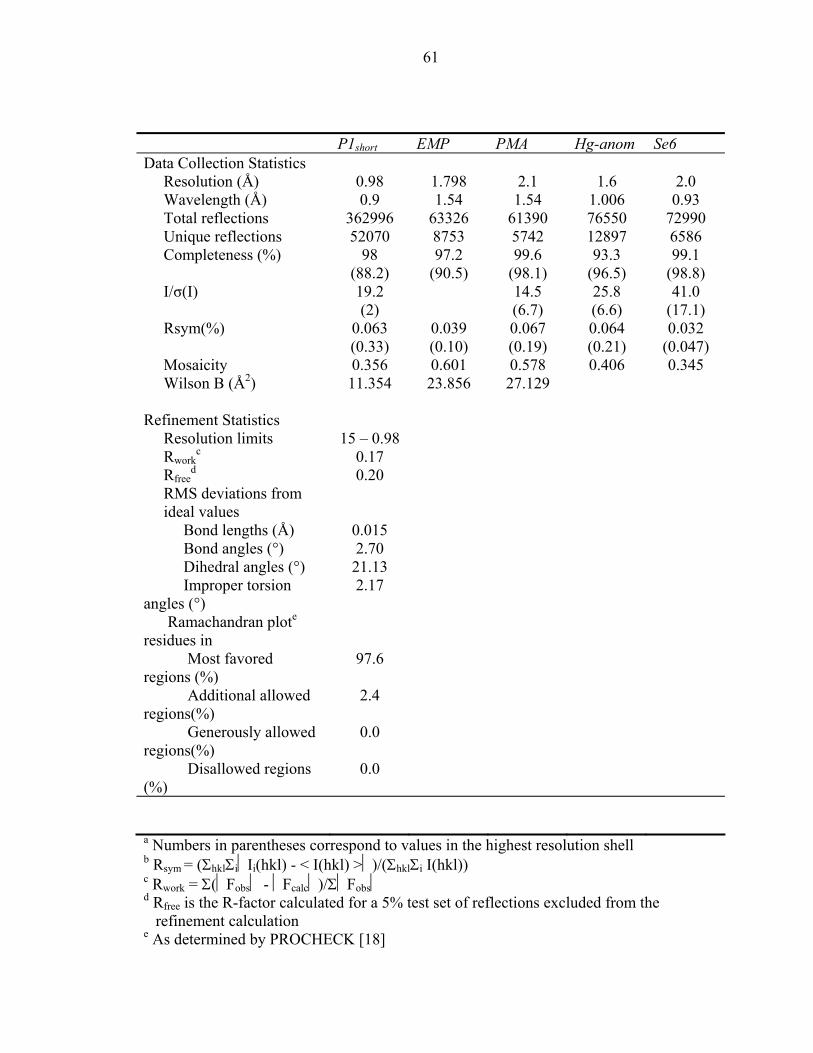
61
P1short EMP PMA Hg-anom Se6 Data Collection Statistics Resolution (Å) 0.98 1.798 2.1 1.6 2.0 Wavelength (Å) 0.9 1.54 1.54 1.006 0.93 Total reflections 362996 63326 61390 76550 72990 Unique reflections 52070 8753 5742 12897 6586 Completeness (%) 98
(88.2) 97.2
(90.5) 99.6
(98.1) 93.3
(96.5) 99.1
(98.8) I/σ(I) 19.2
(2) 14.5
(6.7) 25.8 (6.6)
41.0 (17.1)
Rsym(%) 0.063 (0.33)
0.039 (0.10)
0.067 (0.19)
0.064 (0.21)
0.032 (0.047)
Mosaicity 0.356 0.601 0.578 0.406 0.345 Wilson B (Å2) 11.354 23.856 27.129 Refinement Statistics Resolution limits 15 – 0.98 Rwork
c 0.17 Rfree
d 0.20 RMS deviations from ideal values
Bond lengths (Å) 0.015 Bond angles (°) 2.70 Dihedral angles (°) 21.13 Improper torsion angles (°)
2.17
Ramachandran plote residues in
Most favored regions (%)
97.6
Additional allowed regions(%)
2.4
Generously allowed regions(%)
0.0
Disallowed regions (%)
0.0
a Numbers in parentheses correspond to values in the highest resolution shell b Rsym = (ΣhklΣiIi(hkl) - < I(hkl) >)/(ΣhklΣi I(hkl)) c Rwork = Σ(Fobs - Fcalc)/ΣFobs d Rfree is the R-factor calculated for a 5% test set of reflections excluded from the
refinement calculation e As determined by PROCHECK [18]
62
Table 2: MIR and MAD phasing statistics
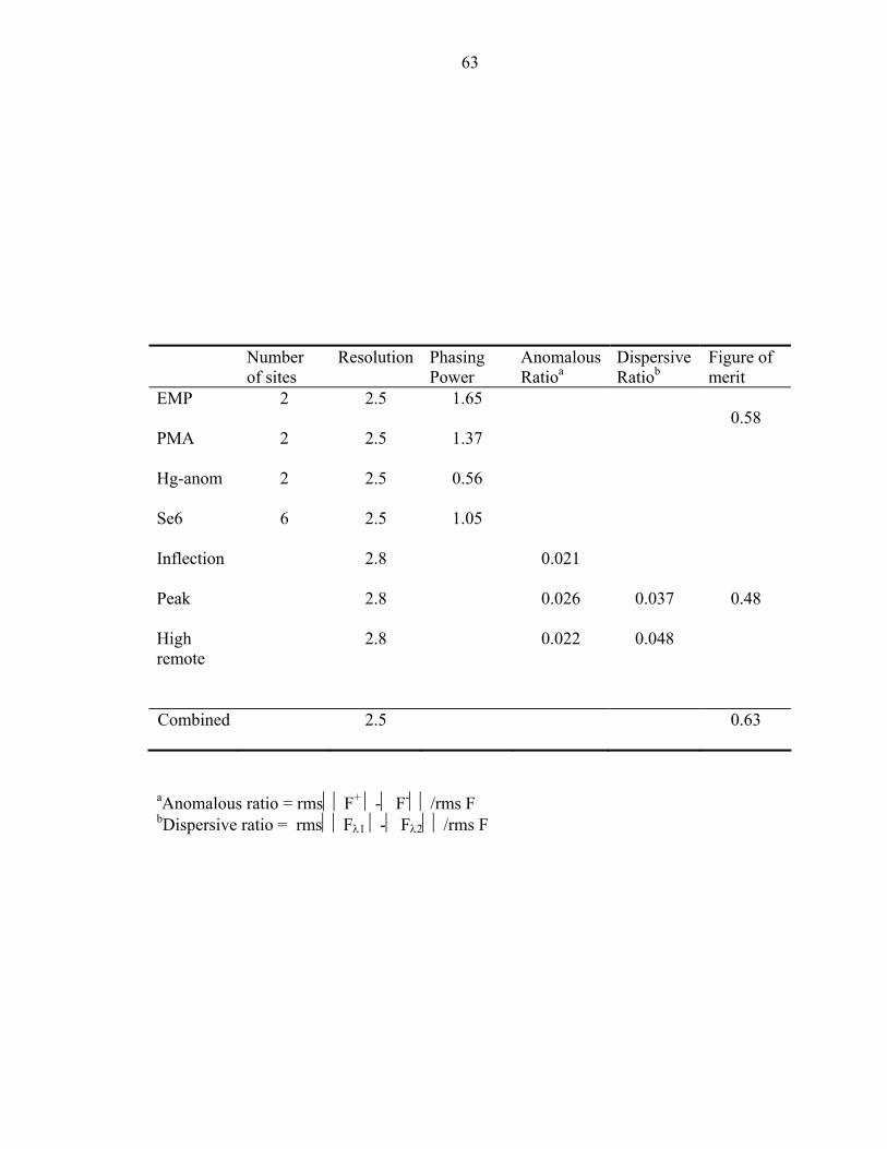
63
Number of sites
Resolution Phasing Power
Anomalous Ratioa
Dispersive Ratiob
Figure of merit
EMP 2 2.5 1.65
PMA 2 2.5 1.37 0.58
Hg-anom 2 2.5 0.56
Se6 6 2.5 1.05
Inflection 2.8 0.021
Peak 2.8 0.026 0.037
High remote
2.8 0.022 0.048
0.48
Combined 2.5 0.63
aAnomalous ratio = rmsF+ - F-/rms F bDispersive ratio = rmsFλ1 - Fλ2/rms F
64
Table 4: Comparison of kinetic and stability parameters between P1 and P1short
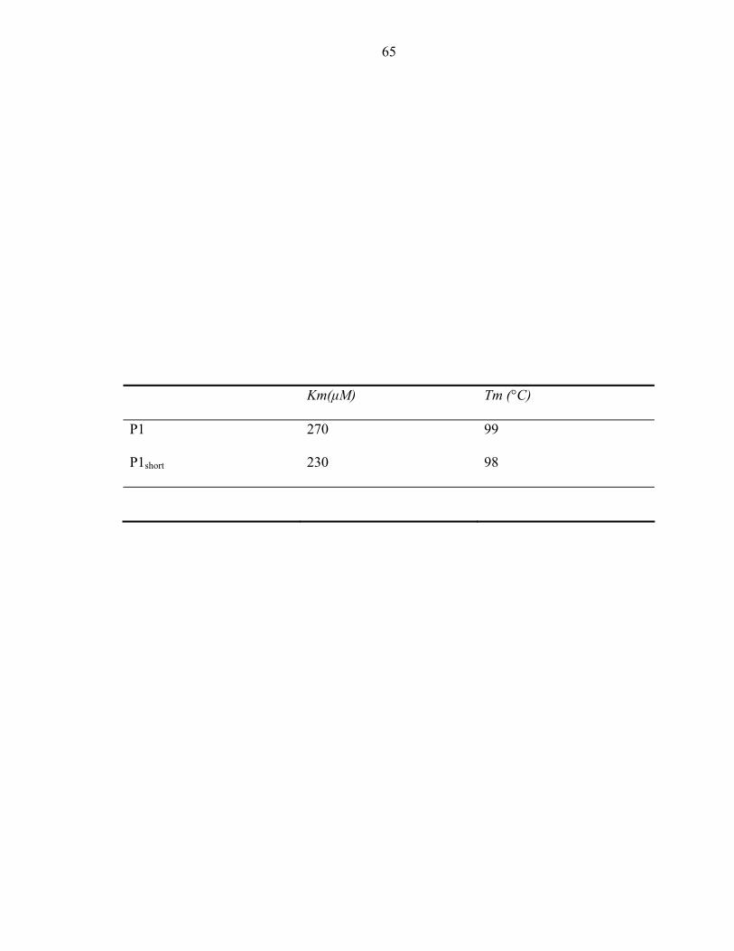
65
Km(µM) Tm (°C)
P1 270 99
P1short 230 98
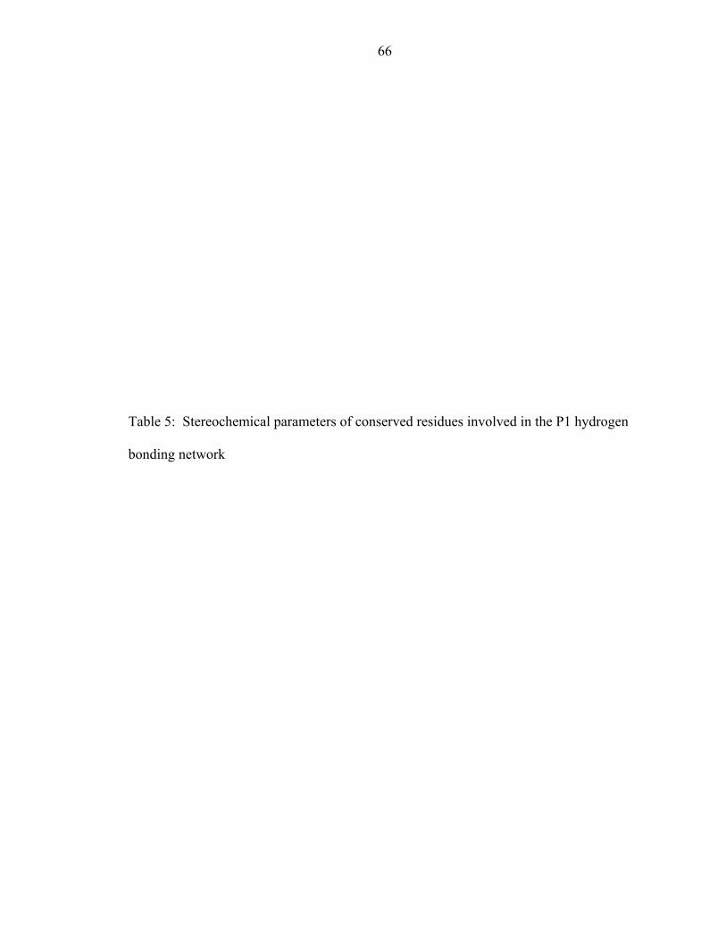
66
Table 5: Stereochemical parameters of conserved residues involved in the P1 hydrogen
bonding network
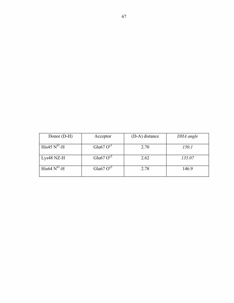
67
Donor (D-H) Acceptor (D-A) distance DHA angle
His45 Nδ1-H Glu67 Oε1 2.70 150.1
Lys48 NZ-H Glu67 Oε2 2.62 135.07
His64 Nδ1-H Glu67 Oε2 2.78 146.9
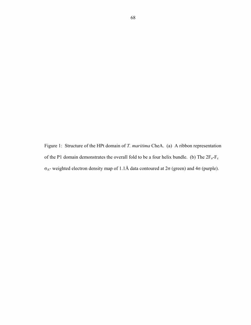
68
Figure 1: Structure of the HPt domain of T. maritima CheA. (a) A ribbon representation
of the P1 domain demonstrates the overall fold to be a four helix bundle. (b) The 2Fo-Fc
σA- weighted electron density map of 1.1Å data contoured at 2σ (green) and 4σ (purple).
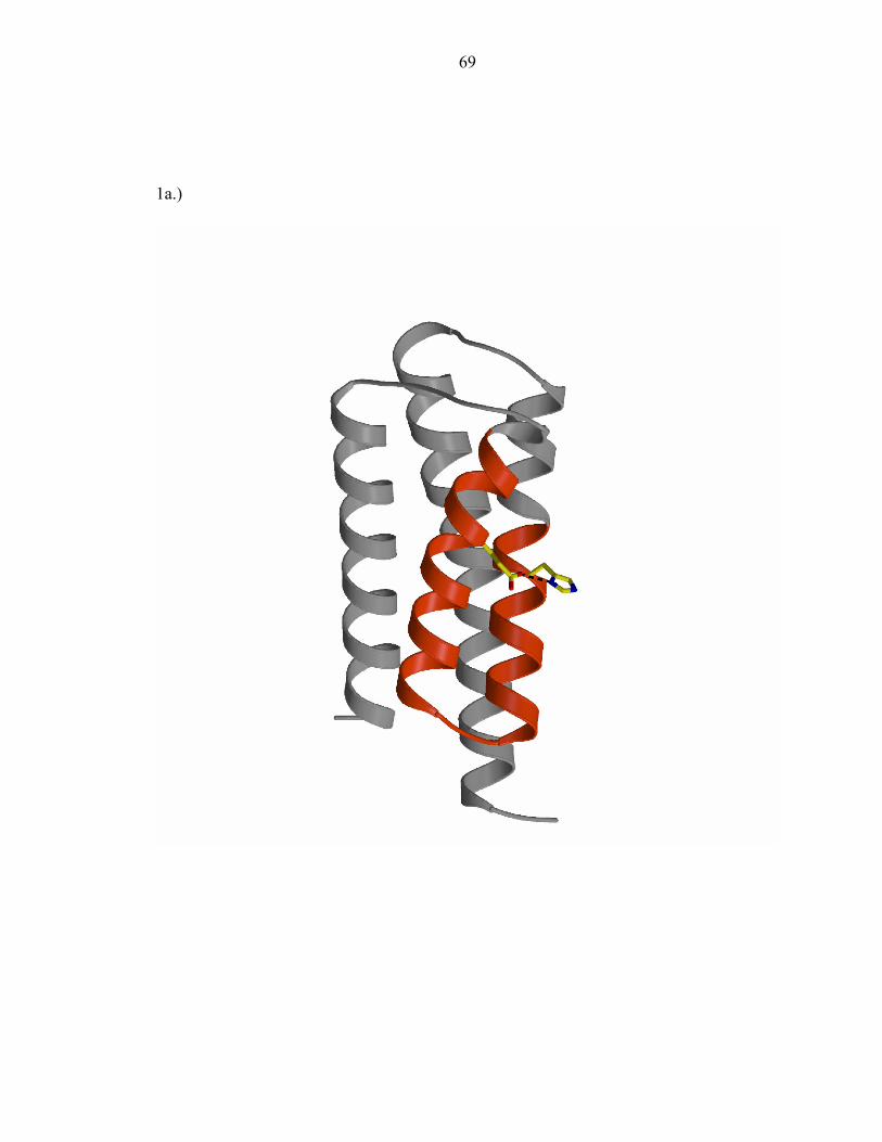
69
1a.)
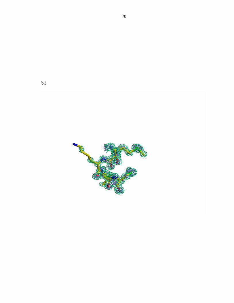
70
b.)
71
Figure 2: (a) The P1 mainchain exists in two conformations (b) Fo-Fc σA- weighted
omit maps contoured at 2σ of a region exhibiting an alternate conformation
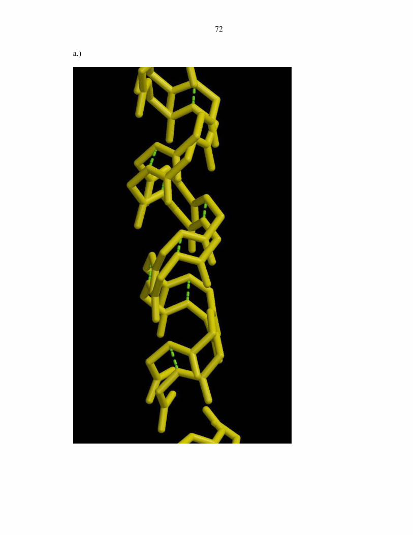
72
a.)
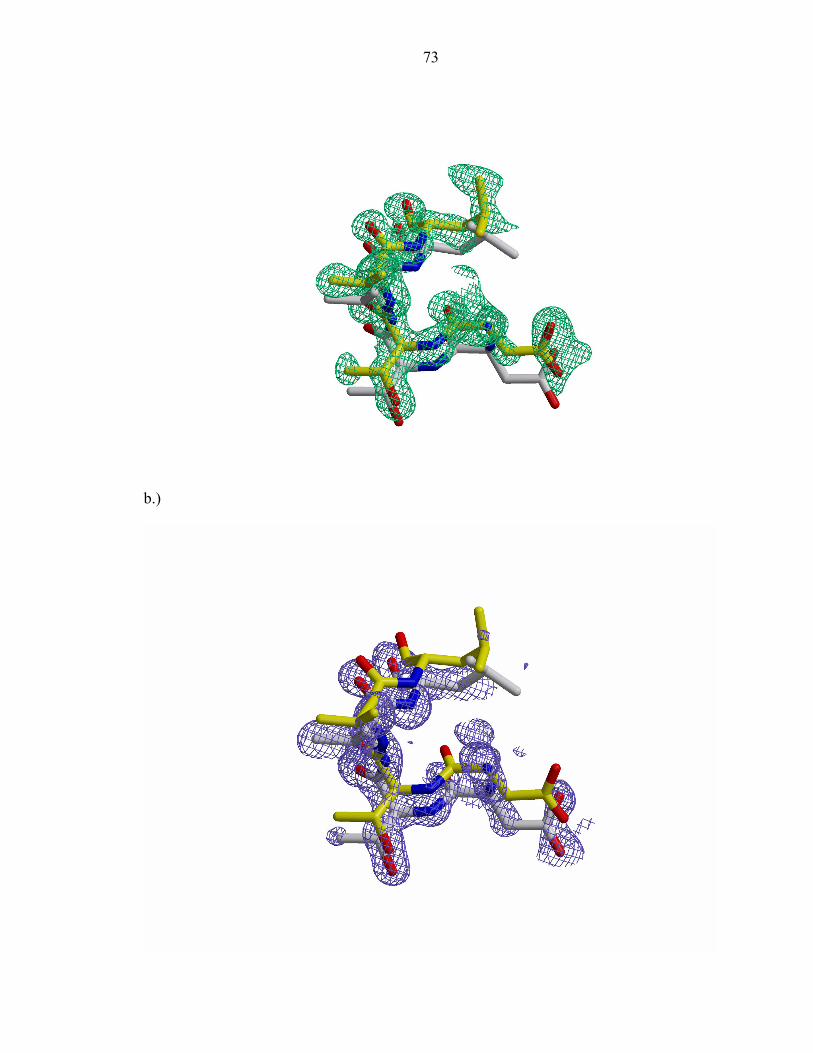
73
b.)
74
Figure 3: The 2Fo-Fc σA- weighted electron density map of the P1 active site contoured at
2.2σ (green) and 3.2σ (purple).
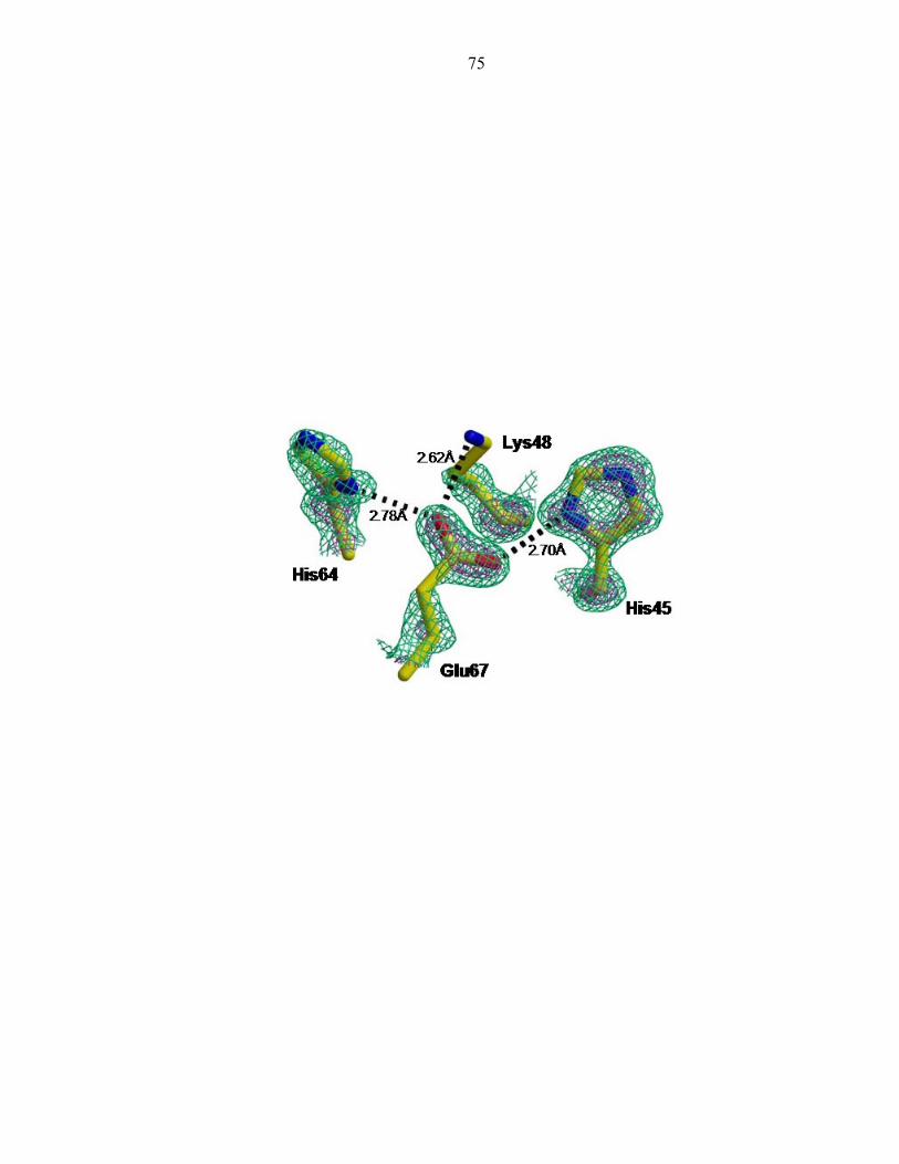
75
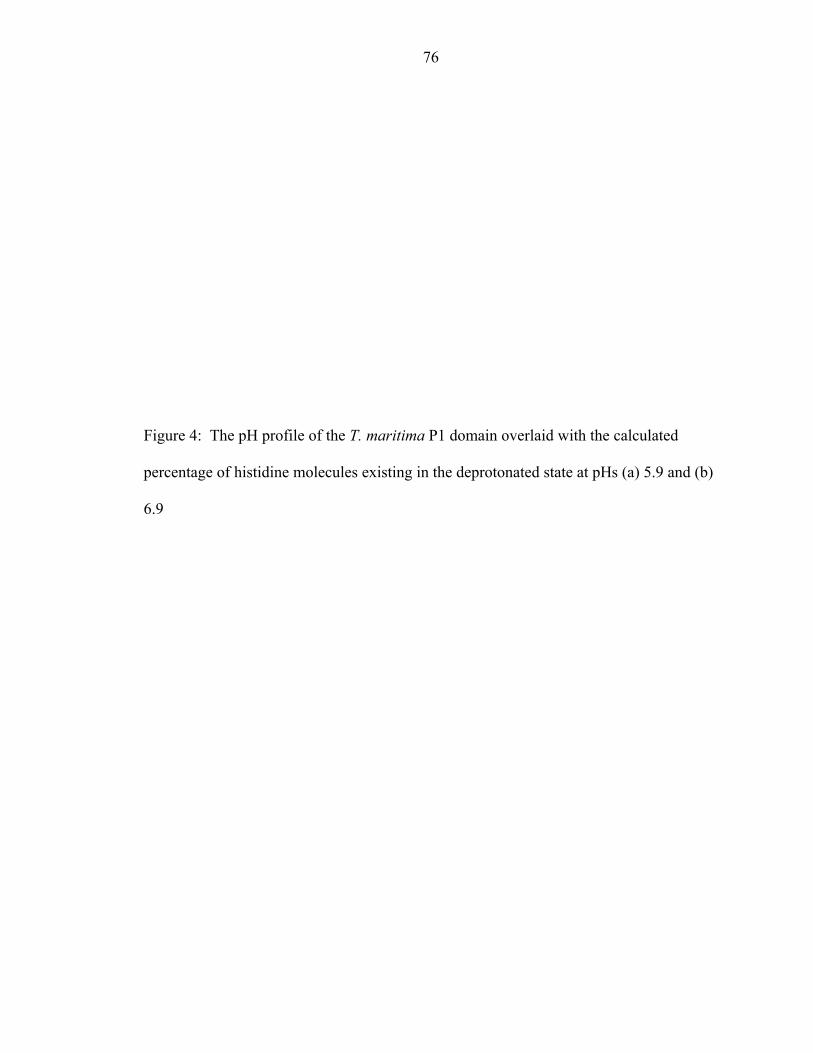
76
Figure 4: The pH profile of the T. maritima P1 domain overlaid with the calculated
percentage of histidine molecules existing in the deprotonated state at pHs (a) 5.9 and (b)
6.9
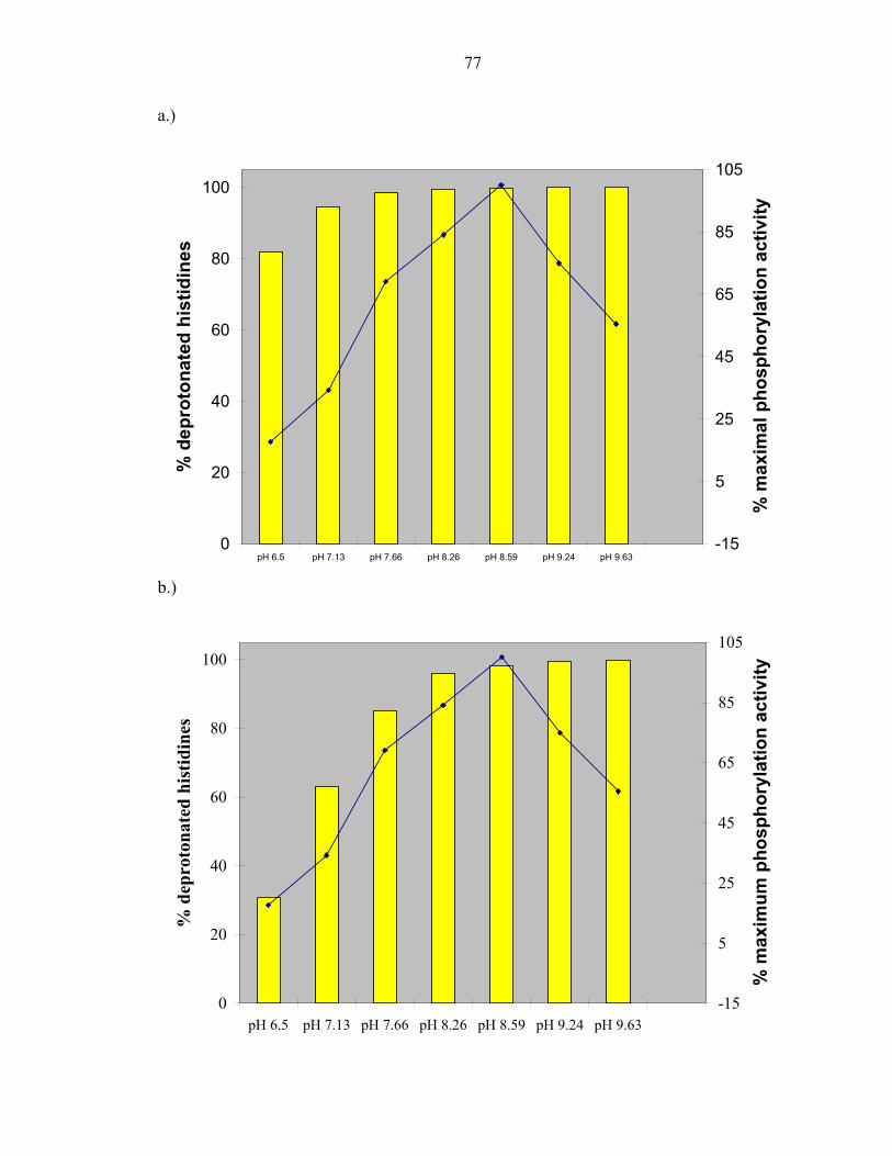
77
a.)
0
20
40
60
80
100
pH 6.5 pH 7.13 pH 7.66 pH 8.26 pH 8.59 pH 9.24 pH 9.63
% d
epro
tona
ted
hist
idin
es
-15
5
25
45
65
85
105
% m
axim
al p
hosp
hory
latio
n ac
tivity
b.)
0
20
40
60
80
100
pH 6.5 pH 7.13 pH 7.66 pH 8.26 pH 8.59 pH 9.24 pH 9.63
% d
epro
tona
ted
hist
idin
es
-15
5
25
45
65
85
105
% m
axim
um p
hosp
hory
latio
n ac
tivity
78
REFERENCES
1. Zhou, H., et al., NMR Studies of the Phosphotransfer Domain of the Histidine
Kinase CheA from Escherichia coli: Assignments, Secondary Structure, General Fold, and Backbone Dynamics. Biochemistry, 1995. 34: p. 13858-13870.
2. Mourey, L., et al., Crystal Structure of the CheA Histidine Phosphotransfer Domain that Mediates Response Regulator Phosphorylation in Bacterial Chemotaxis. The Journal of Biological Chemistry, 2001. 276(33): p. 31074-31082.
3. Zhou, H. and F.W.Dahlquist, Phosphotransfer site of the Chemotaxis-Specific Protein Kinase CheA as Revealed by NMR. Biochemistry, 1997. 36: p. 699-710.
4. Otwinowski, A. and W. Minor, Processing of X-ray diffraction data in oscillation mode. Methods in Enzymolozy, 1997. 276: p. 307-325.
5. Brunger, A.T., Free R-Value - a Novel Statistical Quantity for Assessing the Accuracy of Crystal-Structures. Nature, 1992. 355(6359): p. 472-475.
6. McRee, D.E., XtalView: a visual protein crystallographic software system for X11/Xview. J. Mol. Graph., 1992. 10: p. 44-47.
7. Furey, W. and S. Swaminathan, PHASES: A Program Package for the Processing and Analysis of Diffraction Data from Macromolecules, in ACA meeting Abstracts. 1990, American Crystallographic Association.
8. Crane, B.R. and E.D. Getzoff, Determining phases and anomalous scattering models from the multiwavelength anomalous diffraction of native protein metal clusters. Improved MAD phase error estimates and anomalous- scatterer positions. Acta Crystallographica Section D-Biological Crystallography, 1997. 53: p. 23-40.
9. Collaborative Computational Project, N., The CCP4 suite: programs for protein crystallography. Acta Crystallographica, 1994. D50: p. 760-763.
79
10. Brunger, A.T., et al., Crystallography & NMR system: a new software suite for macromolecular structure determination. Acta Crystallographica Section D, 1998. 54: p. 905-921.
11. Sheldrick, G.M. and T.R. Schneider, SHELXL:high resolution refinement. Methods in Enzymology, 1997. 277: p. 319-343.
12. Wilson, M.A. and A.T. Brunger, The 1.0A Crystal Structure of Ca2+-bound Calmodulin: an Analysis of Disorder and Implications for Functionally Relevant Plasticity. Journal of Molecular Biology, 2000. 301: p. 1237-1256.
13. Gill, S.C. and P.H. Vonhippel, Calculation of Protein Extinction Coefficients from Amino-Acid Sequence Data. Analytical Biochemistry, 1989. 182(2): p. 319-326.
14. Kato, M., et al., Insights into Multistep Phosphorelay from the Crystal Structure of the C-Terminal HPt Domain of ArcB. Cell, 1997. 88: p. 717-723.
15. Kato, M., et al., Refined structure of the histidine-containing phosphotransfer (HPt) domain of the anaerobic sensor kinase ArcB from Escherichia coli at 1.57A resolution. Acta Crystallographica, 1999. D55: p. 1842-1849.
16. Xu, Q. and A.H. West, Conservation of Structure and Function Among Histidine-containing Phosphotransfer (HPt) Domains as Revealed by the Crystal Structure of YPD1. Journal of Molecular Biology, 1999. 292: p. 1039-1050.
17. Ramakrishnan, C. and G. Ramachandran, Stereochemical Criteria for Polypeptide and Protein Chain Conformations .2. Allowed Conformations for a Pair of Peptide Units. Biophysical Journal, 1965. 5(6): p. 909-933.
18. Laskowski, R.A., et al., PROCHECK: a program to check the stereochemical quality of protein structures. J. Appl. Crystallogr., 1993. 26: p. 283-291.
19. Hutchinson, E. and J. Thornton, PROMOTIF: a program to identify and analyze structural motifs in proteins. Protein Science, 1996. 5: p. 212-220.
20. Smith, J.L., et al., Structural Heterogeneity in Protein Crystals. Biochemistry, 1986. 25: p. 5018-5027.
80
21. Morrison, T.B. and J.S. Parkinson, Liberation of an interaction domain from the phosphotransfer region of CheA, a signaling kinase of Excherichia coli. Proc. Natl. Acad. Sci. U.S.A., 1994. 91: p. 5485-5489.
22. Zhou, H., et al., Phosphotransfer and CheY-Binding Domains of the Histidine Autokinase CheA Are Joined by a Flexible Linker. Biochemistry, 1996. 35: p. 433-443.
23. Bilwes, A., et al., Structure of CheA, a Signal-Transducing Histidine Kinase. Cell, 1999. 96: p. 131-141.
24. Deacon, A., et al., The structure of concanavalin A and its bound solvent determined with small-molecule accuracy at 0.94 angstrom resolution. Journal of the Chemical Society-Faraday Transactions, 1997. 93(24): p. 4305-4312.
25. Schuster, II and J.D. Roberts, N-15 Nuclear Magnetic-Resonance Spectroscopy - Effects of Hydrogen-Bonding and Protonation on Nitrogen Chemical-Shifts in Imidazoles. Journal of Organic Chemistry, 1979. 44(22): p. 3864-3867.
26. Conley, M.P., et al., pH Dependence of CheA Autophosphorylation in Escherichia coli. Journal of Bacteriology, 1994. 176(13): p. 3870-3877.
27. Song, H.K., et al., Insights into eukaryotic multistep phosphorelay signal transduction revealed by the crystal structure of Ypd1p from Saccharomyces cerevisiae. Journal of Molecular Biology, 1999. 293(4): p. 753-761.
28. Bilwes, A.M., et al., Nucleotide binding by the histidine kinase CheA. Nature Structural Biology, 2001. 8(4): p. 353-360.
29. Jackson, A.P. and A. Maxwell, Identifying the catalytic residue of the ATPase reaction of DNA gyrase. Proceedings of the National Academy of Sciences, 1993. 90: p. 11232-11236.
81
Chapter 3
The chemical determinants of histidine reactivity in CheA
82
Protein phosphorylation is a key mechanism for intracellular signal transduction
in both eukaryotic and prokaryotic cells. Histidine phosphorylation is central to bacterial
signaling [1-3]. Histidine kinases initiate signal processing, transmitting information
from the extracellular environment and triggering an appropriate response. Since
histidine phosphorylation is prevalent in prokaryotes, and rarely found in higher
eukaryotes, it is of extreme interest to elucidate the determinants of histidine reactivity.
Distinguishing features could lead toward the development of novel antibiotics.
One of the best characterized histidine kinases is CheA, an autophosphorylating
dimeric protein that initiates the phosphorylation cascade which results in chemotaxis, the
directed movement of bacteria in a chemical environment. The structure of CheA’s
catalytic core revealed that it is not similar to the Ser/Thr or Tyr kinases. Instead, CheA
surprisingly resembles the functionally divergent GHL (for GyrB, Hsp90, MutL) ATPase
family [4]. Histidine kinases use ATP as a phosphodonor, transferring its γ-phosphoryl
group to a histidine residue. In contrast, ATPases cleave the ATP γ-phosphoryl group
and release it into water.
CheA and the GHL family of ATPases share a common structural element, a deep
cavity for ATP binding to the active site. In addition to possessing similar topologies, the
sequences of the ATP-binding sites of protein histidine kinases (PHKs) and GHL
ATPases are highly conserved. The edges of the ATP-binding cavities of these proteins
include four regions of sequence similarity: the N, G1, F, and G2 boxes [4, 5].
Despite the striking topological and sequence similarities, some critical
differences exist between these two protein families. An essential glutamate of the GHL
ATPases presumed to be the general base involved in water activation for ATP
83
hydrolysis [6] is replaced by His 405 in CheA. This glutamate is missing from the kinase
domain. However, there is a conserved glutamate in the CheA histidine phosphotransfer
domain, P1, which is in the vicinity of the active site histidine.
CheA phosphorylation activity is sensitive to pH in the range between 6.5 and 10
[7] (Chapter 2 Fig. 4). The phosphoaccepting histidine (His48 in E.coli) was previously
determined by NMR to have a higher than normal pKa of 7.8 and to exist in the normally
unfavored tautomeric form, Nδ1H. [8]. The phosphorylatable histidine remains strongly
hydrogen bonded, even at pHs above its pKa value, via the Nδ1 atom, which serves as a
hydrogen bond donor. However, due to the low resolution of the P1 NMR structure, it
was initially not possible to identify the histidine hydrogen bonding partner [9]. A
crystal structure of the Salmonella P1 domain identified a glutamate residue as a potential
hydrogen bonding partner [10].
In an effort to determine whether the conserved P1 glutamate (Glu67 in
Thermotoga maritima) plays a similar role as the activating glutamate of the GHL
ATPases, in depth structural and biochemical studies were carried out at the 67th position
of the T. maritima histidine phosphotransfer domain. Herein, we explore the putative
catalytic role of Glu67 to the CheA autophosphorylation reaction by site-directed
mutagenesis, protein crystallography, and 2-D NMR techniques.
MATERIALS AND METHODS
84
Protein cloning, expression, and purification
T. maritima CheA, ∆289 (residues 290-671), domain P1 (residues 4-133), and
P1E67Qshort (residues 4-104) were subcloned in the vector pET28(a) (Novagen). The
plasmid was transformed into E. coli strain BL21(DE3) (Novagen) and protein expressed
in 2L TB cultures. Protein purification was achieved by affinity chromatography on
Nickel-NTA beads (Qiagen), followed by an overnight digestion of the His6 tag at 4°C.
The protein was further purified by gel filtration on a superdex 75 or 200 column
(Pharmacia) using a buffer composed of 50 mM Tris pH7.5, 150 mM NaCl and 2 mM
DTT. Centrifugation with an Amicon centriprep concentrator yielded the concentrated
protein. Mutations were made using the QuickChange Site-Directed Mutagenesis Kit
(Stratagene).
Phosphorylation Assays
Initial velocities of full length CheA and its mutants (2-10 µM) or P1(30 µM)
incubated with ∆289(2 µM) were measured in 50 mM Tris pH 8.5, 50 mM KCl, and 2
mM DTT at 50°C. Reactions were initiated upon addition of [γ-32P] ATP. At specific
time intervals, aliquots were quenched with 2X sodium dodecyl sulfate (SDS)
electrophoresis buffer containing 25 mM ethylenediaminetetra acetic acid (EDTA).
Samples were then electrophoresed on 12-18% Criterion Tris-HCl gels (Biorad) using a
Criterion Dodeca Cell (Biorad). Gels were dried under vacuum and phosphorylation
quantified using a Storm phosphoimager (Molecular Dynamics). pH dependence studies
were performed in the same manner over a pH range of 6 to 9.5. The same protocol was
85
used when P1(30 µM) was incubated with ∆289(2 µM) in the presence of a tenfold
excess of P1 mutant (300 µM) or the control BSA(300 µM).
Swarm assays
Tryptone swarm plates consist of 0.25% Difco Bacto Agar in tryptone broth (1%
tryptone, 0.5% NaCl) 100 µg/ml ampicillin, and 5 µM IPTG. Strain RP9538, a derivative
of E.coli K-12 carrying a deletion for the CheA gene, was transformed with pAR1:cheA
derivatives containing the wild-typeor mutant alleles. Tryptone swarm plates were
stabbed in the center with fresh transformants and immediately incubated at 37°C. Ring
diameters were measured after twelve hours. RP9538 and pAR1:cheA were kindly
provided by Prof. Rick Stewart (University of Maryland, College Park).
CD experiments
CD spectra were recorded with an AVIV (Lakewood, NJ) 62A DS spectrometer
equipped with a Peltier-type temperature control system and using a 0.1 cm quartz
cuvette (Wilmad). Samples were prepared in 20 mM NaPi buffer at pH 8. Data were
collected in the far UV (from 250 to 190 nm) with an averaging time of 5 s and a
bandwidth of 1.5 nm.
Crystallization, data collection, structure determination, and refinement
Orthorhombic crystals belonging to the P2221 space group were obtained by
mixing 2 µL of 7-15 mg/ml P1E67Qshort with 2 µL of the reservoir solution (14% PEG
2K, 0.1M NaAc pH 4.5, 0.2M AmAc). Crystals were briefly soaked in a cryoprotectant
solution (36% PEG 8K, 0.1M NaAc pH4.5, and 0.2M AmAc) and then flash frozen.
86
Diffraction data were colleted to 1.1Å resolution at the Cornell University synchrotron
source, CHESS, on beamline F1. Data were processed, scaled, and merged using the
DENZO/SCALEPACK suite of programs [11] (Table1).
The “warpNtrace” feature of ARP/wARP was used to automatically build the
initial protein model [12]. The asymmetric unit consists of one molecule. Iterative
cycles of refinement using the program SHELXL [13] and manual rebuilding of the
model in XtalView [14] yielded an Rwork and Rfree of 0.146 and 0.194, respectively.
Inclusion of all data yielded a crystallographic R factor of 0.148 (Table 1). As previously
observed in the wild-type structure of P1 (Chapter 2 Fig. 2), several discrete residues (14,
16, 39, 51, 54, and 55) exist in alternate conformations. Extended regions displaying
alternate conformations in helices A and D (residues 21-32 and 82-105) are also observed
in the P1E67Qshort crystal structure.
NMR spectroscopy
NMR experiments were performed on a 600 MHz Inova spectrometer equipped
with 1H [13C/15N] pulsed-field gradient probes. NMR samples contained 50mM sodium
phosphate, 0.05% sodium azide and 10% D20. All experiments were recorded at 50˚C.
Assignments for all non-proline backbone amides of wild-type P1 were obtained from the
Dahlquist Lab (Hamel et al. unpublished results). For this work, 3-D 15N-NOESY-
HSQC, 2-D HSQC-aliphatic and 2-D HSQC-aromatic correlation experiments were
acquired on uniformly 15N-labeled proteins [15]. For aliphatic (aromatic) HSQC spectra,
the 15N carrier frequency was set to approximately 121(190) ppm, using a delay of
4.8(50) ms to refocus chemical shift evolution arising from the coupling between the
87
backbone amide proton and nitrogen (carbon-bound proton and imidazole nitrogen). In
all correlation experiments 100(128) complex t1 increments were recorded. Spectral
widths were 1600(4800) Hz in F1 and 8000 Hz in F2. The number of scans in individual
2D experiments ranged from 4 to 128 depending on the signal-to-noise ratio. Quadrature
detection in the 15N dimension used the States-TPPI method [16]. 3D 15N-NOESY-
HSQC spectra were recorded for each mutant and used to verify backbone amide
resonance assignments. These spectra had the same parameters listed above with 64
complex t2 increments, spectral width of 8000 Hz in F3 and 150 ms mixing time. In the
pH titration experiments, a series of spectra were recorded for each protein, ranging from
approximately pH 4 to pH 10. The pH was adjusted with small amounts of HCl or NaOH.
The aliphatic HSQC experiments were used to measure chemical shift changes with pH
of the amides. The aromatic HSQC experiments were used to measure pH-dependent
chemical shift changes of the histidine rings. All data was processed using the FELIX
software package from MSI.
Analysis of NMR data
The protonation state of a nitrogen nucleus results in a unique chemical shift [17-
21]. The charged (type α+) and neutral (type α) protonated imidazole 15N nucleus have
chemical shifts of 176.5 and 167.5 ppm, respectively. The deprotonated 15N (type β)
possesses a chemical shift of 249.5 ppm. These values are according to that of Pelton et
al., which differ from those of Bachovchin [22, 23] (Table 2).
At low pH, the observed chemical shifts reflect that of the pure protonated state,
whereas at high pH, the chemical shifts consist of the weighted averaged of the neutral
88
tautomer shifts [23]. Fast equilibration of the two tautomers, Nδ1H and Nε2H, is observed
in solution [24]. Important information about the predominant histidine tautomeric state
and potential hydrogen bonding interactions can be obtained by comparing the observed
chemical shift values to those obtained from model compounds. Hydrogen bonding
results in changes to the 15N chemical shifts that are similar to those produced by nitrogen
protonation and deprotonation [24]. However, the magnitude is much smaller, up to
10ppm. Another indication of hydrogen bond formation is the predominance of the
normally unfavored tautomer Nδ1H [8, 23, 25].
The pKa values of the imidazole rings were determined by a least squares fit of
the chemical shifts over a range of pH values using the Henderson-Hasselbach equation:
δobs= δ110-pH/(10-pH +10-pKa) + δ210-pKa/(10-pH + 10-pKa) (Eqn 1)
The chemical shifts at the low and high pH limits are δ1 and δ2 respectively. The
observed chemical shift, δobs, at any given pH is assumed to result from the weighted
average of fast exchange between the limiting values δ1 and δ2, respectively. The fraction
of each tautomer present was calculated as previously described [8].
RESULTS
A conserved residue, Glu67, is critical to CheA auto-phosphorylation
As a first step in probing whether GHL ATPases and PHKs share a common
mechanism of ATP cleavage, we investigated the role Glu67 plays in CheA
89
autophosphorylation. A series of mutations at this position were designed to examine
whether hydrogen bonding ability or charge were critical to the phosphorylation reaction.
Using site directed mutagenesis, the conserved residue Glu67 was altered to Gln, Asp,
and Ala.
The first mutation, E67Q, replaces the oxygen moiety of the carboxyl side chain
with an amide. This conservative mutation maintains hydrogen bonding ability, but
removes charge from this location. In addition, other histidine phosphotransfer (HPt)
domains, such as ArcBc and Ypd1, have a glutamine at this position. It was of interest to
probe whether a glutamine residue is interchangeable for a glutamate at this site. The
Asp mutation keeps the charge, but being one carbon shorter than a Glu, could weaken or
abolish a hydrogen bond. The Ala mutation removes both charge and the ability to
hydrogen bond. The effects of a Glu to Ala mutation have already been described for
Salmonella [10]. However, the conservative and semi-conservative mutations reported in
this study investigate the role of Glu67 in further detail by assessing the effect of varying
hydrogen bonding degrees and charge on the phosphorylation reaction of T. maritima
CheA.
To initially characterize each Glu67 mutant, the phosphorylation level of wild-
type protein was compared to that of mutant protein following a thirty-minute incubation
with γ32P ATP in vitro. Mutations were made both in the isolated phosphotransfer
domain, P1, as well as the intact CheA protein. CheA containing the mutations E67Q,
E67D, and E67A showed dramatically reduced autophosphorylation activity (5.7%,
8.4%, and 2.4%, respectively; Figure 1a). An even more dramatic reduction in
90
phosphotransfer activity to P1 was observed, averaging about 1% of wild-type activity
for all mutants (Figure 1b).
The effect of each mutation was further investigated in vivo by measuring the
ability of E. coli mutant strains to migrate in semisolid agar medium [26]. Plasmids
expressing each E67 mutation in intact CheA were transformed into a host strain, ∆cheA,
lacking the CheA allele. All E67 CheA mutants exhibit significantly reduced
chemotactic swarming ability, approximately 20% of that observed with wild-type
(Figure 2). Therefore, a point mutation of a critical residue in CheA can disrupt the
normal chemotaxis signal transduction pathway in intact cells.
To probe whether an increase in pH rescues the activity of the E67 mutants by
making His45 more nucleophilic through deprotonation and therefore more amenable to
phosphorylation, the activity of E67 mutants was monitored as a function of pH at 50ºC.
The phosphotransfer reaction of the mutants is not markedly affected by a change in pH
(Figure 3). Phosphotransfer to the mutant proteins is therefore independent of pH or the
deprotonation of the active site histidine.
Far-UV CD spectra of wild-type P1 and the E67 mutants are virtually identical,
indicating that the mutation does not alter the secondary structure of the phosphotransfer
domain (Figure 4a). In order to probe whether the P1E67Q mutant binds the kinase
domain as P1, it was incubated in a tenfold excess to its wild-type counterpart in the
presence of the kinase fragment ∆289. The time course of this reaction was monitored
and revealed that P1E67Q inhibits the normal progression of phosphotransfer. A tenfold
excess of P1E67Q results in 26% of normal phosphotransfer activity (Figure 4b). To
further investigate whether P1E67Q is specifically inhibiting the phosphorylation
91
reaction, a ten fold excess of a non-specific protein, BSA, was added to a solution
containing P1 and ∆289. 90% of normal activity is retained. Taken together, these data
suggest P1E67Q is a catalytic mutation of the wild-type reaction. Both charge and the
ability to hydrogen bond are important at this position.
The high resolution structure of P1E67Q reveals a disrupted hydrogen bond network
In order to further elucidate the role that Glu67 plays in the phosphotransfer
reaction, the crystal structure of P1E67Qshort (helices A-D) was solved to a resolution of
1.1Å (Table 1). The protein crystal belongs to the orthorhombic P2221 space group with
cell dimensions a= 27.36Å, b= 37.49Å, and c= 87.47Å. Refinement converged to an
Rwork of 14.6% and an Rfree of 19.4%.
At a resolution of 1.1 Å, individual atoms such as N and O can be unambiguously
assigned based on the electron density volume. Additionally, the shape of the electron
density at single and double bonds can be distinguished. As observed in Figure 5, the
glutamine carbonyl exhibits continuous electron density, as expected for a double bond,
between the carbon and oxygen atoms. On the other hand, the nitrogen moiety of the Gln
residue shows discrete density, as expected for a single bond. The high resolution
structure of P1E67Q therefore affords a unique opportunity to see the structural effects of
a single point mutation in atomic detail.
The E67Q structure is almost indistinguishable from that of the native P1
structure, with the exception of a few but critical details. An overlay of the active site
region of wild-typeP1 and P1E67Q reveals the disruption of the hydrogen bond network
involving His45, Glu67, Lys48, and His64 (Figure 6a). At pH 4.5, the Nδ1 position of
92
the phosphoaccepting histidine is within hydrogen bonding distance, 2.82Å, of the Gln67
carbonyl oxygen atom. The slight change in side chain orientation results in the
disturbance of the hydrogen bond interactions to Lys48 and His64 observed in the wild-
type at this position (Figure 6b).
Glu67 tunes the reactivity of the phosphoaccepting His45
In solution, histidine side chains readily exist in three protonation states: a
charged state and two neutral tautomers Nδ1and Nε2, which undergo fast exchange in
solution [23, 24]. Based on the chemical shift value, it is possible to identify hydrogen
bonding interactions, protonation and tautomeric states. (Table 2). In order to evaluate
how a conservative mutation at the 67th position affects the reactivity of the
phosphoaccepting histidine, we expressed 15N labeled P1E67Q. P1E67Q contains three
histidines, the site of phosphorylation, His45, a participant in the hydrogen bond network,
His64, and a residual N-terminal histidine from the histidine tag used for protein
purification. Histidine deprotonation was monitored as a function of pH using two
dimensional NMR techniques.
The pH titration of the P1E67Q His45 imidazole is depicted in Figure 7a. Using
equation 1, its pKa was determined to be 5.8. At pH 3.97, the chemical shift values of
Nδ1 and Nε2 are 178.4 and 174.3ppm, respectively. These values closely correspond to
the expected 176.5 ppm for a charged imidazole nitrogen. At the high pH limit, the
chemical shift values of Nδ1 and Nε2 are 226.7 and 184.5 ppm, respectively (Table 3).
The larger shift of the Nδ1 atom indicates that the phosphoaccepting histidine primarily
93
exists in the Nε2H tautomeric state. In fact, the Nδ1H:Nε2H tautomeric ratio was
calculated to be 1:3.3 (Table 4).
The protonation states of the remaining two histidines were also examined
(Figures 7b-c). His64 is involved in the conserved hydrogen bonding network of P1
(Figure 4b). The Nδ1 atom has a much larger chemical shift, 237.1 ppm, than the
176.1ppm of Nε2 indicating that the Nε2H tautomer is dominant. The pKa value of His64
is 5.9 and the Nδ1H:Nε2H tautomeric ratio was calculated to be 1:4.3 (Table 4). The final
histidine, located at the N-terminus, is a residual amino acid from the histidine tag. It
plays the role of an internal control, serving as a model solvent-exposed histidine residue
(Table4). Its pKa was measured to be 5.9 and its Nδ1H/ Nε2H tautomeric ratio is 1:4.2.
The pH titration of the imidazole 1H chemical shifts for all histidines can be found in
Appendix 1.
DISCUSSION
The activation of a phosphoaccepting histidine residue
Using site-directed mutagenesis, crystallography, and 2-D heteronuclear NMR,
Glu67 was identified as an indispensable residue to the CheA autophosphorylation
reaction. A systematic mutational study investigating whether hydrogen bonding or
charge were important contributing factors to the phosphorylation reaction in vitro and in
vivo revealed that both properties are important (Figures 1-2). A Gln side chain that
maintains hydrogen bonding ability and an Asp side chain that preserves charge at this
location both result in the dramatic inhibition of phosphorylation activity. Interestingly, a
94
Gln residue, although found in the same position in other HPt domains [27, 28], is not
interchangeable for a Glu residue in CheA (Figure 1). These results indicate that a
negatively charged residue that simultaneously acts as a hydrogen bond acceptor of the
phosphoaccepting histidine is required for efficient CheA autophosphorylation to take
place.
Although the E67A mutation in Salmonella was shown to reduce the ATP
phosphotransfer rate, these experiments could not distinguish whether the decrease in
activity was due to the disruption of secondary structure, the loss of binding between the
P1 and the kinase domain, or a catalytic defect [10]. Point mutations at Glu67 do not
perturb the secondary structure content of P1 as evidenced by far-UV circular dichroism
studies (Figure 4a). The crystal structure of P1E67Q further confirms that the overall
structure remains intact. Additionally, phosphorylation of wild-typeP1 by the catalytic
core fragment, ∆289, was inhibited in the presence of an excess of P1E67Q. On the other
hand, an excess of a random protein, bovine serum albumin (BSA), that is not expected to
bind P4, did not affect P1 phosphorylation (Figure 4b). We infer that P1E67Q is
recognizing the kinase domain P4 and acting specifically.
The pH profile of E. coli and T. maritima CheA demonstrated optimal activity at
pH 8.5, consistent with the existence of a higher than normal histidine pKa (Chapter2
Figure 4) [7]. The phosphorylation activity of E67 mutants are independent of pH. In
fact, the phosphorylation level at pH 6.5, where roughly 70% of histidines exist in the
protonated state, is similar to that at pH 8.5 and higher, where greater than 99% of the
histidines are expected to exist in the deprotonated state. Histidine reactivity in E67
95
mutants is unaffected by the histidine protonation state. Hence, the mere deprotonation
of His45 does not lead to histidine phosphorylation (Figure 3).
The atomic resolution structure of P1E67Q provided further insight into the
functional role of Glu67. In changing one atom of a side chain from an oxygen to an
amide, the side chain orientation at the 67th position is altered. This slight change
disrupts the hydrogen bonding network observed in the native structure (Figure 6). The
crystal structure of wild-type P1 demonstrated that Glu67 acts as a hydrogen bond
acceptor to both Lys48 and His64. In P1E67Q, the neutral Gln no longer forms a salt
bridge with a positively charged Lys48. Furthermore, the Gln amide cannot act as a
hydrogen bond acceptor to His64.
Despite the disruption to the intricate hydrogen bonding network the position of
His45 is curiously not altered by this mutation. Glu67 is thus not involved in the
positioning or alignment of the phosphoaccepting histidine. Although structural
perturbations cannot be ruled out as a cause for the decreased efficiency of histidine
phosphorylation, a change in the chemical and electronic properties of the
phosphorylatable histidine is likely most responsible for the drastic change in activity.
The P1E67Q phosphoaccepting histidine exists in the Nε2H tautomeric state (Figure 7a).
Since its phosphorylation activity is dramatically reduced, this indicates that the
phosphoaccepting histidine must exist in the correct tautomer, Nδ1H, in order for it to be
phosphorylated. Curiously, the same strategy is employed by the conserved Asp and His
catalytic residues of the serine proteases.
Ypd1, ArcBc, and P1E67Q share structural and chemical properties. Their Gln
residues have similar side chain orientations and are able to hydrogen bond to the Nδ1H
96
atom of the phosphoaccepting histidine. Interestingly, ArcBc and P1E67Q both
predominantly exist in the Nε2H tautomeric state and have pKas that correspond to that of
a normal solvent exposed histidine [27]. The requirements for histidine phosphorylation
therefore differ between these two proteins (see discussion below). A mere hydrogen
bond with the Nδ1 atom of the phosphoaccepting histidine is not sufficient to
phosphorylate the CheA P1 domain. The histidine must predominantly exist in the Nδ1H
tautomeric state in order for phosphorylation to occur.
The native crystal structure revealed that the electrons of the carboxyl moiety of
Glu67 are resonating (Chapter 2 Figure3). Therefore His45 is acting as a hydrogen bond
donor to a negatively charged oxygen in the case of the wild-type and to a neutral oxygen
in P1E67Q. The disparity in electronegativity between Glu and Gln is likely responsible
for the effect on histidine reactivity. A negatively charged oxygen atom from a
glutamate acts as a stronger base than a neutral glutamine. Glu67 can therefore stabilize
the existence of an otherwise unfavorable Nδ1H histidine tautomer over a large pH range.
Experiments described in this paper have enabled us to clarify the functional role
that Glu67 plays in the phosphorylation reaction. Glu67 does not fix the orientation of
the histidine. Instead, it tunes the reactivity of the phosphorylatable histidine, making it
more nucleophilic. Taken together, the data suggest the P1 domain not only possesses
the nucleophile for phosphate transfer, His45, but also the activating glutamate, Glu70.
Towards understanding the biochemical mechanism of CheA
Despite the fact that CheA is one of the best characterized histidine kinases, a
detailed biochemical mechanism for CheA autophosphorylation has not been elucidated.
97
Based on the results reported in this paper and on the extensive structural [4, 5, 9, 10, 29],
biochemical [30-32], and mutagenesis investigations of CheA [33-37], we are now
beginning to unravel the details of its biochemical mechanism.
The CheA active site spans two domains. All necessary elements for histidine
phosphorylation are contained in the phosphotransfer (P1) and kinase (P4) domains [5]
(Figure 8). Conserved residues from P4 provide stabilizing contacts necessary for ATP
binding and form a pocket that optimally positions the gamma phosphoryl for transfer to
the histidine residue [5, 34]. Amino acids present in the kinase domain have also been
shown to be catalytically important [34]. P1 possesses a residue that tunes the reactivity
of the phosphoaccepting histidine. His45 is made nucleophilic as a result of a hydrogen
bond to the strong base Glu67. Thus, we conclude that the P1 domain provides the
nucleophile for phosphate transfer (His45) and the activating glutamate (Glu67)
completing the GyrB catalytic center.
It therefore appears that in addition to sharing a common fold and ATP binding
site, CheA and GyrB also share a common mechanism. The main difference between
CheA and the GHL family of ATPases is in the molecule that the conserved glutamate
residue activates. In the case of the ATPases, the glutamate is postulated to activate a
water molecule that attacks the γ-phosphoryl group [6]. Alternatively, in CheA, the
glutamate activates the phosphoaccepting histidine by perturbing its pKa and stabilizing
the normally unfavored Nδ1H tautomeric state.
Since some crucial residues for CheA autophosphorylation have been identified,
there is a solid foundation from which to undertake detailed mechanistic studies. It is
clear that the kinase domain binds and positions ATP for phosphotransfer, however the
98
question of exactly how the conserved residues contribute to catalysis has yet to be
answered. The challenge remains in not only deciphering the exact functional role of
conserved residues, but also in determining the role protein dynamics play in the
phosphorylation reaction. Future studies will lead to a deeper understanding of the CheA
mechanism and facilitate the development of novel antibiotic agents.
Mechanisms of phosphotransfer between components in bacterial signaling differ
The phosphorelay schemes of two component systems are highly adaptable
allowing for variation that has likely evolved to optimize specific signaling systems
(Chapter 1 Figure 4). Despite significant diversity in domain organization, the core
structures and activities of these domains are conserved. The phosphorylation of
histidine residues is central to the transmission of environmental information for
prokaryotes [2]. Therefore, it is of key interest to understand the chemical determinants
of histidine reactivity.
Despite the low sequence similarities observed in histidine phosphotransfer
domains of two-component systems, a four-helix bundle is used as a generic scaffold for
the transfer of phosphoryl groups to and from solvent exposed histidines [9, 10, 28, 38,
39]. Furthermore, monomeric HPt domains are reported to be rigid [9, 27] and even upon
phosphorylation, minor structural changes occur that are limited to the region
surrounding the phosphoaccepting histidine [8, 40-45]. Does a common rigid helical
bundle topology imply that HPt domains also share a common biochemical mechanism of
phosphotransfer? More specifically, are the chemical determinants of phosphate flow to
99
a histidine conserved among monomeric HPt domains such as the CheA P1 domain,
ArcBc, and Ypd1? We think not.
We propose that two flavors of phosphotransfer exist for monomeric HPt domains
that can be divided into two categories: kinase acceptors and phosphotransfer acceptors.
Kinase acceptors, exemplified by the CheA P1 domain, accept a γ-phosphoryl group from
ATP. These histidines are chemically distinguishable from those of phosphotransfer
acceptors. The histidine residue of a kinase acceptor must be activated in a specific way.
A hydrogen bond via the Nδ1 atom to a strong base, a glutamate, results in an altered
histidine pKa and in the existence of the normally unfavored Nδ1H tautomeric state of the
histidine. This results in a highly nucleophilic histidine that is phosphorylated at the Nε2
atom.
Bacterial chemotaxis demands a very fast response time, in the order of
milliseconds. Hence, an activated histidine, one that is so to speak “ready to go,” may be
essential for CheA. Its phosphotransfer domain lies distal to the kinase domain. The
binding interaction between the two domains is weak (Chapter 2, Table 3)[32], so when
making contact there is no time to waste.
On the other hand, histidines of phosphotransfer acceptors, like ArcBc and Ypd1,
do not require such reactive histidines. The pKa of the ArcBc phosphoaccepting histidine
was measured and found to be around 6.5, that of a solvent exposed histidine at room
temperature. Moreover, it possesses an almost equal population of each tautomer.
Enough of the unprotonated Nε2 form therefore exists to allow for phosphorylation at this
location [27]. Interestingly, mutations of the conserved Gln residue in ArcBc [46] and
Ypd1[47] affected neither phosphotransfer to or from these proteins. Accepting a
100
phosphoryl group from a phosphorylated aspartyl, a reactive species, does not require the
tuning of the active site histidine. Conversely, CheA necessitates an activated histidine in
order for phosphorylation to occur at the Nε2 site. Glu67 is needed in order for the Nδ1H
tautomer to dominate.
In summary, the phosphotransfer mechanism is by no means common to all
histidine phosphotransfer domains. The details of this mechanism lie in the environment
surrounding the phosphoaccepting histidine. Slight differences in the atomic details of
various phosphorylation sites may result in differences in phosphorylation mechanism
that may be exploited to develop selective drugs. Distinguishing features could lead
toward the development of novel antibiotics specific to a particular regulation point in a
bacterial signal transduction pathway. In addition to further elucidating how bacteria
interpret environmental stimuli, this work could be applied towards the design of
chemically tuned peptide-based catalysts used for the phosphorylation of small molecule
compounds [48, 49].
101
Table 1: Summary of data collection and refinement statistics
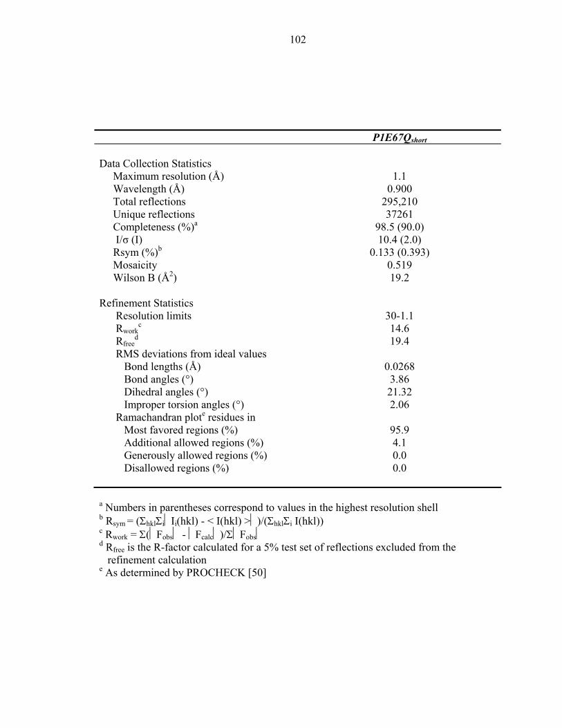
102
P1E67Qshort Data Collection Statistics
Maximum resolution (Å) 1.1 Wavelength (Å) 0.900 Total reflections 295,210 Unique reflections 37261 Completeness (%)a 98.5 (90.0) I/σ (I) 10.4 (2.0) Rsym (%)b 0.133 (0.393) Mosaicity 0.519 Wilson B (Å2) 19.2 Refinement Statistics
Resolution limits 30-1.1 Rwork
c 14.6 Rfree
d 19.4 RMS deviations from ideal values Bond lengths (Å) 0.0268 Bond angles (°) 3.86 Dihedral angles (°) 21.32 Improper torsion angles (°) 2.06 Ramachandran plote residues in Most favored regions (%) 95.9 Additional allowed regions (%) 4.1 Generously allowed regions (%) 0.0 Disallowed regions (%) 0.0 a Numbers in parentheses correspond to values in the highest resolution shell b Rsym = (ΣhklΣiIi(hkl) - < I(hkl) >)/(ΣhklΣi I(hkl)) c Rwork = Σ(Fobs - Fcalc)/ΣFobs d Rfree is the R-factor calculated for a 5% test set of reflections excluded from the
refinement calculation e As determined by PROCHECK [50]
103
Table 2: Nitrogen types and characteristic 15N chemical shifts (ppm)
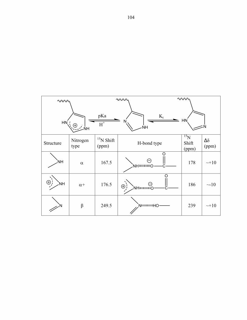
104
NH
HNNH
NN
HNpKa
H+
Kt
Structure Nitrogen type
15N Shift (ppm) H-bond type
15N Shift (ppm)
∆δ (ppm)
NH
α 167.5 NH O C
O
178 ~+10
NH
α+ 176.5 NH O C
O
186 ~-10
N
β 249.5 N HO
239 ~+10

105
Table 3: Expected and observed 15N chemical shifts for P1E67Q histidine residues
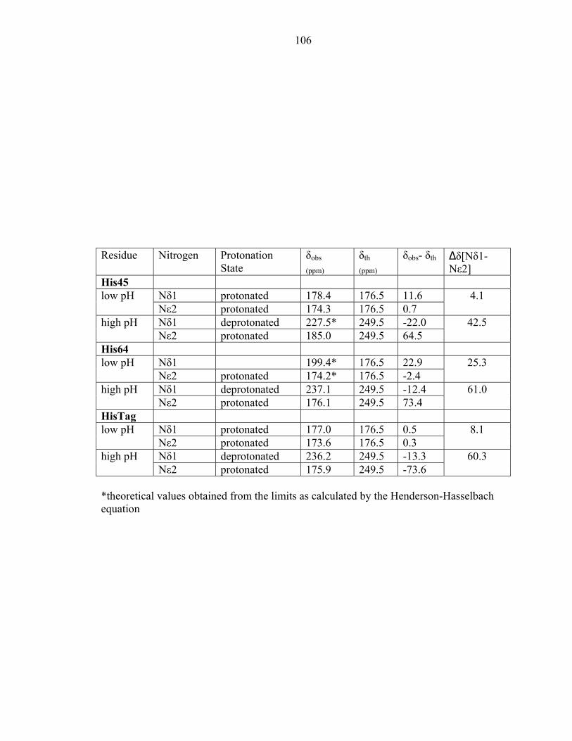
106
Residue Nitrogen Protonation
State δobs
(ppm) δth
(ppm) δobs- δth ∆δ[Nδ1-
Nε2] His45
Nδ1 protonated 178.4 176.5 11.6 low pH Nε2 protonated 174.3 176.5 0.7
4.1
Nδ1 deprotonated 227.5* 249.5 -22.0 high pH Nε2 protonated 185.0 249.5 64.5
42.5
His64 Nδ1 199.4* 176.5 22.9 low pH Nε2 protonated 174.2* 176.5 -2.4
25.3
Nδ1 deprotonated 237.1 249.5 -12.4 high pH Nε2 protonated 176.1 249.5 73.4
61.0
HisTag Nδ1 protonated 177.0 176.5 0.5 low pH Nε2 protonated 173.6 176.5 0.3
8.1
Nδ1 deprotonated 236.2 249.5 -13.3 high pH Nε2 protonated 175.9 249.5 -73.6
60.3
*theoretical values obtained from the limits as calculated by the Henderson-Hasselbach equation
107
Table 5: Apparent pKa values and tautomeric states of P1E67Q histidine residues
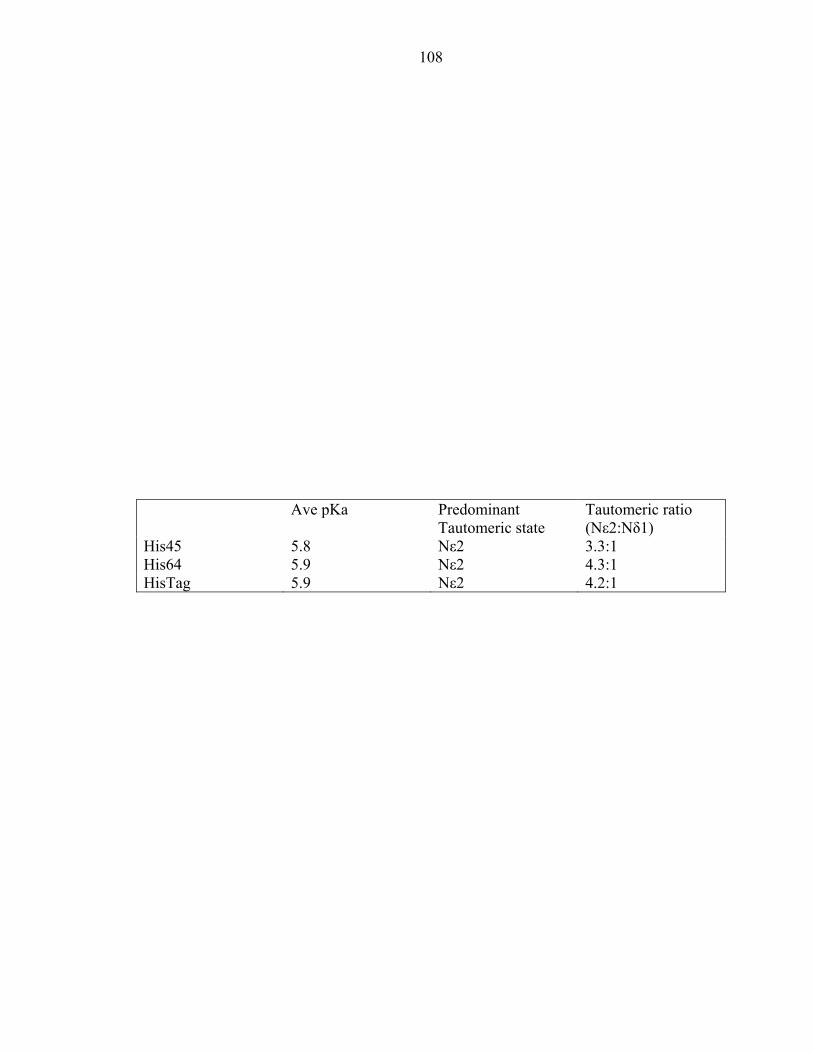
108
Ave pKa Predominant
Tautomeric state Tautomeric ratio (Nε2:Nδ1)
His45 5.8 Nε2 3.3:1 His64 5.9 Nε2 4.3:1 HisTag 5.9 Nε2 4.2:1
109
Figure 1: (a) Relative phosphorylation of CheA and CheA E67 mutants (b) Relative phosphorylation of the isolated P1 domain and its mutants
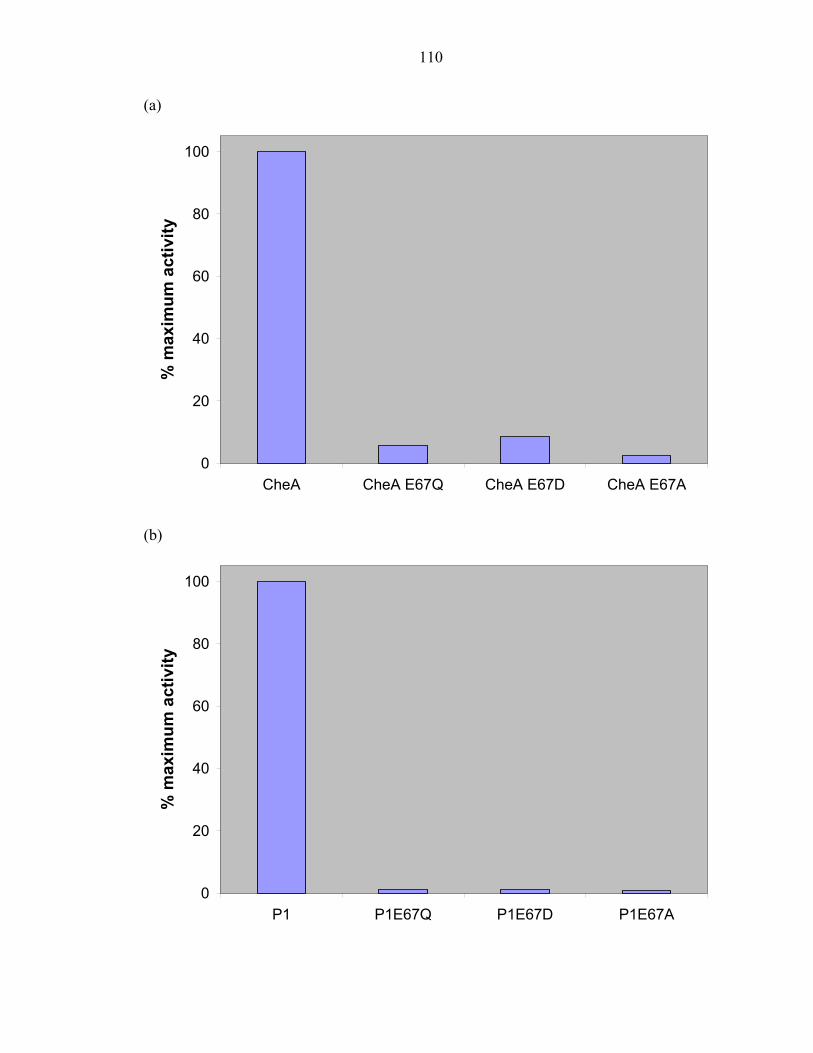
110
(a)
0
20
40
60
80
100
CheA CheA E67Q CheA E67D CheA E67A
% m
axim
um a
ctiv
ity
(b)
0
20
40
60
80
100
P1 P1E67Q P1E67D P1E67A
% m
axim
um a
ctiv
ity
111
Figure 2: (a) Swarm diameter of CheA and CheA E67 mutants. Photographs of swarm assays produced by (b) P1E67A, (c) P1E67D, (d) P1E67Q, and (e) CheA

112
(a)
0
1
2
3
4
5
6
7
P1 P1K48A P1H64A
Swar
m D
iam
eter
(cm
)
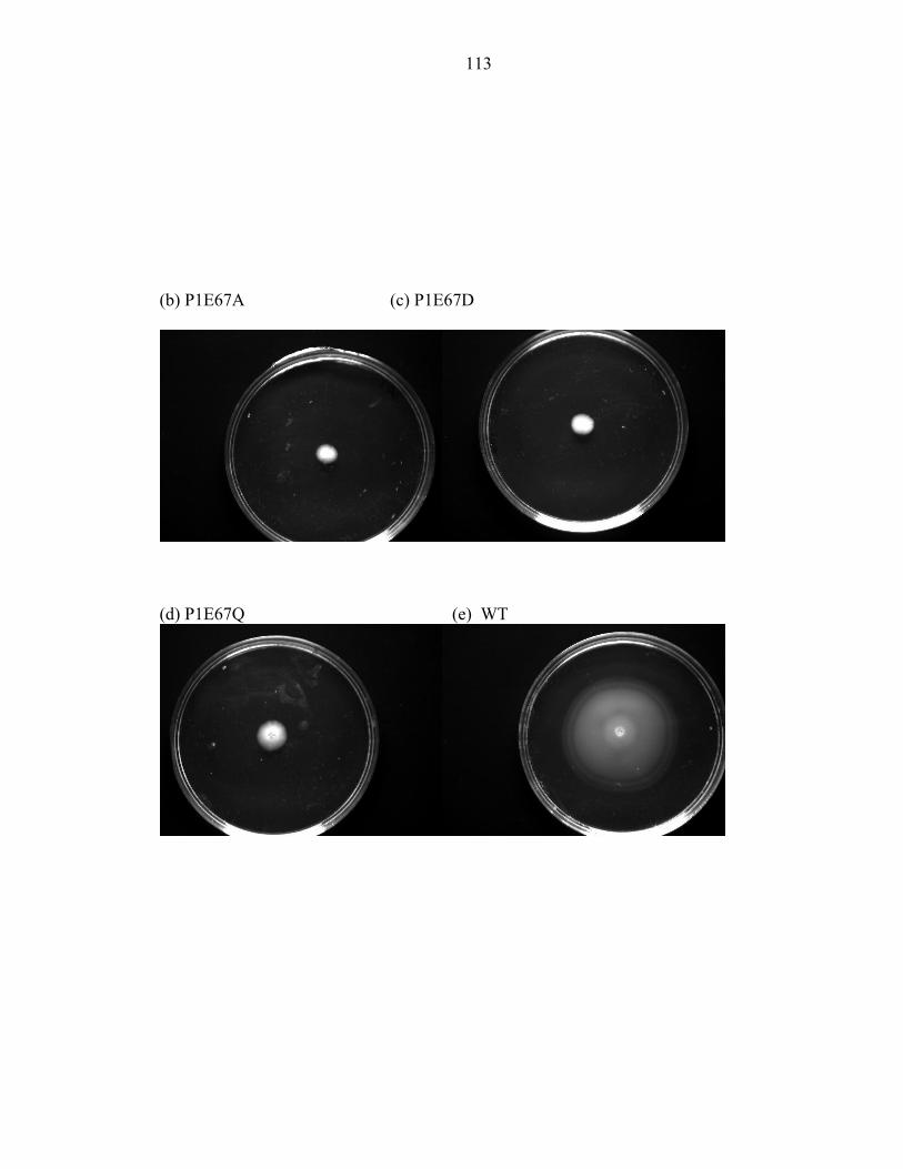
113
(b) P1E67A (c) P1E67D
(d) P1E67Q (e) WT
114
Figure 3: Phosphorylation of mutant proteins CheA E67Q, CheA E67D, and CheA E67A as a function of pH
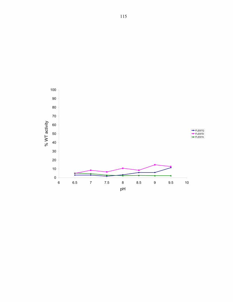
115
0
10
20
30
40
50
60
70
80
90
100
6 6.5 7 7.5 8 8.5 9 9.5 10
pH
% W
T ac
tivity
FLE67QFLE67DFLE67A
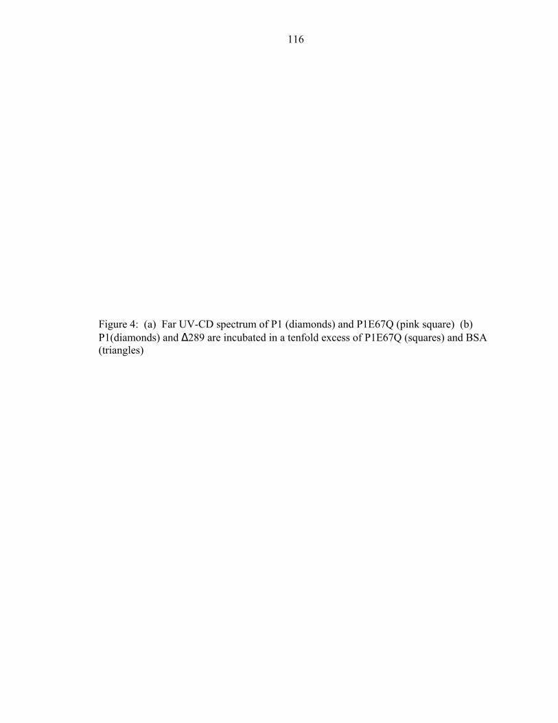
116
Figure 4: (a) Far UV-CD spectrum of P1 (diamonds) and P1E67Q (pink square) (b) P1(diamonds) and ∆289 are incubated in a tenfold excess of P1E67Q (squares) and BSA (triangles)
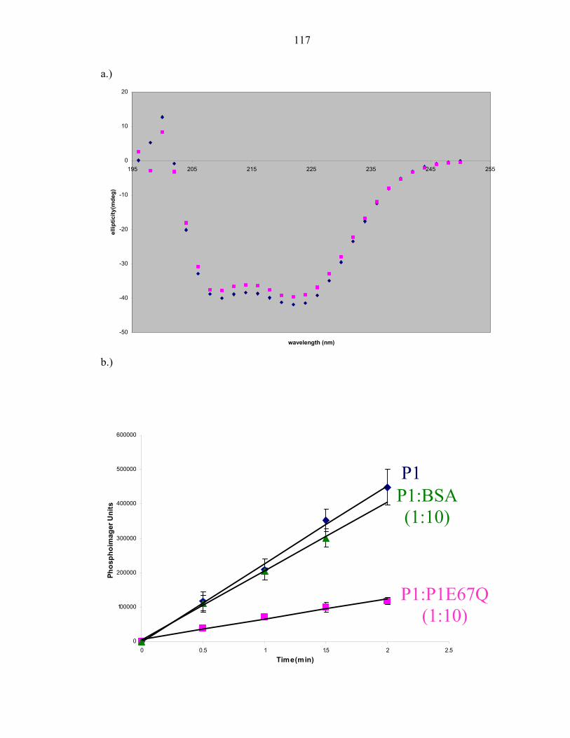
117
a.)
-50
-40
-30
-20
-10
0
10
20
195 205 215 225 235 245 255
wavelength (nm)
ellip
ticity
(mde
g)
b.)
0
100000
200000
300000
400000
500000
600000
0 0.5 1 1.5 2 2.5
Time(min)
Phos
phoi
mag
er U
nits
P1 P1:BSA (1:10)
P1:P1E67Q (1:10)
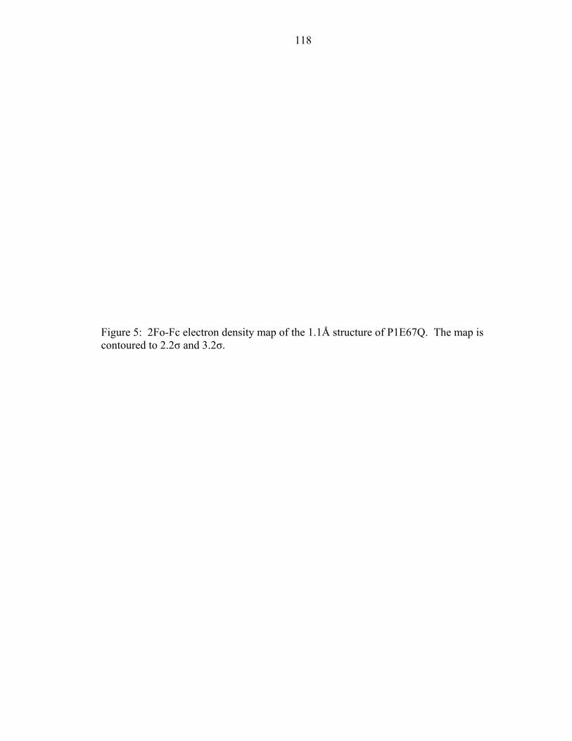
118
Figure 5: 2Fo-Fc electron density map of the 1.1Å structure of P1E67Q. The map is contoured to 2.2σ and 3.2σ.
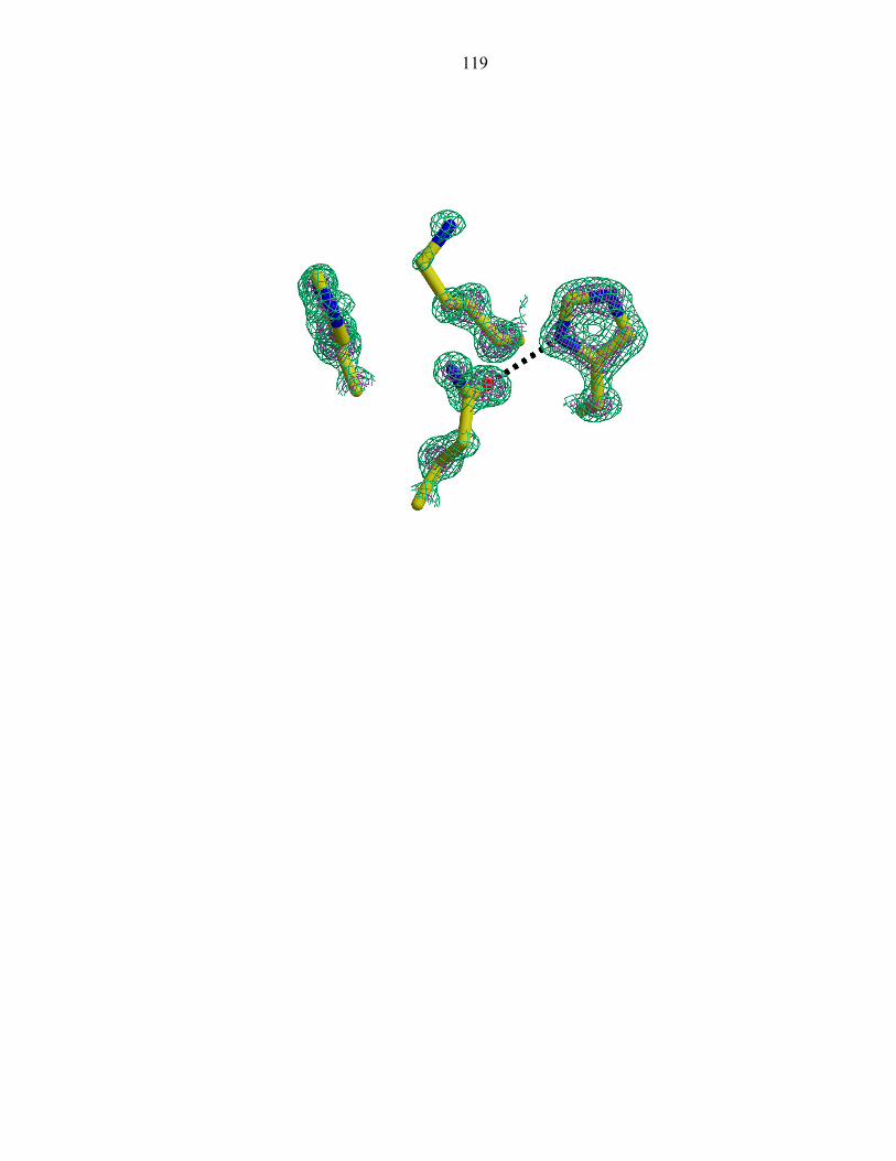
119
120
Figure 6: A conservative point mutation disrupts the native hydrogen bonding network of P1
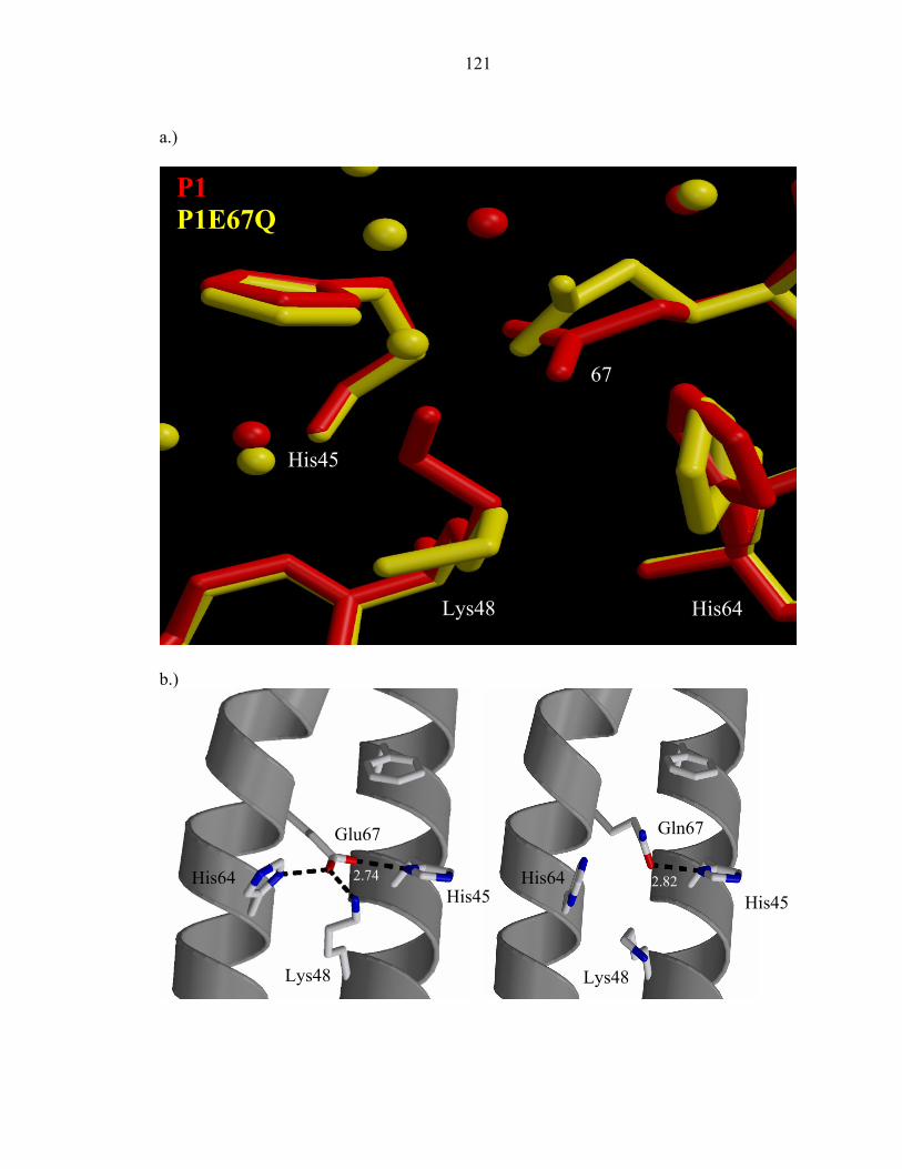
121
a.)
b.)
P1 P1E67Q
His45
67
Lys48 His64
His45
Lys48
Glu67
His64
Gln67
His45
Lys48
His642.74 2.82
122
Figure 7: 15N chemical shifts of P1E67Q (a) His45 (b) His64 and (c) HisTag
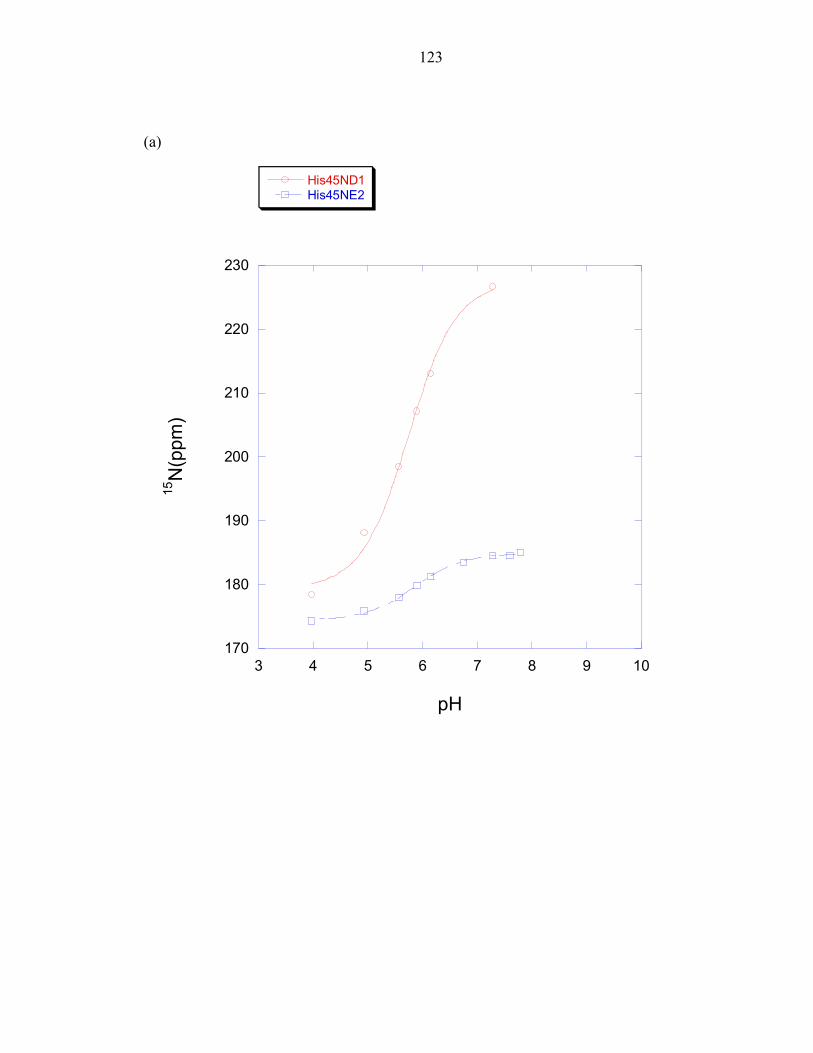
123
(a)
170
180
190
200
210
220
230
3 4 5 6 7 8 9 10
His45ND1His45NE2
15N
(ppm
)
pH
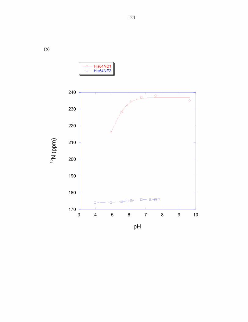
124
(b)
170
180
190
200
210
220
230
240
3 4 5 6 7 8 9 10
His64ND1His64NE2
15N
(ppm
)
pH
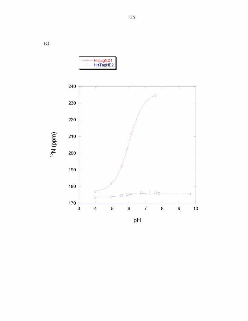
125
(c)
170
180
190
200
210
220
230
240
3 4 5 6 7 8 9 10
HistagND1HisTagNE2
15N
(ppm
)
pH
126
Figure 8: Critical residues to the CheA autophosphorylation reaction are located in the (a) P1 and (b) P4 domains.
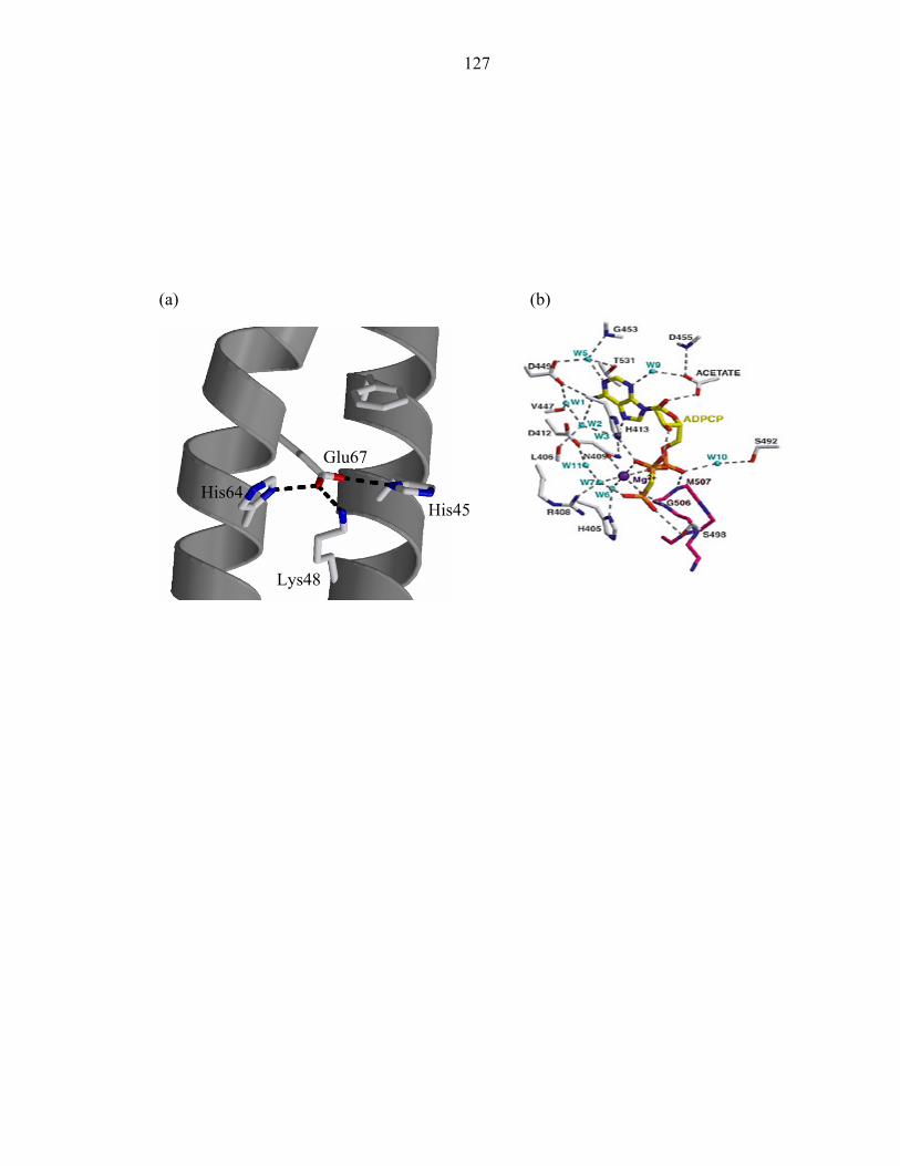
127
(a) (b)
His45
Lys48
Glu67
His64
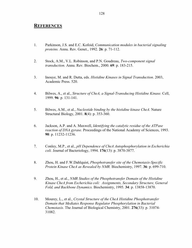
128
REFERENCES
1. Parkinson, J.S. and E.C. Kofoid, Communication modules in bacterial signaling
proteins. Annu. Rev. Genet., 1992. 26: p. 71-112.
2. Stock, A.M., V.L. Robinson, and P.N. Goudreau, Two-component signal transduction. Annu. Rev. Biochem., 2000. 69: p. 183-215.
3. Inouye, M. and R. Dutta, eds. Histidine Kinases in Signal Transduction. 2003, Academic Press. 520.
4. Bilwes, A., et al., Structure of CheA, a Signal-Transducing Histidine Kinase. Cell, 1999. 96: p. 131-141.
5. Bilwes, A.M., et al., Nucleotide binding by the histidine kinase CheA. Nature Structural Biology, 2001. 8(4): p. 353-360.
6. Jackson, A.P. and A. Maxwell, Identifying the catalytic residue of the ATPase reaction of DNA gyrase. Proceedings of the National Academy of Sciences, 1993. 90: p. 11232-11236.
7. Conley, M.P., et al., pH Dependence of CheA Autophosphorylation in Escherichia coli. Journal of Bacteriology, 1994. 176(13): p. 3870-3877.
8. Zhou, H. and F.W.Dahlquist, Phosphotransfer site of the Chemotaxis-Specific Protein Kinase CheA as Revealed by NMR. Biochemistry, 1997. 36: p. 699-710.
9. Zhou, H., et al., NMR Studies of the Phosphotransfer Domain of the Histidine Kinase CheA from Escherichia coli: Assignments, Secondary Structure, General Fold, and Backbone Dynamics. Biochemistry, 1995. 34: p. 13858-13870.
10. Mourey, L., et al., Crystal Structure of the CheA Histidine Phosphotransfer Domain that Mediates Response Regulator Phosphorylation in Bacterial Chemotaxis. The Journal of Biological Chemistry, 2001. 276(33): p. 31074-31082.
129
11. Otwinowski, A. and W. Minor, Processing of X-ray diffraction data in oscillation mode. Methods in Enzymolozy, 1997. 276: p. 307-325.
12. Perrakis, A., R. Morris, and V.S. Lamzin, Automated protein model building combined with iterative structure refinement. Nature Structural Biology, 1999. 6(5): p. 458-463.
13. Sheldrick, G.M. and T.R. Schneider, SHELXL:high resolution refinement. Methods in Enzymology, 1997. 277: p. 319-343.
14. McRee, D.E., XtalView: a visual protein crystallographic software system for X11/Xview. J. Mol. Graph., 1992. 10: p. 44-47.
15. Zhang, O.W., et al., Backbone H-1 and N-15 Resonance Assignments of the N-Terminal Sh3 Domain of Drk in Folded and Unfolded States Using Enhanced- Sensitivity Pulsed-Field Gradient Nmr Techniques. Journal of Biomolecular Nmr, 1994. 4(6): p. 845-858.
16. Marion, D., et al., Rapid Recording of 2d Nmr-Spectra without Phase Cycling - Application to the Study of Hydrogen-Exchange in Proteins. Journal of Magnetic Resonance, 1989. 85(2): p. 393-399.
17. Roberts, J.D., et al., A N-15 Nuclear Magnetic-Resonance Study of the Acid-Base and Tautomeric Equilibria of 4-Substituted Imidazoles and Its Relevance to the Catalytic Mechanism of Alpha-Lytic Protease. Journal of the American Chemical Society, 1982. 104(14): p. 3945-3949.
18. Munowitz, M., et al., Acid-Base and Tautomeric Equilibria in the Solid-State - N-15 Nmr-Spectroscopy of Histidine and Imidazole. Journal of the American Chemical Society, 1982. 104(5): p. 1192-1196.
19. Harbison, G., J. Herzfeld, and R.G. Griffin, N-15 Chemical-Shift Tensors in L-Histidine Hydrochloride Monohydrate. Journal of the American Chemical Society, 1981. 103(16): p. 4752-4754.
20. Blomberg, F., W. Maurer, and H. Ruterjans, Nuclear Magnetic-Resonance Investigation of N-15-Labeled Histidine in Aqueous-Solution. Journal of the American Chemical Society, 1977. 99(25): p. 8149-8159.
130
21. Witanowski, M., et al., Nitrogen-14 Nuclear Magnetic-Resonance of Azoles and Their Benzo Derivatives. Tetrahedron, 1972. 28(3): p. 637-653.
22. Pelton, J.G., et al., Tautomeric States of the Active-Site Histidines of Phosphorylated and Unphosphorylated Iii(Glc), a Signal- Transducing Protein from Escherichia-Coli, Using 2-Dimensional Heteronuclear Nmr Techniques. Protein Science, 1993. 2(4): p. 543-558.
23. Bachovchin, W.W., N-15 Nmr-Spectroscopy of Hydrogen-Bonding Interactions in the Active-Site of Serine Protease - Evidence for a Moving Histidine Mechanism. Biochemistry, 1986. 25(23): p. 7751-7759.
24. Schuster, II and J.D. Roberts, N-15 Nuclear Magnetic-Resonance Spectroscopy - Effects of Hydrogen-Bonding and Protonation on Nitrogen Chemical-Shifts in Imidazoles. Journal of Organic Chemistry, 1979. 44(22): p. 3864-3867.
25. Reynolds, W.F. and C.W. Tzeng, Determination of Preferred Tautomeric Form of Histamine by C-13 Nmr-Spectroscopy. Canadian Journal of Biochemistry, 1977. 55(5): p. 576-578.
26. Wolfe, A. and H. Berg, Migration of bacteria in semisolid agar. Proceedings of the National Academy of Sciences, 1989. 86: p. 6973-6977.
27. Ikegami, T., et al., Solution Structure and Dynamic Character of the Histidine-Containing Phosphotransfer Domain of Anaerobic Sensor Kinase ArcB from Escherichia coli. Biochemistry, 2001. 40: p. 375-386.
28. Xu, Q. and A.H. West, Conservation of Structure and Function Among Histidine-containing Phosphotransfer (HPt) Domains as Revealed by the Crystal Structure of YPD1. Journal of Molecular Biology, 1999. 292: p. 1039-1050.
29. McEvoy, M.M., et al., Nuclear Magnetic Resonance Assignments and Global Fold of a CheY-Binding Domain in CheA, the Chemotaxis-Specific Kinase of Escherichia coli. Biochemistry, 1995. 34: p. 13871-13880.
30. Tawa, P. and R.C. Stewart, Kinetics of Chea Autophosphorylation and Dephosphorylation Reactions. Biochemistry, 1994. 33(25): p. 7917-7924.
131
31. Swanson, R.V., S.C. Schuster, and M.I. Simon, Expression of Chea Fragments Which Define Domains Encoding Kinase, Phosphotransfer, and Chey Binding Activities. Biochemistry, 1993. 32(30): p. 7623-7629.
32. Levit, M., et al., Active site Interference and Asymmetric Activation in the Chemotaxis Protein Histidine Kinase CheA. The Journal of Biological Chemistry, 1996. 271(50): p. 32057-32063.
33. Tawa, P. and R.C. Stewart, Mutational Activation of Chea, the Protein-Kinase in the Chemotaxis System of Escherichia-Coli. Journal of Bacteriology, 1994. 176(14): p. 4210-4218.
34. Hirschman, A., et al., Active Site Mutations in CheA, the Signal-Transducing Protein Kinase of the Chemotaxis System in Escherichia coli. Biochemistry, 2001. 40: p. 13876-13887.
35. Morrison, T.B. and J.S. Parkinson, A fragment liberated from the Escherichia coli CheA kinase that blocks stimulatory, but not inhibitory, chemoreceptor signaling. Journal of Bacteriology, 1997. 179(17): p. 5543-5550.
36. Ellefson, D.D., U. Weber, and A.J. Wolfe, Genetic analysis of the catalytic domain of the chemotaxis- associated histidine kinase CheA. Journal of Bacteriology, 1997. 179(3): p. 825-830.
37. Bourret, R.B., J. Davagnino, and M.I. Simon, The Carboxy-Terminal Portion of the Chea Kinase Mediates Regulation of Autophosphorylation by Transducer and Chew. Journal of Bacteriology, 1993. 175(7): p. 2097-2101.
38. Kato, M., et al., Refined structure of the histidine-containing phosphotransfer (HPt) domain of the anaerobic sensor kinase ArcB from Escherichia coli at 1.57A resolution. Acta Crystallographica, 1999. D55: p. 1842-1849.
39. Kato, M., et al., Insights into Multistep Phosphorelay from the Crystal Structure of the C-Terminal HPt Domain of ArcB. Cell, 1997. 88: p. 717-723.
40. Jones, B.E., P. Rajagopal, and R.E. Klevit, Phosphorylation on histidine is accompanied by localized structural changes in the phosphocarrier protein, HPr from Bacillus subtilis. Protein Science, 1997. 6(10): p. 2107-2119.
132
41. vanNuland, N.A.J., et al., Phosphorylation-induced torsion-angle strain in the active center of HPr, detected by NMR and restrained molecular dynamics refinement. Protein Science, 1996. 5(3): p. 442-446.
42. Pullen, K., et al., Phosphorylation of Serine-46 in Hpr, a Key Regulatory Protein in Bacteria, Results in Stabilization of Its Solution Structure. Protein Science, 1995. 4(12): p. 2478-2486.
43. Vannuland, N.A.J., et al., High-Resolution Structure of the Phosphorylated Form of the Histidine-Containing Phosphocarrier Protein Hpr from Escherichia-Coli Determined by Restrained Molecular-Dynamics from Nmr-Noe Data. Journal of Molecular Biology, 1995. 246(1): p. 180-193.
44. Rajagopal, P., E.B. Waygood, and R.E. Klevit, Structural Consequences of Histidine Phosphorylation - Nmr Characterization of the Phosphohistidine Form of Histidine- Containing Protein from Bacillus-Subtilis and Escherichia-Coli. Biochemistry, 1994. 33(51): p. 15271-15282.
45. Pelton, J.G., et al., Structural Comparison of Phosphorylated and Unphosphorylated Forms of Iii(Glc), a Signal-Transducing Protein from Escherichia-Coli, Using 3-Dimensional Nmr Techniques. Biochemistry, 1992. 31(22): p. 5215-5224.
46. Matsushika, A. and T. Mizuno, Mutational analysis of the histidine-containing phosphotransfer (HPt) signaling domain of the ArcB sensor in Escherichia coli. Bioscience Biotechnology and Biochemistry, 1998. 62(11): p. 2236-2238.
47. Janiak-Spens, F. and A.H. West, Functional roles of conserved amino acid residues surrounding the phosphorylatable histidine of the yeast phosphorelay protein YPD1. Molecular Microbiology, 2000. 37(1): p. 136-144.
48. Sculimbrene, B.R., A.J. Morgan, and S.J. Miller, Enantiodivergence in small-molecule catalysis of asymmetric phosphorylation: Concise total syntheses of the enantiomeric D- myo-inositol-1-phosphate and D-myo-inositol-3-phosphate. Journal of the American Chemical Society, 2002. 124(39): p. 11653-11656.
49. Sculimbrene, B.R. and S.J. Miller, Discovery of a catalytic asymmetric phosphorylation through selection of a minimal kinase mimic: A concise total synthesis of D-myo-inositol-1-phosphate. Journal of the American Chemical Society, 2001. 123(41): p. 10125-10126.
133
50. Laskowski, R.A., et al., PROCHECK: a program to check the stereochemical quality of protein structures. J. Appl. Crystallogr., 1993. 26: p. 283-291.
134
Chapter 4
Mutations in the conserved hydrogen bonding network of the CheA
histidine phosphotransfer domain
135
In-depth structural and biochemical studies of the kinase domain have identified
amino acids involved in CheA autokinase activity and ATP binding [1, 2]. Since the
phosphotransfer domain P1 completes the CheA active site, we are interested in further
elucidating the functions of key residues in this domain. As an extension to previous
NMR [3] and crystallographic studies [4], our laboratory solved an atomic resolution
structure of the phosphotransfer domain from the T. maritima organism (Chapter 2). A
hydrogen bonding network between residues His45, Glu67, Lys48, and His64 was
identified. Since these residues are highly conserved in a domain that exhibits low
sequence similarity, it is of interest to evaluate their contributions to the CheA
autophosphorylation reaction.
Prior NMR studies revealed the phosphoaccepting histidine, His45, exists in the
normally unfavored Nδ1H tautomeric state and exhibits a pKa of 7.8 [5]. This value is
unusually high compared to a pKa of approximately 6.9 for a solvent exposed histidine at
room temperature [6]. These atypical chemical properties were attributed to the
formation of a hydrogen bond between the Nδ1H atom of His45 and an unknown residue.
Based on the structural and sequence similarities between CheA and the ATPase GyrB, a
conserved glutamate residue that is postulated to play a key role in the cleavage of the γ-
phosphoryl group in GyrB was hypothesized to be located in the P1 domain [2, 7].
Subsequent crystal structure analysis of the phosphotransfer domain confirmed the
existence of a hydrogen bond between the Nδ1H atom of His45 and Glu67 [4] (Chapter2).
Furthermore, a thorough characterization of the conserved Glu67 revealed that it
is not only critical to the CheA autophosphorylation reaction but also necessary for the
stabilization of the Nδ1H histidine tautomer and the preservation of an altered pKa
136
(Chapter 3). The conservative mutation, E67Q, promotes a change in the tautomeric state
of the phosphoaccepting histidine to the Nε2H species and lowers the pKa by
approximately one pH unit (Chapter 3 Figure 7a). These changes in the chemical
properties of the histidine residue result in dramatically reduced phosphorylation activity.
Hence, the active site histidine must exist in the correct tautomer in order for it to be
phosphorylated.
We continue to examine the structural, biochemical, and electronic consequences
of disrupting members of the conserved hydrogen bonding network. The native P1
structure shows that Glu67 additionally acts as a hydrogen bond acceptor to two other
conserved residues, Lys48 and His64. In this chapter, mutagenic and biophysical studies
within the context of mutant crystal structures are used to define the functional roles of
Lys48 and His64 in the catalytic mechanism of CheA.
MATERIALS AND METHODS
Cloning, expression, and purification
T. maritima CheA (residues 1-671), ∆289 (residues 290-671), domain P1
(residues 4-133), and a fragment of domain P1 termed P1short (residues 4-105) were
subcloned in the vector pET28(a) (Novagen). Alanine mutants of Lys48 and His64 were
generated using Quickchange mutagenesis (Stratagene). Proteins were expressed in
E.coli strain BL21(DE3) (Novagen) in 2L TB cultures and purified by affinity
chromatography on Nickel-NTA beads (Qiagen), followed by thrombin digestion and gel
filtration as described in the Materials and Methods section of Chapter 2.
137
Phosphorylation assays
Initial velocities of full length CheA, CheA K48A, CheA H64A, and CheA E67Q,
each at a protein concentration of 2 µM, were measured in 50 mM Tris pH 8.5, 50 mM
KCl, 5 mM MgCl2, and 2 mM DTT at 50°C. The initial velocities of full length CheA
and CheA H64A were also compared to one another at room temperature in the same
buffer at pH 7.5. Reactions were initiated upon addition of [γ-32P] ATP. The
phosphorylation assays were carried out in the same manner as described in the Materials
and Methods section of Chapter 3.
Swarm assays
Bacterial strain RP9535, lacking the CheA gene, was transformed with
pAR1:cheA derivatives expressing wild-type CheA, CheA K48A, or CheA H64A.
Tryptone swarm plates were stabbed with the transformants and incubated at 37ºC for 12
hours as described in the Materials and Methods section of Chapter 3.
Circular dichroism measurements
Far UV CD spectra were recorded with an AVIV (Lakewood, NJ) 62A DS
spectrometer equipped with a Peltier-type temperature control system and using a 0.1cm
quartz cuvette (Wilmad). P1K48A, P1H64A, and P1 were at a concentration of 20 µM in
20 mM sodium phosphate buffer at pH 8.0. Protein concentrations were determined by
UV spectrophotometry using extinction coefficients calculated from the number of
tryptophan and tyrosine residues present in each protein [8]. Data were collected
138
between 250 to 190nm using an averaging time of 5 seconds and a bandwidth of 1.5nm at
25ºC.
Determination of phosphohistidine stability
The autophosphorylation reaction of CheA and its mutants was initiated with [γ-
32P] ATP as described above. After adequate phosphorylation levels were achieved, the
reaction was quenched using 2X sodium dodecyl sulfate (SDS) electrophoresis buffer
containing 25 mM ethylenediaminetetra acetic acid (EDTA). The reaction tube was kept
at 50°C and aliquots were taken and placed at 4°C at designated time points. Samples
were then electrophoresed and processed as described in the Materials and Methods
section of Chapter 3.
Crystallization, data collection, and structure refinement
Orthorhombic crystals belonging to the P2221 space group of P1K48Ashort and
P1H64Ashort were obtained via the hanging drop method of crystallization. The protein
fragments used for crystallization were composed of residues 4 -104 of the CheA
histidine phosphotransfer domain. Crystals were generated by mixing 2 µL of 7-15
mg/ml protein with 2 µL of reservoir solution (32% PEG 4K, 0.1M NaAc pH 4.5, 0.2M
AmAc for P1K48Ashort; 30% PEG 4K, 0.1M NaAc pH 4.5, 0.2M AmAc for P1H64Ashort).
Diffraction data were collected on the A1 beamline at the Cornell High Energy
Synchrotron Source (CHESS) using a wavelength of 0.9363Å. P1K48Ashort and
P1H64Ashort crystals diffracted to 1.38Å and 1.4Å, respectively. Data were processed,
scaled, and reduced using the DENZO/SCALEPACK suite of programs [9]. The cell
139
dimensions of P1K48Ashort are x= 27.680Å, y= 37.627Å, and z=87.517Å. The cell
dimensions of P1H64Ashort are x=27.567Å, y=37.618Å, and z= 87.402Å. The structures
of each mutant were refined using SHELXL [10] and the model manually rebuilt in
XtalView [11] as described in the Materials and Methods section of Chapter 3. Details
of data collection and refinement can be found in Table 1.
General NMR methods and data analysis
Uniformly 15N labeled protein P1K48A and P1H64A was expressed and used to
collect NMR data on 500 and 600 MHz Varian UNITY and Inova spectrometers,
respectively. These instruments were equipped with 1H [13C/15N] pulsed-field gradient
probes. Assignment of specific histidine side chain resonances was done by comparison
to wild type values obtained elsewhere (Hamel et al. unpublished results). Procedures
and parameters used for 3D 15N-NOESY-HSQC, 2D HSQC-aliphatic and 2D HSQC-
aromatic correlation experiments over a pH range of 3.5 to 9.8 are described in the
Materials and Methods section of Chapter 3 [12, 13]. The analysis of the NMR data can
also be found there.
RESULTS
Mutations of residues involved in the hydrogen bond network inhibit phosphotransfer
Amino acid residues that participate in the conserved hydrogen bond network
were mutated. The activities of CheA, CheA K48A, CheA H64A, and CheA E67Q were
first compared in vitro at 50°C and pH 8.5. The activity of wild type CheA was too rapid
to be measured under these conditions. However, it was possible to obtain the relative
140
levels of mutant phosphorylation (Figure 1a). CheA H64A is approximately 15-fold
faster than K48A and roughly 270-fold faster than E67Q. At pH 7.5 and room
temperature, the initial velocity of wild type CheA is approximately 60-fold faster than
CheA H64A (Figure 1b). The chemotaxis ability of E. coli mutants CheA K48A and
CheA H64A were also assessed in vivo. Swarm assays show that the chemotaxis
signaling network cannot tolerate mutations of K48 and H64 (Figure 2).
To assess whether these mutations perturbed the secondary structure of the
phosphotransfer domain, we compared the far UV CD spectra of P1H64A and P1K48A
to the wild type P1. The results indicate that P1H64A and P1 have similar spectra
(Figure 3a). However, P1K48A has lost some secondary structure content (Figure 3b),
suggesting that this residue may play a role in stabilizing the helical conformation of the
P1 domain.
In order to determine whether despite the observed structural perturbance,
P1K48A could still recognize the CheA kinase domain, it was incubated in a 10-fold
excess in the presence of P1 and the catalytic fragment ∆289 at room temperature, where
its activity is minimal. P1K48A is indeed capable of inhibiting the phosphorylation of
wild type P1 by ∆289. The wild type reaction is inhibited by over 50% in the presence of
P1K48A (Figure 4).
To further explore putative roles for residues involved in the hydrogen bonding
network, their involvement in stabilizing the phosphohistidine complex was investigated.
Phospho-labeled CheA and mutants were generated by incubation with [γ-32P] ATP. The
reaction was quenched and the phosphorylation level monitored over a period of three
hours. The stability of the phosphoramidate bond is not affected by mutations at Lys48,
141
His64, or Glu67 over this time frame (Figure 5). It is therefore unlikely that these
residues directly interact with the phosphate.
Lys48 and His64 contribute to the structural integrity of the active site region.
In order to fully characterize P1K48A and P1H64A, a structural analysis of these
mutants was performed. Here we report the crystal structures of P1K48Ashort and
P1H64Ashort. The quality of the data and refinement are summarized in Tables 1 and 2.
Neither of these mutations disrupt the general four-helix bundle fold observed in the wild
type P1 [3, 4] (Chapter 2 Figure 3). However, as described below, the area surrounding
the phosphoaccepting histidine is structurally altered.
The replacement of Lys48 with an alanine disrupts the positions of two other
members of the hydrogen bonding network, Glu67 and His64 (Figure 6a). The
discontinuous electron density for these side chains reflects prevalent disorder. The
conformation of Glu67 that we were able to model revealed that the side chain orientation
has swiveled. However, it is still capable of accepting a hydrogen bond from His45. The
structural properties of His64 are also affected by the K48A mutation. His64 exists in at
least two alternate conformations, and no longer hydrogen bonds to Glu67. Lys48
therefore appears to fix the orientation of Glu67 and His64. An overlay of the
P1K48Ashort and native P1 structure surprisingly revealed that the position of the
phosphoaccepting histidine, His45, remains the same (Figure 6b).
The structural changes incurred by the H64A mutation are not as disruptive as
those observed for K48A (Figure 7a). The P1H64Ashort structure revealed that Glu67 is
hydrogen bonded to the Nδ1 atom of His45, but exists in a slightly different position.
142
However, substitution of the His64 side chain for an alanine results in a broken salt
bridge between Lys48 and Glu67 due to displacement of the Lys48 side chain. The
orientation of His45 oncemore remains unchanged (Figure 7b).
Chemical properties of the phosphoaccepting histidine in the mutants K48A and H64A
Alanine substitutions for Lys48 and His64 both diminish the autophosphorylation
of CheA; but do they affect the reactivity of the phosphoaccepting histidine as previously
documented for Glu67 (Chapter 3). NMR spectroscopy is ideally suited to study the
electronic properties of histidine residues. To further explore the functional roles of
Lys48 and His64, the HSQC spectra [14] of 15N labeled proteins at several pH values
were evaluated in the mutants P1K48A and P1H64A. Data points obtained from the
histidine side chains were plotted as a function of pH and fit to equation 1 (Chapter 3)
using least squares fitting. Relevant chemical shifts are listed in Table 2.
The titration curves for the Nε2 and Nδ1 resonances of the P1K48A His45 side
chain are shown in Figure 8. The pKa value for the imidazole ring was determined to be
6.8. The larger shift of the Nε2 atom at high pH reveals that the Nδ1H tautomer is
dominant. This tautomer is stabilized through the hydrogen bond to Glu67. In fact, the
Nδ1:Nε2 tautomeric ratio is 2.6:1, as observed for the native protein (Table 4) (Hamel, et
al. unpublished results)[5].
Identical NMR experiments were performed on the P1H64A mutant. The
resonances of the His45 imidazole side chain also demonstrate a dependence on pH
(Figure 9). The phosphoaccepting histidine exhibits similar properties to the wild type.
It possesses an altered pKa of 6.8 and its chemical shifts at high pH reveal the existence
143
of a predominantly Nδ1H tautomeric state. The Nδ1H:Nε2H ratio was calculated to be
2.6:1. The tautomeric and protonation states of each histidine is listed in Table 3 and
compared to wild type protein and a previously characterized mutant P1E67Q (Chapter
3). The titration curves, chemical shifts, and tautomeric states of the remaining histidines
in P1 can be found in Appendix 1.
DISCUSSION
The work presented in this paper has further elucidated the roles of conserved
residues in the CheA phosphotransfer domain. By implementing mutagenesis studies,
macromolecular protein crystallography, and two-dimensional NMR techniques, we have
further investigated the functional roles of conserved residues in the phosphotransfer
domain. The effects mutations at Lys48 and His64 exert on the structure of the P1
domain and on the chemical properties of the phosphoaccepting histidine are examined.
An intact hydrogen bond network optimizes the histidine phosphotransfer reaction
Mutagenic analysis of His64 revealed that it is the least disruptive of the mutants
studied (Figure 1). Its secondary structure, as revealed by the far UV CD spectrum,
remains intact (Figure 3a). The 1.4Å crystal structure demonstrated that the only major
difference between the native and mutant structure is the position of the Lys48 side chain.
Both the phosphoaccepting histidine, His45, and the activating Glu67 maintain their side
chain orientation. 2-D NMR studies confirmed that the reactivity of His45 is unaltered.
The phosphorylatable histidine exists in the normally unfavored Nδ1H tautomeric state
144
possessing a Nδ1H:Nε2H ratio that is very similar to that of the wild type (Table 3).
Additionally, this mutation does not affect the stability of the phospho-histidine complex
(Figure 5). Hence, the observed decrease in phosphorylation activity of the H64A mutant
can be likely attributed to a decrease in binding affinity to the kinase domain.
The active site residues may form part of the binding interface to the kinase domain
Lys48 appears to play a critical role in stabilizing the active site structure. As
demonstrated by CD and crystallography, Lys48 disrupts the secondary structure of the
phosphotransfer domain (Figures 3b and 6). It is located towards the C-terminus of helix
B and may play a role in stabilizing the negative charge of the helix dipole, thereby
stabilizing the P1 helical structure. Its removal results in residue disorder, as shown by
the alternate conformations of His64 and the discontinuous density for the His64 and
Glu67 side chains.
Despite this disorder in the active site region, the phosphoaccepting histidine
largely retains its original side chain orientation, and most importantly, its reactivity
(Figures 6 and 8). It possesses the same properties as the wild type phosphoaccepting
histidine, exhibiting an altered pKa of 6.8, and favoring the Nδ1H tautomeric state.
Furthermore, it is not involved in stabilizing the tautomeric state of the phosphorylated
histidine. Together, these results imply that Lys48 does not contribute towards the kcat of
the autophosphorylation reaction. Instead, it maintains the structural integrity of the
immediate region surrounding His45 and is perhaps also key to forming the binding
interface to the kinase domain P4.
145
Comparative studies of residues participating in the conserved hydrogen bond network
By exploiting the structural and chemical information obtained in these
experiments, we are better able to interpret the observed differences in phosphorylation
activity between the P1 mutants K48A, H64A, and E67Q. Although the structures of
H64A and E67Q revealed disruption to the native hydrogen bond network, both
structures only exhibited minor perturbations to the side chain orientations of residues
(Figure 7 and Chapter 3 Figure 6). In both structures, the Lys48 side chain swings out of
hydrogen bonding distance from Glu67.
Despite minimal structural perturbations, the phosphorylation of the P1H64A
mutant is approximately 270-fold faster than that of P1E67Q. The major differences
between these two mutants are the tautomeric state of the phosphoaccepting histidine and
the pKa value. As in the wild type P1 domain, His45 of the P1H64A mutant exists in the
Nδ1H tautomeric state and has a pKa of 6.9 at 50°C. The correct orientation of the Lys48
side chain hence contributes to optimal phosphotransfer, perhaps by being an important
residue involved in binding the kinase domain. This infers that although P1E67Q is
largely a kcat mutant, its diminished phosphorylation could also result from some loss of
binding affinity to P4 due to the altered position of Lys48. Further support for this
hypothesis is obtained by comparing the results of competition studies between wild type
P1 and the mutants P1K48A and P1E67Q. P1E67Q acts as a better inhibitor of the P1
phosphorylation reaction than P1K48A (Chapter 3 Figure 4b and Figure 4).
Although the mutation of Lys48 led to the most significant disruption of
secondary structure, it was still more amenable to phosphorylation than E67Q (Figure
1a). Neither P1K48A nor P1E67Q stabilize the phosphorylated histidine. Moreover,
146
they do not affect the phosphotransfer activity to the response regulator CheY [4].
P1K48A shares the same chemical properties of the wild type phosphoaccepting
histidine, meaning the chemical reactivity of His45 is not altered by the disorder in the
neighboring environment. Removal of the positive charge likely affects the binding
interaction; therefore in addition to stabilizing the helical structure, a positive charge at
this precise location may be important for molecular recognition of the kinase domain P4.
Together, these results imply that among the studied mutations of the P1
hydrogen bonding network, E67Q imparts the strongest chemical perturbation and K48A
the greatest structural perturbation to the CheA autophosphorylation reaction. Although
much work remains to be done towards elucidating the CheA mechanism, an emerging
picture is one where the phosphotransfer domain contains a catalytic dyad, a resonating
Glu67 which activates a nucleophilic His45. The active site may be completed by a third
residue, His405, in the kinase domain. A mutation to this residue was found to affect the
kcat of the CheA phosphorylation reaction, but not the Km for ATP [1]. We are slowly
beginning to better understand the structural and chemical determinants of the CheA
autophosphorylation reaction. Further structure-function studies will elucidate the
mechanistic basis for the initiation of the His-Asp phosphorelay in bacterial chemotaxis.
147
Table 1: Data collection and refinement statistics
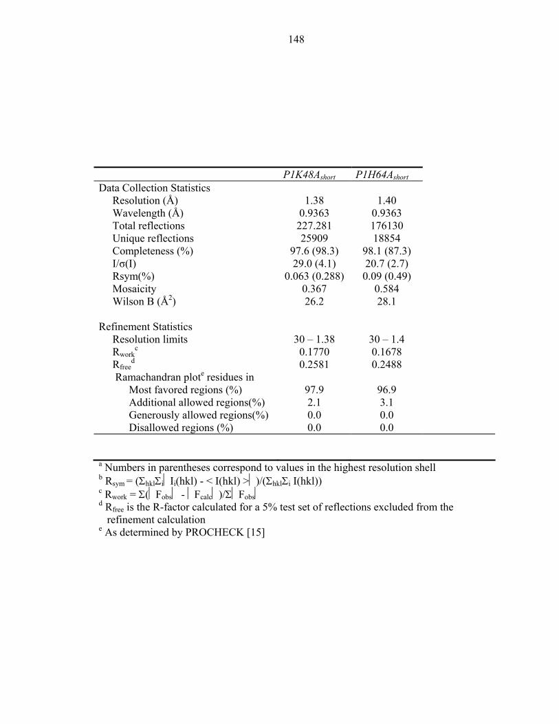
148
P1K48Ashort P1H64Ashort Data Collection Statistics Resolution (Å) 1.38 1.40 Wavelength (Å) 0.9363 0.9363 Total reflections 227.281 176130 Unique reflections 25909 18854 Completeness (%) 97.6 (98.3) 98.1 (87.3) I/σ(I) 29.0 (4.1) 20.7 (2.7) Rsym(%) 0.063 (0.288) 0.09 (0.49) Mosaicity 0.367 0.584 Wilson B (Å2) 26.2 28.1 Refinement Statistics Resolution limits 30 – 1.38 30 – 1.4 Rwork
c 0.1770 0.1678 Rfree
d 0.2581 0.2488 Ramachandran plote residues in Most favored regions (%) 97.9 96.9 Additional allowed regions(%) 2.1 3.1 Generously allowed regions(%) 0.0 0.0 Disallowed regions (%) 0.0 0.0
a Numbers in parentheses correspond to values in the highest resolution shell b Rsym = (ΣhklΣiIi(hkl) - < I(hkl) >)/(ΣhklΣi I(hkl)) c Rwork = Σ(Fobs - Fcalc)/ΣFobs d Rfree is the R-factor calculated for a 5% test set of reflections excluded from the
refinement calculation e As determined by PROCHECK [15]

149
Table 2: Expected and observed 15N chemical shifts for the His45 residue of P1K48A
and P1H64A
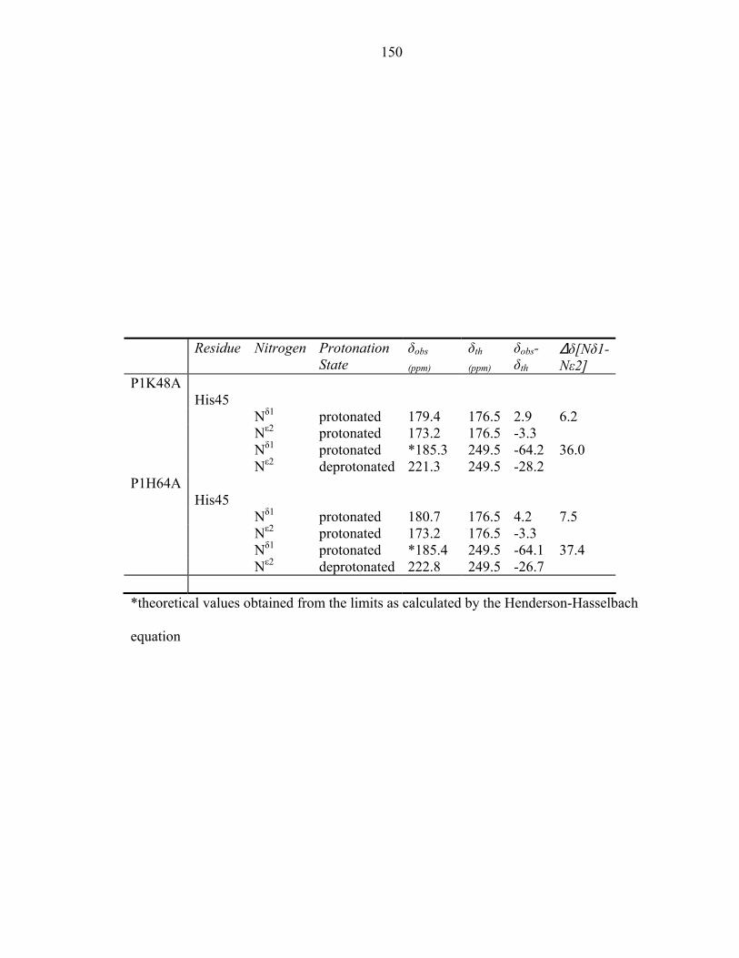
150
Residue Nitrogen Protonation
State δobs
(ppm) δth
(ppm) δobs- δth
∆δ[Nδ1- Nε2]
P1K48A His45 Nδ1 protonated 179.4 176.5 2.9 6.2 Nε2 protonated 173.2 176.5 -3.3 Nδ1 protonated *185.3 249.5 -64.2 36.0 Nε2 deprotonated 221.3 249.5 -28.2 P1H64A His45 Nδ1 protonated 180.7 176.5 4.2 7.5 Nε2 protonated 173.2 176.5 -3.3 Nδ1 protonated *185.4 249.5 -64.1 37.4 Nε2 deprotonated 222.8 249.5 -26.7 *theoretical values obtained from the limits as calculated by the Henderson-Hasselbach
equation
151
Table 3: The pKa values and predominant tautomeric states of His45 in CheA and its
variants
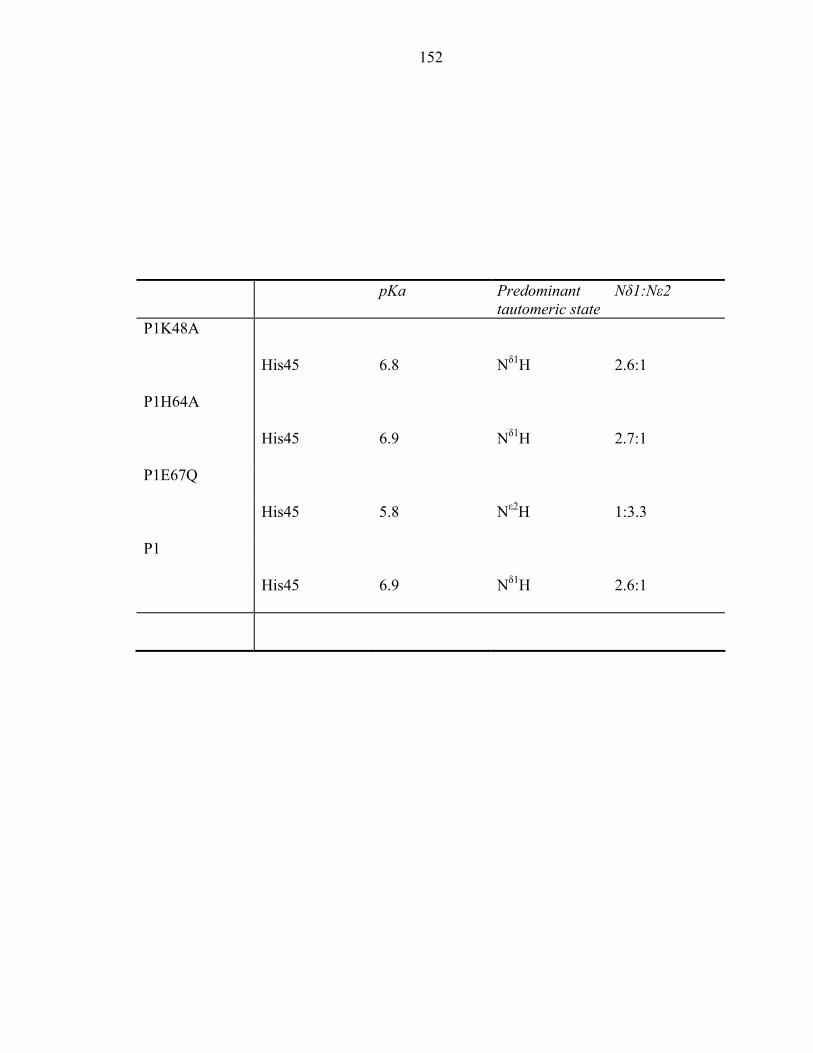
152
pKa Predominant tautomeric state
Nδ1:Nε2
P1K48A
His45 6.8 Nδ1H 2.6:1
P1H64A
His45 6.9 Nδ1H 2.7:1
P1E67Q
His45 5.8 Nε2H 1:3.3
P1
His45 6.9 Nδ1H 2.6:1
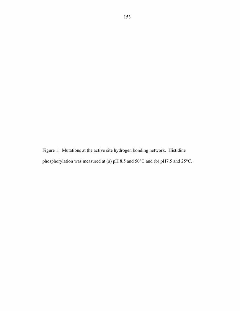
153
Figure 1: Mutations at the active site hydrogen bonding network. Histidine
phosphorylation was measured at (a) pH 8.5 and 50°C and (b) pH7.5 and 25°C.
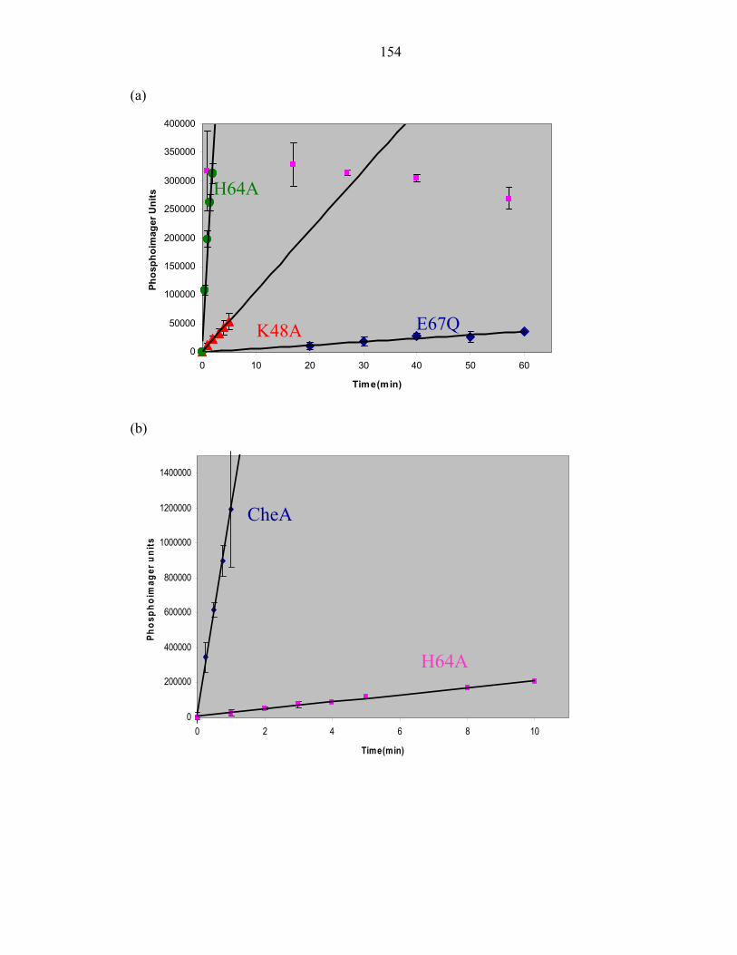
154
(a)
(b)
0
50000
100000
150000
200000
250000
300000
350000
400000
0 10 20 30 40 50 60
Time(min)
Phos
phoi
mag
er U
nits H64A
K48A E67Q
0
200000
400000
600000
800000
1000000
1200000
1400000
0 2 4 6 8 10
Time(min)
Phos
phoi
mag
er u
nits
CheA
H64A
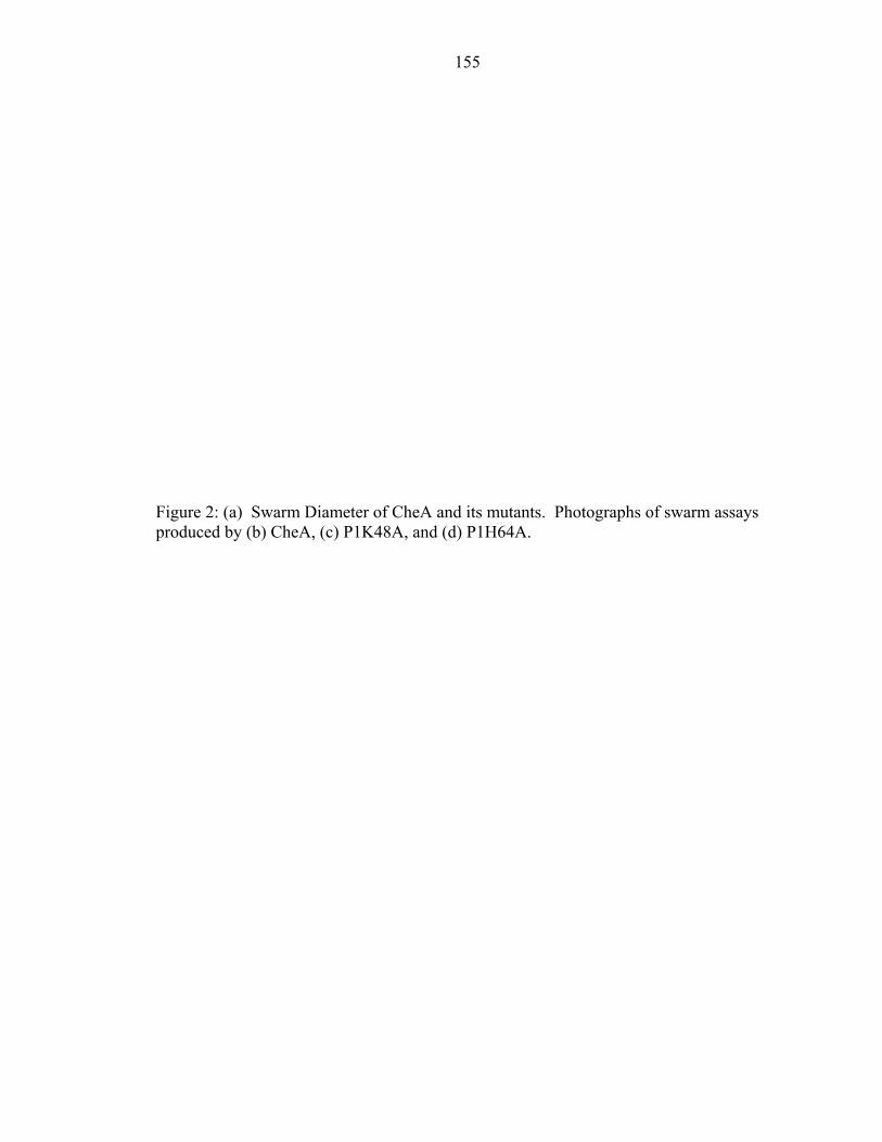
155
Figure 2: (a) Swarm Diameter of CheA and its mutants. Photographs of swarm assays produced by (b) CheA, (c) P1K48A, and (d) P1H64A.
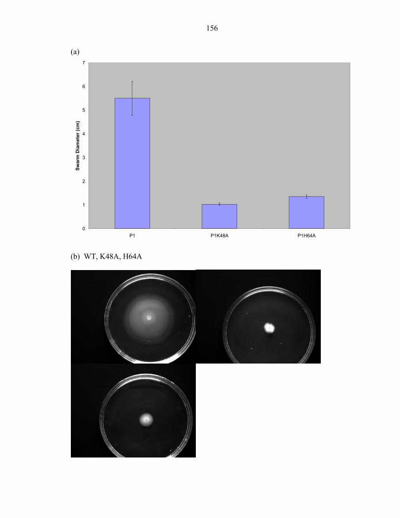
156
(a)
0
1
2
3
4
5
6
7
P1 P1K48A P1H64A
Swar
m D
iam
eter
(cm
)
(b) WT, K48A, H64A
157
Figure 3: Far-UV CD spectra of (a) P1H64A and P1; (b) P1K48A and P1
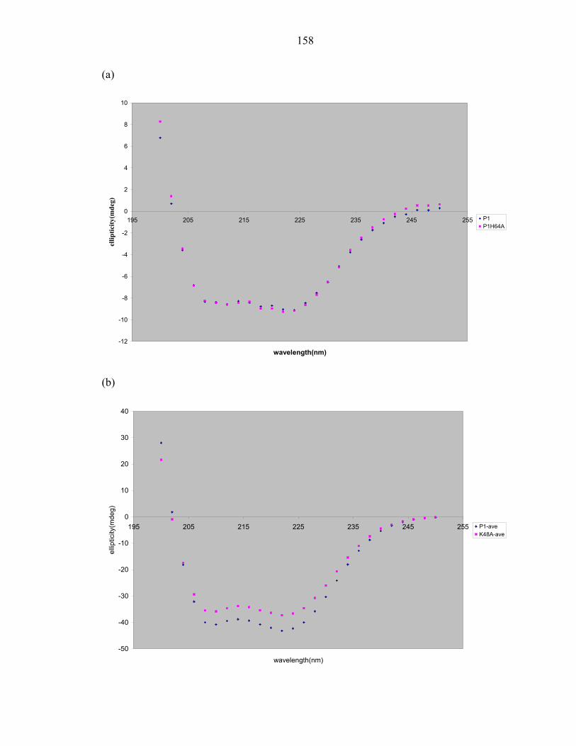
158
(a)
-12
-10
-8
-6
-4
-2
0
2
4
6
8
10
195 205 215 225 235 245 255
wavelength(nm)
ellip
ticity
(mde
g)
P1P1H64A
(b)
-50
-40
-30
-20
-10
0
10
20
30
40
195 205 215 225 235 245 255
wavelength(nm)
ellip
ticity
(mde
g)
P1-aveK48A-ave
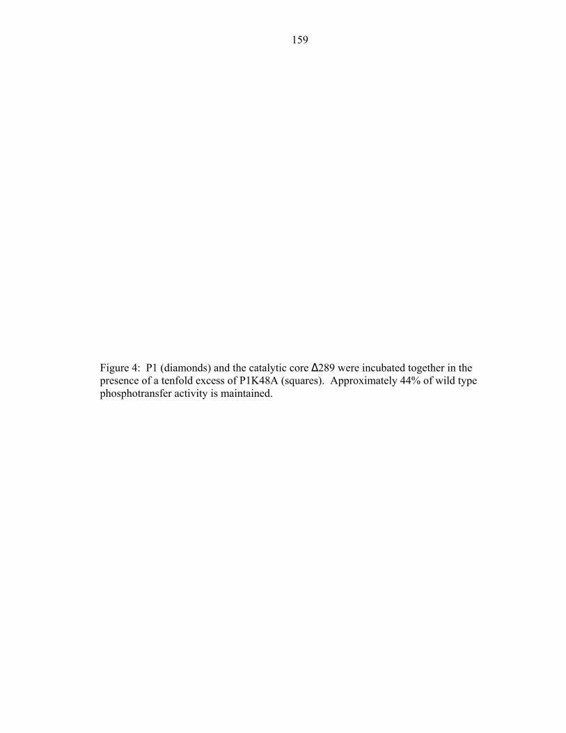
159
Figure 4: P1 (diamonds) and the catalytic core ∆289 were incubated together in the presence of a tenfold excess of P1K48A (squares). Approximately 44% of wild type phosphotransfer activity is maintained.
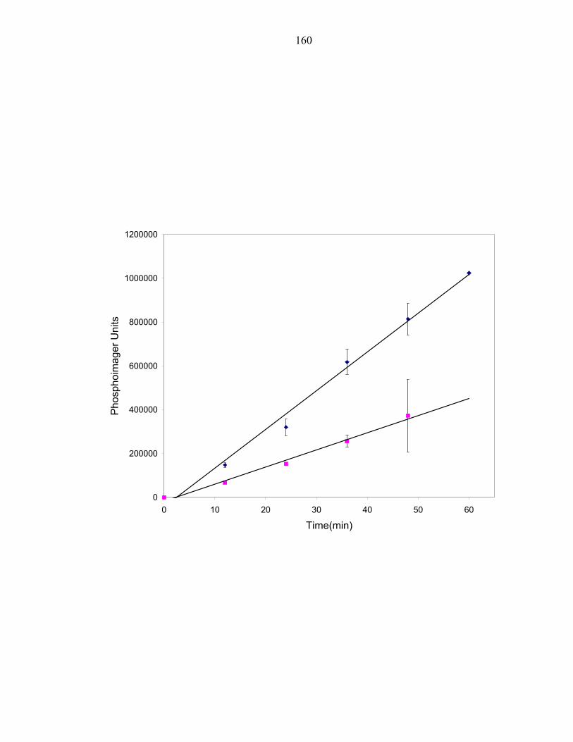
160
0
200000
400000
600000
800000
1000000
1200000
0 10 20 30 40 50 60
Time(min)
Pho
spho
imag
er U
nits
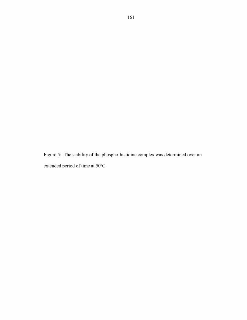
161
Figure 5: The stability of the phospho-histidine complex was determined over an
extended period of time at 50ºC
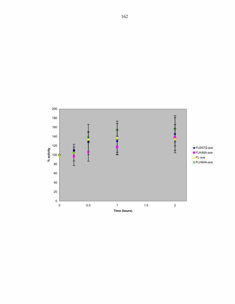
162
0
20
40
60
80
100
120
140
160
180
200
0 0.5 1 1.5 2
Time (hours)
% a
ctiv
ity
FLE67Q-aveFLK48A-aveFL-aveFLH64A-ave
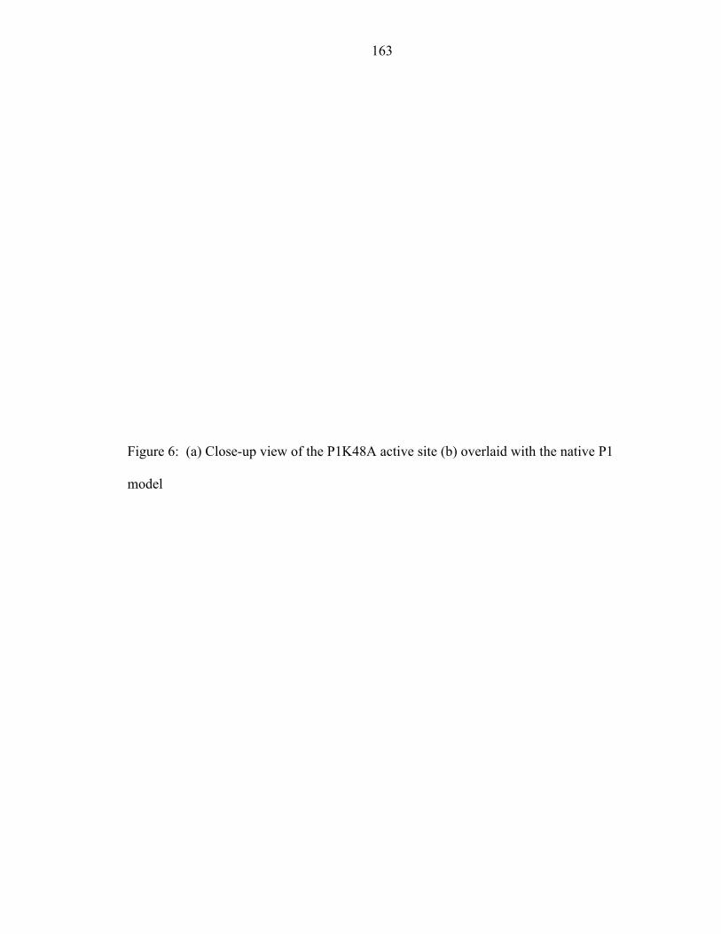
163
Figure 6: (a) Close-up view of the P1K48A active site (b) overlaid with the native P1
model
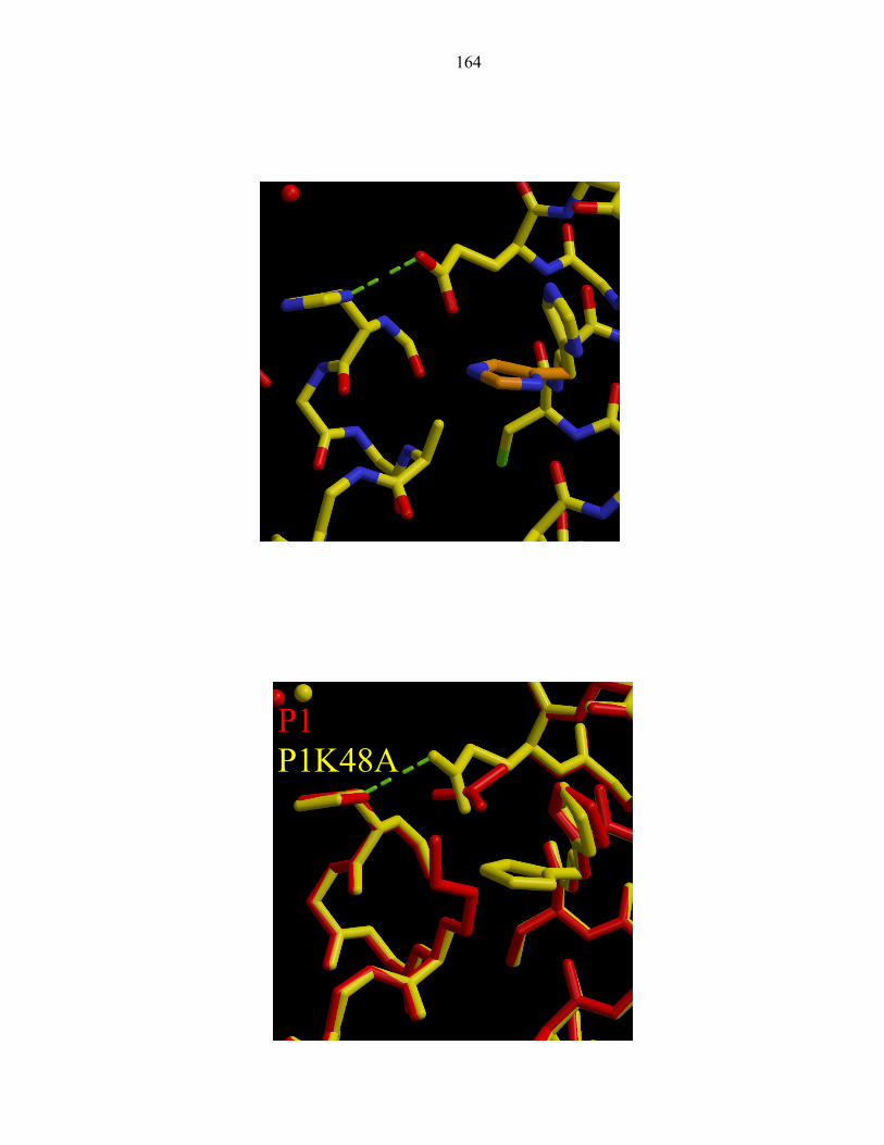
164
P1 P1K48A
165
Figure 7: (a.) Close up view of the P1H64A active site (b.) overlaid with the native P1
model
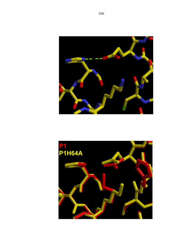
166
P1 P1H64A
167
Figure 8: Chemical shifts of the His45 imidazole 15N nuclei of P1K48A as a function of
pH. The Nε2 atom is blue and the Nδ1 is red.
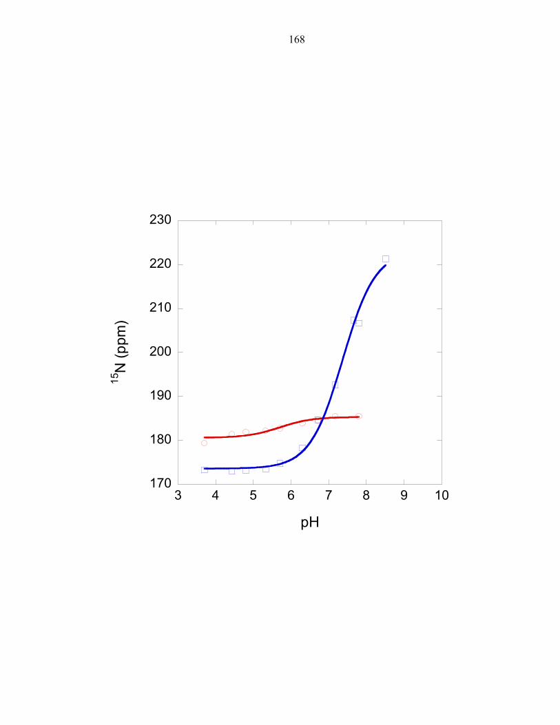
168
170
180
190
200
210
220
230
3 4 5 6 7 8 9 10
15N
(ppm
)
pH

169
Figure 9: Chemical shifts of the imidazole 15N nuclei of P1H64A His45 as a function of
pH. The Nε2 atom is blue and the Nδ1 is red.
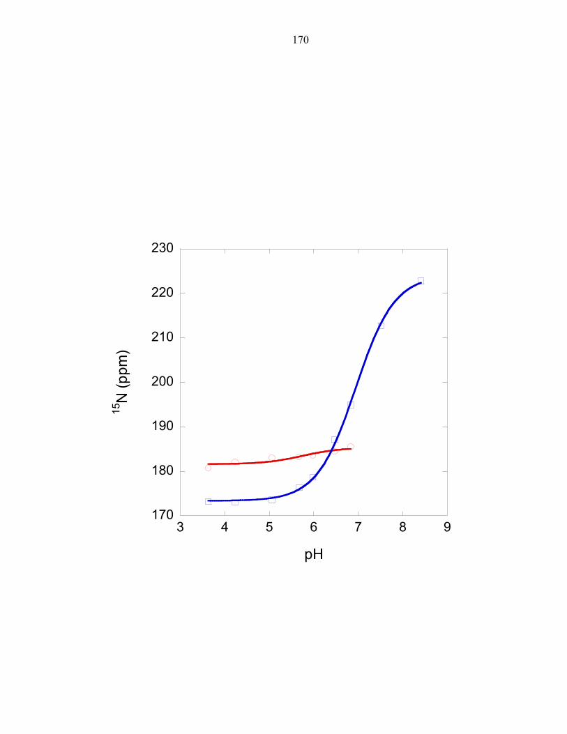
170
170
180
190
200
210
220
230
3 4 5 6 7 8 9
15N
(ppm
)
pH
171
REFERENCES
1. Hirschman, A., et al., Active Site Mutations in CheA, the Signal-Transducing Protein Kinase of the Chemotaxis System in Escherichia coli. Biochemistry, 2001. 40: p. 13876-13887.
2. Bilwes, A.M., et al., Nucleotide binding by the histidine kinase CheA. Nature Structural Biology, 2001. 8(4): p. 353-360.
3. Zhou, H., et al., NMR Studies of the Phosphotransfer Domain of the Histidine Kinase CheA from Escherichia coli: Assignments, Secondary Structure, General Fold, and Backbone Dynamics. Biochemistry, 1995. 34: p. 13858-13870.
4. Mourey, L., et al., Crystal Structure of the CheA Histidine Phosphotransfer Domain that Mediates Response Regulator Phosphorylation in Bacterial Chemotaxis. The Journal of Biological Chemistry, 2001. 276(33): p. 31074-31082.
5. Zhou, H. and F.W.Dahlquist, Phosphotransfer site of the Chemotaxis-Specific Protein Kinase CheA as Revealed by NMR. Biochemistry, 1997. 36: p. 699-710.
6. Markley, J.L., Observation of Histidine Residues in Proteins by Means of Nuclear Magnetic-Resonance Spectroscopy. Accounts of Chemical Research, 1975. 8(2): p. 70-80.
7. Bilwes, A., et al., Structure of CheA, a Signal-Transducing Histidine Kinase. Cell, 1999. 96: p. 131-141.
8. Gill, S.C. and P.H. Vonhippel, Calculation of Protein Extinction Coefficients from Amino-Acid Sequence Data. Analytical Biochemistry, 1989. 182(2): p. 319-326.
9. Otwinowski, A. and W. Minor, Processing of X-ray diffraction data in oscillation mode. Methods in Enzymolozy, 1997. 276: p. 307-325.
10. Sheldrick, G.M. and T.R. Schneider, SHELXL:high resolution refinement. Methods in Enzymology, 1997. 277: p. 319-343.
172
11. McRee, D.E., XtalView: a visual protein crystallographic software system for X11/Xview. J. Mol. Graph., 1992. 10: p. 44-47.
12. Zhang, O.W., et al., Backbone H-1 and N-15 Resonance Assignments of the N-Terminal Sh3 Domain of Drk in Folded and Unfolded States Using Enhanced- Sensitivity Pulsed-Field Gradient Nmr Techniques. Journal of Biomolecular Nmr, 1994. 4(6): p. 845-858.
13. Marion, D., et al., Rapid Recording of 2d Nmr-Spectra without Phase Cycling - Application to the Study of Hydrogen-Exchange in Proteins. Journal of Magnetic Resonance, 1989. 85(2): p. 393-399.
14. Ikegami, T., et al., Solution Structure and Dynamic Character of the Histidine-Containing Phosphotransfer Domain of Anaerobic Sensor Kinase ArcB from Escherichia coli. Biochemistry, 2001. 40: p. 375-386.
15. Laskowski, R.A., et al., PROCHECK: a program to check the stereochemical quality of protein structures. J. Appl. Crystallogr., 1993. 26: p. 283-291.
173
Appendix 1
1H and 15N Chemical shifts of P1 histidine residues
174
Table 1: Expected and observed imidazole 15N chemical shifts for P1K48A and P1H64A
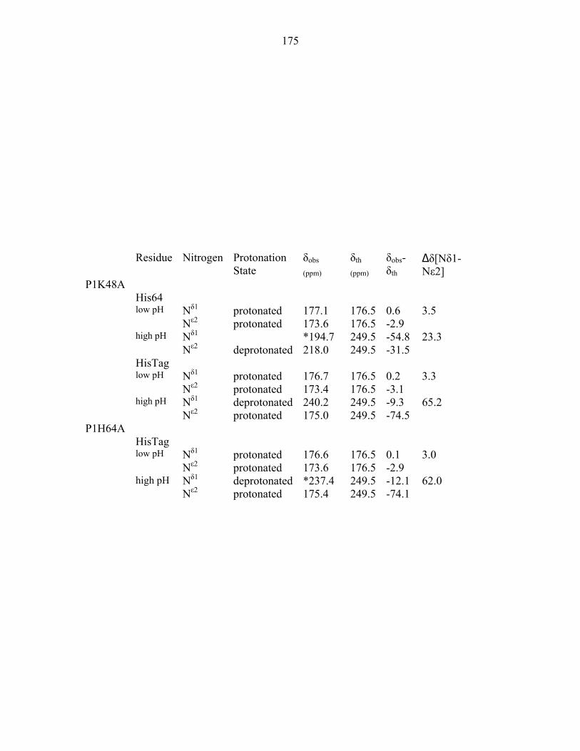
175
Residue Nitrogen Protonation
State δobs
(ppm) δth
(ppm) δobs- δth
∆δ[Nδ1- Nε2]
P1K48A His64 Nδ1 protonated 177.1 176.5 0.6 3.5
low pH Nε2 protonated 173.6 176.5 -2.9
Nδ1 *194.7 249.5 -54.8 23.3
high pH Nε2 deprotonated 218.0 249.5 -31.5
HisTag Nδ1 protonated 176.7 176.5 0.2 3.3
low pH Nε2 protonated 173.4 176.5 -3.1
Nδ1 deprotonated 240.2 249.5 -9.3 65.2
high pH Nε2 protonated 175.0 249.5 -74.5
P1H64A HisTag Nδ1 protonated 176.6 176.5 0.1 3.0
low pH Nε2 protonated 173.6 176.5 -2.9
Nδ1 deprotonated *237.4 249.5 -12.1 62.0
high pH Nε2 protonated 175.4 249.5 -74.1
176
Table 2: The tautomeric states and pKas of histidine residues found in the CheA P1 domain
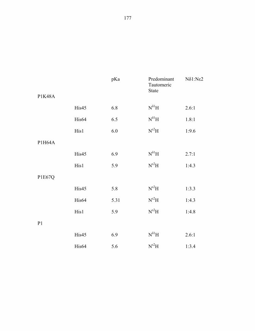
177
pKa Predominant
Tautomeric State
Nδ1:Nε2
P1K48A
His45 6.8 Nδ1H 2.6:1
His64 6.5 Nδ1H 1.8:1
His1 6.0 Nε2H 1:9.6
P1H64A
His45 6.9 Nδ1H 2.7:1
His1 5.9 Nε2H 1:4.3
P1E67Q
His45 5.8 Nε2H 1:3.3
His64 5.31 Nε2H 1:4.3
His1 5.9 Nε2H 1:4.8
P1
His45 6.9 Nδ1H 2.6:1
His64 5.6 Nε2H 1:3.4
178
Figure 1: 1H chemical shifts for the histidines of P1E67Q (a) His45 (b) His64 (c) HisTag
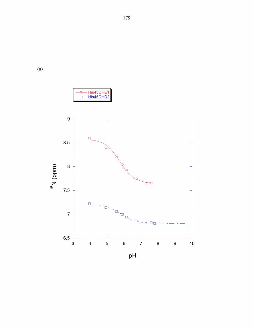
179
(a)
6.5
7
7.5
8
8.5
9
3 4 5 6 7 8 9 10
His45CHE1His45CHD2
15N
(ppm
)
pH
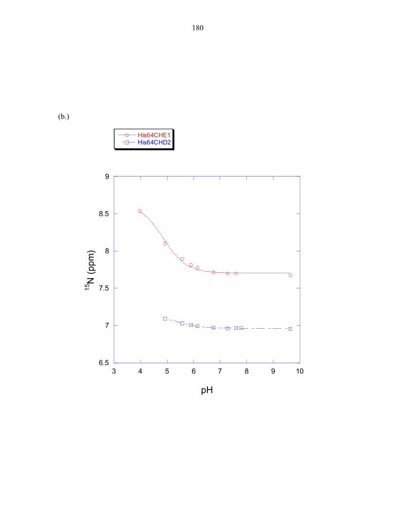
180
(b.)
6.5
7
7.5
8
8.5
9
3 4 5 6 7 8 9 10
His64CHE1His64CHD2
15N
(ppm
)
pH
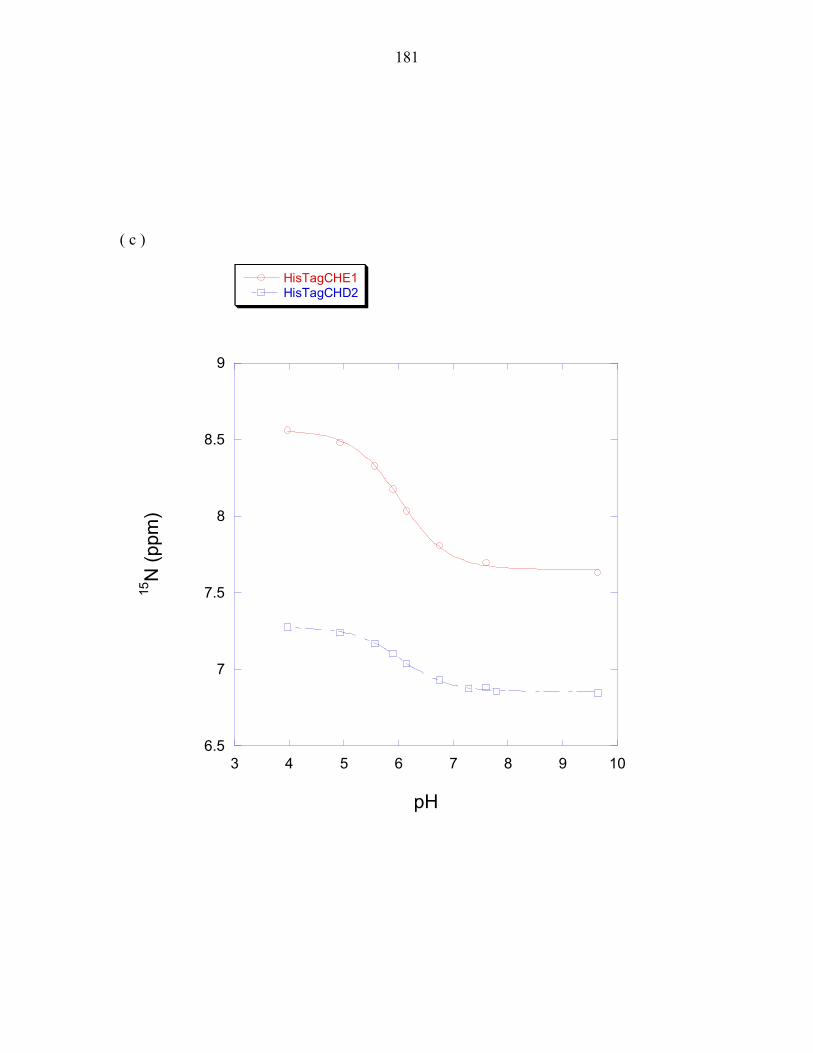
181
( c )
6.5
7
7.5
8
8.5
9
3 4 5 6 7 8 9 10
HisTagCHE1HisTagCHD2
15N
(ppm
)
pH
182
Figure 2: 1H chemical shifts for the histidines of P1K48A (a) His45 (b) His64 (c) HisTag
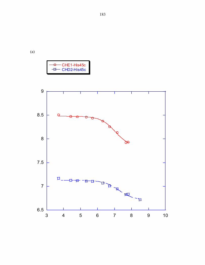
183
(a)
6.5
7
7.5
8
8.5
9
3 4 5 6 7 8 9 10
CHE1-His45cCHD2-His45c
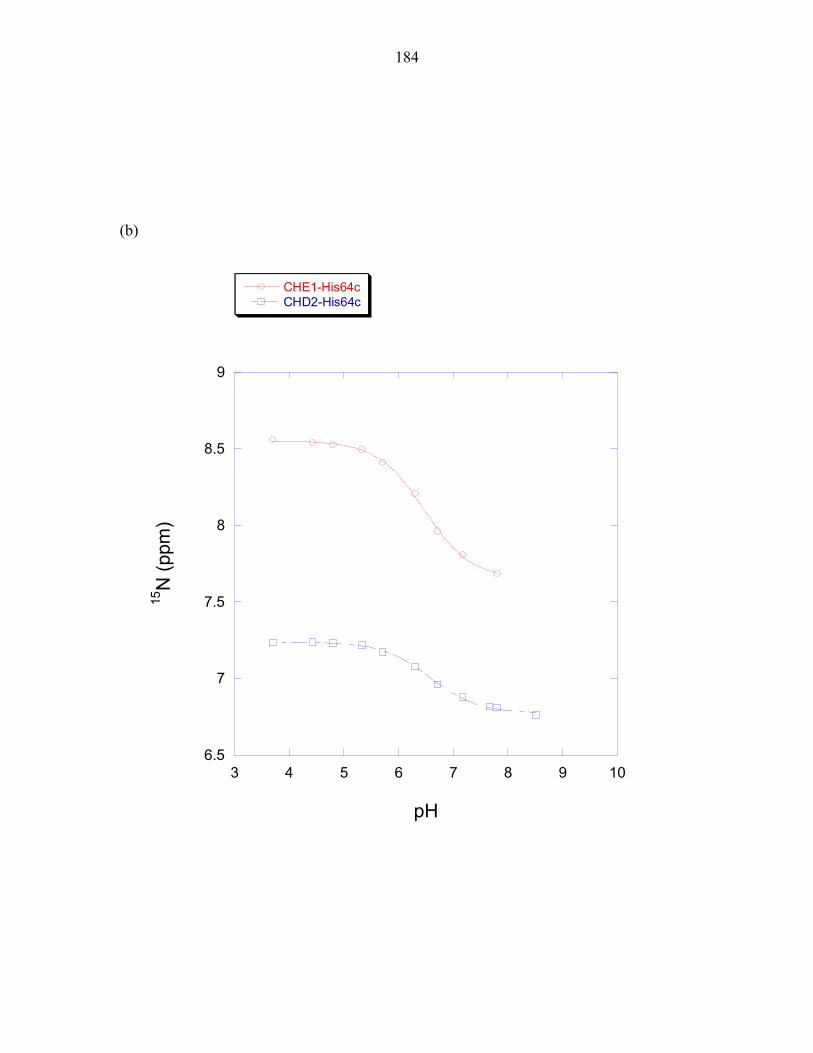
184
(b)
6.5
7
7.5
8
8.5
9
3 4 5 6 7 8 9 10
CHE1-His64cCHD2-His64c
15N
(ppm
)
pH

185
(c)
6.5
7
7.5
8
8.5
9
3 4 5 6 7 8 9 10
CHE1-TagcCHD2-Tagc
15N
(ppm
)
pH
186
Figure 3: 1H chemical shifts for the histidines of P1H64A (a) His45 (b) HisTag
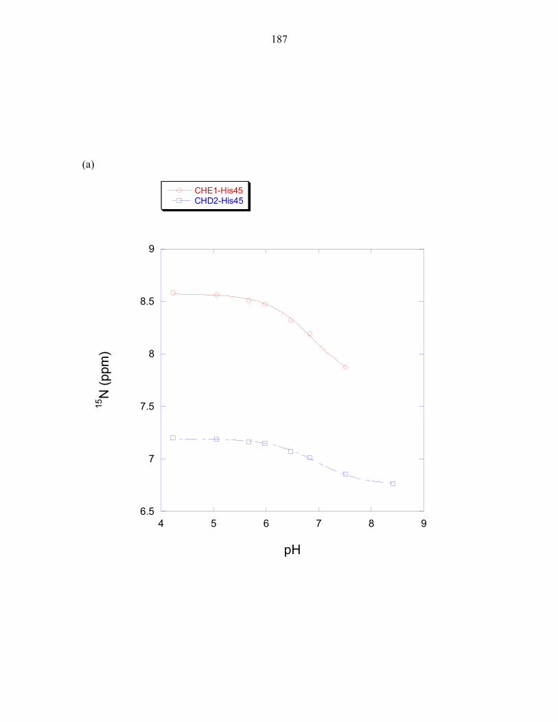
187
(a)
6.5
7
7.5
8
8.5
9
4 5 6 7 8 9
CHE1-His45CHD2-His45
15N
(ppm
)
pH
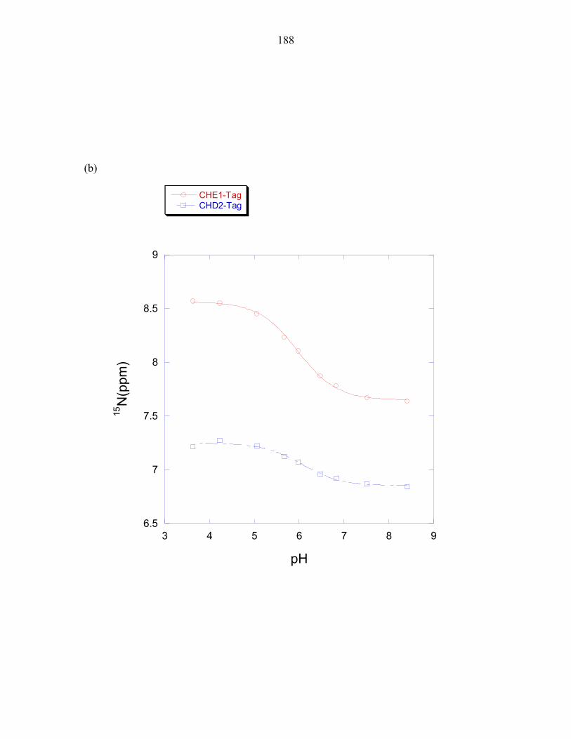
188
(b)
6.5
7
7.5
8
8.5
9
3 4 5 6 7 8 9
CHE1-TagCHD2-Tag
15N
(ppm
)
pH
189
189
Appendix 2
Biochemical Characterization of CheA domains
190
190
The phospho-histidine bond is not stable at high temperatures
The ability of P1 to be phosphorylated by ∆289 was monitored as a function of
temperature (Figure 1a). Although this hyperthermophilic organism has an optimal
growth temperature of 80°C and a maximal growth temperature of 90°C (Huber,
Langworthy et al. 1986), maximal phosphorylation activity was surprisingly observed at
60°C. The stability of the P1 phospho-histidine complex was subsequently tested as a
function of time (Figure 1b). The phospho-histidine bond is stable for up to 3 hours at
25°C and 40°C. However, at 50°C, the bond becomes slightly less stable. A drastic
reduction in the stability of the phospho-histidine complex is observed at temperatures
above 60°C. The bond is rapidly broken as evidenced by the lack of phosphorylation
signal observed after two hours.
191
191
Figure 1: Temperature dependence of phosphorylation activity (a) and the stability of the
P1 phospho-histidine complex (b): The phosphorylation of P1 by ∆289 was allowed to
reach saturation levels. The reaction was then terminated and placed at 4°C. Aliquots of
the phosphorylation reaction were taken at the indicated time points as described in
Materials and Methods.
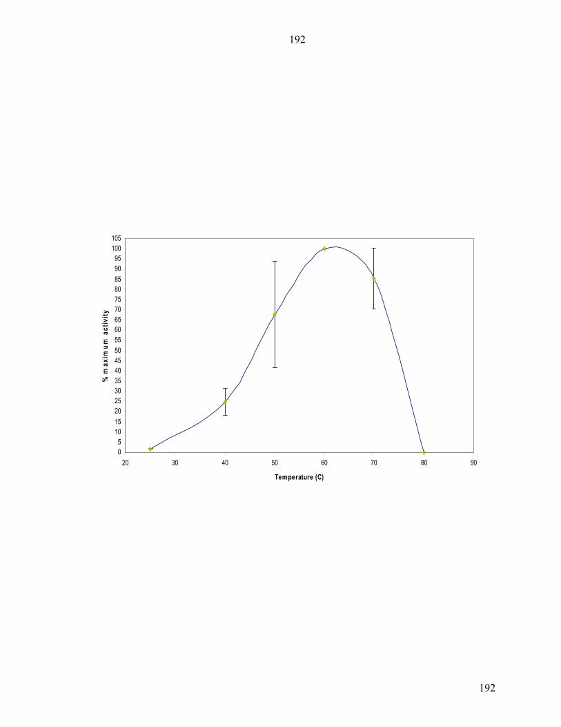
192
192
05101520253035404550556065707580859095100105
20 30 40 50 60 70 80 90
Temperature (C)
% m
axim
um a
ctiv
ity
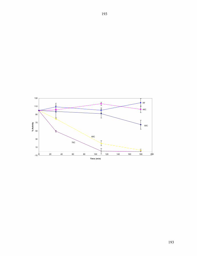
193
193
-10
10
30
50
70
90
110
130
0 20 40 60 80 100 120 140 160 180 200
Time (min)
% A
ctiv
ity
70C
60C
50C
40C
RT
194
194
MATERIALS AND METHODS
Temperature Dependence and Phosphostability Studies
The initial velocities of P1(30µM) phosphorylation by ∆289(2µM) were
measured at 25°C, 40°C, 50°C, 60°C, 70°C and 80°C as described above. To measure
the stability of the phospho-histidine complex, the phosphorylation reaction was
quenched with 4X sodium dodecyl sulfate (SDS) electrophoresis buffer containing 25mM
ethylenediaminetetra acetic acid (EDTA). The reaction tube was kept at the respective
temperature and aliquots were placed at 4°C at designated time points. Samples were
then electrophoresed and processed as previously described.


![Research Article CrystalStructureofL-Histidinium2 ...chloride monohydrate [2], L-histidine tetrafluoroborate [3], L-histidine hydrochloride monohydrate [4], L-histidine hydrofluoride](https://img.pdfslide.us/doc/110x75/60b51c180636315681384205/research-article-crystalstructureofl-histidinium2-chloride-monohydrate-2.jpg)