Embed Size (px)
Citation preview
Proc. Natl. Acad. Sci. USAVol. 89, pp. 2499-2503, March 1992Biophysics
Structure of the histidine-containing phosphocarrier protein HPrfrom Bacillus subtilis at 2.0-A resolutionOSNAT HERZBERG*t, PRASAD REDDY*, SARAH SUTRINA§, MILTON H. SAIER, JR.§, JONATHAN REIZER§,AND GEETA KAPADIA**Maryland Biotechnology Institute, University of Maryland, and tNational Institute of Standards and Technology, Center for Advanced Research inBiotechnology, 9600 Gudelsky Drive, Rockville, MD 20850; and §Department of Biology, University of California, San Diego, La Jolla, CA 92093-0116
Communicated by David R. Davies, December 3, 1991 (received for review September 30, 1991)
ABSTRACT The crystal structure of the histidine-containing phosphocarrier protein (HPr) of the phospho-enolpyruvate:sugar phosphotransferase system (PTS) fromBacillus subtilis has been determined at 2.0-A resolution andrefined to a crystallographic residual error R-factor of 0.150.The secondary-structure folding topology of the molecule isthat of an open-face (i-sandwich formed by four antiparallel(i-strands packed against three a-helices. The active-site his-tidine, His-15, caps the N terminus of the first helix, suggestingthat the helix dipole plays a role in stabilizing the phosphory-lated state of the histidine. A sulfate anion located betweenHis-15 and the neighboring Arg-17 has been identified in theelectron-density map. Association of this negatively chargedspecies with the two key catalytic residues implies that thecrystal structure resembles the phosphorylated state of theprotein. A model of the phosphorylated form of the molecule isproposed, in which the negatively charged phosphoryl groupinteracts with two main-chain nitrogen atoms of the followinghelix and with the guanidinium group of Arg-17. It is alsoproposed that the phosphoryl transfer from HPr to the IIAdomain of the glucose permease involves Arg-17 switchingbetween two salt bridges: one with the phosphorylated histidylofHPr and the other with two aspartyl residues associated withthe active site of the IHA domain of glucose permease, which areaccessible upon complex formation.
The bacterial phosphoenolpyruvate:sugar phosphotransfer-ase system (PTS) first described by Kundig et al. (1) trans-ports some carbohydrates into the cell and simultaneouslyphosphorylates them to initiate their metabolism and preventleakage from the cytoplasm. This system also plays a role inchemotaxis toward PTS sugars and in regulation of theuptake of several non-PTS sugars (for reviews, see refs. 2-5).The system consists of two general energy-coupling compo-nents [enzyme I and a histidine-containing phosphocarrierprotein (HPr)] and of sugar-specific permeases (enzymes II).An enzyme II typically consists of three domains, IIA (alsodesignated enzyme III or factor III), IIB, and IIC (6). A totalof five phosphoryl group transfers occurs along the pathway:phosphoenolpyruvate (PEP) enzyme I -- HPr ---hA-IIIB -* sugar. The IIC domain forms the transmembranechannel for the specific sugar.HPr is a small protein (9 kDa), which is phosphorylated on
the N8 atom of a histidine residue, His-15 (7). In contrast toHPrs from Gram-negative bacteria, HPrs from Gram-positivebacteria are also phosphorylated on Ser46 by an ATP-dependent protein kinase. This phosphoryl group is hydro-lyzed by a HPr(Ser-P) phosphatase. It has been recentlyshown for the Bacillus subtilis system that the phosphoryltransfer from enzyme I to HPr is inhibited "100-fold by theserine phosphorylation (8). HPr(Ser-P) in Gram-positive
bacteria may play a regulatory role in sugar transport,although the nature of this regulation is not clear (for review,see ref. 9).The folding topology ofHPr from Escherichia coli has been
determined by NMR methods (10). This topology differsdrastically from the topology reported for the x-ray crystalstructure (11). Recently, binding of site-directed mutants ofHPr to monoclonal antibodies has shown that theNMR modelis consistent with a continuous surface-binding site. In con-trast, the x-ray model implies discontinuous surface and, thus,is probably not correct (12). The topology of the homologousHPr from B. subtilis (34% identity) has been shown by NMRto resemble that ofE. coliHPr (13, 14). The 'H-NMR spectrumof the B. subtilis protein was found to be sensitive to phos-phorylation of Ser-46. The observed backbone chemical-shiftperturbations extend far away from position 46, which theauthors interpreted as an indication of extensive structuralchanges beyond the mere addition of a negative charge.Moreover, the mutant S46D, in which Ser-46 is substituted byan aspartyl residue, had a similar spectrum to HPr(Ser-P),suggesting a similar conformation for both.We present here the x-ray crystal structure determination
of HPr from B. subtilis at 2.0-A resolution.$EXPERIMENTAL METHODS
Protein Purification and Crystallization. The B. subtilis HPrwas cloned, overproduced, and purified as described (15).Initial crystallographic work was done with the inactivemutant H15A (16). For the heavy-atom derivative work,several cysteine mutants were prepared by site-directedmutagenesis, all of which contained the catalytic His-15.Only one of these mutants (S83C) formed crystals suitable forx-ray work. The S83C HPr is active in the PTS and, therefore,further crystallographic work has been done with this mutant.The crystallization conditions are somewhat different fromthose used to obtain the mutant H15A HPr crystals. Thevapor-diffusion method was used, but the protein/salt solu-tion was equilibrated against reservoirs containing 68-74%saturated ammonium sulfate solution, 0.5% (wt/vol) poly-ethylene glycol 1000, and 100 mM citrate buffer at pH6.2-6.4. The pH of the crystals is close to physiological pHand substantially above the pK. of the active-site histidylresidue (17). The crystals diffract to at least 2.0-A resolution.As the H15A mutant crystals, they belong to space groupP3121 (resolved from P3221 by using the anomalous data ofthe heavy-atom derivatives) with unit cell dimensions of a =b = 47.3 A; c = 61.8 A; a = = 900; y = 120'A.
Structure Determination. The structure was determined byusing the multiple isomorphous replacement (MIR) method.
Abbreviations: PTS, phosphoenolpyruvate:sugar phosphotransfer-ase system; HPr, histidine-containing phosphocarrier protein.tTo whom reprint requests should be addressed.1The atomic coordinates have been deposited in the Protein DataBank, Chemistry Department, Brookhaven National Laboratory,Upton NY 11973 (reference, 1HPR).
2499
The publication costs of this article were defrayed in part by page chargepayment. This article must therefore be hereby marked "advertisement"in accordance with 18 U.S.C. §1734 solely to indicate this fact.
Dow
nloa
ded
by g
uest
on
Janu
ary
25, 2
022
Proc. NatL. Acad. Sci. USA 89 (1992)
Table 1. Heavy-atom derivative statistics
Soaking Risot UniqueCompound* time, days (2.8 A) reflections, no. Sites, no. R, t (fH)/(EH)t
Uranyl acetate (2 mM) 4 0.188 2150 2 0.51 1.92KAu(CN)2 (1 mM) 6 0.164 2185 1 0.52 1.77*For soaking experiments the citrate buffer was replaced by Tris acetate.tRiw= h IIFPHobsI - IFpobslI/lhlFpobsl.Rcen = Xh centnc IIFpHobsI - jFpobs + FHcalcII/Xh centncllFPHobsl - Fpobsjl, where h, Miller indices;obs, observed; calc, calculated; Fp, FPH, and FH, native, derivative, and heavy-atom structure factors,respectively. (EH), rms lack of closure error; (fH), rms heavy-atom scattering.
X-ray intensity data were collected on a Siemens area de-tector mounted on a Siemens 3-axis goniostat. Graphitemonochromated CuKa x-rays were generated with a RigakuRotaflex RU-200BH rotating-anode source. The data wereprocessed with the computer program suite XENGEN (18).Data to 2.0-A resolution were obtained for the S83C protein.The 5083 reflection set is 90% complete, with the shell in the2.1- to 2.0-A range only 58% complete. The agreementbetween symmetry-related reflections (Rsym) is 0.086. Data at2.8-A resolution for two heavy-atom derivatives were alsocollected. The Rsym values for the uranyl and gold derivativeare 0.057 and 0.070, respectively (Bijvoet-related reflectionswere not merged).The multiple isomorphic replacement and solvent-flattening
work was done by using the computer program suite PHASES. 11Heavy-atom binding sites were determined from differencePatterson and difference Fourier maps in the usual manner(19). The positional parameters and occupancies of the heavyatoms were refined by lack of closure error minimization (20).Some statistics for the heavy-atom derivatives are given inTable 1. Although the protein contains an engineered cysteineresidue, no mercurial derivative was obtained (the currentelectron-density map shows that Cys-83 is oxidized). A phaseset was obtained with a mean figure of merit of 0.66. A 2.8-Aresolution electron-density map was computed with centroidphases (21) and measured structure-factor amplitudesweighted by the figure of merit. The map showed continuousstretches of density that were interpretable with some effort.Although the volume occupied by solvent in these crystals israther low (44%), solvent flattening (22) proved useful inimproving the map. The procedure was done as implementedin PHASES. The solvent content was specified for maskingpurposes as only 35%. The average cumulative phase shiftfrom the initial MIR phases was 34°. The solvent-flattenedelectron-density map was easier to interpret and enabledunambiguous tracing of the polypeptide chain consistent withthe known amino acid sequence. Fig. 1 shows a region of themap around the active-site histidyl residue.A molecular model was fitted to the electron-density map
on an Evans and Sutherland (Salt Lake City) PS390 interac-tive graphics system using the program FRODO (23). Thisinitial model consisted of all but the first amino acid residuebecause the expressed protein lacks the N-terminal methio-nine (14).
Structure Refinement. The first stage of structure refine-ment was done with the program X-PLOR (24) by using thesimulated annealing slow-cooling protocol at 3000 K andincluding data between 6.0 and 2.0 A for which structurefactor F 2 2ac(F). The residual error factor R was reducedfrom 0.441 to 0.219 (R = IhI IFoI - FCI I/XhIFoI, where |FO1 andIFI are the observed and calculated structure factor ampli-tudes, respectively). After one cycle of X-PLOR refinement,interactive model building was resumed, displaying two typesof electron-density maps: (i) a map computed with thecoefficients 21FOI - IFJI and calculated phases; (ii) a map
'Furey, W. & Swaminathan, S., 14th American CrystallographicAssociation Meeting, April 8-13, 1990, New Orleans, abstr. PA33.
computed with the coefficients IFOI - IFCI and calculatedphases. Adjustments were made to the model, and densityconsistent with a bound sulfate anion was identified andmodeled.For the next stage of refinement the restrained-parameter
least-squares program of Hendrickson and Konnert (25),which includes a fast Fourier transform calculation (26), wasused. Forty-one cycles of refinement were done with threemanual refitting and the addition of 100 solvent molecules tothe model.The current model of HPr has an R-factor of 0.150 for the
44% reflections between 8.0- and 2.0-A resolution for whichF- 2ar(F) (88% of all measured data). The stereochemicalparameters are well within the range known from crystalstructures of small peptides: the rms distance deviations fromideal values for bond length is 0.023 A and for bond angle is0.043 A.
RESULTS AND DISCUSSIONMolecular Conformation of HPV. HPr forms a classical
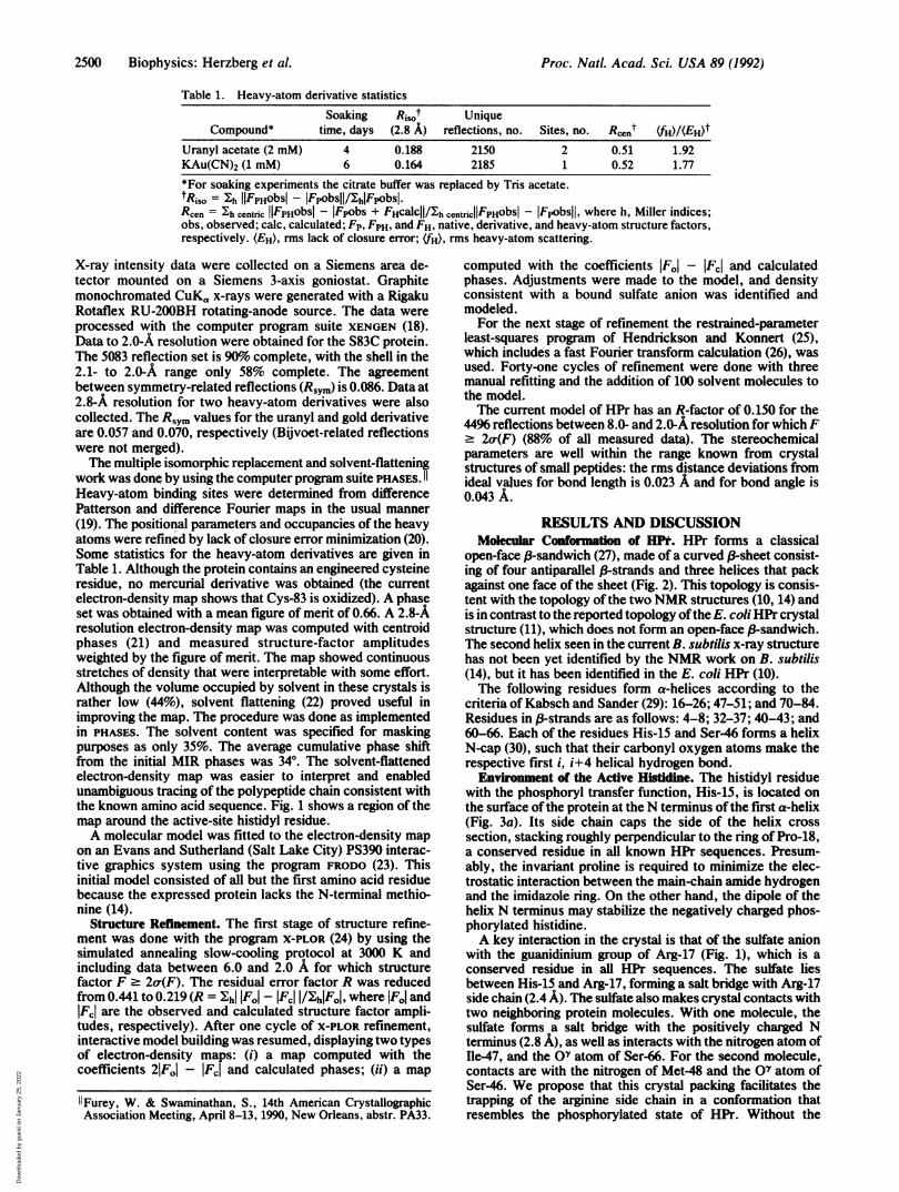
open-face (-sandwich (27), made of a curved (3-sheet consist-ing of four antiparallel (-strands and three helices that packagainst one face of the sheet (Fig. 2). This topology is consis-tent with the topology of the two NMR structures (10, 14) andis in contrast to the reported topology ofthe E. coliHPr crystalstructure (11), which does not form an open-face (-sandwich.The second helix seen in the current B. subtilis x-ray structurehas not been yet identified by the NMR work on B. subtilis(14), but it has been identified in the E. coli HPr (10).The following residues form a-helices according to the
criteria of Kabsch and Sander (29): 16-26; 47-51; and 70-84.Residues in (-strands are as follows: 4-8; 32-37; 40-43; and60-66. Each of the residues His-15 and Ser-46 forms a helixN-cap (30), such that their carbonyl oxygen atoms make therespective first i, i+4 helical hydrogen bond.Environment of the Active Histidine. The histidyl residue
with the phosphoryl transfer function, His-15, is located onthe surface ofthe protein at theN terminus ofthe first a-helix(Fig. 3a). Its side chain caps the side of the helix crosssection, stacking roughly perpendicular to the ring of Pro-18,a conserved residue in all known HPr sequences. Presum-ably, the invariant proline is required to minimize the elec-trostatic interaction between the main-chain amide hydrogenand the imidazole ring. On the other hand, the dipole of thehelix N terminus may stabilize the negatively charged phos-phorylated histidine.A key interaction in the crystal is that of the sulfate anion
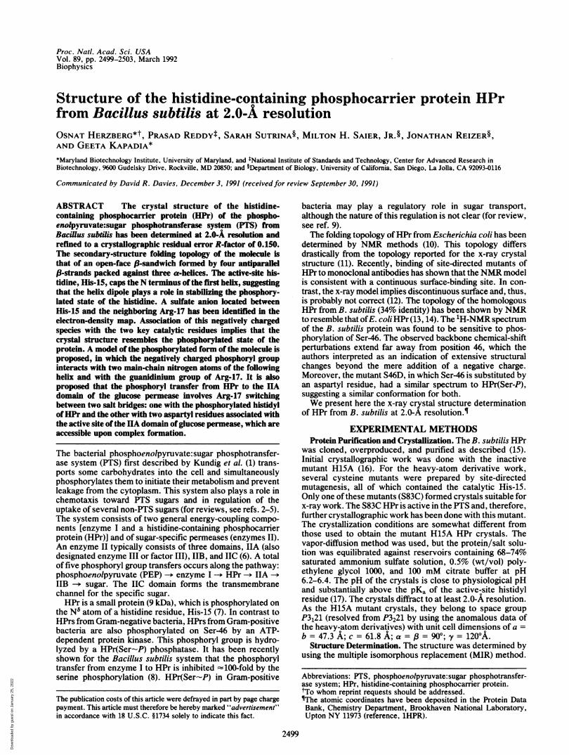
with the guanidinium group of Arg-17 (Fig. 1), which is aconserved residue in all HPr sequences. The sulfate liesbetween His-15 and Arg-17, forming a salt bridge with Arg-17side chain (2.4 A). The sulfate also makes crystal contacts withtwo neighboring protein molecules. With one molecule, thesulfate forms a salt bridge with the positively charged Nterminus (2.8 A), as well as interacts with the nitrogen atom ofIle-47, and the OQ atom of Ser-66. For the second molecule,contacts are with the nitrogen of Met-48 and the OV atom ofSer-46. We propose that this crystal packing facilitates thetrapping of the arginine side chain in a conformation thatresembles the phosphorylated state of HPr. Without the
2500 Biophysics: Herzberg et al.
Dow
nloa
ded
by g
uest
on
Janu
ary
25, 2
022
Proc. Natl. Acad. Sci. USA 89 (1992) 2501
FIG. 1. Stereoscopic view of a region of the elec-tron-density map in vicinity of the active-site His-15.The 2.8-A solvent-flattened multiple isomorphous re-placement map is displayed together with the refinedmodel at 2.0 A. The electron density for the sulfateanion (labeled S04) can be seen already in this map,although it was added to the model only at advancedstage of refinement. Residues from neighboring mole-cules are also shown. (') is added to the labels ofresidues from one molecule (symmetry operator Y, X,-Z + 1), and (") is added to those of a second molecule(symmetry operator X - Y + 1, - Y + 1, -Z + 2/3).
sulfate, the electrostatic interaction of Arg-17 with the main-chain nitrogens at the N terminus of the a-helix would berepulsive.Lack of a salt bridge interaction between His-15 and the
sulfate ion (the shortest distance is 3.9 A) suggests that thehistidine is uncharged, consistent with its role as a nucleophilein the phosphoryl transfer. Although discriminating betweennitrogen and carbon atoms at the resolution of the structuredetermination is impossible, the imidazole-ring orientationwas chosen such that the N5 atom, the target of phosphory-lation, points toward Arg-17 and the sulfate. A hydrogen bondis formed between the Ser-12 OY atom and the N6 atom. Theclose proximity of the N8 atom to the two main-chain nitrogenatoms of the proceeding helix is consistent with it beingdeprotonated, as required for a phosphoryl acceptor.Three hydrophobic side chains on the surface ofthe protein
are located close to the active site: Ile47, Met-48, andMet-51. This hydrophobic patch may indicate the site ofinteraction with other proteins of the PTS. Interestingly, thispatch is flanked by His-15 from one side and by Ser46 fromthe other side (see below).Environment of Ser-46. Ser-46, the target of phosphoryla-
tion by the ATP-dependent HPr kinase in Gram-positivebacteria, is located on the surface at the N terminus of thesecond a-helix (Fig. 4). Its side chain caps the helix such thatthe OY atom interacts with the main-chain nitrogen atom ofGly-49 (2.9 A). Capping of N termini of helices by serineresidues is common in protein structures (30, 31). Aspar-agines and aspartic acids also occupy this position with ahigher frequency than expected. Hydrogen bonding of theside-chain oxygen atom with the main-chain nitrogen atom is
associated with this preference. Presumably, the negativecharge of a phosphorylated serine can be stabilized by thehelix dipole as well. However, modeling shows that whenphosphorylated, the Xi dihedral angle of Ser-46 should berotated by -60° to avoid short contacts of the phosphorylgroup with the helix.
It is an interesting coincidence that the crystal packingbrings the active site of one molecule and Ser-46 of a secondone close to the sulfate anion. This proximity probably has nophysiological significance because no evidence is availablefor dimer formation.
It has been recently shown that the interaction of the B.subtilis HPr with enzyme I is impaired by phosphorylation ofSer-46 (8). The crystal structure shows that phosphorylationof Ser-46 does not require a major rearrangement of thepolypeptide chain, but the charge distribution is obviouslyaltered. The hydrophobic patch formed by Ile-47, Met-48,and Met-51 is flanked by Ser-46 on one side and by the activesite on the other and may be the region of interaction of HPrwith enzyme I (and perhaps with the IIA domains of the PTSpermeases). The introduction of a negative charge on Ser-46-P in the interface formed by the two interacting proteinscould interfere with the protein-protein complex formationbecause of unfavorable electrostatic interactions. Similarly,when His-15 is phosphorylated, the interaction with thekinase may be prevented if the active site forms part of theinterface.Why are HPrs from Gram-positive bacteria phosphory-
lated on Ser-46, whereas those from Gram-negative bacteriaare not? There are three residues in the vicinity of Ser-46 thatare invariant in the known sequences of HPrs from Gram-
FIG. 2. Fold of HPr from B. subtilis. (a) Highlighting secondary-structure motifs. Figure was generated with the computer program RIBBON(28). (b) Stereoscopic representation showing a-carbon positions in the molecule. Every tenth amino acid is labeled.
Biophysics: Herzberg et al.
Dow
nloa
ded
by g
uest
on
Janu
ary
25, 2
022

Proc. Natl. Acad. Sci. USA 89 (1992)
a
S04
b
FIG. 3. Stereoscopic view of the active-site region of HPr. (a) Crystal structure. (b) Model of phosphorylated state. Bonds betweenmain-chain atoms are filled, and those between side-chain atoms are open. Important interactions of side chains of His-15 and Arg-17 are shownby broken lines. The sulfate is labeled S04, and the phosphate is labeled P03.
positive bacteria but differ in the protein from E. coli (4):Asn-43 is a serine in E. coli HPr, Met-48 is a phenylalanine,and Gly-49 is a lysine. Inspection ofthe crystal structure (Fig.
4) shows that the last two replacements would crowd theenvironment of Ser-46, change the charge distribution, andpotentially prevent interaction with the kinase.
FIG. 4. Stereoscopic view of environment of Ser-46. Bonds between main-chain atoms are filled, and bonds between side-chain atoms areopen. Hydrogen bond between the 07 atom of Ser-46 and the main-chain nitrogen atom of Gly-49 is shown by broken line.
2502 Biophysics: Herzberg et al.
Dow
nloa
ded
by g
uest
on
Janu
ary
25, 2
022
Proc. Natl. Acad. Sci. USA 89 (1992) 2503
Model of Phosphorylated His-15. In analogy to our modelingof the phosphorylated state of the IIA domain of the glucosepermease (32), a model for the activated phosphohistidine ofHPr has been derived. The phosphoryl group was built so thatthe phosphorus atom is in the plane of the imidazole ring ofHis-15 (Fig. 3b). In contrast to the IIA domain, where no
adjustments to the protein were required, minor adjustmentshave been made here: the X2 dihedral angle of His-15 was
rotated by 100 to avoid short contact with the main-chainnitrogen atom of Arg-17. This rotation facilitates electrostaticinteractions of the phosphoryl oxygen atom with the twomain-chain nitrogens of Ala-16 and Arg-17. In addition, the Xi
dihedral angle of Arg-17 was rotated by 400, such that a saltbridge was formed with the phosphoryl moiety rather thanwith the sulfate ion, as observed in the crystal structure (Fig.3a).We propose that without a negative charge in the active
site, the side-chain conformation of Arg-17 is altered to avoidthe unfavorable electrostatic interaction with the dipole ofthehelix N terminus. In contrast, the NMR work suggests thatthe side chains of His-15 and Arg-17 interact also in thephosphate-free state (10, 14), implying that Arg-17 doesinteract with the helix N terminus. Perhaps this observationwas made because the two residues are close in sequence,such that the CO atom of Arg-17 is 4.2 A apart from the sidechain of His-15. However, this short distance does not implythat an electrostatic interaction should exist between the twoside chains. Modeling shows that an alternative position ofthe guanidinium group may be obtained simply by changingthe side-chain torsion angles, without invoking a majormain-chain conformational transition. For example, a fullyextended side-chain conformation would be acceptable.Klevit and Waygood (10) observed an interaction betweenArg-17 and the C terminus of E. coli HPr (with either theside-chain or the backbone carboxyl group of Glu-85). Anequivalent interaction has not been reported in the NMRwork on the B. subtilis protein (14). The C terminus of the B.subtilis HPr (Glu-88) is remote from the active site, and theonly negatively charged residue that could play such a role isGlu-84 on the C terminus of the third helix. Modeling showsthat a salt bridge can be formed by modifying the dihedralangles of Arg-17 such that its side chain points away fromHis-15. However, there is no evidence yet that this salt bridgeis essential for maintaining the structural integrity of thephosphate-free form. Any other allowed conformation of theArg-17 side chain in which the charge is solvated and theinteraction with the dipole of the helix N terminus is avoidedwould be equally possible.
Proposed Mechanism for Phosphoryl Transfer. The struc-ture ofHPr, in which the active-site histidine is on the proteinsurface, is consistent with the previously proposed mecha-nism ofthe PTS phosphoryl transfer involving the associativepathway (ref. 33; for review ofphosphoryl transfer pathways,see ref. 34), and with the crystal structure of the IIA domainof the B. subtilis glucose permease (32). The associativepathway is required to avoid atomic clashes at the protein-protein interface. A pentacoordinated intermediate or tran-sition state is implied, with trigonal bipyramidal geometry atthe phosphorus. With the two active-site histidine nitrogenatoms (N8 ofHis-15 in HPr and N- ofHis-83 in the IIA domainof glucose permease) occupying apical positions, the transferwould lead to inversion of configuration at the phosphorus.The electrostatic interactions should be delicately bal-
anced to permit burial of the negatively charged phosphorylgroup at the protein-protein interface. We have identifiedtwo aspartate residues in the IIA domain of the glucosepermease (Asp-31 and Asp-87) associated with the active siteand oriented toward each other (32). Whereas the phospho-histidine of HPr is stabilized by the salt bridge with Arg-17,we propose that the transfer to the IIA domain of glucose
permease is facilitated by a switch to another salt bridge inwhich Arg-17 interacts with the two aspartates of this 11Adomain. The stability of HPr(His-P) is thus reduced, allow-ing the phosphoryl transfer to His-83 of the IIA domain ofglucose permease.
We thank John Moult, Walt Stevens, Gary Gilliland, Der-Ing Liao,and Tom Poulos for many helpful discussions. Computing time for theX-PLOR refinement was provided by the National Institute of Stan-dards and Technology (NIST). Certain commercial equipment, in-struments, and materials are identified in this paper. Such identifica-tion does not imply a recommendation or endorsement by NIST. Thiswork was supported by National Science Foundation Grant DMB-9019340 to O.H. Protein preparation was supported by NationalInstitutes ofHealth Grants R01-AI21702 and RO1-AI14176 to M.H.S.
1. Kundig, W., Ghosh, S. & Roseman, S. (1964) Proc. Natl. Acad. Sci.USA 52, 1067-1074.
2. Postma, P. W. & Lengeler, J. W. (1985) Microbiol. Rev. 49, 232-269.
3. Saier, M. H., Jr. (1985) Mechanisms and Regulation of Carbohy-drate Transport in Bacteria (Academic, New York).
4. Reizer, J., Saier, M. H., Jr., Deutscher, J., Grenier, F., Thompson,J. & Hengstenberg, W. (1988) CRC Crit. Rev. Microbiol. 15,297-338.
5. Meadow, N. D., Fox, D. K. & Roseman, S. (1990) Annu. Rev.Biochem. 59, 497-542.
6. Saier, M. H., Jr., & Reizer, J. (1991) J. Bacteriol., in press.7. Kalbitzer, H. R., Hengstenberg, W., Rosch, P., Muss, P., Berns-
mann, P., Engelmann, R., D6rschug, M. & Deutscher, J. (1982)Biochemistry 21, 2879-2885.
8. Reizer, J., Sutrina, S. L., Wu, L.-F., Deutscher, J., Reddy, P. &Saier, M. H., Jr. (1992) J. Biol. Chem., in press.
9. Reizer, J. (1989) FEMS Microbiol. Rev. 63, 149-156.10. Klevit, R. E. & Waygood, E. B. (1986) Biochemistry 25,7774-7781.11. El-Kabbani, 0. A. L., Waygood, E. B. & Delbaere, L. T. J. (1987)
J. Biol. Chem. 262, 12926-12929.12. Sharma, S., Georges, F., Delbaere, L. T. J., Lee, J. S., Klevit,
R. E. & Waygood, E. B. (1991) Proc. Nat!. Acad. Sci. USA 88,4877-4881.
13. Wittekind, M., Reizer, J., Deutscher, J., Saier, M. H. & Klevit,R. E. (1989) Biochemistry 28, 9908-9912.
14. Wittekind, M., Reizer, J. & Klevit, R. E. (1990) Biochemistry 29,7191-7200.
15. Reizer, J., Sutrina, S. L., Saier, M. H., Jr., Stewart, G. C., Pe-terkofsky, A. & Reddy, P. (1989) EMBO J. 8, 2111-2120.
16. Kapadia, G., Reizer, J., Sutrina, S. L., Saier, M. H., Jr., Reddy, P.& Herzberg, 0. (1990) J. Mol. Biol. 211, 1-2.
17. Hengstenberg, W. & Deutscher, J. (1987) in Sugar TransportedMetabolism in Gram-Positive Bacteria, eds. Reizer, J. & Peterkof-sky, A. (Horwood, Chichester, England), pp. 215-234.
18. Howard, A. J., Gilliland, G. L., Finzel, B. C., Poulos, T., Ohlen-dorf, D. 0. & Salemme, F. R. (1987) J. Appl. Crystallogr. 20,383-387.
19. Blundell, T. L. & Johnson, L. N. (1976) Protein Crystallography(Academic, London).
20. Dickerson, R. E., Weinzierl, J. E. & Palmer, R. A. (1968) ActaCrystallogr. Sect. B 24, 997-1003.
21. Blow, D. M. & Crick, F. H. C. (1959)Acta Crystallogr. 12, 794-802.22. Wang, B. C. (1985) Methods Enzymol. 115, 90-112.23. Jones, T. A. (1982) in Computational Crystallography, ed. Sayre,
D. (Oxford Univ. Press, London), pp. 303-317.24. Brunger, A. T., Kuriyan, J. & Karplus, M. (1987) Science 235,
458-460.25. Hendrickson, W. A. & Konnert, J. H. (1980) in Biomolecular
Structure, Function, Conformation and Evolution, ed. Srinivasan,R. (Pergamon, Oxford), Vol. 1, pp. 43-75.
26. Finzel, B. C. (1987) J. Appl. Crystallogr. 20, 53-55.27. Richardson, J. (1981) Adv. Protein Chem. 34, 167-339.28. Priestle, J. P. (1988) J. Appl. Crystallogr. 21, 572-576.29. Kabsch, W. & Sander, C. (1983) Biopolymers 22, 2577-2637.30. Richardson, J. S. & Richardson, D. C. (1988) Science 240, 1648-
1652.31. Kendrew, J. C., Watson, H. C., Stranberg, B. E., Dickerson, E.,
Phillips, D. C. & Shore, V. C. (1961) Nature (London) 190, 666-670.
32. Liao, D.-I., Kapadia, G., Reddy, P., Saier, M. H., Jr., Reizer, J. &Herzberg, 0. (1991) Biochemistry 30, 9583-9594.
33. Begley, G. S., Hansen, D. E., Jacobson, G. R. & Knowles, J. R.(1982) Biochemistry 21, 5552-5556.
34. Knowles, J. R. (1980) Annu. Rev. Biochem. 49, 877-919.
Biophysics: Herzberg et al.
Dow
nloa
ded
by g
uest
on
Janu
ary
25, 2
022

![Stimuli-responsive poly(ampholyte)s containing L-histidine ... › articles › EPL-0000518_article.pdf · polymerization [25]. The polymer was obtained as follows. A mixture of MHist](https://img.pdfslide.us/doc/110x75/5f1fb74c533efe22a776140f/stimuli-responsive-polyampholytes-containing-l-histidine-a-articles-a.jpg)


![Research Article CrystalStructureofL-Histidinium2 ...chloride monohydrate [2], L-histidine tetrafluoroborate [3], L-histidine hydrochloride monohydrate [4], L-histidine hydrofluoride](https://img.pdfslide.us/doc/110x75/60b51c180636315681384205/research-article-crystalstructureofl-histidinium2-chloride-monohydrate-2.jpg)