Embed Size (px)
Citation preview
Nano Res
1
Highly stable organic fluorescent nanorods for living
cell imaging
Minhuan Lan1, †, Jinfeng Zhang1, †, Xiaoyue Zhu1, Pengfei Wang2(), Xianfeng Chen1(), Chun-Sing
Lee1, and Wenjun Zhang1()
Nano Res., Just Accepted Manuscript • DOI 10.1007/s12274-015-0748-4
http://www.thenanoresearch.com on February 15, 2015
© Tsinghua University Press 2015
Just Accepted
This is a “Just Accepted” manuscript, which has been examined by the peer-review process and has been
accepted for publication. A “Just Accepted” manuscript is published online shortly after its acceptance,
which is prior to technical editing and formatting and author proofing. Tsinghua University Press (TUP)
provides “Just Accepted” as an optional and free service which allows authors to make their results available
to the research community as soon as possible after acceptance. After a manuscript has been technically
edited and formatted, it will be removed from the “Just Accepted” Web site and published as an ASAP
article. Please note that technical editing may introduce minor changes to the manuscript text and/or
graphics which may affect the content, and all legal disclaimers that apply to the journal pertain. In no event
shall TUP be held responsible for errors or consequences arising from the use of any information contained
in these “Just Accepted” manuscripts. To cite this manuscript please use its Digital Object Identifier (DOI®),
which is identical for all formats of publication.
Nano Research
DOI 10.1007/s12274-015-0748-4
Highly stable organic fluorescent nanorods for living
cell imaging
Minhuan Lan1, †, Jinfeng Zhang1, †, Xiaoyue Zhu1, Pengfei
Wang2*, Xianfeng Chen1*, Chun-Sing Lee1, and Wenjun
Zhang1*
1 Center of Super-Diamond and Advanced Films
(COSDAF), Department of Physics and Materials Science,
City University of Hong Kong, Hong Kong SAR, P. R.
China.
2 Key Laboratory of Photochemical Conversion and
Optoelectronic Materials, Technical Institute of Physics
and Chemistry (TIPC), Chinese Academy of Sciences
(CAS), Beijing, 100190, P. R. China.
† These authors contributed equally.
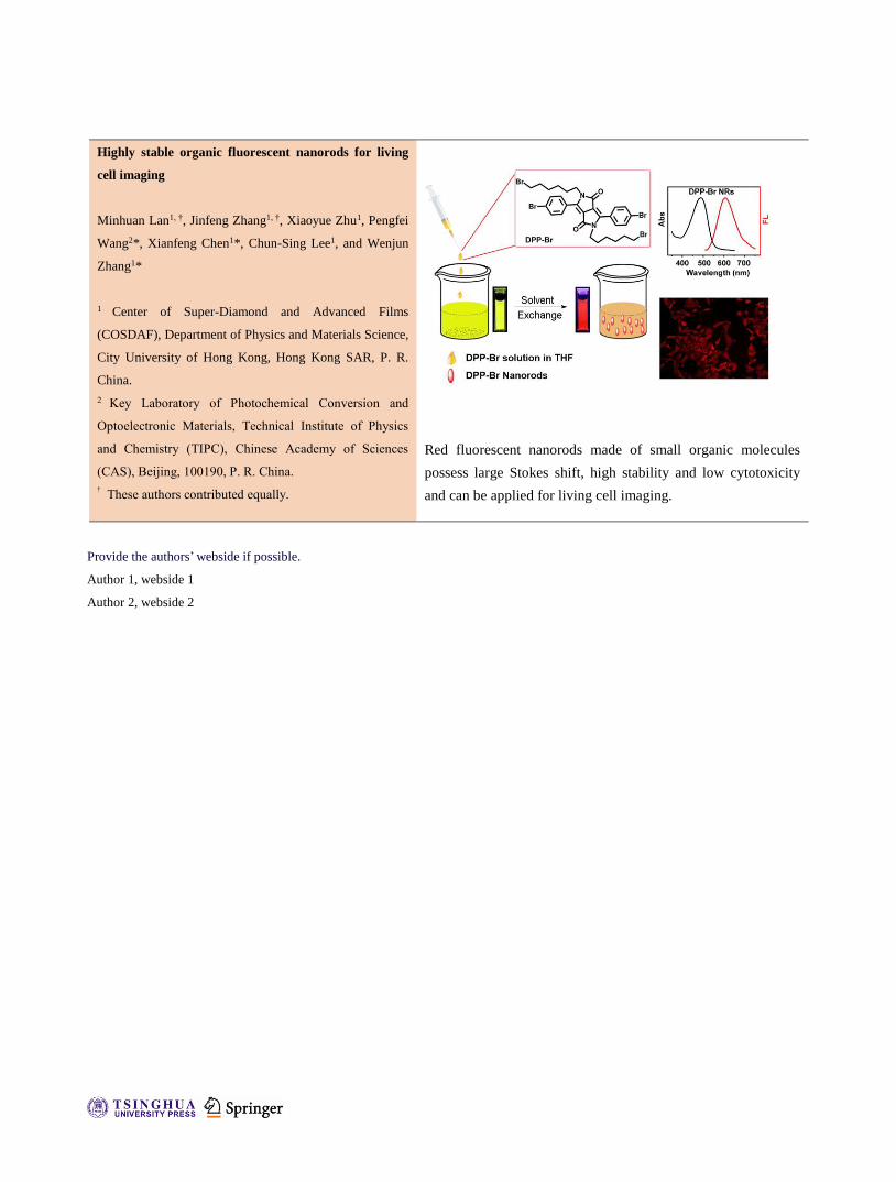
Red fluorescent nanorods made of small organic molecules
possess large Stokes shift, high stability and low cytotoxicity
and can be applied for living cell imaging.
Provide the authors’ webside if possible.
Author 1, webside 1
Author 2, webside 2
Highly stable organic fluorescent nanorods for living
cell imaging
Minhuan Lan1, †, Jinfeng Zhang1, †, Xiaoyue Zhu1, Pengfei Wang2(), Xianfeng Chen1(), Chun-Sing
Lee1, and Wenjun Zhang1()
†
Received: day month year
Revised: day month year
Accepted: day month year
(automatically inserted by
the publisher)
© Tsinghua University Press
and Springer-Verlag Berlin
Heidelberg 2014
KEYWORDS
Cell imaging, organic dye,
fluorescent nanorods,
DPP.
ABSTRACT
Metal-free, organic dye based fluorescent nanorods were prepared through a
simple solvent-exchange route. The as-prepared nanorods possess low toxicity
to living cells and excellent photostability, and are stable in solutions of various
pHs and high ionic strength and with the interference of metal ions. Compared
with the free DPP-Br molecules in THF, these nanorods exhibit larger Stokes
shift and broader absorption spectra and much improved photostability. With
all of these beneficial characteristics, we successfully demonstrated their
application as a good fluorescence probe for bio-imaging.
Nano Research
DOI (automatically inserted by the publisher)
Research Article

| www.editorialmanager.com/nare/default.asp
2 Nano Res.
1. Introduction
Accurate and timely detection of pathological
changes in human tissues provides a basis for early
disease diagnosis and successful treatment [1-3].
Among the emerging disease diagnosis methods,
biomedical imaging technique surpasses traditional
ones (e.g., biological fluids analysis and histological
section technique) in terms of having non-invasive
nature and low side effects and being able to quickly
and directly show the body's internal organizational
structure, shape, and organ function in a very
intuitive way [4, 5]. Therefore, considerable efforts
have been devoted to develop new imaging
approaches to enable precise reveal of pathological
changes. To date, various imaging techniques
including ultrasound imaging, X-ray computed
tomography imaging, nuclear magnetic resonance
imaging, nuclear medical imaging, infrared imaging
and microwave imaging have been developed and
some of them are already being applied in clinical
diagnosis [6-11]. Although effective, these methods
suffer from a number of disadvantages such as
complicated instruments, complex procedures, and
being time-consuming. Therefore, it is highly
desirable to seek further development of new
approaches for efficacious observation of
pathological changes from both scientific and
technical views.
Optical imaging of tissue offers potential
advantages in distinguishing different structures
according to their biological environment [12, 13].
Fluorescence (FL) imaging is one of a rapid emerging
technology which provides real-time in vitro and in
vivo functional imaging information with high
spatial resolution and image contrast, as compared
to many other existing optical imaging techniques
[14-16]. The key factor in fluorescence imaging
technique is the fluorescent material. Many organic
dyes have been demonstrated for cells and tissues
imaging [17, 18]. However, their current applications
are often limited by their poor photostability and
water dispersibility. Although alternative
semiconductor quantum dots are superior to organic
dyes in terms of these two aspects [19-22], the clinical
applications of these agents have been hindered due
to the high cytotoxicity [23-26]. In addition, the
stability and performance of organic dyes and
quantum dots are often affected by pH values and
transition metals ions [27, 28].
Overcoming these limitations would benefit many
advanced applications such as imaging in living cells
and other complex body fluids that contain various
ions or possess extreme pH values, and real-time
monitoring of dynamic biological process [29-31].
There have been various attempts to circumvent this
problem, such as the surface modification of
fluorophore with optimized ligands, and making
fluorescent molecules in the form of nanomaterials
[32, 33]. Among these approaches, organic dye based
nanomaterials combine the advantageous properties
of organic dye and quantum dots including
outstanding optical properties, low cytotoxicity, and
robust chemical inertness, and therefore are defined
as a new class of promising fluorescence materials
[34-36]. In addition, in contrast to semiconductor
quantum dots, the emission of organic dye based
nanomaterials mainly depends on their constituent
molecules. Therefore, they can be fabricated with
different shape and size with constant emission
wavelength. Due to these superior advantages of
organic dye based nanomaterials, various kinds of
nanoparticles (NPs) have been reported for cell
imaging and tumor targeting [37, 38], but it was
recently found that, comparing with spherical
particles, nanorods (NRs) feature a larger
surface/volume ratio which may be advantageous
since additional targeting ligands can aid in cell
binding, cellular uptake, and therapeutic
effectiveness [39, 40]. V. P. Chauhan et al. found that
the shape of nanomaterials is an important aspect in
the design of effective nanomedicines. NRs could
penetrate tumours more rapidly than nanospheres
due to improved transport through pores [41].
Another recent study also found that NRs, as
opposed to spherical NPs, appear to adhere more
effectively to the surface of endothelial of cells,
allowing for drug targeting and imaging to specific
types of cells [42].
Address correspondence to Wenjun Zhang, email: [email protected]; Xianfeng Chen, email: [email protected]; Pengfei Wang, email: [email protected].
www.theNanoResearch.com∣www.Springer.com/journal/12274 | Nano Research
3 Nano Res.
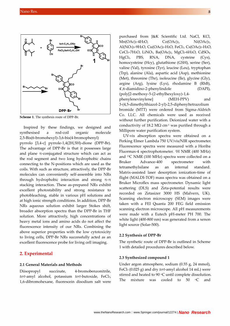
Scheme 1. The synthesis route of DPP-Br.
Inspired by these findings, we designed and
synthesised a rod-coil organic molecule
2,5-Bis(6-bromohexyl)-3,6-bis(4-bromophenyl)
pyrrolo [3,4-c] pyrrole-1,4(2H,5H)-dione (DPP-Br).
The advantage of DPP-Br is that it possesses large
and plane π-conjugated structure which can act as
the rod segment and two long hydrophobic chains
connecting to the N-positions which are used as the
coils. With such as structure, attractively, the DPP-Br
molecules can conveniently self-assemble into NRs
through hydrophobic interaction and strong π-π
stacking interaction. These as-prepared NRs exhibit
excellent photostability and strong resistance to
photobleaching, stable in various pH solutions and
at high ionic strength conditions. In addition, DPP-Br
NRs aqueous solution exhibit larger Stokes shift,
broader absorption spectra than the DPP-Br in THF
solution. More attractively, high concentrations of
heavy metal ions and amino acids do not affect the
fluorescence intensity of our NRs. Combining the
above superior properties with the low cytotoxicity
to living cells, DPP-Br NRs successfully acted as an
excellent fluorescence probe for living cell imaging.
2. Experimental
2.1 General Materials and Methods
Diisopropyl succinate, 4-bromobenzonitrile,
tert-amyl alcohol, potassium tert-butoxide, FeCl3,
1,6-dibromohexane, fluorescein disodium salt were
purchased from J&K Scientific Ltd. NaCl, KCl,
Mn(OAc)2-4H2O, Co(OAc)2, Ni(OAc)2,
Al(NO3)3-9H2O, Cu(OAc)2-H2O, FeCl3, Cs(OAc)2-H2O,
CeCl3-7H2O, LiNO3, Ba(OAc)2, MgCl2-6H2O, CdSO4,
HgCl2, PBS, RNA, DNA, cysteine (Cys),
homocysteine (Hcy), glutathione (GSH), serine (Ser),
valine (Val), tyrosine (Tyr), leucine (Leu), tryptophan
(Trp), alanine (Ala), aspartic acid (Asp), methionine
(Met), threonine (Thr), isoleucine (Ile), glycine (Gly),
argine (Arg), lysine (Lys), rhodamine B (RhB),
4',6-diamidino-2-phenylindole (DAPI),
(poly[2-methoxy-5-(2-ethylhexyloxy)-1,4-
phenylenevinylene] (MEH-PPV) and
3-(4,5-dimethylthiazol-2-yl)-2,5-diphenyltetrazolium
bromide (MTT) were ordered from Sigma-Aldrich
Co. LLC. All chemicals were used as received
without further purification. Deionized water with a
conductivity of 18.2 MΩ cm-1 was purified through a
Millipore water purification system.
UV-vis absorption spectra were obtained on a
Perking Elmer Lambda 750 UV/vis/NIR spectrometer.
Fluorescence spectra were measured with a Horiba
Fluormax-4 spectrophotometer. 1H NMR (400 MHz)
and 13C NMR (100 MHz) spectra were collected on a
Bruker Advance-400 spectrometer with
tetramethylsilane as an internal standard.
Matrix-assisted laser desorption ionization-time of
flight (MALDI-TOF) mass spectra was obtained on a
Bruker Microflex mass spectrometer. Dynamic light
scattering (DLS) and Zeta-potential results were
recorded on Zetasizer 3000 HS (Malvern, UK).
Scanning electron microscopy (SEM) images were
taken with a FEI Quanta 200 FEG field emission
scanning electron microscope. All pH measurements
were made with a Eutech pH-meter PH 700. The
white light (400-800 nm) was generated from a xenon
light source (Solar-500).
2.2 Synthesis of DPP-Br
The synthetic route of DPP-Br is outlined in Scheme
1 with detailed procedures described below.
2.3 Synthesized compound 1
Under argon atmosphere, sodium (0.55 g, 24 mmol),
FeCl3 (0.025 g) and dry tert-amyl alcohol 14 mL) were
stirred and heated to 90 oC until complete dissolution.
The mixture was cooled to 50 oC and

| www.editorialmanager.com/nare/default.asp
4 Nano Res.
4-bromobenzonitrile (2.20 g, 12 mmol) was added
followed by reheating to 90 oC. A solution of succinic
acid dissopropyl ester (1.0 g, 4.8 mmol) in dry
tert-amyl alcohol (5 mL) was dropwisely added in 1
hour. After stirring for 24 hours, 10 mL of acetic acid
was added, and the mixture was heated to 120 oC
and maintained for 1 hour. Then the precipitate was
collected and repeatedly washed with hot water and
methanol, and dried under vacuum at 80 oC. Red
solid was obtained. (Compound 1, 1.55 g, yield:
70%).
2.4 Synthesized DPP-Br
0.9 g compound 1, potassium tert-butoxide (0.5 g)
and N-methyl-2-pyrrolidone (10 mL, NMP) were
mixed and heated to 60 oC. 1,6-dibromohexane (2 mL)
was slowly added and the mixture was stirred at 60 oC for 24 hours. After cooling to room temperature,
50 mL of toluene was added into the reaction
mixture and washed with water to remove the NMP.
The organic solution was concentrated and the crude
product was purified by column chromatography on
silica using dichloromethane as eluent to yield a red
powder. (DPP-Br, 0.6 g. yield: 39%). 1H NMR (CDCl3,
400Hz, ppm) δ 1.22-1.25 (2H), 1.31-1.37 (2H),
1.52-1.57 (2H), 1.74-1.78 (2H), 3.30-3.34 (2H), 3.70-3.74
(2H), 7.65 (8H). 13C NMR (CDCl3, 100Hz, ppm) δ
25.86, 27.30, 29.67, 32.46, 33.56, 41.67, 109.97, 125.95,
126.88, 130.05, 132.36, 147.39, 162.40. MALDI-TOF
Mass spectrum m/z: Calculated: 772; Found: 772.
Scheme 2. Preparation of DPP-Br nanorods.
2.5 Preparation of DPP-Br NRs
DPP-Br NRs were obtained by a co-precipitation
method according to Scheme 2. Two hundred
microliters of 1.5 mg/mL DPP-Br/THF solution was
dropped into 5 mL of aqueous solution by
microsyringe at room temperature under vigorous
stirring. During this process, the DPP-Br molecules
self-assemble into NRs through hydrophobic
interaction and strong π-π stacking interaction. After
stirring for 5 minutes, the THF in the solution was
removed by bubbling nitrogen at room temperature.
Finally, the NRs dispersion was obtained by
centrifugation for further spectra measurements.
2.6 Preparation of MEH-PPV NPs
MEH-PPV NPs were obtained by a co-precipitation
method similar with DPP-Br NRs. Two hundred
microliters of 1 mg/mL MEH-PPV/THF solution was
dropped into 10 mL of aqueous solution by
microsyringe over a period of 5 minutes under
vigorous stirring at room temperature. The THF in
the solution was removed by bubbling nitrogen at
room temperature.
2.7 Cell culture and in vitro imaging studies
A549 cells were obtained from Peking Union Medical
College and cultured in culture media (DMEM/F12
supplemented with 10% FBS, 50 unit/mL penicillin,
and 50 μg/mL of streptomycin) at 37 oC in a
humidified incubator containing 5% CO2. For cell
imaging studies, cells were seeded in a 6-well plate
at a density of 104 cells per well in culture media and
maintained at 37 oC in a 5% CO2/95% air incubator
for 24 hours. Then, the cells were incubated with 100
μL of DPP-Br NRs aqueous solution (20 μM) in
culture media for 4 hours at 37 oC. The cells were
stained with a blue fluorescence nuclei specific dye
DAPI before observation. The imaging was taken
using Nikon fluorescence microscopy.
2.8 MTT assay
A549 cells were seeded in a 96-well plate at a density
of 104 cells per well in culture media and maintained
at 37 oC in a 5% CO2/95% air incubator for 24 hours.
Then, the culture media were removed and the cells
were incubated in culture medium containing
as-prepared DPP-Br NRs or MEH-PPV NPs with
www.theNanoResearch.com∣www.Springer.com/journal/12274 | Nano Research
5 Nano Res.
different concentrations (0~13 μM) for 24 or 48 hours,
and washed with culture medium. An amount of 200
μL of fresh culture medium (without FBS) containing
MTT (20 μL, 5 mg/mL) was then added, followed by
incubating for 4 hours to allow the formation of
formazan crystals. Absorbance was measured at 570
nm. Cell viability values were determined according
to the following formulae: Cell viability (%) = the
absorbance of experimental group/the absorbance of
control group × 100%.
3. Results and Discussion
3.1 Morphology and photochemistry properties of
DPP-Br NRs
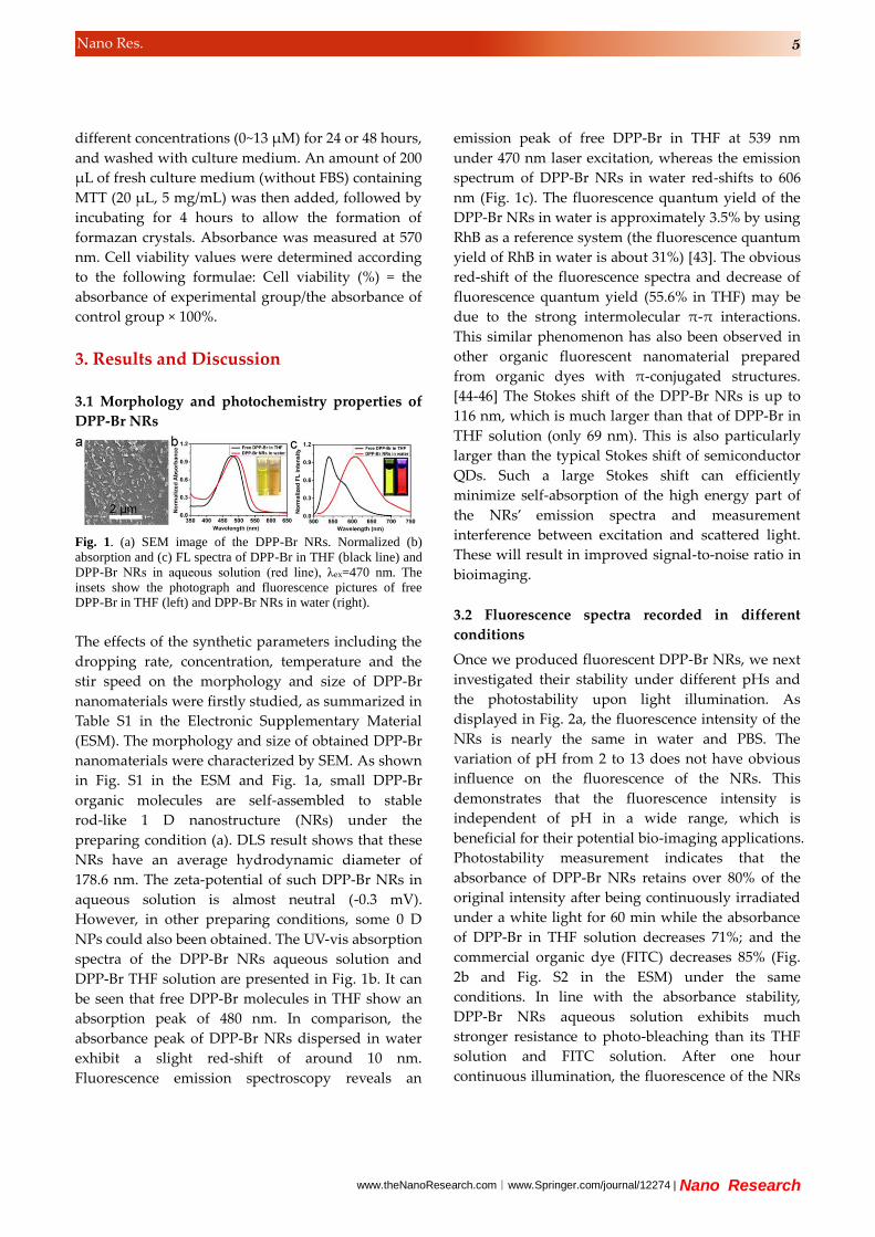
Fig. 1. (a) SEM image of the DPP-Br NRs. Normalized (b)
absorption and (c) FL spectra of DPP-Br in THF (black line) and
DPP-Br NRs in aqueous solution (red line), λex=470 nm. The
insets show the photograph and fluorescence pictures of free DPP-Br in THF (left) and DPP-Br NRs in water (right).
The effects of the synthetic parameters including the
dropping rate, concentration, temperature and the
stir speed on the morphology and size of DPP-Br
nanomaterials were firstly studied, as summarized in
Table S1 in the Electronic Supplementary Material
(ESM). The morphology and size of obtained DPP-Br
nanomaterials were characterized by SEM. As shown
in Fig. S1 in the ESM and Fig. 1a, small DPP-Br
organic molecules are self-assembled to stable
rod-like 1 D nanostructure (NRs) under the
preparing condition (a). DLS result shows that these
NRs have an average hydrodynamic diameter of
178.6 nm. The zeta-potential of such DPP-Br NRs in
aqueous solution is almost neutral (-0.3 mV).
However, in other preparing conditions, some 0 D
NPs could also been obtained. The UV-vis absorption
spectra of the DPP-Br NRs aqueous solution and
DPP-Br THF solution are presented in Fig. 1b. It can
be seen that free DPP-Br molecules in THF show an
absorption peak of 480 nm. In comparison, the
absorbance peak of DPP-Br NRs dispersed in water
exhibit a slight red-shift of around 10 nm.
Fluorescence emission spectroscopy reveals an
emission peak of free DPP-Br in THF at 539 nm
under 470 nm laser excitation, whereas the emission
spectrum of DPP-Br NRs in water red-shifts to 606
nm (Fig. 1c). The fluorescence quantum yield of the
DPP-Br NRs in water is approximately 3.5% by using
RhB as a reference system (the fluorescence quantum
yield of RhB in water is about 31%) [43]. The obvious
red-shift of the fluorescence spectra and decrease of
fluorescence quantum yield (55.6% in THF) may be
due to the strong intermolecular π-π interactions.
This similar phenomenon has also been observed in
other organic fluorescent nanomaterial prepared
from organic dyes with π-conjugated structures.
[44-46] The Stokes shift of the DPP-Br NRs is up to
116 nm, which is much larger than that of DPP-Br in
THF solution (only 69 nm). This is also particularly
larger than the typical Stokes shift of semiconductor
QDs. Such a large Stokes shift can efficiently
minimize self-absorption of the high energy part of
the NRs’ emission spectra and measurement
interference between excitation and scattered light.
These will result in improved signal-to-noise ratio in
bioimaging.
3.2 Fluorescence spectra recorded in different
conditions
Once we produced fluorescent DPP-Br NRs, we next
investigated their stability under different pHs and
the photostability upon light illumination. As
displayed in Fig. 2a, the fluorescence intensity of the
NRs is nearly the same in water and PBS. The
variation of pH from 2 to 13 does not have obvious
influence on the fluorescence of the NRs. This
demonstrates that the fluorescence intensity is
independent of pH in a wide range, which is
beneficial for their potential bio-imaging applications.
Photostability measurement indicates that the
absorbance of DPP-Br NRs retains over 80% of the
original intensity after being continuously irradiated
under a white light for 60 min while the absorbance
of DPP-Br in THF solution decreases 71%; and the
commercial organic dye (FITC) decreases 85% (Fig.
2b and Fig. S2 in the ESM) under the same
conditions. In line with the absorbance stability,
DPP-Br NRs aqueous solution exhibits much
stronger resistance to photo-bleaching than its THF
solution and FITC solution. After one hour
continuous illumination, the fluorescence of the NRs

| www.editorialmanager.com/nare/default.asp
6 Nano Res.
can retain 83% while DPP-Br in THF solution and the
FITC solution decrease 72% (Fig. 2c). In addition,
MEH-PPV NPs with an average diameter of 63 nm
(Fig. S3 in the ESM) were synthesized through
co-precipitation method and their photostability
were further investigated. As shown in Fig. 2b and 2c,
under white light irradiation, the absorbance and the
FL intensity of the NPs decreased to 5%. All above
results indicate that the as-prepared DPP-Br NRs
have excellent photostability. Moreover, the NRs
suspension can be stored for at least one week in air
at room temperature, without the observation of any
precipitates and with only slight loss of fluorescence
intensity (Fig. 2d).
Fig. 2. (a) The effect of pH value on the FL spectra of DPP-Br
NRs aqueous solution. A comparison of (b) photostability and (c)
photo-bleaching of DPP-Br NRs in aqueous solution, DPP-Br in
THF solution, the traditional fluorescein dye (fluorescein sodium,
FITC) and MEH-PPV NPs in aqueous solution. All samples were
continuously irradiated using a 500 W xenon lamp. The
absorbance and FL intensity were normalized. The corresponding
absorption and FL spectra after irradiation 0 to 60 min were
shown in Fig. S2 in the ESM. (d) Time-dependent normalized FL intensity of DPP-Br NRs aqueous solution at 606 nm.
The complexity of intracellular system presents a
great challenge to the living cell imaging. The FL
spectra of DPP-Br NRs were recorded in NaCl
solution with different concentrations to verify the
stability of DPP-Br NRs under high ionic strength
environments. As shown in Fig. 3a, only slight
increase of the FL intensities at 606 nm is observed
even in 1 M NaCl solution. This clearly reveals that
the DPP-Br NRs are stable at high ionic strength
conditions. In addition, heavy metal ions such as
Hg2+ and Cu2+ often quench the fluorescence of
semiconductor QDs and carbon NPs through redox
reactions or electron transfer [47, 48]. Thus, the metal
ions effect on the FL of DPP-Br NRs were carried out
by monitoring the FL intensity of the NRs aqueous
solution at 606 nm in the presence of metal ions that
may coexist in living cells. Remarkably, as shown in
Fig. 3b, trivial changes of the signal are observed
when the NRs are interfered with various types of
heavy metal ions (20 μM). Taking into account that
some biological molecules in biological systems may
affect the fluorescence property of fluorophores,
several typical amino acids, RNA and DNA were
added to the solution to examine the potential
influence. As shown in Fig. 3c, negligible variations
of the FL intensity of DPP-Br NRs aqueous solution
at 606 nm are observed with their exposure to a high
concentration of amino acids (20 μM), DNA or RNA
(0.1 mg/mL). Overall, all of these results indicate that
our DPP-Br NRs have great potential for bioimaging
applications under physiological conditions. It's
worth noting that our DPP-Br NRs show superior
photostability and comparable environment-stability
(pH, high ionic strength, metal ions and biological
molecules) to that of MEH-PPV NPs (a commonly
used organic fluorescent nanomaterial) (Fig. S4 in the
ESM).
Fig. 3. (a) The effect of NaCl concentrations (0, 0.025, 0.05, 0.1,
0.25, 0.5, 1.0 M) on the FL intensities of DPP-Br NRs aqueous
solution. Ic and I0 represent the FL intensity of the DPP-Br NRs
aqueous solution at 606 nm in the presence different
concentration and absence of NaCl, respectively. The effect of (b)
metal ions and (c) amino acids, RNA and DNA on the FL
intensity of DPP-Br NRs solution at 606 nm. IAnaylte and IBlank
represent the FL intensity of DPP-Br NRs aqueous solution at
606 nm in the presence and absence of kinds of anayltes. (d) Cell
viability values (%) estimated by MTT proliferation tests versus
incubation concentrations of DPP-Br NRs (0, 1.6, 3.2, 6.5, 13.0
μM) at 37 oC for 24 hours (black column) and 48 hours (red

www.theNanoResearch.com∣www.Springer.com/journal/12274 | Nano Research
7 Nano Res.
column).
3.3 Cytotoxicity and in vitro cell imaging
In order to assess the NRs for potential biomedical
imaging applications, the cytotoxicity of DPP-Br NRs
to A549 cells was investigated by the standard MTT
assay. The cell viabilities of A549 cells were examined
upon exposure to the DPP-Br NRs with different
concentrations. Fig. 3d shows that the NRs exhibit
low cytotoxicity. The viability of the cells after 24
hours incubation retains about 100% even at a
concentration of 13 μM. When the incubation
extends to 48 hours, the cell viability is still about
90%. This result is similar with MEH-PPV NPs (Fig.
S5 in the ESM).
After confirming that the DPP-Br NRs possess low
cytotoxicity and are stable in physiological
environments, we tested their practical application
for living cell imaging. A549 cells were again chosen
and the nuclei were stained with DAPI (blue
staining). The results are shown in Fig. 4, as expected,
a remarkable intra-cellular red fluorescence is
observed in Fig. 4c. The overlay of the NRs
fluorescence and nuclei staining in Fig. 4d suggests
that the NRs are predominantly delivered to the
cytoplasm of the cells.
Fig. 4. Subcellular localization of DPP-Br NRs monitored by
fluorescence imaging in A549 cells. (a) bright field channel; (b)
blue fluorescence channel (DAPI channel); (c) red fluorescence
channel (DPP-Br NRs channel); (d) Overlap of the above images. Scale bar is 40 μm.
4. Conclusions
In summary, heavy metal free, organic small
molecule based red fluorescence DPP-Br NRs were
prepared through a co-precipitation approach. The
as-prepared NRs are a promising candidate for
living cell imaging due to their combined
superiorities for bioapplications, including
outstanding fluorescence properties (large Stokes
shift, photostability, and high resistance to
photo-bleaching, good stability in wide pH range
and at high ionic strength condition), good water
solubility, and excellent biocompatibility.
Acknowledgements
This work was supported by General Research Fund
of Hong Kong (CityU 104911) and National Natural
Science Foundation of China (NSFC 51372213 and
NSFC 61176007).
Electronic Supplementary Material: Supplementary
material (The detailed preparing conditions of the
obtained nanomaterials and their corresponding
SEM images; photostability and anti-bleaching
measurements; environments stability and
cytotoxicity of MEH-PPV NPs) is available in the
online version of this article at
http://dx.doi.org/10.1007/s12274-***-****-*
(automatically inserted by the publisher). References [1] Huh, Y. M.; Jun, Y. W.; Song, H. T.; Kim, S.; Choi, J. S.;
Lee, J. H.; Yoon, S.; Kim, K. S.; Shin, J. S.; Suh, J. S.;
Cheon, J. In vivo magnetic resonance detection of cancer
by using multifunctional magnetic nanocrystals. J. Am.
Chem. Soc. 2005, 127, 12387-12391.
[2] Haris, M.; Singh, A.; Cai, K.; McArdle, E.; Fenty, M.;
Davatzikos, C.; Trojanowski, J. Q.; Melhem, E. R.; Clark C.
M.; Borthakur, A. T1ρ MRI in Alzheimer’s disease:
detection of pathological changes in medial temporal lobe.
J Neuroimaging 2011, 21, 86-90.
[3] Kaur, S.; Baine, M. J.; Jain, M.; Sasson, A. R.; Batra, S. K.
Early diagnosis of pancreatic cancer: challenges and new
developments. Biomark. Med. 2012, 6, 597-612.
[4] Acharya, R.; Wasserman, R.; Stevens, J.; Hinojosa, C.
Biomedical imaging modalities: a tutorial. Comput. Med.
Imaging. Graph. 1995, 19, 3-25.
[5] Hurtley, S. M.; Helmuth, L. Special Issue on Biological
Imaging. Science, 2003, 300, 75-102.
[6] Stadtmauer, L. A.; Tur-Kaspa, I. Ultrasound Imaging in
Reproductive Medicine, Springer-Verlag London Limited,
2014.
[7] Cierniak, R. X-Ray Computed Tomography in Biomedical
Engineering, Springer-Verlag London Limited, 2011.
[8] Morris, P. G. Nuclear Magnetic Resonance Imaging in

| www.editorialmanager.com/nare/default.asp
8 Nano Res.
Medicine and Biology, Oxford University Press, 1986.
[9] National Research Council (US) and Institute of Medicine
(US) Committee on State of the Science of Nuclear
Medicine, Advancing Nuclear Medicine Through
Innovation, National Academies Press (US), 2007.
[10] Gabriel, S.; Lau, R. W.; Gabriel, C. The dielectric
properties of biological tissues: II. Measurements in the
frequency range 10 Hz to 20 GHz. Phys. Med. Biol. 1996,
41, 2251-2269.
[11] Fear, E. C.; Stuchly, M. A. Microwave detection of breast
cancer. IEEE Trans. Microwave Theory Tech. 2000, 48,
1854-1863.
[12] Luker, G. D.; Luker, K. E. Optical imaging: current
applications and future directions. J. Nucl. Med. 2008, 49,
1-4.
[13] Tromberg, B. J.; Pogue, B. W.; Paulsen, K. D.; Yodh, A. G.;
Boas, D. A.; Cerussi, A. E. Assessing the future of diffuse
optical imaging technologies for breast cancer management.
Med. Phys. 2008, 35, 2443-2451.
[14] van Dam, G. M.; Themelis, G.; Crane, L. M.; Harlaar, N. J.;
Pleijhuis, R. G.; Kelder, W.; Sarantopoulos, A.; de Jong, J.
S.; Arts, H. J.; van der Zee, A. G.; Bart, J.; Low, P. S.;
Ntziachristos, V. Intraoperative tumor-specific fluorescence
imaging in ovarian cancer by folate receptor-α targeting:
first in-human results. Nat. Med. 2011, 17, 1315-1319.
[15] Sevick-Muraca, E. M. Translation of near-infrared
fluorescence imaging technologies: emerging clinical
applications. Annu. Rev. Med. 2012, 63, 217-231.
[16] Dedecker, P.; Mo, G. C. H.; Dertinger, T.; Zhang, J. Widely
accessible method for superresolution fluorescence
imaging of living systems. Proc. Natl. Acad. Sci. U S A
2012, 109, 10909-10914.
[17] Luo, S. L.; Zhang, E. L.; Sun, Y. P.; Cheng, T. M.; Shi, C.
M. A review of NIR dyes in cancer targeting and imaging.
Biomaterials 2011, 32, 7127-7138.
[18] Yuan, L.; Lin, W. Y.; Zheng, K. B.; He, L. W.; Huang, W.
M. Far-red to near infrared analyte-responsive fluorescent
probes based on organic fluorophore platforms for
fluorescence imaging. Chem. Soc. Rev. 2013, 42, 622-661.
[19] Gao, X. H.; Cui, Y. Y.; Levenson, R. M.; Chung, L. W. K.;
Nie, S. M. In vivo cancer targeting and imaging with
semiconductor quantum dots. Nat. Biotechnol. 2004, 22,
969-976.
[20] Michalet, X.; Pinaud, F. F.; Bentolila, L. A.; Tsay, J. M.;
Doose, S.; Li, J. J.; Sundaresan, G.; Wu, A. M.; Gambhir, S.
S.; Weiss, S. Quantum dots for live cells, in vivo imaging,
and diagnostics. Science 2005, 307, 538-544.
[21] Resch-Genger, U.; Grabolle, M.; Cavaliere-Jaricot, S.;
Nitschke, R.; Nann, T. Quantum dots versus organic dyes
as fluorescent labels. Nat. Methods 2008, 5, 763-775.
[22] Yao, J.; Yang, M.; Duan, Y. X. Chemistry, biology, and
medicine of fluorescent nanomaterials and related systems:
new insights into biosensing, bioimaging, genomics,
diagnostics, and therapy. Chem. Rev. 2014, 114,
6130-6178.
[23] Derfus, A. M.; Chan, W. C. W.; Bhatia, S. N. Probing the
cytotoxicity of semiconductor quantum dots. Nano Lett.
2004, 4, 11-18.
[24] Chan, W. H.; Shiao, N. H.; Lu, P. Z. CdSe quantum dots
induce apoptosis in human neuroblastoma cells via
mitochondrial-dependent pathways and inhibition of
survival signals. Toxicol. Lett. 2006, 167, 191-200.
[25] Chen, N.; He, Y.; Su, Y. Y.; Li, X. M.; Huang, Q.; Wang, H.
F.; Zhang, X. Z.; Tai, R. Z.; Fan, C. H. The cytotoxicity of
cadmium-based quantum dots. Biomaterials 2012, 33,
1238-1244.
[26] Tsoi, K. M.; Dai, Q.; Alman, B. A.; Chan, W. C. W. Are
quantum dots toxic? Exploring the discrepancy between
cell culture and animal studies. Acc. Chem. Res. 2013, 46,
662-671.
[27] Liu, Y. S.; Sun, Y. H.; Vernier, P. T.; Liang, C. H.; Chong, S.
Y. C.; Gundersen, M. A. pH-sensitive photoluminescence
of CdSe/ZnSe/ZnS quantum dots in human ovarian cancer
cells. J. Phys. Chem. C 2007, 111, 2872-2878.
[28] Saleh, S. M.; Ali, R.; Wolfbeis, O. S. Quenching of the
luminescence of upconverting luminescent nanoparticles
by heavy metal ions. Chem. Eur. J. 2011, 17, 14611-14617.
[29] Zhang, X.; Servos, M. R.; Liu, J. W. Ultrahigh nanoparticle
stability against salt, pH, and solvent with retained surface
accessibility via depletion stabilization. J. Am. Chem. Soc.
2012, 134, 9910-9913.
[30] An, F. F.; Ye, J.; Zhang, J. F.; Yang, Y. L.; Zheng, C. J.;
Zhang, X. J.; Liu, Z.; Lee, C. S.; Zhang, X. H.
Non-blinking, highly luminescent, pH- and
heavy-metal-ion-stable organic nanodots for bio-imaging. J.
Mater. Chem. B 2013, 1, 3144-3151.
[31] Miao, R.; Mu, L. X.; Zhang, H. Y.; She, G. W.; Zhou, B. J.;
Xu, H. T.; Wang P. F.; Shi, W. S. Silicon nanowire-based
fluorescent nanosensor for complexed Cu2+ and its
bioapplications. Nano Lett. 2014, 14, 3124-3129.
[32] Susumu, K.; Oh, E.; Delehanty, J. B.; Blanco-Canosa, J. B.;
Johnson, B. J.; Jain, V.; IV, W. J. H.; Algar, W. R.;
Boeneman, K.; Dawson, P. E.; Medintz, I. L.
Multifunctional compact zwitterionic ligands for preparing
robust biocompatible semiconductor quantum dots and
gold nanoparticles. J. Am. Chem. Soc. 2011, 133,
9480-9496.
[33] Schieber, C.; Bestetti, A.; Lim, J. P.; Ryan, A. D.; Nguyen,
T. L.; Eldridge, R.; White, A. R.; Gleeson, P. A.; Donnelly,
P. S.; Williams, S. J.; Mulvaney, P. Conjugation of
transferrin to azide-modified CdSe/ZnS core-shell quantum
dots using cyclooctyne click chemistry. Angew. Chem. Int.
Ed. 2012, 51, 10523-10527.
[34] Wu, C. F.; Chiu, D. T. Highly fluorescent semiconducting
polymer dots for biology and medicine. Angew. Chem. Int.
Ed. 2013, 52, 3086-3109.
[35] Feng, L. H.; Zhu, C. L.; Yuan, H. X.; Liu, L. B.; Lv, F. T.;
Wang, S. Conjugated polymer nanoparticles: preparation,
properties, functionalization and biological applications.
Chem. Soc. Rev. 2013, 42, 6620-6633.
[36] Yu, J.; Zhang, X. J.; Hao, X. J.; Zhang, X. H.; Zhou, M. J.;
Lee, C. S.; Chen, X. F. Near-infrared fluorescence imaging
using organic dye nanoparticles. Biomaterials 2014, 35,
3356-3364.
[37] Wu, C. F.; Schneider, T.; Zeigler, M.; Yu, J. B.; Schiro, P.
G.; Burnham, D. R.; McNeill, J. D.; Chiu, D. T.
Bioconjugation of ultrabright semiconducting polymer dots
for specific cellular targeting. J. Am. Chem. Soc. 2010, 132,

www.theNanoResearch.com∣www.Springer.com/journal/12274 | Nano Research
9 Nano Res.
15410-15417.
[38] Wu, C. F.; Hansen, S. J.; Hou, Q.; Yu, J. B.; Zeigler, M.; Jin,
Y. H.; Burnham, D. R.; McNeill, J. D.; Olson, J. M. Chiu,
D. T. Design of highly emissive polymer dot bioconjugates
for in vivo tumor targeting. Angew. Chem. 2011, 123,
3492-3496.
[39] Park, J. H.; Von Maltzahn, G.; Zhang, L.; Derfus, A. M.;
Simberg, D.; Harris, T. J.; Ruoslahti, E.; Bhatia, S. N.;
Sailor, M. J. Systematic surface engineering of magnetic
nanoworms for in vivo tumor targeting. Small 2009, 5,
694-700.
[40] Shin, S.; Gihm, S. H.; Park, C. R.; Kim, S.; Park, S. Y.
Water-soluble fluorinated and PEGylated cyanostilbene
derivative: an amphiphilic building block forming
self-assembled organic nanorods with enhanced
fluorescence emission. Chem. Mater. 2013, 25, 3288-3295.
[41] Chauhan, V. P.; Popović, Z.; Chen, O.; Cui, J.; Fukumura,
D.; Bawendi, M. G.; Jain, R. K. Fluorescent nanorods and
nanospheres for real-time in vivo probing of nanoparticle
shape-dependent tumor penetration. Angew. Chem. Int. Ed.
2011, 50, 11417-11420.
[42] Kolhar, P.; Anselmo, A. C.; Gupta, V.; Pant, K.;
Prabhakarpandian, B.; Ruoslahti, E.; Mitragotri. S. Using
shape effects to target antibody-coated nanoparticles to
lung and brain endothelium. Proc. Natl. Acad. Sci. U S A
2013, 110, 10753-10758.
[43] Magde, D.; Rojas, G. E.; Seybold, P. G. Solvent
dependence of the fluorescence lifetimes of xanthene dyes.
Photochem. Photobiol. 1999, 70, 737-744.
[44] Bao. B. Q.; Tao, N. J.; Yang, D. L.; Yuwen, L. H.; Weng, L.
X.; Fan, Q. L.; Huang, Q.; Wang, L. H. A controllable
approach to development of multi-spectral conjugated
polymer nanoparticles with increased emission for cell
imaging. Chem. Commun. 2013, 49, 10623-10625.
[45] Bao, B. Q.; Ma, M. F.; Chen, J.; Yuwen, L. H.; Weng, L. X.;
Fan, Q. L.; Huang, W.; Wang, L. H. Facile preparation of
multicolor polymer nanoparticle bioconjugates with
specific biorecognition. ACS Appl. Mater. Interfaces 2014,
6, 11129-11135.
[46] Li, M.; Feng, L. H.; Lu, H. Y.; Wang, S.; Chen, C. F.
Tetrahydro[5]helicene-based nanoparticles for
structure-dependent cell fluorescent imaging. Adv. Funct.
Mater. 2014, 24, 4405-4412.
[47] Beaune, G.; Tamang, S.; Bernardin, A.; Bayle-Guillemaud,
P.; Fenel, D.; Schoehn, G.; Vinet, F.; Reiss, P.; Texier, I.
Luminescence of polyethylene glycol coated CdSeTe/ZnS
and InP/ZnS nanoparticles in the presence of copper
cations. ChemPhysChem. 2011, 12, 2247-2254.
[48] Zhou, L.; Lin, Y. H.; Huang, Z. Z.; Ren, J. S.; Qu, X. G.
Carbon nanodots as fluorescence probes for rapid, sensitive,
and label-free detection of Hg2+ and biothiols in complex
matrices. Chem. Commun. 2012, 48, 1147-1149.


www.theNanoResearch.com∣www.Springer.com/journal/12274 | Nano Research
Nano Res.
Electronic Supplementary Material
Highly stable organic fluorescent nanorods for living
cell imaging
Minhuan Lan1, †, Jinfeng Zhang1, †, Xiaoyue Zhu1, Pengfei Wang2(), Xianfeng Chen1(), Chun-Sing
Lee1, and Wenjun Zhang1()
†
Supporting information to DOI 10.1007/s12274-****-****-* (automatically inserted by the publisher)
Preparing
conditions
Concentration of
DPP-Br (mM)
Dropping rate
(second/drop)
Temperature
(oC)
Stir speed
(rpm)
a 2 2 25 1000
b 1 2 25 1000
c 0.5 2 25 1000
d 2 2 25 2000
e 2 2 25 500
f 2 5 25 1000
g 2 Quick injection 25 1000
h 2 2 50 1000
i 2 2 0 1000
Table S1. The detailed preparing conditions of the obtained nanomaterials.
Address correspondence to Wenjun Zhang, email: [email protected]; Xianfeng Chen, email: [email protected]; Pengfei Wang, email: [email protected].

| www.editorialmanager.com/nare/default.asp
Nano Res.
Fig. S1. (a~i) The SEM images of the obtained nanomaterials prepared using condition (a) to (i).
Fig. S2. (a~d) Absorption and (e~h) FL spectra of the DPP-Br NRs in aqueous (a, e), DPP-Br in THF (b, f), FITC
(c,g) and MEH-PPV NPs (d, h) in aqueous solutions after irradiation 0 to 60 min.

www.theNanoResearch.com∣www.Springer.com/journal/12274 | Nano Research
Nano Res.
Fig. S3. (a) SEM image of the MEH-PPV NPs. (b) The particle size distribution histograms of the NPs.
Fig. S4. (a) The effect of pH value on the FL spectra of MEH-PPV NPs aqueous solution (λex=500 nm). (b) The
effect of NaCl concentrations (0, 0.025, 0.05, 0.1, 0.25, 0.5, 1.0 M) on the FL intensities of MEH-PPV NPs
aqueous solution. Ic and I0 represent the FL intensity of the MEH-PPV NPs aqueous solution at 568 nm in the
presence different concentration and absence of NaCl, respectively. The effect of (c) metal ions and (d) amino
acids, RNA and DNA on the FL intensity of MEH-PPV NPs solution at 568 nm. IAnaylte and IBlank represent the
FL intensity of MEH-PPV NPs aqueous solution at 568 nm in the presence and absence of kinds of anayltes.

| www.editorialmanager.com/nare/default.asp
Nano Res.
Fig. S5. Cell viability values (%) estimated by MTT proliferation tests versus incubation concentrations of
MEH-PPV NPs (0, 1.6, 3.2, 6.5, 13.0 μM) at 37 oC for 24 hours (black column) and 48 hours (red column).