Embed Size (px)
Citation preview
antibody isotyping Guide
high performance immunoassays
Table of Contents Antibody Isotyping
Immunoglobulin (Ig) Overview 1
Introduction to Immunoglobulins. . . . . . . . . . . . . 1
1. Immunoglobulin 3
Immunoglobulin Structure . . . . . . . . . . . . . . . . . 3
Antibody Isotypes and Properties . . . . . . . . . . . . . 6
2. Monoclonal Antibody Development 10
Determine Antibody Isotypes With Ease
and Accuracy . . . . . . . . . . . . . . . . . . . . . . . . . 10
Isotyping With the ELISA Technique. . . . . . . . . . . 10
Assess Immune Response After Vaccination
or Infection . . . . . . . . . . . . . . . . . . . . . . . . . . 13
3. Antibody Isotyping Kits 14
Multiple Solutions for the Detection/Quantification
of Soluble Immunoglobulins . . . . . . . . . . . . . . . 14
Product Overview . . . . . . . . . . . . . . . . . . . . . . 19
high performance immunoassays
eBioscience is committed to developing and manufacturing high-
quality, innovative reagents in an ISO certified facility. As a provider of
more than 10,000 products, we empower our customers worldwide
to obtain exceptional results by using reagents that offer a new
standard of excellence in the areas of innovation, quality and value.
For Research Use Only. Not for use in diagnostic or therapeutic procedures.All designated trademarks used are the property of their respective owners.
1
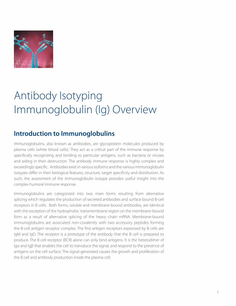
Antibody Isotyping Immunoglobulin (Ig) Overview
Introduction to ImmunoglobulinsImmunoglobulins, also known as antibodies, are glycoprotein molecules produced by plasma cells (white blood cells). They act as a critical part of the immune response by specifically recognizing and binding to particular antigens, such as bacteria or viruses and aiding in their destruction. The antibody immune response is highly complex and exceedingly specific. Antibodies exist in various isoforms and the various immunoglobulin isotypes differ in their biological features, structure, target specificity and distribution. As such, the assessment of the immunoglobulin isotype provides useful insight into the complex humoral immune response.
Immunoglobulins are categorized into two main forms resulting from alternative splicing which regulates the production of secreted antibodies and surface bound B-cell receptors in B cells. Both forms, soluble and membrane-bound antibodies, are identical with the exception of the hydrophobic transmembrane region on the membrane-bound form as a result of alternative splicing of the heavy chain mRNA. Membrane-bound immunoglobulins are associated non-covalently with two accessory peptides forming the B-cell antigen receptor complex. The first antigen receptors expressed by B cells are IgM and IgD. The receptor is a prototype of the antibody that the B cell is prepared to produce. The B cell receptor (BCR) alone can only bind antigens. It is the heterodimer of Igα and Igβ that enables the cell to transduce the signal, and respond to the presence of antigens on the cell surface. The signal generated causes the growth and proliferation of the B cell and antibody production inside the plasma cell.
2
Antibody Isotypes
The various antibodies produced by plasma cells are classified by their isotypes that differ in function and antigen responses. Five major antibody isotypes have been identified in placental mammals:
• IgA
• IgD
• IgE
• IgG
• IgM
Antibody isotypes are categorized according to differences in their amino acid sequence in the constant region (Fc) of the antibody heavy chains.
The antibody isotypes IgG and IgA are further grouped into subclasses (e.g. human IgG1, IgG2, IgG3, IgG4, IgA1 and IgA2) based on additional small differences in their amino acid heavy chain sequences. Determination of individual subclasses is relevant in assessing primary immunodeficiencies or immune responses, especially if the total IgG or IgA concentration is not altered.
In addition to the antibody heavy chains, differences in the amino acid sequence in the constant region of the antibody light chain can be further subclassified. These two types of light chains are referred to as kappa (κ) or lambda (λ). A light chain has two successive domains: one constant and one variable domain.
Isotype Class Switching
Humoral response through specific antibody secretion to address invading bodies and their toxic products is one of the primary functions of B cells in adaptive immunity. Some of these cells undergo a “class switch” that results in the expression of a new antibody isotype. For example, an antibody isotype could switch from an IgM to an antibody of any of the possible classes (e.g. IgG1, IgG4, IgE). During this switch, the constant region of the heavy chain is changed, but the variable regions of the heavy chain and light chain that are specific to the antigen remain unchanged. This switch does not affect the antigen’s specificity, but rather the effector functions that each class of antibody can execute. The antibody class switch is critically dependent on the type of cytokine(s) present. Various cytokines, such as IL-4, IL-5, IFNγ and TGFβ, are known to be responsible for class switching. At a certain stage, the cell will lose its ability to undergo a switch to a class that has been generated before.
3
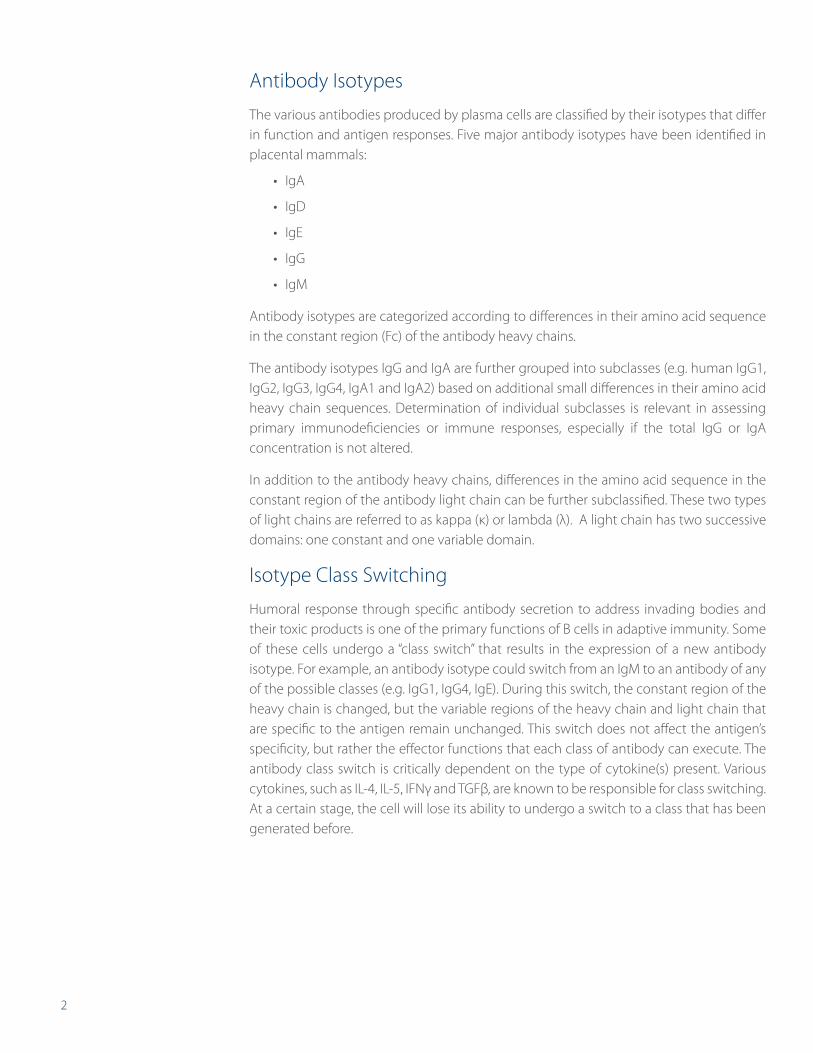
Immunoglobulin
Immunoglobulin StructureAll immunoglobulins, independent of their isotype and specificity, have a common structure with four polypeptide chains:
• Two identical heavy (H) chains with covalently attached oligosaccharide groups
• Two identical, non-glycosylated light (L) chains
Within the immunoglobulin, disulphide bonds form between:
• Two heavy chains
• The heavy chains to the light chains
The disulphide bonds joining the antibody heavy chains are located in a flexible region of the heavy chain known as the hinge region. The effector functions of the antibody, such as placental transport or antigen-dependent cellular toxicity, are determined by the constant region of the heavy chain.
Most species produce two major classes of light chains, kappa and lambda, denoted as κ and λ. The ratio of these chains differs among species though. It is key to note that in naturally occurring, non-engineered antibodies, the light chains within the specific antibody are the same, either both k or both λ.
1
IgM
IgG IgA
Hypervariable Region
LEGEND
Constant Region
Variable Region
Light Chain
Heavy Chain
Oligosaccharide
Hinge Region
Typical Antibody Molecules
4
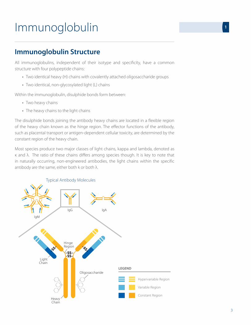
Fragment Antigen Binding (Fab) Region
The Fab regions of the antibody are the two “arms” of the antibody that recognize and bind to antigens in a target specific manner. In general, Fab regions are identical and can each bind to antigens. Each arm is composed of both a variable region (at the end of the antibody) and a constant region for both the heavy and light chain portions of the Fab fragment. Each of the two Fab regions of the antibody, as shown in the figure below, are composed of four domains:
• Two variable domains (VH, VL)
• Two constant domains (CH, CL)
These variable domains are attached to the constant domains. As the name implies, the variable domains vary in their amino acid sequence from one antibody molecule to another, providing the vast diversity the immune system needs to fight foreign invaders. The antigen binding site is formed where a heavy chain variable domain (VH) and a light chain variable domain (VL) come close together. It is the antibody binding sites which have the highest degree of variability between different antibodies.
Hypervariable Region
LEGEND
Constant Region
Variable Region
VL
VH
CH1
N
CLight
Chain
CHeavy Chain
Antigen Binding Site
Oligosaccharide
Fab
Fc
Hinge Region
CL
Fab Fragment, antigen-binding
Fc Fragment, crystallizable
CL Constant domain, Light Chain
CH Constant domain, Heavy Chain
VL Variable domain, Light Chain
VH Variable domain, Heavy Chain
IgG Antibody Molecule
5
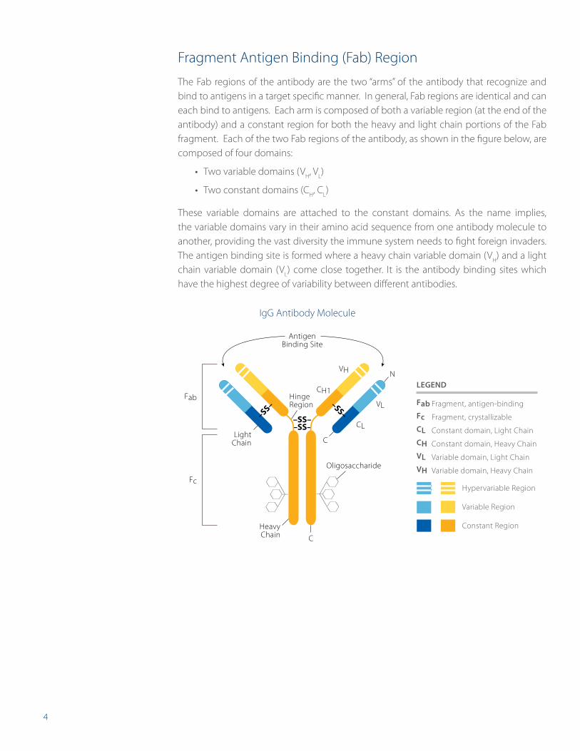
Proteolytic Cleavage of Immunoglobulins
The use of immunoglobulin proteolytic cleavage can be advantageous for various experimental techniques, and as such, is fairly common. Proteolytic treatment with the enzyme papain cleaves the antibody into three fragments of approximately the same size, as described in the figure above (left). Two of these fragments are Fab fragments, which retain the ability to bind antigens. The third fragment, known as fragment crystallizable (Fc), cannot bind with antigens. It is responsible for biological effector functions which include complement fixation and binding to macrophages, natural killer cells and neutrophils.
Treatment with the enzyme pepsin results in a divalent fragment called F(ab‘)2 consisting of the two Fab fragments that are joined by disulfide bonds and a Fc fragment, as shown in the figure above (right).
FabFab
Fc
F(ab’)2
Fc
Proteolytic cleavage by papain Proteolytic cleavage by pepsin
6
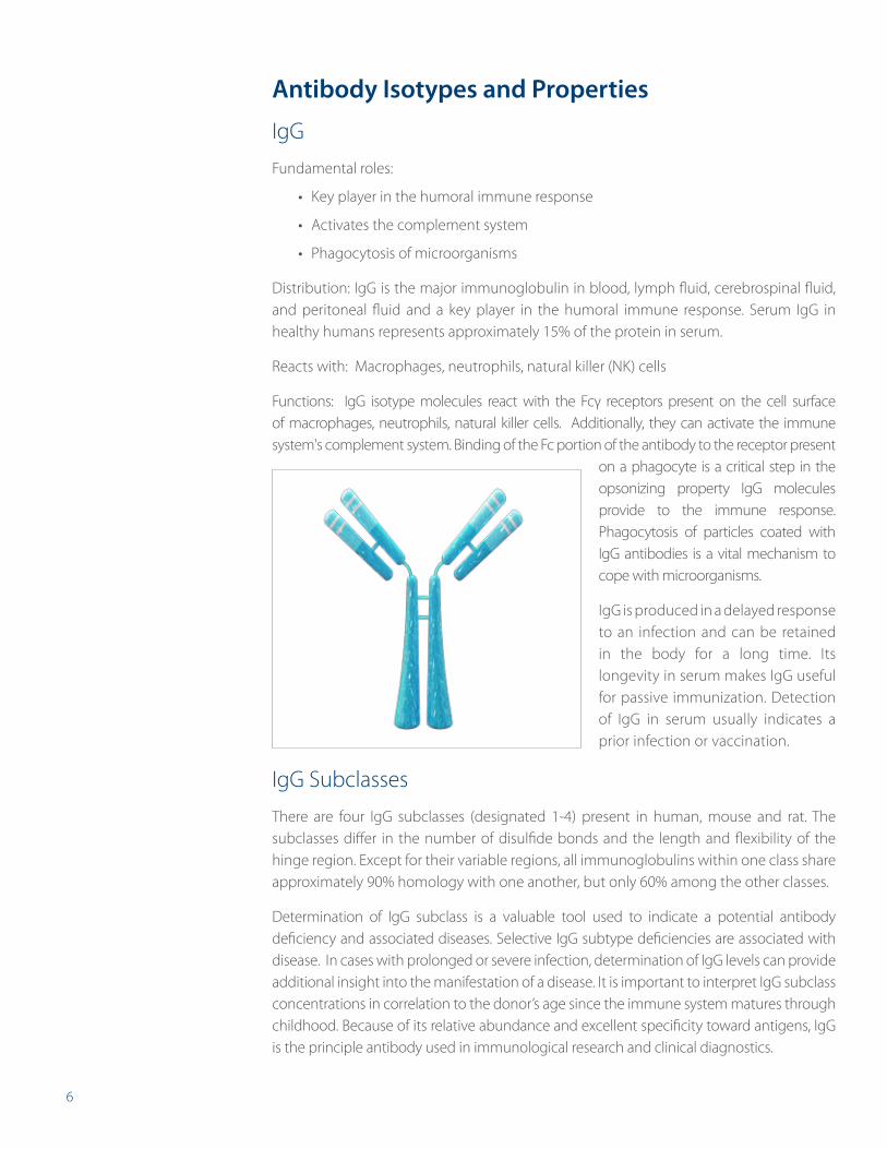
Antibody Isotypes and Properties
IgG
Fundamental roles:
• Key player in the humoral immune response
• Activates the complement system
• Phagocytosis of microorganisms
Distribution: IgG is the major immunoglobulin in blood, lymph fluid, cerebrospinal fluid, and peritoneal fluid and a key player in the humoral immune response. Serum IgG in healthy humans represents approximately 15% of the protein in serum.
Reacts with: Macrophages, neutrophils, natural killer (NK) cells
Functions: IgG isotype molecules react with the Fcγ receptors present on the cell surface of macrophages, neutrophils, natural killer cells. Additionally, they can activate the immune system's complement system. Binding of the Fc portion of the antibody to the receptor present
on a phagocyte is a critical step in the opsonizing property IgG molecules provide to the immune response. Phagocytosis of particles coated with IgG antibodies is a vital mechanism to cope with microorganisms.
IgG is produced in a delayed response to an infection and can be retained in the body for a long time. Its longevity in serum makes IgG useful for passive immunization. Detection of IgG in serum usually indicates a prior infection or vaccination.
IgG Subclasses
There are four IgG subclasses (designated 1-4) present in human, mouse and rat. The subclasses differ in the number of disulfide bonds and the length and flexibility of the hinge region. Except for their variable regions, all immunoglobulins within one class share approximately 90% homology with one another, but only 60% among the other classes.
Determination of IgG subclass is a valuable tool used to indicate a potential antibody deficiency and associated diseases. Selective IgG subtype deficiencies are associated with disease. In cases with prolonged or severe infection, determination of IgG levels can provide additional insight into the manifestation of a disease. It is important to interpret IgG subclass concentrations in correlation to the donor’s age since the immune system matures through childhood. Because of its relative abundance and excellent specificity toward antigens, IgG is the principle antibody used in immunological research and clinical diagnostics.
7
IgG1
IgG1 comprises 60-65% of the total IgG. It is predominantly responsible for the thymus mediated immune response against proteins and polypeptide antigens. IgG1 binds to the Fc-receptor of phagocytic cells and can activate the complement cascade via binding to C1 complex. IgG1 immune response can be measured in newborns and reaches its typical concentration in infancy. A deficiency in IgG1 isotype typically is a sign of a hypogammaglobulinemia.
IgG2
IgG2 comprises 20-25% of the IgG subclass. It is the prevalent immune response against carbohydrate/polysaccharide antigens. Mature concentrations are typically reached around 6-7 years of age. Among all IgG isotypes, a deficiency in IgG2 is the most common and is associated with recurring airway/respiratory infections in infants.
IgG3
IgG3 comprises 5-10% of the total IgG and plays a major role in immune responses against protein/polypeptide antigens. The affinity of IgG3 can be higher than that of IgG1.
IgG4
IgG4 usually comprises less than 4% of the total IgG. It is known to be unable to bind to polysaccharides. Testing for IgG4 has been associated with food allergies. Recent studies have shown that elevated serum levels of IgG4 are found in patients suffering from sclerosing pancreatitis, cholangitis, and interstitial pneumonia caused by infiltrating IgG4 positive plasma cells. The precise role of IgG4 is still relatively unknown.
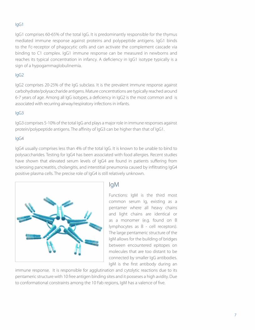
IgM
Functions: IgM is the third most common serum Ig, existing as a pentamer where all heavy chains and light chains are identical or as a monomer (e.g. found on B lymphocytes as B - cell receptors). The large pentameric structure of the IgM allows for the building of bridges between encountered epitopes on molecules that are too distant to be connected by smaller IgG antibodies. IgM is the first antibody during an
immune response. It is responsible for agglutination and cytolytic reactions due to its pentameric structure with 10 free antigen binding sites and it posseses a high avidity. Due to conformational constraints among the 10 Fab regions, IgM has a valence of five.
8
IgM is not as versatile as IgG, but it is of vital importance in complement activation and agglutination. IgM is predominantly found in the lymph fluid and the blood and is a very effective neutralizing agent in the early stages of disease. Elevated levels can be a sign of recent infection or exposure to an antigen.
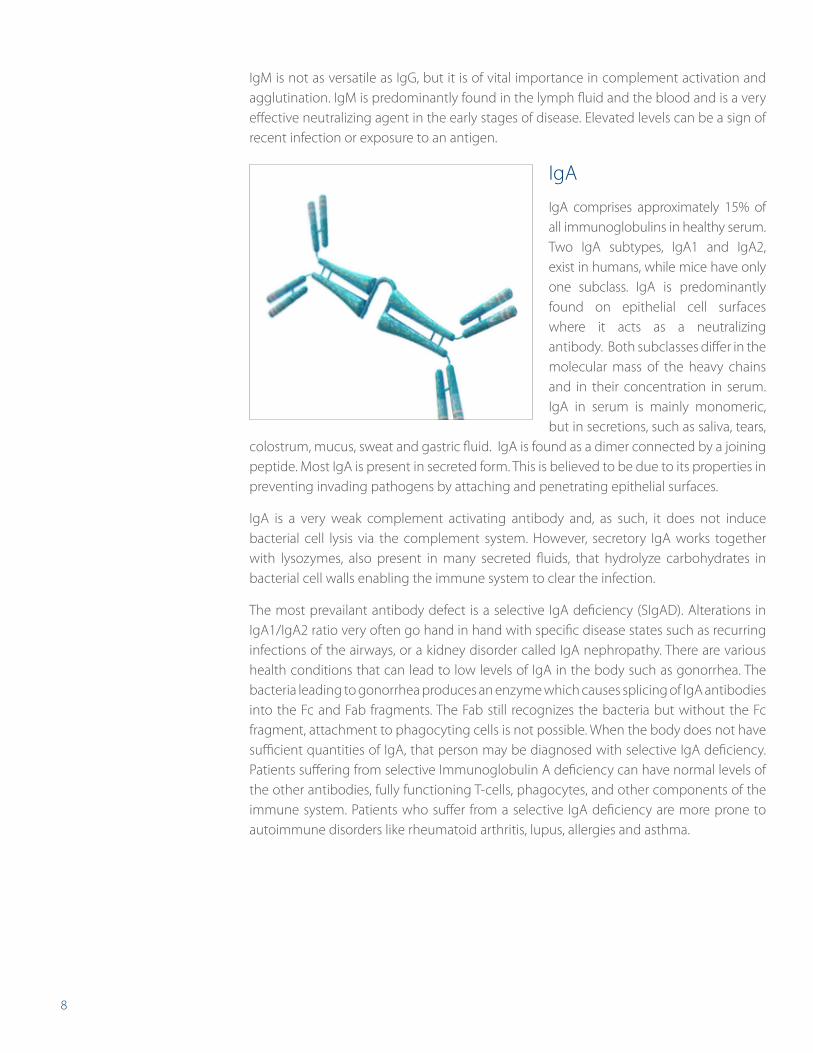
IgA
IgA comprises approximately 15% of all immunoglobulins in healthy serum. Two IgA subtypes, IgA1 and IgA2, exist in humans, while mice have only one subclass. IgA is predominantly found on epithelial cell surfaces where it acts as a neutralizing antibody. Both subclasses differ in the molecular mass of the heavy chains and in their concentration in serum. IgA in serum is mainly monomeric, but in secretions, such as saliva, tears,
colostrum, mucus, sweat and gastric fluid. IgA is found as a dimer connected by a joining peptide. Most IgA is present in secreted form. This is believed to be due to its properties in preventing invading pathogens by attaching and penetrating epithelial surfaces.
IgA is a very weak complement activating antibody and, as such, it does not induce bacterial cell lysis via the complement system. However, secretory IgA works together with lysozymes, also present in many secreted fluids, that hydrolyze carbohydrates in bacterial cell walls enabling the immune system to clear the infection.
The most prevailant antibody defect is a selective IgA deficiency (SIgAD). Alterations in IgA1/IgA2 ratio very often go hand in hand with specific disease states such as recurring infections of the airways, or a kidney disorder called IgA nephropathy. There are various health conditions that can lead to low levels of IgA in the body such as gonorrhea. The bacteria leading to gonorrhea produces an enzyme which causes splicing of IgA antibodies into the Fc and Fab fragments. The Fab still recognizes the bacteria but without the Fc fragment, attachment to phagocyting cells is not possible. When the body does not have sufficient quantities of IgA, that person may be diagnosed with selective IgA deficiency. Patients suffering from selective Immunoglobulin A deficiency can have normal levels of the other antibodies, fully functioning T-cells, phagocytes, and other components of the immune system. Patients who suffer from a selective IgA deficiency are more prone to autoimmune disorders like rheumatoid arthritis, lupus, allergies and asthma.
9
IgA SubclassesIgA1
IgA1 comprises approximately 85% of total IgA concentration in serum. Although IgA1 shows a broad resistance against several proteases, there are some that can cleave at the hinge region. Concerning antigen binding, IgA1 elicits a good immune response to protein antigens and to polysaccharide and lipopolysaccharides to a lesser degree.
IgA2
IgA2 represents only up to 15% of total IgA in serum. It plays a crucial role in the mucosa of the airways, eyes, and in the gastrointestinal tract to protect against polysaccharide and lipopolysaccaride antigens. It also shows strong resistance to proteolysis and many bacterial proteases supporting the importance of IgA2 in fighting bacterial infections.
IgE
Fundamental role: Allergic reactions, parasitic infections, and hypersensitivity reactions. IgE is not fundamental in agglutination or complement activation.
Distribution: widely found in the lungs, skin and mucous membranes.
IgE is the least common serum immunoglobulin. IgE antibodies are predominantly cell bound and bind to allergens. When antigens such as pollen, venoms, fungus, spores, dust mites or pet dander bind with the Fab portion of the IgE attached to the cells, the cells degranulate and release factors like heparin, histamine, proteolytic enzymes, leukotrienes and cytokines. This results in vasodilatation and increased small vessel permeability that causes fluid to escape from capillaries into the tissues and leads to the characteristic symptoms of an allergic reaction. Most of these typical allergic reactions like mucus secretion, sneezing, coughing, or tear production are considered beneficial to expel remaining allergens from the body.
Mouse studies are an important model of research regarding the mechanisms and treatment of allergic responses, and quantification of IgE levels is an important testing parameter.
Studies have shown that conditions such as asthma, rhinitis, eczema, urticaria, dermatitis, and some parasitic infections (e.g. helminthes or tapeworms) lead to increased IgE levels. Binding of eosinophils with Fc receptors to IgE-coated helminths results in killing of the parasite. Low levels of IgE can occur in ataxia-telangiectasia, a rare inherited disease that affects muscle coordination.
10
Monoclonal Antibody Development
Determine Antibody Isotypes With Ease and AccuracyHybridoma screening is one of the most important steps during hybridoma development to ensure that the most productive and specific clones are selected for further evaluation. Whether you are a core facility making monoclonal antibodies every day, or a scientist that is trying to generate a monoclonal antibody for the first time, eBioscience isotyping ELISAs and FlowCytomix™ multiplex assays are an easy solution for istoyping either mouse or rat monoclonal hybridoma cell lines. With less than 1 mL of hybridoma supernatant, you can accurately and specifically identify which heavy and light chain your hybridoma is producing and if it is monoclonal or not. These assay kits are powerful tools to isolate and characterize each potential clone.
Isotyping With the ELISA TechniqueAntibody isotyping is a critical and benefical aspect of hybridoma development. To illustrate the importance of screening at the various development stages of fusion, cloning and subcloning, Tables 1-4 show the use of antibody isotype screening by ELISA after the fusion of mouse myeloma cells with the mouse splenocytes of the immunized mouse.
Cloning
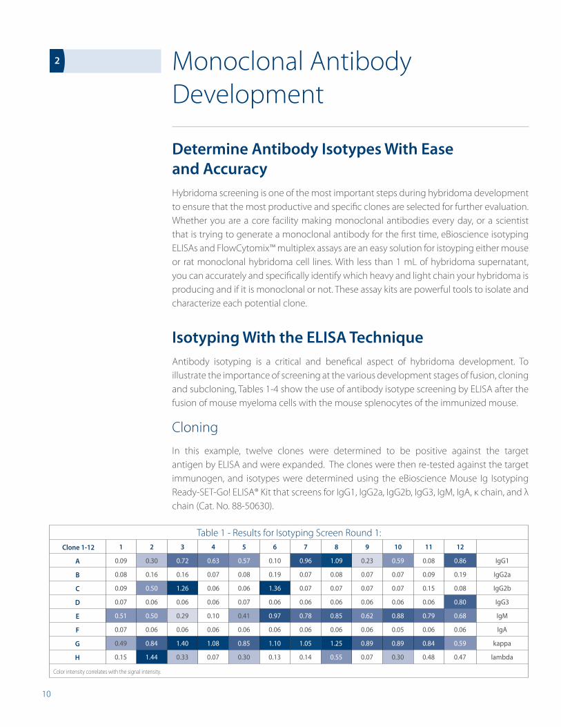
In this example, twelve clones were determined to be positive against the target antigen by ELISA and were expanded. The clones were then re-tested against the target immunogen, and isotypes were determined using the eBioscience Mouse Ig Isotyping Ready-SET-Go! ELISA® Kit that screens for IgG1, IgG2a, IgG2b, IgG3, IgM, IgA, κ chain, and λ chain (Cat. No. 88-50630).
Table 1 - Results for Isotyping Screen Round 1: Clone 1-12 1 2 3 4 5 6 7 8 9 10 11 12
A 0.09 0.30 0.72 0.63 0.57 0.10 0.96 1.09 0.23 0.59 0.08 0.86 IgG1
B 0.08 0.16 0.16 0.07 0.08 0.19 0.07 0.08 0.07 0.07 0.09 0.19 IgG2a
C 0.09 0.50 1.26 0.06 0.06 1.36 0.07 0.07 0.07 0.07 0.15 0.08 IgG2b
D 0.07 0.06 0.06 0.06 0.07 0.06 0.06 0.06 0.06 0.06 0.06 0.80 IgG3
E 0.51 0.50 0.29 0.10 0.41 0.97 0.78 0.85 0.62 0.88 0.79 0.68 IgM
F 0.07 0.06 0.06 0.06 0.06 0.06 0.06 0.06 0.06 0.05 0.06 0.06 IgA
G 0.49 0.84 1.40 1.08 0.85 1.10 1.05 1.25 0.89 0.89 0.84 0.59 kappa
H 0.15 1.44 0.33 0.07 0.30 0.13 0.14 0.55 0.07 0.30 0.48 0.47 lambda
Color intensity correlates with the signal intensity.
2
11
The data in Table 1 reveals it is unlikely the hybridoma cells are monoclonal at this stage. It is much more probable that the antibodies secreted are mixed with the antibodies produced from spleen cells before they were killed off under selection pressure. Therefore, it is not surprising to see multiple isotypes and a strong IgM signal.
Analysis of Table 1:
• Hybridomas only producing IgM are not of interest, so 1 and 11 can be eliminated with confidence.
• Clone 4 is promising as it has a nearly clean IgG1κ isotype and could be monoclonal already.
• Clones 2, 3, 5, 6, 7, 8, 9, 10 and 12 show multiple isoforms. It is necessary to subclone these hybridomas to either confirm their monoclonality or isolate clonal ones.
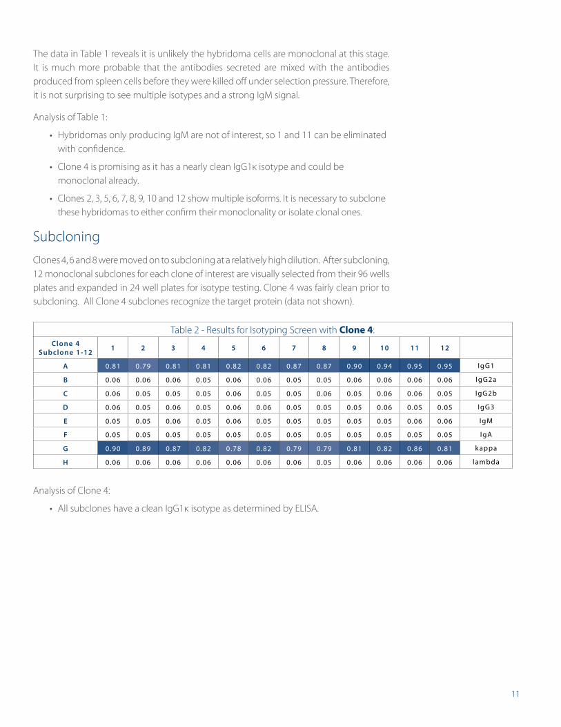
Subcloning
Clones 4, 6 and 8 were moved on to subcloning at a relatively high dilution. After subcloning, 12 monoclonal subclones for each clone of interest are visually selected from their 96 wells plates and expanded in 24 well plates for isotype testing. Clone 4 was fairly clean prior to subcloning. All Clone 4 subclones recognize the target protein (data not shown).
Table 2 - Results for Isotyping Screen with clone 4:Clone 4
Subclone 1-12 1 2 3 4 5 6 7 8 9 10 11 12
A 0.81 0.79 0.81 0.81 0.82 0.82 0.87 0.87 0.90 0.94 0.95 0.95 IgG1
B 0.06 0.06 0.06 0.05 0.06 0.06 0.05 0.05 0.06 0.06 0.06 0.06 IgG2a
C 0.06 0.05 0.05 0.05 0.06 0.05 0.05 0.06 0.05 0.06 0.06 0.05 IgG2b
D 0.06 0.05 0.06 0.05 0.06 0.06 0.05 0.05 0.05 0.06 0.05 0.05 IgG3
E 0.05 0.05 0.06 0.05 0.06 0.05 0.05 0.05 0.05 0.05 0.06 0.06 IgM
F 0.05 0.05 0.05 0.05 0.05 0.05 0.05 0.05 0.05 0.05 0.05 0.05 IgA
G 0.90 0.89 0.87 0.82 0.78 0.82 0.79 0.79 0.81 0.82 0.86 0.81 k appa
H 0.06 0.06 0.06 0.06 0.06 0.06 0.06 0.05 0.06 0.06 0.06 0.06 lambda
Analysis of Clone 4:
• All subclones have a clean IgG1κ isotype as determined by ELISA.
12
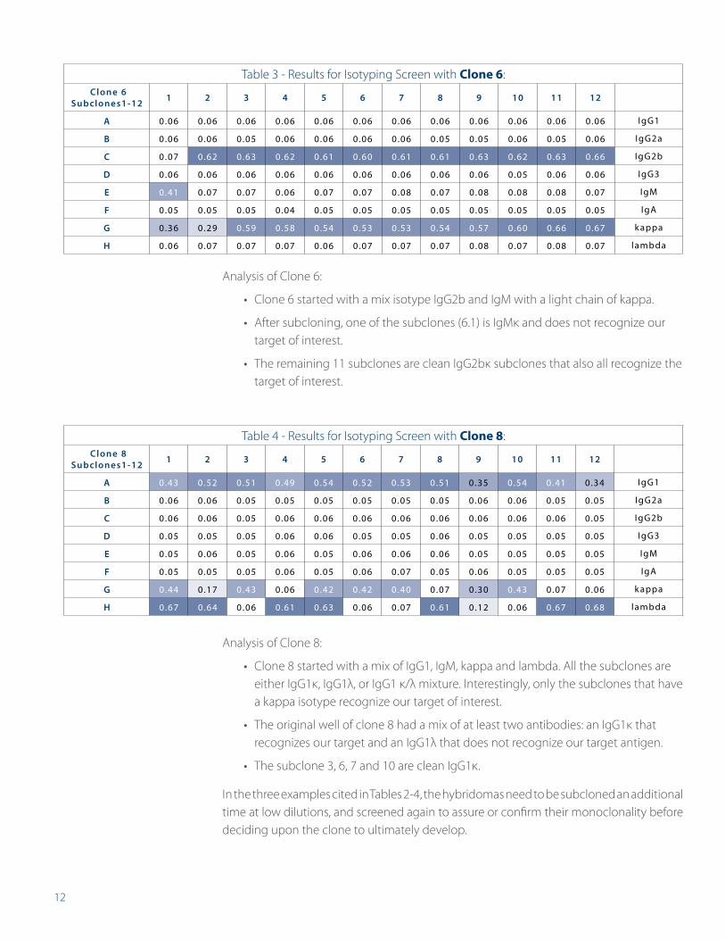
Analysis of Clone 6:
• Clone 6 started with a mix isotype IgG2b and IgM with a light chain of kappa.
• After subcloning, one of the subclones (6.1) is IgMκ and does not recognize our target of interest.
• The remaining 11 subclones are clean IgG2bκ subclones that also all recognize the target of interest.
Analysis of Clone 8:
• Clone 8 started with a mix of IgG1, IgM, kappa and lambda. All the subclones are either IgG1κ, IgG1λ, or IgG1 κ/λ mixture. Interestingly, only the subclones that have a kappa isotype recognize our target of interest.
• The original well of clone 8 had a mix of at least two antibodies: an IgG1κ that recognizes our target and an IgG1λ that does not recognize our target antigen.
• The subclone 3, 6, 7 and 10 are clean IgG1κ.
In the three examples cited in Tables 2-4, the hybridomas need to be subcloned an additional time at low dilutions, and screened again to assure or confirm their monoclonality before deciding upon the clone to ultimately develop.
Table 3 - Results for Isotyping Screen with clone 6: Clone 6
Subclones1-12 1 2 3 4 5 6 7 8 9 10 11 12
A 0.06 0.06 0.06 0.06 0.06 0.06 0.06 0.06 0.06 0.06 0.06 0.06 IgG1
B 0.06 0.06 0.05 0.06 0.06 0.06 0.06 0.05 0.05 0.06 0.05 0.06 IgG2a
C 0.07 0.62 0.63 0.62 0.61 0.60 0.61 0.61 0.63 0.62 0.63 0.66 IgG2b
D 0.06 0.06 0.06 0.06 0.06 0.06 0.06 0.06 0.06 0.05 0.06 0.06 IgG3
E 0.41 0.07 0.07 0.06 0.07 0.07 0.08 0.07 0.08 0.08 0.08 0.07 IgM
F 0.05 0.05 0.05 0.04 0.05 0.05 0.05 0.05 0.05 0.05 0.05 0.05 IgA
G 0.36 0.29 0.59 0.58 0.54 0.53 0.53 0.54 0.57 0.60 0.66 0.67 k appa
H 0.06 0.07 0.07 0.07 0.06 0.07 0.07 0.07 0.08 0.07 0.08 0.07 lambda
Table 4 - Results for Isotyping Screen with clone 8: Clone 8
Subclones1-12 1 2 3 4 5 6 7 8 9 10 11 12
A 0.43 0.52 0.51 0.49 0.54 0.52 0.53 0.51 0.35 0.54 0.41 0.34 IgG1
B 0.06 0.06 0.05 0.05 0.05 0.05 0.05 0.05 0.06 0.06 0.05 0.05 IgG2a
C 0.06 0.06 0.05 0.06 0.06 0.06 0.06 0.06 0.06 0.06 0.06 0.05 IgG2b
D 0.05 0.05 0.05 0.06 0.06 0.05 0.05 0.06 0.05 0.05 0.05 0.05 IgG3
E 0.05 0.06 0.05 0.06 0.05 0.06 0.06 0.06 0.05 0.05 0.05 0.05 IgM
F 0.05 0.05 0.05 0.06 0.05 0.06 0.07 0.05 0.06 0.05 0.05 0.05 IgA
G 0.44 0.17 0.43 0.06 0.42 0.42 0.40 0.07 0.30 0.43 0.07 0.06 k appa
H 0.67 0.64 0.06 0.61 0.63 0.06 0.07 0.61 0.12 0.06 0.67 0.68 lambda
13
Assess Immune Response After Vaccination or InfectionImmunization with vaccines induces the production of antibodies that provide protection against pathogens. To better understand the effects on the immunogenic properties of a vaccine and its adjuvants, eBioscience Antibody Isotyping assays, profiling sera and plasma from humans, mouse and rat, can provide useful insight into immune response.
The examination of individual antibody isotypes first produced following vaccination can help illucidate or indicate a primary immune response to a vaccine. Each antibody isotype possesses unique effector functions to better gauge a vaccine response. IgE is characteristic in allergic reactions, and therefore useful during the assessment of an immune response. If there is no allergic reaction, IgE is usually barely above the limit of detection. A strong IgE response is a negative indicator of a vaccine. Determination of the specific Ig subtype profile can also provide insight into whether the vaccine composition alters or drives the TH1 and/or TH2 profile in the immune response. Presence of elevated IgG1 subclass levels can be a sign for a TH2 mediated immune response, whereas elevated IgG4 levels can be an indicator for a TH1 mediated immune response. Futhermore, the elicitation of IgA can be useful in the assessment of a vaccine that is aimed to provide protection against viral pathogens entering mucosal tissues. In combination with high levels of IgG, this could be a sign for a strong candidate vaccine. Detailed profiling of isotypes following vaccination results in conclusions regarding not only the active pharmaceutical ingredient, but also of the effects of the adjuvants that could be useful in determining vaccine candidates.
Antibody isotyping is relevant while investigating a time course of a specific immune response to determine which route and dose of administration generates the most appropriate immune response. The antibody titer can be easily assessed from boost to boost, indicating the most effective vaccination schedule.
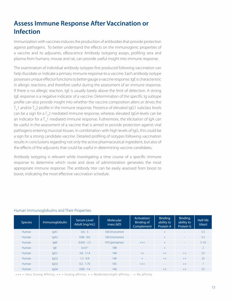
Human Immunoglobulins and Their Properties
Species Immunoglobulin Serum Level Adult [mg/mL]
Molecular mass (kD)
Activation/Binding of
Complement
Binding ability to Protein A
Binding ability to Protein G
Half-life (days)
Human IgA1 0.6 - 3 160 (monomer) - + - 5.5
Human IgA2 0.06 - 0.6 160 (monomer) - + - 5.5
Human IgM 0.045 - 2.5 970 (pentamer) +++ + - 5-10
Human IgE 5x10⁻⁵ 188 - + - 2
Human IgG1 3.8 - 11.4 146 ++ ++ ++ 23
Human IgG2 1.5 - 6.9 146 + ++ ++ 23
Human IgG3 0.2 - 1.70 165 +++ - ++ 7
Human IgG4 0.08 - 1.4 146 - ++ ++ 23
+++ = Ver y Strong Af f in i ty ; ++ = Strong af f in i ty ; + = Moderate/s l ight af f in i ty ; – = No af f in i ty
14
Antibody Isotyping Kits
Multiple Solutions for the Detection/Quantification of Soluble ImmunoglobulinsAntibody Isotyping ELISA Kits are a fast, sensitive and convenient method for quantitation of immunoglobulin isotypes during and after hybridoma development, to assess immune response or to gauge response during vaccination. Compared to membrane-based isotyping tests, eBioscience ELISAs provide an accurate quantitative result with a higher throughput for testing more samples at a time. eBioscience ELISAs guarantee optimal specificity of the antibodies to their respective immunoglobulin isotypes. Antibody levels of individual isotypes can be easily assessed with ELISAs for individual Isotype determination or for multiple isotypes using a single sample. Whether you want to perform a traditional ELISA with pre-coated plates or choose a cost-savings kit with a coat-it-yourself solution, eBioscience has a solution.
Determining Individual Antibody Isotypes
Platinum ELISA Isotyping Kits
• Available for human
• Pre-coated plates
• Serum and plasma samples
• Only one wash and one hour incubation
• Comprehensive Validated Kits
Ready-SET-Go!® ELISA Isotyping Kits
• Available for human, mouse and rat
• All necessary ready-to-use reagents included
• Serum, plasma and cell culture supernatants
• 2, 10, or 20 Plate kit size to suit your experimental size
• Affordable and easy to use
3
15
Determining Multiple Antibody Isotypes
Antibody isotyping often requires the screening of multiple isotypes simultaneously to find the answers that move your research forward. eBioscience offers various assay solutions, with both plate-based ELISAs kits and bead-based assays for the screening of multiple isotypes in parallel.
eBioscience Ig Isotyping Kits are the optimal match for screening your hybridoma in a 96-strip well (8x12) ELISA format. Up to 12 individual samples can be tested for anti-IgG1, IgG2a, IgG2b, IgG2c or IgG3, IgA, and IgM as well as light chain kappa and lambda. To match your individual research needs and budget, choose between the pre-coated (Instant ELISA), a coat-it-yourself (Ready-SET-Go!) isotyping kit, or take it to the next level with FlowCytomix™ bead-based multiplex assays.
Antibody Isotyping ELISA Ready-SET-Go!® Kit
• Value added alternative
• Available for mouse and rat
• Complete assay components included for the end user to coat the ELISA plates and run the assays
• Allows for custom assay tailoring
• Measure 10 to 12 samples per plate
• The inexpensive alternative for the experienced ELISA user
16
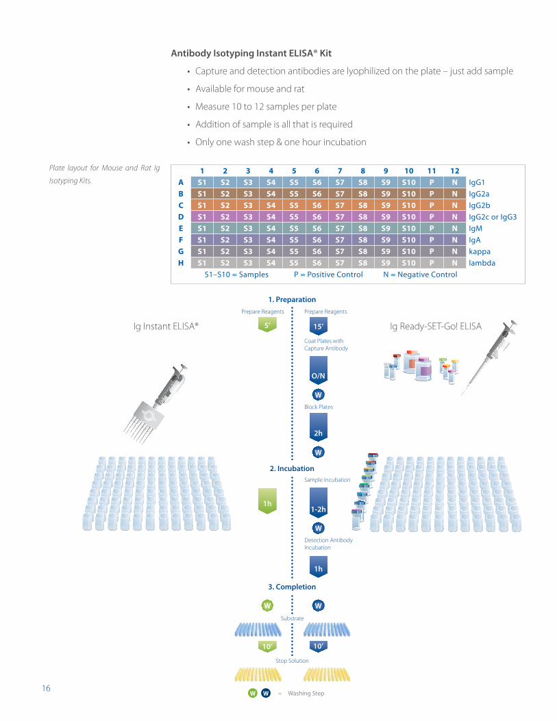
Antibody Isotyping Instant ELISA® Kit
• Capture and detection antibodies are lyophilized on the plate – just add sample
• Available for mouse and rat
• Measure 10 to 12 samples per plate
• Addition of sample is all that is required
• Only one wash step & one hour incubation
1 2 3 4 5 6 7 8 9 10 11 12a S1 S2 S3 S4 S5 S6 S7 S8 S9 S10 p n IgG1B S1 S2 S3 S4 S5 S6 S7 S8 S9 S10 p n IgG2ac S1 S2 S3 S4 S5 S6 S7 S8 S9 S10 p n IgG2bd S1 S2 S3 S4 S5 S6 S7 S8 S9 S10 p n IgG2c or IgG3e S1 S2 S3 S4 S5 S6 S7 S8 S9 S10 p n IgMf S1 S2 S3 S4 S5 S6 S7 S8 S9 S10 p n IgAG S1 S2 S3 S4 S5 S6 S7 S8 S9 S10 p n kappaH S1 S2 S3 S4 S5 S6 S7 S8 S9 S10 p n lambda
S1–S10 = Samples P = Positive Control N = Negative Control
Plate layout for Mouse and Rat Ig
Isotyping Kits.
1. Preparation
2. Incubation
3. Completion
Substrate
Stop Solution
5’
1h
W
10’
15’
1-2h
2h
O/N
W
W
W
W
10’
Prepare Reagents
Coat Plates with Capture Antibody
Prepare Reagents
Sample Incubation
1h
Detection AntibodyIncubation
Block Plates
WW = Washing Step
Ig Instant ELISA® Ig Ready-SET-Go! ELISA
17
Isotyping with FlowCytomix™ Multiplex Assays
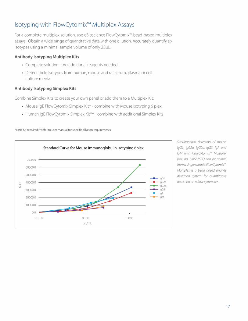
For a complete multiplex solution, use eBioscience FlowCytomix™ bead-based multiplex assays. Obtain a wide range of quantitative data with one dilution. Accurately quantify six isotypes using a minimal sample volume of only 25μL.
Antibody Isotyping Multiplex Kits
• Complete solution – no additional reagents needed
• Detect six Ig isotypes from human, mouse and rat serum, plasma or cell culture media
Antibody Isotyping Simplex Kits
Combine Simplex Kits to create your own panel or add them to a Multiplex Kit:
• Mouse IgE FlowCytomix Simplex Kit† - combine with Mouse Isotyping 6 plex
• Human IgE FlowCytomix Simplex Kit*† - combine with additional Simplex Kits
*Basic Kit required; †Refer to user manual for specific dilution requirements
Standard Curve for Mouse Immunoglobulin Isotyping 6plex
µg/mL
M.F.
I.
10000.0
40000.0
60000.0
20000.0
50000.0
70000.0
0.0
0.010 0.100 1.000
30000.0
lgG1
lgG2b
lgA
lgG2a
lgG3
lgM
Simultaneous detection of mouse
IgG1, IgG2a, IgG2b, IgG3, IgA and
IgM with FlowCytomix™ Multiplex
(cat. no. BMS815FF) can be gained
from a single sample. FlowCytomix™
Multiplex is a bead based analyte
detection system for quantitative
detection on a flow cytometer.
18
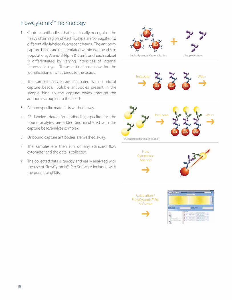
FlowCytomixTM Technology
1. Capture antibodies that specifically recognize the heavy chain region of each isotype are conjugated to differentially-labeled fluorescent beads. The antibody capture beads are differentiated within two bead size populations, A and B (4µm & 5µm), and each subset is differentiated by varying intensities of internal fluorescent dye. These distinctions allow for the identification of what binds to the beads.
2. The sample analytes are incubated with a mix of capture beads. Soluble antibodies present in the sample bind to the capture beads through the antibodies coupled to the beads.
3. All non-specific material is washed away.
4. PE labeled detection antibodies, specific for the bound analytes, are added and incubated with the capture bead/analyte complex.
5. Unbound capture antibodies are washed away.
8. The samples are then run on any standard flow cytometer and the data is collected.
9. The collected data is quickly and easily analyzed with the use of FlowCytomix™ Pro Software included with the purchase of kits.
Incubate
Incubate
Wash
Wash
Calculation / FlowCytomix™ Pro
Software
IgG2a
IgG1 IgG2b
IgG1 IgG2a IgG2b
IgG1 IgG2a IgG2b
Antibody-coated Capture Beads
PE labeled detection Antibodies
Sample Analytes
Flow Cytometric
Analysis
19
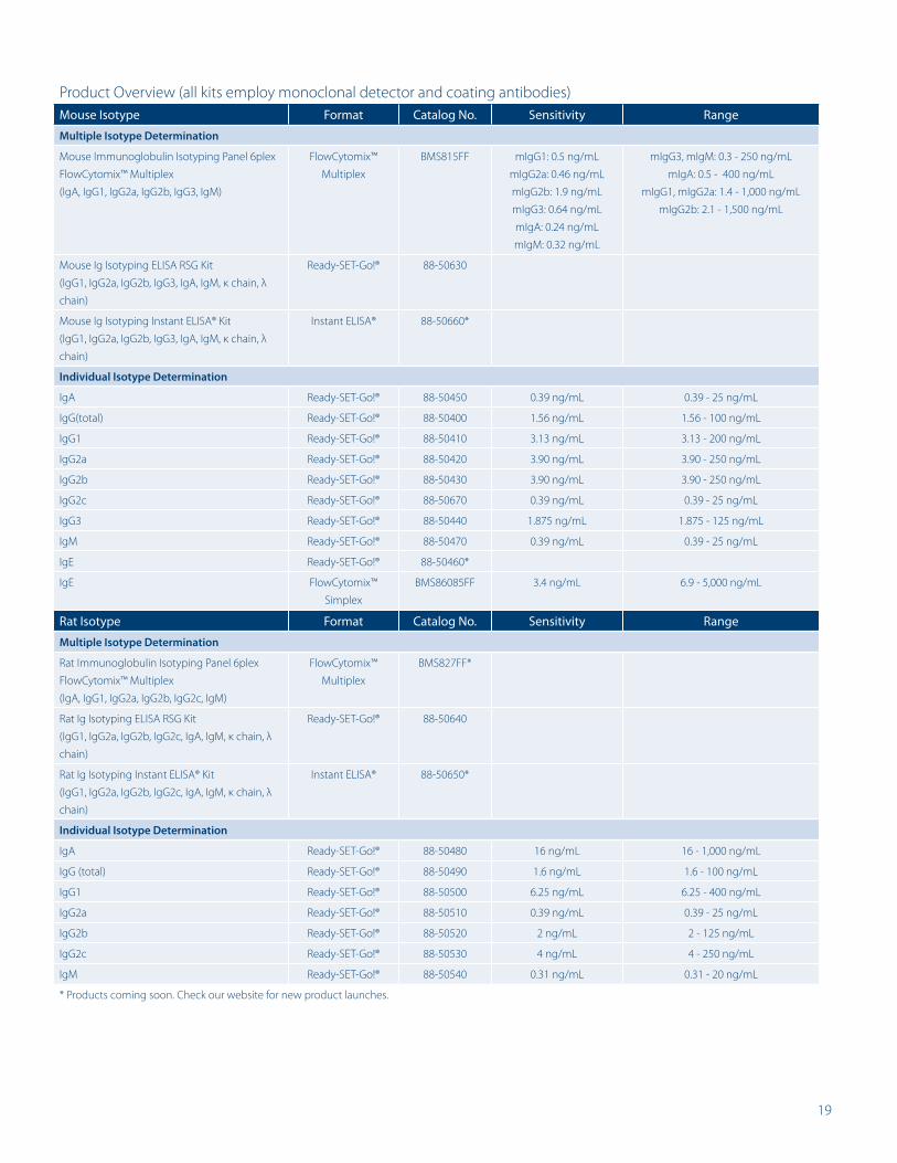
Product Overview (all kits employ monoclonal detector and coating antibodies)Mouse Isotype Format Catalog No. Sensitivity Range
Multiple Isotype Determination
Mouse Immunoglobulin Isotyping Panel 6plex
FlowCytomix™ Multiplex
(IgA, IgG1, IgG2a, IgG2b, IgG3, IgM)
FlowCytomix™
Multiplex
BMS815FF mIgG1: 0.5 ng/mL
mIgG2a: 0.46 ng/mL
mIgG2b: 1.9 ng/mL
mIgG3: 0.64 ng/mL
mIgA: 0.24 ng/mL
mIgM: 0.32 ng/mL
mIgG3, mIgM: 0.3 - 250 ng/mL
mIgA: 0.5 - 400 ng/mL
mIgG1, mIgG2a: 1.4 - 1,000 ng/mL
mIgG2b: 2.1 - 1,500 ng/mL
Mouse Ig Isotyping ELISA RSG Kit
(IgG1, IgG2a, IgG2b, IgG3, IgA, IgM, κ chain, λ
chain)
Ready-SET-Go!® 88-50630
Mouse Ig Isotyping Instant ELISA® Kit
(IgG1, IgG2a, IgG2b, IgG3, IgA, IgM, κ chain, λ
chain)
Instant ELISA® 88-50660*
Individual Isotype Determination
IgA Ready-SET-Go!® 88-50450 0.39 ng/mL 0.39 - 25 ng/mL
IgG(total) Ready-SET-Go!® 88-50400 1.56 ng/mL 1.56 - 100 ng/mL
IgG1 Ready-SET-Go!® 88-50410 3.13 ng/mL 3.13 - 200 ng/mL
IgG2a Ready-SET-Go!® 88-50420 3.90 ng/mL 3.90 - 250 ng/mL
IgG2b Ready-SET-Go!® 88-50430 3.90 ng/mL 3.90 - 250 ng/mL
IgG2c Ready-SET-Go!® 88-50670 0.39 ng/mL 0.39 - 25 ng/mL
IgG3 Ready-SET-Go!® 88-50440 1.875 ng/mL 1.875 - 125 ng/mL
IgM Ready-SET-Go!® 88-50470 0.39 ng/mL 0.39 - 25 ng/mL
IgE Ready-SET-Go!® 88-50460*
IgE FlowCytomix™
Simplex
BMS86085FF 3.4 ng/mL 6.9 - 5,000 ng/mL
Rat Isotype Format Catalog No. Sensitivity Range
Multiple Isotype Determination
Rat Immunoglobulin Isotyping Panel 6plex
FlowCytomix™ Multiplex
(IgA, IgG1, IgG2a, IgG2b, IgG2c, IgM)
FlowCytomix™
Multiplex
BMS827FF*
Rat Ig Isotyping ELISA RSG Kit
(IgG1, IgG2a, IgG2b, IgG2c, IgA, IgM, κ chain, λ
chain)
Ready-SET-Go!® 88-50640
Rat Ig Isotyping Instant ELISA® Kit
(IgG1, IgG2a, IgG2b, IgG2c, IgA, IgM, κ chain, λ
chain)
Instant ELISA® 88-50650*
Individual Isotype Determination
IgA Ready-SET-Go!® 88-50480 16 ng/mL 16 - 1,000 ng/mL
IgG (total) Ready-SET-Go!® 88-50490 1.6 ng/mL 1.6 - 100 ng/mL
IgG1 Ready-SET-Go!® 88-50500 6.25 ng/mL 6.25 - 400 ng/mL
IgG2a Ready-SET-Go!® 88-50510 0.39 ng/mL 0.39 - 25 ng/mL
IgG2b Ready-SET-Go!® 88-50520 2 ng/mL 2 - 125 ng/mL
IgG2c Ready-SET-Go!® 88-50530 4 ng/mL 4 - 250 ng/mL
IgM Ready-SET-Go!® 88-50540 0.31 ng/mL 0.31 - 20 ng/mL
* Products coming soon. Check our website for new product launches.
20
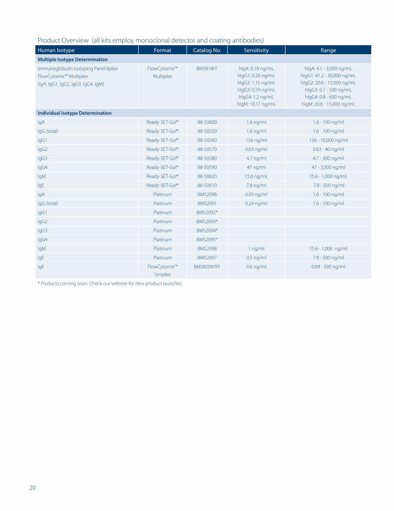
Product Overview (all kits employ monoclonal detector and coating antibodies)Human Isotype Format Catalog No. Sensitivity Range
Multiple Isotype Determination
Immunoglobulin Isotyping Panel 6plex
FlowCytomix™ Multiplex
(IgA, IgG1, IgG2, IgG3, IgG4, IgM)
FlowCytomix™
Multiplex
BMS818FF hIgA: 0.18 ng/mLhIgG1: 0.28 ng/mLhIgG2: 1.15 ng/mLhIgG3: 0.29 ng/mLhIgG4: 1.2 ng/mL
hIgM: 10.17 ng/mL
hIgA: 4.1 - 3,000 ng/mLhIgG1: 41.2 - 30,000 ng/mLhIgG2: 20.6 - 15,000 ng/mL
hIgG3: 0.7 - 500 ng/mLhIgG4: 0.8 - 600 ng/mL
hIgM: 20.6 - 15,000 ng/mL
Individual Isotype Determination
IgA Ready-SET-Go!® 88-50600 1.6 ng/ml 1.6 - 100 ng/ml
IgG (total) Ready-SET-Go!® 88-50550 1.6 ng/ml 1.6 - 100 ng/ml
IgG1 Ready-SET-Go!® 88-50560 156 ng/ml 156 - 10,000 ng/ml
IgG2 Ready-SET-Go!® 88-50570 0.63 ng/ml 0.63 - 40 ng/ml
IgG3 Ready-SET-Go!® 88-50580 4.7 ng/ml 4.7 - 300 ng/ml
IgG4 Ready-SET-Go!® 88-50590 47 ng/ml 47 - 3,000 ng/ml
IgM Ready-SET-Go!® 88-50620 15.6 ng/mL 15.6 - 1,000 ng/mL
IgE Ready-SET-Go!® 88-50610 7.8 ng/ml 7.8 - 500 ng/ml
IgA Platinum BMS2096 0.03 ng/ml 1.6 - 100 ng/ml
IgG (total) Platinum BMS2091 0.24 ng/ml 1.6 - 100 ng/ml
IgG1 Platinum BMS2092*
IgG2 Platinum BMS2093*
IgG3 Platinum BMS2094*
IgG4 Platinum BMS2095*
IgM Platinum BMS2098 1 ng/ml 15.6 - 1,000 ng/ml
IgE Platinum BMS2097 0.5 ng/ml 7.8 - 500 ng/ml
IgE FlowCytomix™
Simplex
BMS82097FF 0.6 ng/ml 0.69 - 500 ng/ml
* Products coming soon. Check our website for new product launches.
HeadquarterseBioscience, Inc.10255 Science Center DriveSan Diego, CA 92121USA
Customers in countries where direct sales are not available may contact their eBioscience distributor listed at www.eBioscience.com/distributors
Q112007e_Isotyping Kits_0412
high performance immunoassays
www.eBioscience.com
Service and Support for direct SaleS
North AmericaTechnical Support:
For Research Products:[email protected] Clinical Products:[email protected]
Customer Service:[email protected]
Fax:858.642.2046
AustriaTechnical Support:
[email protected] Service:
+43 1 796 40 40 [email protected]
Fax:+43 1 796 40 40 400
BelgiumTechnical Support:
[email protected] Service:
+43 1 796 40 40 [email protected]
Fax:+43 1 796 40 40 400
FranceTechnical Support:
[email protected] Service:
0 800 800 [email protected]
Fax:0 800 800 418
GermanyTechnical Support:
[email protected] Service:
+49 69 33 29 64 [email protected]
Fax:+49 69 255 77 335
IrelandTechnical Support:
[email protected] Service:
+44 208 951 [email protected]
Fax:+44 207 900 1559
NetherlandsTechnical Support:
[email protected] Service:
+43 1 796 40 40 [email protected]
Fax:+43 1 796 40 40 400
PolandTechnical Support:
[email protected] Service:
+43 1 796 4040 305 [email protected]
Fax: +43 1 796 4040 400
SwitzerlandTechnical Support:
[email protected] Service:
+41 21 510 [email protected]
Fax:+41 21 510 1216
United KingdomTechnical Support:
[email protected] Service:
+44 208 951 [email protected]
Fax:+44 207 900 1559