Embed Size (px)
Citation preview

OPEN
Heterarchy of transcription factors driving basal andluminal cell phenotypes in human urothelium
Carl Fishwick1,4, Janet Higgins2, Lawrence Percival-Alwyn2, Arianna Hustler1, Joanna Pearson1, Sarah Bastkowski2, Simon Moxon2,5,David Swarbreck2, Chris D Greenman3 and Jennifer Southgate*,1
Cell differentiation is affected by complex networks of transcription factors that co-ordinate re-organisation of the chromatinlandscape. The hierarchies of these relationships can be difficult to dissect. During in vitro differentiation of normal human uro-epithelial cells, formaldehyde-assisted isolation of regulatory elements (FAIRE-seq) and RNA-seq was used to identify alterationsin chromatin accessibility and gene expression changes following activation of the nuclear receptor peroxisome proliferator-activated receptor gamma (PPARc) as a differentiation-initiating event. Regions of chromatin identified by FAIRE-seq, as havingaltered accessibility during differentiation, were found to be enriched with sequence-specific binding motifs for transcriptionfactors predicted to be involved in driving basal and differentiated urothelial cell phenotypes, including forkhead box A1 (FOXA1),P63, GRHL2, CTCF and GATA-binding protein 3 (GATA3). In addition, co-occurrence of GATA3 motifs was observed within subsetsof differentiation-specific peaks containing P63 or FOXA1. Changes in abundance of GRHL2, GATA3 and P63 were observed inimmunoblots of chromatin-enriched extracts. Transient siRNA knockdown of P63 revealed that P63 favoured a basal-likephenotype by inhibiting differentiation and promoting expression of basal marker genes. GATA3 siRNA prevented differentiation-associated downregulation of P63 protein and transcript, and demonstrated positive feedback of GATA3 on PPARG transcript, butshowed no effect on FOXA1 transcript or protein expression. This approach indicates that as a transcriptionally regulatedprogramme, urothelial differentiation operates as a heterarchy, wherein GATA3 is able to co-operate with FOXA1 to driveexpression of luminal marker genes, but that P63 has potential to transrepress expression of the same genes.Cell Death and Differentiation advance online publication, 10 March 2017; doi:10.1038/cdd.2017.10
The nuclear receptor peroxisome proliferator-activated receptorgamma (PPARg) is widely known as an essential and sufficientdriver of adipogenesis,1,2 but it also plays roles in M1 to M2polarisation of macrophages3 and differentiation of humanurothelial cells of the bladder and associated urinary tract.4–6
When grown in vitro in the absence of serum or other nuclearreceptor signalling, non-immortalised normal human urothelial(NHU) cells acquire a proliferative, autocrine epidermal growthfactor receptor (EGFR)-regulated squamous cell phenotype.7,8
RNA microarray studies of NHU cell cultures have shown thatwhen downstream EGFR signalling is blocked, exogenousligand activation of PPARg induces expression of intermediarytranscription factors required for specifying the differentiatedurothelial cell phenotype, including forkhead box A1 (FOXA1),interferon regulatory factor 1 (IRF1), GATA-binding protein 3(GATA3) and E74-like ETS transcription factor 3 (ELF3).9,10 Ofthese, FOXA1 and GATA3 are recognised as pioneer factorscapable of driving changes in chromatin organisation andaccessibility.11 In urothelial carcinoma, FOXA1 andGATA3 havebeen associated with differentiation status12,13 and 8% oftumours were found to carry ELF3 mutations.14 Mouse studieshave identified other transcription factors as determinants ofurothelial specification, including Grainyhead-like transcription
factor 3 (Grhl3),15 Kruppel-like factor (Klf5),16 and Gata4 andGata6,17 but it remains unclear what role these factors play inhuman urothelium.Formaldehyde-assisted isolation of regulatory elements
coupledwith next-generation sequencing (FAIRE-seq)18 exploitsthe propensity of nucleosome-depleted DNA, or ‘open’ chroma-tin, to shear from adjacent nucleosomes during sonication ofnuclear material from formaldehyde-fixed cells. Isolating thissheared DNA from nucleosomal DNA by phase separationenables characterisation of the relative extent of chromatinaccessibility in a genome-wide manner. As transcription factorsbind dynamically to nucleosome-depleted regions, motif match-ingwithin open chromatin, as identified by FAIRE, can be used toclassify transcription factors that actively associate with chroma-tin and define cell phenotype.19–23 FAIRE identifies a comple-mentary but partially distinct set of putative enhancer regionsoutside of gene promoters, as compared to DNase-seq,19 whichuses DNase I enzyme to cleave regions of open chromatin.FAIRE-seq DNA has been shown to be enriched relative toDNase-seq for potential FOXA1-binding sites, which are knownto contribute to urothelial differentiation,9 and chromatin-associated histone H3 monomethylated on lysine 4
1Jack Birch Unit for Molecular Carcinogenesis, Department of Biology, University of York, York YO10 5DD, UK; 2Earlham Institute, Norwich Research Park, Norwich NR47UZ, UK and 3School of Computing Sciences, University of East Anglia, Norwich NR4 7TJ, UK*Corresponding author: J Southgate, Jack Birch Unit for Molecular Carcinogenesis, Department of Biology, University of York, York YO10 5DD UK. Tel: +44 (0)1904 32 8705;Fax: +44 (0) 1904 32 8704; E-mail: [email protected] address: GSK, Medicines Research Center, Gunnels Wood Road, Stevenage SG1 2NY, UK.5Current address: School of Biological Sciences, University of East Anglia, Norwich Research Park, Norwich NR4 7TJ, UK.Received 24.7.16; revised 10.1.17; accepted 11.1.17; Edited by A Ashkenazi
Cell Death and Differentiation (2017), 1–10Official journal of the Cell Death Differentiation Association
www.nature.com/cdd
(H3K4me1), which is associated with genomic enhancersspecific to cell type.To obtain a genome-wide picture of the transcriptional
drivers of different urothelial cell phenotypes, RNA-seq andFAIRE-seq were performed on serially propagated NHU cellcultures from three independent donors at 24 and 144 h timepoints after concurrent EGFR blockade and PPARg activationto induce differentiation,4 alongside time-matched non-differ-entiated vehicle controls. Open chromatin regions unique totreated and control libraries were searched for matches toknown sequence-specific transcription factor-binding motifs,both on a genome-wide basis and proximal to differentiallyexpressed genes. Selected candidate transcriptional regula-tors were validated as modulators of urothelial differentiationusing immunoblots of chromatin-enriched extracts and siRNAknockdown to investigate effects on urothelial phenotype.
Results
Differentially expressed genes and FAIRE-seq peakgenomic distribution. Results obtained from the analysis
of RNA-seq data identified 559 and 463 genes that wereupregulated, and 467 and 158 genes that were down-regulated in differentiation-induced cells relative to time-matched controls at the 24 and 144 h time points (falsediscovery rate (FDR)o0.1), respectively (SupplementaryFigure S1, Supplementary Tables 1A and 1B). Transcriptsupregulated at both time points included the urothelium-restricted differentiation markers uroplakin 1A (UPK1A) andUPK2.24–26 Gene ontology analysis, performed using theGOrilla tool,27 showed that the 122 genes upregulated at bothtime points included genes involved in lipid metabolism(P= 1.16 × 10− 5) and water homeostasis (P= 8.09 ×10×5;Supplementary Table 2), with the latter likely reflecting therole of urothelium as a barrier to urinary solutes.Peak calling using the MACS algorithm on FAIRE-seq data
pooled for the three donor cell lines gave466 000 total peaksrising to 471 000 at 144 h, with a near equal distributionbetween proportions of distinct (control or differentiated) andoverlapping peaks at each time point (Figures 1a and b).Consistent with other investigations into the relationshipbetween DNA enriched by FAIRE and gene expression,19,20
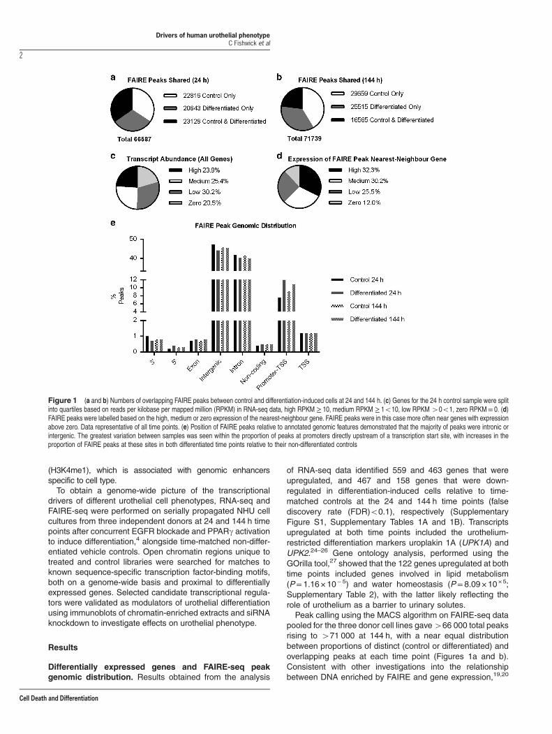
Figure 1 (a and b) Numbers of overlapping FAIRE peaks between control and differentiation-induced cells at 24 and 144 h. (c) Genes for the 24 h control sample were splitinto quartiles based on reads per kilobase per mapped million (RPKM) in RNA-seq data, high RPKM ≥ 10, medium RPKM ≥ 1o10, low RPKM40o1, zero RPKM= 0. (d)FAIRE peaks were labelled based on the high, medium or zero expression of the nearest-neighbour gene. FAIRE peaks were in this case more often near genes with expressionabove zero. Data representative of all time points. (e) Position of FAIRE peaks relative to annotated genomic features demonstrated that the majority of peaks were intronic orintergenic. The greatest variation between samples was seen within the proportion of peaks at promoters directly upstream of a transcription start site, with increases in theproportion of FAIRE peaks at these sites in both differentiated time points relative to their non-differentiated controls
Drivers of human urothelial phenotypeC Fishwick et al
2
Cell Death and Differentiation

when genes were split into quartiles based on normalisedRNA-seq read counts (Figure 1c and Supplementary Table 3),a greater proportion of nearest-neighbour genes to FAIREpeaks had reads per kilobase per mapped million (RPKM)values above zero as compared to total genes (Figure 1d). Inaddition, most FAIRE peaks were intronic or intergenic, and aslight increase in the proportion of peaks associated withpromoters was noted in differentiation-induced cells at bothtime points (Figure 1e and Supplementary Table 3).
Transcription factor motifs enriched in FAIRE peaks. Touncover transcription factors driving cell phenotype in differen-tiated and non-differentiated urothelial cells, sequence-specifictranscription factor-binding motifs enriched in non-overlappingFAIRE peaks at each time point were identified using themotif discovery tool HOMER.28 Motif searching was conductedusing control-specific peak sets as the background for thedifferentiation-specific peak set and vice versa.Previous transcription factor motif matching studies using
open chromatin isolation techniques have observed thatparticular motifs tend to be enriched at sites distal togenes,29 and that within promoter regions, transcription startsites (TSSs) have fewer differences in transcription factormotifs than the rest of the genome.20 As such, FAIRE peaks inTSS promoter regions (−1 kb to +100 bp) were excluded fromall analyses. To highlight any differences between motifsenriched proximal to genes and those found across thegenome, control-specific and differentiation-specific FAIREpeaks were compared as either genome-wide groups, oranalysis was restricted to those located within ± 25 kb of theTSS of differentially regulated genes. Motifs matched byHOMER were filtered for those which occurred in at least≥1.25-fold of the total percentage of regions in the target setas compared to the background set, in order to focus onmotifssignificantly enriched in each experimental situation.20,30 This
approach identified divergent groups of transcription factormotifs across the different regions, with each group containingmatches to motifs from both previously described urothelium-associated factors and others not previously associated withurothelium (Figure 2 and Supplementary Tables 4–12). Denovo motif analysis was less successful than matching toknown motifs, as most matches that were not similar to thosefound in the HOMER database were in low percentages ofpeaks (data not shown).Motifs with the highest fold change in abundance in peaks
specific to control libraries and around downregulated genesat 24 h were dominated by cell cycle-associated transcriptionfactors such as ETS family factors, JUN-AP1, EGR1 andEGR2, and a motif associated with combined binding of theOCT4-SOX2-TCF-NANOG pluripotency factors in mouseembryonic stem cells.31 OCT4 transcripts are expressed byNHU cells, but the pluripotency-associated isoform OCT4A isnot.32 P63, a transcription factor associated with a non-differentiated ‘basal-like’ urothelial cell phenotype in normalcells and carcinoma,33–37 was enriched both proximal todownregulated genes and across the genome at 144 h,whereas STAT6 and ETS motifs were specifically associatedwith peaks ±25 kb of downregulated genes at this time point.Motifs from urothelial differentiation-associated transcrip-
tion factors FOXA1,9 GATA310,12 and PPARg4 were enrichedin differentiation-specific FAIRE peaks within ±25 kb of theTSS of genes with expression upregulated during differentia-tion. PPARg motifs were only enriched around genesupregulated at 24 h, in agreement with observations that itdrives early events during in vitro urothelial differentiationupstream of FOXA1,9 motifs from which were matched at144 h. GATA3, CEBPB and GRHL2 motifs were enrichedaround upregulated genes at both time points. GRHL2 hasbeen implicated in regulation of tight junction complex genes,which are central to barrier formation in several epithelia,38
Figure 2 Summary of known motifs from the HOMER database matched in FAIRE-seq peaks specific to control and differentiation-induced NHU cells. FAIRE-seq peaks frompooled donor data were compared between control and differentiation-induced cells at 24 and 144 h time points, and peaks unique (non-overlapping) to each library weresearched for known sequence motifs in HOMER to generate a genome-wide comparison for all peaks. The same comparison was performed using only peaks found within± 25 kb of the TSS of genes upregulated or downregulated during differentiation at the respective time points
Drivers of human urothelial phenotypeC Fishwick et al
3
Cell Death and Differentiation

including urothelium,6,39 whereas the closely related GRHL3has been associated with urothelial differentiation in themouse.15 CEBPB plays a key role in orchestratingCEBPA andPPARG expression during adipogenesis.2 CEBPB has noknown role in normal human urothelial biology, although othergroups have shown the CEBPB motif to be enriched inpromoters of urothelial carcinoma gene sets,40 and it has beenassociated with urothelial differentiation in mouse.41 ELF5 andELF1 motifs were enriched in regions proximal to up- anddownregulated genes at 144 h, respectively. Although neitherof these has been previously associated with urothelialbiology, the closely related ELF3, whose motif is not in theHOMER database used here, is a driver of differentiation.10
Across the genome, in differentiation-induced cells, motifsfrom the known urothelium-associated transcription factorIRF19 and the closely related motif for IRF2 were enriched at24 h, as were those from CTCF at both time points. As none ofthese motifs were enriched proximal to differentially regulatedgenes, these observations agree with previous studies thatshowed CTCF and IRF1 preferentially bind to regions distal toexpressed genes.29
Co-occurrence of transcription factor motifs in openchromatin. Lineage-determining transcription factors havebeen observed to bind in regions proximal to one anotherduring differentiation.28 Pioneer factors such as FOXA1,which can open repressed regions of chromatin, often bindproximally to differentiation-inducing nuclear receptors.42–44
To determine whether there was co-occurrence of differen-tiation-associated transcription factor motifs within FAIRE-seqpeaks, P63 and FOXA1 motif-containing open chromatinregions specific to control and differentiated cells at each timepoint were searched separately for enriched motifs using thesame approach as for the genome-wide investigation.P63- and FOXA1-containing peaks were enriched with motifs
that overlapped the genome-wide set of peaks, but withsignificant differences (Supplementary Figures 2A, 2B andSupplementary Tables 13–20).Motifs co-occurring within P63- and FOXA1-containing
peaks were largely distinct from one another, but with notableexceptions such as GATA3, GRHL2, P63 and IRF motifs,which co-occurred with both FOXA1 and P63 in differentiation-specific peaks (Supplementary Figure 3).
Chromatin binding of transcription factors with enrichedmotifs. To determine whether transcription factors withenriched motifs and other putative urothelial phenotypeorchestrators reported in the literature were enriched inurothelial chromatin, immunoblots of chromatin extracts weregenerated using urothelial cell cultures from independentlines. PPARg, FOXA1, GRHL2 and GATA3 were enriched inchromatin extracted from differentiated cell cultures, whereasbasal-associated P63 was more abundant in non-differentiated cultures (Figure 3). CTCF and GRHL3 hadsimilar abundance on chromatin from control and differen-tiated cultures. ELF5 and ELF1 detection was not possibledue to poor antibody specificity, but ELF3 was observed to beassociated with chromatin from differentiated cells.
Differentiation-associated transcription factors in nativeurothelium. To determine whether transcription factors withmotifs matched to the non-differentiated or differentiated NHUcell phenotypes were expressed by normal urothelium in situ,immunohistochemistry was performed on human urothelialtissue sections (Figure 4). P63 demonstrated a basal–intermediate cell distribution, with markedly reduced labellingof the most differentiated superficial cells. PPARg, CTCF,GATA3, GRHL2 and FOXA1 were observed to be nuclear inall layers of the urothelium, with GRHL2 and FOXA1 showing
Figure 3 Chromatin extracts showing bound transcription factors that changed in abundance during differentiation. Factors with motifs detected as enriched in differentiation-specific FAIRE peaks, including GRHL2, GATA3, FOXA1 and PPARg, were upregulated in chromatin extracts from differentiation-induced NHU cells from two independentdonors. CTCF and GRHL3 did not change in abundance with differentiation. P63 abundance was reduced after induction of differentiation. Histone H2A is included as a loadingcontrol
Drivers of human urothelial phenotypeC Fishwick et al
4
Cell Death and Differentiation
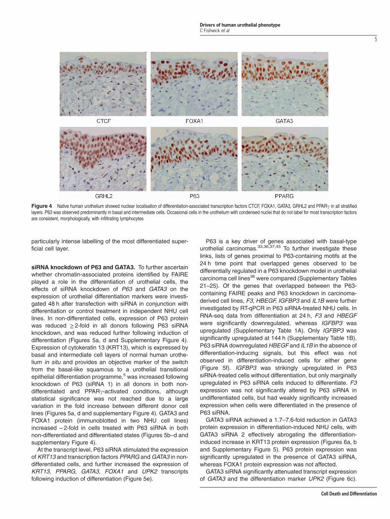
particularly intense labelling of the most differentiated super-ficial cell layer.
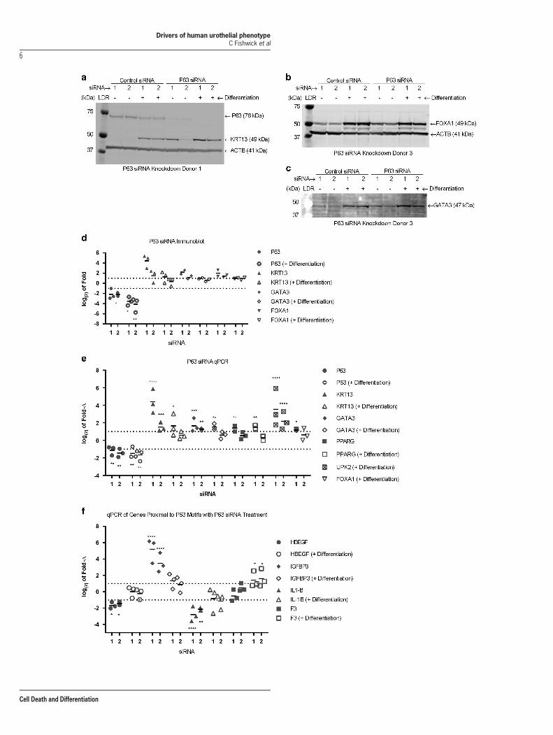
siRNA knockdown of P63 and GATA3. To further ascertainwhether chromatin-associated proteins identified by FAIREplayed a role in the differentiation of urothelial cells, theeffects of siRNA knockdown of P63 and GATA3 on theexpression of urothelial differentiation markers were investi-gated 48 h after transfection with siRNA in conjunction withdifferentiation or control treatment in independent NHU celllines. In non-differentiated cells, expression of P63 proteinwas reduced ≥ 2-fold in all donors following P63 siRNAknockdown, and was reduced further following induction ofdifferentiation (Figures 5a, d and Supplementary Figure 4).Expression of cytokeratin 13 (KRT13), which is expressed bybasal and intermediate cell layers of normal human urothe-lium in situ and provides an objective marker of the switchfrom the basal-like squamous to a urothelial transitionalepithelial differentiation programme,5 was increased followingknockdown of P63 (siRNA 1) in all donors in both non-differentiated and PPARg-activated conditions, althoughstatistical significance was not reached due to a largevariation in the fold increase between different donor celllines (Figures 5a, d and supplementary Figure 4). GATA3 andFOXA1 protein (immunoblotted in two NHU cell lines)increased ~ 2-fold in cells treated with P63 siRNA in bothnon-differentiated and differentiated states (Figures 5b–d andsupplementary Figure 4).At the transcript level, P63 siRNA stimulated the expression
of KRT13 and transcription factors PPARG andGATA3 in non-differentiated cells, and further increased the expression ofKRT13, PPARG, GATA3, FOXA1 and UPK2 transcriptsfollowing induction of differentiation (Figure 5e).
P63 is a key driver of genes associated with basal-typeurothelial carcinomas.33,36,37,45 To further investigate theselinks, lists of genes proximal to P63-containing motifs at the24 h time point that overlapped genes observed to bedifferentially regulated in a P63 knockdown model in urothelialcarcinoma cell lines36 were compared (Supplementary Tables21–25). Of the genes that overlapped between the P63-containing FAIRE peaks and P63 knockdown in carcinoma-derived cell lines, F3, HBEGF, IGFBP3 and IL1B were furtherinvestigated by RT-qPCR in P63 siRNA-treated NHU cells. InRNA-seq data from differentiation at 24 h, F3 and HBEGFwere significantly downregulated, whereas IGFBP3 wasupregulated (Supplementary Table 1A). Only IGFBP3 wassignificantly upregulated at 144 h (Supplementary Table 1B).P63 siRNA downregulatedHBEGF and IL1B in the absence ofdifferentiation-inducing signals, but this effect was notobserved in differentiation-induced cells for either gene(Figure 5f). IGFBP3 was strikingly upregulated in P63siRNA-treated cells without differentiation, but only marginallyupregulated in P63 siRNA cells induced to differentiate. F3expression was not significantly altered by P63 siRNA inundifferentiated cells, but had weakly significantly increasedexpression when cells were differentiated in the presence ofP63 siRNA.GATA3 siRNA achieved a 1.7–7.6-fold reduction in GATA3
protein expression in differentiation-induced NHU cells, withGATA3 siRNA 2 effectively abrogating the differentiation-induced increase in KRT13 protein expression (Figures 6a, band Supplementary Figure 5). P63 protein expression wassignificantly upregulated in the presence of GATA3 siRNA,whereas FOXA1 protein expression was not affected.GATA3 siRNA significantly attenuated transcript expression
of GATA3 and the differentiation marker UPK2 (Figure 6c).
Figure 4 Native human urothelium showed nuclear localisation of differentiation-associated transcription factors CTCF, FOXA1, GATA3, GRHL2 and PPARg in all stratifiedlayers. P63 was observed predominantly in basal and intermediate cells. Occasional cells in the urothelium with condensed nuclei that do not label for most transcription factorsare consistent, morphologically, with infiltrating lymphocytes
Drivers of human urothelial phenotypeC Fishwick et al
5
Cell Death and Differentiation
Drivers of human urothelial phenotypeC Fishwick et al
6
Cell Death and Differentiation
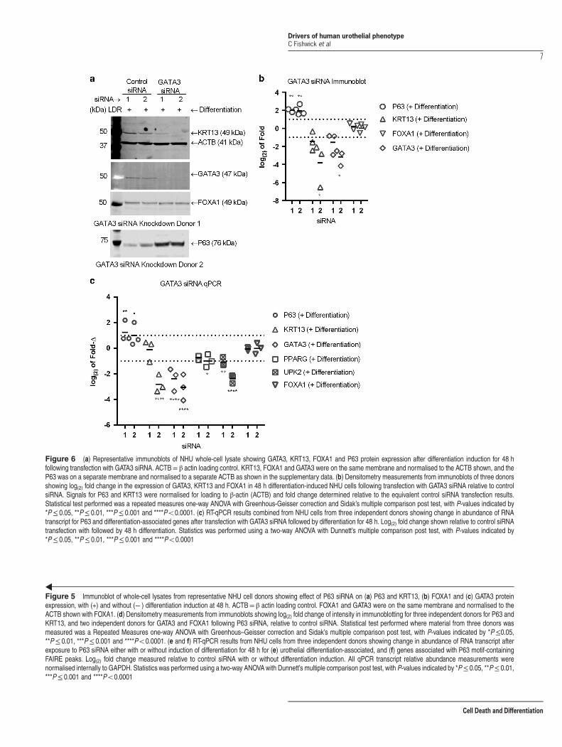
Figure 5 Immunoblot of whole-cell lysates from representative NHU cell donors showing effect of P63 siRNA on (a) P63 and KRT13, (b) FOXA1 and (c) GATA3 proteinexpression, with (+) and without (− ) differentiation induction at 48 h. ACTB= β actin loading control. FOXA1 and GATA3 were on the same membrane and normalised to theACTB shown with FOXA1. (d) Densitometry measurements from immunoblots showing log(2) fold change of intensity in immunoblotting for three independent donors for P63 andKRT13, and two independent donors for GATA3 and FOXA1 following P63 siRNA, relative to control siRNA. Statistical test performed where material from three donors wasmeasured was a Repeated Measures one-way ANOVA with Greenhous–Geisser correction and Sidak’s multiple comparison post test, with P-values indicated by *P≤0.05,**P≤ 0.01, ***P≤ 0.001 and ****Po0.0001. (e and f) RT-qPCR results from NHU cells from three independent donors showing change in abundance of RNA transcript afterexposure to P63 siRNA either with or without induction of differentiation for 48 h for (e) urothelial differentiation-associated, and (f) genes associated with P63 motif-containingFAIRE peaks. Log(2) fold change measured relative to control siRNA with or without differentiation induction. All qPCR transcript relative abundance measurements werenormalised internally to GAPDH. Statistics was performed using a two-way ANOVAwith Dunnett’s multiple comparison post test, with P-values indicated by *P≤ 0.05, **P≤ 0.01,***P≤ 0.001 and ****Po0.0001
Figure 6 (a) Representative immunoblots of NHU whole-cell lysate showing GATA3, KRT13, FOXA1 and P63 protein expression after differentiation induction for 48 hfollowing transfection with GATA3 siRNA. ACTB= β actin loading control. KRT13, FOXA1 and GATA3 were on the same membrane and normalised to the ACTB shown, and theP63 was on a separate membrane and normalised to a separate ACTB as shown in the supplementary data. (b) Densitometry measurements from immunoblots of three donorsshowing log(2) fold change in the expression of GATA3, KRT13 and FOXA1 in 48 h differentiation-induced NHU cells following transfection with GATA3 siRNA relative to controlsiRNA. Signals for P63 and KRT13 were normalised for loading to β-actin (ACTB) and fold change determined relative to the equivalent control siRNA transfection results.Statistical test performed was a repeated measures one-way ANOVA with Greenhous-Geisser correction and Sidak’s multiple comparison post test, with P-values indicated by*P≤ 0.05, **P≤ 0.01, ***P≤ 0.001 and ****Po0.0001. (c) RT-qPCR results combined from NHU cells from three independent donors showing change in abundance of RNAtranscript for P63 and differentiation-associated genes after transfection with GATA3 siRNA followed by differentiation for 48 h. Log(2) fold change shown relative to control siRNAtransfection with followed by 48 h differentiation. Statistics was performed using a two-way ANOVA with Dunnett’s multiple comparison post test, with P-values indicated by*P≤ 0.05, **P≤ 0.01, ***P≤ 0.001 and ****Po0.0001
Drivers of human urothelial phenotypeC Fishwick et al
7
Cell Death and Differentiation

KRT13 transcript was only reduced significantly by GATA3siRNA 2, as with the protein. P63 showed increases intranscript and protein expression with both GATA3 siRNAoligonucleotides. Neither GATA3 siRNA sequence had aneffect on FOXA1 transcript abundance and only siRNA 2showed a small inhibitory effect on PPARG transcriptexpression
Discussion
By comparing transcription factor-binding motifs matchedwithin open chromatin regions in normal human urothelialcells in non-differentiated versus differentiated states, thisstudy provides new insight into the identity and operationalrelationships between transcriptional drivers of urothelial cellphenotype. Of major significance, P63 drives the non-differentiated squamous phenotype assumed by normalhuman urothelial cells maintained in serum-free cultureconditions in the absence of nuclear receptor signalling.siRNA knockdown revealed that P63 maintains this primitiveor ‘basal-like’ phenotype at least in part by inhibitingexpression of transitional epithelial lineage genes includingKRT13 and PPARG.P63 plays an essential role in epithelial tissues during
development, where its absence causes severe dysgenesis ofepithelial tissues, as described in mouse epidermis.46
Changes in expression and somatic mutations of P63 havebeen associated with clinically relevant subtypes of bladdercancer, with P63 identified as a driver of the basal-like cellphenotype in urothelial carcinoma.36 These authors showedthat knockdown of P63 in the established bladder cancer-derived UM-UC14 cell line affected expression of PPARG-influenced genes, including HBEGF, IGFBP3 and IL1B.36
Here these same genes were differentially affected by siRNAknockdown of P63 in NHU cells, implying usage of the samegene networks by normal and cancer cells.In urothelium, PPARg has been identified as a nuclear
receptor whose activation mediates the transition fromsquamous to a differentiated transitional (urothelial) pheno-type. This involves a major shift in gene expression, implying achange in genomic organisation to reflect the transcriptionallandscape of urothelium. We have previously identified anetwork of PPARg-regulated intermediary transcription factorsthat mediate the differentiated urothelial programme, althoughinter-relationships within the network have yet to be estab-lished. In other tissues, such as breast, a role has beenidentified for the so-called pioneer factors FOXA and GATA indefining the tissue-specific genomic organisation. GATA3 andFOXA1 have been shown to act co-operatively in promotingESR1-driven transcription in MCF7 cells, with GATA3 lyingupstream of FOXA1.44 In the current study, GATA3 siRNA incombination with PPARg stimulation prevented downregula-tion of P63 and attenuated expression of intermediate to latedifferentiationmarkers, but did not alter FOXA1 expression. AsFOXA1, P63 and GATA3motifs were all co-enriched within thesame open chromatin associated specifically with differentia-tion, this establishes a basis for a model of the interaction of allthree factors in determining urothelial phenotype wherein P63outcompetes FOXA1 for chromatin-binding sites in theabsence of GATA3. The results from modulating GATA3
expression point to the existence of a heterarchical relation-ship between differentiation drivers, in which transcriptionfactors such as GATA3 are capable of influencing theexpression of phenotypic drivers such as P63 independentlyof other key determining intermediary transcription factors inthe network, including FOXA1.The motif matching performed here identified transcription
factors not previously associated with urothelial differentiation,including CTCF. CTCF was not enriched at the protein levelin chromatin extracts after induction of differentiation, mostprobably because CTCF is a constitutive chromatin-asso-ciated protein that, among other functions, facilitates looping ofchromatin.47–51 The results in this study add to the weight ofevidence that CTCF binding, although widespread and well-conserved in many genomic regions,47–51 shows tissue-specific binding around genes associated with cell phenotype.Our initial analysis of differentially expressed gene tran-
scripts indicated a potential role forGRHL3 in differentiation ofhuman urothelium. However, no differentiation-associatedchanges in GRHL3 protein abundance or localisation wereseen during differentiation, whereas the constitutivelyexpressed GRHL2 gene showed increased protein abun-dance and relocation onto the chromatin of differentiatingcells. Taken with the nuclear localisation of GRHL2 in situ,these data implicate GRHL2 as the more important factor andfurther illustrate that not all differentiation-associated eventsare transcriptionally regulated. GRHL2 has been observed tobe expressed by human urothelium in another recent study52
and is known to reside within a genomic region that iscommonly amplified in aggressive urothelial carcinoma.53
Klf5 is reported to be upstream of Pparg andGrhl3 in mouseurothelial development,16 however KLF5 was not significantlydifferentially expressed in urothelium as determined by RNA-seq, nor were KLF5 motifs enriched in FAIRE peaks analysedherein. This suggests if KLF5 has a role in human urothelialdevelopment, it may function in early urothelial specificationand not be directly associated with regulating genes asso-ciated with mature differentiation stages. Gata4 has beenassociated with urothelial differentiation in mouse.17 However,GATA4 was not detected in RNA-seq data in the current study,where GATA3 transcript was the most highly expressed GATAgene family member detected and, in addition, was the onlyGATA member to be upregulated upon differentiation andassociated with post-differentiation chromatin. These dataimplicate GATA3 rather than GATA4 in the differentiation ofhuman urothelium.
Materials and MethodsIn vitro growth and differentiation of normal human urothelialcells. NHU cells were maintained as finite, serially passaged cell lines, asdescribed previously.54 Cultures were subcultured by trypsinisation and maintainedin keratinocyte serum-free medium containing bovine pituitary extract and epidermalgrowth factor (Gibco, Paisley, UK) and further supplemented with 30 ng/ml choleratoxin (Sigma-Aldrich, Dorset, UK). Differentiation was induced in just-confluent cellcultures using 1 μM troglitazone as PPARg ligand with concurrent 1 μM PD153035to block epidermal growth factor receptor activation.4 Non-differentiated vehiclecontrol (0.1% DMSO) cultures were maintained in parallel and used at the sametime points (24 and 144 h).
RNA-seq sample and library preparation. Cell monolayers weresolubilised in Trizol (Invitrogen, Paisley, UK), using the manufacturer’s protocol for
Drivers of human urothelial phenotypeC Fishwick et al
8
Cell Death and Differentiation

chloroform and isopropanol extraction, and DNA was digested using RNAse-freeDNase I (Ambion, Foster City, CA, USA). Library construction was performed usingTruSeq RNA Sample Prep Kit v2 (Illumina United Kingdom, Great Chesterford, UK).Sequencing was performed using an Illumina HiSeq 2500 sequencer and readsaligned using RSEM55 to the reference UCSC hg19 human genome. Differentialgene expression was performed between control and differentiation-induced cells at24 and 144 h time points using DESeq.56 The results obtained from threeindependent cell lines were treated as replicates and genes with a FDR cutoffo0.1were called significant.
FAIRE-seq sample and library preparation. Cell monolayers were fixedin 1% formaldehyde for 10 min before quenching by addition of glycine to 125 mMfor 5 min and scrape-harvesting in ice-cold PBS with added protease inhibitors.Approximately 5 × 10⁶ cells were lysed and sheared, and open chromatin extractedas described in the FAIRE protocol.57
Motif searching. MACS peak-calling algorithm58 was used to call FAIRE-enriched peaks. Non-overlapping peaks between control and differentiated samplesat each time point were identified using bedtools. HOMER motif discoverysoftware28 was used to discover motifs over-represented in each treatmentcondition, using peaks uniquely present in control cells as the background whensearching the differentiation-induced specific peaks, and vice versa. Motifs identifiedby HOMER as enriched were further filtered by fold change as percentageenrichment above background of ≥ 1.25.
Chromatin enrichment. Cells were fixed and scrape-harvested as for FAIRE,then pelleted cells were subjected to a chromatin enrichment protocol59 withoptional RNase digestion step included.
Antibodies. Anti-FOXA1 (Santa Cruz Biotechnology, Santa Cruz, CA, USA,catalogue no. sc-101058) used at 1:250 for IHC and 1:400 for immunoblot.Anti-CTCF (Cell Signaling, Beverley, MA, USA, catalogue no. 2899) used at 1:250for IHC and 1:1000 for immunoblot. Anti-P63 (Santa Cruz Biotechnologies,catalogue no. sc-8431) used at 1:1000 for IHC and 1:500 for immunoblot. Anti-GRHL2 (Abcam, catalogue no. ab88631) used at 1:150 for IHC and 1:400 forimmunoblot. Anti-PPARg (Santa Cruz, catalogue no. 7273) used at 1:2000 for IHCand 1:500 for immunoblot. Anti-GATA3 (Cell Signalling, catalogue no. 5852) used at1:800 for IHC and 1:1000 for immunoblot. Anti-GRHL3 (Abcam, Cambridge, UK,catalogue no. ab57612) used at 1:500 for immunoblot. Anti-KRT13 (Abnova, TaipeiCity, Taiwan, catalogue no. MAB1864) used at 1:1000 for immunoblot. Anti-BACT(Sigma-Aldrich, St. Louis, MO, USA, catalogue no. AC5441) used at 1:250 000 forimmunoblot).
Conflict of InterestThe authors declare no conflict of interest.
Acknowledgements. Next-generation sequencing and library constructionwere delivered via the BBSRC National Capability in Genomics (BB/J010375/1) at theEarlham Insitute by members of the Platforms and Pipelines Group. CF was aBBSRC-funded CASE student with GSK. All other parts of the study were funded byYork Against Cancer. Jennifer Hinley is thanked for the immunohistochemistry work.
1. Tontonoz P, Hu E, Spiegelman BM. Stimulation of adipogenesis in fibroblasts by PPARγ2, alipid-activated transcription factor. Cell 1994; 79: 1147–1156.
2. Wu Z, Xie Y, Bucher NL, Farmer SR. Conditional ectopic expression of C/EBP beta inNIH-3T3 cells induces PPAR gamma and stimulates adipogenesis. Genes Dev 1995; 9:2350–2363.
3. Lefterova MI, Steger DJ, Zhuo D, Qatanani M, Mullican SE, Tuteja G et al. Cell-specificdeterminants of peroxisome proliferator-activated receptor gamma function in adipocytesand macrophages. Mol Cell Biol 2010; 30: 2078–2089.
4. Varley CL, Stahlschmidt J, Lee WC, Holder J, Diggle C, Selby PJ et al. Role of PPARgammaand EGFR signalling in the urothelial terminal differentiation programme. J Cell Sci 2004;117(Pt 10): 2029–2036.
5. Varley CL, Stahlschmidt J, Smith B, Stower M, Southgate J. Activation of peroxisomeproliferator-activated receptor-gamma reverses squamous metaplasia and induces transi-tional differentiation in normal human urothelial cells. Am J Pathol 2004; 164: 1789–1798.
6. Varley CL, Garthwaite MAE, Cross W, Hinley J, Trejdosiewicz LK, Southgate J. PPAR-regulated tight junction development during human urothelial cytodifferentiation. J CellPhysiol 2006; 208: 407.
7. Varley C, Hill G, Pellegrin S, Shaw NJ, Selby PJ, Trejdosiewicz LK et al. Autocrine regulationof human urothelial cell proliferation and migration during regenerative responses in vitro.Exp Cell Res 2005; 306: 216–229.
8. Rebouissou S, Bernard-Pierrot I, de Reyniès A, Lepage M-L, Krucker C, Chapeaublanc Eet al. EGFR as a potential therapeutic target for a subset of muscle-invasive bladder cancerspresenting a basal-like phenotype. Sci Trans Med 2014; 6: 244ra91.
9. Varley C, Bacon E, Holder J, Southgate J. FOXA1 and IRF-1 intermediary transcriptionalregulators of PPAR-induced urothelial cytodifferentiation. Cell Death Different 2008; 16:103–114.
10. Böck M, Hinley J, Schmitt C, Wahlicht T, Kramer S, Southgate J. Identification of ELF3 as anearly transcriptional regulator of human urothelium. Dev Biol 2014; 386: 321–330.
11. Cirillo LA, Lin FR, Cuesta I, Friedman D, Jarnik M, Zaret KS. Opening of compactedchromatin by early developmental transcription factors HNF3 (FoxA) and GATA-4. Mol Cell2002; 9: 279–289.
12. Higgins JP, Kaygusuz G, Wang L, Montgomery K, Mason V, Zhu SX et al. Placental S100(S100P) and GATA3: markers for transitional epithelium and urothelial carcinoma discoveredby complementary DNA microarray. Am J Surg Pathol 2007; 31: 673–680.
13. DeGraff DJ, Clark PE, Cates JM, Yamashita H, Robinson VL, Yu X et al. Loss of the UrothelialDifferentiation Marker FOXA1 Is Associated with High Grade, Late Stage Bladder Cancerand Increased Tumor Proliferation. PLoS ONE 2012; 7: e36669.
14. The Cancer Genome Atlas Research Network. Comprehensive molecular characterizationof urothelial bladder carcinoma. Nature 2014; 507: 315–322.
15. Yu Z, Mannik J, Soto A, Lin KK, Andersen B. The epidermal differentiation-associatedGrainyhead gene Get1/Grhl3 also regulates urothelial differentiation. EMBO J 2009; 28:1890–1903.
16. Bell SM, Zhang L, Mendell A, Xu Y, Haitchi HM, Lessard JL et al. Kruppel-like factor 5 isrequired for formation and differentiation of the bladder urothelium. Dev Biol 2011; 358: 79–90.
17. Mauney JR, Ramachandran A, Yu RN, Daley GQ, Adam RM, Estrada CR. All-trans retinoicacid directs urothelial specification of murine embryonic stem cells via GATA4/6 signalingmechanisms. PLoS ONE 2010; 5: e11513.
18. Giresi PG, Kim J, McDaniell RM, Iyer VR, Lieb JD. FAIRE (formaldehyde-assisted isolation ofregulatory elements) isolates active regulatory elements from human chromatin. GenomeRes 2007; 17: 877–885.
19. Song L, Zhang Z, Grasfeder LL, Boyle AP, Giresi PG, Lee B-K et al. Open chromatin definedby DNaseI and FAIRE identifies regulatory elements that shape cell-type identity. GenomeRes 2011; 21: 1757–1767.
20. Waki H, Nakamura M, Yamauchi T, Wakabayashi K, Yu J, Hirose-Yotsuya L et al. Globalmapping of cell type-specific open chromatin by FAIRE-seq reveals the regulatory role of theNFI family in adipocyte differentiation. PLoS Genet 2011; 7: e1002311.
21. Gaulton KJ, Nammo T, Pasquali L, Simon JM, Giresi PG, Fogarty MP et al. A map of openchromatin in human pancreatic islets. Nat Genet 2010; 42: 255–259.
22. Murtha M, Strino F, Tokcaer-Keskin Z, Sumru Bayin N, Shalabi D, Xi X et al. ComparativeFAIRE-seq analysis reveals distinguishing features of the chromatin structure of groundstate- and primed-pluripotent cells. Stem Cells 2015; 33: 378–391.
23. Reschen ME, Gaulton KJ, Lin D, Soilleux EJ, Morris AJ, Smyth SS et al. Lipid-inducedepigenomic changes in human macrophages identify a coronary artery disease-associatedvariant that regulates PPAP2B expression through altered C/EBP-beta binding. PLoS Genet2015; 11: e1005061.
24. Sun TT, Liang FX, Wu XR. Uroplakins as markers of urothelial differentiation. Adv Exp MedBiol 1999; 462: 7.
25. Lobban ED, Smith BA, Hall GD, Harnden P, Roberts P, Selby PJ et al. Uroplakingene expression by normal and neoplastic human urothelium. Am J Pathol 1998; 153:1957–1967.
26. Olsburgh J, Harnden P, Weeks R, Smith B, Joyce A, Hall G et al. Uroplakin gene expressionin normal human tissues and locally advanced bladder cancer. J Pathol 2003; 199: 41–49.
27. Eden E, Navon R, Steinfeld I, Lipson D, Yakhini Z. GOrilla: a tool for discovery andvisualization of enriched GO terms in ranked gene lists. BMC Bioinformatics 2009; 10: 48.
28. Heinz S, Benner C, Spann N, Bertolino E, Lin YC, Laslo P et al. Simple combinations oflineage-determining transcription factors prime cis-regulatory elements required formacrophage and B cell identities. Molecular cell 2010; 38: 576–589.
29. Boyle AP, Song L, Lee BK, London D, Keefe D, Birney E et al. High-resolution genome-widein vivo footprinting of diverse transcription factors in human cells. Genome Res 2011; 21:456–464.
30. Mikkelsen TS, Ku M, Jaffe DB, Issac B, Lieberman E, Giannoukos G et al.Genome-wide mapsof chromatin state in pluripotent and lineage-committed cells. Nature 2007; 448: 553–560.
31. Chen X, Xu H, Yuan P, Fang F, Huss M, Vega VB et al. Integration of external signalingpathways with the core transcriptional network in embryonic stem cells. Cell 2008; 133:1106–1117.
32. Wezel F, Pearson J, Kirkwood LA, Southgate J. Differential expression of Oct4 variants andpseudogenes in normal urothelium and urothelial cancer. Am J Pathol 2013; 183:1128–1136.
33. Karni-Schmidt O, Castillo-Martin M, Shen TH, Gladoun N, Domingo-Domenech J,Sanchez-Carbayo M et al. Distinct expression profiles of p63 variants during urothelialdevelopment and bladder cancer progression. Am J Pathol 2011; 178: 1350–1360.
Drivers of human urothelial phenotypeC Fishwick et al
9
Cell Death and Differentiation
34. Feil G, Maurer S, Nagele U, Krug J, Bock C, Sievert KD et al. Immunoreactivity of p63 inmonolayered and in vitro stratified human urothelial cell cultures compared with nativeurothelial tissue. Eur Urol 2008; 53: 1066–1072.
35. Wezel F, Pearson J, Southgate J. Plasticity of in vitro-generated urothelial cells for functionaltissue formation. Tissue Eng Part A 2014; 20: 1358–1368.
36. Choi W, Porten S, Kim S, Willis D, Plimack ER, Hoffman-Censits J et al. Identification ofdistinct basal and luminal subtypes of muscle-invasive bladder cancer with differentsensitivities to frontline chemotherapy. Cancer Cell 2014; 25: 152–165.
37. Tran MN, Choi W, Wszolek MF, Navai N, Lee I-LC, Nitti G et al. The p63 protein isoformΔNp63α inhibits epithelial-mesenchymal transition in human bladder cancer cells role ofMIR-205. J Biol Chem 2013; 288: 3275–3288.
38. Werth M, Walentin K, Aue A, Schonheit J, Wuebken A, Pode-Shakked N et al. Thetranscription factor grainyhead-like 2 regulates the molecular composition of the epithelialapical junctional complex. Development 2010; 137: 3835–3845.
39. Smith NJ, Hinley J, Varley CL, Eardley I, Trejdosiewicz LK, Southgate J. The humanurothelial tight junction: claudin 3 and the ZO-1alpha switch. Bladder 2015; 2: e9.
40. Eriksson P, Aine M, Veerla S, Liedberg F, Sjödahl G, Höglund M. Molecular subtypes ofurothelial carcinoma are defined by specific gene regulatory systems. BMC Med Genomics2015; 8: 25.
41. Dozmorov M, Stone R 2nd, Clifford JL, Sabichi AL, Engles CD, Hauser PJ et al.System level changes in gene expression in maturing bladder mucosa. J Urol 2011; 185:1952–1958.
42. Carroll JS, Meyer CA, Song J, Li W, Geistlinger TR, Eeckhoute J et al. Genome-wideanalysis of estrogen receptor binding sites. Nat Genet 2006; 38: 1289–1297.
43. Lupien M, Eeckhoute J, Meyer C, Wang Q, Zhang Y, Li W et al. FoxA1 translatesepigenetic signatures into enhancer-driven lineage-specific transcription. Cell 2008; 132:958–970.
44. Theodorou V, Stark R, Menon S, Carroll JS. GATA3 acts upstream of FOXA1in mediating ESR1 binding by shaping enhancer accessibility. Genome Res 2013; 23:12–22.
45. Choi W, Shah JB, Tran M, Svatek R, Marquis L, Lee I-L et al. p63 expression defines a lethalsubset of muscle-invasive bladder cancers. PLoS ONE 2012; 7: e30206.
46. Mills AA, Zheng B, Wang X-J, Vogel H, Roop DR, Bradley A. p63 is a p53 homologuerequired for limb and epidermal morphogenesis. Nature 1999; 398: 708–713.
47. Ouboussad L, Kreuz S, Lefevre PF. CTCF depletion alters chromatin structure andtranscription of myeloid-specific factors. J. Mol Cell Biol 2013; 5: 308–322.
48. Dubois-Chevalier J, Oger F, Dehondt H, Firmin FF, Gheeraert C, Staels B et al.A dynamic CTCF chromatin binding landscape promotes DNA hydroxymethylationand transcriptional induction of adipocyte differentiation. Nucleic Acids Res 2014; 42:10943–10959.
49. Heath H, Ribeiro de Almeida C, Sleutels F, Dingjan G, van de Nobelen S, Jonkers I et al.CTCF regulates cell cycle progression of alphabeta T cells in the thymus. EMBO J 2008; 27:2839–2850.
50. Soshnikova N, Montavon T, Leleu M, Galjart N, Duboule D. Functional analysis of CTCFduring mammalian limb development. Dev Cell 2010; 19: 819–830.
51. Sanborn AL, Rao SS, Huang S-C, Durand NC, Huntley MH, Jewett AI et al. Chromatinextrusion explains key features of loop and domain formation in wild-type and engineeredgenomes. Proc Nat Acad Sci USA 2015; 112: E6456–E6465.
52. Riethdorf S, Frey S, Santjer S, Stoupiec M, Otto B, Riethdorf L et al. Diverse expressionpatterns of the EMT suppressor grainyhead‐like 2 (GRHL2) in normal and tumour tissues. IntJ Cancer 2016; 138: 949–963.
53. Hurst CD, Platt FM, Taylor CF, Knowles MA. Novel tumor subgroups of urothelial carcinoma ofthe bladder defined by integrated genomic analysis. Clin Cancer Res 2012; 18: 5865–5877.
54. Southgate J, Hutton KA, Thomas DF, Trejdosiewicz LK. Normal human urothelial cellsin vitro: proliferation and induction of stratification. Lab Invest 1994; 71: 583–594.
55. Li B, Dewey CN. RSEM: accurate transcript quantification from RNA-Seq data with or withouta reference genome. BMC Bioinformatics 2011; 12: 323.
56. Anders S, Huber W. Differential expression analysis for sequence count data. Genome Biol2010; 11: R106.
57. Giresi PG, Lieb JD. Isolation of active regulatory elements from eukaryotic chromatin usingFAIRE (formaldehyde assisted isolation of regulatory elements). Methods 2009; 48: 233–239.
58. Feng J, Liu T, Zhang Y. Using MACS to identify peaks from ChIP‐Seq data.Curr ProtBioinformatics 2011; 214: 1–2.
59. Kustatscher G, Wills KL, Furlan C, Rappsilber J. Chromatin enrichment for proteomics.Nat Protoc 2014; 9: 2090–2099.
This work is licensed under a Creative CommonsAttribution 4.0 International License. The images or
other third party material in this article are included in the article’sCreative Commons license, unless indicated otherwise in the credit line;if the material is not included under the Creative Commons license,users will need to obtain permission from the license holder toreproduce the material. To view a copy of this license, visit http://creativecommons.org/licenses/by/4.0/
r The Author(s) 2017
Supplementary Information accompanies this paper on Cell Death and Differentiation website (http://www.nature.com/cdd)
Drivers of human urothelial phenotypeC Fishwick et al
10
Cell Death and Differentiation