Embed Size (px)
Citation preview
Mechanisms, Diagnosis and Management of Hepatic Encephalopathy CME Ravi Prakash, MD; Kevin D. Mullen, MD
CME Released: 08/10/2010; Valid for credit through 08/10/2011
Abstract and Introduction
Abstract
Hepatic encephalopathy (HE) is a serious neuropsychiatric complication of both acute and chronic liver disease. Symptoms of HE can include confusion, disorientation and poor coordination. A general consensus exists that the synergistic effects of excess ammonia and inflammation cause astrocyte swelling and cerebral edema; however, the precise molecular mechanisms that lead to these morphological changes in the brain are unclear. Cerebral edema occurs to some degree in all patients with HE, regardless of its grade, and could underlie the pathogenesis of this disorder. The different grades of HE can be diagnosed by a number of investigations, including neuropsychometric tests (such as the psychometric hepatic encephalopathy score), brain imaging and clinical scales (such as the West Haven criteria). HE is best managed by excluding other possible causes of encephalopathy alongside identifying and the precipitating cause, and confirming the diagnosis by a positive response to empiric treatment. Empiric therapy for HE is largely based on the principle of reducing the production and absorption of ammonia in the gut through administration of pharmacological agents such as rifaximin and lactulose, which are approved by the FDA for the treatment of HE.
Introduction
Hepatic encephalopathy (HE) is a serious neuropsychiatric complication of both acute and chronic liver disease.[1] This disease encompasses a broad range of neuropsychiatric abnormalities of varying severity: affected patients exhibit alterations in psychomotor, intellectual, cognitive, emotional, behavioral and fine motor functions. HE can be classified as either 'overt' or 'minimal'. Overt HE (OHE) is a syndrome of neurological and neuropsychiatric abnormalities that can be detected by bedside clinical tests. By contrast, patients with minimal HE (MHE) present with normal mental and neurological status upon clinical examination but specific psychometric tests yield abnormal results. A classification system for HE disorders was devised by the Working Party at the 1998 World Congress of Gastroenterology in Vienna, Austria (Figure 1.)[1] This classification has helped to standardize the nomenclature used in HE diagnosis and research worldwide, and has been used throughout this article.

Figure 1. Classification of Hepatic Encephalopathy (HE) Proposed by the Working Party at the 1998 World Congress of Gastroenterology, Vienna, Austria. The Working Party proposed a classification system for HE to standardize the nomenclature used in HE diagnosis. HE can be graded into three types: type A HE is associated with acute liver failure; type B HE is found in patients with portosystemic bypass and no intrinsic hepatocellular disease; type C HE is associated with cirrhosis or portal hypertension or portosystemic shunts. Type C HE can be further divided into three categories: episodic HE (precipitated; spontaneous; recurrent); persistent HE (mild; severe; treatment-dependent); minimal HE.
Historically, the role of ammonia accumulation has dominated explanations of the pathogenesis of HE. Over the past decade, however, evidence has emerged for a role of other concurrent factors (such as inflammation and hyponatremia) in the development of HE.[2-6] Some degree of cerebral edema occurs in all patients with HE, including those with MHE, and astrocyte swelling is thought to have a key role in the disease. The precise molecular mechanisms that cause these changes in the brain of patients with HE are, however, yet to be elucidated.
The burden of disease for cirrhosis is increasing, especially with regard to the rise in the number of patients with hepatitis C or nonalcoholic steatohepatitis. For this reason, recognition of the complications of cirrhosis (including HE) and the need for improved management of patients affected by this disease is imperative.[7-9] HE has a substantial negative effect on quality of life, even in patients with minimal disease.[10] Prasad et al.[11] were the first group to show that treatment of MHE improves patients' quality of life. Moreover, patients with HE have poor navigational skills and an impaired ability

to drive.[12-15] This impairment places these individuals at an increased risk of road traffic violations and accidents.[15,16] Apart from these negative effects on quality of life and daily functioning, patients with HE also have increased mortality.[9,17] Furthermore, diagnosis of MHE has a prognostic implication with regard to the risk of progression to OHE.[17] With increased awareness and improved diagnostic methods, the burden of HE is likely to attain epidemic proportions. This Review therefore considers the pathogenesis, diagnosis and management of HE. The therapies that are effective in the treatment of HE are also discussed along with the available options for long-term management of this disorder.
Pathogenesis
The pathogenesis of HE has not been clearly defined. The general consensus is that elevated levels of ammonia and an inflammatory response work in synergy to cause astrocytes to swell and fluid to accumulate in the brain (cerebral edema), which is thought to explain the symptoms of HE. The precise molecular mechanisms that result in these morphological changes in the brain are yet to be identified.
Ammonia
Ammonia is a byproduct of the metabolism of nitrogen-containing compounds and is involved in a number of metabolic reactions. However, ammonia is toxic at elevated concentrations and must be removed from the body.[18,19] In mammals, ammonia is most commonly eliminated through the formation of urea in the liver. This nontoxic metabolite is water soluble and can be excreted by the kidneys. In patients with acute liver failure, however, brain and muscle cells are involved in the metabolism of ammonia to a greater extent than normal.[19] These 'ammonia sinks' utilize the amino acid glutamate to detoxify ammonia by converting it to glutamine.[20,21]
Accumulation of ammonia has received considerable attention as an explanation for the pathogenesis of HE. In the early 18th century, Nencki, Pavlov and Zaleski demonstrated the development of neuropsychiatric changes in dogs after experimental portacaval fistula surgery (termed Eck fistula) induced the symptoms of HE.[22] The neuropsychiatric symptoms worsened if the dogs were fed meat, which led to the term 'meat intoxication syndrome'.[23] Behavioral alterations in patients with liver dysfunction were formally described later in the 20th century by Phillips and colleagues.[24] In 1991, Lockwood and colleagues demonstrated direct evidence for the role of ammonia in the pathogenesis of HE by using radiolabeled nitrogen in PET imaging studies of patients with severe liver disease and MHE.[25]
Astrocytes are the only cells in the brain that can metabolize ammonia. [19] The enzyme glutamine synthetase (present in the endoplasmic reticulum of astrocytes) is responsible for the conversion of equimolar concentrations of glutamate and ammonia to glutamine.[21] Intracellular levels of glutamine, therefore, increase enormously as the ambient ammonia concentrations rise owing to liver failure. [26] As glutamine is an osmolyte, water moves inside the astrocyte causing it to swell. This swelling leads to cerebral edema and intra-cranial hypertension.[27,28] Administration of methionine sulfoximine (an inhibitor of glutamine synthase) prevents astrocyte swelling in experiments in animals. [29,30] Sudden exposure of astrocytes to high concentrations of ammonia in vitro (equivalent to the levels of ammonia in the brain observed during acute liver failure and thereby, type A HE) results in glutamate release. [31-
33] This release is thought to contribute to increased neuronal activity because glutamate is an excitatory neurotransmitter and is believed to be responsible for the clinical changes observed in patients with type A HE, including agitation, confusion, seizures and coma.
Low-grade cerebral edema and a predominantly neuroinhibitory state (that is, slowing of mental processes) is pathognomonic of type C HE, which is associated with chronic liver disease (Figure 1). [34-
36] In astrocytes, prolonged exposure to increased concentrations of ammonia induces a number of changes. Astrocyte swelling is, in part, compensated for by release of the osmolytes myoinositol and taurine from inside the cell.[34] This homeostatic mechanism results in depletion of intracellular myoinositol stores; low intracellular myoinositol levels are associated with an increased risk of sudden deterioration of HE.[37] The activity of glutamate receptors in the postsynaptic plate are downregulated and glutamate transporters on the astrocyte cell membrane are inactivated. [38] Over time, some of these cells change in shape and form and become 'Alzheimer type II' astrocytes, as observed in both in vitro studies and in human autopsy specimens.[39,40]
Inflammation
Ammonia dysmetabolism cannot single-handedly explain all the neurological changes that are seen in patients with HE.[41] Sepsis is a well-known precipitating factor for decompensation of liver disease in a previously stable patient with cirrhosis (a process now termed acute-on-chronic liver failure). Shawcross et al.[3] studied the effect of induced hyperammonemia in a group of patients with cirrhosis who were admitted to hospital with systemic inflammatory response syndrome (SIRS). [3] Patients with SIRS who were given an oral amino acid solution to induce hyperammonemia achieved worse psychometric test results. However, once SIRS (and the infection) had been successfully treated, and patient's levels of the inflammatory markers tumor necrosis factor (TNF), interleukin (IL)-1 and IL-6 had returned to normal, their psychometric test results did not deteriorate after hyperammonemia was induced.[3] Similar results were obtained when a large population of patients with cirrhosis underwent blood tests (to measure levels of ammonia and inflammatory markers) and psychometric assessment.[2] The presence and severity of MHE were independent of both serum levels of ammonia and the severity of liver disease; however, serum levels of inflammatory markers (such as C-reactive protein, white blood cell count, IL-6) were much higher in patients with MHE than in patients without MHE. [2]
The peripheral immune system communicates with the brain in response to infection and inflammation. Astrocytes and microgial cells release cytokines in response to injury or inflammation. [42] Findings from studies in rats have indicated that the rise in blood levels of TNF that occurs during inflammation stimulates glial cells to secrete the cytokines IL-1 and IL-6.[43] TNF also compromises the endothelial blood-brain barrier and IL-1β affects the integrity of the glial side of the blood-brain barrier. [44,45] Both TNF and IL-6 enhance fluid-phase permeability of isolated brain endothelial cells in vitro, and TNF also increases the diffusion of ammonia into astrocytes.[46]
Neurosteroids
In patients with HE, expression of the 18 kDa translocator protein (also known as the peripheral-type benzodiazepine receptor) is thought to be upregulated in microglial cells that are activated by inflammation. Increased expression of this receptor results in increased mitochondrial synthesis of neuroactive steroids, which are also known as neurosteroids. [47]
Evidence suggests that neurosteroids are involved in the pathogenesis of HE. [48] These compounds are synthesized in the central and peripheral nervous system, either from cholesterol or from steroid precursors (metabolites of steroid hormones produced in the gonads and adrenals). In the brain, neurosteroids are mainly produced by myelinating glial cells (such as astrocytes). [49] Neurosteroid synthesis occurs in the mitochondrial endoplasmic reticulum of astroglial cells. Translocator proteins are situated on the mitochondrial membrane in astrocytes and regulate neurosteroid synthesis. [50,51]

Ammonia and manganese, which accumulate in patients with liver failure, are thought to enhance neurosteroid synthesis by activating these translocator proteins. [6] Findings from brain autopsies have shown an increased density of expression of this protein in patients with cirrhosis. [52,53] In addition, Cagnin et al.[47] demonstrated increased densities of translocated protein expression in the brains of patients with MHE by using a specific ligand that binds to this protein in PET imaging studies. [47] Neurosteroids are positive allosteric modulators of the GABAA receptor; they increase influx of chloride ions and thereby enhance GABAergic tone. These effects are responsible for some clinical sequelae in patients with type C HE.[54]
Oxidative and Nitrosative Stress
Enhanced production of reactive nitrogen species (RNS) and reactive oxygen species (ROS) occurs in cultured astrocytes (isolated from rats) that are exposed to ammonia, inflammatory cytokines, hyponatremia or benzodiazepines.[5,55] This process is dependent on levels of calcium and occurs through N-methyl-D-aspartate receptor pathways.[56] Hilgier et al.[57] demonstrated overstimulation of N-methyl-D-aspartate receptors in the rat brain after intravenous administration of ammonium chloride. Acute swelling of astrocytes has also been observed when these cells are exposed to ROS or RNS in vitro.[58] In 2006, Albrecht and Norenberg proposed a 'Trojan horse' hypothesis to account for the toxic effect of glutamine in astrocytes.[59] These researchers suggested that glutamine formed in the cytoplasm enters the mitochondrial matrix and is cleaved to release ammonia while still inside the mitochondria. This intramitochondrial ammonia is then thought to mediate release of ROS and RNS through calcium-dependent pathways.[59] Evidence indicating a close association and interplay between astrocyte swelling and ROS is now growing.[5] Apart from astrocyte swelling, ROS are also involved in nitration of tyrosine residues in intracellular proteins.[55,58] Tyrosine nitration affects transastrocytic substrate transport and selective degradation of the permeability of the blood-brain barrier, which ultimately promotes astrocyte swelling and cerebral edema.[29]
Manganese
Manganese is a neurotoxin that preferentially accumulates in the basal ganglia. Manganese deposition has been detected by MRI in the basal ganglia of patients with cirrhosis and in rats with an extensive portacaval shunt[61-63] and has been shown to resolve with normalization of liver function.[64,65] Manganese is thought to induce changes in astrocytes of the basal ganglia that promote the formation of Alzheimer type II astrocytes. This neurotoxin is also involved in stimulation of translocator proteins on astrocytes, which further enhances neurosteroid synthesis and GABAergic tone.[8] Preferential deposition of manganese in the basal ganglia might explain the Parkinsonian symptoms (such as tremors) seen in some patients with HE.[66]
Diagnosis
The approach to HE comprises exclusion of other causes of encephalopathy, identification of the precipitating cause and a trial of empiric treatment for HE (Figure 2). A rapid response to this empiric treatment confirms a diagnosis of HE, whereas lack of response within 72 h indicates that further treatment options should be considered (as discussed below).

Figure 2. Management of Patients with Hepatic Encephalopathy (HE). A tripartite strategy is useful in the management of patients with HE. Other potential causes of encephalopathy, such as CNS sepsis, cerebral edema and hypoxia, must be excluded and the precipitating cause of HE identified before a firm diagnosis of HE can be made. in patients with cirrhosis, factors such as sepsis, drugs and dietary protein overload can precipitate HE. When HE is suspected, simultaneous, empiric treatment for HE should be initiated to improve the patient’s symptoms. Any improvement in HE symptoms after initiation of therapy indicates that the diagnosis of HE is correct. A lack of improvement indicates that another explanation for the symptoms should be sought. *Predominantly observed in patients with acute liver failure.Differential Diagnosis and Underlying Causes
Several conditions have similar symptoms to HE (Figure 2) and exclusion of these causes of encephalopathy is imperative for the correct management of patients with HE. If patients show evidence of changes in mental status, exclusion of subdural hematoma is vital as patients with cirrhosis have coagulopathies and an increased risk of falls.[67] Cirrhosis is associated with an increased risk of sepsis, sepsis-related organ failure and death. [68] Medication-induced adverse effects are very common in patients with cirrhosis, as the liver is the site of first-pass metabolism for most drugs and this organ is dysfunctional in these patients. As well as excluding conditions that can mimic HE, diagnosis of this disease involves the identification of potential precipitating causes. These two processes are a key aspect of the management of patients with suspected HE. Most patients with chronic liver disease have at least one (and often multiple, coexisting) simultaneous precipitating factors capable of inducing an episode of HE.

Many different assessments, including brain imaging and neuropsychometric tests, can be used to diagnose HE ( Table 1 ). OHE is conventionally graded by clinical scales such as the West Haven criteria.[69] MHE, by contrast, can only be diagnosed by specific neuropsychometric tests.[1] A wide spectrum of neurological and neuropsychological abnormalities exists in patients with cirrhosis, which stretches from no HE at one end to severe HE and coma at the other. [70]
Table 1. Advantages and Disadvantages of Diagnostic Tests for Hepatic Encephalopathy
Clinical Scales for Grading HE

A number of scales have been devised for the diagnosis of HE; the first of its kind was proposed by Parson-Smith and colleagues in 1957.[71] For patients with moderate to severe HE, the Glasgow Coma Scale can also be employed.[72]
West Haven Criteria. In 1977, Conn et al.[69] developed the West Haven criteria ( Table 1 ), which have been used in a number of studies of HE.[69] This scale semiquantitatively grades a patient's mental state by means of subjective assessments of behavior, intellectual function, alteration of consciousness and neuromuscular function. The original version of the West Haven criteria comprised four grades, ranging from grade 1 (which includes symptoms such as a trivial lack of awareness) through to grade 4 (which implies an unresponsive patient in a coma). However, the findings of studies utilizing this scale demonstrated substantial variability between observers in their assessments of low grades of HE. In 2004, Amodio and colleagues, therefore, proposed a modification to the West Haven criteria that introduced objective scales for the assessment of the individual components of the criteria in patients with HE.[73,74] Further studies are needed to determine whether this version of the West Haven criteria should be implemented for general use in patients with suspected HE.
Table 1. Advantages and Disadvantages of Diagnostic Tests for Hepatic Encephalopathy


Hepatic Encephalopathy Scoring Algorithm. Characterization of the different grades of HE is especially important in the design of therapeutic trials. The HE Scoring Algorithm (HESA) was originally devised by Hassanein et al.[75] for use in a multicenter study that assessed the utility of extracorporeal albumin dialysis in the treatment of patients with HE. [75] This algorithm is particularly useful for assessing patients with low grades of HE, as minimal variability was evident between the scores given at the different sites participating in the study ( Table 1 ). [75]
Table 1. Advantages and Disadvantages of Diagnostic Tests for Hepatic Encephalopathy
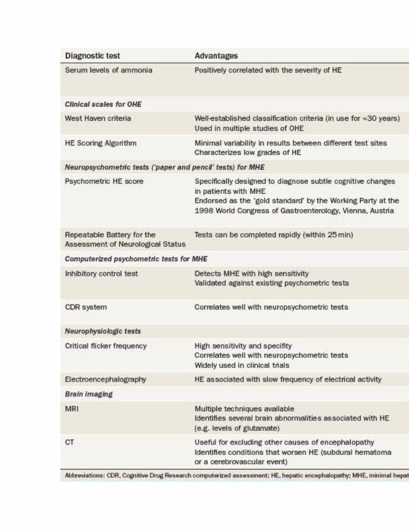

Neuropsychometric Tests
Neuropsychometric tests (including 'paper and pencil' tests and computerized tests) are employed to identify impairments in domains such as visuospatia functioning, attention, processing speed and response inhibition. Over time, these tests have proven to be sensitive to the changes associated with MHE. However, these tests are not without their limitations ( Table 1 ).
Table 1. Advantages and Disadvantages of Diagnostic Tests for Hepatic Encephalopathy


PHES. The psychometric HE Score (PHES) is a battery of neuropsychometric tests that was specifically designed to diagnose the subtle cognitive changes that characterize MHE in patients with cirrhosis ( Table 1 and Box 1 ).[76,77] Normative data for comparison were initially obtained in Germany, followed by Italy and Spain.[73,76,78] Abnormal test results are strongly correlated with changes found on functional brain neuroimaging scans. The Working Party of the 1998 World Congress of Gastroenterology, Vienna, Austria, endorsed this test as the official 'gold standard' for the diagnosis of MHE. Notably, apart from the PHES, no other psychometric tests for HE have been validated by comparisons with normative data. This lack of validation remains an important area for further research in the field of MHE. The PHES has failed to gain popularity in the USA, however, owing to the lack of US-specific normative data and limited availability of the testing system.
Table 1. Advantages and Disadvantages of Diagnostic Tests for Hepatic Encephalopathy


Box 1. The Psychometric Hepatic Encephalopathy Score
RBANS. The Repeatable Battery for the Assessment of Neurological Status (RBANS) was issued to diagnose neurocognitive disorders such as dementia, traumatic brain injury, stroke, multiple sclerosis and bipolar disorder in the USA. A subset of the RBANS tests is used in the USA instead of the PHES. This modified version of the RBANS was designed to focus specifically on the cognitive changes that occur in patients with OHE.[77] The modified RBANS test has now been used in multiple studies in the USA and has proven to be effective in screening patients for MHE. [79,80] Like the PHES, the RBANS is a paper and pencil test that takes about 20-25 min to administer. In addition to the domains assessed in the PHES, the RBANS test scores patients' visual, verbal and working memory.
Computerized Psychometric Tests
A number of computerized psychometric tests have been developed in the past 5 years that, once approved by a large consensus group, have the potential to revolutionize the assessment of patients with HE.
Inhibitory Control Test. The inhibitory control test seems to be the most popular of the currently available computerized tests for HE. This test assesses two different cognitive domains that are affected in patients with MHE—response inhibition and attention.[81,82] The principle of this test is based on 'targets' and 'lures'. Patients are shown a series of different alphabet sequences that flash on the computer screen one after another, and are expected to respond when 'X' is followed by 'Y' and vice versa—a so-called 'target. However, patients are told not to respond to a 'lure'—when 'X' followed by 'X' or 'Y' is followed by 'Y'. A lure response greater than five (out of 40 attempts) detects MHE with high sensitivity.[81,82] This test has been validated against conventional standard psychometric tests and, in a trial of probiotic therapy for HE, has shown close correlations with other HE scores and improvements in test results in patients who responded to therapy.[82] However, most studies that used the inhibitory control test have been published by a single group of investigators and have been conducted only in either Wisconsin or Virginia, USA. Nonetheless, this test shows promise and might ultimately be approved for the diagnosis of MHE.
CDR Computerized Assessment System. The eponymous computerized assessment system from Cognitive Drug Research Ltd (CDR), Goring-on-Thames, UK, was devised specifically for neuropsychiatric profiling of patients with cirrhosis and MHE.[83] This battery consists of seven tests that comprise over 50 parallel forms of each task and comprehensively measure power of attention, continuity of attention, quality of episodic memory, quality of continuous memory and speed of

memory. This test has shown good correlation with the gold-standard PHES test and is popular in the UK.[83]
Electrophysiological Assessments
Critical Flicker Frequency Test. The critical flicker frequency test was validated for the assessment of patients with HE in 2002[84] and for the assessment of patients with HE who were undergoing transjugular intrahepatic portosystemic shunt (TIPS) placement in 2009. [85] The principle of this test is based on the fact that retinal glial cells in patients with HE undergo similar changes (that is, swelling) to those seen in cerebral glial cells, termed hepatic retinopathy. The test involves showing the patient light pulses, initially at a frequency of 60 Hz, which is gradually reduced in 0.1 Hz decrements, once per second. The critical flicker frequency represents the frequency at which discrete light pulses are first perceived. The test results are not influenced by sex, occupation or education level, and are only slightly dependent on age. Furthermore, the test results correlate positively with those of paper and pencil neuropsychometric tests.[84,85] Moreover, a critical flicker frequency of below 39 Hz diagnoses MHE with high sensitivity and specificity. This tool has been widely used to assess HE in therapeutic trials.
Electroencephalography. Electroencephalography is an excellent tool for diagnosing HE in the research setting. HE is associated with a decreased mean frequency of electrical activity in the brain, and the diagnostic sensitivity for HE of this finding ranges between 43% and 100% in published studies.[73] New advances in electroencephalography, such as artificial neural network expert system software and short epoch, dominant activity and cluster analysis, could prove useful for the diagnosis of HE, although they require further validation in clinical trials. [86]
Brain Imaging
Cerebral edema in patients with HE is increasingly being detected by MRI. [36] Many MRI techniques can identify low-grade cerebral edema ( Table 2 ). A CT scan of the head is useful to identify conditions that could either mimic or exacerbate HE, such as subdural hematoma or a cerebrovascular event ( Table 2 ).
Table 2. Brain Imaging Modalities for Diagnosis of Hepatic Encephalopathy

Table 2. Brain Imaging Modalities for Diagnosis of Hepatic Encephalopathy
Measurement of Serum Ammonia Levels
Levels of arterial and venous ammonia correlate positively with the severity of HE. [87] However, routine measurement of ammonia levels in the blood is not recommended, as the results of the test would not change either the approach to diagnosis or management of a patient with suspected HE. Additionally, this test can be challenging to perform in the clinic, as the blood sample should be collected from a stasis-free vein (that is, without using a tourniquet and taking care not to cause turbulence or hemolysis) and must be immediately transported on ice to the laboratory to be analyzed within 20 min.
Treatment
Many treatment options are available for patients with OHE (as described below), but no evidence currently supports the treatment of MHE. Most patients show clinical signs of improvement in the symptoms of HE within 24-48 h of initiation of treatment (both empiric therapy and treatment of the underlying causes). Serum levels of ammonia might lag behind the clinical response. However, if HE persists after 72 h of treatment, the following possibilities must be explored: other causes of encephalopathy might have been missed or inadequately treated; a precipitating factor might have been missed, treated inadequately or remain uncorrected; effective empiric therapy has not been instituted or has been given to excess (in the case of lactulose, as discussed below).
Pharmacological Therapy
Correction of the underlying factors that precipitate HE might in itself help to resolve the disease, but predicting a patient's response to treatment is difficult. Inadequate response to treatment can be due to a combination of factors. All patients should, therefore, receive empiric therapy for HE as other possible diagnoses are being excluded. Empiric therapy is based on the principle of reducing the

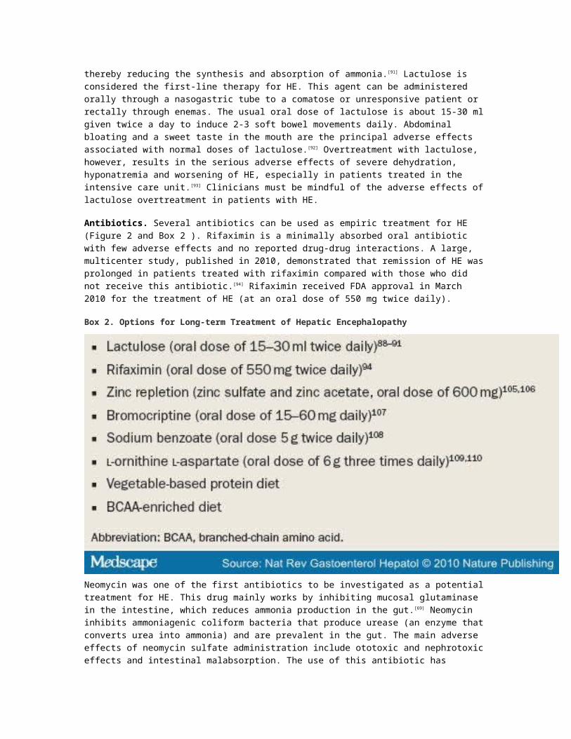
production and absorption of ammonia in the gut—a number of agents are beneficial for this purpose ( Box 2 ).
Box 2. Options for Long-term Treatment of Hepatic Encephalopathy
Nonabsorbable Disaccharides. Nonabsorbable disaccharides include lactulose and lactitol (an analog of lactulose that is not available in USA).[88-90] In addition to having a laxative effect, lactulose and lactitol reduce the colonic pH and interfere with mucosal uptake of glutamine in the gut, thereby reducing the synthesis and absorption of ammonia.[91] Lactulose is considered the first-line therapy for HE. This agent can be administered orally through a nasogastric tube to a comatose or unresponsive patient or rectally through enemas. The usual oral dose of lactulose is about 15-30 ml given twice a day to induce 2-3 soft bowel movements daily. Abdominal bloating and a sweet taste in the mouth are the principal adverse effects associated with normal doses of lactulose. [92] Overtreatment with lactulose, however, results in the serious adverse effects of severe dehydration, hyponatremia and worsening of HE, especially in patients treated in the intensive care unit. [93] Clinicians must be mindful of the adverse effects of lactulose overtreatment in patients with HE.
Antibiotics. Several antibiotics can be used as empiric treatment for HE (Figure 2 and Box 2 ). Rifaximin is a minimally absorbed oral antibiotic with few adverse effects and no reported drug-drug interactions. A large, multicenter study, published in 2010, demonstrated that remission of HE was prolonged in patients treated with rifaximin compared with those who did not receive this antibiotic. [94] Rifaximin received FDA approval in March 2010 for the treatment of HE (at an oral dose of 550 mg twice daily).
Box 2. Options for Long-term Treatment of Hepatic Encephalopathy

Neomycin was one of the first antibiotics to be investigated as a potential treatment for HE. This drug mainly works by inhibiting mucosal glutaminase in the intestine, which reduces ammonia production in the gut.[69] Neomycin inhibits ammoniagenic coliform bacteria that produce urease (an enzyme that converts urea into ammonia) and are prevalent in the gut. The main adverse effects of neomycin sulfate administration include ototoxic and nephrotoxic effects and intestinal malabsorption. The use of this antibiotic has declined over the past few years, owing to the availability of safer antibiotics such as rifaximin.
Nutritional Interventions
Skeletal muscle metabolizes ammonia in patients with chronic liver disease. [19-21] Loss of lean body mass depletes this 'ammonia sink' and increases the ammonia load to the brain, thereby worsening HE. A strong consensus, therefore, exists that patients with cirrhosis should receive a high-protein diet. The European Society for Parenteral and Enteral Nutrition recommended, in 2006, that patients with cirrhosis must eat at least 1.2 g/kg of protein daily.[95,96] They also recommended that the diet of patients with cirrhosis should be supplemented with branched-chain amino acids (BCAAs) and vegetable protein once HE has developed.
Branched-chain Amino Acids. BCAAs have been studied extensively and a recent meta-analysis has shown that patients with cirrhosis who receive BCAAs are more likely to recover from HE than those who do not receive this supplement.[97] BCAAs improve levels of serum albumin, increase progression-free survival and reduce both the number of hospitalizations and the length of hospital stays in patients with cirrhosis.[97-99] These amino acids can be administered orally as well as intravenously; however, their use can be limited by poor availability and high costs.
Vegetable-based Protein. Vegetable-based protein is better tolerated by patients with cirrhosis than meat-based protein. Vegetable-based protein foods have a high fiber content, which increases intestinal transit time and colonic motility and enhances intestinal nitrogen clearance. [99] Vegetable protein also reduces colonic pH, which prevents ammonia from being absorbed in the gut. The

adverse effects of a predominantly vegetarian diet include abdominal bloating and flatulence. To reduce these effects and increase a patient's protein intake vegetable protein can be combined with dairy products, such as milk and cheese.
Long-term Management
Outpatient Management of HE. After an episode of HE has resolved, patients with cirrhosis tend to remain on empiric therapy for an indefinite period of time or until they undergo liver transplantation. The goals of therapy at this stage are to prevent recurrent episodes of HE and to ensure a reasonable quality of life.[100]
Lactulose and rifaximin are popular choices for ongoing therapy in patients who have experienced an episode of HE ( Box 2 ). Adherence of patients to lactulose therapy on a long-term basis is limited by its gastrointestinal adverse effects. Patients must also be educated with regard to the need to monitor the consistency of their bowel movements and use appropriate dose titration when being treated with lactulose. Rifaximin, by contrast, has emerged as an effective treatment strategy to prevent recurrence of HE in a multicenter study published in 2010.[94] Patients who had previously experienced at least two episodes of HE and were in remission were randomly assigned to receive either rifaximin or placebo and were followed up for 6 months. More than 90% of participants in this study were treated with lactulose in addition to rifaximin or placebo. The patients in the rifaximin group maintained their remission from HE more effectively than did patients in the placebo group, and rifaximin was associated with improved tolerability and decreased adverse effects compared with placebo. [94] Rifaximin is, therefore, likely to dominate treatment strategy for HE in the future.
Box 2. Options for Long-term Treatment of Hepatic Encephalopathy
Persistent HE. Orthotopic liver transplantation is the ultimate solution for patients affected by HE. However, HE has lost ground as an indication for liver transplantation in the era of the Model for End-stage Liver Disease (MELD) score. Stewart et al.[101] reported that the severity of HE before liver transplantation is inversely correlated with the duration of survival after transplantation, and that the
prognosis of patients who undergo transplantation for HE is worse than that indicated by their MELD score.[101] A crucial issue, therefore, is that the work-up of a patient who is eligible for liver transplantation should be initiated at the earliest opportunity after an acute episode of HE.
Surgical portosystemic shunting or TIPS placement worsens HE because ammonia in the gut circulation can then bypass first-pass metabolism in the liver and go directly to the brain. [102] Symptoms of HE that result from portosystemic shunting are amenable to shunt closure or a reduction in stent diameter, but these procedures are indicated only if the patient is not a candidate for transplantation. Alternative treatments for the underlying condition that prompted shunt placement (that is, recurrent variceal bleeding or refractory ascites) should be considered to prevent the development of HE.
Conclusions
HE is a neuropsychiatric complication of cirrhosis. Ammonia is recognized as a crucial component in the pathogenesis of HE, but other factors such as inflammation, neurosteroids and manganese are also implicated in the development of the disease. OHE can be diagnosed clinically and mild to moderate grades of the disease might be present in a considerable proportion of ambulatory patients with cirrhosis. Patients with MHE have normal findings on clinical examination, but abnormal psychometric test results.
Several computerized psychometric tests are being developed to aid the clinical diagnosis of HE and enable clinicians to screen patients with cirrhosis for this condition in an outpatient setting. Serum levels of ammonia have limited value in the diagnosis of HE, despite being an indicator of disease severity. Empiric treatment should be started in all patients with OHE. The main goals of outpatient management of patients who have previously experienced an episode of HE is focused on the maintenance of remission and on ensuring that they have a reasonable quality of life. Rifaximin and lactulose have both received FDA approval for the maintenance of remission in patients who have previously been affected by HE. An early work-up for liver transplantation is beneficial for the management of patients with HE who are candidates for this procedure.
Approval of computerized psychometric tests by the Hepatic Encephalopathy Consensus group for the diagnosis of MHE is on the horizon. This approval will facilitate widespread implementation of screening of patients with cirrhosis for HE in an outpatient setting. Furthermore, cyclic GMP and related molecules hold promise as biomarkers of MHE, which could revolutionize the future diagnosis and management of these patients.[103,104] Translational research that involves advanced MRI techniques and proton spectroscopy might resolve controversial issues in the pathogenesis of HE. Although preliminary studies have shown that patients with MHE derive benefit from treatment, results of large multicenter trials in this setting are awaited. Identification of a cost-effective and well-tolerated agent for the treatment of HE remains a challenge.
Key Points
Hepatic encephalopathy (HE) is a serious neuropsychiatric complication of acute and chronic liver disease
Inflammation and raised levels of ammonia in the blood (owing to diminished clearance of ammonia by the liver) underlie the pathogenesis of HE
Some degree of cerebral edema is observed in all grades of HE
The occurrence of any neuropsychiatric manifestation in patients with liver disease should be treated as HE unless proven otherwise
An acute episode of HE is managed by a tripartite strategy: ruling out other causes of encephalopathy, identifying the precipitating cause and initiating empiric therapy
Rifaximin and lactulose are the only two medications approved by FDA for long-term treatment of HE
Work-up for liver transplantation must be initiated as early as possible


![Hepatic Encephalopathy in Chronic Liver Disease: 2014 ... · ascites [7]. Overt hepatic encephalopathy is also reported in Overt hepatic encephalopathy is also reported in subjects](https://img.pdfslide.us/doc/110x75/5d489aa688c993047d8b91d5/hepatic-encephalopathy-in-chronic-liver-disease-2014-ascites-7-overt.jpg)