Embed Size (px)
Citation preview

RESEARCH Open Access
Fractalkine suppression during hepaticencephalopathy promotesneuroinflammation in miceMatthew McMillin1,2, Stephanie Grant1,2, Gabriel Frampton1,2, Sarah Andry3, Adam Brown3
and Sharon DeMorrow1,2*
Abstract
Background: Acute liver failure is associated with numerous systemic consequences including neurologicaldysfunction, termed hepatic encephalopathy, which contributes to mortality and is a challenge to manage in theclinic. During hepatic encephalopathy, microglia activation and neuroinflammation occur due to dysregulated cellsignaling and an increase of toxic metabolites in the brain. Fractalkine is a chemokine that is expressed primarily inneurons and through signaling with its receptor CX3CR1 on microglia, leads to microglia remaining in a quiescentstate. Fractalkine is often suppressed during neuropathies that are characterized by neuroinflammation. However,the expression and subsequent role of fractalkine on microglia activation and the pathogenesis of hepaticencephalopathy due to acute liver failure is unknown.
Methods: Hepatic encephalopathy was induced in mice via injection of azoxymethane (AOM) or saline for controls.Subsets of these mice were implanted with osmotic minipumps that infused soluble fractalkine or saline into thelateral ventricle of the brain. Neurological decline and the latency to coma were recorded in these mice, and brain,serum, and liver samples were collected. Neurons or microglia were isolated from whole brain samples usingimmunoprecipitation. Liver damage was assessed using hematoxylin and eosin staining and by measuringserum liver enzyme concentrations. Fractalkine and CX3CR1 expression were assessed by real-time PCR,and proinflammatory cytokine expression was assessed using ELISA assays.
Results: Following AOM administration, fractalkine expression is suppressed in the cortex and in isolated neuronscompared to vehicle-treated mice. CX3CR1 is suppressed in isolated microglia from AOM-treated mice. Solublefractalkine infusion into the brain significantly reduced neurological decline in AOM-treated mice compared tosaline-infused AOM-treated mice. Infusion of soluble fractalkine into AOM-treated mice reduced liver damage,lessened microglia activation, and suppressed expression of chemokine ligand 2, interleukin-6, and tumornecrosis factor alpha compared to saline-infused mice.
Conclusions: These findings suggest that fractalkine-mediated signaling is suppressed in the brain following thedevelopment of hepatic encephalopathy. Supplementation of AOM-treated mice with soluble fractalkine led toimproved outcomes, which identifies this pathway as a possible therapeutic target for the management ofhepatic encephalopathy following acute liver injury.
Keywords: Acute liver failure, Azoxymethane, CX3CL1, CX3CR1, CCL2(Continued on next page)
* Correspondence: [email protected] of Internal Medicine, Texas A&M Health Science Center, Collegeof Medicine, Temple, TX, USA2Central Texas Veterans Healthcare System, 1901 S. 1st Street, Building 205,Temple, TX 76504, USAFull list of author information is available at the end of the article
© 2016 The Author(s). Open Access This article is distributed under the terms of the Creative Commons Attribution 4.0International License (http://creativecommons.org/licenses/by/4.0/), which permits unrestricted use, distribution, andreproduction in any medium, provided you give appropriate credit to the original author(s) and the source, provide a link tothe Creative Commons license, and indicate if changes were made. The Creative Commons Public Domain Dedication waiver(http://creativecommons.org/publicdomain/zero/1.0/) applies to the data made available in this article, unless otherwise stated.
McMillin et al. Journal of Neuroinflammation (2016) 13:198 DOI 10.1186/s12974-016-0674-8
(Continued from previous page)
Abbreviations: ADAMs, A disintegrin and metalloproteinase proteins; ALT, Alanine aminotransferasase;AOM, Azoxymethane; CCL2, Chemokine ligand 2; DAPI, 4′,6-Diamidino-2-phenylindole; ERK1/2, Extracellularsignal-regulated kinase 1/2; GLAST, Glutamate aspartate transporter; ICV, Intracerebroventricular; IL, Interleukin;RT-PCR, Real-time PCR; sFKN, Soluble fractalkine; TNFα, Tumor necrosis factor-α
BackgroundAcute liver failure in Europe and the USA is causedprimarily by acetaminophen-induced drug toxicity butcan also be the result of viral hepatitis, toxin ingestion,non-acetaminophen-induced drug toxicity, and numerousother rare causative factors [1]. Following the developmentof acute liver failure, a variety of extrahepatic complicationscan develop including renal, respiratory, immunological,and neurological dysfunction. Of these complications,neurological dysfunction accounts for around 25 % of themortality associated with this disease state and remains asignificant challenge for clinical management of this disease[2]. These neurological complications are termed hepaticencephalopathy and have a variety of neuropsychiatric pre-sentations from asymptomatic to severe cognitive declineand coma with implications on liver transplant priority,patient quality of life, and survival [3, 4].A growing body of evidence implicates neuroinflam-
mation and microglia activation involvement with thedevelopment of hepatic encephalopathy. Elevations ofthe proinflammatory cytokines interleukin (IL)-1β, IL-6,and tumor necrosis factor alpha (TNFα) are observed inboth patients and in rodent models of acute liver failure[5, 6]. During hepatic encephalopathy, the use of anti-inflammatory therapeutics, such as minocycline, has beenshown to reduce neuroinflammation and neurological dys-function [7]. The production of pathogenic proinflamma-tory cytokines in the brain is a consequence of microgliaactivation, which can occur via a variety of signals includ-ing signal transduction by chemokines. We have previouslyshown that chemokine ligand 2 (CCL2) is one pathogenicchemotactic cytokine that is elevated in neurons duringhepatic encephalopathy due to acute liver failure and con-tributes to both microglia activation and the elevation ofIL-1β and IL-6 [8]. The receptors for CCL2 and otherchemokines are primarily localized to microglia in thebrain and therefore chemokine signaling is important ininitiating the activation of microglia during states of neuro-inflammation. Together, this implicates neuroinflammationand chemokine-signaling pathways as causative factors inthe progression of hepatic encephalopathy.Fractalkine, also known as CX3CL1, is the only member
of the CX3C class of chemokines. In the brain, fractalkineis highly expressed in neurons but can be induced in astro-cytes if they are treated with TNFα or interferon-γ [9, 10].
Fractalkine in its native form is a transmembrane proteinthat can be cleaved by cathepsin S or a disintegrin andmetalloproteinase proteins (ADAMs) [11, 12]. Upon cleav-age, fractalkine is released in its soluble form that containsthe extracellular N-terminal chemokine domain. Solublefractalkine can then bind its receptor, CX3CR1, which is aG-protein coupled receptor that is localized to microgliaand is thought to keep microglia in a quiescent state [9, 13].Fractalkine/CX3CR1 signaling has been demonstrated innumerous studies to dampen neuroinflammation, withfractalkine signaling being shown to reduce IL-1β secretionin lipopolysaccharide-treated microglia [14]. This has alsobeen shown in vivo as rats with cerebral ischemia adminis-tered CX3CR1 siRNA were found to have increased micro-glia activation and proinflammatory cytokine expressioncompared to ischemic controls [15]. In addition to this,in a variety of inflammatory conditions there is a recipro-cal relationship between fractalkine and CCL2, thoughwhether these chemokines have a direct effect on eachother or are inversely affected by an extraneous signal isunknown [16, 17].At this time, there have been no studies investigating
fractalkine in the context of neuroinflammation associatedwith hepatic encephalopathy. Therefore, the aims of thisstudy were to assess the expression of fractalkine and itsinteraction with inflammatory signaling in a murine modelof hepatic encephalopathy due to acute liver failure and todetermine how fractalkine contributes to the neurologicalcomplications associated with this disease state.
MethodsMaterialsFractalkine antibodies were purchased from Abcam(Cambridge, MA). Antibodies against IBA1 were purchasedfrom Wako Chemicals USA (Richmond, VA). NeuNantibodies were ordered from Millipore (Billerica, MA).Antibodies against CD11b and glutamate aspartate trans-porter (GLAST) were purchased from Miltenyi Biotec (SanDiego, CA). Phosphorylated and total extracellular signal-regulated kinase 1/2 (pERK1/2 and tERK1/2) antibodieswere purchased from Cell Signaling (Danvers, MA). Mousesoluble fractalkine was purchased from R&D Systems(Minneapolis, MN). Mouse fracktalkine, CCL2, IL-6, andTNFα ELISAs were purchased from R&D Systems. Allreal-time PCR (RT-PCR) primers were purchased from
McMillin et al. Journal of Neuroinflammation (2016) 13:198 Page 2 of 12
SABiosciences (Frederick, MD). Total bilirubin was assayedusing an ELISA kit from Cusabio (Wuha, China). All otherchemicals and reagents were purchased from Sigma-Aldrich (St. Louis, MO) unless otherwise noted and were ofthe highest grade available.
Mouse model of hepatic encephalopathyMale C57Bl/6 mice (20–25 g; Charles River Laboratories,Wilmington, MA) were used in all in vivo experimentsusing methodology described previously [8, 18, 19]. Allanimal experiments were performed following approvalfrom Baylor Scott & White Health Institutional AnimalCare and Use Committee (Temple, TX). Mice were pro-vided free access to water and rodent chow and werehoused in constant temperature, humidity, and 12-h light-dark cycling. Acute liver failure was induced via a singleintraperitoneal injection of 100 mg/kg of azoxymethane(AOM) as previously described [18, 19]. In parallel,soluble fractalkine was administered to the brain via intra-cerebroventricular (ICV) infusion (200 ng/day) for 3 daysprior to injection of AOM as previously described [18].This dose was selected to maintain concentrations offractalkine in the cerebrospinal fluid at concentrationsthat are at or above those reported by other researchers[20]. Care was taken to minimize the effects of anesthesiaon hepatic metabolism in ICV mice by the following: (1)all mice were anesthetized with isoflurane to reach sur-gical plane with the subsequent surgeries taking appro-ximately 30 min to complete per mouse and (2) AOMinjection was delayed for 3 days after the ICV surgery toallow for residual anesthesia to be eliminated from thebody. After AOM injection, mice were placed on heatingpads adjusted to 37 °C and monitored for signs of neuro-logical decline. To reduce the impact of hypoglycemia anddehydration, cage floors were supplied with hydrogel androdent chow and after 12 h and every subsequent 4 h,mice were injected subcutaneously with 5 % dextrose in250 μl of saline. If mice underwent a 20 % weight loss orgreater, they were removed from the study.At 12 h following injection (and every 2 h thereafter),
body temperature, weight, and neurological assessmentswere measured. Neurological functioning was assessed bymeasuring the pinna reflex, corneal reflex, tail flexion reflex,escape response reflex, righting reflex, and ataxia whichwere scored on a scale of 0 (no reflex) to 2 (intact reflex).The neurological score at each time point was defined asthe summation of these reflex scores. In addition, time tocoma (defined as a loss of all reflexes) was recorded. Tissuewas flash frozen and collected at coma for further analyses.Mice used for histochemical studies were transcardially per-fused with PBS followed by 4 % paraformaldehyde. Wholebrains were removed and placed into 4 % paraformaldehydefor 24 h, after which they were moved to a 30 % sucrose
solution for cryoprotection. Brains were frozen and sec-tioned using a cryostat for immunofluorescence imaging.
Cell isolation and flow cytometryFresh whole brains from adult C57Bl/6 mice were homo-genized using an automated homogenizer from MiltenyiBiotec using solutions provided in the Neural TissueDissociation Kit (Miltenyi Biotec). Following dissociationinto a single-cell suspension, cells were passed through LScolumns (Miltenyi Biotec) containing beads coated withCD11b antibodies (to isolate microglia) or GLAST anti-bodies (to isolate astrocytes) localized to the columns. Theremaining cells not bound to the columns were kept asthe neuron-enriched fraction. LS columns were washed toremove the CD11b-bound cells or the GLAST-boundcells. Cells were subsequently spun down and homoge-nized for subsequent experiments.
Liver histology and biochemistryParaffin-embedded livers were cut into 3-μm sectionsand mounted onto positively charged slides (VWR,Radnor, PA). Slides were deparaffinized and stained withHematoxylin QS (Vector Laboratories, Burlingame, CA)for 1 min, followed by staining for 1 min with eosin Y(Amresco, Solon, OH), and rinsed in 95 % ethanol. Theslides were then dipped into 100 % ethanol and sub-sequently through two xylene washes. Coverslips weremounted onto the slides using Vectamount mounting media(Vector Laboratories). The slides were viewed and imagedusing an Olympus BX40 microscope with an OlympusDP25 imaging system (Olympus, Center Valley, PA).Serum alanine aminotransferasase (ALT) and bilirubin
were assessed using commercially available kits. Alanineaminotransferase measurement was performed using afluorimetric activity assay. Total bilirubin was assayedusing a total bilirubin ELISA. All assays and subsequentanalyses were performed according to the instructionsprovided by the manufacturers.
Real-time PCRRNA was extracted from flash frozen tissue or isolatedneural cells, and RT-PCR was performed as previouslydescribed using commercially available primers designedagainst mouse fractalkine, CX3CR1, and glyceraldehyde3-phosphate dehydrogenase [21]. A ΔΔCT analysis wasperformed using vehicle-treated tissue or isolated corticalneurons/microglia as controls for subsequent experiments[22, 23]. Data for all experiments are expressed as meanrelative mRNA levels ± SEM.
ImmunofluorescenceFree-floating immunostaining was performed on brainsections using anti-IBA1 immunoreactivity to detectmorphology and relative staining of microglia. In addition,
McMillin et al. Journal of Neuroinflammation (2016) 13:198 Page 3 of 12
fractalkine and NeuN immunostaining were performed.Immunoreactivity was visualized using fluorescent second-ary antibodies labeled with Dylight 488 or Cy3 and counter-stained with ProLong© Gold Antifade Reagent containing4′,6-diamidino-2-phenylindole (DAPI). Slides were viewedand imaged using a Leica Microsystems TCS SP5-Xinverted confocal microscope (Buffalo Grove, IL). Quantifi-cation of IBA-1 field staining was performed by convertingimages to grayscale, inverting their color and quantifyingthe number of stained pixels with ImageJ software. Thevalues for the vehicle group were normalized to 1 with thisreference being used for all other quantifications of IBA1field staining. Z-stack images were created by performing a30-μm merge of images through the entire brain sectionusing ImageJ software.
Cytokine and chemokine ELISAsCortex tissue from all treatment groups was homogenizedusing a Miltenyi Biotec gentleMACS™ Dissociator, andtotal protein was quantified using a Pierce™ BCA ProteinAssay kit (ThermoFisher Scientific, Waltham, MA). Cap-ture antibodies against CCL2, IL-6, TNFα, or fractalkinewere incubated overnight in 96-well plates. Each ELISAwas performed according to the instructions providedfrom R&D Systems, and the total input protein for eachsample was 50 μg. Absorbance was read using a Spectra-Max® M5 plate reader from Molecular Devices (Sunnyvale,CA). Data are reported as CCL2, IL-6, TNFα, or fractalk-ine concentration per milligram of total lysate protein.
ImmunoblottingImmunoblots were performed as previously described [8]with minor modifications. For western blots, 10 % sodiumdodecyl sulfate-polyacrylamide gel electrophoresis gelswere loaded with 20 μg of protein diluted in Laemmlibuffer. Specific primary antibodies against pERK1/2 andtERK1/2 were used along with appropriate fluorescentsecondary antibodies (LI-COR, Lincoln, NE). All imagingwas performed using an Odyssey 9120 Infrared ImagingSystem (LI-COR). Data are expressed as fold change influorescent band intensity of pERK1/2 divided by tERK1/2. The values of the vehicle ICV saline group was used asa baseline and set to a relative protein expression value of1. All band intensity quantifications were performed usingImageJ software (National Institutes of Health, Bethesda,MD). Data for all experiments were expressed as meanrelative protein ± SEM (n = 4).
Statistical analysisAll statistical analyses were performed using GraphpadPrism software (Graphpad Software, La Jolla, CA). Resultswere expressed as mean ± SEM. For data passing norma-lity tests, significance was established using the Student’st test when differences between two groups were analyzed,
and analysis of variance when differences between threeor more groups were compared, followed by the appropri-ate post hoc test. For neurological score analyses, two-wayanalysis of variance analysis and a subsequent Bonferronipost hoc test were performed. If tests for normality failed,two groups were compared with a Mann-Whitney U testor a Kruskal-Wallis ranked analysis when more than twogroups were analyzed. Differences were considered signifi-cant when the p value was less than 0.05.
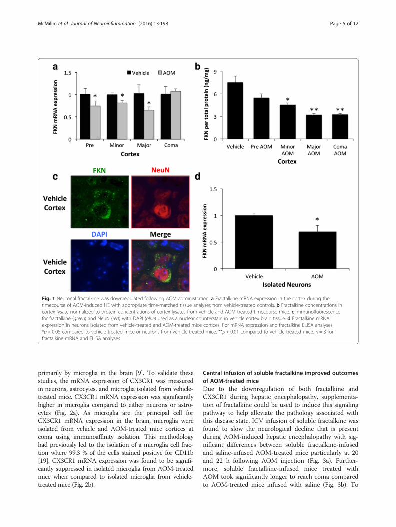
ResultsNeuronal fractalkine was downregulated followingAOM administrationC57Bl/6 mice were administered the hepatotoxin AOM toinduce acute liver failure and the development of hepaticencephalopathy. The mouse AOM model of hepatic en-cephalopathy is characterized by a prodromal phase thattransitions into mild cognitive deficits and quickly pro-gresses to coma [24]. Fractalkine mRNA expression wasdownregulated in AOM-treated mice compared to time-matched vehicle controls prior to the development ofneurological decline (pre), when minor neurological declinewas evident (minor) and when major neurological declinewas present (major) but had expression similar to controlswhen the mice reach coma (Fig. 1a). The expression offractalkine was not altered in the vehicle groups over thetime period studied (data not shown); therefore, all vehiclegroups used in the remainder of this study were time-matched to coma AOM-treated mice. The suppression offractalkine mRNA expression observed in AOM-treatedmice correlated with a parallel reduction in fractalkineprotein in cortex lysates throughout AOM-induced neuro-logical decline (Fig. 1b). The expression of fractalkine hasbeen reported in multiple cell types in the brain, and there-fore, immunofluorescence for fractalkine was performed.This staining demonstrated that the highest expression offractalkine was in neurons as a high degree of co-stainingwas found with the neuron marker NeuN (Fig. 1c). In orderto determine if AOM treatment led to reduced productionof neuronal fractalkine, neurons were isolated from thecortex of vehicle and AOM-treated C57Bl/6 mice using im-munoprecipitation. We have previously utilized this tech-nique and found that the neuron-enriched cell fraction had99.7 % of the cells stain positive for CD90, which indicatesthat this methodology provides a relatively pure neuron cellfraction from whole brain homogenates [19]. FractalkinemRNA expression was significantly downregulated inisolated neurons from AOM-treated mice compared toneurons isolated from vehicle-treated mice (Fig. 1d).
CX3CR1 was downregulated in microglia followingAOM-induced hepatic encephalopathyFractalkine transduces its signal through binding CX3CR1,which has been shown in previous studies to be expressed
McMillin et al. Journal of Neuroinflammation (2016) 13:198 Page 4 of 12
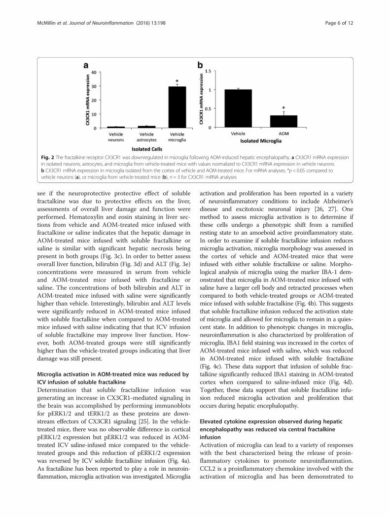
primarily by microglia in the brain [9]. To validate thesestudies, the mRNA expression of CX3CR1 was measuredin neurons, astrocytes, and microglia isolated from vehicle-treated mice. CX3CR1 mRNA expression was significantlyhigher in microglia compared to either neurons or astro-cytes (Fig. 2a). As microglia are the principal cell forCX3CR1 mRNA expression in the brain, microglia wereisolated from vehicle and AOM-treated mice cortices atcoma using immunoaffinity isolation. This methodologyhad previously led to the isolation of a microglia cell frac-tion where 99.3 % of the cells stained positive for CD11b[19]. CX3CR1 mRNA expression was found to be signifi-cantly suppressed in isolated microglia from AOM-treatedmice when compared to isolated microglia from vehicle-treated mice (Fig. 2b).
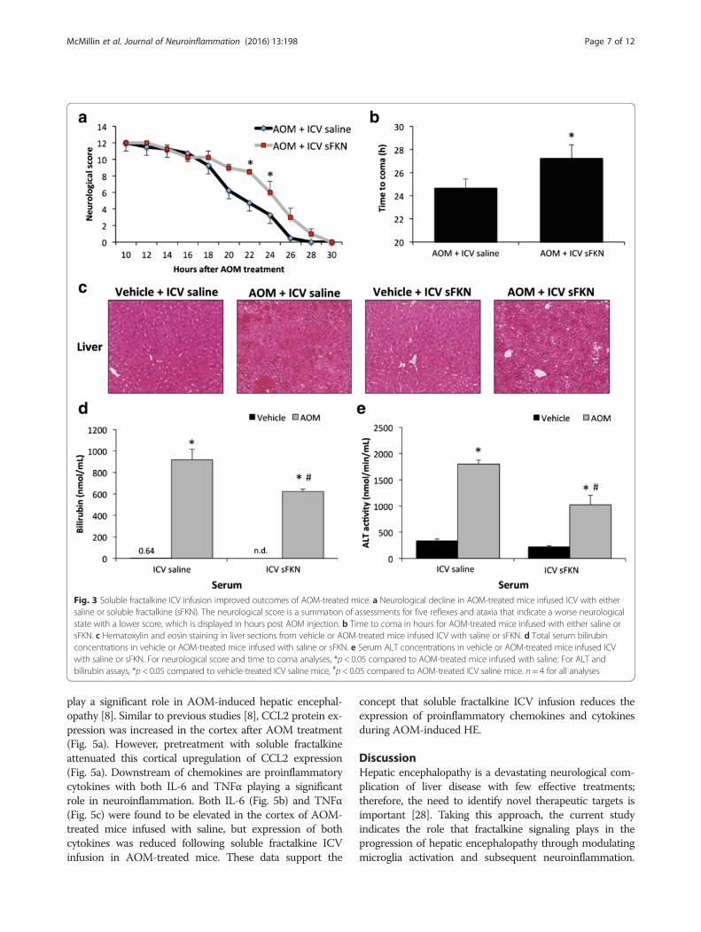
Central infusion of soluble fractalkine improved outcomesof AOM-treated miceDue to the downregulation of both fractalkine andCX3CR1 during hepatic encephalopathy, supplementa-tion of fractalkine could be used to induce this signalingpathway to help alleviate the pathology associated withthis disease state. ICV infusion of soluble fractalkine wasfound to slow the neurological decline that is presentduring AOM-induced hepatic encephalopathy with sig-nificant differences between soluble fractalkine-infusedand saline-infused AOM-treated mice particularly at 20and 22 h following AOM injection (Fig. 3a). Further-more, soluble fractalkine-infused mice treated withAOM took significantly longer to reach coma comparedto AOM-treated mice infused with saline (Fig. 3b). To
Fig. 1 Neuronal fractalkine was downregulated following AOM administration. a Fractalkine mRNA expression in the cortex during thetimecourse of AOM-induced HE with appropriate time-matched tissue analyses from vehicle-treated controls. b Fractalkine concentrations incortex lysate normalized to protein concentrations of cortex lysates from vehicle and AOM-treated timecourse mice. c Immunofluorescencefor fractalkine (green) and NeuN (red) with DAPI (blue) used as a nuclear counterstain in vehicle cortex brain tissue. d Fractalkine mRNAexpression in neurons isolated from vehicle-treated and AOM-treated mice cortices. For mRNA expression and fractalkine ELISA analyses,*p < 0.05 compared to vehicle-treated mice or neurons from vehicle-treated mice, **p < 0.01 compared to vehicle-treated mice. n = 3 forfractalkine mRNA and ELISA analyses
McMillin et al. Journal of Neuroinflammation (2016) 13:198 Page 5 of 12
see if the neuroprotective protective effect of solublefractalkine was due to protective effects on the liver,assessments of overall liver damage and function wereperformed. Hematoxylin and eosin staining in liver sec-tions from vehicle and AOM-treated mice infused withfractalkine or saline indicates that the hepatic damage inAOM-treated mice infused with soluble fractalkine orsaline is similar with significant hepatic necrosis beingpresent in both groups (Fig. 3c). In order to better assessoverall liver function, bilirubin (Fig. 3d) and ALT (Fig. 3e)concentrations were measured in serum from vehicleand AOM-treated mice infused with fractalkine orsaline. The concentrations of both bilirubin and ALT inAOM-treated mice infused with saline were significantlyhigher than vehicle. Interestingly, bilirubin and ALT levelswere significantly reduced in AOM-treated mice infusedwith soluble fractalkine when compared to AOM-treatedmice infused with saline indicating that that ICV infusionof soluble fractalkine may improve liver function. How-ever, both AOM-treated groups were still significantlyhigher than the vehicle-treated groups indicating that liverdamage was still present.
Microglia activation in AOM-treated mice was reduced byICV infusion of soluble fractalkineDetermination that soluble fractalkine infusion wasgenerating an increase in CX3CR1-mediated signaling inthe brain was accomplished by performing immunoblotsfor pERK1/2 and tERK1/2 as these proteins are down-stream effectors of CX3CR1 signaling [25]. In the vehicle-treated mice, there was no observable difference in corticalpERK1/2 expression but pERK1/2 was reduced in AOM-treated ICV saline-infused mice compared to the vehicle-treated groups and this reduction of pERK1/2 expressionwas reversed by ICV soluble fractalkine infusion (Fig. 4a).As fractalkine has been reported to play a role in neuroin-flammation, microglia activation was investigated. Microglia
activation and proliferation has been reported in a varietyof neuroinflammatory conditions to include Alzheimer’sdisease and excitotoxic neuronal injury [26, 27]. Onemethod to assess microglia activation is to determine ifthese cells undergo a phenotypic shift from a ramifiedresting state to an amoeboid active proinflammatory state.In order to examine if soluble fractalkine infusion reducesmicroglia activation, microglia morphology was assessed inthe cortex of vehicle and AOM-treated mice that wereinfused with either soluble fractalkine or saline. Morpho-logical analysis of microglia using the marker IBA-1 dem-onstrated that microglia in AOM-treated mice infused withsaline have a larger cell body and retracted processes whencompared to both vehicle-treated groups or AOM-treatedmice infused with soluble fractalkine (Fig. 4b). This suggeststhat soluble fractalkine infusion reduced the activation stateof microglia and allowed for microglia to remain in a quies-cent state. In addition to phenotypic changes in microglia,neuroinflammation is also characterized by proliferation ofmicroglia. IBA1 field staining was increased in the cortex ofAOM-treated mice infused with saline, which was reducedin AOM-treated mice infused with soluble fractalkine(Fig. 4c). These data support that infusion of soluble frac-talkine significantly reduced IBA1 staining in AOM-treatedcortex when compared to saline-infused mice (Fig. 4d).Together, these data support that soluble fractalkine infu-sion reduced microglia activation and proliferation thatoccurs during hepatic encephalopathy.
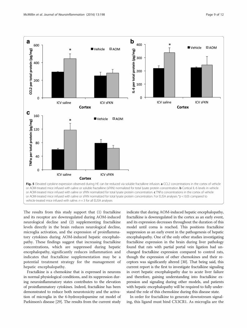
Elevated cytokine expression observed during hepaticencephalopathy was reduced via central fractalkineinfusionActivation of microglia can lead to a variety of responseswith the best characterized being the release of proin-flammatory cytokines to promote neuroinflammation.CCL2 is a proinflammatory chemokine involved with theactivation of microglia and has been demonstrated to
Fig. 2 The fractalkine receptor CX3CR1 was downregulated in microglia following AOM-induced hepatic encephalopathy. a CX3CR1 mRNA expressionin isolated neurons, astrocytes, and microglia from vehicle-treated mice with values normalized to CX3CR1 mRNA expression in vehicle neurons.b CX3CR1 mRNA expression in microglia isolated from the cortex of vehicle and AOM-treated mice. For mRNA analyses, *p < 0.05 compared tovehicle neurons (a), or microglia from vehicle-treated mice (b), n = 3 for CX3CR1 mRNA analyses
McMillin et al. Journal of Neuroinflammation (2016) 13:198 Page 6 of 12
play a significant role in AOM-induced hepatic encephal-opathy [8]. Similar to previous studies [8], CCL2 protein ex-pression was increased in the cortex after AOM treatment(Fig. 5a). However, pretreatment with soluble fractalkineattenuated this cortical upregulation of CCL2 expression(Fig. 5a). Downstream of chemokines are proinflammatorycytokines with both IL-6 and TNFα playing a significantrole in neuroinflammation. Both IL-6 (Fig. 5b) and TNFα(Fig. 5c) were found to be elevated in the cortex of AOM-treated mice infused with saline, but expression of bothcytokines was reduced following soluble fractalkine ICVinfusion in AOM-treated mice. These data support the
concept that soluble fractalkine ICV infusion reduces theexpression of proinflammatory chemokines and cytokinesduring AOM-induced HE.
DiscussionHepatic encephalopathy is a devastating neurological com-plication of liver disease with few effective treatments;therefore, the need to identify novel therapeutic targets isimportant [28]. Taking this approach, the current studyindicates the role that fractalkine signaling plays in theprogression of hepatic encephalopathy through modulatingmicroglia activation and subsequent neuroinflammation.
Fig. 3 Soluble fractalkine ICV infusion improved outcomes of AOM-treated mice. a Neurological decline in AOM-treated mice infused ICV with eithersaline or soluble fractalkine (sFKN). The neurological score is a summation of assessments for five reflexes and ataxia that indicate a worse neurologicalstate with a lower score, which is displayed in hours post AOM injection. b Time to coma in hours for AOM-treated mice infused with either saline orsFKN. c Hematoxylin and eosin staining in liver sections from vehicle or AOM-treated mice infused ICV with saline or sFKN. d Total serum bilirubinconcentrations in vehicle or AOM-treated mice infused with saline or sFKN. e Serum ALT concentrations in vehicle or AOM-treated mice infused ICVwith saline or sFKN. For neurological score and time to coma analyses, *p < 0.05 compared to AOM-treated mice infused with saline. For ALT andbilirubin assays, *p < 0.05 compared to vehicle-treated ICV saline mice, #p < 0.05 compared to AOM-treated ICV saline mice. n = 4 for all analyses
McMillin et al. Journal of Neuroinflammation (2016) 13:198 Page 7 of 12
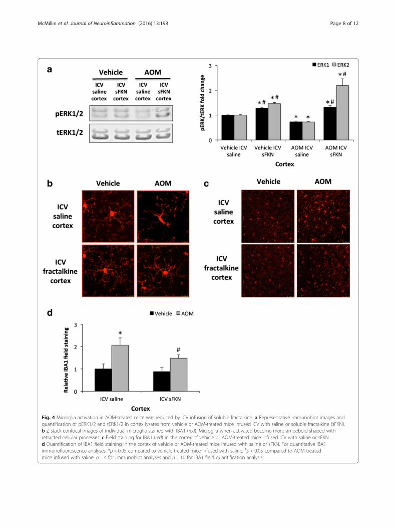
Fig. 4 Microglia activation in AOM-treated mice was reduced by ICV infusion of soluble fractalkine. a Representative immunoblot images andquantification of pERK1/2 and tERK1/2 in cortex lysates from vehicle or AOM-treated mice infused ICV with saline or soluble fractalkine (sFKN).b Z-stack confocal images of individual microglia stained with IBA1 (red). Microglia when activated become more amoeboid shaped withretracted cellular processes. c Field staining for IBA1 (red) in the cortex of vehicle or AOM-treated mice infused ICV with saline or sFKN.d Quantification of IBA1 field staining in the cortex of vehicle or AOM-treated mice infused with saline or sFKN. For quantitative IBA1immunofluorescence analyses, *p < 0.05 compared to vehicle-treated mice infused with saline, #p < 0.05 compared to AOM-treatedmice infused with saline. n = 4 for immunoblot analyses and n = 10 for IBA1 field quantification analysis
McMillin et al. Journal of Neuroinflammation (2016) 13:198 Page 8 of 12
The results from this study support that (1) fractalkineand its receptor are downregulated during AOM-inducedneurological decline and (2) supplementing fractalkinelevels directly in the brain reduces neurological decline,microglia activation, and the expression of proinflamma-tory cytokines during AOM-induced hepatic encephalo-pathy. These findings suggest that increasing fractalkineconcentrations, which are suppressed during hepaticencephalopathy, significantly reduces inflammation andindicates that fractalkine supplementation may be apotential treatment strategy for the management ofhepatic encephalopathy.Fractalkine is a chemokine that is expressed in neurons
in normal physiological conditions, and its suppression dur-ing neuroinflammatory states contributes to the elevationof proinflammatory cytokines. Indeed, fractalkine has beendemonstrated to reduce both neurotoxicity and the activa-tion of microglia in the 6-hydroxydopamine rat model ofParkinson’s disease [29]. The results from the current study
indicate that during AOM-induced hepatic encephalopathy,fractalkine is downregulated in the cortex as an early event,and its expression decreases throughout the duration of thismodel until coma is reached. This positions fractalkinesuppression as an early event in the pathogenesis of hepaticencephalopathy. One of the only other studies investigatingfractalkine expression in the brain during liver pathologyfound that rats with partial portal vein ligation had un-changed fractalkine expression compared to control rats,though the expression of other chemokines and their re-ceptors was significantly altered [30]. That being said, thiscurrent report is the first to investigate fractalkine signalingin overt hepatic encephalopathy due to acute liver failureand therefore, gaining understanding into fractalkine ex-pression and signaling during other models, and patientswith hepatic encephalopathy will be required to fully under-stand the role of this chemokine during this disease state.In order for fractalkine to generate downstream signal-
ing, this ligand must bind CX3CR1. As microglia are the
Fig. 5 Elevated cytokine expression observed during HE can be reduced via soluble fractalkine infusion. a CCL2 concentrations in the cortex of vehicleor AOM-treated mice infused with saline or soluble fractalkine (sFKN) normalized for total lysate protein concentration. b Cortical IL-6 levels in vehicleor AOM-treated mice infused with saline or sFKN normalized for total lysate protein concentration. c TNFα concentrations in the cortex of vehicleor AOM-treated mice infused with saline or sFKN normalized for total lysate protein concentration. For ELISA analyses *p < 0.05 compared tovehicle-treated mice infused with saline. n = 3 for all ELISA analyses
McMillin et al. Journal of Neuroinflammation (2016) 13:198 Page 9 of 12
primary source of CX3CR1, the gene expression of this re-ceptor was assessed in isolated microglia from vehicle andAOM-treated mice. CX3CR1 mRNA was downregulatedin microglia isolated from the cortices of AOM-treatedmice when compared to control-treated mice. Because ofthe technical difficulty and low yield in isolating microgliafrom adult mouse brains, analysis of CX3CR1 protein wasnot possible in this study. Furthermore, fractalkine, via theactivation of CX3CR1 leads to the downstream phospho-rylation of ERK1/2. Our data support a suppression offractalkine-induced ERK1/2 activation after AOM. Takentogether, our data indicate that the fractalkine/CX3CR1signaling axis is suppressed as an early event duringAOM-induced hepatic encephalopathy that persists untilcoma. A similar suppression of fractalkine signaling hasbeen reported during various other neuropathies. Forexample, during neuroinflammation associated with scra-pie infection in hamsters, microglia activation occurs anddownregulation of fractalkine and its receptor is observed[31]. Similarly, in mice with genetic knockout of CX3CR1,there is reduced expression of IL-1β and TNFα as well asreduced infarct size following focal cerebral ischemiacompared to wild-type mice [32]. These effects have alsobeen observed in the clinic, as lower plasma fractalkineconcentrations have been observed in patients who haveworse 6-month outcomes following stroke [33]. Similarfindings regarding fractalkine were observed in the currentstudy as ICV infusion of soluble fractalkine slowed theneurological decline and reduced the elevated concentra-tions CCL2, IL-1β, and TNFα that are associated withAOM-induced hepatic encephalopathy. That being said,fractalkine appears to play an immunomodulatory role thatcan reduce, but not completely reverse, the pathology ofAOM-treated mice. AOM-induced hepatic encephalopathyis characterized by elevations of ammonia and bile acids asa result of liver injury and both of these contribute to the pro-gression of this disorder [34]. Therefore, fractalkinesupplementation may provide an even greater therapeuticbenefit if used in conjunction with other therapies for themanagement of acute liver failure or hepatic encephalopathy.Contrary to the data reported here, some reports in the
brain, and a few investigating other inflammatory diseases,suggest that fractalkine can promote the infiltration ofimmune cells and generate inflammation. One of thesestudies demonstrates that increased fractalkine expressionduring experimental autoimmune encephalomyelitis, amouse model of multiple sclerosis, promotes lymphocyteentry into the brain further exacerbating disease severity[35]. Further support of this is demonstrated in theα-synuclein-induced inflammation model of Alzheimer’sdisease, where the use of CX3CR1 knockout mice hasbeen demonstrated to reduce inflammation during thisdisease state compared to wild-type mice [13]. These dif-ferential effects during various neuropathies indicate that
fractalkine signaling may generate different effects depen-ding on the disease state and the pathology that contributesto the progression of the disease. Of interest is thatCX3CR1 was initially thought to respond to fractalkinealone; however, more recently, CX3XR1 was found to alsobind chemokine ligand 26, making the findings usingCX3CR1 knockout mice difficult to interpret with respectto specific fractalkine signaling [36].Neuroinflammation has been associated with a variety
of neuropathies including multiple sclerosis, stroke, andAlzheimer’s disease [37]. In regard to hepatic encepha-lopathy, microglia activation has been observed duringhepatic devascularization, toxic liver injury, and biliarycirrhosis [38–40]. Elevations of ammonia occur duringhepatic encephalopathy and have been demonstrated topromote microglia activation in rats fed an ammonium-enriched diet indicating that ammonia may generate someof its effects on hepatic encephalopathy pathogenesisthrough exacerbating neuroinflammation [41]. During hep-atic encephalopathy, microglia activation and subsequentneuroinflammation is thought to be primarily driven byincreased concentrations of ammonia, lactate, manganese,or glutamate in the brain [42]. Currently, many of theseeffects are not well understood, but recent studies havebegun to expand on how the initiation of neuroinflamma-tion may occur. Neuroinflammation can be induced bychemokine signaling, such as through CCL2, which isupregulated during AOM-induced hepatic encephalopathyand chemokine receptor 2 or chemokine receptor 4 antag-onist treatment improved outcomes in AOM-treated mice[8]. Similar findings were observed during bile duct ligationin mice where cerebral concentrations of CCL2 were ele-vated and were associated with monocyte infiltration in thebrain, which was eliminated in CCL2 or chemokine recep-tor 2 knockout mice [40]. Of interest is that there appearsto be an inverse relationship between CCL2 and fractalkineduring hepatic encephalopathy. Gaining a greater under-standing of the mechanism that generates these signalingevents may provide better understanding of the initiators ofinflammation during this disease state. This study was thefirst to report that supplementing fractalkine during hepaticencephalopathy suppresses CCL2 concentrations in thecortex, which indicates that the increase of CCL2 observedduring AOM-induced hepatic encephalopathy is dependentupon the suppression of fractalkine. Therefore, there issupport that chemokine signaling plays a significant role inthe initiation of inflammation during hepatic encephalo-pathy, though more studies are needed to fully understandthese signaling pathways and their roles in the pathogenesisof this disorder.As AOM-induced hepatic encephalopathy is the result
of acute liver injury, the finding that soluble fractalkineinfusion directly into the brain conferred some protectionto the liver is interesting and unexpected. Due to fractalkine
McMillin et al. Journal of Neuroinflammation (2016) 13:198 Page 10 of 12
being administered directly into the lateral ventricle of thebrain and at a low concentration, one would logically inferthat fractalkine infusion should have little effect on the liver.Recent studies have identified a variety of findings thatsupport that this treatment could influence the liver. Thefirst is that the blood-brain barrier becomes permeable atthe later stages AOM-induced hepatic encephalopathy,which could allow small amounts of soluble fractalkine toenter the circulation at the end stages of this model [43].That being said, fractalkine signaling during liver disease isgenerally thought to contribute to hepatic inflammation. Inliver sections from patients with fibrosis and cirrhosis, ithas been reported that hepatic stellate cells upregulateADAMs and subsequently lead to the release of solublefractalkine that recruits inflammatory cells to the liver andcontributes to increased liver pathology [44]. This contri-bution to liver injury has also been shown in primarybiliary cirrhosis where fractalkine triggers the infiltration ofCX3CR1 expressing immune cells to the liver that promoteinflammation [45]. Therefore, the protective effects of sol-uble fractalkine ICV infusion on the liver may be a result ofimproved neurological function. The connection betweenbrain pathology and liver dysfunction is beginning to be-come recognized as patients with stroke and traumaticbrain injury have elevations of liver enzymes followinginjury [46, 47]. That being said, this change in serum trans-aminases and liver enzymes observed during neuropathiesis not well classified and gaining an understanding of brain-liver axis signaling will require more studies.
ConclusionsThe results presented suggest that following liver failureand the development of hepatic encephalopathy, fractalk-ine expression is reduced which contributes to the neuro-logical decline associated with this disease. Direct cranialinfusion of soluble fractalkine was found to slow neuro-logical decline, reduce microglia activation, and decreasethe concentrations of proinflammatory cytokines in thecortex. Therefore, treatments aimed at increasing fractalk-ine concentrations may be a potential therapeutic strategyfor patients with hepatic encephalopathy.
AcknowledgementsThis material is the result of work supported with resources and the use offacilities at the Central Texas Veterans Health Care System, Temple, Texas.The content is the responsibility of the author(s) alone and does notnecessarily reflect the views or policies of the Department of VeteransAffairs or the United States Government.
FundingThis study was funded by an NIH R01 award (DK082435) awarded toDr. DeMorrow, a VA Merit award (BX002638-01) from the United StatesDepartment of Veterans Affairs Biomedical Laboratory Research andDevelopment Service (BLR&D) to Dr. DeMorrow, a Baylor Scott & WhiteHealth Research Mentorship Award (#150156) to Dr. Andry, and a BaylorScott & White Health Research Mentorship Award (#150408) to Dr. Brown.
Availability of data and materialsNo project specific resources (cell lines, plasmids, or mouse strains) wereused in this study. All reagents used are commercially available. Raw datafor these studies will be made to interested parties available upon request.
Authors’ contributionsMM, SG, GF, SA, AB, and SD performed the technical work and data analysis.MM, GF, and SD performed the statistical analyses. MM and SD conceivedthe study, designed and coordinated the experiments, and drafted themanuscript. All authors have read and approved the final manuscript.
Competing interestsThe authors declare that they have no competing interests.
Consent for publicationNot applicable.
Ethics approval and consent to participateAll animal experiments were performed following approval from Baylor Scott& White Health Institutional Animal Care and Use Committee (Temple, TX).
Author details1Department of Internal Medicine, Texas A&M Health Science Center, Collegeof Medicine, Temple, TX, USA. 2Central Texas Veterans Healthcare System,1901 S. 1st Street, Building 205, Temple, TX 76504, USA. 3Department ofInternal Medicine, Baylor Scott & White Health, 2401 S. 31st Street, Temple,TX 76508, USA.
Received: 24 December 2015 Accepted: 17 August 2016
References1. Ostapowicz G, Fontana RJ, Schiodt FV, Larson A, Davern TJ, Han SH,
McCashland TM, Shakil AO, Hay JE, Hynan L, et al. Results of a prospectivestudy of acute liver failure at 17 tertiary care centers in the United States.Ann Intern Med. 2002;137:947–54.
2. Bernal W, Auzinger G, Dhawan A, Wendon J. Acute liver failure. Lancet.2010;376:190–201.
3. Butterworth RF. Hepatic encephalopathy: a central neuroinflammatorydisorder? Hepatology. 2011;53:1372–6.
4. Ferenci P, Lockwood A, Mullen K, Tarter R, Weissenborn K, Blei AT.Hepatic encephalopathy—definition, nomenclature, diagnosis, andquantification: final report of the working party at the 11th WorldCongresses of Gastroenterology, Vienna, 1998. Hepatology. 2002;35:716–21.
5. Wright G, Shawcross D, Olde Damink SW, Jalan R. Brain cytokine flux inacute liver failure and its relationship with intracranial hypertension.Metab Brain Dis. 2007;22:375–88.
6. Bemeur C, Qu H, Desjardins P, Butterworth RF. IL-1 or TNF receptor genedeletion delays onset of encephalopathy and attenuates brain edema inexperimental acute liver failure. Neurochem Int. 2010;56:213–5.
7. Jiang W, Desjardins P, Butterworth RF. Cerebral inflammation contributes toencephalopathy and brain edema in acute liver failure: protective effect ofminocycline. J Neurochem. 2009;109:485–93.
8. McMillin M, Frampton G, Thompson M, Galindo C, Standeford H,Whittington E, Alpini G, DeMorrow S. Neuronal CCL2 is upregulated duringhepatic encephalopathy and contributes to microglia activation andneurological decline. J Neuroinflammation. 2014;11:121.
9. Harrison JK, Jiang Y, Chen S, Xia Y, Maciejewski D, McNamara RK, Streit WJ,Salafranca MN, Adhikari S, Thompson DA, et al. Role for neuronally derivedfractalkine in mediating interactions between neurons and CX3CR1-expressing microglia. Proc Natl Acad Sci U S A. 1998;95:10896–901.
10. Yoshida H, Imaizumi T, Fujimoto K, Matsuo N, Kimura K, Cui X, Matsumiya T,Tanji K, Shibata T, Tamo W, et al. Synergistic stimulation, by tumor necrosisfactor-alpha and interferon-gamma, of fractalkine expression in humanastrocytes. Neurosci Lett. 2001;303:132–6.
11. Fonovic UP, Jevnikar Z, Kos J. Cathepsin S generates soluble CX3CL1(fractalkine) in vascular smooth muscle cells. Biol Chem. 2013;394:1349–52.
12. Hurst LA, Bunning RA, Sharrack B, Woodroofe MN. siRNA knockdown ofADAM-10, but not ADAM-17, significantly reduces fractalkine sheddingfollowing pro-inflammatory cytokine treatment in a human adult brainendothelial cell line. Neurosci Lett. 2012;521:52–6.
McMillin et al. Journal of Neuroinflammation (2016) 13:198 Page 11 of 12
13. Thome AD, Standaert DG, Harms AS. Fractalkine signaling regulates theinflammatory response in an alpha-synuclein model of Parkinson disease.PLoS One. 2015;10:e0140566.
14. Lyons A, Lynch AM, Downer EJ, Hanley R, O'Sullivan JB, Smith A, Lynch MA.Fractalkine-induced activation of the phosphatidylinositol-3 kinase pathwayattentuates microglial activation in vivo and in vitro. J Neurochem.2009;110:1547–56.
15. Briones TL, Woods J, Wadowska M. Chronic neuroinflammation andcognitive impairment following transient global cerebral ischemia: role offractalkine/CX3CR1 signaling. J Neuroinflammation. 2014;11:13.
16. Koyama Y, Kotani M, Sawamura T, Kuribayashi M, Konishi R, Michinaga S.Different actions of endothelin-1 on chemokine production in rat culturedastrocytes: reduction of CX3CL1/fractalkine and an increase in CCL2/MCP-1and CXCL1/CINC-1. J Neuroinflammation. 2013;10:51.
17. Zimmermann HW, Trautwein C, Tacke F. Functional role of monocytes andmacrophages for the inflammatory response in acute liver injury.Front Physiol. 2012;3:56.
18. McMillin M, Galindo C, Pae HY, Frampton G, Di Patre PL, Quinn M,Whittington E, DeMorrow S. Gli1 activation and protection against hepaticencephalopathy is suppressed by circulating transforming growth factorbeta1 in mice. J Hepatol. 2014;61:1260–6.
19. McMillin M, Frampton G, Tobin R, Dusio G, Smith J, Shin H, Newell-Rogers K,Grant S, DeMorrow S. TGR5 signaling reduces neuroinflammation duringhepatic encephalopathy. J Neurochem. 2015;135:565–76.
20. Rancan M, Bye N, Otto VI, Trentz O, Kossmann T, Frentzel S,Morganti-Kossmann MC. The chemokine fractalkine in patients withsevere traumatic brain injury and a mouse model of closed head injury.J Cereb Blood Flow Metab. 2004;24:1110–8.
21. Frampton G, Invernizzi P, Bernuzzi F, Pae HY, Quinn M, Horvat D, Galindo C,Huang L, McMillin M, Cooper B, et al. Interleukin-6-driven progranulinexpression increases cholangiocarcinoma growth by an Akt-dependentmechanism. Gut. 2012;61:268–77.
22. DeMorrow S, Francis H, Gaudio E, Venter J, Franchitto A, Kopriva S, Onori P,Mancinelli R, Frampton G, Coufal M, et al. The endocannabinoidanandamide inhibits cholangiocarcinoma growth via activation of thenoncanonical Wnt signaling pathway. Am J Physiol Gastrointest LiverPhysiol. 2008;295:G1150–1158.
23. Livak KJ, Schmittgen TD. Analysis of relative gene expression data usingreal-time quantitative PCR and the 2(-Delta Delta C(T)) Method. Methods.2001;25:402–8.
24. Matkowskyj KA, Marrero JA, Carroll RE, Danilkovich AV, Green RM, Benya RV.Azoxymethane-induced fulminant hepatic failure in C57BL/6 J mice:characterization of a new animal model. Am J Physiol. 1999;277:G455–462.
25. Cambien B, Pomeranz M, Schmid-Antomarchi H, Millet MA, Breittmayer V,Rossi B, Schmid-Alliana A. Signal transduction pathways involved in solublefractalkine-induced monocytic cell adhesion. Blood. 2001;97:2031–7.
26. Puga DA, Tovar CA, Guan Z, Gensel JC, Lyman MS, McTigue DM, PopovichPG. Stress exacerbates neuron loss and microglia proliferation in a ratmodel of excitotoxic lower motor neuron injury. Brain Behav Immun.2015;49:246–54.
27. Marlatt MW, Bauer J, Aronica E, van Haastert ES, Hoozemans JJ, Joels M,Lucassen PJ. Proliferation in the Alzheimer hippocampus is due to microglia,not astroglia, and occurs at sites of amyloid deposition. Neural Plast.2014;2014:693851.
28. Cash WJ, McConville P, McDermott E, McCormick PA, Callender ME,McDougall NI. Current concepts in the assessment and treatment ofhepatic encephalopathy. QJM. 2010;103:9–16.
29. Pabon MM, Bachstetter AD, Hudson CE, Gemma C, Bickford PC. CX3CL1reduces neurotoxicity and microglial activation in a rat model of Parkinson'sdisease. J Neuroinflammation. 2011;8:9.
30. Merino J, Aller MA, Rubio S, Arias N, Nava MP, Loscertales M, Arias J, Arias JL.Gut-brain chemokine changes in portal hypertensive rats. Dig Dis Sci.2011;56:2309–17.
31. Xie WL, Shi Q, Zhang J, Zhang BY, Gong HS, Guo Y, Wang SB, Xu Y, Wang K,Chen C, et al. Abnormal activation of microglia accompanied with disruptedCX3CR1/CX3CL1 pathway in the brains of the hamsters infected withscrapie agent 263K. J Mol Neurosci. 2013;51:919–32.
32. Denes A, Ferenczi S, Halasz J, Kornyei Z, Kovacs KJ. Role of CX3CR1(fractalkine receptor) in brain damage and inflammation inducedby focal cerebral ischemia in mouse. J Cereb Blood Flow Metab.2008;28:1707–21.
33. Donohue MM, Cain K, Zierath D, Shibata D, Tanzi PM, Becker KJ. Higherplasma fractalkine is associated with better 6-month outcome fromischemic stroke. Stroke. 2012;43:2300–6.
34. McMillin M, Frampton G, Quinn M, Ashfaq S, de los Santos 3rd M, Grant S,DeMorrow S. Bile acid signaling is involved in the neurological decline in amurine model of acute liver failure. Am J Pathol. 2016;186:312–23.
35. Mills JH, Alabanza LM, Mahamed DA, Bynoe MS. Extracellular adenosinesignaling induces CX3CL1 expression in the brain to promote experimentalautoimmune encephalomyelitis. J Neuroinflammation. 2012;9:193.
36. Nakayama T, Watanabe Y, Oiso N, Higuchi T, Shigeta A, Mizuguchi N, KatouF, Hashimoto K, Kawada A, Yoshie O. Eotaxin-3/CC chemokine ligand 26 is afunctional ligand for CX3CR1. J Immunol. 2010;185:6472–9.
37. Janssen B, Vugts DJ, Funke U, Molenaar GT, Kruijer PS, van Berckel BN,Lammertsma AA, Windhorst AD. Imaging of neuroinflammation inAlzheimer’s disease, multiple sclerosis and stroke: recent developments inpositron emission tomography. Biochim Biophys Acta. 2016;1862(3):425–41.
38. Jiang W, Desjardins P, Butterworth RF. Direct evidence for centralproinflammatory mechanisms in rats with experimental acuteliver failure: protective effect of hypothermia. J Cereb BloodFlow Metab. 2009;29:944–52.
39. Rangroo Thrane V, Thrane AS, Chang J, Alleluia V, Nagelhus EA, NedergaardM. Real-time analysis of microglial activation and motility in hepatic andhyperammonemic encephalopathy. Neuroscience. 2012;220:247–55.
40. D'Mello C, Le T, Swain MG. Cerebral microglia recruit monocytes into thebrain in response to tumor necrosis factoralpha signaling during peripheralorgan inflammation. J Neurosci. 2009;29:2089–102.
41. Rodrigo R, Cauli O, Gomez-Pinedo U, Agusti A, Hernandez-Rabaza V,Garcia-Verdugo JM, Felipo V. Hyperammonemia induces neuroinflammationthat contributes to cognitive impairment in rats with hepaticencephalopathy. Gastroenterology. 2010;139:675–84.
42. van Rossum D, Hanisch UK. Microglia. Metab Brain Dis. 2004;19:393–411.43. McMillin MA, Frampton GA, Seiwell AP, Patel NS, Jacobs AN, DeMorrow S.
TGFbeta1 exacerbates blood-brain barrier permeability in a mouse modelof hepatic encephalopathy via upregulation of MMP9 and downregulationof claudin-5. Lab Invest. 2015;95:903–13.
44. Bourd-Boittin K, Basset L, Bonnier D, L'Helgoualc'h A, Samson M, Theret N.CX3CL1/fractalkine shedding by human hepatic stellate cells: contributionto chronic inflammation in the liver. J Cell Mol Med. 2009;13:1526–35.
45. Sasaki M, Miyakoshi M, Sato Y, Nakanuma Y. Chemokine-chemokinereceptor CCL2-CCR2 and CX3CL1-CX3CR1 axis may play a role in theaggravated inflammation in primary biliary cirrhosis. Dig Dis Sci.2014;59:358–64.
46. Fox A, Sanderlin JB, McNamee S, Bajaj JS, Carne W, Cifu DX. Elevated liverenzymes following polytraumatic injury. J Rehabil Res Dev. 2014;51:869–74.
47. Muscari A, Collini A, Fabbri E, Giovagnoli M, Napoli C, Rossi V, Vizioli L,Bonfiglioli A, Magalotti D, Puddu GM, Zoli M. Changes of liver enzymes andbilirubin during ischemic stroke: mechanisms and possible significance.BMC Neurol. 2014;14:122.
• We accept pre-submission inquiries
• Our selector tool helps you to find the most relevant journal
• We provide round the clock customer support
• Convenient online submission
• Thorough peer review
• Inclusion in PubMed and all major indexing services
• Maximum visibility for your research
Submit your manuscript atwww.biomedcentral.com/submit
Submit your next manuscript to BioMed Central and we will help you at every step:
McMillin et al. Journal of Neuroinflammation (2016) 13:198 Page 12 of 12


![Hepatic Encephalopathy in Chronic Liver Disease: 2014 ... · ascites [7]. Overt hepatic encephalopathy is also reported in Overt hepatic encephalopathy is also reported in subjects](https://img.pdfslide.us/doc/110x75/5d489aa688c993047d8b91d5/hepatic-encephalopathy-in-chronic-liver-disease-2014-ascites-7-overt.jpg)