Embed Size (px)
Citation preview
Hendra Virus Infection
Prevention Advice
Hendra Virus Interagency Technical Working Group
Biosecurity Queensland, Australian Veterinary Association, Queensland Health, Workplace Health
& Safety Queensland
(October 2014)
Q
ueen
slan
d G
over
nmen
t
1
Contents Acknowledgements ............................................................................................................................................. 2
1.1 INTRODUCTION ........................................................................................................... 3
1.2 SCOPE .......................................................................................................................... 3
1.3 AIM ................................................................................................................................ 3
1.4 PURPOSE ..................................................................................................................... 3
1.5 OBJECTIVES ................................................................................................................ 4
2 SUMMARISED LITERATURE REVIEW ........................................................................... 4
3 CLASSIFICATION AND TERMINOLOGY ....................................................................... 8
3.1 Classification of Hendra virus status in horses ..................................................................................... 8
3.2 Classifications of humans exposed to Hendra virus ............................................................................. 8
4 CLASSIFICATION LEVELS AND DISEASE TRANSMISSION TERMINOLOGY ........... 8
4.1 Animal Health Terminology ...................................................................................................................... 9
4.2 Human Health Terminology .................................................................................................................... 18
5 INFECTION CONTROL ADVICE ................................................................................... 23
5.1 Animal-Human Infection Control ........................................................................................................... 23 5.1.1 Horse Vaccination.................................................................................................................................... 23 5.1.2 Rationale for Infection Control Recommendations .................................................................................. 24 Table 1: Animal-Human Infection Control Recommendations ......................................................................... 25 Table 2: General Infection Control Advice and Resources for Veterinarians and Animal Handlers ................ 28 Table 3: Human Health Infection Control Advice and Resources .................................................................... 31
6 HENDRA VIRUS EXPOSURE ASSESSMENT .............................................................. 33
7 REFERENCES ............................................................................................................... 34

2
Acknowledgements
This document has been developed by a consortium of human and animal health professionals from Biosecurity Queensland, Australian Veterinary Association (AVA), Equine Veterinarians Australia (EVA), Queensland Health (QH) and Workplace Health and Safety Queensland (WHSQ), and special thanks to Dr Peter Reid.
3
1.1 Introduction Hendra virus (HeV) is a recently emerged, highly pathogenic, zoonotic virus and both HeV and the closely related Nipah virus (NiV) are members of the Henipavirus genus within the family Paramyxoviridae. Current evidence indicates that the four species of Australian flying foxes within the Pteropus genus are the only known natural reservoir of HeV. There have been sporadic incidents of HeV infection in horses and rare cases of HeV transmission from horses to humans. In 2011, HeV neutralising antibodies were detected in a dog that resided on a property where HeV infection had been confirmed in horses. In 2013 a dog was confirmed to be infected with HeV on a property in New South Wales. Both cases of HeV infection in dogs were on properties where HeV infection had been confirmed in horses and followed close contact between the dog and horse. The level of risk to humans and other susceptible animals posed by dogs is under further experimental investigation.
The advice provided in this document is based on the best available evidence gathered since HeV was first identified in 1994, as well as an extrapolation of evidence gathered from NiV outbreaks overseas. As the human health implications of infection with HeV are significant and the evidence pertaining to disease transmission is incomplete, the precautionary principle underpins the advice within this document.
This document also includes information on the HeV vaccine for horses released in November 2012. Appropriate vaccination of horses can provide a public health and workplace health and safety benefit by the ability to protect horses from infection and to break the transmission cycle of infection from horses to humans.
1.2 Scope This document focuses specifically on HeV infection risks for humans and includes:
• a review of relevant literature • infection control management advice underpinned by available evidence • background for determining the level of a person’s contact with an infected horse and
subsequent risk of infection • descriptions and classifications of agency specific technical terms used by Biosecurity
Queensland, Australian Veterinary Association, Queensland Health and Workplace Health and Safety Queensland.
This document does not include medical advice for treatment of infected people.
1.3 Aim To prevent HeV infection in humans
1.4 Purpose The purpose of this document is:
• to collate the best available evidence-based advice on risk management regarding the prevention of HeV infection; and
• to inform the content of relevant publications for human and animal health care providers as well as advice for the public.
4
1.5 Objectives 1. To provide technical advice based on best available evidence on measures needed
to prevent human infection with HeV, taking into consideration:
• the likelihood that a horse may be infectious • the likelihood of human infection as a result of exposure to an infected horse • the consequences of infection
and/or
• the likelihood a person may be infectious • the likelihood of human infection as a result of exposure to an infected person • the consequences of infection.
2. To facilitate a systematic process for determining the level of exposure a person has had to a suspect or infectious horse to inform the subsequent management of that person.
2 Summarised literature review • The exact route of transmission of HeV from flying foxes to horses has not been positively
identified but possible modes have been suggested by experimental infections and from investigations of natural infection. HeV has been isolated from the urine, saliva, faeces and uterine fluids of flying foxes. Equine infection from flying foxes is thought to occur via contact or droplet transmission when a horse is exposed to pasture or discarded fruit contaminated with flying fox body fluids and/or excreta 1-4. Transmission to horses by inhalation of infected droplets via the nasal route is also plausible 5-7.
• Serological surveillance of Australian flying foxes has found antibodies to HeV in all four mainland Pteropus species – the black, grey headed, little red and spectacled flying foxes 1-4 and pteropid bats have been confirmed as the reservoir hosts of henipaviruses. There is no evidence that other animals may act as a natural reservoir for HeV 8.
• Experimental research on other animal species has found that cats, pigs, hamsters, ferrets, African Green Monkeys, and guinea pigs can be infected with HeV and develop fulminant clinical disease9-13. An experimental study in 2012 has demonstrated that mice can develop encephalitis following intra-nasal exposure without detectable viraemia or significant systemic involvement 5. In one challenge experiment in 1995 , rats, rabbits and a dog developed antibodies but no clinical signs, when inoculated with HeV by the subcutaneous route, whilst chickens and mice did not develop disease or seroconvert when similarly challenged 14. There is no evidence of direct HeV infection from flying foxes to humans despite over 800 notified bat bites or scratches to humans between 1999 and 2009 15 and serological testing of bat carers 16. However, the potential for this exists based on extrapolation of NiV transmission to humans directly from Pteropus bats in Bangladesh, where transmission may have occurred through consumption of raw date palm sap contaminated with bat urine or saliva 17.
• A three year longitudinal study in flying foxes found that HeV is not present continuously in flying fox colonies, and that the level of excretion in any particular colony fluctuates over time. The study indicated that flying foxes can be infected and excrete virus at any time of the year and that spill-over of virus to horses requires factors other than just the presence of virus 18.
• HeV is a negative sense, single stranded RNA virus and exists as a quasispecies with the ability of different genetic variants, or the same variant, to circulate in the flying fox population at any time or place. Although newly developed molecular diagnostic tests are highly sensitive and specific, this viral feature can at times represent a challenge for these tests 19. HeV isolates
5
sequenced from flying foxes and spillover events to date have shown close similarity. A study of the genome sequences of five HeV isolates obtained from the 2011 outbreaks found that the multiple spillover events were not the result of a single strain but were caused by multiple strains with sequence variations at a similar level to those observed from previous incidents. The data collectively indicated that spillover events did not correlate with particular HeV isolates, suggesting that host and/or environmental factors are the primary determinants of flying fox - horse spillover 20. It should be noted however that direct sequencing of different NiV isolates has revealed that while strains isolated from different hosts (human, pig and bat) in Malaysia were very similar at genome sequence level, the NiV variants present in Bangladesh and India demonstrated greater genetic diversity 19,21.
• Since 1994 when HeV was first identified, up until 2010, one to two HeV incidents in horses at most were detected each year. Since 2011 there have been multiple HeV incidents in horses with a wider geographical distribution within Queensland and New South Wales.
• HeV infection in horses has presented with variable clinical signs, with fever and increased heart rate common in acute cases 2,22-30. Typically there is a rapid deterioration in acutely infected horses with respiratory and/or neurological clinical signs. HeV infection has been detected in some instances when there have been no or minimal clinical signs, including absence of fever.
• HeV has been detected in horse blood and oral and nasal secretions and HeV genetic material has been detected in urine, faeces and a wide range of body tissues of infected horses 13,23,26,27,31. It is possible HeV could be present in any body fluids.
• An experimental study with horses using the HeV/2008/Redlands isolate has shown that viral RNA could be detected continually in nasal swabs from as early as two days post exposure, which was up to five days before the onset of clinical signs indicating that systemic spread of the virus may be preceded by local viral replication in the nasal cavity or oropharynx. The data indicates that nasal secretions of asymptomatic horses may pose a transmission risk during the early phase of disease that precedes viraemia, fever, or other discernible clinical signs of HeV infection. However, the increasing gene copy number recovered over time also suggests that the risk provided by these animals is relatively low compared with animals in the immediate pre-symptomatic and symptomatic stages of infection. Duration and type of exposure also contributes to infection risk. The febrile and then symptomatic horse, particularly those late in the disease process are likely to shed more virus from a variety of excretions and pose a higher risk of disease transmission. Conducting necropsies poses a higher risk of infection transmission because of the potential for gross contamination and the handling of sharp instruments 27 32
• The infectious dose of HeV for humans is unknown. Epidemiological evidence from at least one of the seven known human infections with HeV suggests human infection most plausibly occurred from a horse in the late incubation period, i.e. up to 72 hours before onset of clinical signs in the horse 33.
• Based on epidemiological evidence, HeV infected horses are considered to present the most significant level of risk of infection to humans up to 72 hours before the onset of clinical signs and up to and including post-mortem examination (if conducted) and safe disposal of the carcass. Because experimental studies have detected viral RNA from two infected horses up to five days before the onset of clinical signs 27,32 consideration should also be given to assessing the theoretical risk of human infection from high risk procedures undertaken in the five day period preceding onset of clinical signs in the horse. Uncertainty around the time of onset of clinical signs in a horse may mean determining the period humans were potentially exposed, needs to be flexible.
6
• As a horse may be potentially infectious before clinical signs of illness are apparent 27 32 or have an asymptomatic infection 2, the blood and body fluids of all horses should be considered potentially infectious.
• There have been seven confirmed human cases of HeV between 1994 and October 2013, four of whom have died 33-40. All seven human cases had close contact with an infected horse. There have been no known human asymptomatic seroconversions to HeV.
• Five of the confirmed human cases of HeV were either veterinarians or veterinary assistants who had either performed or assisted with procedures involving the respiratory tract of infectious horses; or necropsies of horses33-38.
• Other people have had high level exposure to blood or body fluids from infected horses and did not become infected. Therefore a range of factors such as the duration, frequency and type of exposure and the susceptibility of the host could play an important part in whether a person becomes infected.
• The exact mode of transmission of HeV between horses and from horses to humans is unknown but is thought to occur via contact or droplet transmission 2,13,23,27,31,33,35-41. Disease epidemiology in humans to date is consistent with infection being caused by direct contact with nasal or oral secretions and by other equine tissues and fluids, or by droplet transmission. However, procedures that generate aerosols may pose a risk of infection42 Respiratory droplets spread from horses may present a risk of infection to people in close proximity to the horse, with most droplets thought to settle within five metres43.
• While the exact route of transmission in humans is not known, experimental infection in different animal species suggests that infection can be efficiently initiated after intranasal or intratracheal challenge 42. Recent research in hamsters found that although the end stage of HeV and NiV-Malaysia disease looked very similar, NiV-Malaysia but not HeV replicated in the tracheal and bronchial epithelium early during infection. The difference in location of virus replication is a potential mechanism of the difference in transmission efficiency observed in humans. It was hypothesised that NiV-Bangladesh which is transmitted efficiently among humans replicates abundantly in the human upper respiratory tract compared to NiV-Malaysia, whereas inefficiently transmitted HeV replicates primarily in the lower respiratory tract 42.
• Studies have shown that henipaviruses are able to efficiently replicate in epithelial cells derived from the bronchi and the small airways of the human respiratory tract, suggesting that HeV and NiV have the potential for human-to-human transmission through aerosols 44.
• HeV is a fragile enveloped virus and is very susceptible outside the host to elevations of temperature, changes in pH, and desiccation. It usually has a short life (hours) outside the body and is rendered non-infectious by soap and detergents. However, in some experimental circumstances the virus can survive longer. For example in one experiment it survived more than four days at 22 degrees Celsius in pH neutral bat urine. In another experiment virus survival on mango flesh ranged from less than two hours to more than two days depending on temperature, pH and desiccation 45. Survival on fruit pulp and in fruit juice varied depending on type and pH of the fruit. Viruses incubated in lychee juice showed greater persistence than either pawpaw or mango juice with two to three fold longer half-lives and survival for more than three days. Fomites could therefore pose a HeV transmission risk.
• Henipavirus studies have shown differences in host responses depending on the cell types and virus strains. The latter is of particular interest since strain variations have been observed between the NiV outbreaks in Malaysia and Bangladesh, correlating with differences in outcome of disease. Similarly, the more recent equine cases of HeV have been associated with a higher prevalence of neurological disease and were associated with strain variations. It is hypothesised that these genetic changes may result in differences in cell and/or tissue tropism 44.
7
• In humans there is evidence of relapsing HeV encephalitis 34,35,38,46. This is also the case in NiV human infections where acute, relapsed and late onset encephalitis also occur 47-49 but there is no evidence to date indicating long term persistence of infectious enveloped virus or that shedding of infectious virus occurs in patients who have recovered and later relapsed or suffered late onset encephalitis. A recent study 50 found no evidence of prolonged HeV shedding by two recovered patients. The time interval from resolution of acute infection in Henipaviruses to the first symptoms of relapsing encephalitis may vary from weeks to years
34,35,38,46-48.
• An April 2013 publication followed a survey of bats in south-east Asia, north Queensland and far north Queensland. Samples from spectacled flying foxes near Cairns in June and November of 2005 followed spillover of HeV to a horse and subsequently a human in October 2004. Neither HeV nor NiV RNA was detected in any of the animals sampled. Antibodies to HeV, NiV or both viruses were detected by virus neutralisation test (VNT) in 119 of 180 animals sampled. Of the animals testing positive on VNT, based on neutralising antibody titre, 52 (43.7%) indicated exposure to HeV, 8 (6.7%) indicated exposure to NiV, and 59 (49.5%) showed equivocal titres. Possible explanations for the equivocal titres and those that suggest exposure to NiV include: that the immunological response may vary among individual bats such that a fourfold or higher titre to HeV was not present in all bats following exposure to HeV; exposure of the sampled bats to a different henipavirus from that of HeV; co-infection, or subsequent infection, of bats with HeV and another henipavirus had taken place; the HeV strain used in the VNT is antigenically different to the HeV strain that the bats had been exposed to. The recent detection of Cedar virus in Australian bats may support the second and third explanations above. The study also found clear evidence of the presence of henipaviruses in non-Pteropus species in Australasia 51. Because of these findings and the distribution and flight range of different fruit bats species carrying NiV in south-east Asia and Papua New Guinea the authors signal the risk posed to Australia of the introduction of Nipah virus.
8
3 Classification and Terminology 3.1 Classification of Hendra virus status in horses
Classification of live or dead horses based on likelihood of having HeV infection.
Level 1 (L1) – Confirmed HeV infection.
Level 2 (L2) – Suspect – High level suspicion of HeV infection
Level 3 (L3) – Exclusion – sick horse; HeV cannot be ruled out
Level 4 (L4) – Healthy – No apparent clinical signs or epidemiological evidence of HeV infection
3.2 Classifications of humans exposed to Hendra virus
Case – person confirmed with HeV infection.
Contact – person who has had contact with an infected horse or human during the infectious period. Contacts will be sub-classified by the Public Health Medical Officer according to their level of exposure.
4 Classification levels and disease transmission terminology While there are similarities across human and animal health care, each specific service has its own language and culture. In some instances similar terminology may have different meanings to different agencies. It is particularly important when dealing with zoonoses for a common understanding of language to be shared between animal and human health care agencies.
The purpose of this section is to provide clarity on key terminology used by animal and human health care agencies.
9
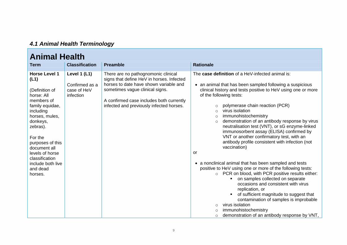
4.1 Animal Health Terminology
Animal Health Term Classification Preamble Rationale
Horse Level 1 (L1)
(Definition of horse: All members of family equidae, including horses, mules, donkeys, zebras).
For the purposes of this document all levels of horse classification include both live and dead horses.
Level 1 (L1)
Confirmed as a case of HeV infection
There are no pathognomonic clinical signs that define HeV in horses. Infected horses to date have shown variable and sometimes vague clinical signs.
A confirmed case includes both currently infected and previously infected horses.
The case definition of a HeV-infected animal is: • an animal that has been sampled following a suspicious
clinical history and tests positive to HeV using one or more of the following tests:
o polymerase chain reaction (PCR) o virus isolation o immunohistochemistry o demonstration of an antibody response by virus
neutralisation test (VNT), or sG enzyme-linked immunosorbent assay (ELISA) confirmed by VNT or another confirmatory test, with an antibody profile consistent with infection (not vaccination)
or • a nonclinical animal that has been sampled and tests
positive to HeV using one or more of the following tests: o PCR on blood, with PCR positive results either:
on samples collected on separate occasions and consistent with virus replication, or
of sufficient magnitude to suggest that contamination of samples is improbable
o virus isolation o immunohistochemistry o demonstration of an antibody response by VNT,
10
Animal Health Term Classification Preamble Rationale
or sG ELISA confirmed by VNT or another confirmatory test, with an antibody profile consistent with infection (not vaccination).
Where positive results are obtained from nonclinical animals sampled at anatomical sites that are susceptible to environmental contamination (e.g. nasal cavity, oral cavity, rectum), or from samples where contamination cannot reasonably be excluded, confirmation of infection by demonstration of an antibody response is necessary. An animal for which testing has not been possible or for which testing is inconclusive, but there is compelling epidemiological evidence that the animal is/was infected (e.g. confirmed human infection following contact with an animal with clinical signs and history suggestive of HeV infection), would also meet the case definition.
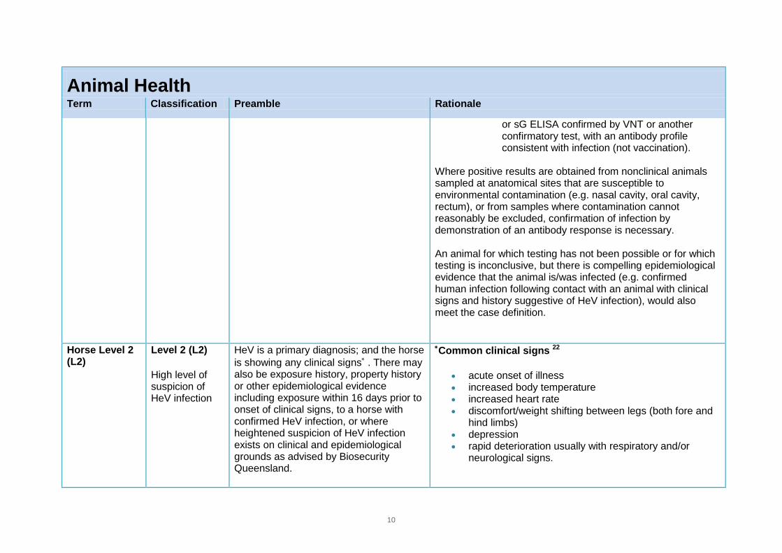
Horse Level 2 (L2)
Level 2 (L2)
High level of suspicion of HeV infection
HeV is a primary diagnosis; and the horse is showing any clinical signs∗ . There may also be exposure history, property history or other epidemiological evidence including exposure within 16 days prior to onset of clinical signs, to a horse with confirmed HeV infection, or where heightened suspicion of HeV infection exists on clinical and epidemiological grounds as advised by Biosecurity Queensland.
∗Common clinical signs 22
• acute onset of illness • increased body temperature • increased heart rate • discomfort/weight shifting between legs (both fore and
hind limbs) • depression • rapid deterioration usually with respiratory and/or
neurological signs.
11
Animal Health Term Classification Preamble Rationale
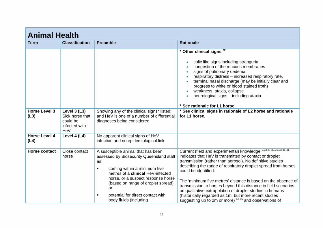
* Other clinical signs 22
• colic like signs including stranguria • congestion of the mucous membranes • signs of pulmonary oedema • respiratory distress – increased respiratory rate, • terminal nasal discharge (may be initially clear and
progress to white or blood stained froth) • weakness, ataxia, collapse • neurological signs – including ataxia
* See rationale for L1 horse Horse Level 3 (L3)
Level 3 (L3) Sick horse that could be infected with HeV
Showing any of the clinical signs* listed; and HeV is one of a number of differential diagnoses being considered.
* See clinical signs in rationale of L2 horse and rationale for L1 horse.
Horse Level 4 (L4)
Level 4 (L4) No apparent clinical signs of HeV infection and no epidemiological link.
Horse contact
Close contact horse
A susceptible animal that has been assessed by Biosecurity Queensland staff as:
coming within a minimum five metres of a clinical HeV-infected horse, or a suspect response horse (based on range of droplet spread); or
potential for direct contact with body fluids (including
Current (field and experimental) knowledge 2,23,27,30,31,33,35-41 indicates that HeV is transmitted by contact or droplet transmission (rather than aerosol). No definitive studies describing the range of respiratory droplet spread from horses could be identified.
The ‘minimum five metres’ distance is based on the absence of transmission to horses beyond this distance in field scenarios, on qualitative extrapolation of droplet studies in humans (historically regarded as 1m, but more recent studies suggesting up to 2m or more) 52-55 and observations of
12
Animal Health Term Classification Preamble Rationale
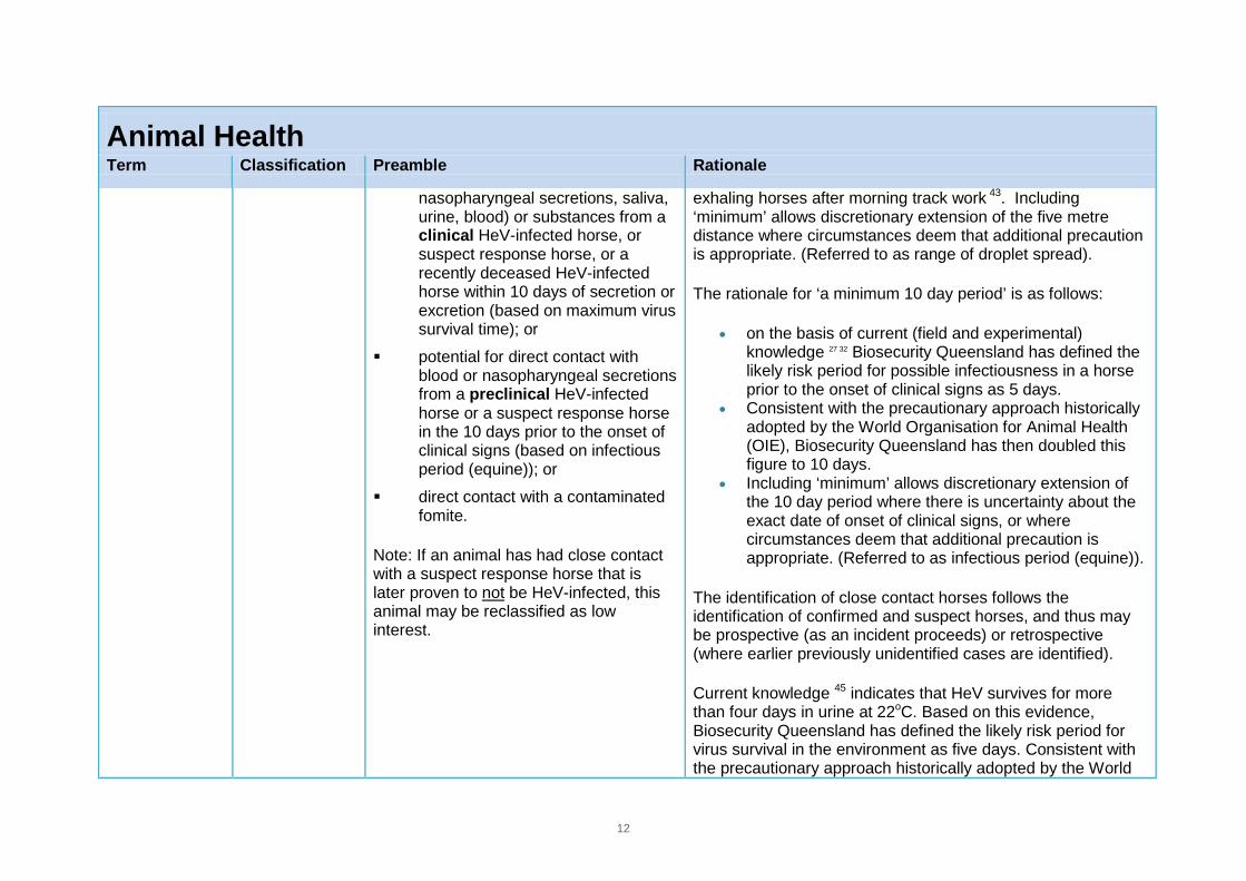
nasopharyngeal secretions, saliva, urine, blood) or substances from a clinical HeV-infected horse, or suspect response horse, or a recently deceased HeV-infected horse within 10 days of secretion or excretion (based on maximum virus survival time); or
potential for direct contact with blood or nasopharyngeal secretions from a preclinical HeV-infected horse or a suspect response horse in the 10 days prior to the onset of clinical signs (based on infectious period (equine)); or
direct contact with a contaminated fomite.
Note: If an animal has had close contact with a suspect response horse that is later proven to not be HeV-infected, this animal may be reclassified as low interest.
exhaling horses after morning track work 43. Including ‘minimum’ allows discretionary extension of the five metre distance where circumstances deem that additional precaution is appropriate. (Referred to as range of droplet spread).
The rationale for ‘a minimum 10 day period’ is as follows:
• on the basis of current (field and experimental) knowledge 27 32 Biosecurity Queensland has defined the likely risk period for possible infectiousness in a horse prior to the onset of clinical signs as 5 days.
• Consistent with the precautionary approach historically adopted by the World Organisation for Animal Health (OIE), Biosecurity Queensland has then doubled this figure to 10 days.
• Including ‘minimum’ allows discretionary extension of the 10 day period where there is uncertainty about the exact date of onset of clinical signs, or where circumstances deem that additional precaution is appropriate. (Referred to as infectious period (equine)).
The identification of close contact horses follows the identification of confirmed and suspect horses, and thus may be prospective (as an incident proceeds) or retrospective (where earlier previously unidentified cases are identified).
Current knowledge 45 indicates that HeV survives for more than four days in urine at 22oC. Based on this evidence, Biosecurity Queensland has defined the likely risk period for virus survival in the environment as five days. Consistent with the precautionary approach historically adopted by the World
13
Animal Health Term Classification Preamble Rationale
Organisation for Animal Health (OIE), Biosecurity Queensland has then doubled this figure to 10 days. (Referred to as maximum virus survival time).
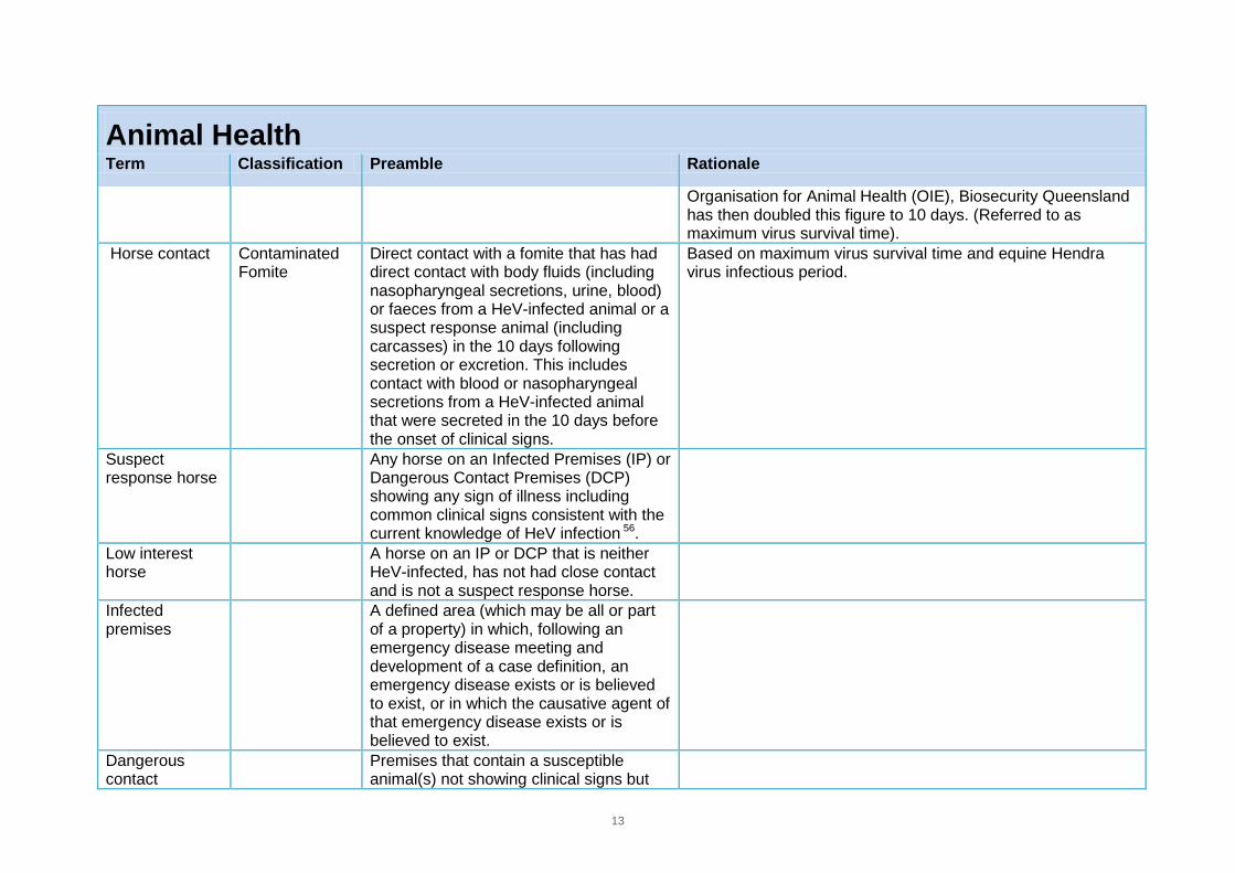
Horse contact Contaminated Fomite
Direct contact with a fomite that has had direct contact with body fluids (including nasopharyngeal secretions, urine, blood) or faeces from a HeV-infected animal or a suspect response animal (including carcasses) in the 10 days following secretion or excretion. This includes contact with blood or nasopharyngeal secretions from a HeV-infected animal that were secreted in the 10 days before the onset of clinical signs.
Based on maximum virus survival time and equine Hendra virus infectious period.
Suspect response horse
Any horse on an Infected Premises (IP) or Dangerous Contact Premises (DCP) showing any sign of illness including common clinical signs consistent with the current knowledge of HeV infection 56.
Low interest horse
A horse on an IP or DCP that is neither HeV-infected, has not had close contact and is not a suspect response horse.
Infected premises
A defined area (which may be all or part of a property) in which, following an emergency disease meeting and development of a case definition, an emergency disease exists or is believed to exist, or in which the causative agent of that emergency disease exists or is believed to exist.
Dangerous contact
Premises that contain a susceptible animal(s) not showing clinical signs but
14
Animal Health Term Classification Preamble Rationale
premises that, following a risk assessment are considered to contain a close contact animal(s) or contaminated animal products, wastes or features that present an unacceptable risk to the response if not addressed.
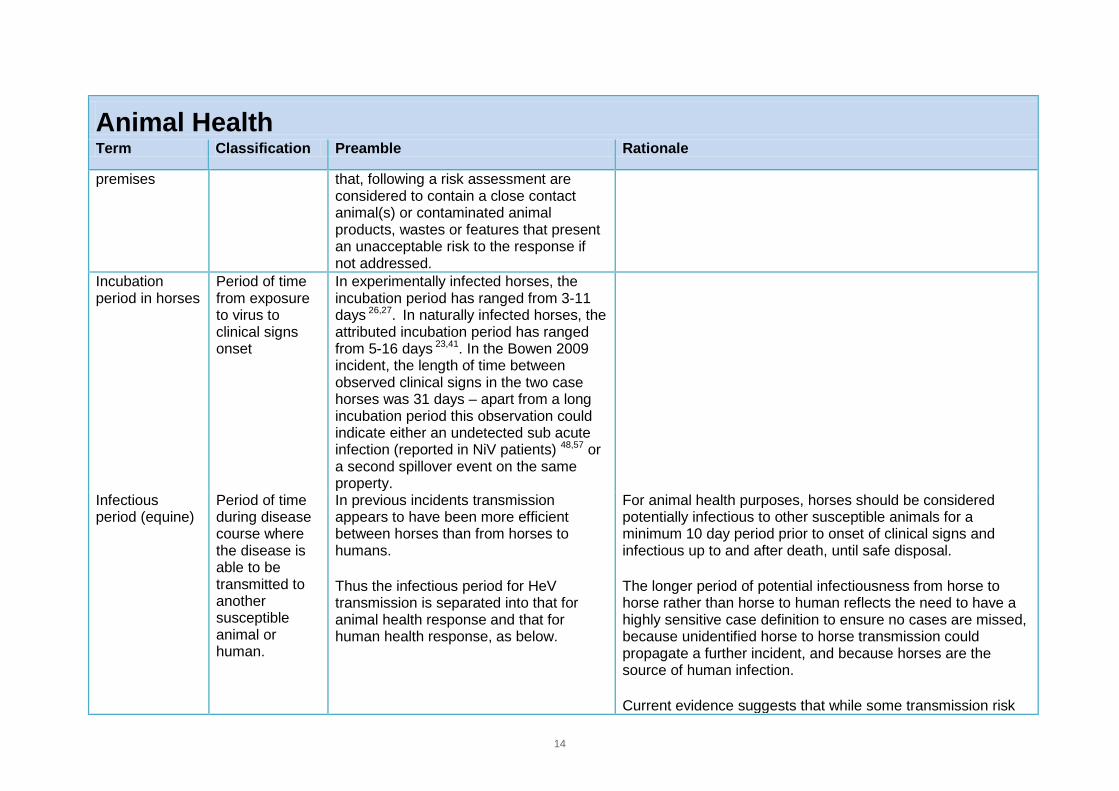
Incubation period in horses
Period of time from exposure to virus to clinical signs onset
In experimentally infected horses, the incubation period has ranged from 3-11 days 26,27. In naturally infected horses, the attributed incubation period has ranged from 5-16 days 23,41. In the Bowen 2009 incident, the length of time between observed clinical signs in the two case horses was 31 days – apart from a long incubation period this observation could indicate either an undetected sub acute infection (reported in NiV patients) 48,57 or a second spillover event on the same property.
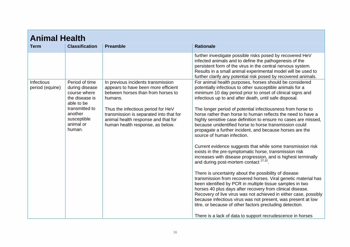
Infectious period (equine)
Period of time during disease course where the disease is able to be transmitted to another susceptible animal or human.
In previous incidents transmission appears to have been more efficient between horses than from horses to humans.
Thus the infectious period for HeV transmission is separated into that for animal health response and that for human health response, as below.
For animal health purposes, horses should be considered potentially infectious to other susceptible animals for a minimum 10 day period prior to onset of clinical signs and infectious up to and after death, until safe disposal.
The longer period of potential infectiousness from horse to horse rather than horse to human reflects the need to have a highly sensitive case definition to ensure no cases are missed, because unidentified horse to horse transmission could propagate a further incident, and because horses are the source of human infection.
Current evidence suggests that while some transmission risk
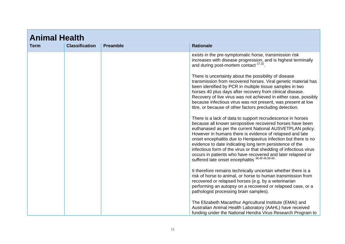
15
Animal Health Term Classification Preamble Rationale
exists in the pre-symptomatic horse, transmission risk increases with disease progression, and is highest terminally and during post-mortem contact 27,32.
There is uncertainty about the possibility of disease transmission from recovered horses. Viral genetic material has been identified by PCR in multiple tissue samples in two horses 40 plus days after recovery from clinical disease. Recovery of live virus was not achieved in either case, possibly because infectious virus was not present, was present at low titre, or because of other factors precluding detection.
There is a lack of data to support recrudescence in horses because all known seropositive recovered horses have been euthanased as per the current National AUSVETPLAN policy. However in humans there is evidence of relapsed and late onset encephalitis due to Henipavirus infection but there is no evidence to date indicating long term persistence of the infectious form of the virus or that shedding of infectious virus occurs in patients who have recovered and later relapsed or suffered late onset encephalitis 38,48 46,58-60 .
It therefore remains technically uncertain whether there is a risk of horse to animal, or horse to human transmission from recovered or relapsed horses (e.g. by a veterinarian performing an autopsy on a recovered or relapsed case, or a pathologist processing brain samples).
The Elizabeth Macarthur Agricultural Institute (EMAI) and Australian Animal Health Laboratory (AAHL) have received funding under the National Hendra Virus Research Program to
16
Animal Health Term Classification Preamble Rationale
further investigate possible risks posed by recovered HeV infected animals and to define the pathogenesis of the persistent form of the virus in the central nervous system. Results in a small animal experimental model will be used to further clarify any potential risk posed by recovered animals.
Infectious period (equine)
Period of time during disease course where the disease is able to be transmitted to another susceptible animal or human.
In previous incidents transmission appears to have been more efficient between horses than from horses to humans.
Thus the infectious period for HeV transmission is separated into that for animal health response and that for human health response, as below.
For animal health purposes, horses should be considered potentially infectious to other susceptible animals for a minimum 10 day period prior to onset of clinical signs and infectious up to and after death, until safe disposal.
The longer period of potential infectiousness from horse to horse rather than horse to human reflects the need to have a highly sensitive case definition to ensure no cases are missed, because unidentified horse to horse transmission could propagate a further incident, and because horses are the source of human infection.
Current evidence suggests that while some transmission risk exists in the pre-symptomatic horse, transmission risk increases with disease progression, and is highest terminally and during post-mortem contact 27,32.
There is uncertainty about the possibility of disease transmission from recovered horses. Viral genetic material has been identified by PCR in multiple tissue samples in two horses 40 plus days after recovery from clinical disease. Recovery of live virus was not achieved in either case, possibly because infectious virus was not present, was present at low titre, or because of other factors precluding detection.
There is a lack of data to support recrudescence in horses
17
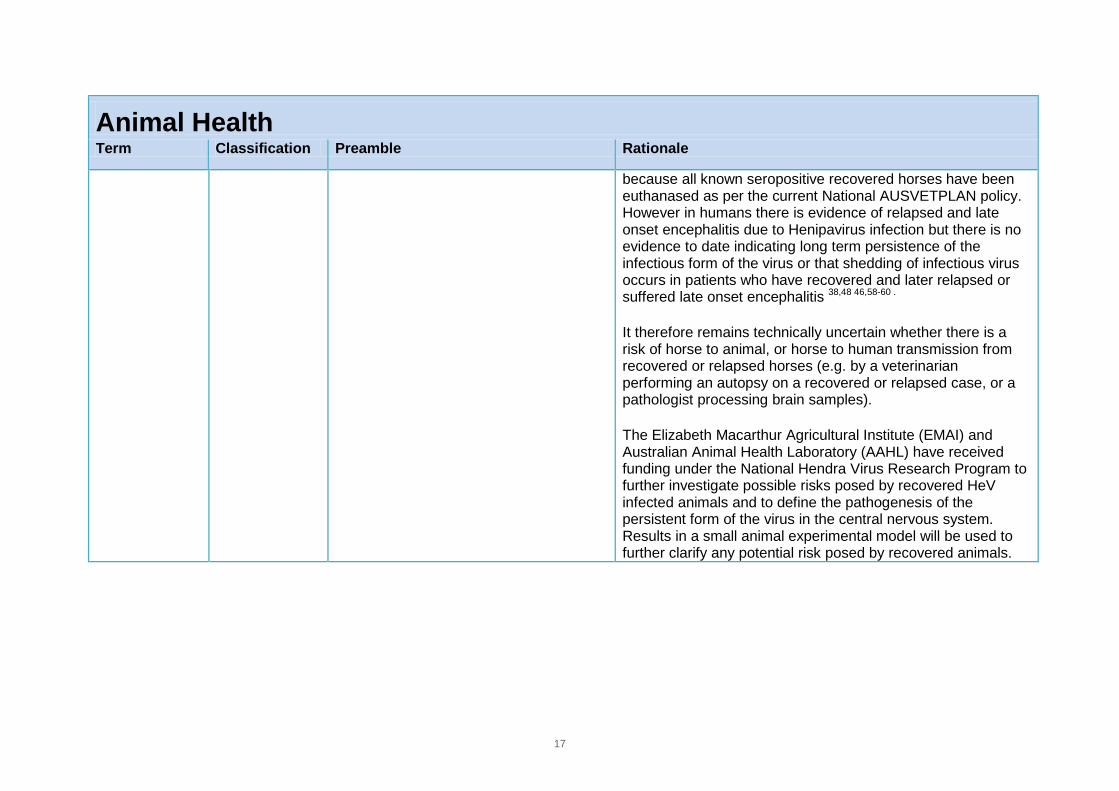
Animal Health Term Classification Preamble Rationale
because all known seropositive recovered horses have been euthanased as per the current National AUSVETPLAN policy. However in humans there is evidence of relapsed and late onset encephalitis due to Henipavirus infection but there is no evidence to date indicating long term persistence of the infectious form of the virus or that shedding of infectious virus occurs in patients who have recovered and later relapsed or suffered late onset encephalitis 38,48 46,58-60 .
It therefore remains technically uncertain whether there is a risk of horse to animal, or horse to human transmission from recovered or relapsed horses (e.g. by a veterinarian performing an autopsy on a recovered or relapsed case, or a pathologist processing brain samples).
The Elizabeth Macarthur Agricultural Institute (EMAI) and Australian Animal Health Laboratory (AAHL) have received funding under the National Hendra Virus Research Program to further investigate possible risks posed by recovered HeV infected animals and to define the pathogenesis of the persistent form of the virus in the central nervous system. Results in a small animal experimental model will be used to further clarify any potential risk posed by recovered animals.
18
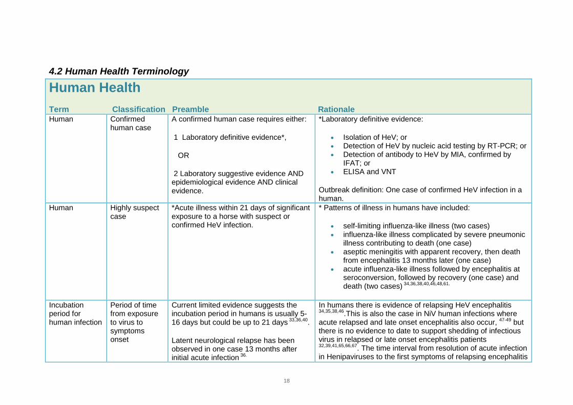
4.2 Human Health Terminology
Human Health Term Classification Preamble Rationale Human Confirmed
human case A confirmed human case requires either:
1 Laboratory definitive evidence*,
OR
2 Laboratory suggestive evidence AND epidemiological evidence AND clinical evidence.
*Laboratory definitive evidence:
• Isolation of HeV; or • Detection of HeV by nucleic acid testing by RT-PCR; or • Detection of antibody to HeV by MIA, confirmed by
IFAT; or • ELISA and VNT
Outbreak definition: One case of confirmed HeV infection in a human.
Human Highly suspect case
*Acute illness within 21 days of significant exposure to a horse with suspect or confirmed HeV infection.
* Patterns of illness in humans have included:
• self-limiting influenza-like illness (two cases) • influenza-like illness complicated by severe pneumonic
illness contributing to death (one case) • aseptic meningitis with apparent recovery, then death
from encephalitis 13 months later (one case) • acute influenza-like illness followed by encephalitis at
seroconversion, followed by recovery (one case) and death (two cases) 34,36,38,40,46,48,61.
Incubation period for human infection
Period of time from exposure to virus to symptoms onset
Current limited evidence suggests the incubation period in humans is usually 5-16 days but could be up to 21 days 33,36,40.
Latent neurological relapse has been observed in one case 13 months after initial acute infection 36.
In humans there is evidence of relapsing HeV encephalitis
34,35,38,46.This is also the case in NiV human infections where acute relapsed and late onset encephalitis also occur, 47-49 but there is no evidence to date to support shedding of infectious virus in relapsed or late onset encephalitis patients
32,39,41,65,66,67. The time interval from resolution of acute infection in Henipaviruses to the first symptoms of relapsing encephalitis
19
Human Health Term Classification Preamble Rationale
may vary from weeks to years 34,35,38,46-48
There have been no known human asymptomatic seroconversions of HeV and a recent study 50 found no evidence of prolonged HeV shedding by two recovered patients.
Standard contact and droplet precautions should be implemented for management of suspected or confirmed human cases. Airborne precautions are routinely practised for aerosol generating procedures42,44. Consultation with the infectious disease physician is recommended 62.
Infectious period for human to human transmission
Period of time during disease course where the disease is able to be transmitted to another person
Unknown, no evidence of human to human transmission of HeV to date.
While there is no evidence to date that human to human transmission of HeV occurs; it is desirable to avoid close contact with blood, body fluids/secretions of a symptomatic human case. HeV RNA has been detected in serum, urine and nasopharyngeal secretions of HeV infected patients 33.
Standard contact and droplet precautions should be implemented for management of suspected or confirmed human cases. Airborne precautions are routinely practised for aerosol generating procedures 42,44. Consultation with the infectious disease physician is recommended 62.
Note: Direct human to human transmission, including healthcare associated infection of the closely related NiV has occurred overseas 64 57,65-69. NiV has been isolated in the respiratory secretions and urine of infected patients but is more difficult to isolate following the appearance of detectable serum IgM antibody 70.
20
Human Health Term Classification Preamble Rationale
Human to human transmission of NiV seen in Bangladesh/India generally occurred from fatal cases late in the course of disease from severe pulmonary involvement; however there was a lack of data from patients earlier in the course of the disease process.
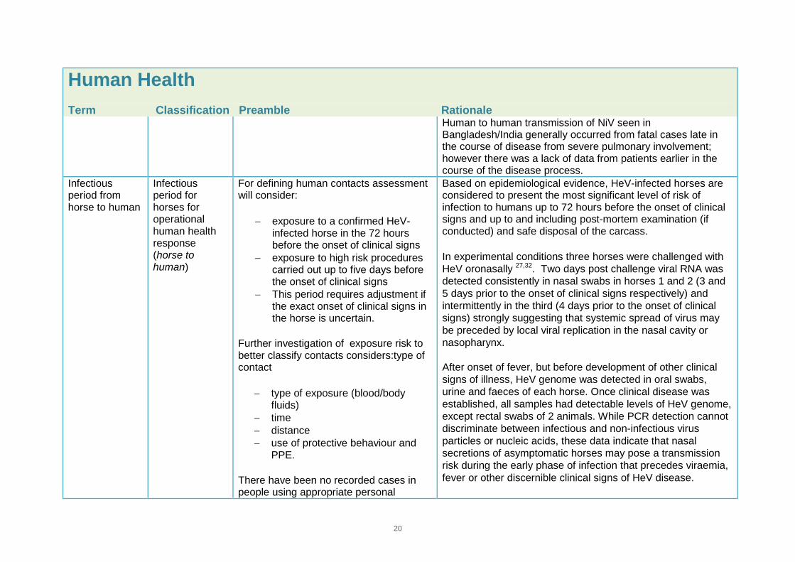
Infectious period from horse to human
Infectious period for horses for operational human health response (horse to human)
For defining human contacts assessment will consider:
− exposure to a confirmed HeV-infected horse in the 72 hours before the onset of clinical signs
− exposure to high risk procedures carried out up to five days before the onset of clinical signs
− This period requires adjustment if the exact onset of clinical signs in the horse is uncertain.
Further investigation of exposure risk to better classify contacts considers:type of contact
− type of exposure (blood/body fluids)
− time − distance − use of protective behaviour and
PPE.
There have been no recorded cases in people using appropriate personal
Based on epidemiological evidence, HeV-infected horses are considered to present the most significant level of risk of infection to humans up to 72 hours before the onset of clinical signs and up to and including post-mortem examination (if conducted) and safe disposal of the carcass.
In experimental conditions three horses were challenged with HeV oronasally 27,32. Two days post challenge viral RNA was detected consistently in nasal swabs in horses 1 and 2 (3 and 5 days prior to the onset of clinical signs respectively) and intermittently in the third (4 days prior to the onset of clinical signs) strongly suggesting that systemic spread of virus may be preceded by local viral replication in the nasal cavity or nasopharynx. After onset of fever, but before development of other clinical signs of illness, HeV genome was detected in oral swabs, urine and faeces of each horse. Once clinical disease was established, all samples had detectable levels of HeV genome, except rectal swabs of 2 animals. While PCR detection cannot discriminate between infectious and non-infectious virus particles or nucleic acids, these data indicate that nasal secretions of asymptomatic horses may pose a transmission risk during the early phase of infection that precedes viraemia, fever or other discernible clinical signs of HeV disease.

21
Human Health Term Classification Preamble Rationale
protective equipment (PPE).
Infected horses are potentially infectious till death by disease or euthanasia and safe disposal of the carcass.
The Hendra virus exposure assessment form provides a framework for assessing human exposure to HeV: http://www.health.gov.au/internet/main/publishing.nsf/Content/cdna-song-hendra-appendix4
It is currently unclear if recovered or sub-clinical horse cases pose an infection risk to humans and further research is in progress. AUSVETPLAN HeV Response Policy Brief currently states that such horses should be euthanased 71. The issue of transmission risk from recovered animals will be reviewed at a national level in late 2013.
The febrile and then symptomatic horses pose a greater transmission risk from nasal and other secretions such as blood and urine than asymptomatic horses. Conducting procedures such as endoscopy, nasal lavage and necropsy pose a higher risk of infection transmission because of the potential for gross contamination and the handling of sharp instrument27,32,72. Consideration should also be given to assessing the theoretical risk of human infection from high risk procedures undertaken in the five day period preceding onset of clinical signs in the horse. Uncertainty around the time of onset of clinical signs in a horse may mean determining the period humans were potentially exposed, needs to be flexible.
There is uncertainty about the possibility of disease transmission from recovered horses. Viral genetic material has been identified by PCR in multiple tissue samples in two horses 40 plus days after recovery from clinical disease. Recovery of infectious virus was not achieved in either case, possibly because infectious virus was not present, was present at low titre, or because of other factors precluding detection.
There is a lack of data to support the recrudescence in horses because all known seropositive recovered horses have been euthanased as per the current National AUSVETPLAN policy.
In humans there is evidence of relapsed and late onset encephalitis due to Henipavirus infection however there is no evidence indicating long term persistence of infectious enveloped virus or that shedding of infectious virus occurs in patients who have recovered and later relapsed or suffered late onset encephalitis47,49,59,60,73,74. A recent study 50 found no
22
Human Health Term Classification Preamble Rationale
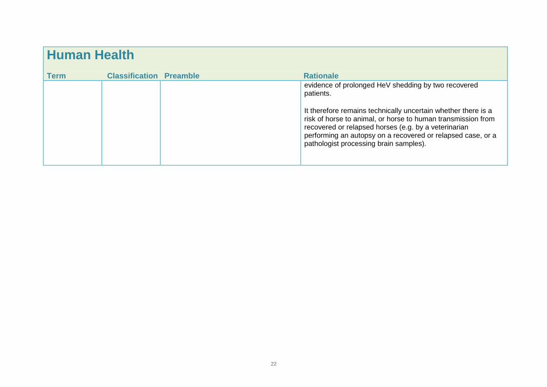
evidence of prolonged HeV shedding by two recovered patients.
It therefore remains technically uncertain whether there is a risk of horse to animal, or horse to human transmission from recovered or relapsed horses (e.g. by a veterinarian performing an autopsy on a recovered or relapsed case, or a pathologist processing brain samples).
23
5 Infection Control Advice Objective 1: To provide technical advice based on best available evidence on measures needed to prevent human infection with HeV. The infection control practices recommended during contact with a horse to reduce the risk of unprotected contact with a horse’s blood/body fluids are based on the potential for severe morbidity or death should human infection occur.
5.1 Animal-Human Infection Control
5.1.1 Horse Vaccination A HeV soluble G (sG) subunit vaccine was released commercially onto the Australian market on 1 November 2012 following evaluation and the issue of a Minor Use Permit by the Australian Pesticides and Veterinary Medicines Authority (APVMA). The horse vaccine was developed subsequent to research which demonstrated the subunit sG vaccine's ability to prevent henipavirus disease and virus transmission or shedding in small animal models 75-79 . Research on horses conducted at CSIRO's Australian Animal Health Laboratory (AAHL) demonstrated that the adjuvanted vaccine containing either 100 µg or 50 µg sG in a prime-boost regime resulted in seroconversion in all vaccinated horses. In the initial efficacy trials, there was no evidence of viral shedding by immunised horses after HeV challenge, as reflected by PCR negative test results on all daily clinical samples. Following euthanasia of immunized horses (7 to 9 days post challenge, and 1-3 days after clinical signs first became apparent in control animals), there was no evidence of HeV viral replication in any tissue of immunised horses collected at post mortem examination, after what would be expected to be the period of acute infection. To demonstrate the immunogenicity of the vaccine under field conditions two trials were conducted. Horses were given two single doses of the vaccine by intramuscular injection on Days 0 and 21. All vaccinated animals in both trials seroconverted, confirming that two doses of vaccine given three weeks apart are sufficient to generate an antibody response in horses from four months of age. Serum neutralising antibody levels on day 42 (three weeks after the second dose of vaccine) in all vaccinated horses from both trials were equivalent to those seen in the earlier efficacy studies by challenge as described above, in which vaccinated horses were shown to be protected from challenge with HeV. At approximately six months post-vaccination, when horses were challenged with HeV via the intranasal route, all were protected from clinical signs of HeV disease. In addition, virus was not reisolated from any clinical samples (collected pre-and post-mortem) from any of the horses, and there was no evidence of virus spreading beyond the site of administration (i.e. the upper respiratory tract). In non-immunised (surrogate control) ferrets, viral infection was detected and all succumbed to acute HeV infection.
24
In summary all vaccinated animals were protected from disease against a virulent challenge with HeV for up to six months following the initial course of two vaccinations 21 days apart, and transmission or shedding of virus was prevented. The vaccine is the single most effective way of reducing the risk of Hendra virus infection in horses and provides a work health and safety and public health benefit by the vaccine's ability to not only protect horses from infection but also to break the cycle of virus transmission from horses to humans. Widespread uptake of the horse vaccine has the potential to significantly reduce the number and risk of human exposures. The Australian Veterinary Association recommends that all horses, particularly those horses in identified risk areas or situations, are vaccinated according to APVMA permit requirements, veterinary and manufacturer's recommendations. Further research at AAHL has been conducted to determine the level of immunity at 12 months.
5.1.2 Rationale for Infection Control Recommendations Based on epidemiological evidence, HeV infected horses are considered to present the most significant level of risk of infection to humans up to 72 hours before the onset of clinical signs and up to and including post-mortem examination (if conducted) and safe disposal of the carcass. Because experimental studies have detected viral RNA from two infected horses up to five days before the onset of clinical signs 27,32 consideration should also be given to assessing the theoretical risk of human infection from high risk procedures undertaken in the five day period preceding onset of clinical signs in the horse. Uncertainty around the time of onset of clinical signs in a horse may mean determining the period humans were potentially exposed, needs to be flexible. HeV infection in horses has presented with variable clinical signs, with fever and increased heart rate common in acute cases 1,2,8,14,22,23,26-30. Typically there is a rapid deterioration in acutely infected horses with respiratory and/or neurological clinical signs, however HeV infection has been detected in some instances when there have been no or minimal clinical signs, including absence of fever. It should be noted that approximately 20 per cent of HeV infected horses can survive acute HeV infection. The febrile and then symptomatic horse, particularly those late in the disease process are likely to shed more virus from a variety of excretions and pose a higher risk of disease transmission. Conducting necropsies poses a higher risk of infection transmission because of the potential for gross contamination and the handling of sharp instruments7,27. If HeV cannot be ruled out as a diagnosis, risk controls should be implemented before anyone contacts a sick horse, not after initial examination.
25
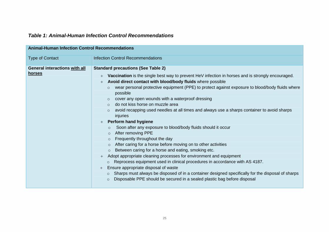
Table 1: Animal-Human Infection Control Recommendations
Animal-Human Infection Control Recommendations
Type of Contact Infection Control Recommendations
General interactions with all horses
Standard precautions (See Table 2)
+ Vaccination is the single best way to prevent HeV infection in horses and is strongly encouraged. + Avoid direct contact with blood/body fluids where possible
o wear personal protective equipment (PPE) to protect against exposure to blood/body fluids where possible
o cover any open wounds with a waterproof dressing o do not kiss horse on muzzle area o avoid recapping used needles at all times and always use a sharps container to avoid sharps
injuries + Perform hand hygiene
o Soon after any exposure to blood/body fluids should it occur o After removing PPE o Frequently throughout the day o After caring for a horse before moving on to other activities o Between caring for a horse and eating, smoking etc.
+ Adopt appropriate cleaning processes for environment and equipment o Reprocess equipment used in clinical procedures in accordance with AS 4187.
+ Ensure appropriate disposal of waste o Sharps must always be disposed of in a container designed specifically for the disposal of sharps o Disposable PPE should be secured in a sealed plastic bag before disposal
26
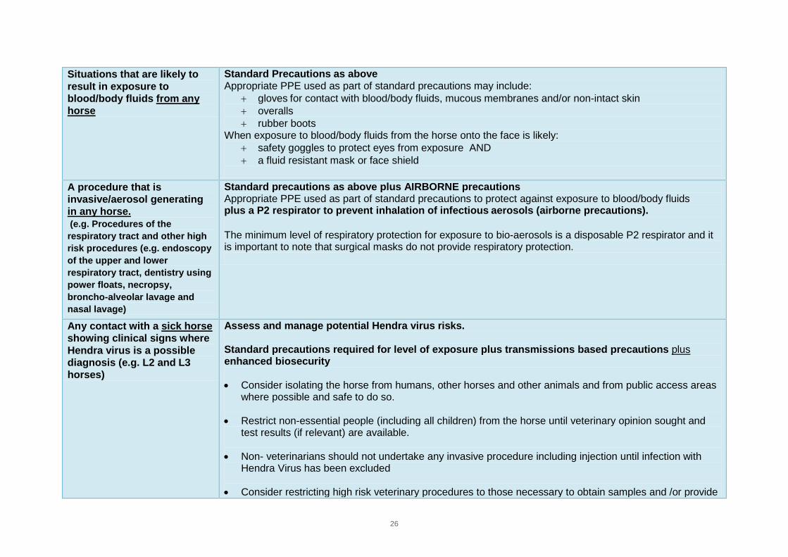
Situations that are likely to result in exposure to blood/body fluids from any horse
Standard Precautions as above Appropriate PPE used as part of standard precautions may include:
+ gloves for contact with blood/body fluids, mucous membranes and/or non-intact skin + overalls + rubber boots
When exposure to blood/body fluids from the horse onto the face is likely: + safety goggles to protect eyes from exposure AND + a fluid resistant mask or face shield
A procedure that is invasive/aerosol generating in any horse. (e.g. Procedures of the respiratory tract and other high risk procedures (e.g. endoscopy of the upper and lower respiratory tract, dentistry using power floats, necropsy, broncho-alveolar lavage and nasal lavage)
Standard precautions as above plus AIRBORNE precautions Appropriate PPE used as part of standard precautions to protect against exposure to blood/body fluids plus a P2 respirator to prevent inhalation of infectious aerosols (airborne precautions). The minimum level of respiratory protection for exposure to bio-aerosols is a disposable P2 respirator and it is important to note that surgical masks do not provide respiratory protection.
Any contact with a sick horse showing clinical signs where Hendra virus is a possible diagnosis (e.g. L2 and L3 horses)
Assess and manage potential Hendra virus risks. Standard precautions required for level of exposure plus transmissions based precautions plus enhanced biosecurity
• Consider isolating the horse from humans, other horses and other animals and from public access areas where possible and safe to do so.
• Restrict non-essential people (including all children) from the horse until veterinary opinion sought and test results (if relevant) are available.
• Non- veterinarians should not undertake any invasive procedure including injection until infection with Hendra Virus has been excluded
• Consider restricting high risk veterinary procedures to those necessary to obtain samples and /or provide
27
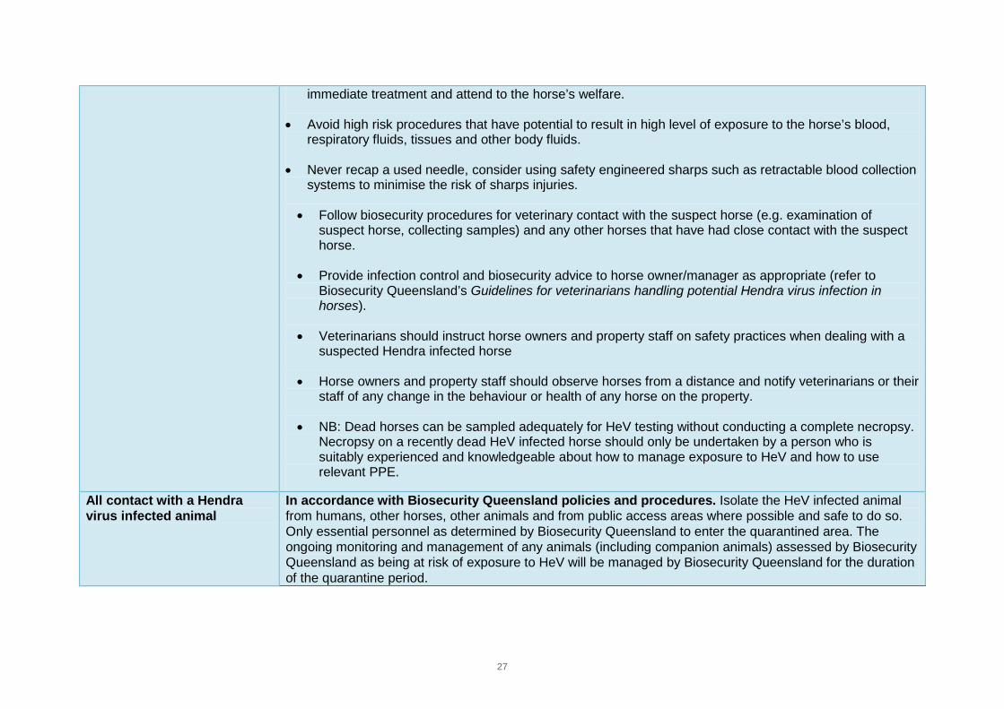
immediate treatment and attend to the horse’s welfare.
• Avoid high risk procedures that have potential to result in high level of exposure to the horse’s blood, respiratory fluids, tissues and other body fluids.
• Never recap a used needle, consider using safety engineered sharps such as retractable blood collection systems to minimise the risk of sharps injuries.
• Follow biosecurity procedures for veterinary contact with the suspect horse (e.g. examination of suspect horse, collecting samples) and any other horses that have had close contact with the suspect horse.
• Provide infection control and biosecurity advice to horse owner/manager as appropriate (refer to Biosecurity Queensland’s Guidelines for veterinarians handling potential Hendra virus infection in horses).
• Veterinarians should instruct horse owners and property staff on safety practices when dealing with a suspected Hendra infected horse
• Horse owners and property staff should observe horses from a distance and notify veterinarians or their staff of any change in the behaviour or health of any horse on the property.
• NB: Dead horses can be sampled adequately for HeV testing without conducting a complete necropsy. Necropsy on a recently dead HeV infected horse should only be undertaken by a person who is suitably experienced and knowledgeable about how to manage exposure to HeV and how to use relevant PPE.
All contact with a Hendra virus infected animal
In accordance with Biosecurity Queensland policies and procedures. Isolate the HeV infected animal from humans, other horses, other animals and from public access areas where possible and safe to do so. Only essential personnel as determined by Biosecurity Queensland to enter the quarantined area. The ongoing monitoring and management of any animals (including companion animals) assessed by Biosecurity Queensland as being at risk of exposure to HeV will be managed by Biosecurity Queensland for the duration of the quarantine period.
28
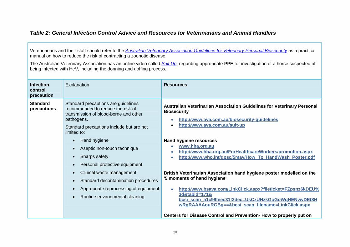
Table 2: General Infection Control Advice and Resources for Veterinarians and Animal Handlers
Veterinarians and their staff should refer to the Australian Veterinary Association Guidelines for Veterinary Personal Biosecurity as a practical manual on how to reduce the risk of contracting a zoonotic disease. The Australian Veterinary Association has an online video called Suit Up, regarding appropriate PPE for investigation of a horse suspected of being infected with HeV, including the donning and doffing process.
Infection control precaution
Explanation Resources
Standard precautions
Standard precautions are guidelines recommended to reduce the risk of transmission of blood-borne and other pathogens.
Standard precautions include but are not limited to:
• Hand hygiene
• Aseptic non-touch technique
• Sharps safety
• Personal protective equipment
• Clinical waste management
• Standard decontamination procedures
• Appropriate reprocessing of equipment
• Routine environmental cleaning
Australian Veterinarian Association Guidelines for Veterinary Personal Biosecurity
• http://www.ava.com.au/biosecurity-guidelines • http://www.ava.com.au/suit-up
Hand hygiene resources
• www.hha.org.au • http://www.hha.org.au/ForHealthcareWorkers/promotion.aspx • http://www.who.int/gpsc/5may/How_To_HandWash_Poster.pdf
British Veterinarian Association hand hygiene poster modelled on the '5 moments of hand hygiene'
• http://www.bsava.com/LinkClick.aspx?fileticket=FZpsnz6kDEU%3d&tabid=171& bcsi_scan_a1c99feec31f2dec=UsCzUHzkGoGoWqHENvwDEt8HwRgRAAAAouRGBg==&bcsi_scan_filename=LinkClick.aspx
Centers for Disease Control and Prevention- How to properly put on
29
and take off a disposable respirator • http://www.cdc.gov/niosh/docs/2010-133/
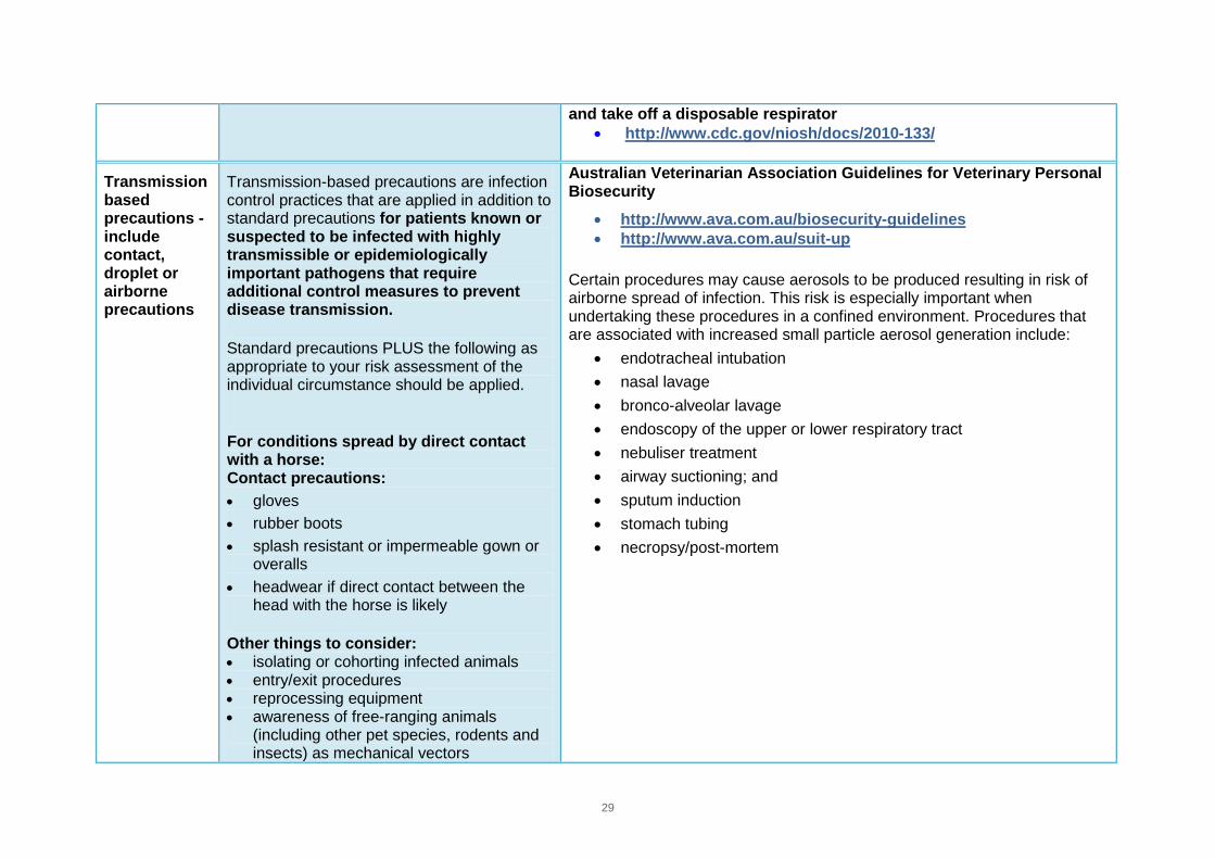
Transmission based precautions - include contact, droplet or airborne precautions
Transmission-based precautions are infection control practices that are applied in addition to standard precautions for patients known or suspected to be infected with highly transmissible or epidemiologically important pathogens that require additional control measures to prevent disease transmission.
Standard precautions PLUS the following as appropriate to your risk assessment of the individual circumstance should be applied.
For conditions spread by direct contact with a horse: Contact precautions: • gloves • rubber boots • splash resistant or impermeable gown or
overalls • headwear if direct contact between the
head with the horse is likely
Other things to consider: • isolating or cohorting infected animals • entry/exit procedures • reprocessing equipment • awareness of free-ranging animals
(including other pet species, rodents and insects) as mechanical vectors
Australian Veterinarian Association Guidelines for Veterinary Personal Biosecurity
• http://www.ava.com.au/biosecurity-guidelines • http://www.ava.com.au/suit-up
Certain procedures may cause aerosols to be produced resulting in risk of airborne spread of infection. This risk is especially important when undertaking these procedures in a confined environment. Procedures that are associated with increased small particle aerosol generation include:
• endotracheal intubation • nasal lavage • bronco-alveolar lavage • endoscopy of the upper or lower respiratory tract • nebuliser treatment • airway suctioning; and • sputum induction • stomach tubing • necropsy/post-mortem
30
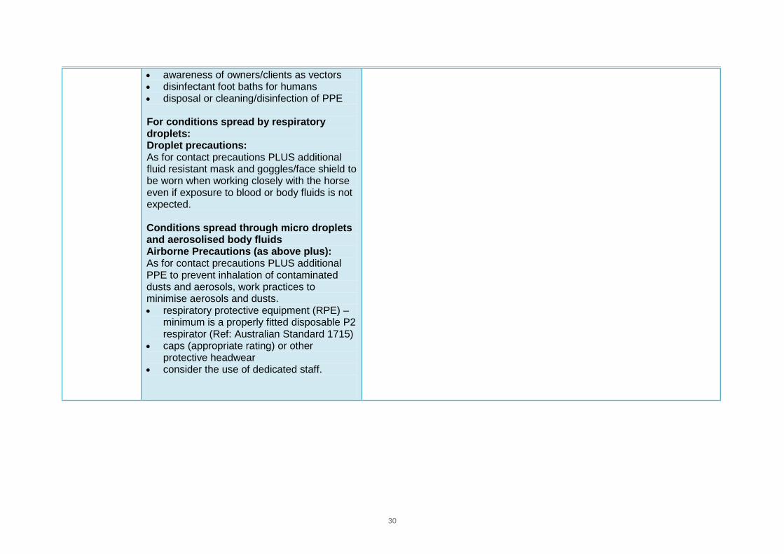
• awareness of owners/clients as vectors • disinfectant foot baths for humans • disposal or cleaning/disinfection of PPE For conditions spread by respiratory droplets: Droplet precautions: As for contact precautions PLUS additional fluid resistant mask and goggles/face shield to be worn when working closely with the horse even if exposure to blood or body fluids is not expected. Conditions spread through micro droplets and aerosolised body fluids Airborne Precautions (as above plus): As for contact precautions PLUS additional PPE to prevent inhalation of contaminated dusts and aerosols, work practices to minimise aerosols and dusts. • respiratory protective equipment (RPE) –
minimum is a properly fitted disposable P2 respirator (Ref: Australian Standard 1715)
• caps (appropriate rating) or other protective headwear
• consider the use of dedicated staff.
31
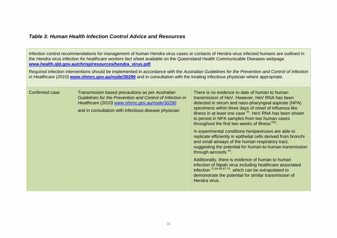
Table 3: Human Health Infection Control Advice and Resources
Infection control recommendations for management of human Hendra virus cases or contacts of Hendra virus infected humans are outlined in the Hendra virus infection for healthcare workers fact sheet available on the Queensland Health Communicable Diseases webpage. www.health.qld.gov.au/chrisp/resources/hendra_virus.pdf. Required infection interventions should be implemented in accordance with the Australian Guidelines for the Prevention and Control of Infection in Healthcare (2010) www.nhmrc.gov.au/node/30290 and in consultation with the treating infectious physician where appropriate. Confirmed case Transmission based precautions as per Australian
Guidelines for the Prevention and Control of Infection in Healthcare (2010) www.nhmrc.gov.au/node/30290
and in consultation with infectious disease physician
There is no evidence to date of human to human transmission of HeV. However, HeV RNA has been detected in serum and naso-pharyngeal aspirate (NPA) specimens within three days of onset of influenza like illness in at least one case 35. HeV RNA has been shown to persist in NPA samples from two human cases throughout the first two weeks of illness (35).
In experimental conditions henipaviruses are able to replicate efficiently in epithelial cells derived from bronchi and small airways of the human respiratory tract, suggesting the potential for human-to-human transmission through aerosols 44. Additionally, there is evidence of human to human infection of Nipah virus including healthcare associated infection 21,64,65,67-70. which can be extrapolated to demonstrate the potential for similar transmission of Hendra virus.
32
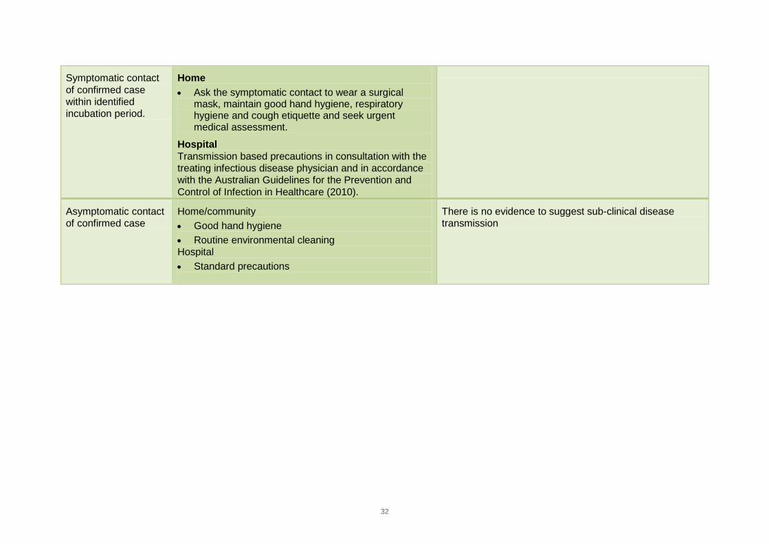
Symptomatic contact of confirmed case within identified incubation period.
Home • Ask the symptomatic contact to wear a surgical
mask, maintain good hand hygiene, respiratory hygiene and cough etiquette and seek urgent medical assessment.
Hospital Transmission based precautions in consultation with the treating infectious disease physician and in accordance with the Australian Guidelines for the Prevention and Control of Infection in Healthcare (2010).
Asymptomatic contact of confirmed case
Home/community • Good hand hygiene • Routine environmental cleaning Hospital • Standard precautions
There is no evidence to suggest sub-clinical disease transmission
33
6 Hendra virus Exposure Assessment Objective: To facilitate a systematic process for determining the level of exposure a person has had to a suspect or infected horse to inform the subsequent management of that person.
In the occurrence of the identification of suspected and confirmed HeV infected horses Biosecurity Queensland and Queensland Health will work together to identify all people who possibly had contact with blood or body fluids, tissue or faeces from an infectious horse. This is in accordance with the current Memorandum of Understanding - Incident management of threats to human or animal health and Multi-agency Coordination Standard Operating Procedure.
People identified at risk of HeV infection will be assessed and managed in accordance with the Hendra virus National guidelines for public health units http://www.health.gov.au/internet/main/publishing.nsf/Content/cdna-song-hendra.htm
A monoclonal antibody to Hendra virus, developed in the US, is available on special access for compassionate reasons for treatment of people assessed by a public health medical officer as having high level exposure to Hendra virus. A phase one safety trial of the monoclonal antibody is expected to be completed in 2015.
Contacts will be provided with resources as required such as Queensland Health Hendra virus fact sheet for contacts and any other appropriate specific resources as outlined in the appendices of the Hendra virus National guidelines for public health units and the Queensland Government Interagency Hendra Virus Communications Framework http://www.health.qld.gov.au/ph/documents/cdb/hendra_comms_fwk.pdf
These tasks will be performed by public health medical officers or nurses who have the skills and knowledge to identify risks for transmission on a case by case basis, given that all risks may not be explicitly covered in the current assessment tool.
34
7 References 1. Field H, Mackenzie JS, Daszak P. Henipaviruses: emerging paramyxoviruses associated with fruit bats. Wildlife and
Emerging Zoonotic Diseases: The Biology, Circumstances and Consequences of Cross-Species Transmission 2007:133-159.
2. Field H, Young P, Yob JM, et al. The natural history of Hendra and Nipah viruses. Microbes and infection 2001;3:307-314.
3. Young P, Field H, Halpin K. Identification of likely natural hosts for equine morbillivirus. Communicable Diseases Intelligence 1996;20:476.
4. Plowright RK, Field HE, Smith C, et al. Reproduction and nutritional stress are risk factors for Hendra virus infection in little red flying foxes (Pteropus scapulatus). Proc Biol Sci 2008;275:861-869.
5. Dups J, Middleton D, Yamada M, et al. A New Model for Hendra Virus Encephalitis in the Mouse. PloS one 2012;7:e40308.
6. Rudd PA, Cattaneo R, von Messling V. Canine distemper virus uses both the anterograde and the hematogenous pathway for neuroinvasion. Journal of virology 2006;80:9361-9370.
7. Mori I, Komatsu T, Takeuchi K, et al. Parainfluenza virus type 1 infects olfactory neurons and establishes long-term persistence in the nerve tissue. Journal of general virology 1995;76:1251-1254.
8. Halpin K, Hyatt AD, Fogarty R, et al. Pteropid bats are confirmed as the reservoir hosts of henipaviruses: a comprehensive experimental study of virus transmission. American Journal of Tropical Medicine and Hygiene 2011;85:946.
9. Li M, Embury-Hyatt C, Weingartl HM. Experimental inoculation study indicates swine as a potential host for Hendra virus. Veterinary research 2010;41:33.
10. Geisbert TW, Feldmann H, Broder CC. Animal challenge models of henipavirus infection and pathogenesis. Henipavirus: Springer, 2012;153-177.
11. Hooper PT, Westbury HA, Russell GM. The lesions of experimental equine morbillivirus disease in cats and guinea pigs. Vet Pathol 1997;34:323-329.
12. Westbury HA, Hooper PT, Brouwer SL, et al. Susceptibility of cats to equine morbillivirus. Aust Vet J 1996;74:132-134. 13. Williamson M, Hooper P, Selleck P, et al. Transmission studies of Hendra virus (equine morbilli‐virus) in fruit bats, horses
and cats. Australian Veterinary Journal 2008;76:813-818. 14. Westbury H, Hooper P, Selleck P, et al. Equine morbillivirus pneumonia: susceptibility of laboratory animals to the virus.
Australian Veterinary Journal 1995;72:278-279. 15. Queensland Health. Unpublished data, Notifiable Conditions System (NOCS). 2000-2010. 16. Selvey L, Taylor R, Arklay A, et al. Screening of bat carers for antibodies to equine morbillivirus. Communicable Diseases
Intelligence 1996;20:477-477. 17. Luby SP, Rahman M, Hossain MJ, et al. Foodborne transmission of Nipah virus, Bangladesh. Emerging Infectious
Diseases 2006;12:1888.
35
18. Field H, de Jong C, Melville D, et al. Hendra Virus Infection Dynamics in Australian Fruit Bats. PloS one 2011;6:e28678. 19. Wang L-F, Daniels P. Diagnosis of Henipavirus Infection: Current Capabilities and Future Directions In: Lee B,Rota PA,
eds. Henipavirus: Springer Berlin Heidelberg, 2012;179-196. 20. Smith I, Broos A, de Jong C, et al. Identifying Hendra virus diversity in pteropid bats. PLoS One 2011;6:e25275. 21. Lo MK, Lowe L, Hummel KB, et al. Characterization of Nipah virus from outbreaks in Bangladesh, 2008–2010. Emerging
infectious diseases 2012;18:248. 22. Biosecurity Queensland. Guidelines for Veterinarians Handling Potential Hendra Virus Infection in Horses. 4th ed. The
State of Queensland:Brisbane, 2010. 23. Field H, Schaaf K, Kung N, et al. Hendra virus outbreak with novel clinical features, Australia. Emerging Infectious
Diseases 2010;16:338. 24. Field HE, Breed AC, Shield J, et al. Epidemiological perspectives on Hendra virus infection in horses and flying foxes.
Aust Vet J 2007;85:268-270. 25. Field HE, Barratt PC, Hughes RJ, et al. A fatal case of Hendra virus infection in a horse in north Queensland: clinical and
epidemiological features. Aust Vet J 2000;78:279-280. 26. Hooper PT, Ketterer PJ, Hyatt AD, et al. Lesions of experimental equine morbillivirus pneumonia in horses. Vet Pathol
1997;34:312-322. 27. Middleton D. Initial experimental characterisation of HeV (Redland Bay 2008) infection in horses. Online: CSIRO, 2009. 28. Murray K, Selleck P, Hooper P, et al. A morbillivirus that caused fatal disease in horses and humans. Science (New York,
NY) 1995;268:94. 29. Rogers RJ, Douglas IC, Baldock FC, et al. Investigation of a second focus of equine morbillivirus infection in coastal
Queensland. Aust Vet J 1996;74:243-244. 30. Williamson MM, Hooper PT, Selleck PW, et al. Transmission studies of Hendra virus (equine morbillivirus) in fruit bats,
horses and cats. Aust Vet J 1998;76:813-818. 31. Australian Biosecurity CRC Research update: Hendra virus., 2009. 32. Marsh GA, Haining J, Hancock TJ, et al. Experimental infection of horses with Hendra
virus/Australia/horse/2008/Redlands. Emerging Infectious Diseases 2011;17:2232. 33. Playford EG, McCall B, Smith G, et al. Human Hendra virus encephalitis associated with equine outbreak, Australia,
2008. Emerg Infect Dis 2010;16:219-223. 34. Allworth T, O’Sullivan J, Selvey L, et al. Equine morbillivirus in Queensland. Comm Dis Intell 1995;19:575. 35. Anonymous. Another case of equine morbillivirus disease in Australia. Emerging Infectious Diseases 1996;2:71-72. 36. Hanna JN, McBride WJ, Brookes DL, et al. Hendra virus infection in a veterinarian. Med J Aust 2006;185:562-564. 37. McCormack JG, Allworth AM, Selvey LA, et al. Transmissibility from horses to humans of a novel paramyxovirus, equine
morbillivirus (EMV). J Infect 1999;38:22-23. 38. O'sullivan J, Allworth A, Paterson D, et al. Fatal encephalitis due to novel paramyxovirus transmitted from horses. The
Lancet 1997;349:93-95.
36
39. Paterson DL, Murray PK, McCormack JG. Zoonotic disease in Australia caused by a novel member of the paramyxoviridae. Clin Infect Dis 1998;27:112-118.
40. Selvey LA, Wells RM, McCormack JG, et al. Infection of humans and horses by a newly described morbillivirus. Med J Aust 1995;162:642-645.
41. Baldock F, Douglas I, Halpin K, et al. Epidemiological investigations into the 1994 equine morbillivirus outbreaks in Queensland, Australia. Singapore Veterinary Journal 1996;20:57-61.
42. Rockx B, Wang L-F. Zoonotic henipavirus transmission. Journal of clinical virology: the official publication of the Pan American Society for Clinical Virology 2013.
43. Reid P. Personal communication. Brisbane, 2010. 44. Escaffre O, Borisevich V, Carmical JR, et al. Henipavirus Pathogenesis in Human Respiratory Epithelial Cells. Journal of
virology 2013;87:3284-3294. 45. Fogarty R, Halpin K, Hyatt AD, et al. Henipavirus susceptibility to environmental variables. Virus Res 2008;132:140-144. 46. Wong KT, Robertson T, Ong BB, et al. Human Hendra virus infection causes acute and relapsing encephalitis.
Neuropathol Appl Neurobiol 2009;35:296-305. 47. Chong HT, Tan CT. Relapsed and late-onset Nipah encephalitis, a report of three cases. Neurol J Southeast Asia
2003;8:109-112. 48. Tan CT, Goh KJ, Wong KT, et al. Relapsed and late‐onset Nipah encephalitis. Annals of Neurology 2002;51:703-708. 49. Wong SC, Ooi MH, Wong MN, et al. Late presentation of Nipah virus encephalitis and kinetics of the humoral immune
response. J Neurol Neurosurg Psychiatry 2001;71:552-554. 50. Taylor C, Playford EG, McBride WJ, et al. No evidence of prolonged Hendra virus shedding by 2 patients, Australia.
Emerging infectious diseases 2012;18:2025-2027. 51. Breed AC, Meers J, Sendow I, et al. The Distribution of Henipaviruses in Southeast Asia and Australasia: Is Wallace’s
Line a Barrier to Nipah Virus? PloS one 2013;8:e61316. 52. Lavoie J. Cloutier Y. Lara J. Marchand G. Chemical substances and biological agents. Studies and research projects.
Technical guide RG-501. Guide on respiratory protection against bioaerosols. Recommendations on its selection and use. Montreal: IRSST:, 2007.
53. Lenhart SW, Seitz T, Trout D, et al. Issues affecting respirator selection for workers exposed to infectious aerosols: emphasis on healthcare settings. Applied Biosafety 2004;9:20-36.
54. Siegel JD, Rhinehart E, Jackson M, et al. 2007 Guideline for Isolation Precautions: Preventing Transmission of Infectious Agents in Health Care Settings. Am J Infect Control 2007;35:65-6164.
55. Stuart RL, Cheng AC, Marshall CL, et al. ASID (HICSIG) position statement: infection control guidelines for patients with influenza-like illnesses, including pandemic (H1N1) influenza 2009, in Australian health care facilities. Med J Aust 2009;191:454-458.
56. Biosecurity Queensland. Biosecurity Policy:Hendra Virus Response: Infected Premises and Dangerous Contact Premises Version 0.6.
37
57. Tan K, Sarji SA, Tan C, et al. Patients with asymptomatic Nipah virus infection may have abnormal cerebral MR imaging. Neurol J Southeast Asia 2000;5:69-73.
58. Suhailah Abdullah L-YC, 3Kartini Rahmat, 1Khean Jin Goh, 1Chong Tin., Tan CT. Late-onset Nipah virus encephalitis 11 years after the initial outbreak: A case report. Neurology Asia 2012;17:71 – 74.
59. Goh KJ, Tan CT, Chew NK, et al. Clinical features of Nipah virus encephalitis among pig farmers in Malaysia. New England Journal of Medicine 2000;342:1229-1235.
60. Wong K, Tan C. Clinical and pathological manifestations of human henipavirus infection. Henipavirus: Springer, 2012;95-104.
61. Selvey L, Sheridan J. Outbreak of Severe Respiratory Disease in Humans and Horses Due to a Previously Unrecognized Paramyxovirus*. Journal of Travel Medicine 1995;2:275-275.
62. Queensland Health. Hendra virus infection for healthcare workers In: Health Q, ed. Brisbane, 2010. 63. Zoonotic henipavirus transmission. J Clin Virol 2013;18:00054-00051. 64. Gurley ES, Montgomery JM, Hossain MJ, et al. Person-to-person transmission of Nipah virus in a Bangladeshi
community. Emerging Infectious Diseases 2007;13:1031. 65. Homaira N, Rahman M, Hossain MJ, et al. Nipah virus outbreak with person-to-person transmission in a district of
Bangladesh, 2007. Epidemiology and Infection 2010;138:1630-1636. 66. Icddr B. Person-to-person transmission of Nipah virus during outbreak in Faridpur District, 2004. Health and Science
Bulletin 2004;2:5-9. 67. Luby SP, Hossain MJ, Gurley ES, et al. Recurrent zoonotic transmission of Nipah virus into humans, Bangladesh, 2001-
2007. Emerg Infect Dis 2009;15:1229-1235. 68. Luby SP, Gurley ES, Hossain MJ. Transmission of human infection with Nipah virus. Clin Infect Dis 2009;49:1743-1748. 69. Tan CT, Tan KS. Nosocomial transmissibility of Nipah virus. J Infect Dis 2001;184:1367-1367. 70. Chua K, Lam S, Goh K, et al. The presence of Nipah virus in respiratory secretions and urine of patients during an
outbreak of Nipah virus encephalitis in Malaysia. Journal of Infection 2001;42:40-43. 71. Animal Health Australia. Australian Emergency Veterinary Plan AUSVETPLAN Response Policy Brief version 3.5 Hendra
virus 2013. 72. Marsh GA, Wang LF. Hendra and Nipah viruses: why are they so deadly? Current Opinion in Virology 2012. 73. Paterson DL, Murray PK, McCormack JG. Zoonotic disease in Australia caused by a novel member of the
paramyxoviridae. Clinical infectious diseases 1998;27:112-118. 74. Hazelton B, Ba Alawi F, Kok J, et al. Hendra virus: a one health tale of flying foxes, horses and humans. Future
microbiology 2013;8:461-474. 75. Broder CC. Henipavirus outbreaks to antivirals: the current status of potential therapeutics. Current Opinion in Virology
2012. 76. Pallister J, Middleton D, Wang L-F, et al. A recombinant Hendra virus G glycoprotein-based subunit vaccine protects
ferrets from lethal Hendra virus challenge. Vaccine 2011;29:5623-5630.
38
77. McEachern JA, Bingham J, Crameri G, et al. A recombinant subunit vaccine formulation protects against lethal Nipah virus challenge in cats. Vaccine 2008;26:3842-3852.
78. Mungall BA, Middleton D, Crameri G, et al. Feline model of acute Nipah virus infection and protection with a soluble glycoprotein-based subunit vaccine. Journal of virology 2006;80:12293-12302.
79. Broder C, Geisbert T, Xu K, et al. Immunization Strategies Against Henipaviruses. Current topics in microbiology and immunology 2012.