Embed Size (px)
Citation preview
Vet. Res. 38 (2007) 153–180 153c© INRA, EDP Sciences, 2007DOI: 10.1051/vetres:2006053
Review article
Bovine respiratory syncytial virus infection
Jean-Francois Va, Geraldine Tb*
a IVI-Animal Health, Lärkbacken, 740 20 Vänge, Uppsala, Swedenb Institute for Animal Health, Compton, Newbury, Berkshire RG20 7NN, United Kingdom
(Received 6 April 2006; accepted 18 July 2006)
Abstract – Bovine respiratory syncytial virus (BRSV) belongs to the pneumovirus genus withinthe family Paramyxoviridae and is a major cause of respiratory disease in young calves. BRSV isenveloped and contains a negative sense, single-stranded RNA genome encoding 11 proteins. Thevirus replicates predominantly in ciliated respiratory epithelial cells but also in type II pneumocytes.It appears to cause little or no cytopathology in ciliated epithelial cell cultures in vitro, suggestingthat much of the pathology is due to the host’s response to virus infection. RSV infection inducesan array of pro-inflammatory chemokines and cytokines that recruit neutrophils, macrophages andlymphocytes to the respiratory tract resulting in respiratory disease. Although the mechanisms re-sponsible for induction of these chemokines and cytokines are unclear, studies on the closely relatedhuman (H)RSV suggest that activation of NF-κB via TLR4 and TLR3 signalling pathways is in-volved. An understanding of the mechanisms by which BRSV is able to establish infection andinduce an inflammatory response has been facilitated by advances in reverse genetics, which haveenabled manipulation of the virus genome. These studies have demonstrated an important role forthe non-structural proteins in anti-interferon activity, a role for a virokinin, released during prote-olytic cleavage of the fusion protein, in the inflammatory response and a role for the SH and thesecreted form of the G protein in establishing pulmonary infection. Knowledge gained from thesestudies has also provided the opportunity to develop safe, stable, live attenuated virus vaccine can-didates.
BRSV / pathogenesis / respiratory disease / cattle / vaccines
Table of contents
1. Introduction ...................................................................................................... 1542. The virion ......................................................................................................... 155
2.1. BRSV proteins ........................................................................................... 1552.1.1. Non-structural proteins NS1 and NS2 ................................................... 1552.1.2. Small hydrophobic SH protein ............................................................ 1552.1.3. Glycoprotein G ................................................................................ 1562.1.4. Fusion F protein ............................................................................... 1572.1.5. Nucleocapsid proteins ....................................................................... 1582.1.6. Matrix proteins ................................................................................ 159
2.2. Virus replication ......................................................................................... 1592.3. Antigenic and genetic subgroups of BRSV ....................................................... 159
* Corresponding author: [email protected]
Article available at http://www.edpsciences.org/vetres or http://dx.doi.org/10.1051/vetres:2006053
154 J.-F. Valarcher et al.
3. Epidemiology and clinical signs of disease............................................................... 1603.1. Epidemiology............................................................................................. 1603.2. Clinical signs of disease and pathology ............................................................ 161
4. Pathogenesis of BRSV......................................................................................... 1624.1. The role of the NS proteins in the pathogenesis of BRSV..................................... 1634.2. The role of the F protein in the pathogenesis of BRSV ....................................... 1644.3. The role of the G protein in the pathogenesis of BRSV........................................ 1654.4. The role of the SH protein............................................................................. 1674.5. The role of viral proteins in determining host-range specificity ............................. 167
5. Prevention, control and vaccination ........................................................................ 1685.1. NS deletion mutants as live vaccines ............................................................... 1695.2. FCS-2 cleavage mutants or ∆p27 mutants as live vaccines.................................... 1695.3. G protein mutants as live vaccines .................................................................. 1695.4. SH deletion mutants as live vaccines ............................................................... 1705.5. Other strategies for the development of live attenuated BRSV vaccine candidates ..... 170
6. Conclusions ...................................................................................................... 170
1. INTRODUCTION
Bovine respiratory syncytial virus(BRSV) is an enveloped, non-segmented,negative-stranded RNA virus and is a ma-jor cause of respiratory disease in youngcalves [128]. BRSV is closely related tohuman (H)RSV, which is a major causeof respiratory disease in young children,and the epidemiology and pathogenesisof infection with these viruses are sim-ilar [156]. These features make BRSVinfection in calves a good model forthe study of HRSV. In return, findingsobtained by studying HRSV in vivo inhumans, in small animal models or in vitrohave allowed a better understanding ofsome virological and pathogenic char-acteristics of its counterpart in bovines.Although these viruses are very similar inmany aspects, there are differences, suchas their level of replication in vitro and invivo in their respective hosts, differencesin the peptide released as a result ofcleavage at the two furin cleavage sites ofthe F protein and the relative functions ofNS1 and NS2 in the inhibition of type Iinterferon induction.
HRSV and BRSV are members of thePneumovirus genus within the subfam-ily Pneumovirinae, family Paramyxoviri-
dae of the order Mononegavirales [26].Other pneumoviruses include pneumoniavirus of mice (PVM), ovine (O)RSV andcaprine (C)RSV. Although the RS virusesare structurally and antigenically related,BRSV and CRSV are the most closely re-lated [2, 82, 123, 147].
By electron microscopy, the morphol-ogy of the RSV virions appears to be eithervery pleomorphic, with a shape roughlyrounded and a diameter between 150 and35 nm, or filamentous with a length thatcan reach 5 µm and a diameter between60 and 100 nm [147]. Regardless of thestructure, each infectious particle containsa single functional copy of the genome.In contrast to HRSV, but similar to 80%of caprine virions, BRSV virions are orga-nized in a network and viral particles arelinked to each other by bridges of 12 ±3 nm [10]. The roles of these structures re-main unexplained.
In contrast to HRSV, several vaccinesagainst BRSV are available on the mar-ket. However, the development of a secondgeneration of BRSV vaccines with greaterefficacy in the presence of maternal an-tibodies and which induce more durableprotection would be facilitated by a greaterunderstanding of the pathogenesis of the
Bovine respiratory syncytial virus infection 155
virus. In this paper, after describing thecharacteristics of the virus and the diseaseinduced by BRSV, we will review some re-cent findings on host-pathogen interactionsthat could provide a basis for the future de-velopment of safe, stable, live attenuatedvirus vaccines.
2. THE VIRION
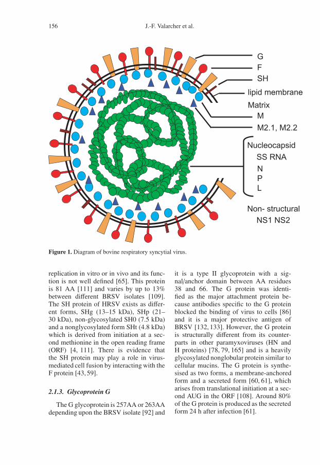
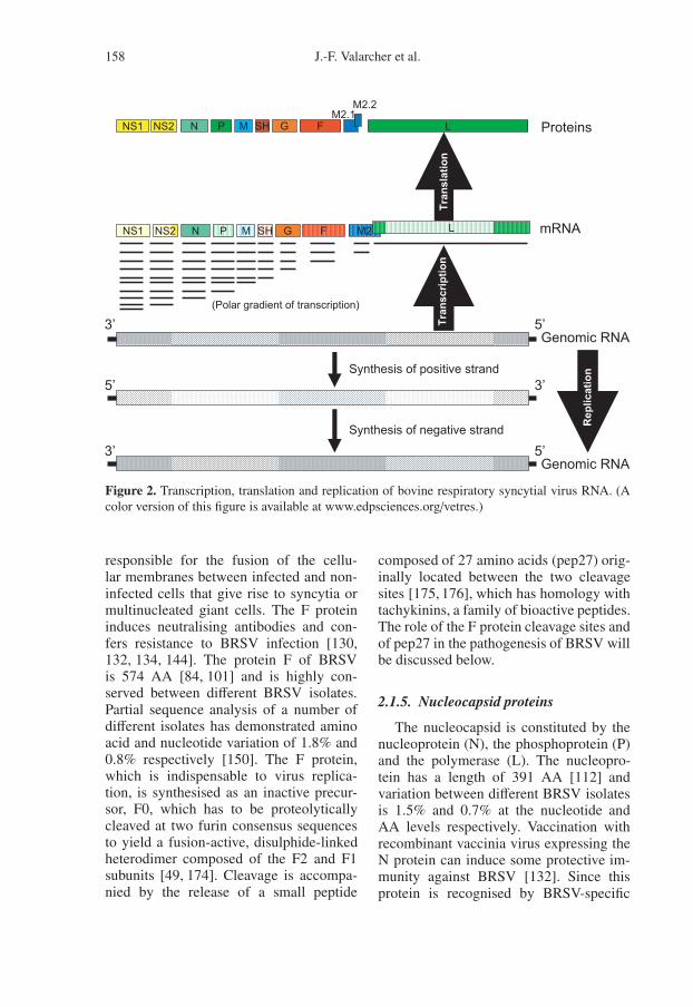
The BRSV virion consists of a lipidenvelope, derived from the host plasmamembrane, containing three virally en-coded transmembrane surface glycopro-teins, which are organised separately intospikes on the surface of the virion (Fig. 1).These glycoproteins are the large glyco-protein (G), the fusion protein (F) andthe small hydrophobic protein (SH) [33].The envelope encloses a helical nucle-ocapsid, which consists of the nucleo-protein (N), phosphoprotein (P), the vi-ral RNA-dependent polymerase protein (L)and a genomic RNA of around 15000nucleotides. In addition, there is a ma-trix M protein that is thought to form alayer on the inner face of the envelopeand a transcriptional anti-termination fac-tor M2-1. The genome also encodes anRNA regulatory protein M2-2 and twonon-structural proteins, NS1 and NS2 [33](Fig. 1). In addition, the viral particle con-tains cellular proteins, such as actin, whichhas been demonstrated on the surface ofHRSV [148], caveolin-1 [18] and MHCclass I molecules1. Thus, BRSV propa-gated in bovine cells can be neutralised bymonoclonal antibodies specific for bovineMHC class I.
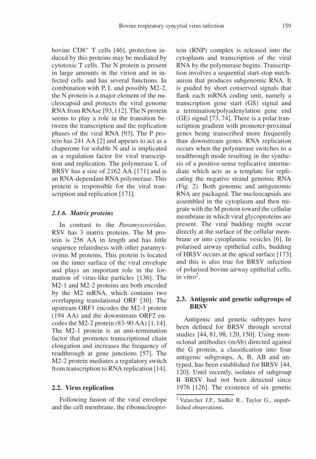
The genomic RNA is the template forreplication and transcription. The genomicRNA, which is transcribed in a sequen-tial fashion from the 3’ end, encodes tenmRNA. There is a polar transcription gra-dient such that 3’-terminal genes are tran-scribed more frequently than those at the 5’
1 Taylor G., unpublished observations.
end (Fig. 2). The 10 mRNA are then trans-lated into 11 viral proteins. Some proper-ties of the BRSV proteins and their aminoacid (AA) identity with HRSV proteinsare shown in Table I. The Pneumovirusgenome is characterised by the existence oftwo non-structural proteins, NS1 and NS2,and a transcriptional overlap between M2and L that lead to the synthesis of M2-1and M2-2 proteins.
2.1. BRSV proteins2.1.1. Non-structural proteins NS1 and
NS2
One of the major differences betweenthe pneumoviruses and the other Paramy-xoviridae is the presence of two non-structural (NS) proteins, NS1 and NS2,which have 136 AA and 124 AA, respec-tively [102]. The genes encoding thesetwo proteins are abundantly transcribed invirus-infected cells, however, the proteinsare detected only in trace amounts in pu-rified virions. There is evidence that theHRSV NS1 protein coprecipitates with theM protein [42], and is a strong inhibitorof viral RNA transcription and replica-tion [7]. The NS2 protein also appears tobe a transcriptional inhibitor but at a lowerlevel than the NS1 protein [7]. The NS2protein colocalizes with the P and N pro-teins in infected cells [164] but does notcoprecipitate with any viral protein [42].These proteins are not essential for virusreplication in vitro, although the growthof recombinant HRSV and BRSV lackingone or other of these proteins is attenu-ated in cell culture [20,115,137]. The NS1and NS2 proteins play an important rolein regulating IFNα/β and their role in thepathogenesis and host-range restriction ofBRSV is discussed below.
2.1.2. Small hydrophobic SH protein
The SH protein is a short integral mem-brane protein and is not essential for virus
156 J.-F. Valarcher et al.
Figure 1. Diagram of bovine respiratory syncytial virus.
replication in vitro or in vivo and its func-tion is not well defined [65]. This proteinis 81 AA [111] and varies by up to 13%between different BRSV isolates [109].The SH protein of HRSV exists as differ-ent forms, SHg (13–15 kDa), SHp (21–30 kDa), non-glycosylated SH0 (7.5 kDa)and a nonglycosylated form SHt (4.8 kDa)which is derived from initiation at a sec-ond methionine in the open reading frame(ORF) [4, 111]. There is evidence thatthe SH protein may play a role in virus-mediated cell fusion by interacting with theF protein [43, 59].
2.1.3. Glycoprotein G
The G glycoprotein is 257AA or 263AAdepending upon the BRSV isolate [92] and
it is a type II glycoprotein with a sig-nal/anchor domain between AA residues38 and 66. The G protein was identi-fied as the major attachment protein be-cause antibodies specific to the G proteinblocked the binding of virus to cells [86]and it is a major protective antigen ofBRSV [132, 133]. However, the G proteinis structurally different from its counter-parts in other paramyxoviruses (HN andH proteins) [78, 79, 165] and is a heavilyglycosylated nonglobular protein similar tocellular mucins. The G protein is synthe-sised as two forms, a membrane-anchoredform and a secreted form [60, 61], whicharises from translational initiation at a sec-ond AUG in the ORF [108]. Around 80%of the G protein is produced as the secretedform 24 h after infection [61].
Bovine respiratory syncytial virus infection 157
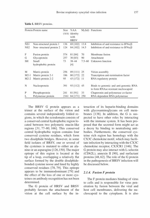
Table I. BRSV proteins.
Protein Protein name Size(AA)
%AAidentitywithHRSV
Mr(kd) Functions
NS1NS2
Non-structural protein 1Non- structural protein 2
136124
69 [102]84 [102]
13.814.5
Inhibition of and resistance to IFNα/βInhibition of and resistance to IFNα/β
FGSH
Fusion proteinGlycoproteinSmallhydrophobic protein
57425773
81 [84]30 [83]38–44[4]
70907.5–60
Membrane fusionAttachmentUnknown function
MM2-1M2-2
Matrix proteinMatrix protein 2-1Matrix protein 2-2
25618695
89 [111]80 [172]43 [172]
252211
Virion assemblyTrancription anti-termination factorRNA regulatory protein
N Nucleoprotein 391 93 [112] 45 Binds to genomic and anti-genomic RNAto form RNAse-resistant nucleocapsid
PL
PhosphoproteinPolymerase protein
2412161
81 [91]84 [171]
33250
Chaperonin and polymerase co-factorRNA dependent RNA polymerase
The BRSV G protein appears as atrimer at the surface of the virion andcontains several independently folded re-gions, in which the ectodomain consists ofa conserved central hydrophobic region lo-cated between two polymeric mucin-likeregions [31, 77–80, 166]. This conservedcentral hydrophobic region contains fourconserved cysteine residues, which formtwo disulphide bridges. However, in somefield isolates of BRSV, one or several ofthe cysteines is mutated to either an ala-nine or an asparagine [126,150]. The majorepitope of this region is located at thetip of a loop, overlapping a relatively flatsurface formed by the double disulphide-bonded cysteine noose and lined by highlyconserved residues [78, 79]. This epitopeappears to be immunodominant [79] andthe effect of the loss of one or more cys-teines on antibody recognition has not beendetermined.
The G protein of HRSV and BRSVprobably favours the attachment of thevirion at the cell surface by the in-
teraction of its heparin-binding domainswith glycosaminoglycans on cell mem-branes [138]. In addition, the G is sus-pected to have other roles by interactingwith the immune system. It has been pro-posed that the secreted form might act asa decoy by binding to neutralising anti-bodies. Furthermore, the conserved cys-teine rich region has homology with theCX3C chemokine motif, which may facili-tate infection by interacting with the CX3Cchemokine receptor, CX3CR1 [146]. TheG protein may also interact with L-selectin(CD62L), annexin II [90], and surfactantproteins [48, 62]. The role of the G proteinin the pathogenesis of BRSV infection willbe discussed below.
2.1.4. Fusion F protein
The F protein mediates binding of virusto cells and is responsible for virus pen-etration by fusion between the viral andhost cell membranes, delivering the nu-cleocapsid to the cytoplasm. It is also
158 J.-F. Valarcher et al.
Figure 2. Transcription, translation and replication of bovine respiratory syncytial virus RNA. (Acolor version of this figure is available at www.edpsciences.org/vetres.)
responsible for the fusion of the cellu-lar membranes between infected and non-infected cells that give rise to syncytia ormultinucleated giant cells. The F proteininduces neutralising antibodies and con-fers resistance to BRSV infection [130,132, 134, 144]. The protein F of BRSVis 574 AA [84, 101] and is highly con-served between different BRSV isolates.Partial sequence analysis of a number ofdifferent isolates has demonstrated aminoacid and nucleotide variation of 1.8% and0.8% respectively [150]. The F protein,which is indispensable to virus replica-tion, is synthesised as an inactive precur-sor, F0, which has to be proteolyticallycleaved at two furin consensus sequencesto yield a fusion-active, disulphide-linkedheterodimer composed of the F2 and F1subunits [49, 174]. Cleavage is accompa-nied by the release of a small peptide
composed of 27 amino acids (pep27) orig-inally located between the two cleavagesites [175, 176], which has homology withtachykinins, a family of bioactive peptides.The role of the F protein cleavage sites andof pep27 in the pathogenesis of BRSV willbe discussed below.
2.1.5. Nucleocapsid proteins
The nucleocapsid is constituted by thenucleoprotein (N), the phosphoprotein (P)and the polymerase (L). The nucleopro-tein has a length of 391 AA [112] andvariation between different BRSV isolatesis 1.5% and 0.7% at the nucleotide andAA levels respectively. Vaccination withrecombinant vaccinia virus expressing theN protein can induce some protective im-munity against BRSV [132]. Since thisprotein is recognised by BRSV-specific
Bovine respiratory syncytial virus infection 159
bovine CD8+ T cells [46], protection in-duced by this proteins may be mediated bycytotoxic T cells. The N protein is presentin large amounts in the virion and in in-fected cells and has several functions. Incombination with P, L and possibly M2-2,the N protein is a major element of the nu-cleocapsid and protects the viral genomeRNA from RNAse [93,112]. The N proteinseems to play a role in the transition be-tween the transcription and the replicationphases of the viral RNA [93]. The P pro-tein has 241 AA [2] and appears to act as achaperone for soluble N and is implicatedas a regulation factor for viral transcrip-tion and replication. The polymerase L ofBRSV has a size of 2162 AA [171] and isan RNA-dependant RNA polymerase. Thisprotein is responsible for the viral tran-scription and replication [171].
2.1.6. Matrix proteins
In contrast to the Paramyxoviridae,RSV has 3 matrix proteins. The M pro-tein is 256 AA in length and has littlesequence relatedness with other paramyx-ovirus M proteins. This protein is locatedon the inner surface of the viral envelopeand plays an important role in the for-mation of virus-like particles [136]. TheM2-1 and M2-2 proteins are both encodedby the M2 mRNA, which contains twooverlapping translational ORF [30]. Theupstream ORF1 encodes the M2-1 protein(194 AA) and the downstream ORF2 en-codes the M2-2 protein (83-90 AA) [1,14].The M2-1 protein is an anti-terminationfactor that promotes transcriptional chainelongation and increases the frequency ofreadthrough at gene junctions [57]. TheM2-2 protein mediates a regulatory switchfrom transcription to RNA replication [14].
2.2. Virus replication
Following fusion of the viral envelopeand the cell membrane, the ribonucleopro-
tein (RNP) complex is released into thecytoplasm and transcription of the viralRNA by the polymerase begins. Transcrip-tion involves a sequential start-stop mech-anism that produces subgenomic RNA. Itis guided by short conserved signals thatflank each mRNA coding unit, namely atranscription gene start (GS) signal anda termination/polyadenylation gene end(GE) signal [73, 74]. There is a polar tran-scription gradient with promoter-proximalgenes being transcribed more frequentlythan downstream genes. RNA replicationoccurs when the polymerase switches to areadthrough mode resulting in the synthe-sis of a positive-sense replicative interme-diate which acts as a template for repli-cating the negative strand genomic RNA(Fig. 2). Both genomic and antigenomicRNA are packaged. The nucleocapsids areassembled in the cytoplasm and then mi-grate with the M protein toward the cellularmembrane in which viral glycoproteins arepresent. The viral budding might occurdirectly at the surface of the cellular mem-brane or into cytoplasmic vesicles [6]. Inpolarised airway epithelial cells, buddingof HRSV occurs at the apical surface [173]and this is also true for BRSV infectionof polarised bovine airway epithelial cells,in vitro2.
2.3. Antigenic and genetic subgroups ofBRSV
Antigenic and genetic subtypes havebeen defined for BRSV through severalstudies [44, 81, 98, 120, 150]. Using mon-oclonal antibodies (mAb) directed againstthe G protein, a classification into fourantigenic subgroups, A, B, AB and un-typed, has been established for BRSV [44,120]. Until recently, isolates of subgroupB BRSV had not been detected since1976 [126]. The existence of six genetic
2 Valarcher J.F., Sadler R., Taylor G., unpub-lished observations.
160 J.-F. Valarcher et al.
subgroups based on G and of five basedon F or N has also been established [150].This classification showed a spatial cluster-ing of BRSV isolates that has been con-firmed by studies including isolates col-lected in many countries [126, 154, 170].The degree of genetic variability of BRSVis limited being less than 15%, which isless than that observed within one sub-group of HRSV [105]. The evolution ofBRSV appears to be continuous and ithas been proposed that evolution may bedriven by selective pressure as a result ofthe immune response induced by vaccina-tion [150].
The biological significance of these sub-groups is not known. However, polyclonalsera obtained from calves vaccinated withthe BRSV G protein from subgroup Avirus recognised a different subgroup ABRSV but not a subgroup B or an untypedisolate [45]. Furthermore, recognition of asubgroup AB virus was less than that of thesubgroup A isolate. Thus, mutations in theimmunodominant region (AA 174–188) ofthe G protein may contribute to the lack ofcross-protection between vaccine and fieldisolates. This might be relevant for the de-velopment of subunit vaccines.
3. EPIDEMIOLOGY AND CLINICALSIGNS OF DISEASE
3.1. Epidemiology
Although cattle are the natural host ofBRSV, it is possible that other speciessuch as ovine, caprine, bison, chamoixor camelids may play an epidemiologi-cal role in certain circumstances [29, 40,107, 113, 157]. The distribution of BRSVis worldwide and the virus has been iso-lated from cattle in Europe, America andAsia [63, 100, 122]. The virus causes regu-lar winter outbreaks of respiratory diseasein cattle [127]. A seroprevalence of 30–70% have been detected in cattle [3,41,54].The frequency of BRSV infections is very
high and the virus might be responsible formore than 60% of the epizootic respiratorydiseases observed in dairy herds [9,41,149]and up to 70% in beef herds [27, 114,127]. The frequency of BRSV infectionsis correlated to the density of the cattlepopulation in an area [41] and the ageof the animal. Indeed more than 70% ofbeef calves were infected with BRSV bythe age of nine months in England [127]and in cattle less than one year old inThe Netherlands [70]. BRSV antibodies incalves between 5 and 11 months of agewere detected in 35% of dairy herds (n =118) in a Swedish study [54]. The fre-quency of infection in adults is difficultto assess because of the high BRSV sero-prevalence in this category of animals.
Severe clinical signs are mainly ob-served in calves [70, 127, 160], but mightalso be observed in adult cattle [41]. Thehigher frequency of clinical signs inducedby BRSV in young calves compared withadults can be explained by the level of spe-cific immunity following frequent expo-sure to the virus. Indeed clinical signs areusually observed in cattle of all ages whenBRSV is introduced in herds where mostof the animals are naïve to the virus andare observed only in calves when the viruscirculates regularly in the herd [156]. Ma-ternally derived antibodies provide at leastpartial protection against clinical signs af-ter natural and experimental BRSV infec-tion [11, 68–70]. Although virus sheddinghas occasionally been detected upon ex-perimental BRSV re-infection, little or noclinical disease is observed in reinfectedanimals [69, 109, 134, 145]. Similar to ob-servations made for HRSV [67], exacer-bated clinical signs have been observedfollowing a natural BRSV infection inanimals immunised with inactivated vac-cines [5, 47, 119].
BRSV infection is associated with ahigh morbidity (60 to 80%) and mortalitycan reach up to 20% in some outbreaks.Clinical disease caused by BRSV is mainly
Bovine respiratory syncytial virus infection 161
diagnosed in the autumn and winter intemperate climate zones [127]. AlthoughBRSV infection occurs mainly in theseseasons [155], it might also occur in thesummer [41]. BRSV is mainly transmittedby direct contact between infected animalsor by aerosol [94] but it cannot be excludedthat it might also be spread by humansacting as a passive vector as observed forHRSV [55]. Some data indicate that BRSVmay persist in infected animals [39, 140,151]. However attempts to demonstratere-excretion of BRSV from previously in-fected animals by treatment with 3-methylindol, BVDV, BHV1 or dexamethasonehave failed [158] and transmission of virusfrom carriers to susceptible animals has notbeen proven.
3.2. Clinical signs of diseaseand pathology
The incubation period for BRSV is esti-mated to be between 2 and 5 days. BRSV-infection may either be asymptomatic,limited to the upper airways or involveboth the upper (URT) and lower respira-tory tracts (LRT). URT disease is char-acterised by a cough with a seromucoidnasal and ocular discharge. In more severeinfections, there is slight depression andanorexia, a decrease in milk yield in lactat-ing cows, hyperthermia, polypnea (respira-tory rate ≥ 60 movements per min) and anabdominal dyspnea. On auscultation of thelung abnormal breathing sounds caused bybronchopneumonia or bonchiolitis mightbe detected [159]. Animals may developsevere respiratory distress with a grunt-ing expiration and breathing through anopen mouth with the neck stretched andthe head down, with saliva poring on thefloor and with the tongue out. In these an-imals, pulmonary emphysema and oedemawith some crackles and wheezes may bedetected [12] and in some cases subcuta-neous emphysema might occur [12, 19].
At necropsy, a broncho-interstitialpneumonia may be observed [19, 161].Areas of the cranio-ventral parts of thelung are consolidated and a mucopurulentdischarge may be seen from the bronchusand small bronchi. The caudo-dorsal partsof the lungs are often distended because ofinterlobular, lobular and sub-pleural em-physematic lesions [19]. Tracheobronchialand mediastinal lymph nodes may beenlarged, oedematous and sometimeshaemorrhagic. If bacterial super-infectionsoccur, the lung parenchyma is moreswollen and consolidated and fibrin orsuppurative bronchopneumonia may beobserved.
Microscopic lesions are characterisedby a proliferative and exudative bron-chiolitis with accompanying alveolar col-lapse and a peribronchiolar infiltration bymononuclear cells [141]. Necrosis of theepithelium and apoptotic epithelial cells,which may be phagocytosed by neigh-bouring cells, can be seen [162]. Giantcells or syncytia may be present, ei-ther free in the bronchi lumen, in thebronchiolar epithelium or in the alveo-lar walls and lumina [162]. The lumenof bronchi, bronchioles and the alveoliare often obstructed by cellular debrisconsisting mostly of neutrophils, desqua-mated epithelial cells, macrophages andsometimes eosinophils [162] and may beaggravated by bronchiolar repair and re-organisation [71]. Eosinophils and lym-phocytes (CD4+, CD8+ and WC1+γ/δ Tcells) are also observed in the laminapropria [141, 143, 162]. Alveolar changesare marked by an interstitial pneumoniaand atelectasis in the consolidated areasand there may be severe emphysema andoedema with a rupture of alveolar wallsin the caudo-dorsal area of the lung. Thepresence of microscopic changes in thecaudo-dorsal area are rarely associatedwith the presence of BRSV antigen, syn-cytia or bronchiolitis [71]. An alveolar ep-ithelisation with a pneumocyte hyperplasia
162 J.-F. Valarcher et al.
contributes to the enlargement of the alve-olar septa with the cell infiltration. Hyalinemembranes may be present in the alveolifollowing inflammation and pneumocytenecrosis [19].
4. PATHOGENESIS OF BRSV
BRSV replicates primarily in the su-perficial layer of the respiratory ciliatedepithelium and replication can also be de-tected in type II pneumocytes [161, 162].Although BRSV is cytopathic in tissue cul-ture, little or no cytopathic effects are seenfollowing infection of differentiated bovineairway epithelial cell cultures, in vitro3. Asimilar lack of obvious cytopathology hasbeen observed in human airway epithelialcell cultures infected with HRSV [173],suggesting that the host response to virusinfection plays a major role in RSV patho-genesis.
HRSV infection of human airway ep-ithelial cells and alveolar macrophages re-sults in activation of NF-κB which leads tothe induction of inflammatory chemokinesand cytokines, such as RANTES (CCL5),MIP-1α (CCL3), MCP-1 (CCL2), eotaxin(CCL11), IL-8 (CXCL8), TNF-α, inter-leukin (IL)-6, IL-1 etc. [15, 52, 53, 58, 95,99], which contribute to inflammation byrecruiting neutrophils, macrophages andlymphocytes to the airways. Although lesswell studied, BRSV infection induces asimilar up-regulation of pro-inflammatorychemokines and cytokines in the bovinelung. Thus, increased levels of mRNAfor IL-12, IFNγ, TNFα, IL-6, IL-18,IL-8, RANTES, MCP-1, MIP-1α, IFNαand IFNβ have been detected in pneu-monic lesions from BRSV-infected gno-tobiotic calves4. The molecular mecha-
3 Sadler R., Valarcher J.-F., Hibbert L., TaylorG., unpublished observations.4 Hibbert L., Valarcher J.-F., Taylor G., unpub-lished observations.
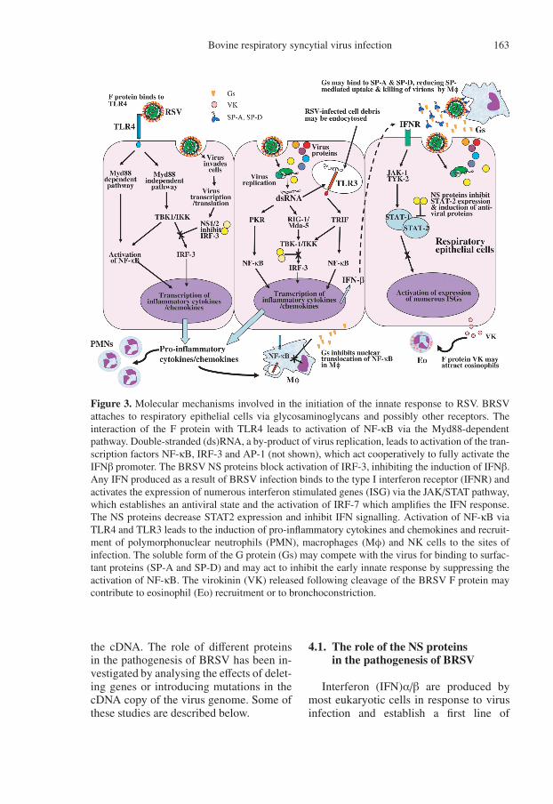
nisms involved in RSV-induced activationof NF-κB and initiation of the innate re-sponse are complex and appear to be me-diated, at least in part by the interactionof the F protein with Toll-like receptor4 (TLR4) [75] and by the interaction ofdsRNA with TLR3 [110] (Fig. 3). Al-though TLR4 is expressed at high levels bymacrophages and dendritic cells (DC) it isexpressed only at very low levels on airwayepithelial cells. However, HRSV infectionincreases TLR4 expression on human air-way epithelial cells and increases theirresponsiveness to LPS [96]. Studies inBALB/c mice infected with HRSV demon-strated two peaks of NF-κB activation. Theimmediate response following RSV inoc-ulation was TLR4-dependent [52] and thesecond peak, which required RSV repli-cation was mediated via TLR3 signallingpathways [110]. It is likely, but has notbeen formally demonstrated, that BRSVinfection induces activation of NF-κB andinduction of pro-inflammatory cytokinesby pathways similar to those demonstratedfor HRSV.
An understanding of the mechanisms bywhich BRSV is able to establish infectionin the bovine respiratory infection, inducean inflammatory response and respiratorydisease has been greatly facilitated by ad-vances in reverse genetics. This involvesthe production of infectious virus fromcloned cDNA [32, 37]. The recovery ofRSV from cDNA requires co-expression incell culture of a complete copy of the vi-ral RNA genome and the N, P, M2-1 andL proteins engineered to be expressed bybacteriophage T7 RNA polymerase. As de-scribed above, these are the constituents ofthe nucleocapsid and polymerase complex,which is the minimum unit of infectivityfor mononegaviruses. The expressed viralcomponents assemble and result in a pro-ductive infection. The recombinant virusproduced in this way is identical to thebiologically-derived virus except for what-ever mutations have been introduced into
Bovine respiratory syncytial virus infection 163
Figure 3. Molecular mechanisms involved in the initiation of the innate response to RSV. BRSVattaches to respiratory epithelial cells via glycosaminoglycans and possibly other receptors. Theinteraction of the F protein with TLR4 leads to activation of NF-κB via the Myd88-dependentpathway. Double-stranded (ds)RNA, a by-product of virus replication, leads to activation of the tran-scription factors NF-κB, IRF-3 and AP-1 (not shown), which act cooperatively to fully activate theIFNβ promoter. The BRSV NS proteins block activation of IRF-3, inhibiting the induction of IFNβ.Any IFN produced as a result of BRSV infection binds to the type I interferon receptor (IFNR) andactivates the expression of numerous interferon stimulated genes (ISG) via the JAK/STAT pathway,which establishes an antiviral state and the activation of IRF-7 which amplifies the IFN response.The NS proteins decrease STAT2 expression and inhibit IFN signalling. Activation of NF-κB viaTLR4 and TLR3 leads to the induction of pro-inflammatory cytokines and chemokines and recruit-ment of polymorphonuclear neutrophils (PMN), macrophages (Mφ) and NK cells to the sites ofinfection. The soluble form of the G protein (Gs) may compete with the virus for binding to surfac-tant proteins (SP-A and SP-D) and may act to inhibit the early innate response by suppressing theactivation of NF-κB. The virokinin (VK) released following cleavage of the BRSV F protein maycontribute to eosinophil (Eo) recruitment or to bronchoconstriction.
the cDNA. The role of different proteinsin the pathogenesis of BRSV has been in-vestigated by analysing the effects of delet-ing genes or introducing mutations in thecDNA copy of the virus genome. Some ofthese studies are described below.
4.1. The role of the NS proteinsin the pathogenesis of BRSV
Interferon (IFN)α/β are produced bymost eukaryotic cells in response to virusinfection and establish a first line of
164 J.-F. Valarcher et al.
defence. Transcription of IFNα/β is me-diated by the transcription factors IRF-3,NF-κB and AP-1. Once secreted, IFNα/βbind to cell surface receptors and activatethe JAK/STAT signalling pathway whichinduces further production of IFN and anarray of IFN-stimulated genes, includingones that establish an antiviral state [50](Fig. 3). In order to establish infection,viruses have evolved a variety of mecha-nisms to counteract the IFNα/β response.BRSV and HRSV are poor inducers ofIFNα/β and are resistant to the antivi-ral effects of IFNα/β [8, 115]. The abilityto regulate the IFNα/β response is me-diated by the NS proteins of BRSV andHRSV [16, 17, 115, 124, 152]. The BRSVNS2 protein appears to have a greater in-hibitory effect on IFNα/β than the NS1protein, which is the converse of that de-scribed for HRSV [124, 152]. The BRSVand HRSV NS proteins prevent inductionof IFNα/β and the establishment of anantiviral state by interfering with the ac-tivation of IRF-3 [17, 125] (Fig. 3). Inaddition, the HRSV NS proteins inhibitIFNα/β signalling by inducing a decreasein Stat2 expression [87, 106] (Fig. 3). Fur-ther studies have demonstrated that the NSgenes may also play a role in activation ofNF-κB. Thus, activation of NF-κB in Verocells, which lack IFNα/β structural genes,infected with rHRSV lacking NS2 (∆NS2)or lacking both NS1 and NS2 (∆NS1/2)was significantly lower than that in cellsinfected with wild-type HRSV [125]. Therole of the BRSV NS proteins in NF-κBactivation is not known.
Although the NS proteins are not essen-tial for virus replication in vitro, growthof recombinant BRSV lacking one or theother of these proteins is attenuated in cellculture [20, 115, 152]. Furthermore, repli-cation of NS deletion mutants of BRSVin young calves is highly attenuated [152].Thus, following intranasal (i.n.) and intra-tracheal (i.t.) inoculation of 2 week-old,gnotobiotic calves with rBRSV ∆NS1 or
rBRSV ∆NS2, only low titres of viruscould be isolated from the nasopharynxfor only 1 to 2 days and virus could notbe detected in the lungs at post-mortem,6 to 7 days after infection. BRSV lackingboth NS1 and NS2 was even more atten-uated and virus could not be recoveredfrom calves at any time post-infection. Incontrast to calves infected with wild-typerBRSV, neither macroscopic nor micro-scopic lung lesions could be detected inany of the calves inoculated with any ofthe NS deletion mutants. These observa-tions highlight the critical role of IFNα/βin the innate response of the bovine respi-ratory tract against BRSV infection.
4.2. The role of the F proteinin the pathogenesis of BRSV
Cleavage of the BRSV F0 proteinby a furin endoprotease occurs at twosites, FCS-1 (RKRR136) and FCS-2(RAR/KR109), and results in the forma-tion of F1 and F2 subunits linked by adisulphide bridge and in the release of anN-glycosylated peptide of 27 amino-acids(pep27) [49, 174]. Cleavage at both sites isrequired for efficient syncytium formation.In BRSV-infected cells, pep27 is furthersubjected to post-translational modifica-tions and is converted into virokinin, amember of the tachykinin family, whichincludes substance P, neurokinins A and B,hemokinin and endokinin A and B [176].Virokinin induces smooth muscle contrac-tion, in vitro, and may therefore contributeto bronchoconstriction in vivo. Usingrecombinant (r)BRSV with mutations inFCS-2 (K108N/K109/N) that abolishedcleavage at this site or in which pep27was deleted (∆p27), neither FCS-2 norpep27 was found to be essential for virusreplication in vitro [72, 176]. However,mutant BRSV in which cleavage was abol-ished at FCS-2 did not grow as efficientlyas the parental wild-type virus duringearly replication cycles. Furthermore,
Bovine respiratory syncytial virus infection 165
both rBRSV (108/109) and ∆p27 showedreduced syncytium formation in cellculture [176].
Despite the differences seen in vitrowith these viruses, they replicated to sim-ilar levels as the wild-type rBRSV inthe bovine upper and lower respiratorytract [153]. However, both the FCS-2 and∆p27 mutants induced significantly lesspulmonary inflammation compared withthe parental wild-type rBRSV [153]. Fur-thermore, calves infected with the F mu-tant viruses showed a marked reduction inthe numbers of eosinophils in the laminapropria of the large bronchioles suggest-ing that the virokinin may play a role ineosinophil recruitment (Fig. 3). Neverthe-less, the virokinin did not have any directchemoattractant proprieties on inflamma-tory cells, in vitro [153]. The chemokinesRANTES (CCL5) and MIP-1α (CCL3) arepotent attractants for human eosinophilsand there is a correlation between levelsof MIP-1α and eosinophil cationic proteinin the lower airways of infants with severeHRSV disease [58]. However, there wereno significant differences in mRNA for ei-ther RANTES or MIP-1α in the lungs ofcalves infected with the F mutant viruses,suggesting that these chemokines may notbe involved in eosinophil recruitment in thecalf [153].
The primary sequence of virokinin isconserved in all BRSV isolates studied todate [176], which suggests that it providessome selective advantage to the virus.Eosinophil products might be expected tohave a detrimental effect on the virus, how-ever these products might damage respira-tory mucosa and induce ciliostasis, whichcould favour viral replication. In contrast toBRSV, the 27-mer peptide produced duringthe maturation of HRSV F does not con-tain a tachykinin motif [176] suggestingthat virokinin may be an additional factorto chemokines involved in eosinophil re-cruitment in BRSV infection and/or maycontribute to species specificity.
BRSV infection is associated with areduction in mitogen-induced lymphocyteproliferation in both calves and lambs [66,121, 168]. BRSV-infected cells are able toinhibit mitogen-induced lymphocyte pro-liferation, in vitro, and this effect wasfound to be mediated by direct contact ofthe lymphocytes with the F protein [116].The precise mechanisms responsible forthis inhibition are not known, however,contact with the F protein resulted in a de-fect or delay in the transit of lymphocytesfrom G0/G1 to S-phase. Although the im-plications of this suppressive effect for thepathogenesis of BRSV infection are notclear, it may contribute to a decrease ineffector function of CD8+ T cells in therespiratory tract, which has been reportedin HRSV-infected mice [28] and/or mayinfluence the generation of BRSV-specificmemory T cells.
4.3. The role of the G proteinin the pathogenesis of BRSV
As mentioned above, the G protein wasthought to be the major attachment proteinof RSV. However, recombinant BRSV andHRSV lacking the G protein (∆G), andor the SH protein, which is the other sur-face glycoprotein, can be rescued and suchviruses replicate efficiently in cell cul-ture [65, 135]. Thus, the F protein alone issufficient to mediate attachment and fusionin the absence of G and SH. Whereasthere is evidence that for HRSV, expres-sion of the G protein enhances binding ofvirus to tissue culture cells, cell-to-cell fu-sion and virion assembly and release [135],this was dependent upon the tissue cul-ture cells [138] and a similar effect onBRSV replication in MDBK cells couldnot be demonstrated [65]. Analysis of thereplication of rBRSV ∆G in differentiated,bovine ciliated airway epithelial cell cul-tures has shown that although the viruscan infect these cells, replication is slightly
166 J.-F. Valarcher et al.
attenuated when compared with that ofwild-type rBRSV5.
In contrast to the limited effects of theG protein on virus replication in vitro, ex-pression of the G protein is essential forsignificant replication of BRSV and HRSVin vivo [118, 138]. Thus, following i.n. in-oculation of calves within the first weekof life with ∆G rBRSV, virus could not bere-isolated from the nasopharynx, althoughsome virus replication was detected byRT-PCR, whereas the parental wild-typevirus reached peak titres of approximately104 pfu/mL [118]. In these studies, the ef-fects of ∆G virus on lower respiratory tractinfection were not investigated. Howeverin mice infected with ∆G HRSV, althoughvirus could not be recovered from the nasalturbinates, it could be isolated from thelungs of about 60% of the mice at titres thatwere 1 000-fold less than that from miceinfected with wild-type HRSV [138].
In order to understand the role of the Gprotein in the pathogenesis of RSV infec-tion, further recombinant viruses express-ing only the membrane-anchored (Gm) oronly the secreted form of the G (Gs) pro-tein have been studied. Viruses express-ing Gm were produced by introducing apoint mutation in the second ATG of theviral G ORF encoding Met-48 to ATC en-coding Ile-48 and viruses expressing Gswere made by deleting the first 141 nu-cleotide segment encoding the cytoplasmicand part of the transmembrane domains ofthe G protein [138]. Recombinant virusesexpressing only Gm or only Gs replicatedas efficiently as the wild-type virus in cellculture6 [138]. As shown for HRSVGs in
5 Sadler R., Valarcher J.-F., Taylor G., unpub-lished observations.6 Sadler R., Buchholz U., Taylor G., The se-creted form of the G protein is a virulence de-terminant of bovine respiratory syncytial virus(BRSV), in: Abstracts of the 156th meeting ofthe Society for General Microbiology, 2005,pp. 80.
mice [138], studies in gnotobiotic calveshave demonstrated that rBRSVGs is highlyattenuated in the lungs and moderately at-tenuated in the nasopharynx, with peaktitres in the nasopharynx approximately100-fold less than that of the wild-typevirus. Therefore BRSV only expressing Gsdoes not appear to be as attenuated as ∆Gvirus suggesting that the function of Gmcan be supplied, at least in part, by the se-creted form of G. In studies investigatingthe replication of HRSVGm in the murinerespiratory tract, HRSVGm replicated innasal turbinates and lungs as efficiently asthe wild-type virus [138], whereas in an-other study, the replication of this virusin the lungs was reduced 10-fold [89]. Incattle, although rBRSVGm replicated asefficiently as the wild-type virus in the up-per airways, titres in the bronchoalveolarlavage (BAL) were 10-fold lower and therewas little or no replication of the Gm virusin the lung parenchyma. Furthermore, incontrast to calves infected with wild-typerBRSV, animals infected with the Gm virusdid not develop gross pneumonic lesionsand microscopic lesions were minimal.These studies suggest that the secretedform of the G protein is important in es-tablishing infection in the lower respiratorytract.
The mechanisms by which Gs medi-ates the establishment of LRT infection arenot known, but it may be that this formof G binds to surfactant proteins in thelower respiratory tract, reducing their ef-fects on the virion itself (Fig. 3). Supportfor this suggestion comes from the obser-vations that susceptibility to severe HRSVinfection in infants is linked to polymor-phisms in SP-A and SP-D genes [76, 88]and SP-A deficient mice have more se-vere HRSV infection than their wild-typelittermates [85]. Furthermore, there is ev-idence that the HRSV G protein can sup-press TLR4-mediated cytokine productionby monocytes and macrophages by inhibit-ing nuclear translocation of NF-κB [104]
Bovine respiratory syncytial virus infection 167
(Fig. 3). The mechanisms by which the Gprotein mediates this effect are not known.However, the conserved cysteine-rich re-gion of the G protein has homology withthe fourth domain of the TNF receptor [80]and may inhibit components of the in-nate response by binding to TNFα or anunknown TNF homologue. Studies withrHRSV lacking the cysteine-rich region ofthe G protein suggest that although thisregion is not required for efficient viralreplication in mice [139], it may play arole in suppressing the anti-viral T-cellresponse [56]. As mentioned previously,field isolates of BRSV have been identifiedthat lack one or more of the cysteines in thecentral conserved region (see Sect. 2.1.3),but there is no information on the virulenceof these isolates. Studies are in progress todetermine the role of the central conservedcysteine-rich region in the pathogenesis ofBRSV in calves.
4.4. The role of the SH protein
The role played by the SH protein dur-ing RSV replication is unclear. BRSVlacking the SH protein replicates as effi-ciently as the wild-type virus in cell cul-ture [65]. However, there is some evidencefrom studies on HRSV that the SH pro-tein may have a negative effect on virusfusion in cell culture [135]. When inocu-lated into mice, ∆SH HRSV resembled theparental wild-type virus in the efficiencyof its replication in the lungs, whereas itreplicated 10-fold less efficiently in thenasal turbinates [22]. These observationscontrast with those of rBRSV ∆SH inchimpanzees where virus replication wassimilar to that of the wild-type virus in thenose but was reduced 40-fold in tracheallavage [167]. Preliminary studies in calvesinfected with rBRSV ∆SH indicate that al-though the SH protein does not influencevirus replication in the nasopharynx, it isimportant in establishing lower respiratory
tract infection7. Thus like Gs, the SH pro-tein may suppress some component(s) ofthe innate response important in mediatingresistance of the lung to BRSV.
4.5. The role of viral proteins indetermining host-range specificity
Although closely related, BRSV andHRSV display a highly restricted hostrange in vivo. Thus, there are no reportsof BRSV infection in humans and thereis little or no replication of BRSV inchimpanzees following experimental in-fection [21]. In contrast, although HRSVdoes not replicate very efficiently in thebovine nasopharynx, it replicates moder-ately well in the lungs and induces somepneumonic lesions following simultaneousi.n. and i.t. inoculation of young gnotobi-otic calves [142]. Studies on the role ofdifferent viral proteins in determining host-range restriction have demonstrated thatthe F and G proteins contribute to hostrange restriction but are not the major de-terminants [21]. Thus, whereas HRSV andBRSV replicated more efficiently in hu-man and bovine cells respectively, rBRSVin which the F and G proteins had beenreplaced with those from HRSV exhibitedintermediate growth characteristics in a hu-man cell line and grew better than eitherparent in a bovine cell line. Furthermore,the chimaeric virus was more competentthan BRSV for replication in chimpanzees,but remained highly restricted comparedwith HRSV.
Studies of HRSV and BRSV infec-tion of differentiated respiratory epithelialcells, peripheral blood lymphocytes andmacrophages also showed a pronouncedhost-range restriction [117]. Using recom-binant HRSV and BRSV expressing chi-maeric F proteins assembled from BRSVor HRSV F1 and F2 subunits, the speciesspecificity correlated with the origin of the
7 Taylor G., unpublished observations.
168 J.-F. Valarcher et al.
F2 subunit [117]. Although the HRSV andBRSV G proteins have only 30% aminoacid identity [83], the G protein did not ap-pear to contribute to host-range restriction.
The NS proteins also appear to con-tribute to the host-range restriction ofHRSV and BRSV. In a recombinant BRSVin which the NS genes were replacedwith those from HRSV, the exchangedgenes could fully substitute for BRSVNS1/NS2 in IFNα/β-negative cells. How-ever, in IFN-competent bovine cells, repli-cation of rBRSV expressing HRSV NS1and NS2 was attenuated [16]. Taken to-gether, these studies indicate that host-range restriction of HRSV and BRSV isdependent upon the actions of several pro-teins, including the F and NS proteins.
5. PREVENTION, CONTROLAND VACCINATION
Since the peak incidence of severeBRSV disease is between 2 and 6 months,an effective BRSV vaccine must be ca-pable of stimulating an effective immuneresponse within the first months of life.The presence of maternally-derived, RSV-neutralising, serum antibodies poses amajor obstacle to successful vaccinationat this time. Furthermore, there is evi-dence from studies in man that vaccina-tion can exacerbate RSV disease. Thus,a formalin-inactivated (FI)-HRSV vaccinenot only failed to protect infants againstHRSV infection but increased the sever-ity of respiratory disease when they be-came infected [64,67]. Vaccine-augmentedBRSV respiratory disease has been re-produced experimentally in calves [5, 47]and severe BRSV disease has been re-ported in calves vaccinated with a β-propiolactone-inactivated virus [119]. It islikely that a parenterally-administered, in-activated virus vaccine would not be effec-tive in inducing a mucosal IgA antibody re-sponse, which would help to limit infectionof the respiratory tract, and would not be
effective in priming BRSV-specific CD8+
T cells, which are important in eliminatingvirus [131]. Furthermore, there is evidencethat the FI-HRSV induced antibodies werepoorly neutralising [97]. Therefore, the ab-sence of a strong mucosal and systemicprotective immune response left the vac-cinees susceptible to natural HRSV infec-tion, whereupon the expression of viralantigens initiated an immunopathogenicresponse. It has been suggested that thisimmunopathogenic immune response mayhave been mediated by the deposition ofimmune complexes and complement acti-vation in the lungs and/or the inductionof a strong Th-2 biased immune responsewhich resulted in the exaggerated recruit-ment of other inflammatory cells into thelungs [35, 36, 51, 103,163].
Whatever the mechanisms of vaccine-augmented disease may be, it has beenproposed that since natural infection withRSV does not predispose to severe diseaseupon subsequent exposure to the virus,a live attenuated virus vaccine would in-duce the most appropriate protective im-mune response. Furthermore, the mucosalroute of vaccination is more resistant tothe immunosuppressive effects of maternalantibodies than the parenteral route of ad-ministration [13, 38]. Whilst it is possibleto generate live, attenuated viruses by pas-sage in cell culture, it has been difficult toproduce a genetically stable HRSV withan appropriate balance between attenua-tion and immunogenicity [34, 169]. Fur-thermore, we have increased the virulenceof a BRSV isolate by sequential passage ingnotobiotic calves8. The ability to recoverinfectious recombinant BRSV from cDNAhas greatly facilitated the production oflive, attenuated, genetically stable vaccinecandidates. Deletion of non-essential genesrepresents an attractive option for produc-tion of a live, attenuated virus vaccine,since they should be particularly refractory
8 Taylor G., unpublished observations.
Bovine respiratory syncytial virus infection 169
to reversion and may be suitable as markervaccines.
5.1. NS deletion mutants as live vaccines
As described above, the replication ofNS deletion mutants of BRSV in thebovine respiratory tract is highly atten-uated and does not result in the devel-opment of a pulmonary inflammatory re-sponse. Despite the poor replication of theNS deletion mutants, infection with eitherthe ∆NS1 or the ∆NS2 mutant induced aBRSV-specific antibody response, primedBRSV-specific CD4+ T cells and inducedprotection against a subsequent challengewith a virulent strain of BRSV [152].Although there were no detectable dif-ferences in the ability of the ∆NS1 orthe ∆NS2 mutants to replicate in thebovine respiratory tract, the ∆NS2 mu-tant induced higher titres of neutralisingserum antibodies, higher titres of BRSV-specific IgG2 antibodies, greater primingof BRSV-specific IFNγ-producing CD4+ Tcells and greater protection against a sub-sequent BRSV infection than the ∆NS1mutant. Since IFNα/β have profound im-munomodulatory effects and can enhancethe adaptive immune response, it has beensuggested that the greater immunogenicityof the ∆NS2 mutant is related to the greaterability of this virus to induce IFNα/β com-pared with the ∆NS1 virus. Although the∆NS2 mutant was highly attenuated andimmunogenic, it has not yet been evaluatedin calves with maternal antibodies and it ispossible that it will be too attenuated to in-duce an effective immune response in suchanimals.
5.2. FCS-2 cleavage mutants or ∆p27mutants as live vaccines
Although disruption of furin-mediatedcleavage at FCS-2 or deletion of pep27 didnot affect virus replication in the bovine
respiratory tract, viruses with these muta-tions induced little or no pulmonary in-flammation, suggesting that they may beideal live vaccine candidates. However,tachykinins are potent immunomodulatorsand it is possible that loss of expression ofthe virokinin may affect the induction ofimmunity. Studies in calves inoculated i.n.and i.t. demonstrated that neither disrup-tion of furin-mediated cleavage at FCS-2nor the loss of pep27 influenced the in-duction of BRSV-specific serum antibod-ies, as detected by ELISA, priming ofBRSV-specific T cells, nor the inductionof a protective immune response in youngcalves, 6 weeks after mucosal vaccina-tion [153]. However, disruption of furin-mediated cleavage at FCS-2, did appear toinfluence the induction of BRSV-specificneutralising antibodies, which were 10-fold lower than those induced by either∆p27 or wild-type rBRSV. Thus, incom-plete cleavage of the BRSV F protein ap-pears to influence both the magnitude andthe duration of neutralising antibodies.
5.3. G protein mutants as live vaccines
Recombinant BRSV lacking the G pro-tein (∆G) appears to be highly attenuatedin calves inoculated via the i.n. route.Nevertheless, mucosal immunisation withthe ∆G virus induced serum neutralisingantibodies, although the titres were 4 to32-fold lower than those induced by theparental wild-type virus [118]. Followingchallenge with a virulent strain of BRSV,there was a significant reduction in virustitres in both the nasopharynx and the lungsof calves previously infected with the ∆Gvirus. However, protection against chal-lenge was not as great as that induced bythe parental wild-type virus. These studiessuggest that the ∆G virus may be too atten-uated to induce a fully protective immuneresponse. Furthermore, the G protein is amajor protective antigen and should ideallybe present in a BRSV vaccine.
170 J.-F. Valarcher et al.
In contrast to the ∆G rBRSV, virusexpressing only the membrane-anchoredform of the G protein replicated as effi-ciently as the wild-type virus in the na-sopharynx of calves but was attenuatedin the lungs. Inoculation of calves by thei.n. and i.t. routes with rBRSVGm in-duced a serum antibody response and T-cell response indistinguishable from thatinduced by wild-type rBRSV6. Further-more, the Gm virus induced complete pro-tection against subsequent challenge witha virulent strain of BRSV. These findingssuggest that the rBRSVGm has promise asa live, attenuated virus vaccine candidate.However, it differs from wild-type BRSVby a single point mutation and the abilityof this virus to revert to virulence on re-peated passages in calves is not known.
5.4. SH deletion mutants as livevaccines
As described previously, rBRSV ∆SHreplicates as efficiently as wild-type virusin the bovine nasopharynx but is attenuatedin the lungs making this virus a suitablevaccine candidate. Although the immuno-genic potential of ∆SH virus has not yetbeen evaluated in calves, inoculation ofchimpanzees by the i.n. and i.t. routes withHRSV ∆SH induced serum neutralisingantibodies comparable to those induced bywild-type HRSV [167]. However, in thisstudy the chimpanzees were not challengedwith virulent HRSV.
5.5. Other strategies for thedevelopment of live attenuatedBRSV vaccine candidates
Deletion of M2-2 in HRSV produceda virus that was attenuated and immuno-genic in chimpanzees and a similar dele-tion mutant of rBRSV may also be a suit-able vaccine candidate for calves. Apartfrom deleting non-essential genes, it is
also possible to target a specific proteinand replace charged amino acids with non-charged ones. This has been done withthe HRSV L protein and a number of themutations were attenuating [129]. Anotherstrategy is to alter the order of the viralgenes. Gene transcription in RSV, has a po-lar gradient such that genes proximal to the3’ promoter are expressed more efficientlythan downstream genes (see Fig. 2). Rear-rangement of the gene order might yieldsub-optimal ratios of proteins and attenu-ate the virus. This method has been use forBRSV and we have demonstrated that al-tering the positions of the BRSV F and Gproteins to positions 3 and 4 in the genomeinstead of positions 7 and 8 (see Fig. 2)resulted in increased expression of the Fand G proteins in vitro, attenuation of thevirus in young calves and induced pro-tection against subsequent challenge withvirulent BRSV9. It may also be possible tointroduce a gene encoding for a cytokinesuch as IL-2, IFNγ or GMCSF into theBRSV genome. This has been done forHRSV and although such viruses were at-tenuated in mice, their ability to induceantibodies and/or prime T cells was similarto that of wild-type HRSV [23–25]. Sim-ilarly, introduction of an extra gene suchas green fluorescent protein into the vi-ral genome has been shown to attenuateBRSV in young calves (unpublished obser-vations). Recombinant BRSV expressingbovine IL-2 or IL-4 have been producedby replacing the F protein peptide pep27coding sequence with that of the bovine cy-tokine [72]. However, these recombinantshave not been tested in cattle.
6. CONCLUSIONS
The ability to manipulate the genomeof BRSV has increased our understand-ing of the role of different proteins in the
9 Taylor G., Valarcher J.-F., Buchholz U.,manuscript in preparation.
Bovine respiratory syncytial virus infection 171
pathogenesis of this virus in calves andhas provided opportunities for the devel-opment of stable, live attenuated virus vac-cines, administered by the mucosal route.This approach to vaccination may be moreeffective at inducing both mucosal and sys-temic immunity of longer duration com-pared to those that are on the market. Allof the studies described in this review haveused mutant viruses derived from BRSVstrain ATue51908 [20,115], which appearsto be attenuated when compared with morevirulent BRSV strains. Thus, although theparental rBRSV strain ATue51908 repli-cates in both the nasopharynx and lungsand induces pneumonic lesions, it does notinduce clinical signs of disease. Therefore,using this virus, it has not been possi-ble to determine the role of various viralproteins in the development of clinical res-piratory disease. For example, it was notpossible to determine the effects of vi-rokinin, produced as a result of cleavageof the F protein, on the development ofbronchoconstriction in calves. Neverthe-less, studies using mutants of this strainof BRSV have demonstrated the role ofthe NS proteins in inhibiting the IFNα/βresponse, a potential role for virokinin ineosinophil recruitment and a role for theSH protein and the secreted form of theG protein in establishing lower respiratorytract infections. In addition, a number ofthese mutants induced an immune responseand protection against experimental BRSVinfection comparable to that induced bywild-type BRSV and are therefore suitablevaccine candidates for the control of BRSVrespiratory disease in the field.
REFERENCES
[1] Ahmadian G., Chambers P., Easton A.J.,Detection and characterization of proteinsencoded by the second ORF of the M2 geneof pneumoviruses, J. Gen. Virol. (1999)80:2011–2016.
[2] Alansari H., Potgieter L.N., Molecularcloning and sequence analysis of the phos-
phoprotein, nucleocapsid protein, matrixprotein and 22K (M2) protein of the ovinerespiratory syncytial virus, J. Gen. Virol.(1994) 75:3597–3601.
[3] Ames T.R., The epidemiology of BRSV in-fection, Vet. Med. (1993) 88:881–885.
[4] Anderson K., King A.M., Lerch R.A.,Wertz G.W., Polylactosaminoglycan mod-ification of the respiratory syncytial virussmall hydrophobic (SH) protein: a con-served feature among human and bovinerespiratory syncytial viruses, Virology(1992) 191:417–430.
[5] Antonis A.F., Schrijver R.S., Daus F.,Steverink P.J., Stockhofe N., Hensen E.J.,Langedijk J.P., van der Most R.G., Vaccine-induced immunopathology during bovinerespiratory syncytial virus infection: ex-ploring the parameters of pathogenesis, J.Virol. (2003) 77:12067–12073.
[6] Arslanagic E., Matsumoto M., SuzukiK., Nerome K., Tsutsumi H., Hung T.,Maturation of respiratory syncytial viruswithin HEp-2 cell cytoplasm, Acta Virol.(1996) 40:209–214.
[7] Atreya P.L., Peeples M.E., Collins P.L., TheNS1 protein of human respiratory syncytialvirus is a potent inhibitor of minigenometranscription and RNA replication, J. Virol.(1998) 72:1452–1461.
[8] Atreya P.L., Kulkarni S., Respiratory syn-cytial virus strain A2 is resistant to theantiviral effects of type I interferons and hu-man MxA, Virology (1999) 261:227–241.
[9] Baker J.C., Ames T.R., Markham R.J.,Seroepizootiologic study of bovine respira-tory syncytial virus in a dairy herd, Am. J.Vet. Res. (1986) 47:240–245.
[10] Belanger F., Berthiaume L., Alain R.,Lussier G., Trudel M., Electron micro-scopic evidence for bridges between bovinerespiratory syncytial virus particles, J. Gen.Virol. (1988) 69:1421–1424.
[11] Belknap E., Baker J.C., Patterson J.S.,Walker R.D., Haines D.M., Clark E.G., Therole of passive immunity in bovine res-piratory syncytial virus-infected calves, J.Infect. Dis. (1991) 163:470–476.
[12] Belknap E.B., Recognizing the clinicalsigns of BRSV infection, Vet. Med. (1993)88:883–887.
[13] Belshe R.B., Van Voris L.P., Mufson M.A.,Parenteral administration of live respiratorysyncytial virus vaccine: results of a fieldtrial, J. Infect. Dis. (1982) 145:311–319.
172 J.-F. Valarcher et al.
[14] Bermingham A., Collins P.L., The M2-2protein of human respiratory syncytial virusis a regulatory factor involved in the bal-ance between RNA replication and tran-scription, Proc. Natl. Acad. Sci. USA(1999) 96:11259–11264.
[15] Bitko V., Velazquez A., Yang L., Yang Y.C.,Barik S., Transcriptional induction of mul-tiple cytokines by human respiratory syncy-tial virus requires activation of NF-κB andis inhibited by sodium salicylate and as-pirin, Virology (1997) 232:369–378.
[16] Bossert B., Conzelmann K.-K., Respiratorysyncytial virus (RSV) nonstructural (NS)proteins as host range determinants: achimeric bovine RSV with NS genes fromhuman RSV is attenuated in interferon-competent bovine cells, J. Virol. (2002)76:4287–4293.
[17] Bossert B., Marozin S., Conzelmann K.K.,Nonstructural proteins NS1 and NS2 ofbovine respiratory syncytial virus block ac-tivation of interferon regulatory factor 3, J.Virol. (2003) 77:8661–8668.
[18] Brown G., Aitken J., Rixon H.W.M.,Sugrue R.J., Caveolin-1 is incorporated intomature respiratory syncytial virus particlesduring virus assembly on the surface ofvirus-infected cells, J. Gen. Virol. (2002)83:611–621.
[19] Bryson D.E., Necroscopy findings asso-ciated with BRSV pneumonia, Vet. Med.(1993) 88:894–899.
[20] Buchholz U.J., Finke S., Conzelmann K.-K., Generation of bovine respiratory syncy-tial virus (BRSV) from cDNA:BRSV NS2is not essential for virus replication in tissueculture, and the human RSV leader regionacts as a functional BRSV genome pro-moter, J. Virol. (1999) 73:251-259.
[21] Buchholz U.J., Granzow H., Schuldt K.,Whitehead S.S., Murphy B.R., Collins P.L.,Chimeric bovine respiratory syncytial viruswith glycoprotein gene substitutions fromhuman respiratory syncytial virus (HRSV):Effects on host range and evaluation asa live-attenuated HRSV vaccine, J. Virol.(2000) 74:1187–1199.
[22] Bukreyev A., Whitehead S.S., MurphyB.R., Collins P.L., Recombinant respiratorysyncytial virus from which the entire SHgene has been deleted grows efficiently incell culture and exhibits site-specific atten-uation in the respiratory tract of mice, J.Virol. (1997) 71:8973–8982.
[23] Bukreyev A., Whitehead S.S., BukreyevaN., Murphy B.R., Collins P.L., Interferonγ expressed by a recombinant respiratorysyncytial virus attenuates virus replicationin mice without compromising immuno-genicity, Proc. Natl. Acad. Sci. USA (1999)96:2367–2372.
[24] Bukreyev A., Whitehead S.S., Prussin C.,Murphy B.R., Collins P.L., Effect of co-expression of interleukin-2 by recombinantrespiratory syncytial virus on virus repli-cation, immunogenicity, and production ofother cytokines, J. Virol. (2000) 74:7151–7157.
[25] Bukreyev A., Belyakov I.M., BerzofskyJ.A., Murphy P.M., Collins P.L.,Granulocyte colony-stimulating factorexpressed by recombinant respiratorysyncytial virus attenuates viral replicationand increases the level of pulmonaryantigen-presenting cells, J. Virol. (2001)75:12128–12140.
[26] Bunt A.A., Milne R.G., Sayaya T.,Verbeek M., Vetten H.J., Walsh J.A.,Paramyxoviridae, in: Fauquet C.M., MayoM.A., Maniloff J., Desselberger U., BallL.A. (Eds.), Virus taxonomy, Eigth re-port of the International Committee onTaxonomy of Viruses, Elsevier, AcademicPress, London, 2005, pp. 655–671.
[27] Caldow G.L., Edwards S., Nixon P., PetersA.R., Associations between viral infectionand respiratory disease in young beef bulls,Vet. Rec. (1988) 122:529–531.
[28] Chang J., Braciale T.J., Respiratory syncy-tial virus infection suppresses lung CD8+
T-cell effector activity and peripheral CD8+
T-cell memory in the respiratory tract, Nat.Med. (2002) 8:54–60.
[29] Citterio C.V., Luzzago C., Sala M., SironiG., Gatti P., Gaffuri A., Lanfranchi P.,Serological study of a population of alpinechamois (Rupicapra r rupicapra) affectedby an outbreak of respiratory disease, Vet.Rec. (2003) 153:592–596.
[30] Collins P.L., Hill M.G., Johnson P.R., Thetwo open reading frames of the 22K mRNAof human respiratory syncytial virus: se-quence comparison of antigenic subgroupsA and B and expression in vitro, J. Gen.Virol. (1990) 71:3015–3020.
[31] Collins P.L., Mottet G., Oligomerizationand post-translational processing of gly-coprotein G of human respiratory syncy-tial virus: altered O-glycosylation in the
Bovine respiratory syncytial virus infection 173
presence of brefeldin A, J. Gen. Virol.(1992) 73:849–863.
[32] Collins P.L., Hill M.G., Camargo E.,Grosfeld H., Chanock R.M., Murphy B.R.,Production of infectious human respiratorysyncytial virus from cloned cDNA confirmsan essential role for the transcription elon-gation factor from the 5’ proximal openreading frame of the M2 mRNA in geneexpression and provides a capability forvaccine development, Proc. Natl. Acad. Sci.USA (1995) 92:11563–11567.
[33] Collins P.L., Chanock R.M., Murphy B.R.,Respiratory syncytial virus, in: Kripe D.M.,Howley P.M. (Eds.), Fields virology, 4thed., Lippincott Williams and Wilkins,Philadelphia, 2001, pp. 1443–1485.
[34] Collins P.L., Murphy B.R., New genera-tion live vaccines against human respiratorysyncytial virus designed by reverse genet-ics, Proc. Am. Thorac. Soc. (2005) 2:166–173.
[35] Connors M., Kulkarni A.B., Firestone C.-Y., Holmes K.L., Morse H.C. III, SotnkovA.V., Murphy B.R., Pulmonary histopathol-ogy induced by respiratory syncytial virus(RSV) challenge of formalin-inactivatedRSV-immunised BALB/c mice is abrogatedby depletion of CD4+ T-cells, J. Virol.(1992) 66:7444–7451.
[36] Connors M., Giese N.A., Kulkarni A.B.,Firestone C.Y., Morse H.C., Murphy B.R.,Enhanced pulmonary histopathology in-duced by respiratory syncytial virus (RSV)challenge of formalin-inactivated RSV-immunized BALB/c mice is abrogated bydepletion of interleukin-4 (IL-4) and IL-10,J. Virol. (1994) 68:5321–5325.
[37] Conzelmann K.K., Reverse genetics ofmononegavirales, Curr. Top. Microbiol.Immunol. (2004) 283:1–41.
[38] Crowe J.E. Jr., Bui P.T., Siber G.R.,Elkins W.R., Chanock R.M., Murphy B.R.,Cold-passaged, temperature-sensitive mu-tants of human respiratory syncytial virus(RSV) are highly attenuated, immuno-genic, and protective in seronegative chim-panzees, even when RSV antibodies are in-fused shortly before immunization, Vaccine(1995) 13:847–855.
[39] De Jong M.C.M., Van der Poel W.H.M.,Kramps J.A., Brand A., Van OirschotJ.T., Quantitative investigation of popula-tion persistence and recurrent outbreaks ofbovine respiratory syncytial virus on dairyfarms, Am. J. Vet. Res. (1996) 57:628–633.
[40] Dunbar M.R., Jessup D.A., Evermann J.F.,Foreyt W.J., Seroprevalence of respira-tory syncytial virus in free-ranging bighornsheep, J. Am. Vet. Med. Assoc. (1985)187:1173–1174.
[41] Elvander M., Severe respiratory disease indairy cows caused by infection with bovinerespiratory syncytial virus, Vet. Rec. (1996)138:101–105.
[42] Evans J.E., Cane P.A., Pringle C.R.,Expression and characterisation of NS1 andNS2 proteins of respiratory syncytial virus,Virus Res. (1996) 43:155–161.
[43] Feldman S.A., Crim R.L., Audet S.A.,Beeler J.A., Human respiratory syncytialvirus surface glycoproteins F, G and SHform an oligomeric complex, Arch. Virol.(2001) 146:2369–2383.
[44] Furze J., Wertz G., Lerch R., Taylor G.,Antigenic heterogenicity of the attach-ment protein of bovine respiratory syncytialvirus, J. Gen. Virol. (1994) 75:363–370.
[45] Furze J.M., Roberts S.R., Wertz G.W.,Taylor G., Antigenically distinct G gly-coproteins of BRSV strains share a highdegree of genetic homogeneity, Virology(1997) 231:48–58.
[46] Gaddum R.M., Cook R.S., Furze J.M., EllisS.A., Taylor G., Recognition of bovine res-piratory syncytial virus proteins by bovineCD8+ T lymphocytes, Immunology (2003)108:220–229.
[47] Gershwin L.J., Schelegle E.S., GuntherR.A., Anderson M.L., WoolumsA.R., Larochelle D.R., Boyle G.A.,Friebertshauser K.E., Singer R.S., Abovine model of vaccine enhanced res-piratory syncytial virus pathophysiology,Vaccine (1998) 16:1225–1236.
[48] Ghildyal R., Hartley C., Varrasso A.,Meanger J., Voelker D.R., Anders E.M.,Mills J., Surfactant protein A binds to thefusion glycoprotein of respiratory syncytialvirus and neutralizes virion infectivity, J.Infect. Dis. (1999) 180:2009–2013.
[49] Gonzalez-Reyes L., Ruiz-Arguello M.B.,Garcia-Barreno B., Calder L., Lopez J.A.,Albar J.P., Skehel J.J., Wiley D.C., MeleroJ.A., Cleavage of the human respiratorysyncytial virus fusion protein at two distinctsites is required for activation of membranefusion, Proc. Natl. Acad. Sci. USA (2001)98:9859–9864.
[50] Goodbourn S., Didcock L., RandallR.E., Interferons: cell signalling, immune
174 J.-F. Valarcher et al.
modulation, antiviral responses and viruscountermeasures, J. Gen. Virol. (2000)81:2341–2364.
[51] Graham B.S., Henderson G.S., Tang Y.W.,Lu X., Neuzil K.M., Colley D.G., Primingimmunization determines T helper cytokinemRNA expression patterns in lungs of micechallenged with respiratory syncytial virus,J. Immunol. (1993) 151:2032–2040.
[52] Haeberle H.A., Takizawa R., Casola A.,Brasier A.R., Dieterich H.J., Van RooijenN., Gatalica Z., Garofalo R.P., Respiratorysyncytial virus-induced activation of nu-clear factor-kappaB in the lung involvesalveolar macrophages and toll-like recep-tor 4-dependent pathways, J. Infect. Dis.(2002) 186:1199–1206.
[53] Haeberle H.A., Casola A., Gatalica Z.,Petronella S., Dieterich H.J., Ernst P.B.,Brasier A.R., Garofalo R.P., IκB kinase isa critical regulator of chemokine expres-sion and lung inflammation in respiratorysyncytial virus infection, J. Virol. (2004)78:2232–2241.
[54] Hagglund S., Svensson C., Emanuelson U.,Valarcher J.F., Alenius S., Dynamics ofvirus infections involved in the bovine res-piratory disease complex in Swedish dairyherds, Vet. J. (2006) 172:320–328.
[55] Hall C.B., Douglas R.G. Jr., Geiman J.M.,Possible transmission by fomites of respi-ratory syncytial virus, J. Infect. Dis. (1980)141:98–102.
[56] Harcourt J., Alvarez R., Jones L.P.,Henderson C., Anderson L.J., Tripp R.A.,Respiratory syncytial virus G protein andG protein CX3C motif adversely affectCX3CR1+ T cell responses, J. Immunol.(2006) 176:1600–1608.
[57] Hardy R.W., Wertz G.W., The product ofthe respiratory syncytial virus M2 geneORF1 enhances readthrough of intergenicjunctions during viral transcription, J. Virol.(1998) 72:520–526.
[58] Harrison A.M., Bonville C.A., RosenbergH.F., Domachowske J.B., Respiratory syn-cytial virus-induced chemokine expressionin the lower airways: eosinophil recruit-ment and degranulation, Am. J. Respir. Crit.Care Med. (1999) 159:1918–1924.
[59] Heminway B.R., Yu Y., Tanaka Y., PerrineK.G., Gustafson E., Bernstein J.M.,Galinski M.S., Analysis of respiratorysyncytial virus F, G, and SH proteins in cellfusion, Virology (1994) 200:801–805.
[60] Hendricks D.A., Baradaran K., McIntoshK., Patterson J.L., Appearance of a solubleform of the G protein of respiratory syncy-tial virus in fluids of infected cells, J. Gen.Virol. (1987) 68:1705–1714.
[61] Hendricks D.A., McIntosh K., PattersonJ.L., Further characterization of the solubleform of the G glycoprotein of respiratorysyncytial virus, J. Virol. (1988) 62:2228–2233.
[62] Hickling T.P., Bright H., Wing K., GowerD., Martin S.L., Sim R.B., Malhotra R.,A recombinant trimeric surfactant proteinD carbohydrate recognition domain in-hibits respiratory syncytial virus infectionin vitro and in vivo, Eur. J. Immunol. (1999)29:3478–3484.
[63] Inaba Y., Tanaka Y., Sato K., Omori T.,Matumoto M., Bovine respiratory syn-cytial virus – Studies on an outbreakin Japan, 1968–1969, Jpn. J. Microbiol.(1972) 16:373–383.
[64] Kapikian A.Z., Mitchell R.H., ChanockR.M., Shvedoff R.A., Stewart C.E., An epi-demiologic study of altered clinical reac-tivity to respiratory syncytial (RS) virusinfection in children previously vaccinatedwith an inactivated RS virus vaccine, Am.J. Epidemiol. (1969) 89:405–421.
[65] Karger A., Schmidt U., Buchholz U.J.,Recombinant bovine respiratory syncytialvirus with deletions of the G or SH genes: Gand F proteins bind heparin, J. Gen. Virol.(2001) 82:631–640.
[66] Keles I., Woldehiwet Z., Murray R.D.,In vitro studies on mechanisms of immuno-suppression associated with bovine respi-ratory syncytial virus, J. Comp. Pathol.(1998) 118:337–345.
[67] Kim H.W., Canchola J.G., Brandt C.D.,Pyles G., Chanock R.M., Jensen K., ParrottR.H., Respiratory syncytial virus diseasein infants despite prior administrationof antigenic inactivated vaccine, Am. J.Epidemiol. (1969) 89:422–434.
[68] Kimman T.G., Westenbrink F., Straver P.J.,Van Zaane D., Schreuder B.E., Isotype-specific ELISAs for the detection of anti-bodies to bovine respiratory syncytial virus,Res. Vet. Sci. (1987) 43:180–187.
[69] Kimman T.G., Westenbrink F., SchreuderB.E., Straver P.J., Local and systemic anti-body response to bovine respiratory syncy-tial virus infection and reinfection in calveswith and without maternal antibodies, J.Clin. Microbiol. (1987) 25:1097–1106.
Bovine respiratory syncytial virus infection 175
[70] Kimman T.G., Zimmer G.M., WestenbrinkF., Mars J., van Leeuwen E., Epidemio-logical study of bovine respiratory syncytialvirus infections in calves: influence of ma-ternal antibodies on the outcome of disease,Vet. Rec. (1988) 123:104–109.
[71] Kimman T.G., Straver P.J., Zimmer G.M.,Pathogenesis of naturally acquired bovinerespiratory syncytial virus infection incalves: morphologic and serologic findings,Am. J. Vet. Res. (1989) 50:684–693.
[72] Konig P., Giesow K., Schuldt K., BuchholzU.J., Keil G.M., A novel protein expressionstrategy using recombinant bovine respira-tory syncytial virus (BRSV): modificationsof the peptide sequence between the twofurin cleavage sites of the BRSV fusionprotein yield secreted proteins, but affectprocessing and function of the BRSV fu-sion protein, J. Gen. Virol. (2004) 85:1815–1824.
[73] Kuo L., Grosfeld H., Cristina J., Hill M.G.,Collins P.L., Effects of mutations in thegene-start and gene-end sequence motifson transcription of monocistronic and di-cistronic minigenomes of respiratory syn-cytial virus, J. Virol. (1996) 70:6892–6901.
[74] Kuo L., Fearns R., Collins P.L., Analysisof the gene start and gene end signals ofhuman respiratory syncytial virus: quasi-templated initiation at position 1 of theencoded mRNA, J. Virol. (1997) 71:4944–4953.
[75] Kurt-Jones E.A., Popova L., Kwinn L.,Haynes L.M., Jones L.P., Tripp R.A., WalshE.E., Freeman M.W., Golenbock D.T.,Anderson L.J., Finberg R.W., Pattern recog-nition receptors TLR4 and CD14 mediateresponse to respiratory syncytial virus, Nat.Immunol. (2000) 1:398–401.
[76] Lahti M., Lofgren J., Marttila R., RenkoM., Klaavuniemi T., Haataja R., RametM., Hallman M., Surfactant protein D genepolymorphism associated with severe res-piratory syncytial virus infection, Pediatr.Res. (2002) 51:696–699.
[77] Lambert D.M., Role of oligosaccharidesin the structure and function of respira-tory syncytial virus glycoproteins, Virology(1988) 164:458–466.
[78] Langedijk J.P.M., Schaaper W.M.M.,Meloen R.H., van Oirschot J.T., Proposedthree-dimensional model for the attachmentprotein G or respiratory syncytial virus, J.Gen. Virol. (1996) 77:1249–1257.
[79] Langedijk J.P., Meloen R.H., Taylor G.,Furze J.M., van Oirschot J.T., Antigenicstructure of the central conserved region ofprotein G of bovine respiratory syncytialvirus, J. Virol. (1997) 71:4055–4061.
[80] Langedijk J.P., de Groot B.L., BerendsenH.J., van Oirschot J.T., Structural homol-ogy of the central conserved region ofthe attachment protein G of respiratorysyncytial virus with the fourth subdomainof 55-kDa tumor necrosis factor receptor,Virology (1998) 243:293–302.
[81] Larsen L.E., Uttenthal A., ArctanderP., Tjornehoj K., Viuff B., RontvedC., Ronshold L., Alexandersen S.,Blixenkrone-Moller M., Serologicaland genetic characterisation of bovinerespiratory syncytial virus (BRSV) in-dicates that Danish isolates belong tothe intermediate subgroup: no evidenceof a selective effect on the variability ofG protein nucleotide sequence by priorcell culture adaption and passages in cellculture or calves, Vet. Microbiol. (1998)62:265–279.
[82] Lehmkuhl H.D., Smith M.H., CutlipR.C., Morphogenesis and ultrastructure ofcaprine respiratory syncytial virus, Arch.Virol. (1980) 65:269–276.
[83] Lerch R.A., Anderson K., Wertz G.W.,Nucleotide sequence analysis and expres-sion from recombinant vectors demonstratethat the attachment protein G of bovinerespiratory syncytial virus is distinct fromthat of human respiratory syncytial virus, J.Virol. (1990) 64:5559–5569.
[84] Lerch R.A., Anderson K., Amann V.L.,Wertz G.W., Nucleotide sequence analysisof the bovine respiratory syncytial virus fu-sion protein mRNA and expression froma recombinant vaccinia virus, Virology(1991) 181:118–131.
[85] LeVine A.M., Gwozdz J., Stark J., BrunoM., Whitsett J., Korfhagen T., Surfactantprotein-A enhances respiratory syncytialvirus clearance in vivo, J. Clin. Invest.(1999) 103:1015–1021.
[86] Levine S., Klaiber Franco R., Paradiso P.R.,Demonstration that glycoprotein G is theattachment protein of respiratory syncytialvirus, J. Gen. Virol. (1987) 68:2521–2524.
[87] Lo M.S., Brazas R.M., Holtzman M.J.,Respiratory syncytial virus nonstructuralproteins NS1 and NS2 mediate inhibition ofStat2 expression and alpha/beta interferon
176 J.-F. Valarcher et al.
responsiveness, J. Virol. (2005) 79:9315–9319.
[88] Lofgren J., Ramet M., Renko M., MarttilaR., Hallman M., Association between sur-factant protein A gene locus and severerespiratory syncytial virus infection in in-fants, J. Infect. Dis. (2002) 185:283–289.
[89] Maher C.F., Hussell T., Blair E., RingC.J., Openshaw P.J., Recombinant respira-tory syncytial virus lacking secreted gly-coprotein G is attenuated, non-pathogenicbut induces protective immunity, MicrobesInfect. (2004) 6:1049–1055.
[90] Malhotra R., Ward M., Bright H., PriestR., Foster M.R., Hurle M., Blair E., BirdM., Isolation and characterisation of poten-tial respiratory syncytial virus receptor(s)on epithelial cells, Microbes Infect. (2003)5:123–133.
[91] Mallipeddi S.K., Samal S.K., Sequencecomparison between the phosphoproteinmRNAs of human and bovine respiratorysyncytial viruses identifies a divergent do-main in the predicted protein, J. Gen. Virol.(1992) 73:2441–2444.
[92] Mallipeddi S.K., Samal S.K., Sequencevariability of the glycoprotein gene ofbovine respiratory syncytial virus, J. Gen.Virol. 74 (1993) 2001–2004.
[93] Mallipeddi S.K., Lupiani B., Samal S.K.,Mapping the domains on the phospho-protein of bovine respiratory syncytialvirus required for N-P interaction using atwo-hybrid system, J. Gen. Virol. (1996)77:1019–1023.
[94] Mars M.H., Bruschke C.J., van OirschotJ.T., Airborne transmission of BHV1,BRSV, and BVDV among cattle is pos-sible under experimental conditions, Vet.Microbiol. (1999) 66:197–207.
[95] Miller A.L., Bowlin T.L., Lukacs N.W.,Respiratory syncytial virus-induced chemo-kine production: linking viral replication tochemokine production in vitro and in vivo,J. Infect. Dis. (2004) 189:1419–1430.
[96] Monick M.M., Yarovinsky T.O.,Powers L.S., Butler N.S., Carter A.B.,Gudmundsson G., Hunninghake G.W.,Respiratory syncytial virus up-regulatesTLR4 and sensitizes airway epithelialcells to endotoxin, J. Biol. Chem. (2003)278:53035–53044.
[97] Murphy B.R., Prince G.A., Walsh E.E.,Kim H.W., Parrott R.H., HemmingV.G., Rodriguez W.J., Chanock R.M.,
Dissociation between serum neutralizingand glycoprotein antibody responses of in-fants and children who received inactivatedrespiratory syncytial virus vaccine, J. Clin.Microbiol. (1986) 24:197–202.
[98] Nettleton P.F., Gilray J.A., Caldow G.,Gidlow J.R., Durkovic B., Vilcek S., Recentisolates of bovine respiratory syncytialvirus from Britain are more closely relatedto isolates from USA than to earlier Britishand current mainland European isolates, J.Vet. Med. B Infect. Dis. Vet. Public Health(2003) 50:196–199.
[99] Noah T., Becker S., Chemokines in nasalsecretions of normal adults experimentallyinfected with respiratory syncytial virus,Clin. Immunol. (2000) 97:43–49.
[100] Paccaud M.F., Jacquier C.L., A respira-tory syncytial virus of bovine origin, Arch.Gesamte Virusforsch. (1970) 30:327–342.
[101] Pastey M.K., Samal S.K., Structure and se-quence comparison of bovine respiratorysyncytial virus fusion protein, Virus Res.(1993) 29:195–202.
[102] Pastey M.K., Samal S.K., Nucleotide se-quence analysis of the non-structural NS1(1C) and NS2 (1B) protein genes of bovinerespiratory syncytial virus, J. Gen. Virol.(1995) 76:193–197.
[103] Polack F.P., Teng M.N., Collins P.L.,Prince G.A., Exner M., Regele H., LirmanD.D., Rabold R., Hoffman S.J., Karp C.L.,Kleeberger S.R., Wills-Karp M., KarronR.A., A role for immune complexes in en-hanced respiratory syncytial virus disease,J. Exp. Med. (2002) 196:859–865.
[104] Polack F.P., Irusta P.M., Hoffman S.J.,Schiatti M.P., Melendi G.A., Delgado M.F.,Laham F.R., Thumar B., Hendry R.M.,Melero J.A., Karron R.A., Collins P.L.,Kleeberger S.R., The cysteine-rich regionof respiratory syncytial virus attachmentprotein inhibits innate immunity elicited bythe virus and endotoxin, Proc. Natl. Acad.Sci. USA (2005) 102:8996–9001.
[105] Prozzi D., Walravens K., Langedijk J.P.,Daus F., Kramps J.A., Letesson J.J.,Antigenic and molecular analyses of thevariability of bovine respiratory syncytialvirus G glycoprotein, J. Gen. Virol. (1997)78:359–366.
[106] Ramaswamy M., Shi L., Varga S.M., BarikS., Behlke M.A., Look D.C., Respiratorysyncytial virus nonstructural protein 2specifically inhibits type I interferon signal
Bovine respiratory syncytial virus infection 177
transduction, Virology (2006) 344:328–339.
[107] Rivera H., Madewell B.R., Ameghino E.,Serologic survey of viral antibodies in thePeruvian alpaca (Lama pacos), Am. J. Vet.Res. (1987) 48:189–191.
[108] Roberts S.R., Lichtenstein D., Ball L.A.,Wertz G.W., The membrane-associated andsecreted forms of the respiratory syncytialvirus attachment glycoprotein G are synthe-sized from alternative initiation codons, J.Virol. (1994) 68:4538–4546.
[109] Rontved C.M., Tjornehoj K., Viuff B.,Larsen L.E., Godson D.L., Ronsholt L.,Alexandersen S., Increased pulmonary se-cretion of tumor necrosis factor-alpha incalves experimentally infected with bovinerespiratory syncytial virus, Vet. Immunol.Immunopathol. (2000) 76:199–214.
[110] Rudd B.D., Burstein E., Duckett C.S.,Li X., Lukacs N.W., Differential rolefor TLR3 in respiratory syncytial virus-induced chemokine expression, J. Virol.(2005) 79:3350–3357.
[111] Samal S.K., Zamora M., Nucleotide se-quence analysis of a matrix and smallhydrophobic protein dicistronic mRNA ofbovine respiratory syncytial virus demon-strates extensive sequence divergence ofthe small hydrophobic protein from that ofhuman respiratory syncytial virus, J. Gen.Virol. (1991) 72:1715–1720.
[112] Samal S.K., Zamora M., McPhillips T.H.,Mohanty S.B., Molecular cloning and se-quence analysis of bovine respiratory syn-cytial virus mRNA encoding the ma-jor nucleocapsid protein, Virology (1991)180:453–456.
[113] Sausker E.A., Dyer N.W., Seroprevalenceof OHV-2, BVDV, BHV-1, and BRSV inranch-raised bison (Bison bison), J. Vet.Diagn. Invest. (2002) 14:68–70.
[114] Schelcher F., Salat O., Bezille P., EspinasseJ., Approche seroépidémiologique des trou-bles respiratoires épizootiques des veauxd’Aveyron : rôle du virus respiratoire syn-cytial, Rev. Med. Vet. (1990) 141:117–123.
[115] Schlender J., Bossert B., Buchholz U.,Conzelmann K.-K., Bovine respiratory syn-cytial virus nonstructural proteins NS1 andNS2 cooperatively antagonize alpha/betainterferon-induced antiviral response, J.Virol. (2000) 74:8234–8242.
[116] Schlender J., Walliser G., Fricke J.,Conzelmann K.-K., Respiratory syncytial
virus fusion protein mediates inhibitionof mitogen-induced T-cell proliferation bycontact, J. Virol. (2002) 76:1163–1170.
[117] Schlender J., Zimmer G., Herrler G.,Conzelmann K.-K., Respiratory syncytialvirus (RSV) fusion protein subunit F2, notattachment protein G, determines the speci-ficity of RSV infection, J. Virol. (2003)77:4609–4616.
[118] Schmidt U., Breyer J., Polster U., GershwinL.J., Buchholz U., Mucosal immunizationwith live recombinant bovine respiratorysyncytial virus (BRSV) and recombinantBRSV lacking the envelope glycoprotein Gprotects against challenge with wild-typeBRSV, J. Virol. (2002) 76:12355–12359.
[119] Schreiber P., Matheise J.P., Dessy F.,Heimann M., Letesson J.J., Coppe P.,Collard A., High mortality rate associ-ated with bovine respiratory syncytial virus(BRSV) infection in Belgian white bluecalves previously vaccinated with an inac-tivated vaccine, J. Vet. Med. B Infect. Dis.Vet. Public Health (2000) 47:535–550.
[120] Schrijver R.S., Daus F., Kramps J.A.,Langedijk J.P., Buijs R., Middel W.G.,Taylor G., Furze J., Huyben M.W., vanOirschot J.T., Subgrouping of bovine res-piratory syncytial virus strains detected inlung tissue, Vet. Microbiol. (1996) 53:253–260.
[121] Sharma R., Woldehiwet Z., Depression oflymphocyte responses to phytohaemagglu-tinin in lambs experimentally infected withbovine respiratory syncytial virus, Res. Vet.Sci. (1991) 50:152–156.
[122] Smith M.H., Frey M.L., Dierks R.E.,Isolation and characterisation of a bovinesyncytial virus, Vet. Rec. (1974) 94:599.
[123] Smith M.H., Lehmkhul H.D., PhillipsS.M.I., Isolation and characterization ofrespiratory syncytial virus from goats, Am.Assoc. Vet. Lab. Diagn. (1979) 22:259–269.
[124] Spann K.M., Tran K.C., Chi B., Rabin R.L.,Collins P.L., Suppression of the inductionof alpha, beta, and lambda interferons bythe NS1 and NS2 proteins of human res-piratory syncytial virus in human epithe-lial cells and macrophages, J. Virol. (2004)78:4363–4369.
[125] Spann K.M., Tran K.C., Collins P.L.,Effects of nonstructural proteins NS1 andNS2 of human respiratory syncytial viruson interferon regulatory factor 3, NF-κB,
178 J.-F. Valarcher et al.
and proinflammatory cytokines, J. Virol.(2005) 79:5353–5362.
[126] Spilki F.R., Almeida R.S., DominguesH.G., D’Arce R.C., Ferreira H.L.,Campalans J., Costa S.C., Arns C.W.,Phylogenetic relationships of Brazilianbovine respiratory syncytial virus isolatesand molecular homology modeling ofattachment glycoprotein, Virus Res. (2006)116:30–37.
[127] Stott E.J., Thomas L.H., Collins A.P.,Crouch S., Jebbett J., Smith G.S., LutherP.D., Caswell R., A survey of virus in-fections of the respiratory tract of cattleand their association with disease, J. Hyg.(Lond.) (1980) 85:257–270.
[128] Stott E.J., Taylor G., Respiratory syncy-tial virus. Brief review, Arch. Virol. (1985)84:1–52.
[129] Tang R.S., Nguyen N., Zhou H., Jin H.,Clustered charge-to-alanine mutagenesis ofhuman respiratory syncytial virus L poly-merase generates temperature-sensitiveviruses, Virology (2002) 302:207–216.
[130] Taylor G., Stott E.J., Furze J., Ford J., SoppP., Protective epitopes on the fusion proteinof respiratory syncytial virus recognized bymurine and bovine monoclonal antibodies,J. Gen. Virol. (1992) 73:2217–2223.
[131] Taylor G., Thomas L.H., Wyld S.G., FurzeJ., Sopp P., Howard C.J., Role of T-lymphocyte subsets in recovery from respi-ratory syncytial virus infection in calves, J.Virol. (1995) 69:6658–6664.
[132] Taylor G., Thomas L.H., Furze J.M., CookR.S., Wyld S.G., Lerch R., Hardy R., WertzG.W., Recombinant vaccinia viruses ex-pressing the F, G or N, but not the M2,protein of bovine respiratory syncytial virus(BRSV) induce resistance to BRSV chal-lenge in the calf and protect against thedevelopment of pneumonic lesions, J. Gen.Virol. (1997) 78:3195–3206.
[133] Taylor G., Rijsewijk F.A.M., ThomasL.H., Wyld S.G., Gaddum R.M., CookR.S., Morrison W.I., Hensen E., vanOirschot J.T., Keil G., Resistance to bovinerespiratory syncytial virus (BRSV) in-duced in calves by a recombinant bovineherpesvirus-1 expressing the attachmentglycoprotein of BRSV, J. Gen. Virol. (1998)79:1759–1767.
[134] Taylor G., Bruce C., Barbet A.F., WyldS.G., Thomas L.H., DNA vaccinationagainst respiratory syncytial virus in youngcalves, Vaccine (2005) 23:1242–1250.
[135] Techaarpornkul S., Baretto N., PeeplesM.E., Functional analysis of recombinantrespiratory syncytial virus deletion mu-tants lacking the small hydrophobic and/orattachment glycoprotein gene, J. Virol.(2001) 75:6825–6834.
[136] Teng M.N., Collins P.L., Identification ofthe respiratory syncytial virus proteins re-quired for formation and passage of helper-dependent infectious particles, J. Virol.(1998) 72:5707–5716.
[137] Teng M.N., Collins P.L., Altered growthcharacteristics of recombinant respiratorysyncytial viruses which do not produce NS2protein, J. Virol. (1999) 73:466–473.
[138] Teng M.N., Whitehead S.S., Collins P.L.,Contribution of the respiratory syncytialvirus G glycoprotein and its secreted andmembrane-bound forms to virus replica-tion in vitro and in vivo, Virology (2001)289:283–296.
[139] Teng M.N., Collins P.L., The central con-served cystine noose of the attachment Gprotein of human respiratory syncytial virusis not required for efficient viral infectionin vitro or in vivo, J. Virol. (2002) 76:6164–6171.
[140] Thomas L.H., Stott E.J., Jones P.W., JebbettN.J., Collins A.P., The possible role of res-piratory syncytial virus and Pasteurella spp.in calf respiratory disease, Vet. Rec. (1980)107:304–307.
[141] Thomas L.H., Stott E.J., Collins A.P.,Jebbett J., Experimental pneumonia in gno-tobiotic calves produced by respiratory syn-cytial virus, Br. J. Exp. Pathol. (1984)65:19–28.
[142] Thomas L.H., Stott E.J., Collins A.P.,Crouch S., Jebbett J., Infection of gnotobi-otic calves with a bovine and human isolateof respiratory syncytial virus. Modificationof the response by dexamethasone, Arch.Virol. (1984) 79:67–77.
[143] Thomas L.H., Cook R.S., Howard C.J.,Gaddum R.M., Taylor G., Influence of se-lective T-lymphocyte depletion on the lungpathology of gnotobiotic calves and the dis-tribution of different T-lymphocyte subsetsfollowing challenge with bovine respira-tory syncytial virus, Res. Vet. Sci. (1996)61:38–44.
[144] Thomas L.H., Cook R.S., Wyld S.G., FurzeJ.M., Taylor G., Passive protection ofgnotobiotic calves using monoclonal anti-bodies directed at different epitopes on the
Bovine respiratory syncytial virus infection 179
fusion protein of bovine respiratory syncy-tial virus, J. Infect. Dis. (1998) 177:874–880.
[145] Tjornehoj K., Uttenthal A., Viuff B., LarsenL.E., Rontved C., Ronsholt L., An exper-imental infection model for reproductionof calf pneumonia with bovine respiratorysyncytial virus (BRSV) based on one com-bined exposure of calves, Res. Vet. Sci.(2003) 74:55–65.
[146] Tripp R.A., Jones L.P., Haynes L.M., ZhengH.Q., Murphy P.M., Anderson L.J., CX3Cchemokine mimicry by respiratory syncy-tial virus G glycoprotein, Nat. Immunol.(2001) 2:732–738.
[147] Trudel M., Nadon F., Simard C., BelangerF., Alain R., Seguin C., Lussier G.,Comparison of caprine, human and bovinestrains of respiratory syncytial virus, Arch.Virol. (1989) 107:141–149.
[148] Ulloa L., Serra R., Asenjo A., VillanuevaN., Interactions between cellular actin andhuman respiratory syncytial virus (HRSV),Virus Res. (1998) 53:13–25.
[149] Uttenthal A., Jensen N.P., Blom J.Y., Viralaetiology of enzootic pneumonia in Danishdairy herds: diagnostic tools and epidemi-ology, Vet. Rec. (1996) 139:114–117.
[150] Valarcher J.-F., Schelcher F., Bourhy H.,Evolution of bovine respiratory syncytialvirus, J. Virol. (2000) 74:10714–10728.
[151] Valarcher J.F., Bourhy H., Lavenu A.,Bourges-Abella N., Roth M., AndreolettiO., Ave P., Schelcher F., Persistent in-fection of B lymphocytes by bovine res-piratory syncytial virus, Virology (2001)291:55–67.
[152] Valarcher J.-F., Furze J., Wyld S., CookR., Conzelmann K.-K., Taylor G., Role oftype I interferons in the attenuation and im-munogenicity of recombinant bovine respi-ratory syncytial viruses (BRSV) lacking NSproteins, J. Virol. (2003) 77:8426–8439.
[153] Valarcher J.F., Furze J.M., Wyld S.G., CookR., Zinkernagel R.M., Herrler G., TaylorG., Bovine respiratory syncytial virus lack-ing the virokinin or with a mutation in fu-rin cleavage site RA(R/K)R109 induces lesspulmonary inflammation without impedingthe induction of protective immunity incalves, J. Gen. Virol. (2006) 87:1659–1667.
[154] Valentova V., Antonis A.F., Kovarcik K.,Restriction enzyme analysis of RT-PCRamplicons as a rapid method for detection
of genetic diversity among bovine respira-tory syncytial virus isolates, Vet. Microbiol.(2005) 108:1–12.
[155] Van der Poel W.H.M., Kramps J.A., MiddelW.G.J., Van Oirschot J.T., Brand A.,Diagnosis of bovine respiratory syncytialvirus infections: a longitudinal epidemio-logical study in dairy herds, Arch. Virol.(1993) 133:309–321.
[156] Van der Poel W.H.M., Brand A., KrampsA., van Oirschot J.T., Respiratory syncytialvirus infections in human beings and in cat-tle, J. Infect. Dis. (1994) 29:215–228.
[157] Van der Poel W.H.M., Langedijk J.P.M.,Kramps J.A., Middel W.G.J., Brand A., VanOirschot J.T., Bovine respiratory syncy-tial virus antibodies in non-bovine species,Arch. Virol. (1995) 140:1549–1555.
[158] Van der Poel W.H.M., Langedijk J.P.M.,Kramps J.A., Middel W.G.J., Brand A.,Van Oirschot J.T., Serological indicationfor persistence of bovine respiratory syncy-tial virus in cattle and attempts to detect thevirus, Arch. Virol. (1997) 142:1682–1696.
[159] Verhoeff J., Van der Ban M., vanNieuwstadt A.P., Bovine respiratorysyncytial virus infections in young dairycattle: clinical and haematological findings,Vet. Rec. (1984) 114:9–12.
[160] Verhoeff J., van Nieuwstadt A.P., BRSvirus, PI3 virus and BHV1 infections ofyoung stock on self-contained dairy farms:epidemiological and clinical findings, Vet.Rec. (1984) 114:288–293.
[161] Viuff B., Uttenthal A., Tegtmeier C.,Alexandersen S., Sites of replication ofbovine respiratory syncytial virus in nat-urally infected calves as determined byin situ hybridisation, Vet. Pathol. (1996)33:383–390.
[162] Viuff B., Tjornehoj K., Larsen L.E.,Rontved C.M., Uttenthal A., Ronsholt L.,Alexandersen S., Replication and clearanceof respiratory syncytial virus. Apoptosis isan important pathway of virus clearanceafter experimental infection with bovinerespiratory syncytial virus, Am. J. Pathol.(2002) 161:2195–2207.
[163] Waris M.E., Tsou C., Erdman D.D., ZakiS.R., Anderson L.J., Respiratory syncytialvirus infection in BALB/c mice previouslyimmunized with formalin-inactivated virusinduces enhanced pulmonary inflamma-tory response with a predominant Th2-like
180 J.-F. Valarcher et al.
cytokine pattern, J. Virol. (1996) 70:2852–2860.
[164] Weber E., Humbert B., Streckert H.-J.,Werchau H., Nonstructural protein 2 (NS2)of respiratory syncytial virus (RSV) de-tected by an antipeptide serum, Respiration(1995) 62:27–33.
[165] Wertz G.W., Collins P.L., Huang Y.,Gruber C., Levine S., Ball L.A., Nucleotidesequence of the G protein gene of humanrespiratory syncytial virus reveals an un-usual type of membrane protein, Proc. Natl.Acad. Sci. USA (1985) 82:4075–4079.
[166] Wertz G.W., Krieger M., Ball L.A.,Structure and cell surface maturation of theattachment glycoprotein of human respira-tory syncytial virus in a cell line deficient inO glycosylation, J. Virol. (1989) 63:4767–4776.
[167] Whitehead S.S., Bukreyev A., Teng M.N.,Firestone C.-Y., St. Claire M., Elkins W.R.,Collins P.L., Murphy B.R., Recombinantrespiratory syncytial virus bearing a dele-tion of either the NS2 or SH gene is at-tenuated in chimpanzees, J. Virol. (1999)73:3438–3442.
[168] Woldehiwet Z., Sharma R., Evidence ofimmunosuppression by bovine respiratorysyncytial virus, Scand. J. Immunol. (1992)Suppl. 11:75–80.
[169] Wright P.F., Belshe R.B., Kim H.W., VanVoris L.P., Chanock R.M., Administrationof a highly attenuated, live respiratory syn-cytial virus vaccine to adults and children,Infect. Immun. (1982) 37:397–400.
[170] Yaegashi G., Seimiya Y.M., Seki Y.,Tsunemitsu H., Genetic and antigenic anal-yses of bovine respiratory syncytial virus
detected in Japan, J. Vet. Med. Sci. (2005)67:145–150.
[171] Yunus A.S., Collins P.L., Samal S.K.,Sequence analysis of a functional poly-merase (L) gene of bovine respiratorysyncytial virus: determination of minimaltrans-acting requirements for RNA replica-tion, J. Gen. Virol. (1998) 79:2231–2238.
[172] Zamora M., Samal S.K., Sequence analysisof M2 mRNA of bovine respiratory syncy-tial virus obtained from an F-M2 dicistronicmRNA suggests structural homology withthat of human respiratory syncytial virus, J.Gen. Virol. (1992) 73:737–741.
[173] Zhang L., Peeples M.E., Boucher R.C.,Collins P.L., Pickles R.J., Respiratory syn-cytial virus infection of human airway ep-ithelial cells is polarized, specific to ciliatedcells, and without obvious cytopathology, J.Virol. (2002) 76:5654–5666.
[174] Zimmer G., Budz L., Herrler G., Proteolyticactivation of respiratory syncytial virusfusion protein, J. Biol. Chem. (2001)276:31642–31650.
[175] Zimmer G., Conzelmann K.-K., HerrlerG., Cleavage at the furin consensus se-quence RAK/KR109 and presence of theintervening peptide of the respiratory syn-cytial virus fusion protein are dispensablefor virus replication in cell culture, J. Virol.(2002) 76:9218–9224.
[176] Zimmer G., Rohn M., McGregor G.P.,Schemann M., Conzelmann K.K., HerrlerG., Virokinin, a bioactive peptide of thetachykinin family, is released from the fu-sion protein of bovine respiratory syncytialvirus, J. Biol. Chem. (2003) 278:46854–46861.