Embed Size (px)
Citation preview
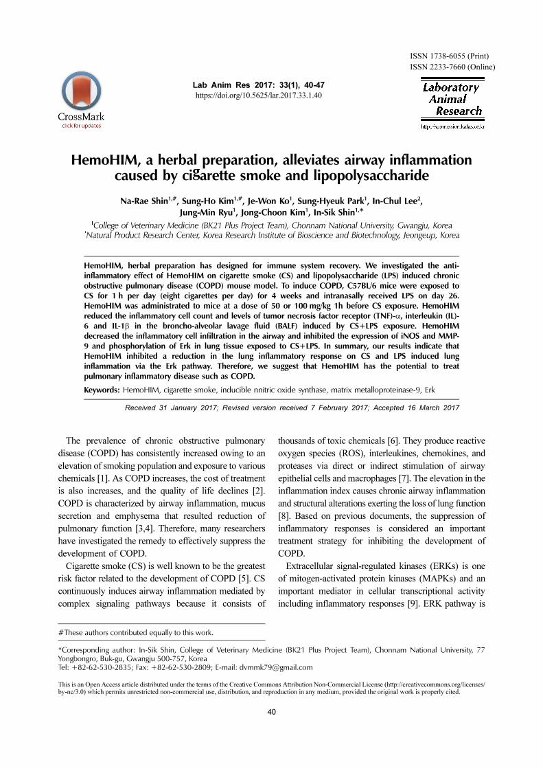
40
Lab Anim Res 2017: 33(1), 40-47
https://doi.org/10.5625/lar.2017.33.1.40
ISSN 1738-6055 (Print)
ISSN 2233-7660 (Online)
HemoHIM, a herbal preparation, alleviates airway inflammation caused by cigarette smoke and lipopolysaccharide
Na-Rae Shin1,#, Sung-Ho Kim1,#, Je-Won Ko1, Sung-Hyeuk Park1, In-Chul Lee2,Jung-Min Ryu1, Jong-Choon Kim1, In-Sik Shin1,*
1College of Veterinary Medicine (BK21 Plus Project Team), Chonnam National University, Gwangju, Korea2Natural Product Research Center, Korea Research Institute of Bioscience and Biotechnology, Jeongeup, Korea
HemoHIM, herbal preparation has designed for immune system recovery. We investigated the anti-inflammatory effect of HemoHIM on cigarette smoke (CS) and lipopolysaccharide (LPS) induced chronicobstructive pulmonary disease (COPD) mouse model. To induce COPD, C57BL/6 mice were exposed toCS for 1 h per day (eight cigarettes per day) for 4 weeks and intranasally received LPS on day 26.HemoHIM was administrated to mice at a dose of 50 or 100 mg/kg 1h before CS exposure. HemoHIMreduced the inflammatory cell count and levels of tumor necrosis factor receptor (TNF)-α, interleukin (IL)-6 and IL-1β in the broncho-alveolar lavage fluid (BALF) induced by CS+LPS exposure. HemoHIMdecreased the inflammatory cell infiltration in the airway and inhibited the expression of iNOS and MMP-9 and phosphorylation of Erk in lung tissue exposed to CS+LPS. In summary, our results indicate thatHemoHIM inhibited a reduction in the lung inflammatory response on CS and LPS induced lunginflammation via the Erk pathway. Therefore, we suggest that HemoHIM has the potential to treatpulmonary inflammatory disease such as COPD.
Keywords: HemoHIM, cigarette smoke, inducible nnitric oxide synthase, matrix metalloproteinase-9, Erk
Received 31 January 2017; Revised version received 7 February 2017; Accepted 16 March 2017
The prevalence of chronic obstructive pulmonary
disease (COPD) has consistently increased owing to an
elevation of smoking population and exposure to various
chemicals [1]. As COPD increases, the cost of treatment
is also increases, and the quality of life declines [2].
COPD is characterized by airway inflammation, mucus
secretion and emphysema that resulted reduction of
pulmonary function [3,4]. Therefore, many researchers
have investigated the remedy to effectively suppress the
development of COPD.
Cigarette smoke (CS) is well known to be the greatest
risk factor related to the development of COPD [5]. CS
continuously induces airway inflammation mediated by
complex signaling pathways because it consists of
thousands of toxic chemicals [6]. They produce reactive
oxygen species (ROS), interleukines, chemokines, and
proteases via direct or indirect stimulation of airway
epithelial cells and macrophages [7]. The elevation in the
inflammation index causes chronic airway inflammation
and structural alterations exerting the loss of lung function
[8]. Based on previous documents, the suppression of
inflammatory responses is considered an important
treatment strategy for inhibiting the development of
COPD.
Extracellular signal-regulated kinases (ERKs) is one
of mitogen-activated protein kinases (MAPKs) and an
important mediator in cellular transcriptional activity
including inflammatory responses [9]. ERK pathway is
#These authors contributed equally to this work.
*Corresponding author: In-Sik Shin, College of Veterinary Medicine (BK21 Plus Project Team), Chonnam National University, 77Yongbongro, Buk-gu, Gwangju 500-757, KoreaTel: +82-62-530-2835; Fax: +82-62-530-2809; E-mail: [email protected]
This is an Open Access article distributed under the terms of the Creative Commons Attribution Non-Commercial License (http://creativecommons.org/licenses/by-nc/3.0) which permits unrestricted non-commercial use, distribution, and reproduction in any medium, provided the original work is properly cited.
Protecive effects of HemoHIM, a herbal preparation in COPD model 41
Lab Anim Res | March, 2017 | Vol. 33, No. 1
activated by various stimuli and CS is a powerful
stimulus for ERK activation [10]. During the development
of COPD, ERK activation produced pro-inflammatory
cytokines, such as tumor necrosis factor (TNF)-α,
interleukin (IL)-1β and IL-6 and matrix metalloproteinases
(MMPs), which aggravates airway inflammation and
destroy normal alveolar structure [11-13].
HemoHIM, a herbal preparation is designed to recover
immune system and commercially used in South Korea.
It consists of three herb; Angelica Radix, Cnidium
Rhizoma and Paeonia Radix [14]. HemoHIM has been
reported to improve the immune system in patients
undergoing chemotherapy and have an anti-inflammatory
effect in carrageenan-induced paw edema, downregulate
Th1-like immune responses in fractionated γ-irradiated
mice, and have an anti-diabetic effect in a streptozotocin-
induced diabetic model [15-18]. However, there has not
been a study on the protective effects of HemoHIM on
lung inflammation induced by CS and LPS exposure.
Therefore, we examined the anti-inflammatory response
of HemoHIM in the lung using a CS and LPS-induced
model. To confirm the possible mechanism of HemoHIM,
we investigated the expression and production of lung
inflammatory mediators due to CS and LPS exposure.
Materials and Methods
Animals
Specific pathogen-free male C57BL/6N mice (20~25
g, six to eight weeks-old) were purchased from the
Samtako Co. (Osan, Korea). They were housed in
groups of nine under standard conditions (temperature
22±2oC, humidity 55±5%, 12-h-light/dark cycle) with
food and water. All experimental procedures were
approved by the Institutional Animal Care and Use
Committee of the Chonnam National University.
Induction of CS and LPS in C57BL/6 mice and drug
administration
The CS was generated from 3R4F research cigarette
(Kentuchy reference cigarette, University of Kentuchy,
USA), containing 11.0 mg of total particulate matter, 9.4
mg of tar, and 0.76 mg of nicotine per cigarette. Exposure
to CS (one puff/min, 35 mL puff volum over 2 seconds,
every 60 seconds, 8 cigarettes per day) was conducted
using cigarette smoke generator (Daehan Biolink, Republic
of Korea). The mice were exposed to CS for 1 h in a
chamber (50 cm×30 cm×30 cm) for 28 days. LPS were
intranasally instilled (10 μg dissolved in 50 μL distilled
water) under anesthesia on day 26. The HemoHIM was
obtained from Korea Institute of Oriental Medicine
(Daejeon, Republic of Korea). HemoHIM was administered
to mice at doses of 50 or 100 mg/kg by oral gavage 1 h
before CS exposure for 28 days. A positive control
group was administered roflumilast (Sigma-Aldrich, St,
Louis, MO, USA, 10 mg/kg) which is a PDE-4 inhibitor
and manufactured for treatment COPD.
Collection of bronchoalveolar lavage fluid (BALF)
Forty-eight hours after the last intranasal LPS
administration, the mice were sacrificed via an intra-
peritoneal injection of zoletil 50 (25 mg/kg; Virbac korea.
Co., Seoul, Korea), and a tracheostomy was performed
according to a previous study [19]. To obtain the
broncho-alveolar lavage fluid (BALF), ice-cold PBS
(0.7 mL) was infused into the lung and withdrawn via
tracheal cannulation. This process was repeated once
(total volume 1.4 mL). To determine the differential cell
counts, 100 μL of BALF was centrifuged onto slides
using a Cytospin (Hanil Science Industrial, Seoul,
Korea). The slides were dried, and the cells were fixed
and stained using Diff-Quik staining reagent (B4132-1A;
IMEB Inc., Deerfield, IL) according to the manufacturer’s
instructions. The supernatant obtained from the BALF
was stored at −70oC for biochemical analysis.
Measurement of pro-inflammatory mediator in BALF
The pro-inflammatory mediators in the BALF were
measured using ELISA kits (R&D System, Minneapolis,
MN, USA) according to the manufacturer’s protocols.
The plates were incubated for 10 min in the dark, and the
absorbance was measured at 450 nm using a microplate
reader (Bio-Rad, Hercules, CA, Laboratories).
Immunoblotting
The lung tissue was homogenized (1/10 w/v) using a
homogenizer in a Tissue Lysis/Extraction reagent (Sigma-
Aldrich, St, Louis, MO, USA) that contained a protease
inhibitor cocktail (Sigma-Aldrich). Protein concentrations
were determined using Bradford reagent (Bio-Rad).
Equal amounts of the total protein (30 μg) were resolved
by 10% SDS-polyacrylamide gel electrophoresis and
transferred to nitrocellulose membranes. The membranes
were incubated with blocking solution (5% skim milk)
followed by overnight incubation at 4oC with the
appropriate primary antibody. The following primary
42 Na-Rae Shin et al.
Lab Anim Res | March, 2017 | Vol. 33, No. 1
antibodies and dilutions were used: anti-β-actin (1:2000
dilution; Cell Signaling, Danvers, MA, USA), anti-
pERK (1:1000 dilution; Cell Signaling), anti-ERK
(1:1000 dilution; Cell Signaling) and anti-iNOS (1:1000
dilution; Santa Cruz Biotechnology, MA, USA). The
blots were washed three times with Tris-buffered saline
containing Tween 20 (TBST) and then incubated with a
1:10000 dilution of horseradish peroxidase (HRP)-
conjugated secondary antibody (Jackson Immuno Research,
West Grove, PA, USA) for 30 min at room temperature.
The blots were then washed three times with TBST and
then developed using an enhanced chemiluminescence
(ECL) kit (Thermo Fisher Scientific, Carlsbad, CA,
USA).
Gelatin zymography
SDSPAGE zymography was performed according to
previous study (Shin et al., 2014) to determine gelatinase
activitiy. Briefly, zymogram gels comprised of 10%
SDS-PAGE containing 1% gelatin were used as the
MMP substrate. The gels were washed in 2.5% Triton
X-100 for 1 h to remove SDS and then incubated at 37oC
for 16 h in developing buffer (1M Tris-HCl, pH 7.5 with
CaCl2). Thereafter, gels were stained with 25% methanol/
8% acetic acid containing Coomassie Brilliant Blue.
Gelatinase activity was visualized as white bands on a
blue background that represented the areas of proteolysis.
Lung tissue histopathology
The lung tissue was fixed in 4% (v/v) paraformaldehyde,
embedded in paraffin, sectioned at 4-μm thickness, and
stained with hematoxylin and eosin (H&E_ solution;
Sigma-Aldrich) to estimate inflammation.
Immunohistochemical slides were deparaffinized,
dehydrated, washed in PBS containing 0.05% tween 20
(PBS-T), and incubated for 20 min at room temperature
with goat serum to block nonspecific staining. The slides
were incubated for 2 h at room temperature with primary
mouse anti-mouse MMP-9 antibody (diluted 1:100,
Abcam). After incubation, they were washed three times,
incubated for 1 h at room temperature with a biotinylated
secondary antibody, and then incubated with an avidin-
biotin-peroxidase complex (Vector Laboratories, Burlin-
game, CA, USA) for 1 h at room temperature. Then, the
slides were washed with PBS-T and incubated with
diaminobenzidine (DAB, Abcam) for an additional
5 min.
Statistical analysis
The data are expressed as the means±standard
deviation (SD). Statistical significance was determined
using an analysis of variance (ANOVA) followed by a
multiple comparison test with Dunnet’s adjustment. P
values <0.05 were considered significant.
Results
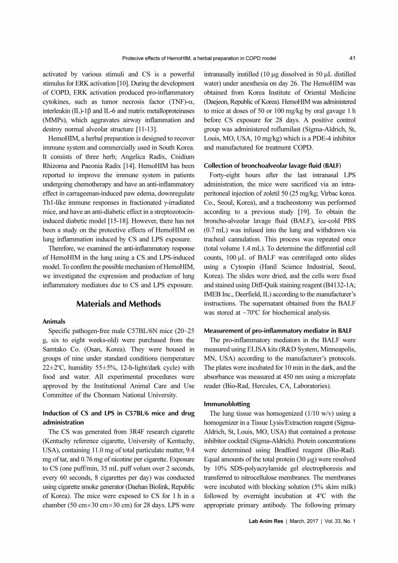
HemoHim reduce the number of inflammatory cells
in BALF induced by CS and LPS exposure
The number of inflammatory cells in BALF was
increased in CS and LPS exposed mice compared with
vehicle control mice. Specifically, CS and LPS exposure
markedly increased the number of neutrophils in BALF
compared to control. In HemoHim treated mice, however,
the number of neutrophils in BALF decreased in a dose-
dependent manner compared to CS and LPS exposed
mice (Figure 1).
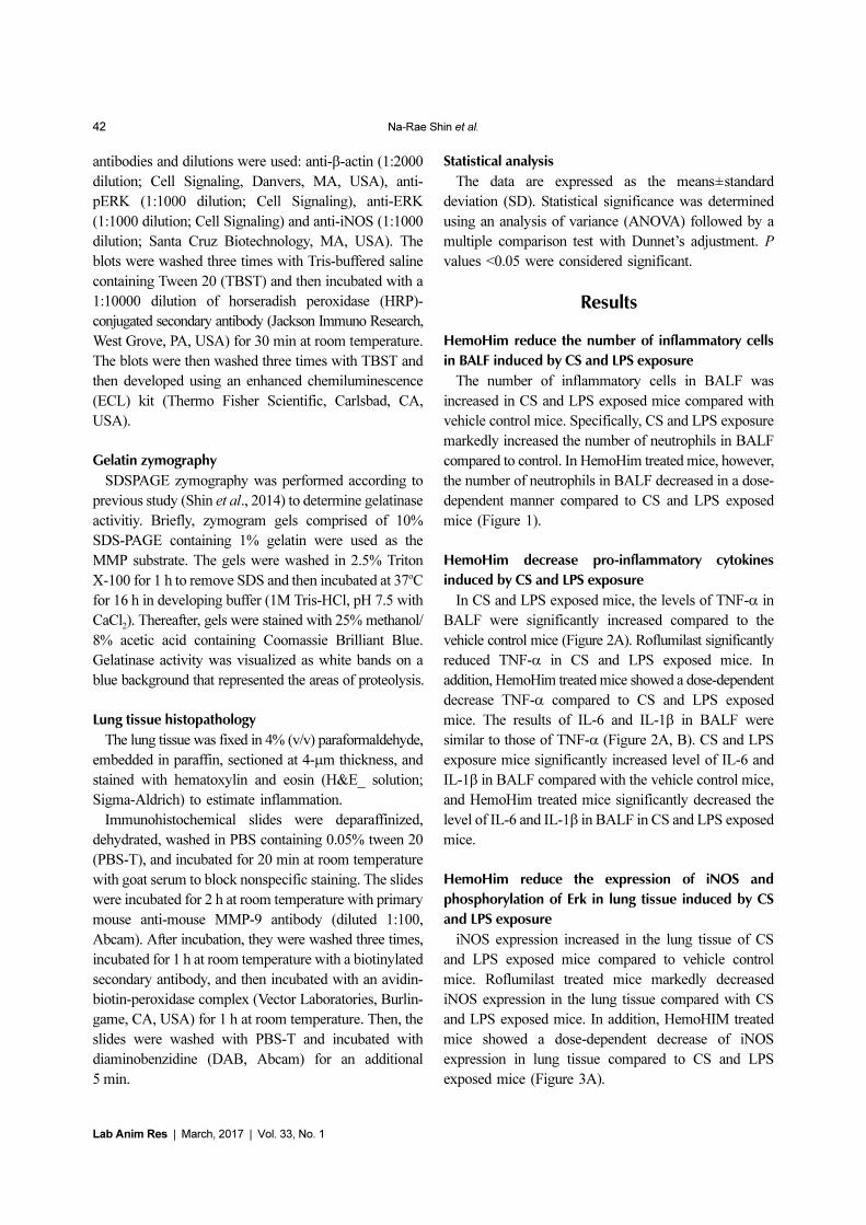
HemoHim decrease pro-inflammatory cytokines
induced by CS and LPS exposure
In CS and LPS exposed mice, the levels of TNF-α in
BALF were significantly increased compared to the
vehicle control mice (Figure 2A). Roflumilast significantly
reduced TNF-α in CS and LPS exposed mice. In
addition, HemoHim treated mice showed a dose-dependent
decrease TNF-α compared to CS and LPS exposed
mice. The results of IL-6 and IL-1β in BALF were
similar to those of TNF-α (Figure 2A, B). CS and LPS
exposure mice significantly increased level of IL-6 and
IL-1β in BALF compared with the vehicle control mice,
and HemoHim treated mice significantly decreased the
level of IL-6 and IL-1β in BALF in CS and LPS exposed
mice.
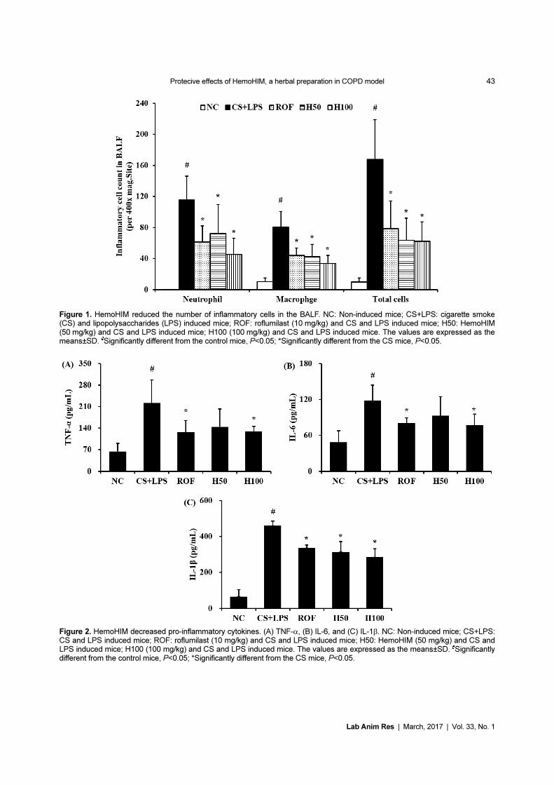
HemoHim reduce the expression of iNOS and
phosphorylation of Erk in lung tissue induced by CS
and LPS exposure
iNOS expression increased in the lung tissue of CS
and LPS exposed mice compared to vehicle control
mice. Roflumilast treated mice markedly decreased
iNOS expression in the lung tissue compared with CS
and LPS exposed mice. In addition, HemoHIM treated
mice showed a dose-dependent decrease of iNOS
expression in lung tissue compared to CS and LPS
exposed mice (Figure 3A).
Protecive effects of HemoHIM, a herbal preparation in COPD model 43
Lab Anim Res | March, 2017 | Vol. 33, No. 1
Figure 1. HemoHIM reduced the number of inflammatory cells in the BALF. NC: Non-induced mice; CS+LPS: cigarette smoke(CS) and lipopolysaccharides (LPS) induced mice; ROF: roflumilast (10 mg/kg) and CS and LPS induced mice; H50: HemoHIM(50 mg/kg) and CS and LPS induced mice; H100 (100 mg/kg) and CS and LPS induced mice. The values are expressed as themeans±SD. #Significantly different from the control mice, P<0.05; *Significantly different from the CS mice, P<0.05.
Figure 2. HemoHIM decreased pro-inflammatory cytokines. (A) TNF-α, (B) IL-6, and (C) IL-1β. NC: Non-induced mice; CS+LPS:CS and LPS induced mice; ROF: roflumilast (10 mg/kg) and CS and LPS induced mice; H50: HemoHIM (50 mg/kg) and CS andLPS induced mice; H100 (100 mg/kg) and CS and LPS induced mice. The values are expressed as the means±SD. #Significantlydifferent from the control mice, P<0.05; *Significantly different from the CS mice, P<0.05.
44 Na-Rae Shin et al.
Lab Anim Res | March, 2017 | Vol. 33, No. 1
In comparison to vehicle control mice, the phosphory-
lation of Erk was significantly increased in CS and LPS
exposed mice. HemoHIM markedly and dose-dependently
decreased phosphorylation of Erk in CS and LPS
exposed mice (Figure 3B).
HemoHim decrease inflammatory responses in lung
tissue induced by CS and LPS exposure
CS and LPS exposed mice exhibited extensive
inflammatory cell infiltration into the lung tissue (Figure
4). Inflammatory cells mainly accumulating in peribronchial
and alveolar lesions. In contrast, roflumilast treated mice
decreased inflammatory cell infiltration into lung tissue
induced by CS and LPS exposure. Similarly, inflammatory
cell infiltration was significantly reduced in a dose-
dependent manner in HemoHim treated mice compared
to CS and LPS exposed mice.
HemoHim reduce the expression and activity of
MMP-9 in lung tissue induced by CS and LPS exposure
MMP-9 expression in lung tissue was markedly
increased in CS and LPS exposed mice compared to
Figure 3. HemoHIM inhibited the iNOS and phosphorylation of ERK expression in lung tissue. (A) Expression of iNOS. (B)Phosphorylation of ERK. (C and D) Quantitative analysis of iNOS expression and phosphorylation of ERK expression. NC: Non-induced mice; CS+LPS: CS and LPS induced mice; ROF: roflumilast (10 mg/kg) and CS and LPS induced mice; H50: HemoHIM(50 mg/kg) and CS and LPS induced mice; H100 (100 mg/kg) and CS and LPS induced mice. The values are expressed as themeans±SD. #Significantly different from the control mice, P<0.05; *Significantly different from the CS mice, P<0.05.
Figure 4. HemoHIM reduced inflammatory cell infiltration induced by CS and LPS exposure. Hematoxylin and eosin (H&E) stainingshowed inflammatory infiltration in the peribronchial region and alveolar region. NC: Non-induced mice; CS+LPS: CS and LPSinduced mice; ROF: roflumilast (10 mg/kg) and CS and LPS induced mice; H50: HemoHIM (50 mg/kg) and CS and LPS inducedmice; H100 (100 mg/kg) and CS and LPS induced mice. The values are expressed as the means±SD. #Significantly different fromthe control mice, P<0.05; *Significantly different from the CS mice, P<0.05.
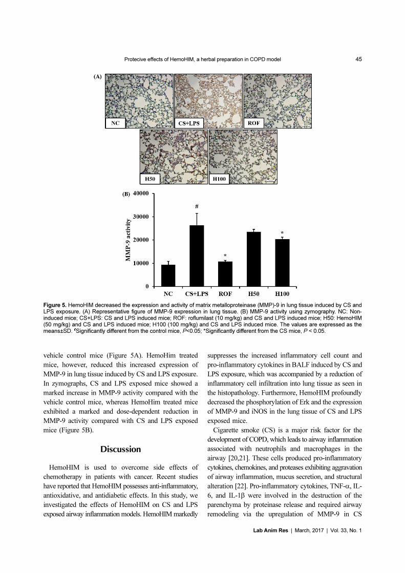
Protecive effects of HemoHIM, a herbal preparation in COPD model 45
Lab Anim Res | March, 2017 | Vol. 33, No. 1
vehicle control mice (Figure 5A). HemoHim treated
mice, however, reduced this increased expression of
MMP-9 in lung tissue induced by CS and LPS exposure.
In zymographs, CS and LPS exposed mice showed a
marked increase in MMP-9 activity compared with the
vehicle control mice, whereas HemoHim treated mice
exhibited a marked and dose-dependent reduction in
MMP-9 activity compared with CS and LPS exposed
mice (Figure 5B).
Discussion
HemoHIM is used to overcome side effects of
chemotherapy in patients with cancer. Recent studies
have reported that HemoHIM possesses anti-inflammatory,
antioxidative, and antidiabetic effects. In this study, we
investigated the effects of HemoHIM on CS and LPS
exposed airway inflammation models. HemoHIM markedly
suppresses the increased inflammatory cell count and
pro-inflammatory cytokines in BALF induced by CS and
LPS exposure, which was accompanied by a reduction of
inflammatory cell infiltration into lung tissue as seen in
the histopathology. Furthermore, HemoHIM profoundly
decreased the phosphorylation of Erk and the expression
of MMP-9 and iNOS in the lung tissue of CS and LPS
exposed mice.
Cigarette smoke (CS) is a major risk factor for the
development of COPD, which leads to airway inflammation
associated with neutrophils and macrophages in the
airway [20,21]. These cells produced pro-inflammatory
cytokines, chemokines, and proteases exhibiting aggravation
of airway inflammation, mucus secretion, and structural
alteration [22]. Pro-inflammatory cytokines, TNF-α, IL-
6, and IL-1β were involved in the destruction of the
parenchyma by proteinase release and required airway
remodeling via the upregulation of MMP-9 in CS
Figure 5. HemoHIM decreased the expression and activity of matrix metalloproteinase (MMP)-9 in lung tissue induced by CS andLPS exposure. (A) Representative figure of MMP-9 expression in lung tissue. (B) MMP-9 activity using zymography. NC: Non-induced mice; CS+LPS: CS and LPS induced mice; ROF: roflumilast (10 mg/kg) and CS and LPS induced mice; H50: HemoHIM(50 mg/kg) and CS and LPS induced mice; H100 (100 mg/kg) and CS and LPS induced mice. The values are expressed as themeans±SD. #Significantly different from the control mice, P<0.05; *Significantly different from the CS mice, P < 0.05.
46 Na-Rae Shin et al.
Lab Anim Res | March, 2017 | Vol. 33, No. 1
induced in vitro and in vivo models [23-25]. Therefore,
inhibition of pro-inflammatory cytokines is important
for attenuation of CS and LPS induced airway
inflammation.
In this study, CS and LPS exposed mice showed
marked increases in inflammatory cell counts, TNF-α,
IL-6, and IL-1β in BALF compared to the controls.
However, HemoHIM treated mice exhibited a significant
reduction in these pathophysiological factors in comparison
to CS and LPS exposed mice. In addition, these events
were accompanied by the reduction in histopathological
alteration of lung tissue. CS- and LPS-exposed mice
showed the extensive infiltration of inflammatory cells
into the lung tissue, whereas HemoHim-treated mice
exhibited a reduction in the histopathological alteration
induced by CS and LPS exposure. Based on these
results, HemoHIM may have an anti-inflammatory effect
on airway inflammation mediated by CS exposure.
ERK is a MAPK transcription factor and plays a key
role in the expression of various inflammatory genes
such as MMP-9 and iNOS [19,26]. Previous studies
have shown an increase in ERK with MMP-9 in CS and
LPS induced mice models and CS condensate-stimulated
cells [19]. CS stimulated the phosphorylation of Erk in
airway epithelial cells, macrophages, and neutrophils,
which eventually elevates the MMP-9 and iNOS
expression [19,27].
MMP-9 is involved in airway inflammatory responses
and the destruction of normal lung tissue via degradation
of collagen and gelatin. iNOS is associated with the
initiation and aggravation of airway inflammation via
the elevation of nitric oxide production in CS induced
airway inflammation [10,28]. This signaling was observed
in COPD clinical trials. Patients with COPD increased
the phosphorylation of Erk, MMP-9, and iNOS expression
in their sputum and lavage [28-31]. Our results show that
CS and LPS exposed mice increased phosphorylation of
ERK with elevated MMP-9 and iNOS expression in
their lung tissue compared to the controls. However,
HemoHim treated mice exhibited a marked reduction in
the phosphorylation of Erk with decreases in MMP-9
and iNOS expression in the lung tissue in comparison to
CS and LPS exposed mice. These results suggest that the
therapeutic effects of HemoHIM on CS and LPS
exposed airway inflammation are closely associated with
a reduction in MMP-9 and iNOS expression via the
suppression of Erk phosphorylation in CS and LPS
exposed lung tissue.
In conclusion, we evaluated the anti-inflammatory
effects of HemoHIM on airway inflammation induced
by CS and LPS exposure. HemoHIM significantly
reduced the inflammatory cells and pro-inflammatory
cytokines in BALF induced by CS and LPS exposure.
HemoHIM decreased the elevated expression of iNOS
and MMP-9 induced by CS and LPS exposure in lung
tissue. These effects may be linked to the inhibition of
ERK phosphorylation. Therefore, our study suggests that
HemoHIM has the potential to treat airway inflammatory
diseases, such as COPD induced by CS exposure.
Acknowledgments
This work was supported by the National Research
Foundation of Korea (NRF) grant funded by the
Ministry of Science, ICT & Future Planning (grant
number: NRF-016R1C1B2008818).
Conflict of interests The authors declare that there is
no financial conflict of interests to publish these results.
References
1. Tang W, Shen Z, Guo J, Sun S. Screening of long non-codingRNA and TUG1 inhibits proliferation with TGF- induction inpatients with COPD. Int J Chron Obstruct Pulmon Dis 2016; 11:2951-2964.
2. Korpershoek Y, Vervoort S, Nijssen L, Trappenburg J,Schuurmans MJ. Factors influencing exacerbation-related self-management in patients with COPD: a qualitative study. Int JChron Obstruct Pulmon Dis 2016; 11: 2977-2990.
3. Boehme SA, Franz-Bacon K, Ludka J, DiTirro DN, Ly TW,Bacon KB. MAP3K19 Is Overexpressed in COPD and Is aCentral Mediator of Cigarette Smoke-Induced PulmonaryInflammation and Lower Airway Destruction. PLoS One 2016;11(12): e0167169
4. Yang J, Yu HM, Zhou XD, Huang HP, Han Zh, Kolosov VP,Perelman JM. Cigarette smoke induces mucin hypersecretion andinflammatory response through the p66shc adaptor protein-mediated mechanism in human bronchial epithelial cells. MolImmunol 2016; 69: 86-98.
5. Lin K, Liu S, Shen Y, Li Q. Berberine attenuates cigarette smoke-induced acute lung inflammation. Inflammation 2013; 36(5):1079-1086.
6. Lee J, Taneja V, Vassallo R. Cigarette smoking and inflammation:cellular and molecular mechanisms. J Dent Res 2012; 91(2): 142-149.
7. Xue WH, Shi XQ, Liang SH, Zhou L, Liu KF, Zhao J. EmodinAttenuates Cigarette Smoke Induced Lung Injury in a MouseModel via Suppression of Reactive Oxygen Species Production. JBiochem Mol Toxicol 2015; 29(11): 526-532.
8. Ge LT, Liu YN, Lin XX, Shen HJ, Jia YL, Dong XW, Sun Y, XieQM. Inhalation of ambroxol inhibits cigarette smoke-inducedacute lung injury in a mouse model by inhibiting the Erk pathway.Int Immunopharmacol 2016; 33: 90-98.
9. Schmitt KR, Diestel A, Lehnardt S, Schwartlander R, Lange PE,Berger F, Ullrich O, Abdul-Khaliq H. Hypothermia suppressesinflammation via ERK signaling pathway in stimulated microglial
Protecive effects of HemoHIM, a herbal preparation in COPD model 47
Lab Anim Res | March, 2017 | Vol. 33, No. 1
cells. J Neuroimmunol 2007; 189(1-2): 7-16.10. Shin IS, Ahn KS, Shin NR, Jeon CM, Kwon OK, Chin YW, Lee
K, Oh SR. Homoegonol attenuates the asthmatic responsesinduced by ovalbumin challenge. Arch Pharm Res 2014; 37(9):1201-1210.
11. Cho A, Graves J, Reidy MA. Mitogen-activated protein kinasesmediate matrix metalloproteinase-9 expression in vascular smoothmuscle cells. Arterioscler Thromb Vasc Biol 2000; 20(12): 2527-2532.
12. Lee IT, Yang CM. Inflammatory signalings involved in airwayand pulmonary diseases. Mediators Inflamm 2013; 2013: 791231.
13. Doz E, Noulin N, Boichot E, Guénon I, Fick L, Le Bert M,Lagente V, Ryffel B, Schnyder B, Quesniaux VF, Couillin I.Cigarette smoke-induced pulmonary inflammation is TLR4/MyD88 and IL-1R1/MyD88 signaling dependent. J Immunol2008; 180(2): 1169-1178.
14. Park HR, Jo SK, Jung U, Yee ST. Restoration of the immunefunctions in aged mice by supplementation with a new herbalcomposition, HemoHIM. Phytother Res 2008; 22(1): 36-42.
15. Jo SK, Lee HJ, Kim SR, Kim JC, Bae CS, Jung U, Park HR, JangJS, Kim SH. Antiinflammatory activity of an herbal preparation(HemoHIM) in rats. Phytother Res 2007: 21(7): 625-628.
16. Park HR, Jo SK, Choi NH, Jung U. HemoHIM ameliorates thepersistent down-regulation of Th1-like immune responses infractionated γ-irradiated mice by modulating the IL-12p70-STAT4signaling pathway. Radiat Res 2012; 177(5): 676-684.
17. Kim JJ, Cho HW, Park HR, Jung U, Jo SK, Yee ST. Preventativeeffect of an herbal preparation (HemoHIM) on development ofairway inflammation in mice via modulation of Th1/2 cellsdifferentiation. PLoS One 2013; 8(7): e68552.
18. Kim JJ, Choi J, Lee MK, Kang KY, Paik MJ, Jo SK, Jung U, ParkHR, Yee ST. Immunomodulatory and Antidiabetic Effects of aNew Herbal Preparation (HemoHIM) on Streptozotocin-InducedDiabetic Mice. Evid Based Complement Alternat Med 2014;2014: 461685.
19. Shin IS, Shin NR, Park JW, Jeon CM, Hong JM, Kwon OK, KimJS, Lee IC, Kim JC, Oh SR, Ahn KS. Melatonin attenuatesneutrophil inflammation and mucus secretion in cigarette smoke-induced chronic obstructive pulmonary diseases via the suppressionof Erk-Sp1 signaling. J Pineal Res 2015; 58(1): 50-60.
20. Lee H, Park JR, Kim EJ, Kim WJ, Hong SH, Park SM, Yang SR.Cigarette smoke-mediated oxidative stress induces apoptosis viathe MAPKs/STAT1 pathway in mouse lung fibroblasts. ToxicolLett 2016; 240(1): 140-148.
21. Lee KH, Lee CH, Jeong J, Jang AH, Yoo CG. Neutrophil Elastase
Differentially Regulates Interleukin 8 (IL-8) and Vascular EndothelialGrowth Factor (VEGF) Production by Cigarette Smoke Extract. JBiol Chem 2015; 290(47): 28438-28445.
22. Gernez Y, Tirouvanziam R, Chanez P. Neutrophils in chronicinflammatory airway diseases: can we target them and how? EurRespir J 2010; 35(3): 467-469.
23. Jeong SH, Park JH, Kim JN, Park YH, Shin SY, Lee YH, KyeYC, Son SW. Up-regulation of TNF-alpha secretion by cigarettesmoke is mediated by Egr-1 in HaCaT human keratinocytes. ExpDermatol 2010; 19(8): e206-212.
24. Wang H, Yang T, Shen Y, Wan C, Li X, Li D, Liu Y, Wang T, XuD, Wen F, Ying B. Ghrelin Inhibits Interleukin-6 ProductionInduced by Cigarette Smoke Extract in the Bronchial EpithelialCell Via NF-κB Pathway. Inflammation 2016; 39(1): 190-198.
25. Markovics JA, Araya J, Cambier S, Somanath S, Gline S, JablonsD, Hill A, Wolters PJ, Nishimura SL. Interleukin-1beta inducesincreased transcriptional activation of the transforming growthfactor-beta-activating integrin subunit beta8 through alteringchromatin architecture. J Biol Chem 2011; 286(42): 36864-36874.
26. Cha SM, Cha JD, Jang EJ, Kim GU, Lee KY. SophoraflavanoneG prevents Streptococcus mutans surface antigen I/II-inducedproduction of NO and PGE2 by inhibiting MAPK-mediatedpathways in RAW 264.7 macrophages. Arch Oral Biol 2016; 68:97-104.
27. Grzela K, Litwiniuk M, Zagorska W, Grzela T. AirwayRemodeling in Chronic Obstructive Pulmonary Disease andAsthma: the Role of Matrix Metalloproteinase-9. Arch ImmunolTher Exp (Warsz) 2016; 64(1): 47-55.
28. Jiang WT, Liu XS, Xu YJ, Ni W, Chen SX. Expression of NitricOxide Synthase Isoenzyme in Lung Tissue of Smokers with andwithout Chronic Obstructive Pulmonary Disease. Chin Med J(Engl) 2015; 128(12): 1584-1589.
29. Mercer PF, Shute JK, Bhowmik A, Donaldson GC, Wedzicha JA,Warner JA. MMP-9, TIMP-1 and inflammatory cells in sputumfrom COPD patients during exacerbation. Respir Res 2005; 6:151.
30. Roh GS, Yi CO, Cho YJ, Jeon BT, Nizamudtinova IT, Kim HJ,Kim JH, Oh YM, Huh JW, Lee JH, Hwang YS, Lee SD, Lee JD.Anti-inflammatory effects of celecoxib in rat lungs with smoke-induced emphysema. Am J Physiol Lung Cell Mol Physiol 2010;299(2): L184-191.
31. Hesslinger C, Strub A, Boer R, Ulrich WR, Lehner MD, Braun C.Inhibition of inducible nitric oxide synthase in respiratorydiseases. Biochem Soc Trans 2009; 37(Pt 4): 886-891.