Embed Size (px)
Citation preview
Zhang et al. BMC Cardiovasc Disord (2021) 21:207 https://doi.org/10.1186/s12872-021-02012-7
RESEARCH ARTICLE
Circ_0004104 knockdown alleviates oxidized low-density lipoprotein-induced dysfunction in vascular endothelial cells through targeting miR-328-3p/TRIM14 axis in atherosclerosisChi Zhang, Liyue Wang and Ying Shen*
Abstract
Background: Circular RNAs have shown important regulatory roles in cardiovascular diseases, containing athero-sclerosis (AS). We intended to explore the role of circ_0004104 in AS using oxidized low-density lipoprotein (ox-LDL)-induced vascular endothelial cells and its associated mechanism.
Methods: Real-time quantitative polymerase chain reaction and Western blot assay were conducted to analyze RNA levels and protein levels, respectively. Cell viability, apoptosis, angiogenic ability and inflammatory response were assessed by 3-(4,5-Dimethylthiazol-2-yl)-2,5-Diphenyltetrazolium Bromide (MTT) assay, flow cytometry, capillary-like network formation assay and enzyme-linked immunosorbent assay, respectively. Cell oxidative stress was assessed using commercial kits. Dual-luciferase reporter assay, RNA immunoprecipitation assay and RNA-pull down assay were performed to verify the intermolecular interaction.
Results: ox-LDL exposure up-regulated the level of circ_0004104 in HUVECs. ox-LDL exposure suppressed cell viability and angiogenic ability whereas promoted the apoptosis, inflammation and oxidative stress of HUVECs partly through up-regulating circ_0004104. MicroRNA-328-3p (miR-328-3p) was confirmed as a target of circ_0004104. MiR-328-3p interference largely reversed circ_0004104 silencing-mediated effects in HUVECs upon ox-LDL exposure. MiR-328-3p interacted with the 3′ untranslated region of tripartite motif 14, and circ_0004104 positively regulated TRIM14 expression by sponging miR-328-3p. TRIM14 overexpression largely overturned miR-328-3p accumulation-induced influences in HUVECs upon ox-LDL exposure.
Conclusion: Circ_0004104 knockdown attenuated ox-LDL-induced dysfunction in HUVECs via miR-328-3p-mediated regulation of TRIM14.
Keywords: Atherosclerosis, Oxidized low-density lipoprotein, Circ_0004104, MiR-328-3p, TRIM14
© The Author(s) 2021. Open Access This article is licensed under a Creative Commons Attribution 4.0 International License, which permits use, sharing, adaptation, distribution and reproduction in any medium or format, as long as you give appropriate credit to the original author(s) and the source, provide a link to the Creative Commons licence, and indicate if changes were made. The images or other third party material in this article are included in the article’s Creative Commons licence, unless indicated otherwise in a credit line to the material. If material is not included in the article’s Creative Commons licence and your intended use is not permitted by statutory regulation or exceeds the permitted use, you will need to obtain permission directly from the copyright holder. To view a copy of this licence, visit http:// creat iveco mmons. org/ licen ses/ by/4. 0/. The Creative Commons Public Domain Dedication waiver (http:// creat iveco mmons. org/ publi cdoma in/ zero/1. 0/) applies to the data made available in this article, unless otherwise stated in a credit line to the data.
BackgroundMany types of cells are involved in the pathogenetic pro-cess of atherosclerosis (AS), containing vascular endothe-lial cells and smooth muscle cells [1]. Endothelial injury
theory is one of the mainstream theories of atheroscle-rosis pathogenesis, and it considers artery atheromatous plaque as the product of endothelial injury [2]. Oxidized low-density lipoprotein (ox-LDL) is a crucial risk fac-tor that is responsible for AS initiation [3]. Therefore, we established AS cell model using ox-LDL-treated HUVECs to find the pivotal molecules that were involved in AS pathogenesis in vitro.
Open Access
*Correspondence: [email protected] of Cardiology, The Puren Hospital, No. 218, Changqing First Road, Jianghan District, Wuhan 430081, Hubei, China
Page 2 of 12Zhang et al. BMC Cardiovasc Disord (2021) 21:207
Circular RNAs (circRNAs) are endogenous RNAs without 5′ or 3′ polarity [4, 5]. CircRNAs have been demonstrated to modulate the pathological pro-cess of human malignancies [6, 7]. Also, accumu-lating studies have identified the important roles of circRNAs in AS. For instance, Li et al. found that circ_0003575 knockdown accelerated the proliferation ability and tube formation capacity of HUVECs [8]. Liu et al. demonstrated that circ_0003204 suppressed proliferation and angiogenesis of ox-LDL-treated HUVECs [9]. Circ_0004104 was reported to be nota-bly up-regulated in patients diagnosed with coronary artery disease [10]. However, the role and mechanism of circ_0004104 in AS progression remain largely unknown.
MicroRNAs (miRNAs) reversely modulate gene expression by interacting with the 3′ untranslated region (3′UTR) of messenger RNAs (mRNAs), causing translational repression or degradation of mRNAs [11, 12]. Dysregulation of miRNAs was associated with AS progression [13]. We concentrated on the biological significance of miR-328-3p in AS progression, which was predicted to be a candidate downstream miRNA of circ_0004104 by bioinformatic database. Guo et al. claimed that miR-328-3p attenuated ox-LDL-mediated dysfunction in HUVECs [14]. Nevertheless, the work-ing mechanism of miR-328-3p in AS still needs to be further clarified.
Tripartite motif 14 (TRIM14) is one of the members of TRIM family [15]. TRIM14 exerted an oncogenic role in many human malignancies [16–19]. In addition, Huang et al. demonstrated that TRIM14 could acceler-ate the activation of endothelium through activating NF-κB signaling [20]. TRIM14 was predicted by bio-informatic database to be a downstream gene of miR-328-3p, and the working mechanism of TRIM14 in AS progression was investigated.
We initially explored the role of circ_0004104 in AS cell model. Subsequently, the working mechanism of circ_0004104 was explored through bioinformatic anal-ysis and rescue experiments.
MethodsCell lineHuman umbilical vein endothelial cells (HUVECs) acquired from Chinese Academy of Medical Sciences, Shanghai institute Cell Bank (Shanghai, China) were cultivated in Dulbecco’s modified Eagle’s medium (DMEM; Gibco, Carlsbad, CA, USA) plus 10% fetal bovine serum (FBS, Hyclone, Carlsbad, CA, USA) and 1% antibiotics (Gibco) under 37℃ humidified atmos-phere with 5% CO2.
AS cell modelHUVECs were exposed to 100 μg/mL ox-LDL (Solarbio, Beijing, China) for 24 h to establish AS cell model as pre-viously reported [8, 14].
Real‑time quantitative polymerase chain reaction (RT‑qPCR)RNA samples were isolated using Trizol reagent (Invitro-gen, Carlsbad, CA, USA). Complementary DNA (cDNA) was synthesized using the miScript Reverse Transcrip-tion kit (for miRNA; Qiagen, Valencia, CA, USA) and RevertAid First Strand cDNA Synthesis Kit (for circRNA and mRNA; Invitrogen). cDNA was amplified via the SYBR™ Green PCR Master Mix (Invitrogen). The primers purchased from Sangon Biotech (Shanghai, China) were shown in Table 1. Relative abundance of circ_0004104, SPARC and TRIM14 was analyzed using the 2−ΔΔCt method with glyceraldehyde-3-phosphate dehydrogenase (GAPDH) as reference, while the fold change of miR-328-3p was calculated using the 2−ΔΔCt method with U6 as reference.
Cyclization validationRNA samples (2 μg) were incubated with 3 U/μg RNase R (Epicentre Technologies, Madison, WI, USA), and RNA levels were determined by RT-qPCR.
Actinomycin D treatmentTranscription inhibitor Actinomycin D (2 mg/mL; Sigma, St. Louis, MO, USA) was added to the culture medium, and RNA levels were examined by RT-qPCR at specific time points.
Table 1 Specific primers in RT-qPCR assay
Gene Direction (5′–3′) Sequence
circ_0004104 Forward 5′-AGA CCT GTG ACC TGG ACA ATG-3′
Reverse 5′-GTG CAC TTT GTG GCA AAG AA-3′
SPARC Forward 5′-GGT ATC TGT GGG AGC TAA TC-3′
Reverse 5′-CTG GTG GGG TCC TGG CAC AC-3′
miR-328-3p Forward 5′-CCT CTC TGC CCT TCCG-3′
Reverse 5′-GAA CAT GTC TGC GTA TCT C-3′
TRIM14 Forward 5′-GAG GTC GGA GCT TGT CGA G-3′
Reverse 5′-TTC TTG GCT GAG TTT CTG CAC-3′
U6 Forward 5′-CTC GCT TCG GCA GCACA-3′
Reverse 5′-AAC GCT TCA CGA ATT TGC GT-3′
GAPDH Forward 5′-AAG AAG GTG GTG AAG CAG GC-3′
Reverse 5′-GTC AAA GGT GGA GGA GTG GG-3′
Page 3 of 12Zhang et al. BMC Cardiovasc Disord (2021) 21:207
Oligonucleotides or plasmids transfectionEctopic expression plasmid of circ_0004104 (circ_0004104), pLCDH-cir empty vector (vector), small interfering RNA against circ_0004104 (si-circ_0004104), negative control of siRNA (si-NC), TRIM14 overexpression plasmid (TRIM14) and empty vector (pcDNA) were purchased from Sangon Biotech, and mimics of miR-328-3p (miR-328-3p), miR-NC, inhibitor of miR-328-3p (anti-miR-328-3p) and anti-miR-NC were acquired from Genepharma (Shanghai, China). All oligonucleotides or plasmids were trans-fected into HUVECs with Lipofectamine 3000 reagent (Invitrogen).
3‑(4,5‑Dimethylthiazol‑2‑yl)‑2,5‑Diphenyltetrazolium Bromide (MTT) assayAt specific time points, HUVECs were incubated with MTT reagent (Sigma) for 4 h. Afterwards, a total of 200 μL dimethyl sulfoxide (DMSO; Sigma) was added to dissolve the formazan products after discarding cell supernatant. The absorbance (490 nm) was determined by the microplate reader (Bio-Rad, Hercules, CA, USA).
Flow cytometryHUVECs were simultaneously stained with Annexin V-fluorescein isothiocyanate (Annexin V-FITC) and propidium iodide (PI) of the Cell Apoptosis Detection Kit (Qiagen). The apoptotic percentage of HUVECs was evaluated by the flow cytometer (BD Biosciences, San Jose, CA, USA).
Angiogenic capacity analysis via capillary‑like network formation assayHUVECs were plated onto Matrigel (BD Biosciences)-pre-coated 96-well cell culture plates (3 × 10⁴ cells/well). After culturing for 48 h, the average number of branches of each node was analyzed.
Western blot assayHUVECs were disrupted using whole cell lysis buffer (Beyotime, Shanghai, China). Protein samples (35 μg) were loaded onto sodium dodecyl sulfate–polyacryla-mide gel electrophoresis (SDS-PAGE) and transferred onto polyvinylidene difluoride (PVDF) membrane (Millipore, Billerica, MA, USA). After sealing with 5% bovine serum albumin (BSA; Sangon Biotech), immunoblot assay was applied through incubating the membrane with the diluted primary antibodies and the horse radish peroxidase (HRP) conjugated sec-ondary antibody (Abcam). Immuno-reactive signals were determined by the enhanced chemiluminescent (ECL) chromogenic substrate (Beyotime). The primary
antibodies contained anti-Cleaved-caspase 3 (anti-Cleaved-casp3, ab32042, Abcam, Cambridge, MA, USA), anti-vascular endothelial growth factor A (anti-VEGFA, ab52917, Abcam), anti-TRIM14 (SAB1410027, Sigma) and anti-GAPDH (ab8245, Abcam).
Enzyme‑linked immunosorbent assay (ELISA)The culture supernatant of HUVECs was collected to assess the release of tumor necrosis factor α (TNF-α) and interleukin 1β (IL-1β) using commercial Human TNF-α/IL-1β Quantikine ELISA Kit (R&D Systems, Minneapolis, MN, USA).
Determination of cell oxidative stressCell oxidative stress was analyzed through measuring the production of superoxide dismutase (SOD) and malon-dialdehyde (MDA) using their corresponding commercial kits (Jiancheng Biotech, Nanjing, China).
Bioinformatic analysisStarBase database (http:// starb ase. sysu. edu. cn) was uti-lized to predict circ_0004104-miRNAs interactions and miR-328-3p-mRNAs interactions.
Dual‑luciferase reporter assayThe fragment of circ_0004104 or the 3′UTR fragment of TRIM14, including the miR-328-3p-binding sequence, was inserted into psiCHECK2 luciferase plasmid (Pro-mega, Madison, WI, USA) to generate circ_0004104 wt and TRIM14 3′UTR wt. Meanwhile, mutated counter-parts were constructed to generate circ_0004104 mut and TRIM14 3′UTR mut. HUVECs were seeded onto 12-well plates and co-transfected with luciferase plasmids and miR-NC or miR-328-3p. After 48-h transfection, the relative luciferase intensities were determined using the Dual-Luciferase Reporter Assay Kit (Promega).
RNA immunoprecipitation (RIP) assayRIP experiment was employed to confirm the binding relation between circ_0004104 and miR-328-3p with Magna RIP™ RNA-Binding Protein Immunoprecipita-tion Kit (Millipore). Cell extracts were prepared using RIP lysis buffer, and anti-Argonaute2 (anti-Ago2; Mil-lipore) or anti-Immunoglobulin G (anti-IgG; Millipore)-pre-coated magnetic beads were incubated with cell lysates. The levels of enriched RNAs were measured by RT-qPCR.
RNA‑pull down assayCell lysates (2 μg) were incubated with 100 pmol Bio-miR-NC, Bio-miR-328-3p-mut or Bio-miR-328-3p-wt. The reaction mixture was then incubated with 100 μL
Page 4 of 12Zhang et al. BMC Cardiovasc Disord (2021) 21:207
agarose beads (Millipore) for 1 h. The retrieved RNAs were measured by RT-qPCR.
Statistical analysisAll experiments were repeated for three times. Statisti-cal analysis was carried out using GraphPad Prism 7.0 software (GraphPad, La Jolla, CA, USA). Data were rep-resented as mean ± standard deviation (SD). The differ-ences were analyzed by Student’s t-test (two groups) or one-way analysis of variance (ANOVA) (more than two groups). Differences were identified as statistically signifi-cant with the P value of less than 0.05.
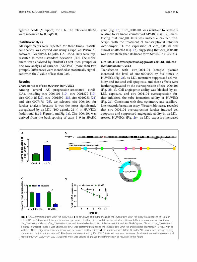
ResultsCharacteristics of circ_0004104 in HUVECsAmong several AS progression-associated circR-NAs, including circ_0004104 [10], circ_0001879 [10], circ_0001445 [22], circ_0001599 [23], circ_0010283 [24] and circ_0007478 [25], we selected circ_0004104 for further analysis because it was the most significantly upregulated by ox-LDL (100 μg/mL, 24 h) in HUVECs (Additional file 1: Figure 1 and Fig. 1a). Circ_0004104 was derived from the back-splicing of exon 6–9 in SPARC
gene (Fig. 1b). Circ_0004104 was resistant to RNase R relative to its linear counterpart SPARC (Fig. 1c), mani-festing that circ_0004104 was indeed a circular tran-script. With the treatment of transcriptional inhibitor Actinomycin D, the expression of circ_0004104 was almost unaffected (Fig. 1d), suggesting that circ_0004104 was more stable than its linear form SPARC in HUVECs.
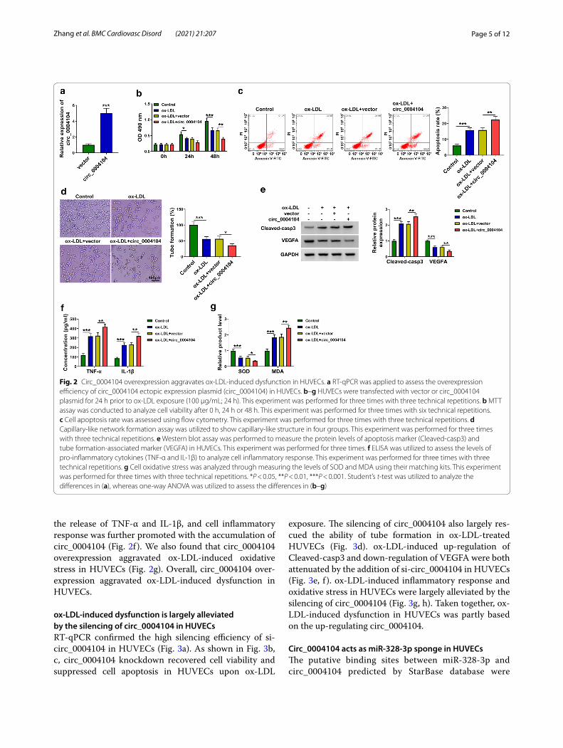
Circ_0004104 overexpression aggravates ox‑LDL‑induced dysfunction in HUVECsTransfection with circ_0004104 ectopic plasmid increased the level of circ_0004104 by five times in HUVECs (Fig. 2a). ox-LDL treatment suppressed cell via-bility and induced cell apoptosis, and these effects were further aggravated by the overexpression of circ_0004104 (Fig. 2b, c). Cell angiogenic ability was blocked by ox-LDL exposure, and circ_0004104 overexpression fur-ther inhibited the tube formation ability of HUVECs (Fig. 2d). Consistent with flow cytometry and capillary-like network formation assay, Western blot assay revealed that circ_0004104 overexpression further induced cell apoptosis and suppressed angiogenic ability in ox-LDL-treated HUVECs (Fig. 2e). ox-LDL exposure increased
Fig. 1 Characteristics of circ_0004104 in HUVECs. a RT-qPCR was applied to measure the level of circ_0004104 in HUVECs exposed to 100 μg/mL ox-LDL for 24 h or not. This experiment was performed for three times with three technical repetitions. b The chromosomal localization of circ_0004104 was shown. Circ_0004104 was derived from the back-splicing of the exon 6, 7, 8 and 9 in SPARC gene. c To test if circ_0004104 was a circular transcript, RNase R was utilized. RT-qPCR was performed to analyze the levels of circ_0004104 and its linear counterpart (SPARC) with or without RNase R digestion. This experiment was performed for three times. d The stability of circ_0004104 and SPARC was tested through adding transcription inhibitor Actinomycin D. RNA levels were examined by RT-qPCR. This experiment was performed for three times with three technical repetitions. **P < 0.01, ***P < 0.001. Student’s t-test was utilized to analyze the differences in all results of in this figure
Page 5 of 12Zhang et al. BMC Cardiovasc Disord (2021) 21:207
the release of TNF-α and IL-1β, and cell inflammatory response was further promoted with the accumulation of circ_0004104 (Fig. 2f ). We also found that circ_0004104 overexpression aggravated ox-LDL-induced oxidative stress in HUVECs (Fig. 2g). Overall, circ_0004104 over-expression aggravated ox-LDL-induced dysfunction in HUVECs.
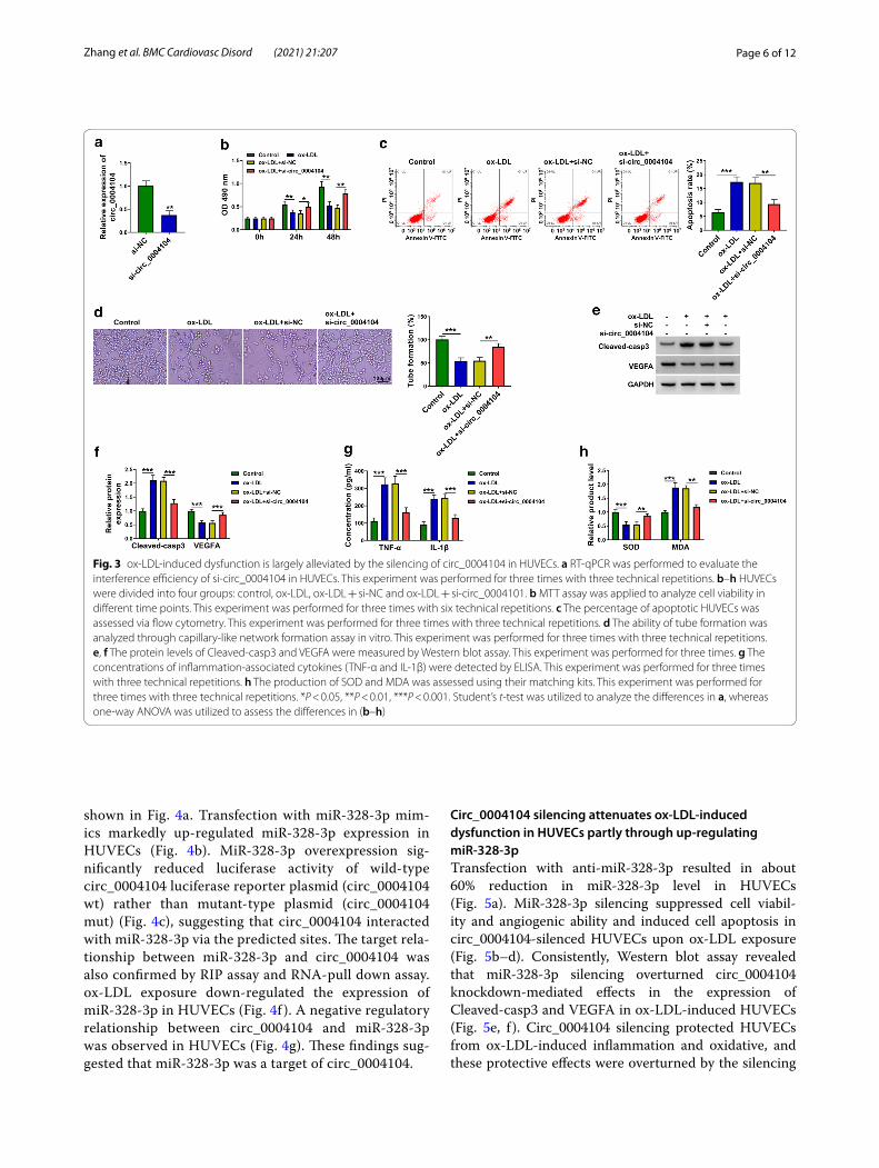
ox‑LDL‑induced dysfunction is largely alleviated by the silencing of circ_0004104 in HUVECsRT-qPCR confirmed the high silencing efficiency of si-circ_0004104 in HUVECs (Fig. 3a). As shown in Fig. 3b, c, circ_0004104 knockdown recovered cell viability and suppressed cell apoptosis in HUVECs upon ox-LDL
exposure. The silencing of circ_0004104 also largely res-cued the ability of tube formation in ox-LDL-treated HUVECs (Fig. 3d). ox-LDL-induced up-regulation of Cleaved-casp3 and down-regulation of VEGFA were both attenuated by the addition of si-circ_0004104 in HUVECs (Fig. 3e, f ). ox-LDL-induced inflammatory response and oxidative stress in HUVECs were largely alleviated by the silencing of circ_0004104 (Fig. 3g, h). Taken together, ox-LDL-induced dysfunction in HUVECs was partly based on the up-regulating circ_0004104.
Circ_0004104 acts as miR‑328‑3p sponge in HUVECsThe putative binding sites between miR-328-3p and circ_0004104 predicted by StarBase database were
Fig. 2 Circ_0004104 overexpression aggravates ox-LDL-induced dysfunction in HUVECs. a RT-qPCR was applied to assess the overexpression efficiency of circ_0004104 ectopic expression plasmid (circ_0004104) in HUVECs. b–g HUVECs were transfected with vector or circ_0004104 plasmid for 24 h prior to ox-LDL exposure (100 μg/mL; 24 h). This experiment was performed for three times with three technical repetitions. b MTT assay was conducted to analyze cell viability after 0 h, 24 h or 48 h. This experiment was performed for three times with six technical repetitions. c Cell apoptosis rate was assessed using flow cytometry. This experiment was performed for three times with three technical repetitions. d Capillary-like network formation assay was utilized to show capillary-like structure in four groups. This experiment was performed for three times with three technical repetitions. e Western blot assay was performed to measure the protein levels of apoptosis marker (Cleaved-casp3) and tube formation-associated marker (VEGFA) in HUVECs. This experiment was performed for three times. f ELISA was utilized to assess the levels of pro-inflammatory cytokines (TNF-α and IL-1β) to analyze cell inflammatory response. This experiment was performed for three times with three technical repetitions. g Cell oxidative stress was analyzed through measuring the levels of SOD and MDA using their matching kits. This experiment was performed for three times with three technical repetitions. *P < 0.05, **P < 0.01, ***P < 0.001. Student’s t-test was utilized to analyze the differences in (a), whereas one-way ANOVA was utilized to assess the differences in (b–g)
Page 6 of 12Zhang et al. BMC Cardiovasc Disord (2021) 21:207
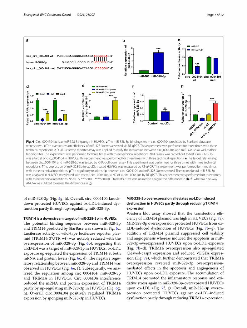
shown in Fig. 4a. Transfection with miR-328-3p mim-ics markedly up-regulated miR-328-3p expression in HUVECs (Fig. 4b). MiR-328-3p overexpression sig-nificantly reduced luciferase activity of wild-type circ_0004104 luciferase reporter plasmid (circ_0004104 wt) rather than mutant-type plasmid (circ_0004104 mut) (Fig. 4c), suggesting that circ_0004104 interacted with miR-328-3p via the predicted sites. The target rela-tionship between miR-328-3p and circ_0004104 was also confirmed by RIP assay and RNA-pull down assay. ox-LDL exposure down-regulated the expression of miR-328-3p in HUVECs (Fig. 4f ). A negative regulatory relationship between circ_0004104 and miR-328-3p was observed in HUVECs (Fig. 4g). These findings sug-gested that miR-328-3p was a target of circ_0004104.
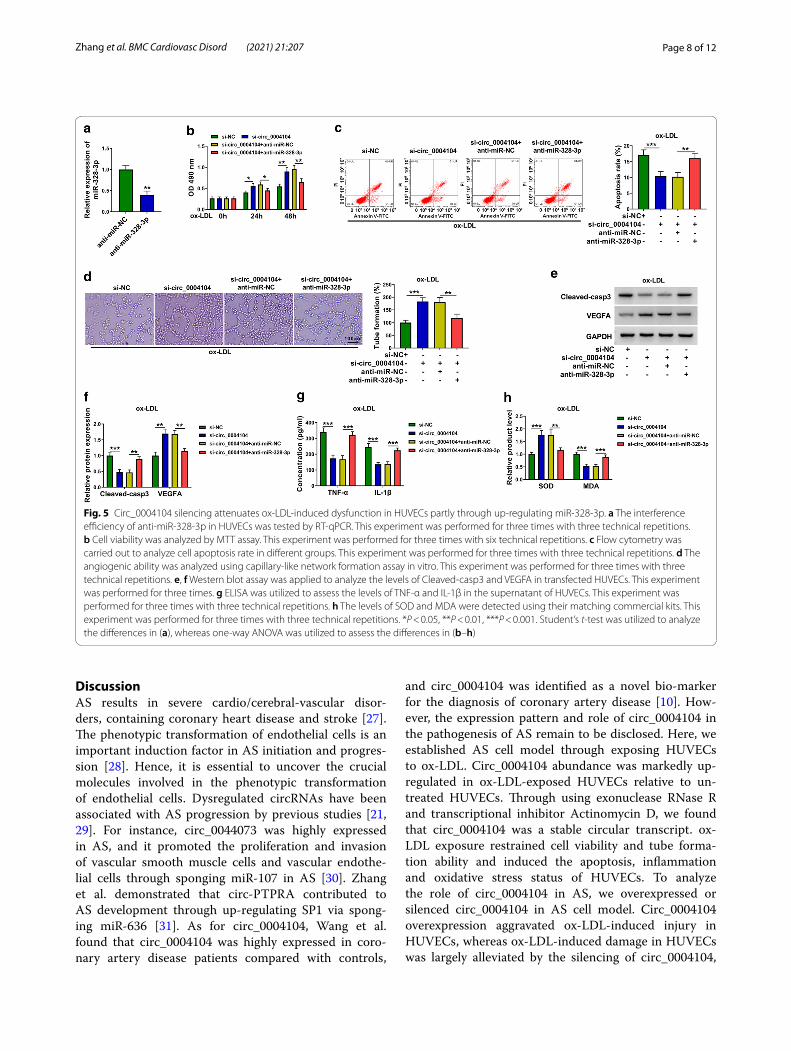
Circ_0004104 silencing attenuates ox‑LDL‑induced dysfunction in HUVECs partly through up‑regulating miR‑328‑3pTransfection with anti-miR-328-3p resulted in about 60% reduction in miR-328-3p level in HUVECs (Fig. 5a). MiR-328-3p silencing suppressed cell viabil-ity and angiogenic ability and induced cell apoptosis in circ_0004104-silenced HUVECs upon ox-LDL exposure (Fig. 5b–d). Consistently, Western blot assay revealed that miR-328-3p silencing overturned circ_0004104 knockdown-mediated effects in the expression of Cleaved-casp3 and VEGFA in ox-LDL-induced HUVECs (Fig. 5e, f ). Circ_0004104 silencing protected HUVECs from ox-LDL-induced inflammation and oxidative, and these protective effects were overturned by the silencing
Fig. 3 ox-LDL-induced dysfunction is largely alleviated by the silencing of circ_0004104 in HUVECs. a RT-qPCR was performed to evaluate the interference efficiency of si-circ_0004104 in HUVECs. This experiment was performed for three times with three technical repetitions. b–h HUVECs were divided into four groups: control, ox-LDL, ox-LDL + si-NC and ox-LDL + si-circ_0004101. b MTT assay was applied to analyze cell viability in different time points. This experiment was performed for three times with six technical repetitions. c The percentage of apoptotic HUVECs was assessed via flow cytometry. This experiment was performed for three times with three technical repetitions. d The ability of tube formation was analyzed through capillary-like network formation assay in vitro. This experiment was performed for three times with three technical repetitions. e, f The protein levels of Cleaved-casp3 and VEGFA were measured by Western blot assay. This experiment was performed for three times. g The concentrations of inflammation-associated cytokines (TNF-α and IL-1β) were detected by ELISA. This experiment was performed for three times with three technical repetitions. h The production of SOD and MDA was assessed using their matching kits. This experiment was performed for three times with three technical repetitions. *P < 0.05, **P < 0.01, ***P < 0.001. Student’s t-test was utilized to analyze the differences in a, whereas one-way ANOVA was utilized to assess the differences in (b–h)
Page 7 of 12Zhang et al. BMC Cardiovasc Disord (2021) 21:207
of miR-328-3p (Fig. 5g, h). Overall, circ_0004104 knock-down protected HUVECs against ox-LDL-induced dys-function partly through up-regulating miR-328-3p.
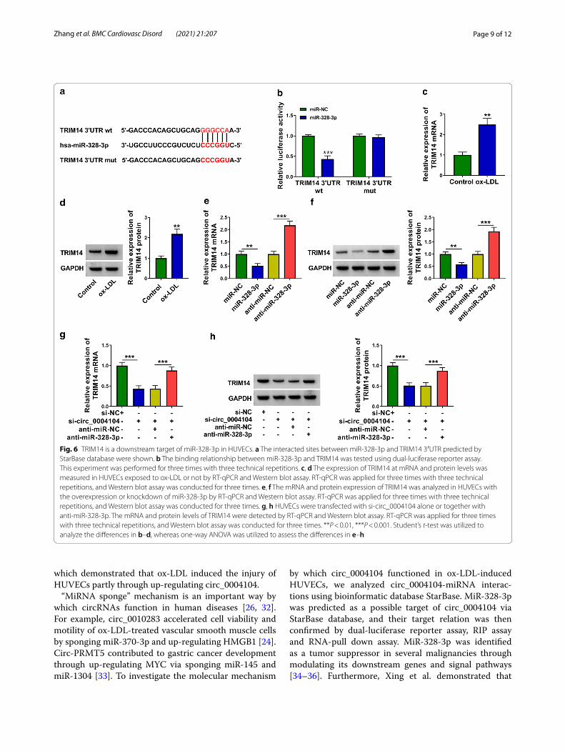
TRIM14 is a downstream target of miR‑328‑3p in HUVECsThe potential binding sequence between miR-328-3p and TRIM14 predicted by StarBase was shown in Fig. 6a. Luciferase activity of wild-type luciferase reporter plas-mid (TRIM14 3′UTR wt) was notably reduced with the overexpression of miR-328-3p (Fig. 6b), suggesting that TRIM14 was a target of miR-328-3p in HUVECs. ox-LDL exposure up-regulated the expression of TRIM14 at both mRNA and protein levels (Fig. 6c, d). The negative regu-latory relationship between miR-328-3p and TRIM14 was observed in HUVECs (Fig. 6e, f ). Subsequently, we ana-lyzed the regulation among circ_0004104, miR-328-3p and TRIM14 in HUVECs. Circ_0004104 interference reduced the mRNA and protein expression of TRIM14 partly by up-regulating miR-328-3p in HUVECs (Fig. 6g, h). Overall, circ_0004104 positively regulated TRIM14 expression by sponging miR-328-3p in HUVECs.
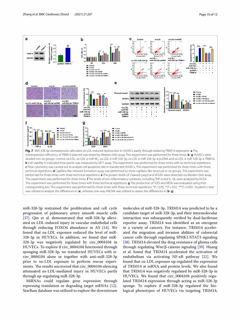
MiR‑328‑3p overexpression alleviates ox‑LDL‑induced dysfunction in HUVECs partly through reducing TRIM14 expressionWestern blot assay showed that the transfection effi-ciency of TRIM14 plasmid was high in HUVECs (Fig. 7a). MiR-328-3p overexpression protected HUVECs from ox-LDL-induced dysfunction of HUVECs (Fig. 7b–g). The addition of TRIM14 plasmid suppressed cell viability and angiogenesis whereas induced the apoptosis in miR-328-3p-overexpressed HUVECs upon ox-LDL exposure (Fig. 7b–d). TRIM14 overexpression also up-regulated Cleaved-casp3 expression and reduced VEGFA expres-sion (Fig. 7e), which further demonstrated that TRIM14 overexpression reversed miR-328-3p overexpression-mediated effects in the apoptosis and angiogenesis of HUVECs upon ox-LDL exposure. The accumulation of TRIM14 promoted the inflammatory response and oxi-dative stress again in miR-328-3p-overexpresed HUVECs upon ox-LDL (Fig. 7f, g). Overall, miR-328-3p overex-pression protected HUVECs against ox-LDL-induced dysfunction partly through reducing TRIM14 expression.
Fig. 4 Circ_0004104 acts as miR-328-3p sponge in HUVECs. a The miR-328-3p-binding sites in circ_0004104 predicted by StarBase database were shown. b The overexpression efficiency of miR-328-3p was assessed via RT-qPCR. This experiment was performed for three times with three technical repetitions. c Dual-luciferase reporter assay was applied to verify the interaction between circ_0004104 and miR-328-3p as well as their binding sites. This experiment was performed for three times with three technical repetitions. d RIP assay was carried out to test if miR-328-3p was a target of circ_0004104 in HUVECs. This experiment was performed for three times with three technical repetitions. e The target relationship between circ_0004104 and miR-328-3p was tested by RNA-pull down assay. This experiment was performed for three times with three technical repetitions. f The expression of miR-328-3p in ox-LDL-treated HUVECs was measured by RT-qPCR. This experiment was performed for three times with three technical repetitions. g The regulatory relationship between circ_0004104 and miR-328-3p was tested. The expression of miR-328-3p was analyzed in HUVECs transfected with vector, circ_0004104, si-NC or si-circ_0004104 by RT-qPCR. This experiment was performed for three times with three technical repetitions. *P < 0.05, **P < 0.01, ***P < 0.001. Student’s t-test was utilized to analyze the differences in (b–f), whereas one-way ANOVA was utilized to assess the differences in (g)
Page 8 of 12Zhang et al. BMC Cardiovasc Disord (2021) 21:207
DiscussionAS results in severe cardio/cerebral-vascular disor-ders, containing coronary heart disease and stroke [27]. The phenotypic transformation of endothelial cells is an important induction factor in AS initiation and progres-sion [28]. Hence, it is essential to uncover the crucial molecules involved in the phenotypic transformation of endothelial cells. Dysregulated circRNAs have been associated with AS progression by previous studies [21, 29]. For instance, circ_0044073 was highly expressed in AS, and it promoted the proliferation and invasion of vascular smooth muscle cells and vascular endothe-lial cells through sponging miR-107 in AS [30]. Zhang et al. demonstrated that circ-PTPRA contributed to AS development through up-regulating SP1 via spong-ing miR-636 [31]. As for circ_0004104, Wang et al. found that circ_0004104 was highly expressed in coro-nary artery disease patients compared with controls,
and circ_0004104 was identified as a novel bio-marker for the diagnosis of coronary artery disease [10]. How-ever, the expression pattern and role of circ_0004104 in the pathogenesis of AS remain to be disclosed. Here, we established AS cell model through exposing HUVECs to ox-LDL. Circ_0004104 abundance was markedly up-regulated in ox-LDL-exposed HUVECs relative to un-treated HUVECs. Through using exonuclease RNase R and transcriptional inhibitor Actinomycin D, we found that circ_0004104 was a stable circular transcript. ox-LDL exposure restrained cell viability and tube forma-tion ability and induced the apoptosis, inflammation and oxidative stress status of HUVECs. To analyze the role of circ_0004104 in AS, we overexpressed or silenced circ_0004104 in AS cell model. Circ_0004104 overexpression aggravated ox-LDL-induced injury in HUVECs, whereas ox-LDL-induced damage in HUVECs was largely alleviated by the silencing of circ_0004104,
Fig. 5 Circ_0004104 silencing attenuates ox-LDL-induced dysfunction in HUVECs partly through up-regulating miR-328-3p. a The interference efficiency of anti-miR-328-3p in HUVECs was tested by RT-qPCR. This experiment was performed for three times with three technical repetitions. b Cell viability was analyzed by MTT assay. This experiment was performed for three times with six technical repetitions. c Flow cytometry was carried out to analyze cell apoptosis rate in different groups. This experiment was performed for three times with three technical repetitions. d The angiogenic ability was analyzed using capillary-like network formation assay in vitro. This experiment was performed for three times with three technical repetitions. e, f Western blot assay was applied to analyze the levels of Cleaved-casp3 and VEGFA in transfected HUVECs. This experiment was performed for three times. g ELISA was utilized to assess the levels of TNF-α and IL-1β in the supernatant of HUVECs. This experiment was performed for three times with three technical repetitions. h The levels of SOD and MDA were detected using their matching commercial kits. This experiment was performed for three times with three technical repetitions. *P < 0.05, **P < 0.01, ***P < 0.001. Student’s t-test was utilized to analyze the differences in (a), whereas one-way ANOVA was utilized to assess the differences in (b–h)
Page 9 of 12Zhang et al. BMC Cardiovasc Disord (2021) 21:207
which demonstrated that ox-LDL induced the injury of HUVECs partly through up-regulating circ_0004104.
“MiRNA sponge” mechanism is an important way by which circRNAs function in human diseases [26, 32]. For example, circ_0010283 accelerated cell viability and motility of ox-LDL-treated vascular smooth muscle cells by sponging miR-370-3p and up-regulating HMGB1 [24]. Circ-PRMT5 contributed to gastric cancer development through up-regulating MYC via sponging miR-145 and miR-1304 [33]. To investigate the molecular mechanism
by which circ_0004104 functioned in ox-LDL-induced HUVECs, we analyzed circ_0004104-miRNA interac-tions using bioinformatic database StarBase. MiR-328-3p was predicted as a possible target of circ_0004104 via StarBase database, and their target relation was then confirmed by dual-luciferase reporter assay, RIP assay and RNA-pull down assay. MiR-328-3p was identified as a tumor suppressor in several malignancies through modulating its downstream genes and signal pathways [34–36]. Furthermore, Xing et al. demonstrated that
Fig. 6 TRIM14 is a downstream target of miR-328-3p in HUVECs. a The interacted sites between miR-328-3p and TRIM14 3′UTR predicted by StarBase database were shown. b The binding relationship between miR-328-3p and TRIM14 was tested using dual-luciferase reporter assay. This experiment was performed for three times with three technical repetitions. c, d The expression of TRIM14 at mRNA and protein levels was measured in HUVECs exposed to ox-LDL or not by RT-qPCR and Western blot assay. RT-qPCR was applied for three times with three technical repetitions, and Western blot assay was conducted for three times. e, f The mRNA and protein expression of TRIM14 was analyzed in HUVECs with the overexpression or knockdown of miR-328-3p by RT-qPCR and Western blot assay. RT-qPCR was applied for three times with three technical repetitions, and Western blot assay was conducted for three times. g, h HUVECs were transfected with si-circ_0004104 alone or together with anti-miR-328-3p. The mRNA and protein levels of TRIM14 were detected by RT-qPCR and Western blot assay. RT-qPCR was applied for three times with three technical repetitions, and Western blot assay was conducted for three times. **P < 0.01, ***P < 0.001. Student’s t-test was utilized to analyze the differences in b–d, whereas one-way ANOVA was utilized to assess the differences in e–h
Page 10 of 12Zhang et al. BMC Cardiovasc Disord (2021) 21:207
miR-328-3p restrained the proliferation and cell cycle progression of pulmonary artery smooth muscle cells [37]. Qin et al. demonstrated that miR-328-3p allevi-ated ox-LDL-induced injury in vascular endothelial cells through reducing FOXO4 abundance in AS [14]. We found that ox-LDL exposure reduced the level of miR-328-3p in HUVECs. In addition, we found that miR-328-3p was negatively regulated by circ_0004104 in HUVECs. To explore if circ_0004104 functioned through sponging miR-328-3p, we transfected HUVECs with si-circ_0004104 alone or together with anti-miR-328-3p prior to ox-LDL exposure to perform rescue experi-ments. The results uncovered that circ_0004104 silencing attenuated ox-LDL-mediated injury in HUVECs partly through up-regulating miR-328-3p.
MiRNAs could regulate gene expression through repressing translation or degrading target mRNAs [12]. StarBase database was utilized to explore the downstream
molecules of miR-328-3p. TRIM14 was predicted to be a candidate target of miR-328-3p, and their intermolecular interaction was subsequently verified by dual-luciferase reporter assay. TRIM14 was identified as an oncogene in a variety of cancers. For instance, TRIM14 acceler-ated the migration and invasion abilities of colorectal cancer cells through regulating SPHK1/STAT3 signaling [38]. TRIM14 elevated the drug resistance of glioma cells through regulating Wnt/β-catenin signaling [39]. Huang et al. found that TRIM14 accelerated the activation of endothelium via activating NF-κB pathway [21]. We found that ox-LDL exposure up-regulated the expression of TRIM14 at mRNA and protein levels. We also found that TRIM14 was negatively regulated by miR-328-3p in HUVECs. We found that circ_0004104 positively regu-lated TRIM14 expression through acting as miR-328-3p sponge. To explore if miR-328-3p regulated the bio-logical phenotypes of HUVECs via targeting TRIM14,
Fig. 7 MiR-328-3p overexpression alleviates ox-LDL-induced dysfunction in HUVECs partly through reducing TRIM14 expression. a The overexpression efficiency of TRIM14 plasmid was tested by Western blot assay. This experiment was performed for three times. b–g HUVECs were divided into six groups: control, ox-LDL, ox-LDL + miR-NC, ox-LDL + miR-328-3p, ox-LDL + miR-328-3p + pcDNA and ox-LDL + miR-328-3p + TRIM14. b Cell viability in indicated time points was measured by MTT assay. This experiment was performed for three times with six technical repetitions. c Flow cytometry was carried out to analyze cell apoptosis rate in transfected HUVECs. This experiment was performed for three times with three technical repetitions. d Capillary-like network formation assay was performed to show capillary-like structure in six groups. This experiment was performed for three times with three technical repetitions. e The protein levels of Cleaved-casp3 and VEGFA were detected via Western blot assay. This experiment was performed for three times. f The levels of pro-inflammatory cytokines, including TNF-α and IL-1β, were analyzed by ELISA. This experiment was performed for three times with three technical repetitions. g The production of SOD and MDA was evaluated using their corresponding kits. This experiment was performed for three times with three technical repetitions. *P < 0.05, **P < 0.01, ***P < 0.001. Student’s t-test was utilized to analyze the differences in (a), whereas one-way ANOVA was utilized to assess the differences in (b–g)
Page 11 of 12Zhang et al. BMC Cardiovasc Disord (2021) 21:207
we performed compensation experiments. The results revealed that miR-328-3p protected HUVECs against ox-LDL-mediated damage in HUVECs partly through down-regulating TRIM14.
ConclusionsIn conclusion, our study demonstrated that circ_0004104 contributed to ox-LDL-induced injury of HUVECs partly through targeting miR-328-3p/TRIM14 axis. Blockage of circ_0004104 might be a potential strategy to attenuate the abnormal phenotypes of vascular endothelial cells in AS.
Supplementary InformationThe online version contains supplementary material available at https:// doi. org/ 10. 1186/ s12872- 021- 02012-7.
Additional file 1: Figure 1. The expression of AS progression-associated circRNAs in HUVECs upon ox-LDL exposure. RT-qPCR was applied to ana-lyze the levels of circ_0004104, circ_0001879, circ_0001445, circ_0001599, circ_0010283 and circ_0007478 in HUVECs induced by ox-LDL. This experiment was performed for three times with three technical repeti-tions. *P < 0.05, **P < 0.01. Student’s t-test was utilized to analyze the differences.
AcknowledgementsNone.
Authors’ contributionsCZ designed and performed the research; CZ, LW, YS analyzed the data; CZ wrote the manuscript. All authors read and approved the final manuscript.
FundingNone.
Availability of data and materialsThe datasets used and/or analysed during the current study are available from the corresponding author on reasonable request.
Declarations
Ethics approval and consent to participateNot applicable.
Consent for publicationNot applicable.
Competing interestsThe authors declare that they have no conflict of interest.
Received: 27 January 2021 Accepted: 13 April 2021
References 1. Weber C, Noels H. Atherosclerosis: current pathogenesis and therapeutic
options. Nat Med. 2011;17(11):1410–22. 2. Mannarino E, Pirro M. Endothelial injury and repair: a novel theory for
atherosclerosis. Angiology. 2008;59(2 Suppl):69s–72s. 3. Di Pietro N, Formoso G, Pandolfi A. Physiology and pathophysiology of
oxLDL uptake by vascular wall cells in atherosclerosis. Vascul Pharmacol. 2016;84:1–7.
4. Chen LL, Yang L. Regulation of circRNA biogenesis. RNA Biol. 2015;12(4):381–8.
5. Zhang HD, Jiang LH, Sun DW, Hou JC, Ji ZL. CircRNA: a novel type of biomarker for cancer. Breast Cancer. 2018;25(1):1–7.
6. Meng S, Zhou H, Feng Z, Xu Z, Tang Y, Li P, Wu M. CircRNA: functions and properties of a novel potential biomarker for cancer. Mol Cancer. 2017;16(1):94.
7. Yin Y, Long J, He Q, Li Y, Liao Y, He P, Zhu W. Emerging roles of circRNA in formation and progression of cancer. J Cancer. 2019;10(21):5015–21.
8. Li CY, Ma L, Yu B. Circular RNA hsa_circ_0003575 regulates oxLDL induced vascular endothelial cells proliferation and angiogenesis. Biomed Phar-macother. 2017;95:1514–9.
9. Liu H, Ma X, Mao Z, Shen M, Zhu J, Chen F. Circular RNA has_circ_0003204 inhibits oxLDL-induced vascular endothelial cell proliferation and angio-genesis. Cell Signal. 2020;70:109595.
10. Wang L, Shen C, Wang Y, Zou T, Zhu H, Lu X, Li L, Yang B, Chen J, Chen S, et al. Identification of circular RNA Hsa_circ_0001879 and Hsa_circ_0004104 as novel biomarkers for coronary artery disease. Atheroscle-rosis. 2019;286:88–96.
11. Ebert MS, Sharp PA. Roles for microRNAs in conferring robustness to biological processes. Cell. 2012;149(3):515–24.
12. Fabian MR, Sonenberg N, Filipowicz W. Regulation of mRNA translation and stability by microRNAs. Annu Rev Biochem. 2010;79:351–79.
13. Raitoharju E, Oksala N, Lehtimäki T. MicroRNAs in the atherosclerotic plaque. Clin Chem. 2013;59(12):1708–21.
14. Qin X, Guo J. MicroRNA-328-3p protects vascular endothelial cells against oxidized low-density lipoprotein induced injury via targeting forkhead box protein O4 (FOXO4) in atherosclerosis. Med Sci Monit. 2020;26:e921877.
15. Galkina E, Ley K. Immune and inflammatory mechanisms of atherosclero-sis. Annu Rev Immunol. 2009;27:165–97.
16. Liang H, Liao M, Zhao W, Zheng X, Xu F, Wang H, Huang J. CXCL16/ROCK1 signaling pathway exacerbates acute kidney injury induced by ischemia-reperfusion. Biomed Pharmacother. 2018;98:347–56.
17. Qiao CY, Qiao TY, Jin H, Liu LL, Zheng MD, Wang ZL. LncRNA KCNQ1OT1 contributes to the cisplatin resistance of tongue cancer through the KCNQ1OT1/miR-124-3p/TRIM14 axis. Eur Rev Med Pharmacol Sci. 2020;24(1):200–12.
18. Wang T, Ren Y, Liu R, Ma J, Shi Y, Zhang L, Bu R. miR-195-5p suppresses the proliferation, migration, and invasion of oral squamous cell carcinoma by targeting TRIM14. Biomed Res Int. 2017;2017:7378148.
19. Deng Y, Zhu H, Xiao L, Liu C, Meng X. Circ_0005198 enhances temozo-lomide resistance of glioma cells through miR-198/TRIM14 axis. Aging (Albany NY). 2020;12:2198–211.
20. Huang X, Li Y, Li X, Fan D, Xin HB, Fu M. TRIM14 promotes endothe-lial activation via activating NF-κB signaling pathway. J Mol Cell Biol. 2020;12(3):176–89.
21. Bayoumi AS, Aonuma T, Teoh JP, Tang YL, Kim IM. Circular noncoding RNAs as potential therapies and circulating biomarkers for cardiovascular diseases. Acta Pharmacol Sin. 2018;39(7):1100–9.
22. Vilades D, Martínez-Camblor P, Ferrero-Gregori A, Bär C, Lu D, Xiao K, Vea À, Nasarre L, Sanchez Vega J, Leta R, et al. Plasma circular RNA hsa_circ_0001445 and coronary artery disease: Performance as a biomarker. FASEB J. 2020;34(3):4403–14.
23. Li S, Hu W, Deng F, Chen S, Zhu P, Wang M, Chen X, Wang Y, Hu X, Zhao B, et al. Identification of circular RNA hsa_circ_0001599 as a novel biomarker for large-artery atherosclerotic stroke. DNA Cell Biol. 2021;40(3):457–68.
24. Ding P, Ding Y, Tian Y, Lei X. Circular RNA circ_0010283 regulates the via-bility and migration of oxidized low-density lipoprotein-induced vascular smooth muscle cells via an miR-370-3p/HMGB1 axis in atherosclerosis. Int J Mol Med. 2020;46(4):1399–408.
25. Wang X, Bai M. CircTM7SF3 contributes to oxidized low-density lipo-protein-induced apoptosis, inflammation and oxidative stress through targeting miR-206/ASPH axis in atherosclerosis cell model in vitro. BMC Cardiovasc Disord. 2021;21(1):51.
26. Panda AC. Circular RNAs act as miRNA sponges. Adv Exp Med Biol. 2018;1087:67–79.
27. Libby P, Bornfeldt KE, Tall AR. Atherosclerosis: successes, surprises, and future challenges. Circ Res. 2016;118(4):531–4.
28. Gimbrone MA Jr, García-Cardeña G. Endothelial cell dysfunction and the pathobiology of atherosclerosis. Circ Res. 2016;118(4):620–36.
Page 12 of 12Zhang et al. BMC Cardiovasc Disord (2021) 21:207
• fast, convenient online submission
•
thorough peer review by experienced researchers in your field
• rapid publication on acceptance
• support for research data, including large and complex data types
•
gold Open Access which fosters wider collaboration and increased citations
maximum visibility for your research: over 100M website views per year •
At BMC, research is always in progress.
Learn more biomedcentral.com/submissions
Ready to submit your researchReady to submit your research ? Choose BMC and benefit from: ? Choose BMC and benefit from:
29. Altesha MA, Ni T, Khan A, Liu K, Zheng X. Circular RNA in cardiovascular disease. J Cell Physiol. 2019;234(5):5588–600.
30. Shen L, Hu Y, Lou J, Yin S, Wang W, Wang Y, Xia Y, Wu W. CircRNA-0044073 is upregulated in atherosclerosis and increases the proliferation and inva-sion of cells by targeting miR-107. Mol Med Rep. 2019;19(5):3923–32.
31. Zhang LL. CircRNA-PTPRA promoted the progression of atherosclerosis through sponging with miR-636 and upregulating the transcription fac-tor SP1. Eur Rev Med Pharmacol Sci. 2020;24(23):12437–49.
32. Hansen TB, Jensen TI, Clausen BH, Bramsen JB, Finsen B, Damgaard CK, Kjems J. Natural RNA circles function as efficient microRNA sponges. Nature. 2013;495(7441):384–8.
33. Du W, Li D, Guo X, Li P, Li X, Tong S, Tong J, Kuang L, Liang D. Circ-PRMT5 promotes gastric cancer progression by sponging miR-145 and miR-1304 to upregulate MYC. Artif Cells Nanomed Biotechnol. 2019;47(1):4120–30.
34. Yan T, Ye XX. MicroRNA-328-3p inhibits the tumorigenesis of bladder cancer through targeting ITGA5 and inactivating PI3K/AKT pathway. Eur Rev Med Pharmacol Sci. 2019;23(12):5139–48.
35. Shi J, An G, Guan Y, Wei T, Peng Z, Liang M, Wang Y. miR-328-3p mediates the anti-tumor effect in osteosarcoma via directly targeting MMP-16. Cancer Cell Int. 2019;19:104.
36. Ma W, Ma CN, Zhou NN, Li XD, Zhang YJ. Up-regulation of miR-328-3p sensitizes non-small cell lung cancer to radiotherapy. Sci Rep. 2016;6:31651.
37. Xing Y, Zheng X, Fu Y, Qi J, Li M, Ma M, Wang S, Li S, Zhu D. Long noncod-ing RNA-maternally expressed gene 3 contributes to hypoxic pulmonary hypertension. Mol Ther. 2019;27(12):2166–81.
38. Jin Z, Li H, Hong X, Ying G, Lu X, Zhuang L, Wu S. TRIM14 promotes colorectal cancer cell migration and invasion through the SPHK1/STAT3 pathway. Cancer Cell Int. 2018;18:202.
39. Tan Z, Song L, Wu W, Zhou Y, Zhu J, Wu G, Cao L, Song J, Li J, Zhang W. TRIM14 promotes chemoresistance in gliomas by activating Wnt/β-catenin signaling via stabilizing Dvl2. Oncogene. 2018;37(40):5403–15.
Publisher’s NoteSpringer Nature remains neutral with regard to jurisdictional claims in pub-lished maps and institutional affiliations.