Embed Size (px)
Citation preview

Hematology
Jan Bazner-ChandlerCPNP, CNS, MSN, RN

Blood Blood is the fluid of life Blood is composed of:
Plasma RBC WBC Platelets

Plasma Plasma consists of:
90% water.
10 % solutes: albumin, electrolytes and proteins.
Proteins consist of clotting factors, globulins, circulating antibodies and fibrinogen.

Red Blood Cells RBC’s travel through the body delivering
oxygen and removing waste. RBC’s are red because they contain a protein
chemical called hemoglobin which is bright red in color.
Hemoglobin contains iron, making it an excellent vehicle for transporting oxygen and carbon dioxide.

RBC’s
Average life cycle is 120 days.
The bones are continually producing new cells.

White Blood Cells The battling blood cells. The white blood cells are continually on the
look out for signs of disease. When a germ appears the WBC will:
Produce protective antibodies. Surround it and devour the bacteria.

WBC’s WBC life span is from a few days to a few
weeks. WBC’s will increase when fighting infection. Neutropenia – abnormal percentage of
neutrophils compared to total white blood cells; decreases a child’s ability to fight pathologic bacteria

Platelets
Platelets are irregularly-shaped, colorless bodies that are present in blood.
Their sticky surface lets them form clots to stop bleeding.

Blood Values CBC with differential and platelet count.
Hgb: Normal levels are 11 to 16 g / dl Panic levels are:
Less than 5 g / dl More than 20 g / dl

Hematocrit Normal hematocrit levels are 35 to 44%.
Panic levels: Hmct less than 15 % Hmct greater than 60%

Blood Tests Hemoglobin and hematocrit used to screen for
anemia. The CBC with differential would be used to
help diagnose a specific disorder. A bone marrow aspiration would be the most
conclusive in determining cause of anemia – aplastic / leukemia.
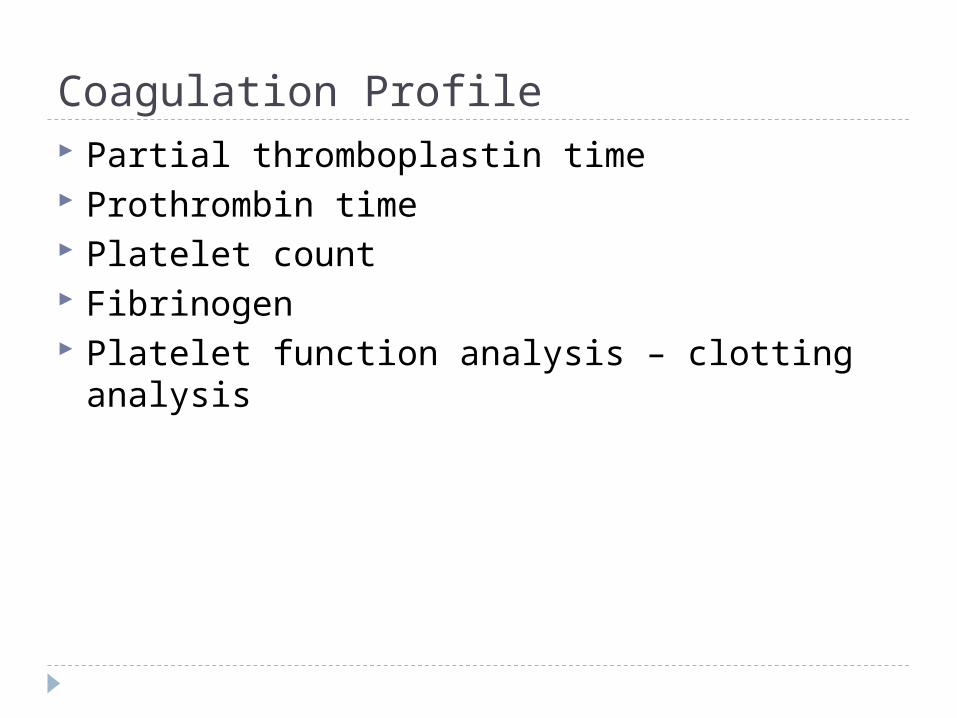
Coagulation Profile Partial thromboplastin time Prothrombin time Platelet count Fibrinogen Platelet function analysis – clotting analysis

Bone MarrowBone marrow is the spongy substance found in
the center of the bones. • It manufactures bone marrow stem cells,
which in turn produce blood cells.• Red blood cells – carry oxygen to tissue• Platelets – help blood to clot • White blood cells – fight infection

Bone Marrow Aspiration

Bone Marrow Transplant Donor is placed under anesthesia. Marrow is aspirated out of the iliac crest. Marrow is filtered and treated to remove bits
of bone and other unwanted cells and debris, transferred to a blood bag, and is infused into the patient’s blood just like at transfusion.

Treatment Modalities Transfusion:
Packed red blood cells – anemia Platelets – platelet dysfunction Fresh frozen plasma – coagulation factors

Blood Transfusions 3 types of transfusion reactions
Hemolytic Allergic Febrile

Hemolytic Reaction Refers to an immune response against
transfused blood cells. Antigens, on the surface of red blood cells, are
recognized as “foreign proteins” and can stimulate B lymphocytes to produce antibodies to the red blood cell antigens.

Hemolytic reaction Flank pain Fever Chills Bloody urine Rash Low blood pressure Dizziness / fainting

Nursing Management Stop the blood transfusion. Start normal saline infusion. Take vital signs with blood pressure Call the MD Obtain blood sample and urine specimen. Return blood to blood bank. Document

Febrile Reaction Often occurs after multiple blood transfusions. Symptoms:fever, chills, and diaphoresis. Interventions:
Slow transfusion and administer antipyretic. Administer antipyretic prior to administration.

Allergic Reaction
Symptoms: rash, urticaria, respiratory distress, or anaphylaxis.
Interventions: administer
antihistamine before transfusion
Physician may order washed rbc’s

Hematologic Conditions
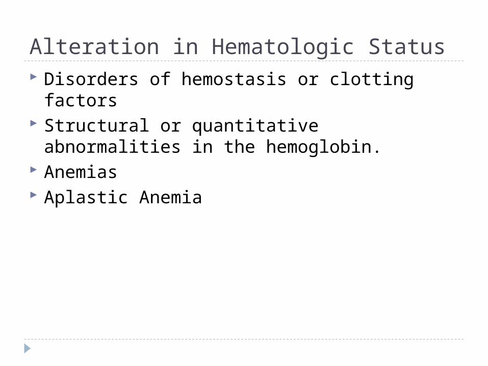
Alteration in Hematologic Status Disorders of hemostasis or clotting factors Structural or quantitative abnormalities in the
hemoglobin. Anemias Aplastic Anemia

Genetic Implications The following have a genetic link: implications
for genetic screening and fetal diagnosis Sickle cell anemia Thalassemia Hemophilia

Bleeding Disorders Three types Hemophilia: males only
Type A most common – factor VIII deficiency Type B - lack of factor IX (Christmas Disease) Type C – lack of factor XI
Von Willebrand Disease – 1% of population – men or women – prolonged bleeding time

Hemophilia Type A Hemophilia type A is the deficiency of clotting
factor VIII. A serious blood disorder Affects 1 in 10,000 males in the US Autoimmune disorder with lowered level of
clotting factor All races and socio economic groups affected
equally

Hemophilia Hemophilia is a sex-linked hereditary bleeding
disorder Transmitted on the X chromosome Female is the carrier Women do not suffer from the disease itself

Historical Perspective First recorded case in Talmud Jewish text by
an Arab physician – documentation of two brothers with bleeding after circumcision.
Queen Victoria is carrier and spread the disease through the male English royalty.

Goals of Care Goals of care:
Provide factor VIII (IX) to aid blood in clotting. To decrease transmission of infectious agents in
blood products; hepatitis & AIDS. Future: gene therapy to increase production of
clotting factor.

Assessment Circumcision may produce prolonged
bleeding. As child matures and becomes more active
the incidence of bleeding due to trauma increases
May be mild, moderate or severe Bleeding into joint spaces, hemarthrosis Most dangerous bleed would be intracranial

Diagnosis Presenting symptoms Prolonged activated aPTT and decreased
levels of factor VIII or IX. Genetic testing to identify carriers

Pharmacologic Interventions Products used to treat hemophilia are:
Fresh frozen plasma and cryoprecipitate which are from single blood donors and require special freezing
Second generation of factor VIII are made with animal or human proteins
Fibrin glue – mixture of fibrogen and thrombin can be applied topically on a wound to stop minor bleeding

Multidisciplinary Interventions
Replace the factor as ordered by physician. Manage pain utilizing analgesics as ordered –
no salicylate products. Maintaining joint integrity during acute phase:
immobilization, elevation, ice. Physical therapy to prevent flexion contraction
and to strengthen muscles and joints. Provide opportunities for normal growth and
development.

Family Education Medic-Alert bracelet Injury prevention appropriate for age Signs and symptoms of internal bleeding or
hemarthrosis Dental checkups Medication administration

Long Term Complications 20% develop neutralizing antibodies that
make replacement products less effective. Gene therapy providing continuous production
of the deficient clotting factor could be the next major advance in hemophilia treatment.

Disseminated Intravascular Coagulation or DIC
DIC is an acquired coagulopathy that is characterized by both thrombosis and hemorrhage.
DIC is not a primary disorder but occurs as a result of a variety of alterations in health.

Assessment The most obvious clinical feature of DIC is
bleeding. Renal involvement = hematuria Pulmonary involvement = hemoptysis,
tachypnea, dyspnea and chest pain. Cutaneous involvement = petechiae,
ecchymosis, jaundice, acrocyanosis and gangrene.

Multidisciplinary Interventions Treatment of the precipitating disorder. Supportive care with administration of platelet
concentration and fresh frozen plasma and coagulation factors.
Administration of heparin (controversial in children).
Heparin potentates anti-thrombin III which inhibits thrombin and further development of thrombosis.

Assessment Rigorous ongoing assessment of all body
systems Monitor bleeding No rectal temps Avoid trauma to delicate tissue areas All injections sites and IV sites need to be
treated like an arterial stick.

Prognosis Depends on the underlying disorder and the
severity of the DIC.

ITP Idiopathic thrombocytopenic purpura
Idiopathic = cause is unknown Thrombocytopenic = blood does not have enough
platelets Purpura = excessive bleeding / bruising

Immune Thrombocytopenic Purpura Antibodies destroy platelets Antibodies see platelets as bacteria and work
to eliminate them ITP is preceded by a viral illness
URI Varicella Smallpox / measles vaccine Mononucleosis Flu

Two Types Acute: occurs in children between 1 and 6
years Chronic (continuous or recurrent): older than
10 years and female

Symptoms Random purpura Epistaxis, hematuria, hematemesis, and
menorrhagia Petechiae and hemorrhagic bullae in mouth

Diagnostic Tests Low platelet count Peripheral blood smear Antiplatelet antibodies
Normal platelet count: 150,000 to 400,000

Pharmacologic Interventions For severe cases (platelet count < 20,000)
IV gamma globulin for 2-5 days to block antibody production, reduce autoimmune problem and increase platelet count
Corticosteroids to enhance vascular stability, increase platelet survival
IV anti-D to stimulate platelet production
Sickle Cell Disease Autosomal recessive disorder Defect in hemoglobin molecule Cells become sickle shaped and rigid Lose ability to adapt shape to surroundings. Sickling may be triggered by fever and
emotional or physical stress


Pathophysiology When exposed to diminished levels of oxygen,
the hemoglobin in the RBC develops a sickle or crescent shape; the cells are rigid and obstruct capillary blood flow, leading to congestion and tissue hypoxia; clinically, this hypoxia causes additional sickling and extensive infarctions.


Whaley & Wong Text

Body Systems Affected by SS Brain: CVA – paralysis - death Eyes: retinopathy – blindness Lungs: pneumonia Abdomen: pain, hepatomegaly, splenomegaly
(medical emergency due to possible rupture Skeletal: joint pain, bone pain – osteomyelitis Skin: chronic ulcers – poor wound healing

Vaso-occlusive Crisis Presents depending on location of vaso-
occlusion Bone pain – back, knees, shoulders, elbows Acute chest syndrome – Chest pain / pneumonia Acute abdominal pain Cerebrovascular accident – stoke symptoms Priapism – nonsexual erection of penis

Splenic Sequestration Sudden and quickly progressing splenic
enlargement Left upper quadrant pain and vomiting Abdominal distension, pallor, dyspnea,
tachycardia Hypovolemia and shock Decrease hemoglobin / hematocrit and
increased reticulocyte count

Aplastic Crisis Diminished production and increased
destruction of red blood cells Triggered by viral infection or depletion of
folic acid Signs include profound anemia, pallor

Diagnostic Screening Newborn screening is mandatory in the United
States. Screening done between 24 and 72 hours of
age. Verification of results are made by the
healthcare provider at first office visit.

Newborn Screening – Follow-up Contact family with screening results Refer to specialists if positive Evaluate infant for slenomegaly Initiate daily penicillin VK (125 mg po bid)
prophylaxis as recommended Educate regarding risk of sepsis, or signs of
splenic sequestration

Nursing Management - Hospital Increase tissue perfusion
Oxygen / bedrest Blood transfusion / antibiotics if ordered
Pain management – meperidine (Demerol) contraindicated
Hydration IV fluids as ordered Oral intake of fluids

Nursing Management - Home Care• Adequate nutrition• Emotional Support• Discharge instructions
• Information about disease management• Daily folic acid• Control of triggers• Prophylactic antibiotics • Immunizations / Pneumococcal

Patient Education Necessity of following plan of care Signs and symptoms of impending crisis. Signs and symptoms of infection Preventing hypoxia from physical and
emotional stress Proving adequate rest

Thalassemia A group of inherited diseases of the blood that
affect the ability to produce hemoglobin. Occurs most frequently in people of Italian,
Greek, Middle Eastern, Southern Asian and African Ancestry.

Two Types of Thalassemia Alpha or beta depending on which part of an
oxygen-carrying protein the red blood cell is lacking.
Child may have mild to severe form of the disease.
Cooley’s anemia is severe form of the disease.

Tests for Thalassemia Blood tests and family genetics study to show
whether an individual has Thalassemia or is a carrier.
Prenatal testing at 11th week of pregnancy using chorionic villi sampling.
Amniocentesis around the 16th week of pregnancy
Assisted reproductive therapy

Clinical ManifestationsThe child may be:
pale and listless have poor appetite enlarged spleen, liver and heart bones become thin and brittle
Heart failure and infection are the major cause of death.

Interdisciplinary Interventions Frequent blood transfusions to keep
hemoglobin levels near normal “iron chelators” to rid body of excess iron
from numerous blood transfusions Bone marrow transplant: only possible for a
small minority of patients who have a suitable bone marrow donor

Iron Deficiency Anemia Most common nutritional deficiency Depletion of iron stores due to
Inadequate dietary intake Impaired iron absorption

American Academy of Pediatrics Recommends screening for iron deficiency at
1 year of age Screening of high risk children (low socio-
economic status or poor dietary intake)

What a problem? IDA can have long lasting effects on motor
development, mental development and auditory and visual function.

IDA Occurs in children experiencing:
Rapid physical growth Low iron intake Inadequate iron absorption Loss of blood

Assessment Associated with low oxygenation of tissue:
Pallor Fatigue Shortness or breath Irritability Intolerance of physical work / exercise

Diagnostic Tests Initial screen is hemoglobin
Hemoglobin levels less than 8 g/dL
Decreased levels of Serum Iron or Total Iron Binding or Serum Ferritin
Microcytic and hypochromic red blood cells

Pharmacologic Treatment Iron supplementation
Given in a.m. on an empty stomach To avoid staining of teeth, give using a syringe,
dropper or straw Instruct caretaker that child may have dark-
colored stools

Management Infants younger than 12 months should be on
formula with iron Solid foods – introduced about 6 months of
age Rice cereal fortified with iron Pureed Vegetables, meats
Infants 12 months or older Decrease intake of milk Increase solid foods
Children: iron fortified cereals, meat, green leafy vegetables, yogurt, cheeses, low-fat or non-fat milk
Teenagers: reduce junk food

Aplastic Anemia Acquired or inherited
Normal production of blood cells in the bone marrow is absent or decreased.
A marked decrease in RBC’s, WBC’s and platelets.

Causes Exposure to drugs Exposure to chemicals Exposure to toxins Infection Idiopathic in nature Inherited: Fanconi’s anemia

Blood Characteristics
Neutophil less than 500
Platelet less than 20,000
Hemoglobin less than 7
Reticulocytes 1%
Nursing Diagnosis?
Bone marrow reveals hypo-cellular and fatty marrow.

Management Immunosuppressive therapy
Antithymocyte globulin Administered IV over 4 days Response seen within 3 months
Bone Marrow Transplant

Neonatal Hyperbilirubinemia
Hyperbilirubinemia is defined as excessive levels of bilirubin in the blood.
Alert: Jaundice that occurs within the first 24 hours of life or after 2 weeks of life signifies an abnormal physiologic process.
Kernicterus: bilirubin encephalopathy

JaundiceYellowing of the skin – occurs when bilirubin is
deposited into the subcutaneous tissue and it becomes visible when the serum bilirubin levels exceed 7.0 mg/dL.

Normal Physiologic Jaundice Occurs in 45% to 65% of all healthy newborns
in the first 3 to 5 days of life Occurs more frequently in infants less than 38
weeks gestation Breast fed infants have a higher incidence of
jaundice (due to dehydration and caloric deprivation)
Assessment Very yellow or orange skin tones
(beginning at the head and spreading to the toes)
Increased sleepiness, so much that it is hard to wake the baby
High-pitched cry Poor sucking or nursing Weakness, limpness, or floppiness

Photo Therapy

Fiberoptic Blanket

Multidisciplinary Interventions Monitor bilirubin levels Assess activity level – muscle tone – infant
reflexes Encourage po intake: May need to
supplement with formula if inadequate breastfeeding
Weight daily to assess hydration status Monitor stools – amount and number Cover eyes while under bili-lights Facilitate parent - infant bonding
Loss of moro or startle reflex can indicate possible brain damage due to Kernicterus