Embed Size (px)
Citation preview

Hematology for Family Practice
When to treat and when to refer
Karen deGenevieve MSN, FNP,BC OCN
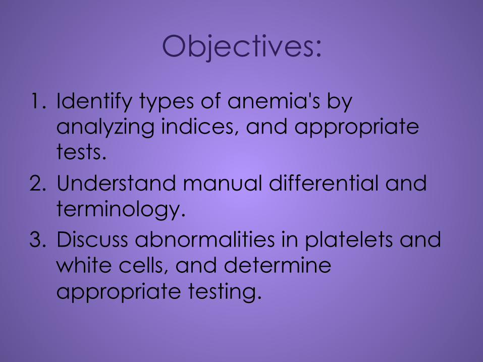
Objectives:
1. Identify types of anemia's by analyzing indices, and appropriate tests.
2. Understand manual differential and terminology.
3. Discuss abnormalities in platelets and white cells, and determine appropriate testing.
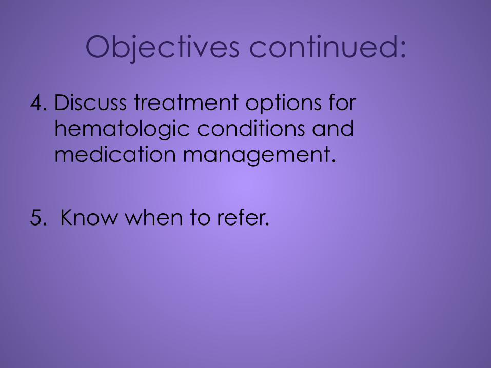
Objectives continued:
4. Discuss treatment options for hematologic conditions and medication management.
5. Know when to refer.
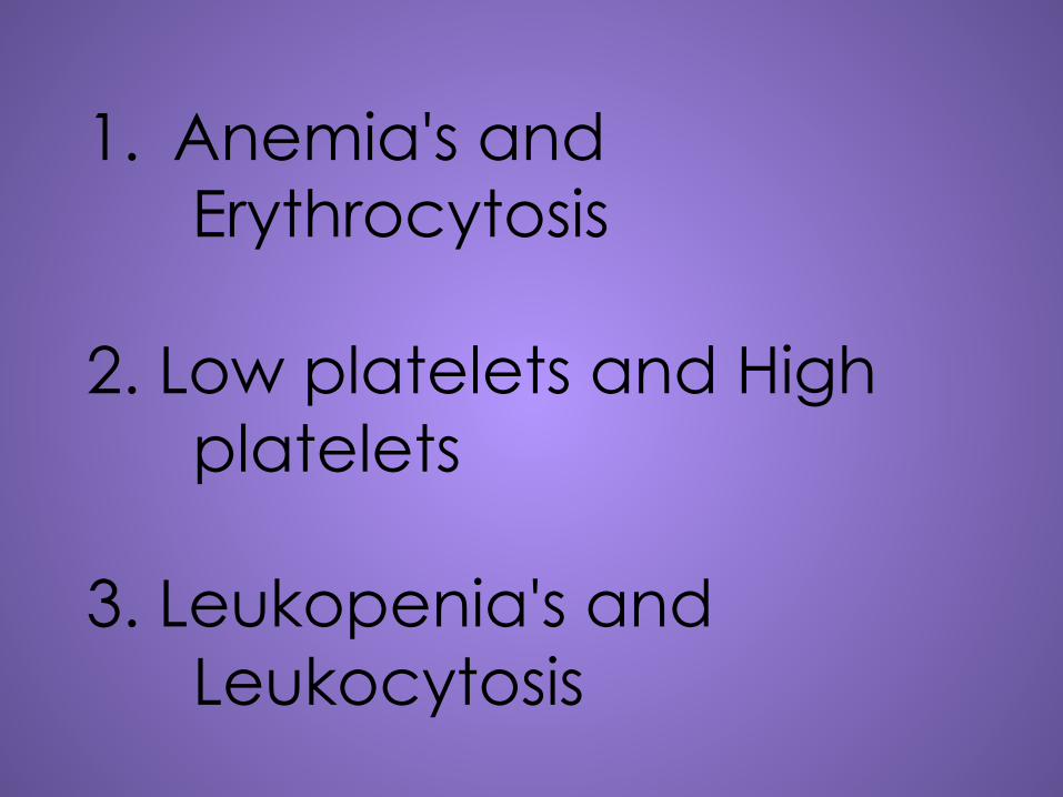
1. Anemia's and Erythrocytosis
2. Low platelets and High platelets
3. Leukopenia's and Leukocytosis
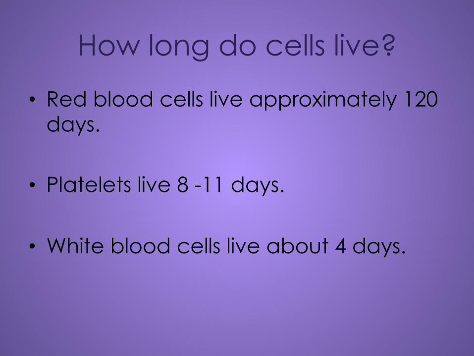
How long do cells live?
• Red blood cells live approximately 120 days.
• Platelets live 8 -11 days.
• White blood cells live about 4 days.

There are millions of RBCs in just one drop of blood. People who
live at higher altitudes have more (like in the mountains of Peru).
They are produced in the bone marrow of large bones at a rate of 2 million per second. In the minute it took you to read this, you made
120 million of them!


Anemia’s And Erythrocytosis
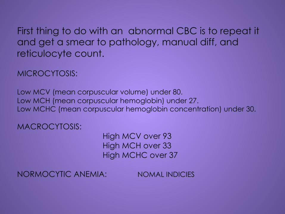
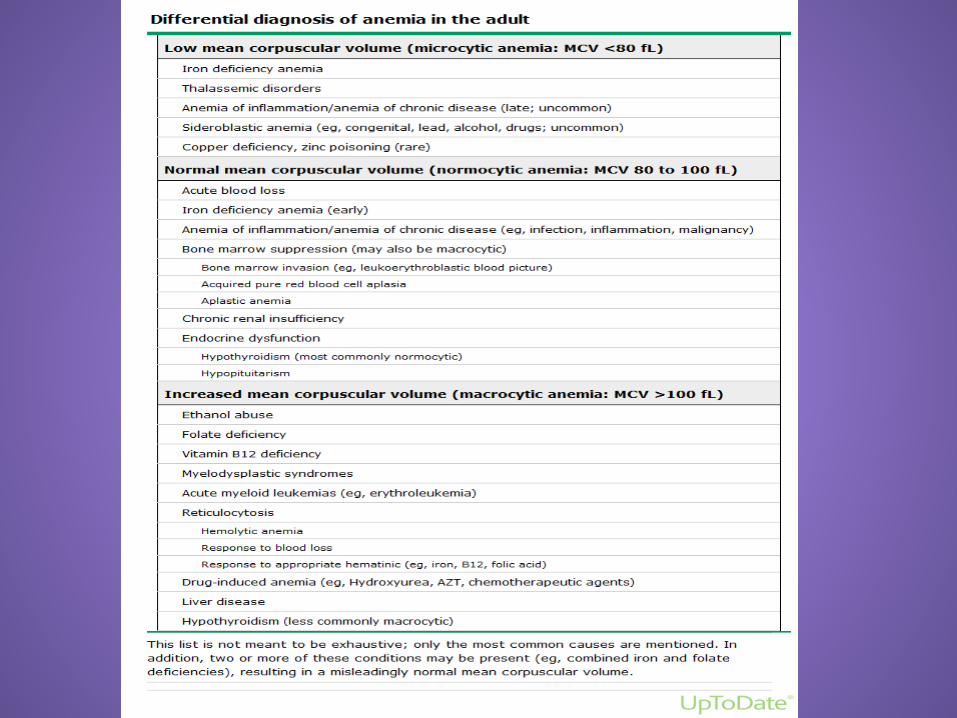
First thing to do with an abnormal CBC is to repeat it and get a smear to pathology, manual diff, and reticulocyte count. MICROCYTOSIS: Low MCV (mean corpuscular volume) under 80. Low MCH (mean corpuscular hemoglobin) under 27. Low MCHC (mean corpuscular hemoglobin concentration) under 30. MACROCYTOSIS:
High MCV over 93 High MCH over 33 High MCHC over 37
NORMOCYTIC ANEMIA: NOMAL INDICIES
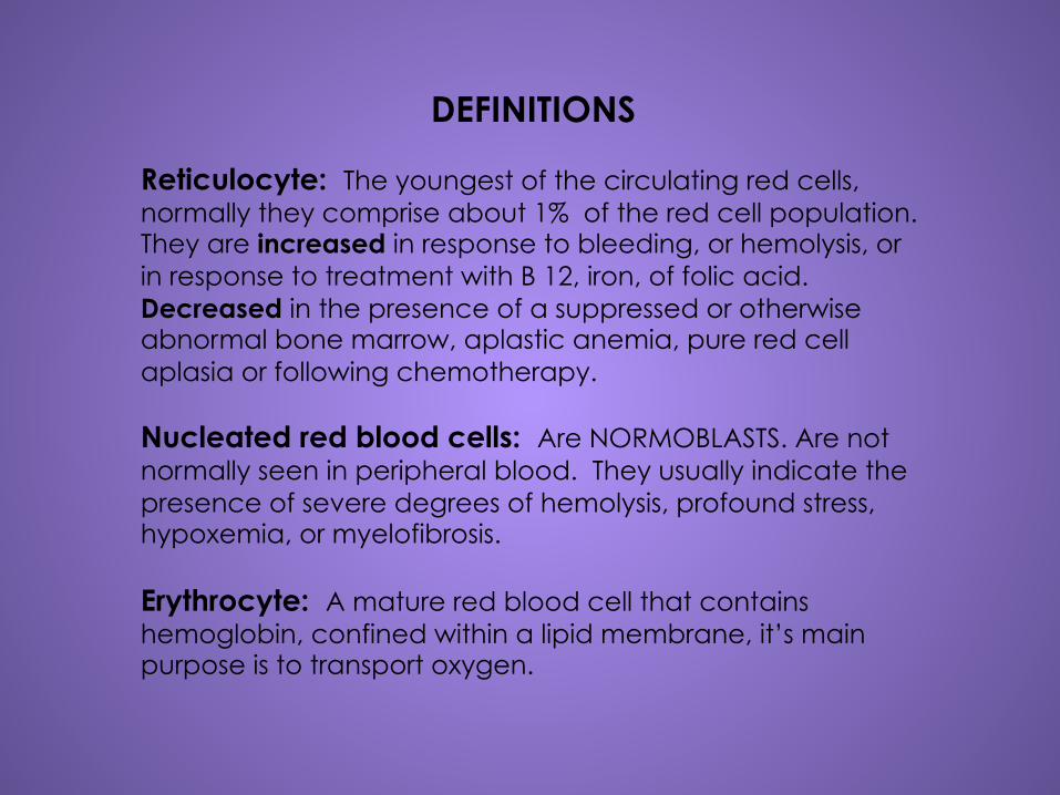
DEFINITIONS Reticulocyte: The youngest of the circulating red cells, normally they comprise about 1% of the red cell population. They are increased in response to bleeding, or hemolysis, or in response to treatment with B 12, iron, of folic acid. Decreased in the presence of a suppressed or otherwise abnormal bone marrow, aplastic anemia, pure red cell aplasia or following chemotherapy. Nucleated red blood cells: Are NORMOBLASTS. Are not normally seen in peripheral blood. They usually indicate the presence of severe degrees of hemolysis, profound stress, hypoxemia, or myelofibrosis. Erythrocyte: A mature red blood cell that contains hemoglobin, confined within a lipid membrane, it’s main purpose is to transport oxygen.
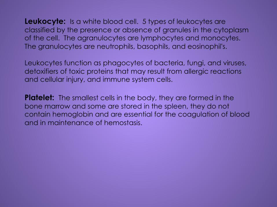
Leukocyte: Is a white blood cell. 5 types of leukocytes are classified by the presence or absence of granules in the cytoplasm of the cell. The agranulocytes are lymphocytes and monocytes. The granulocytes are neutrophils, basophils, and eosinophil's. Leukocytes function as phagocytes of bacteria, fungi, and viruses, detoxifiers of toxic proteins that may result from allergic reactions and cellular injury, and immune system cells. Platelet: The smallest cells in the body, they are formed in the bone marrow and some are stored in the spleen, they do not contain hemoglobin and are essential for the coagulation of blood and in maintenance of hemostasis.
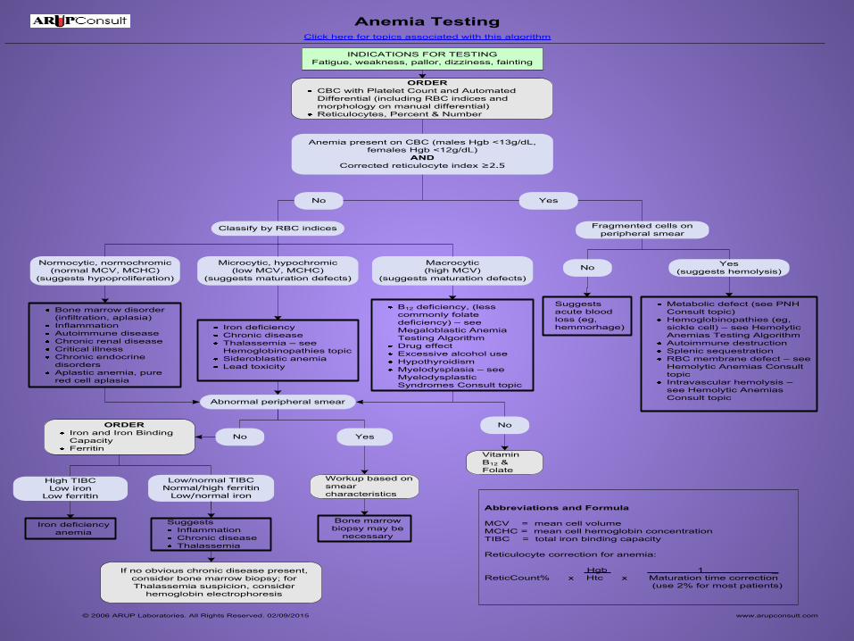
INDICATIONS FOR TESTINGFatigue, weakness, pallor, dizziness, fainting
Anemia TestingClick here for topics associated with this algorithm
Anemia present on CBC (males Hgb <13g/dL, females Hgb <12g/dL)
ANDCorrected reticulocyte index ≥2.5
ORDERCBC with Platelet Count and Automated Differential (including RBC indices and morphology on manual differential)Reticulocytes, Percent & Number
© 2006 ARUP Laboratories. All Rights Reserved. 02/09/2015 www.arupconsult.com
Abbreviations and Formula
MCV = mean cell volumeMCHC = mean cell hemoglobin concentrationTIBC = total iron binding capacity
Reticulocyte correction for anemia:
Hgb 1 _ReticCount% x Htc x Maturation time correction
(use 2% for most patients)
Classify by RBC indices
No
Bone marrow disorder (infiltration, aplasia)InflammationAutoimmune diseaseChronic renal diseaseCritical illnessChronic endocrine disordersAplastic anemia, pure red cell aplasia
Iron deficiencyChronic diseaseThalassemia – see Hemoglobinopathies topicSideroblastic anemiaLead toxicity
B12 deficiency, (less commonly folate deficiency) – see Megaloblastic Anemia Testing AlgorithmDrug effectExcessive alcohol useHypothyroidismMyelodysplasia – see Myelodysplastic Syndromes Consult topic
Normocytic, normochromic(normal MCV, MCHC)
(suggests hypoproliferation)
Microcytic, hypochromic(low MCV, MCHC)
(suggests maturation defects)
Macrocytic(high MCV)
(suggests maturation defects)
ORDERIron and Iron Binding CapacityFerritin
SuggestsInflammationChronic diseaseThalassemia
Low/normal TIBCNormal/high ferritin
Low/normal iron
Iron deficiency anemia
High TIBCLow iron
Low ferritin
Abnormal peripheral smear
Fragmented cells on peripheral smear
Suggests acute blood loss (eg, hemmorhage)
Metabolic defect (see PNH Consult topic)Hemoglobinopathies (eg, sickle cell) – see Hemolytic Anemias Testing AlgorithmAutoimmune destructionSplenic sequestrationRBC membrane defect – see Hemolytic Anemias Consult topicIntravascular hemolysis –see Hemolytic Anemias Consult topic
Yes
No Yes(suggests hemolysis)
No Yes
Bone marrow biopsy may be
necessary
Workup based on smear characteristics
If no obvious chronic disease present, consider bone marrow biopsy; for Thalassemia suspicion, consider
hemoglobin electrophoresis
No
Vitamin B12 & Folate
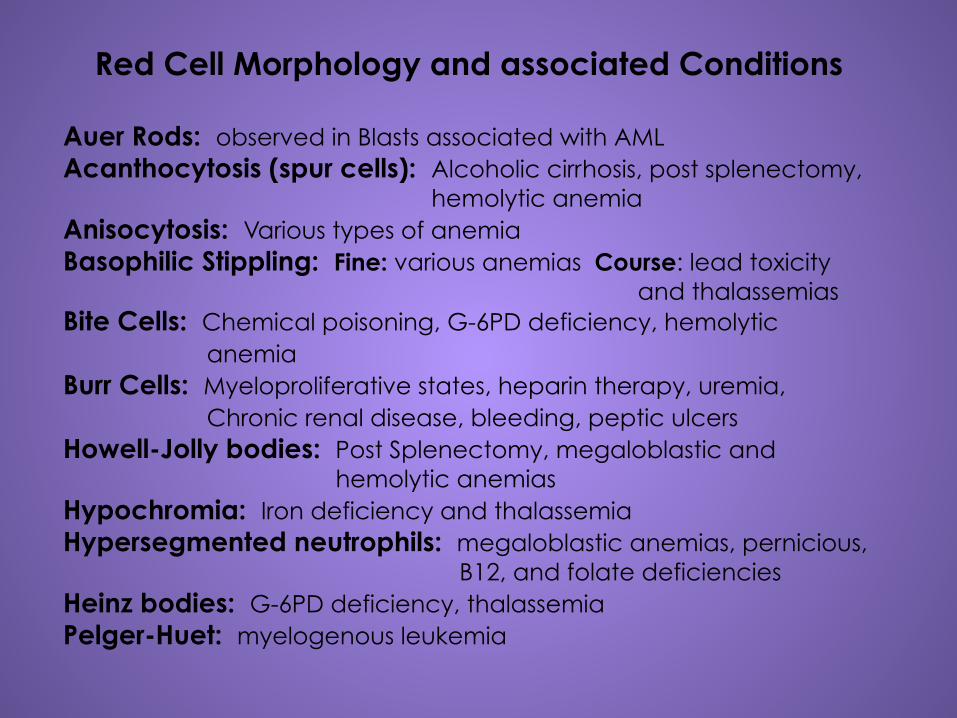
Red Cell Morphology and associated Conditions Auer Rods: observed in Blasts associated with AML Acanthocytosis (spur cells): Alcoholic cirrhosis, post splenectomy,
hemolytic anemia Anisocytosis: Various types of anemia Basophilic Stippling: Fine: various anemias Course: lead toxicity
and thalassemias Bite Cells: Chemical poisoning, G-6PD deficiency, hemolytic
anemia Burr Cells: Myeloproliferative states, heparin therapy, uremia,
Chronic renal disease, bleeding, peptic ulcers Howell-Jolly bodies: Post Splenectomy, megaloblastic and
hemolytic anemias Hypochromia: Iron deficiency and thalassemia Hypersegmented neutrophils: megaloblastic anemias, pernicious,
B12, and folate deficiencies Heinz bodies: G-6PD deficiency, thalassemia Pelger-Huet: myelogenous leukemia

Dohle bodies: (toxic granulation are usually seen together) Acute infection, pneumonia, scarlet fever, measles, septicemia, pregnancy, burns
Reactive lymphocytes: (Downey cells) mono, CMV, viral hepatitis, chronic inflammatory disease
Smudge Cells: atypical lymphocytosis, CML Schistocytosis: cardiac valve disease, DIC, severe burns, uremia Spherocytes: (helmet cells) hereditary spherocytosis, thermal
injuries, immune and hemolytic diseases, TTP, DIC Rouleaux: multiple myeloma, elevated protein Target cells: chronic liver disease, iron deficiency, post splenectomy Tear drop cells: Thalassemias, pernicious anemia, Myeloproliferative disorders. Band Neutrophils: normal 5-11% increased # = LEFT SHIFT (stress,
infection, Myeloproliferative disease) Basophils: <2% are normal. Allergic reaction, hypothyroid, chronic
hemolytic anemia, post splenectomy Eosinophils: increased in asthma, hay fever, extensive skin lesions,
parasitic infections. Decreased in shock, severe burns, and severe infections.
Metamyelocyte: Myelocytic hyperplasia Myelocyte: CML, AML Plasma cells: Not usually seen in peripheral blood. Chronic
infections, autoimmune disorders, alcoholic liver disease. Monocytes: increased in chronic neutropenia, IBD, chronic infection,
CMV, TB an can be elevated in AMML.
Evaluating Anemia
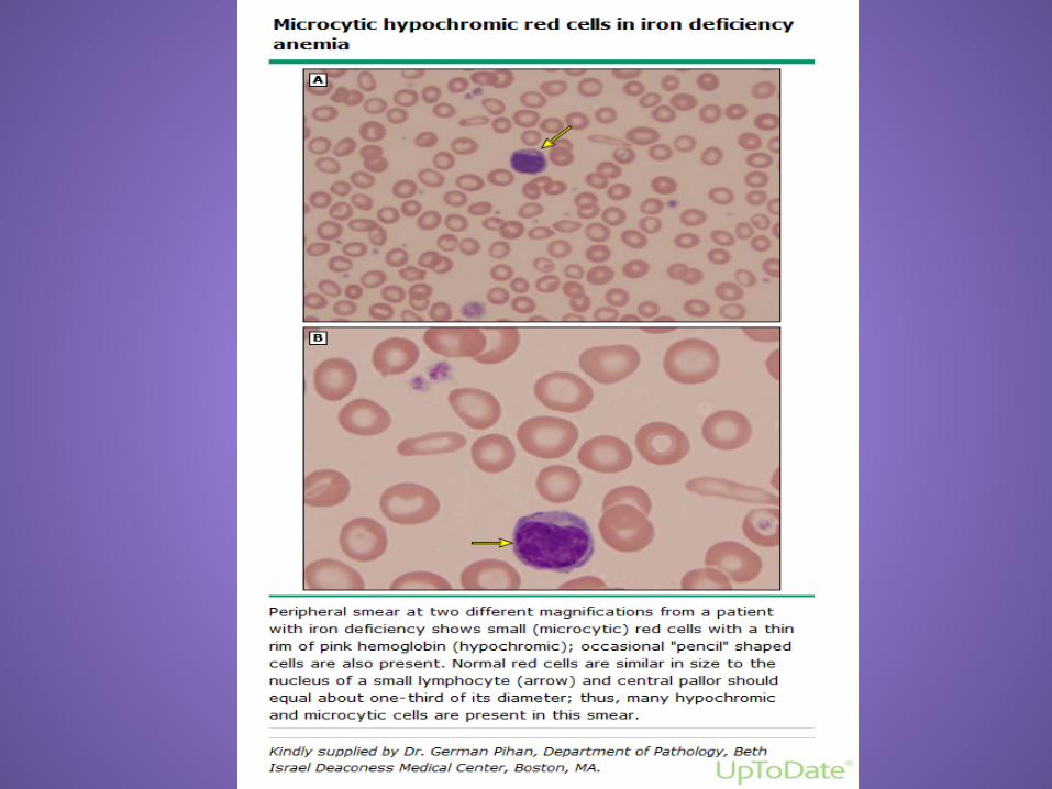
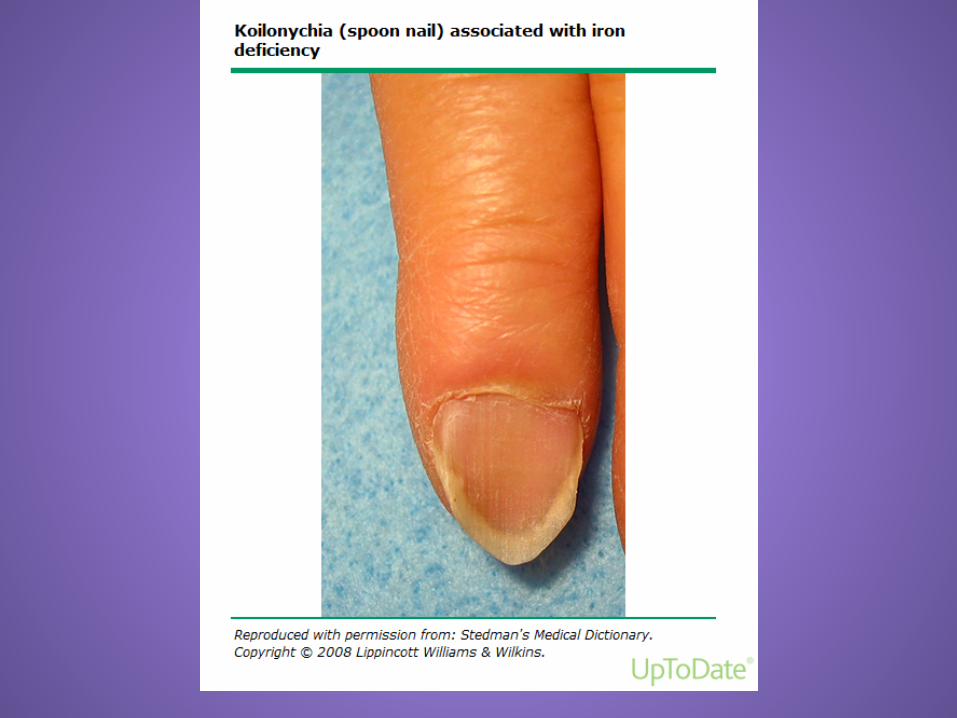
Number one reason for microcytic anemia is bleeding, either GU or GI. Ask the right questions. A good physical exam and a good history is essential to your investigation. Don’t forget family history.

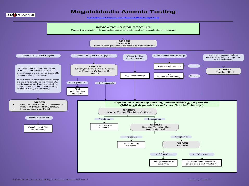
Megaloblastic Anemia TestingClick here for topics associated with this algorithm
Occasionally, clinician may find normal levels of B12 in symptomatic patients (usually neurologic symptoms)
MMA and homocysteine may be appropriate to confirm B12deficiency, as homocysteine may have a role in detecting folate or B12 deficiency
INDICATIONS FOR TESTINGPatient presents with megaloblastic anemia and/or neurologic symptoms
ORDERVitamin B12
Folate (for patient with known risk factors)
ORDERMethylmalonic Acid, Serum
or Plasma (Vitamin B12 Status)
ORDERIntrinsic Factor Blocking Antibody
ORDERGastric Parietal Cell
Antibody, IgG
ORDERGastrin
Folate deficiency
© 2006 ARUP Laboratories. All Rights Reserved. Revised 02/09/2015 www.arupconsult.com
Optional antibody testing when MMA >0.4 µmol/L (MMA >0.4 µmol/L confirms B12 deficiency )
Vitamin B12 >400 pg/mL Vitamin B12 100-400 pg/mL Low folate levels only
Not pernicious
anemia
Pernicious anemia
Pernicious anemia
Not pernicious anemia
Pernicious anemia(indirect confirmation)
Positive
<100 pg/mL
Negative
Positive
>100 pg/mL
Negative
Nofolate deficiency
Low or normal folate levels and high suspicion
for deficiency
ORDERFolate, RBC
ORDERMethylmalonic Acid, Serum or Plasma (Vitamin B12 Status)Homocysteine, Total
<0.4 µmol/L >0.4 µmol/L
Low
Normal
Vitamin B12 <100 pg/mL
B12 deficiency
Both elevated
Confirmed B12 deficiency
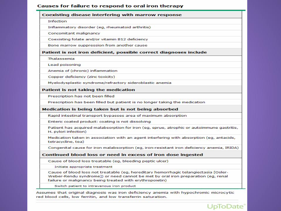

Iron preparations: Ferrous gluconate orally is less likely to cause GI upset and is more tolerated than ferrous sulfate. It is equally absorbable with less side effects. Comes in may strengths and is generally OTC. Severe iron deficiency may require 325 mg TID. Most patients don’t take it as directed for a variety of reasons. Nausea and constipation are the biggest reasons. I never order ferrous sulfate, for those reasons. There are many conditions that can interfere with oral iron absorption and or cause iron deficiency: Being older, poor tolerance of oral iron preparations Inflammatory bowel disease, ulcerative colitis Gastric surgery and gastric bypass H. Pylori, autoimmune gastritis and celiac disease. Chronic kidney disease and dialysis Cancer patients

IV iron preparations: (use them when patients cannot tolerate oral) AVOID IM: It’s painful, stains the buttocks, and has variable absorption. Case reports have also described development of sarcomas. Iron Dextran (Infed): Black Box warnings for anaphylaxis, requires pre medications and takes long to give. Usually including premeds and test dose, 4-6 hours. Dosing is by weight and Hgb. (Chart) can be up to 1.5 Gms. More than a Gram doesn’t work any better. Ferumoxytol (Feraheme): Given in 2 doses, one week apart. 510 mg Often given with premeds and has an increase in second dose reactions. Iron sucrose (Venofer): Should have a test dose. Given in multiple doses, not over 300 mg. Used in CKD, and in the setting of dialysis. Ferric carboxymaltose (Injectafer): is a colloidal iron hydroxide complex with a tighter binding of elemental iron. It’s a 15 minute infusion and doesn’t require premeds and is given in NSS 750 mg in 2 doses, one week apart.
Monitoring: For chronic iron deficiency anemia patients that require ongoing IV iron treatments, monthly CBC’s and iron studies including ferritin. Treat again when ferritin goes below 50. Oral iron treatment F/U should be checked monthly during replacement until repleted. Continue oral iron up to 3-6 months after normalization of iron levels to replete iron stores. When ferritin is normalized, a trial off iron for 3 months and recheck CBC, iron, TIBC and ferritin. If the cause of the iron deficiency has been treated, no further iron should be necessary. (normalization of periods, post uterine oblation, GI bleed is successfully treated, etc.)
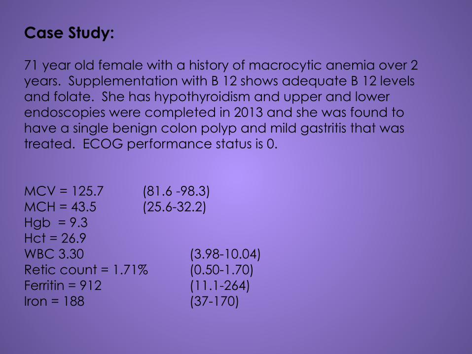
Case Study: 71 year old female with a history of macrocytic anemia over 2 years. Supplementation with B 12 shows adequate B 12 levels and folate. She has hypothyroidism and upper and lower endoscopies were completed in 2013 and she was found to have a single benign colon polyp and mild gastritis that was treated. ECOG performance status is 0. MCV = 125.7 (81.6 -98.3) MCH = 43.5 (25.6-32.2) Hgb = 9.3 Hct = 26.9 WBC 3.30 (3.98-10.04) Retic count = 1.71% (0.50-1.70) Ferritin = 912 (11.1-264) Iron = 188 (37-170)
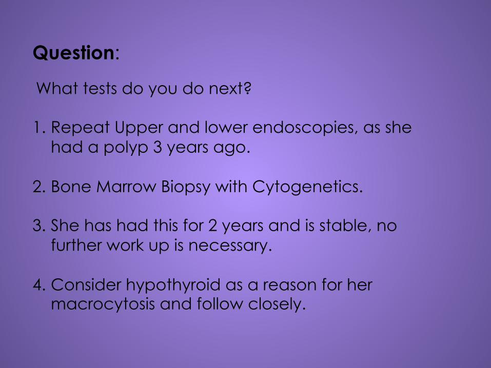
Question:
What tests do you do next? 1. Repeat Upper and lower endoscopies, as she
had a polyp 3 years ago.
2. Bone Marrow Biopsy with Cytogenetics.
3. She has had this for 2 years and is stable, no further work up is necessary.
4. Consider hypothyroid as a reason for her macrocytosis and follow closely.

71 Y/O female with megaloblastic anemia Bone Marrow Biopsy showed: Severely increased iron stores present, ringed sideroblasts present. Increased cellularity, no evidence of metastatic neoplasm. Comment: On BMB from pathologist The patient developed anemia starting in 2013. She is not B 12 or folate deficient, and she is taking levothyroxine. Hypothyroidism may be associated with megaloblastic anemia; however, there is increased particulate iron without blast increase in her marrow. Cytogenetics was negative for myelodysplastic syndrome. Treatment: For now, she should be followed frequently with blood counts and no treatment is needed at this time. Should she continue to drop her blood counts or become symptomatic, then a trial of erythropoietin could be initiated.
INDICATIONS FOR TESTINGPatient with anemia and evidence of hemolysis
Hemolytic Anemias TestingClick here for topics associated with this algorithm
ORDERCBC with Platelet Count and Automated DifferentialReticulocytesLactate Dehydrogenase HaptoglobinBilirubin
Heinz bodies
© 2006 ARUP Laboratories. All Rights Reserved. Revised 05/14/2015 www.arupconsult.com
Presence of the following may provide clues to the etiology of the anemia Increased reticulocyte countAbnormal peripheral smear
Polychromasia, spherocytes, schistocytes, sickle cells, stomatocytes, Heinz bodies, basophilic stippling, unusual red cell inclusions, and agglutination
Note: lack of any of the above does not rule out hemolytic anemia
Proceed based on above findings
Schistocytes, thrombocytopenia
Spherocytes, pyropoikilocytes,
elliptocytes or acanthocytes
ConsiderDICTTPHELLPHUSMechanical cardiac valveVasculitisMalignant hypertension
DIC
Pregnant
Consider HELLP
ORDEROsmotic Fragility,
Erythrocyte (usually positive)
Acquired No
Direct Coombs (Anti-Human
Globulin)
IgG+
Autoimmune hemolytic anemia (consider drug induced, hemolytic disease of the
newborn, autoimmune disease)
Consider one or more of the following tests
Isopropanol heat stability testingGlucose-6-Phosphate Dehydrogenase (G6PD) 2 MutationsEnzymes of glutathione cycle
Polychromasia without other reproducible
morphologic abnormality
Consider one or more of the following tests
Pyruvate kinase HexokinaseGlucose phosphate isomerase
Sickle cells
Basophilic stippling Acquired
No
Consider serum lead level testing
Congenital 5' nucleotidase
deficiency
Consider lead
poisoning
Consider 5' nucleotidase testing
Polychromasia only with or
without platelet decrease
Consider PNH
ORDERPNH, High Sensitivity, RBC and WBC testing
Agglutination
Consider cold
agglutinins disease
ORDERDirect
Coombs (Anti-Human
Globulin)
Positive for complement
Cold agglutinins
disease
Cold agglutinins
testing
Unusual red cell inclusions
If infection suspected, consider
malaria, bartonella (oroya fever), babesia
Yes
Recluse spider venom, clostridium
sepsis
+C3
Cold agglutinins disease, paroxysmal cold hemoglobinuria (PCH)
Yes
YesNo
Confirm PCH with Donath Landsteiner testing
Microangiopathic RBC destruction
ConsiderRBC membrane
disorder (hereditary
spherocytosis, hereditary
elliptocytosis, autoimmune hemolysis)
Consider Sickle cell disease –diverse genotypes: SS, SC,
SE, Sβ thalassemia, S Lepore
High-performance liquid chromatography (HPLC)
ConsiderPyruvate kinase deficiencyHexokinase deficiencyOther enzyme defects
ConsiderGlucose-6-Phosphate dehydrogenase deficiencyUnstable hemoglobin defectsGlutathione metabolism defectsHemoglobin H disease
For hemoglobin disorders, consider HPLC, genetic testing
Consider molecular
testing
ORDERRBC Band 3
Protein Reduction in Hereditary
Spherocytosis
Warm autoimmune
hemolytic anemia
No
Yes
ORDERD-Dimer
Increased
ADAMTS13 activity <10%
TTP
NormalClinical presentation consistent with TMA
ORDERADAMTS13 Activity
ORE. coli Shiga-like Toxin by EIA(dependent on presentation)
Normal
Atypical HUS
Positive Shiga toxin
HUS
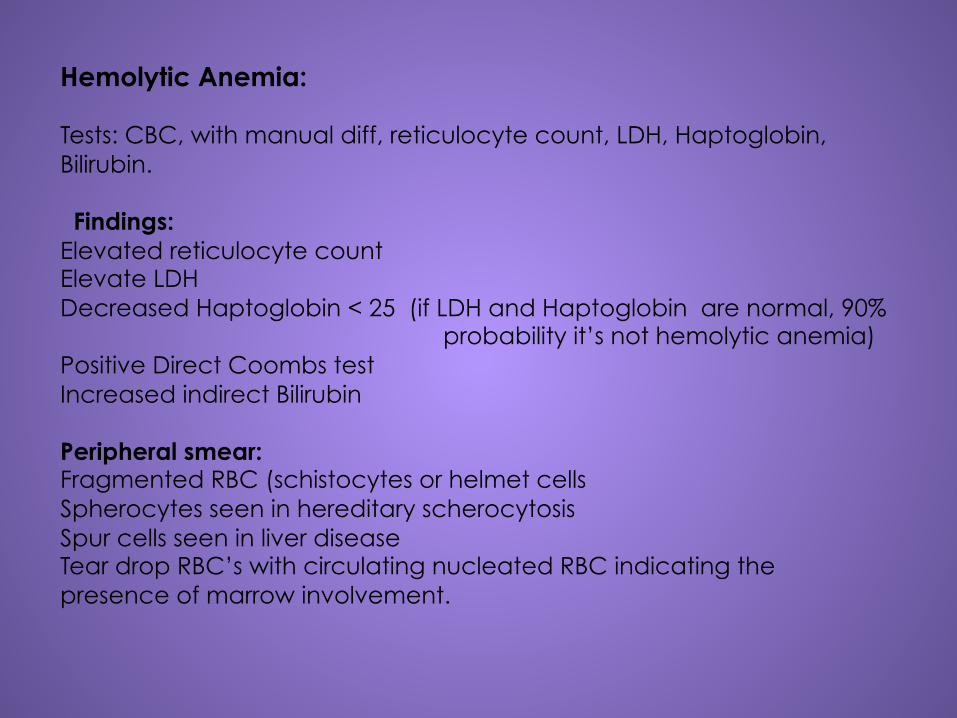
Hemolytic Anemia: Tests: CBC, with manual diff, reticulocyte count, LDH, Haptoglobin, Bilirubin. Findings: Elevated reticulocyte count Elevate LDH Decreased Haptoglobin < 25 (if LDH and Haptoglobin are normal, 90%
probability it’s not hemolytic anemia) Positive Direct Coombs test Increased indirect Bilirubin Peripheral smear: Fragmented RBC (schistocytes or helmet cells Spherocytes seen in hereditary scherocytosis Spur cells seen in liver disease Tear drop RBC’s with circulating nucleated RBC indicating the presence of marrow involvement.

Treatment for Hemolytic Anemia (Autoimmune):
Diagnosis – Accurate diagnosis of warm agglutinin autoimmune hemolytic anemia (AIHA) requires documentation of the presence of red cell destruction (hemolysis) along with demonstration of the presence of an autoantibody or complement on the surface of the patient's red cells. Indications for treatment – Most patients with AIHA present with an acute onset of severe hemolysis with symptomatic anemia, requiring immediate treatment. In patients with underlying cardiac disease, AIHA can present as a medical emergency, requiring immediate packed red cell transfusion. Initial treatment – Once the diagnosis of symptomatic warm agglutinin AIHA is confirmed, we recommend immediate institution of treatment with glucocorticoids over splenectomy, Poorly responsive, severe, or resistant disease Second-line treatment – For symptomatic patients not responding to glucocorticoids, or for those who require large doses to maintain their response (eg, >15 mg/day)

CONTINUED For adults, it is preferred splenectomy over Rituximab, as it is the only modality with potential for long-term cure, while rituximab is the treatment of choice for adults who either are not surgical candidates or refuse surgery. Third-line treatment – For those who have failed treatment with both splenectomy and rituximab, should institute immunosuppressive or cytotoxic agents such as azathioprine (Imuran), cyclophosphamide, or cyclosporine.
Obviously you have already referred to Hematology!

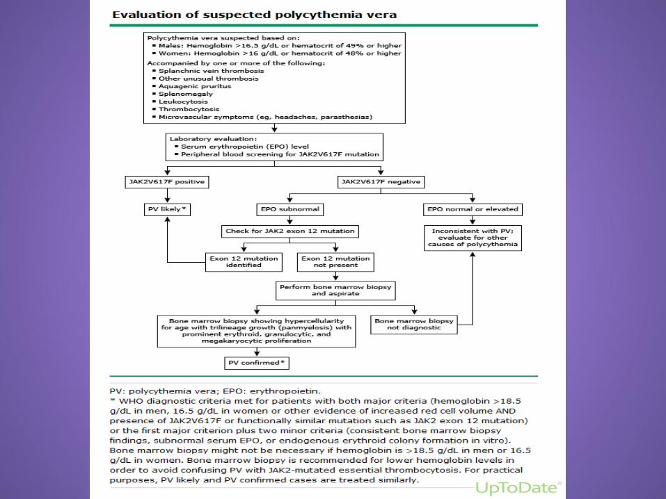
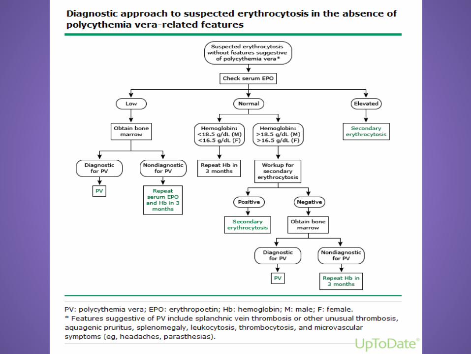
Erythrocytosis
Polycythemia
Primary or Secondary
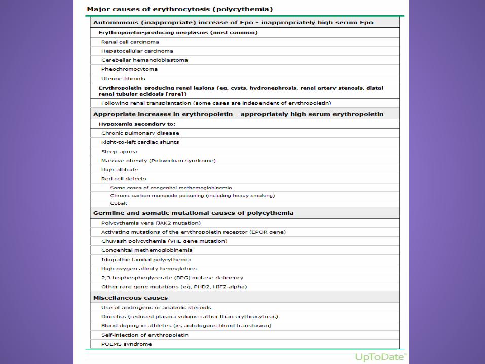
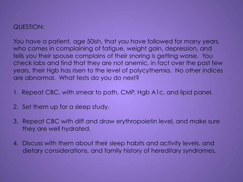
QUESTION: You have a patient, age 50ish, that you have followed for many years, who comes in complaining of fatigue, weight gain, depression, and tells you their spouse complains of their snoring is getting worse. You check labs and find that they are not anemic, in fact over the past few years, their Hgb has risen to the level of polycythemia. No other indices are abnormal. What tests do you do next? 1. Repeat CBC, with smear to path, CMP, Hgb A1c, and lipid panel. 2. Set them up for a sleep study.
3. Repeat CBC with diff and draw erythropoietin level, and make sure they are well hydrated.
4. Discuss with them about their sleep habits and activity levels, and dietary considerations, and family history of hereditary syndromes.
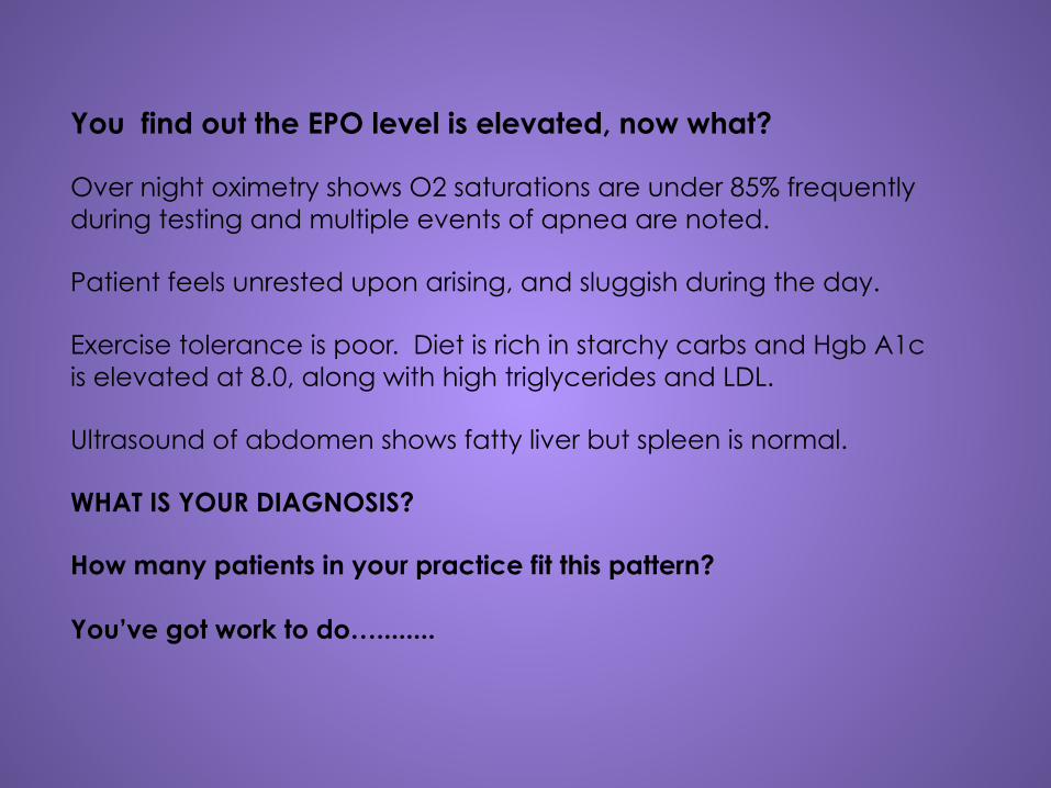
You find out the EPO level is elevated, now what? Over night oximetry shows O2 saturations are under 85% frequently during testing and multiple events of apnea are noted. Patient feels unrested upon arising, and sluggish during the day. Exercise tolerance is poor. Diet is rich in starchy carbs and Hgb A1c is elevated at 8.0, along with high triglycerides and LDL. Ultrasound of abdomen shows fatty liver but spleen is normal. WHAT IS YOUR DIAGNOSIS? How many patients in your practice fit this pattern? You’ve got work to do…........

Let’s Talk White Cells
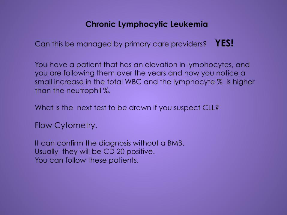
Chronic Lymphocytic Leukemia
Can this be managed by primary care providers? YES! You have a patient that has an elevation in lymphocytes, and you are following them over the years and now you notice a small increase in the total WBC and the lymphocyte % is higher than the neutrophil %. What is the next test to be drawn if you suspect CLL? Flow Cytometry. It can confirm the diagnosis without a BMB. Usually they will be CD 20 positive. You can follow these patients.
Platelets, Thrombocytes those tiny little critters that keep us from bleeding out!
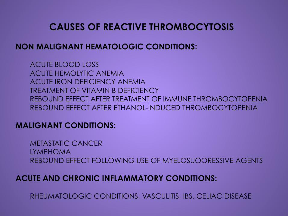
CAUSES OF REACTIVE THROMBOCYTOSIS NON MALIGNANT HEMATOLOGIC CONDITIONS:
ACUTE BLOOD LOSS ACUTE HEMOLYTIC ANEMIA ACUTE IRON DEFICIENCY ANEMIA TREATMENT OF VITAMIN B DEFICIENCY REBOUND EFFECT AFTER TREATMENT OF IMMUNE THROMBOCYTOPENIA REBOUND EFFECT AFTER ETHANOL-INDUCED THROMBOCYTOPENIA
MALIGNANT CONDITIONS:
METASTATIC CANCER LYMPHOMA REBOUND EFFECT FOLLOWING USE OF MYELOSUOORESSIVE AGENTS
ACUTE AND CHRONIC INFLAMMATORY CONDITIONS:
RHEUMATOLOGIC CONDITIONS, VASCULITIS, IBS, CELIAC DISEASE
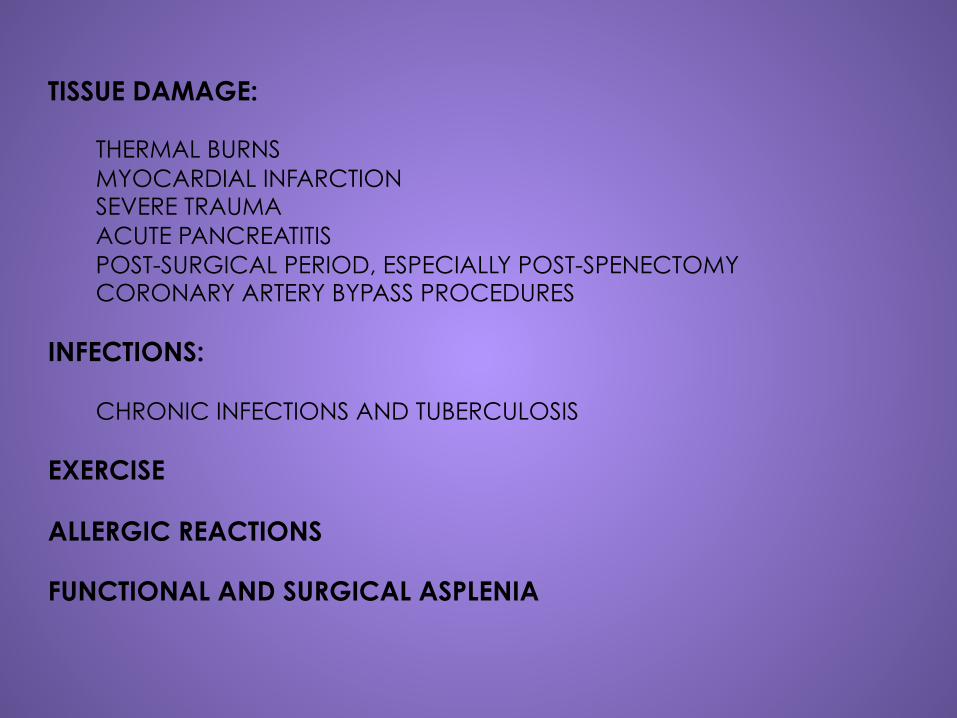
TISSUE DAMAGE:
THERMAL BURNS MYOCARDIAL INFARCTION SEVERE TRAUMA ACUTE PANCREATITIS POST-SURGICAL PERIOD, ESPECIALLY POST-SPENECTOMY CORONARY ARTERY BYPASS PROCEDURES
INFECTIONS:
CHRONIC INFECTIONS AND TUBERCULOSIS EXERCISE ALLERGIC REACTIONS FUNCTIONAL AND SURGICAL ASPLENIA

REACTION TO MEDICATIONS:
VINCRISTINE EPINEPHERINE, GLUCOCORTICOIDS INTERLEUKIN-1B ALL-TRANS RETINOIC ACID THROMBOPOIETIN, THROMBOPOITIN MIMETICS LOW MOLECULAR WEIGHT HEPARINS (ENOXAPARIN)

MEDICATIONS
THOSE PESKY DRUGS

Hydroxyurea: Used mostly these days for Essential Thrombocythemia. It can be used in CML. Dosing is 500 mg tablets titrated to keep the platelet count below 400K. Monitoring CBC’s should be weekly at first and then changed to every 2 weeks until stabilization occurs. It can drop Hgb and WBC’s so titration can be tricky. Usually changes in dosing shouldn’t be sooner than every 2 weeks as it takes that long to stabilize on a new dose. It is an antineoplastic agent and is carcinogenic. Advise sun protection and monitor for malignancies. Adjustments for lower Creatinine clearance. Most people tolerate it without side effects. Causes macrocytosis
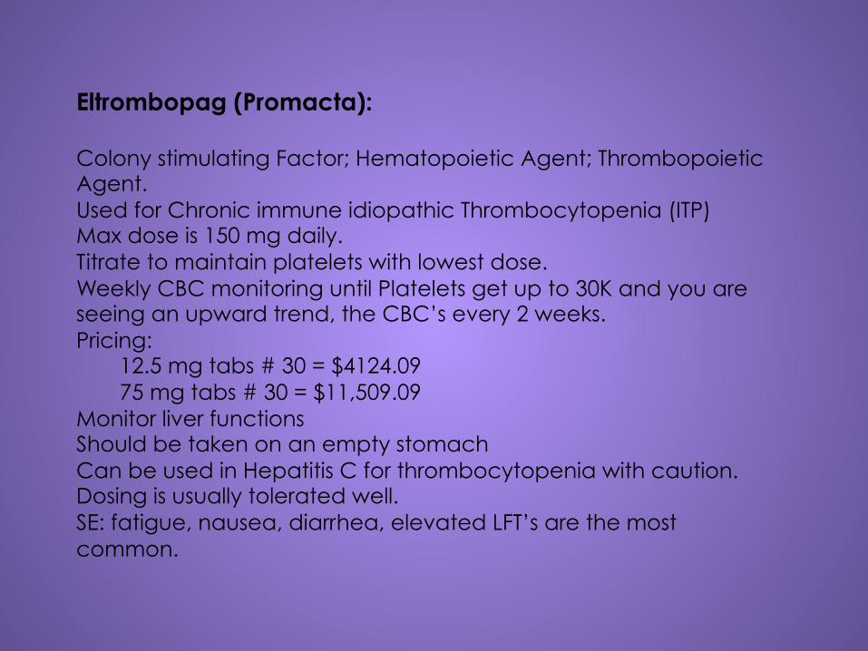
Eltrombopag (Promacta): Colony stimulating Factor; Hematopoietic Agent; Thrombopoietic Agent. Used for Chronic immune idiopathic Thrombocytopenia (ITP) Max dose is 150 mg daily. Titrate to maintain platelets with lowest dose. Weekly CBC monitoring until Platelets get up to 30K and you are seeing an upward trend, the CBC’s every 2 weeks. Pricing:
12.5 mg tabs # 30 = $4124.09 75 mg tabs # 30 = $11,509.09
Monitor liver functions Should be taken on an empty stomach Can be used in Hepatitis C for thrombocytopenia with caution. Dosing is usually tolerated well. SE: fatigue, nausea, diarrhea, elevated LFT’s are the most common.
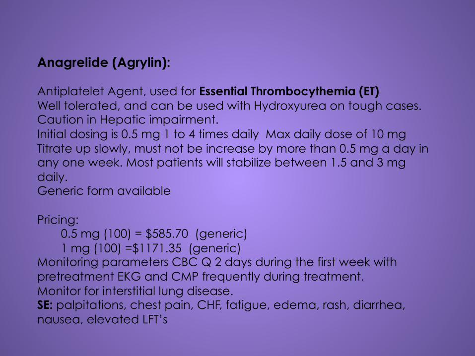
Anagrelide (Agrylin): Antiplatelet Agent, used for Essential Thrombocythemia (ET) Well tolerated, and can be used with Hydroxyurea on tough cases. Caution in Hepatic impairment. Initial dosing is 0.5 mg 1 to 4 times daily Max daily dose of 10 mg Titrate up slowly, must not be increase by more than 0.5 mg a day in any one week. Most patients will stabilize between 1.5 and 3 mg daily. Generic form available Pricing:
0.5 mg (100) = $585.70 (generic) 1 mg (100) =$1171.35 (generic)
Monitoring parameters CBC Q 2 days during the first week with pretreatment EKG and CMP frequently during treatment. Monitor for interstitial lung disease. SE: palpitations, chest pain, CHF, fatigue, edema, rash, diarrhea, nausea, elevated LFT’s
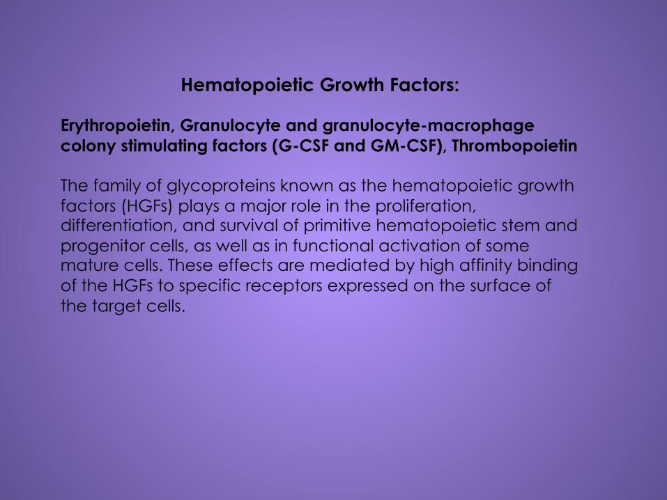
Hematopoietic Growth Factors: Erythropoietin, Granulocyte and granulocyte-macrophage colony stimulating factors (G-CSF and GM-CSF), Thrombopoietin The family of glycoproteins known as the hematopoietic growth factors (HGFs) plays a major role in the proliferation, differentiation, and survival of primitive hematopoietic stem and progenitor cells, as well as in functional activation of some mature cells. These effects are mediated by high affinity binding of the HGFs to specific receptors expressed on the surface of the target cells.
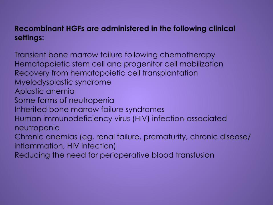
Recombinant HGFs are administered in the following clinical settings: Transient bone marrow failure following chemotherapy Hematopoietic stem cell and progenitor cell mobilization Recovery from hematopoietic cell transplantation Myelodysplastic syndrome Aplastic anemia Some forms of neutropenia Inherited bone marrow failure syndromes Human immunodeficiency virus (HIV) infection-associated neutropenia Chronic anemias (eg, renal failure, prematurity, chronic disease/inflammation, HIV infection) Reducing the need for perioperative blood transfusion
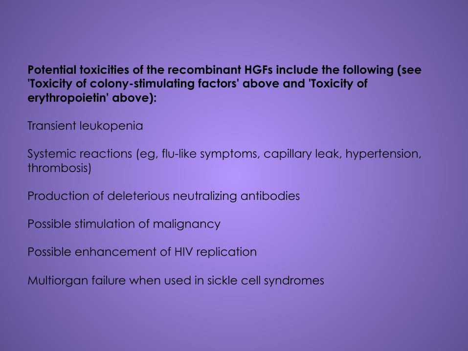
Potential toxicities of the recombinant HGFs include the following (see 'Toxicity of colony-stimulating factors' above and 'Toxicity of erythropoietin' above): Transient leukopenia Systemic reactions (eg, flu-like symptoms, capillary leak, hypertension, thrombosis) Production of deleterious neutralizing antibodies Possible stimulation of malignancy Possible enhancement of HIV replication Multiorgan failure when used in sickle cell syndromes

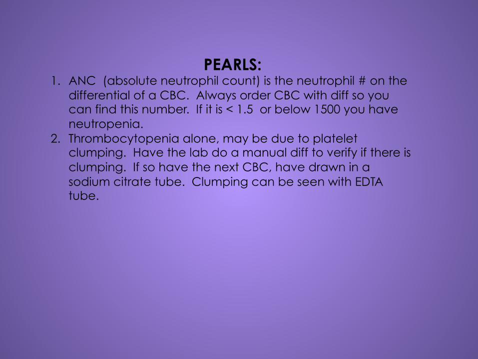
PEARLS: 1. ANC (absolute neutrophil count) is the neutrophil # on the
differential of a CBC. Always order CBC with diff so you can find this number. If it is < 1.5 or below 1500 you have neutropenia.
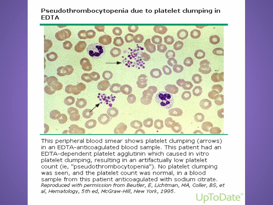
2. Thrombocytopenia alone, may be due to platelet clumping. Have the lab do a manual diff to verify if there is clumping. If so have the next CBC, have drawn in a sodium citrate tube. Clumping can be seen with EDTA tube.
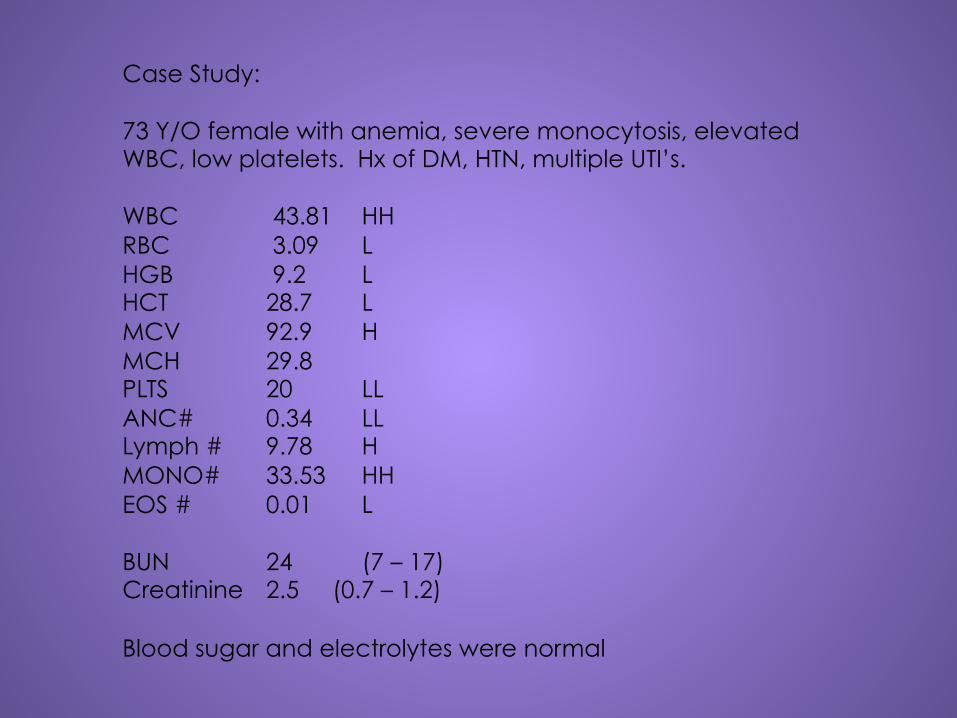
Case Study: 73 Y/O female with anemia, severe monocytosis, elevated WBC, low platelets. Hx of DM, HTN, multiple UTI’s. WBC 43.81 HH RBC 3.09 L HGB 9.2 L HCT 28.7 L MCV 92.9 H MCH 29.8 PLTS 20 LL ANC# 0.34 LL Lymph # 9.78 H MONO# 33.53 HH EOS # 0.01 L BUN 24 (7 – 17) Creatinine 2.5 (0.7 – 1.2) Blood sugar and electrolytes were normal
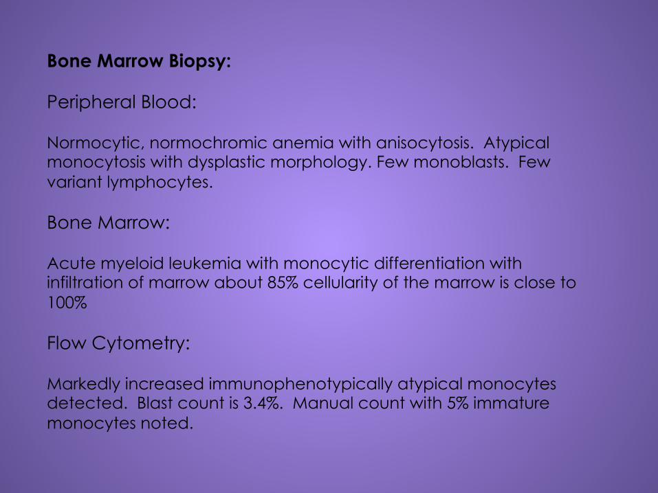
Bone Marrow Biopsy: Peripheral Blood: Normocytic, normochromic anemia with anisocytosis. Atypical monocytosis with dysplastic morphology. Few monoblasts. Few variant lymphocytes. Bone Marrow: Acute myeloid leukemia with monocytic differentiation with infiltration of marrow about 85% cellularity of the marrow is close to 100% Flow Cytometry: Markedly increased immunophenotypically atypical monocytes detected. Blast count is 3.4%. Manual count with 5% immature monocytes noted.
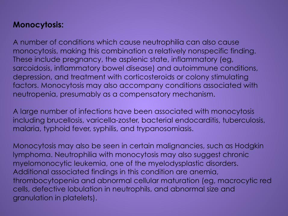
Monocytosis: A number of conditions which cause neutrophilia can also cause monocytosis, making this combination a relatively nonspecific finding. These include pregnancy, the asplenic state, inflammatory (eg, sarcoidosis, inflammatory bowel disease) and autoimmune conditions, depression, and treatment with corticosteroids or colony stimulating factors. Monocytosis may also accompany conditions associated with neutropenia, presumably as a compensatory mechanism. A large number of infections have been associated with monocytosis including brucellosis, varicella-zoster, bacterial endocarditis, tuberculosis, malaria, typhoid fever, syphilis, and trypanosomiasis. Monocytosis may also be seen in certain malignancies, such as Hodgkin lymphoma. Neutrophilia with monocytosis may also suggest chronic myelomonocytic leukemia, one of the myelodysplastic disorders. Additional associated findings in this condition are anemia, thrombocytopenia and abnormal cellular maturation (eg, macrocytic red cells, defective lobulation in neutrophils, and abnormal size and granulation in platelets).
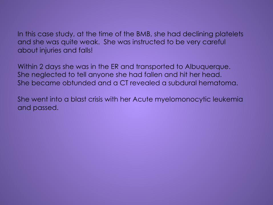
In this case study, at the time of the BMB, she had declining platelets and she was quite weak. She was instructed to be very careful about injuries and falls! Within 2 days she was in the ER and transported to Albuquerque. She neglected to tell anyone she had fallen and hit her head. She became obtunded and a CT revealed a subdural hematoma. She went into a blast crisis with her Acute myelomonocytic leukemia and passed.
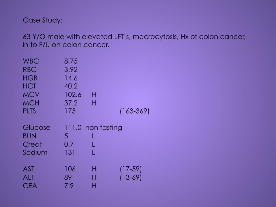
Case Study: 63 Y/O male with elevated LFT’s, macrocytosis, Hx of colon cancer, in to F/U on colon cancer. WBC 8.75 RBC 3.92 HGB 14.6 HCT 40.2 MCV 102.6 H MCH 37.2 H PLTS 175 (163-369) Glucose 111.0 non fasting BUN 5 L Creat 0.7 L Sodium 131 L AST 106 H (17-59) ALT 89 H (13-69) CEA 7.9 H

Ordered the following: Restaging CT Chest, Abd, and pelvis Hepatitis panel Colonoscopy All were negative. Patient smokes 2 pks per day Patient drinks 12 pk beer per day CEA is elevated in smokers Macrocytosis and elevated liver functions from alcohol intake.
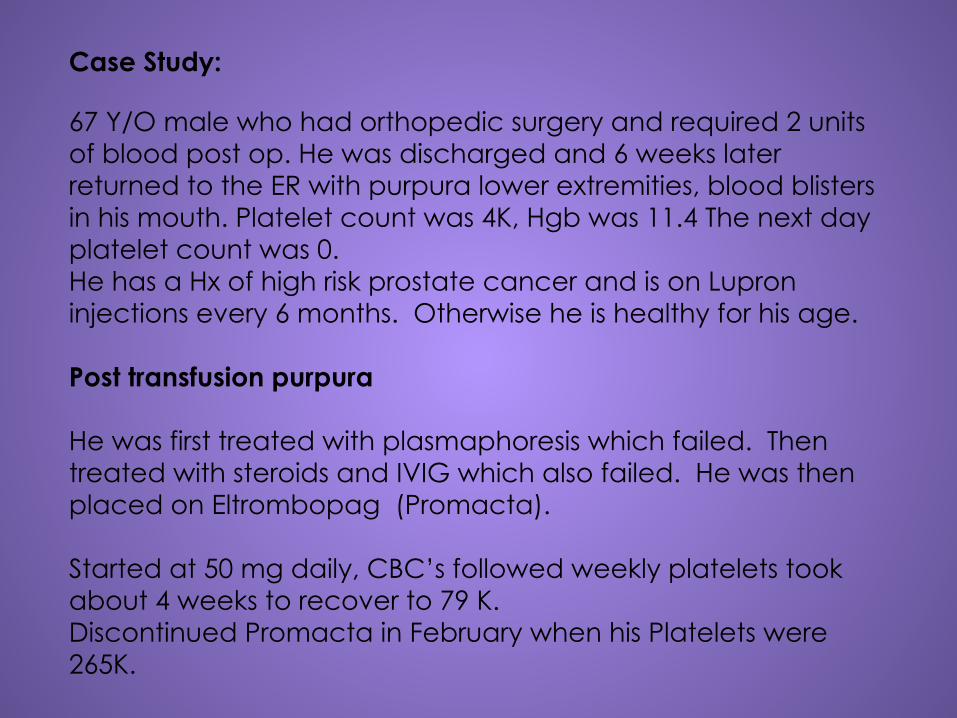
Case Study: 67 Y/O male who had orthopedic surgery and required 2 units of blood post op. He was discharged and 6 weeks later returned to the ER with purpura lower extremities, blood blisters in his mouth. Platelet count was 4K, Hgb was 11.4 The next day platelet count was 0. He has a Hx of high risk prostate cancer and is on Lupron injections every 6 months. Otherwise he is healthy for his age. Post transfusion purpura He was first treated with plasmaphoresis which failed. Then treated with steroids and IVIG which also failed. He was then placed on Eltrombopag (Promacta). Started at 50 mg daily, CBC’s followed weekly platelets took about 4 weeks to recover to 79 K. Discontinued Promacta in February when his Platelets were 265K.

Hemochromatosis TestingClick here for topics associated with this algorithm
INDICATIONS FOR TESTINGSuspicion of hemochromatosis (family history, compatible symptoms)
ORDERIron and Iron Binding Capacity
Note: Test includes serum transferrin saturation (STS)AND
Ferritin (SF)
No further testing at this point
Repeat STS and SF tests
Repeat STS and SF tests at 2-year intervals
Secondary iron
overloadIf both elevated, do liver biopsy
FOR ADULTS, ORDERHemochromatosis (HFE) 3
Mutations(accounts for >90% of mutations in
Caucasians)
C282Y/C282Y C282Y/H63DC282Y/S65CC282Y/wt H63D/H63D
H63D/S65C
Ferritin <1000 μg/LAspartate aminotransferase (AST) normal
Liver biopsy with hepatic iron concentration Phlebotomy Monitor STS
Family screening
Monitor STSConsider liver biopsy
Consider alternative gene testing (TFr2, FPN) ORDER
FerritinAND
Aspartate Aminotransferase, Serum or Plasma
Treat underlying
cause
Hemochromatosis confirmed
Ferritin ≥1000 μg/LAspartate aminotransferase (AST) abnormal
STS ≥45% and/or elevated SF
STS <45% and normal SF
STS ≥45% SF: elevated for age and sex
(esp. if >2x normal)
© 2006 ARUP Laboratories. All Rights Reserved. Revised 01/18/2012 www.arupconsult.com
No Yes
FOR PEDIATRICS, CONSIDER
HJV (HFE2) gene sequencing(accounts for >90% of cases)
CONSIDERHAMP (HEPC) gene
sequencing(accounts for <10% of cases)
Positive Negative
Hemochromatosis confirmed

AND
Plasma Cell DyscrasiasClick here for topics associated with this algorithm
INDICATIONS FOR TESTINGBone pain, recurrent infections, anemia,
lytic lesions on plain film
Perform baseline screeningCBC with Platelet Count and Automated DifferentialMetabolic profile, which should include
Calcium, Serum or PlasmaBUN/Creatinine, Serum or PlasmaProtein, Total, Serum or PlasmAlbumin, Serum or Plasma by Spectrophotometry
Lactate Dehydrogenase, Serum or Plasma
Rule outChronic infections such as HIVImmunoglobulin deficiencies such as Common Variable Immunodeficiency (CVID)Chronic inflammatory processes such as systemic lupus erythematous, liver diseaseOther malignancies
If negative for other diseases
ORDERProtein Electrophoresis with Reflex to Immunofixation Electrophoresis Monoclonal Protein Detection, Quantitation and Characterization, IgA, IgG, and IgM, Serum24 hr urine protein electrophoresis
ANDMonoclonal Protein Detection Quantitation & Characterization, SPEP, IFE, IgA, IgG, IgM, Serum (Protein electrophoresis with reflex testing may occasionally miss IgA MGUS or multiple myeloma [MM])Immunofixation Electrophoresis, Qualitative, Gel
Serum M protein ≥3 g/dL
Abnormal baseline testing or suspicion for multiple myeloma (MM)
ORDERSerum free light chain ratio (Kappa/Lambda Quantitative
Free Light Chains with Ratio, Serum) to diagnose oligosecretory myeloma and non-secretory myeloma
Normal FLC ratio
Abnormal FLC ratio
Monoclonal gammopathy of undetermined significance
(MGUS)
Repeat evaluation in 3-6 months
ORDERBone marrow biopsy
Skeletal survey
<10% plasma cells
Likely MGUS
≥10% plasma cells
<10% plasma cells No bone lesions presentNormal baseline testing
≥10% plasma cells* Bone lesions presentAbnormal baseline
testing
Asymptomatic (smoldering)
MM
Repeat evaluation in
3 months
MM
ORDERBone marrow biopsy
Skeletal survey
Repeat evaluation in 3-6 months
© 2006 ARUP Laboratories. All Rights Reserved. 02/17/2015 www.arupconsult.com
No Yes
YesNo
MM
Asymptomatic (smoldering) MM *This criterion is unnecessary
if bone lesions are present
Bone lessions
Repeat evaluation in 3 months
No Yes
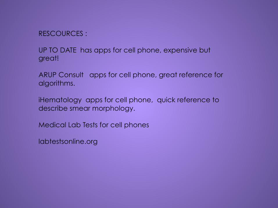
RESCOURCES : UP TO DATE has apps for cell phone, expensive but great! ARUP Consult apps for cell phone, great reference for algorithms. iHematology apps for cell phone, quick reference to describe smear morphology. Medical Lab Tests for cell phones labtestsonline.org

WHEN TO REFER 1. PANCYTOPENIA 2. PLATELETS TREND DOWN OVER TIME AND ARE STAYING
UNDER 100 K 3. YOU CAN’T FIND A REASON FOR IRON DEFICIENCY 4. UNEXPLAINED LEUKOCYTOSIS 5. UNEXPLAINED ADENOPATHY…. GET IT BIOPSIED! 6. INTOLERANCE TO ORAL IRON AND PERSISTANCE OF
IRON DEFICIENCY WITH NEGATIVE WORKUP 7. YOU HAVE A BAD FEELING AND TOO MAY ABNORMALS
ON THE SMEAR... MY ADVICE: GET TO KNOW YOUR LOCAL HEMATOLOGIST AND ASK FOR ADVICE. THEY MAY HAVE A FRIENDLY NP TO TALK TO. SHE OR HE MAY HAVE GOOD ADVICE!!!