Embed Size (px)
Citation preview
Case scenarios in Pediatric Hematology and
Oncology
Dr. Zainul Aabideen Consultant, Pediatric Hematology and Oncology
Burjeel Hospital
* To discuss and learn initial features of childhood leukemia. * To discuss about differential diagnosis of iron deficiency anemia. * Understand about thalassemia carrier. * Discuss and learn treatment of iron deficiency anemia. * Discuss what could you do in acute severe anemia
Objectives
Dr. Zainul Aabideen; Consultant, Pediatric Hematology and Oncology; Burjeel Hospital
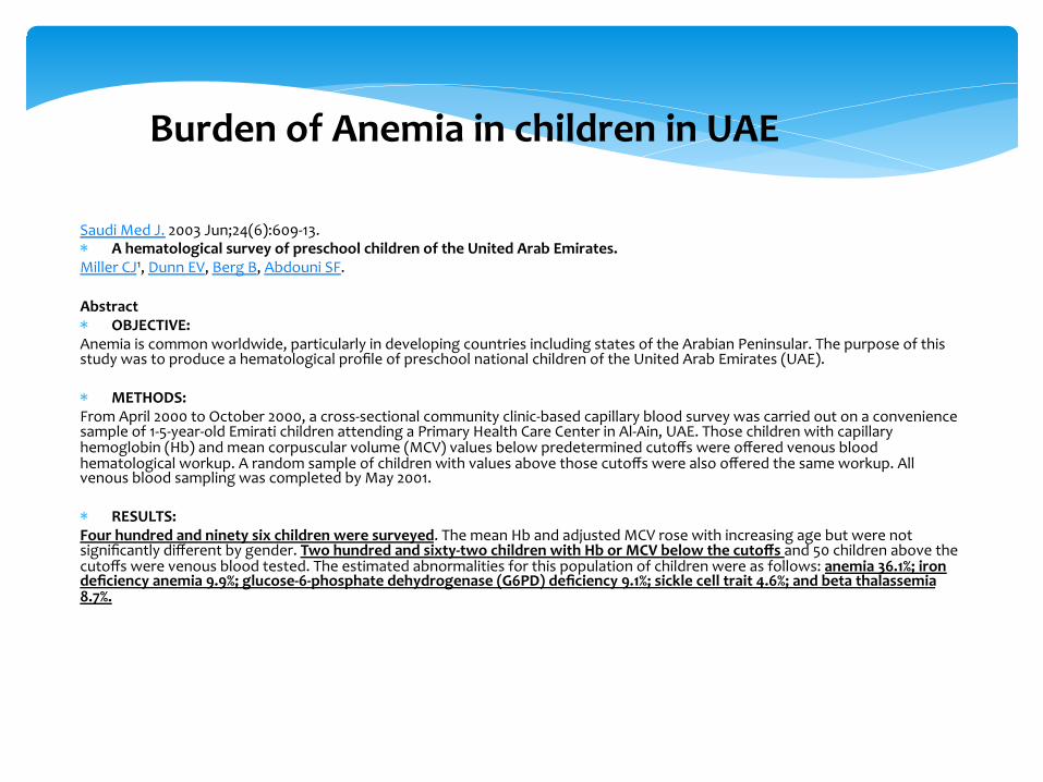
Thank you Saudi Med J. 2003 Jun;24(6):609-‐13. * A hematological survey of preschool children of the United Arab Emirates. Miller CJ1, Dunn EV, Berg B, Abdouni SF. Abstract * OBJECTIVE: Anemia is common worldwide, particularly in developing countries including states of the Arabian Peninsular. The purpose of this study was to produce a hematological profile of preschool national children of the United Arab Emirates (UAE). * METHODS: From April 2000 to October 2000, a cross-‐sectional community clinic-‐based capillary blood survey was carried out on a convenience sample of 1-‐5-‐year-‐old Emirati children attending a Primary Health Care Center in Al-‐Ain, UAE. Those children with capillary hemoglobin (Hb) and mean corpuscular volume (MCV) values below predetermined cutoffs were offered venous blood hematological workup. A random sample of children with values above those cutoffs were also offered the same workup. All venous blood sampling was completed by May 2001. * RESULTS: Four hundred and ninety six children were surveyed. The mean Hb and adjusted MCV rose with increasing age but were not significantly different by gender. Two hundred and sixty-‐two children with Hb or MCV below the cutoffs and 50 children above the cutoffs were venous blood tested. The estimated abnormalities for this population of children were as follows: anemia 36.1%; iron deficiency anemia 9.9%; glucose-‐6-‐phosphate dehydrogenase (G6PD) deficiency 9.1%; sickle cell trait 4.6%; and beta thalassemia 8.7%.
Burden of Anemia in children in UAE
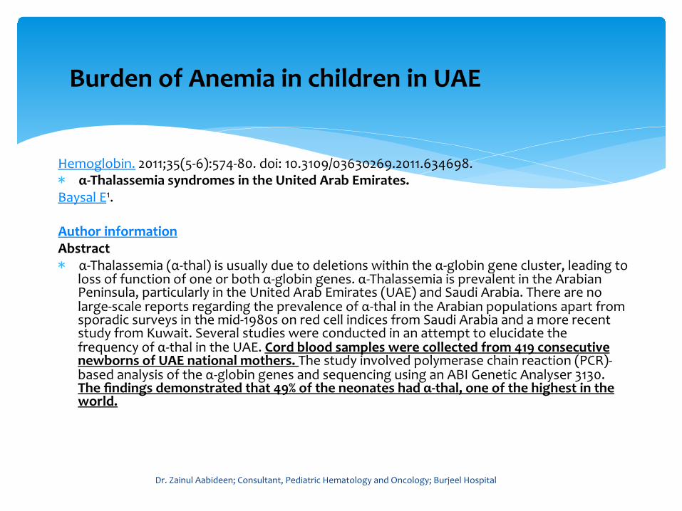
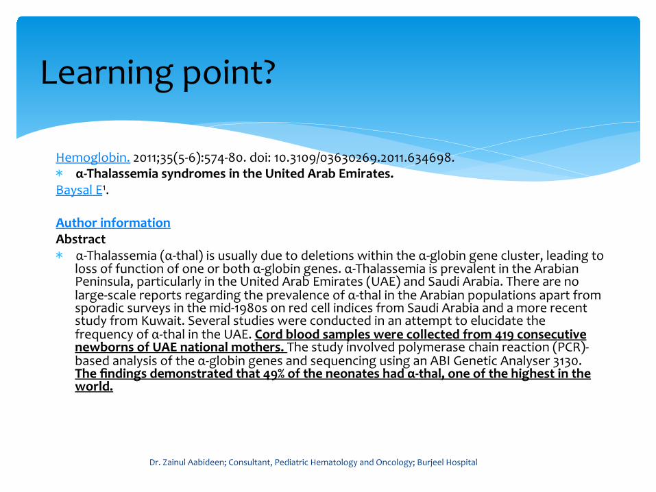
Hemoglobin. 2011;35(5-‐6):574-‐80. doi: 10.3109/03630269.2011.634698. * α-‐Thalassemia syndromes in the United Arab Emirates. Baysal E1. Author information Abstract * α-‐Thalassemia (α-‐thal) is usually due to deletions within the α-‐globin gene cluster, leading to
loss of function of one or both α-‐globin genes. α-‐Thalassemia is prevalent in the Arabian Peninsula, particularly in the United Arab Emirates (UAE) and Saudi Arabia. There are no large-‐scale reports regarding the prevalence of α-‐thal in the Arabian populations apart from sporadic surveys in the mid-‐1980s on red cell indices from Saudi Arabia and a more recent study from Kuwait. Several studies were conducted in an attempt to elucidate the frequency of α-‐thal in the UAE. Cord blood samples were collected from 419 consecutive newborns of UAE national mothers. The study involved polymerase chain reaction (PCR)-‐based analysis of the α-‐globin genes and sequencing using an ABI Genetic Analyser 3130. The findings demonstrated that 49% of the neonates had α-‐thal, one of the highest in the world.
Dr. Zainul Aabideen; Consultant, Pediatric Hematology and Oncology; Burjeel Hospital
Burden of Anemia in children in UAE
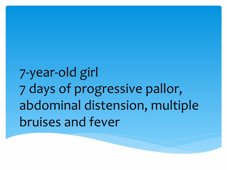
7-‐year-‐old girl 7 days of progressive pallor, abdominal distension, multiple bruises and fever
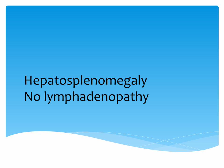
Hepatosplenomegaly No lymphadenopathy
What investigations we could do at this point?
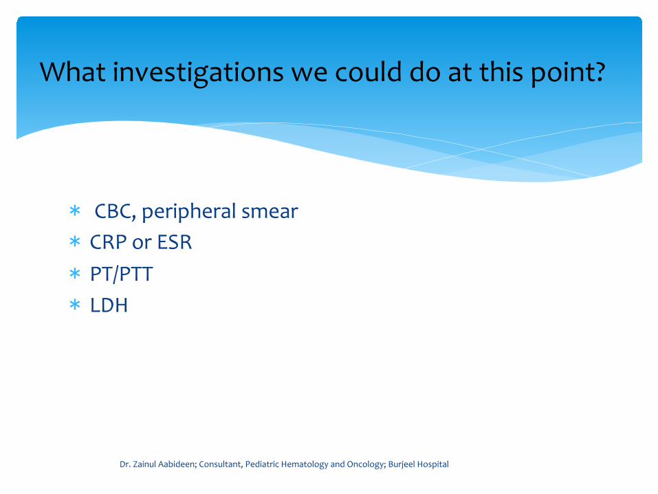
* CBC, peripheral smear * CRP or ESR * PT/PTT * LDH
What investigations we could do at this point?
Dr. Zainul Aabideen; Consultant, Pediatric Hematology and Oncology; Burjeel Hospital
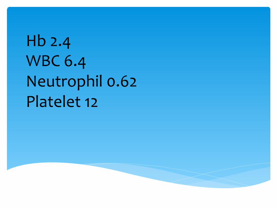
Hb 2.4 WBC 6.4 Neutrophil 0.62 Platelet 12
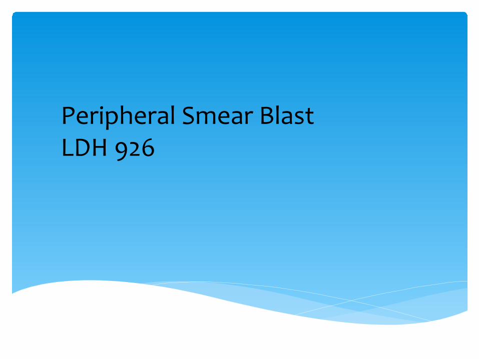
Peripheral Smear Blast LDH 926
Bone Marrow Examination reveals this child has Acute Lymphoblastic Leukemia
What we can learn from this experience
• How the symptoms and signs develop in ALL?
• What are the clinical features of ALL? • What are the differential diagnosis of ALL in children?
• What are the basic investigations you could do ?
Dr. Zainul Aabideen; Consultant, Pediatric Hematology and Oncology; Burjeel Hospital

Clinical Features
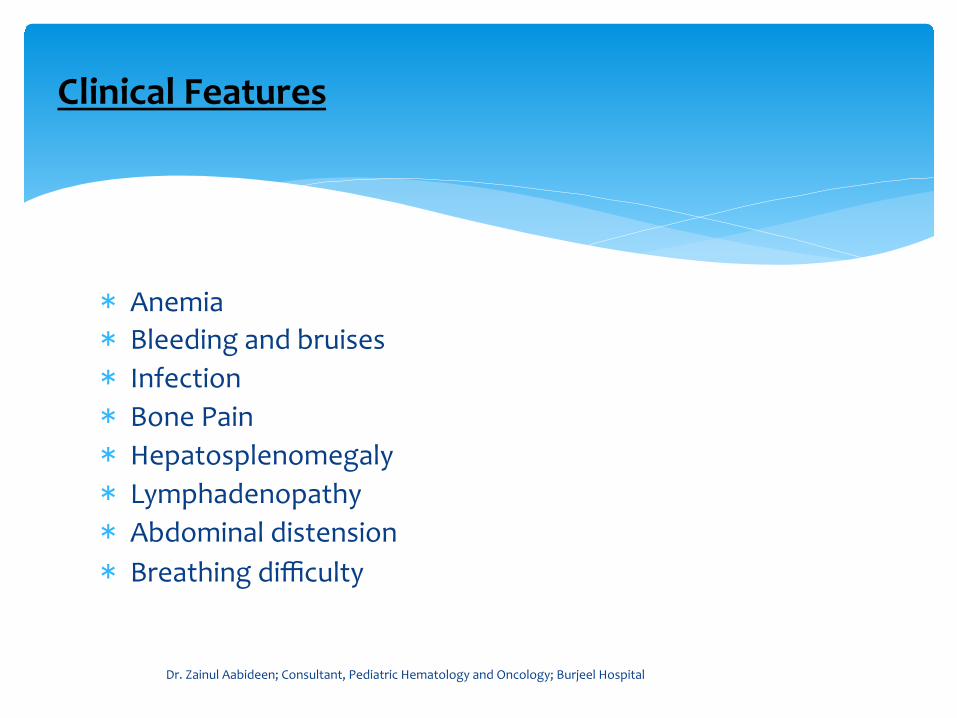
* Anemia * Bleeding and bruises * Infection * Bone Pain * Hepatosplenomegaly * Lymphadenopathy * Abdominal distension * Breathing difficulty
Clinical Features
Dr. Zainul Aabideen; Consultant, Pediatric Hematology and Oncology; Burjeel Hospital
Differential diagnosis
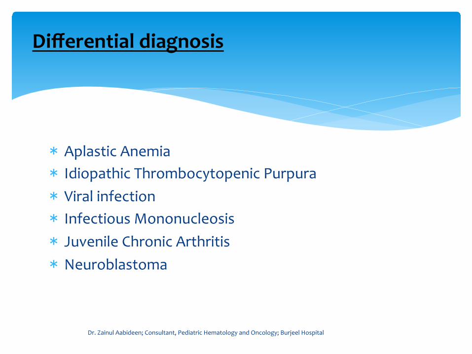
* Aplastic Anemia * Idiopathic Thrombocytopenic Purpura * Viral infection * Infectious Mononucleosis * Juvenile Chronic Arthritis * Neuroblastoma
Differential diagnosis
Dr. Zainul Aabideen; Consultant, Pediatric Hematology and Oncology; Burjeel Hospital
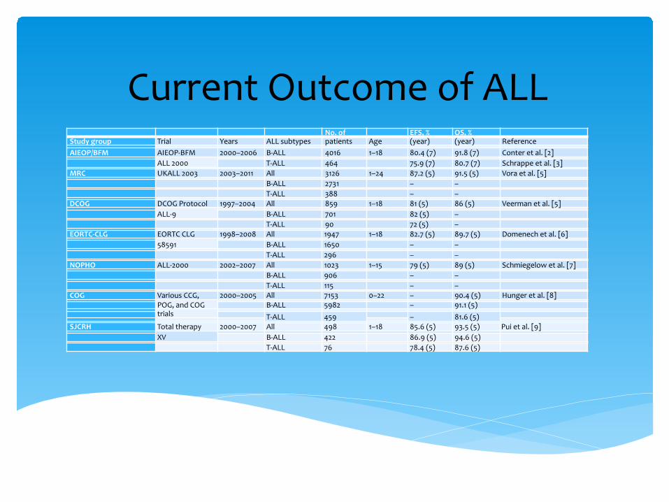
Current Outcome of ALL
No. of EFS, % OS, % Study group Trial Years ALL subtypes patients Age (year) (year) Reference
AIEOP/BFM AIEOP-‐BFM 2000–2006 B-‐ALL 4016 1–18 80.4 (7) 91.8 (7) Conter et al. [2]
ALL 2000
T-‐ALL 464 75.9 (7) 80.7 (7) Schrappe et al. [3] MRC UKALL 2003 2003–2011 All 3126 1–24 87.2 (5) 91.5 (5) Vora et al. [5]
B-‐ALL 2731 – – T-‐ALL 388 – – DCOG DCOG Protocol 1997–2004 All 859 1–18 81 (5) 86 (5) Veerman et al. [5]
ALL-‐9
B-‐ALL 701 82 (5) – T-‐ALL 90 72 (5) – EORTC-‐CLG EORTC CLG 1998–2008 All 1947 1–18 82.7 (5) 89.7 (5) Domenech et al. [6]
58591
B-‐ALL 1650 – – T-‐ALL 296 – – NOPHO ALL-‐2000 2002–2007 All 1023 1–15 79 (5) 89 (5) Schmiegelow et al. [7]
B-‐ALL 906 – – T-‐ALL 115 – – COG Various CCG, 2000–2005 All 7153 0–22 – 90.4 (5) Hunger et al. [8]
POG, and COG
B-‐ALL 5982 – 91.1 (5) trials T-‐ALL 459 – 81.6 (5) SJCRH Total therapy 2000–2007 All 498 1–18 85.6 (5) 93.5 (5) Pui et al. [9]
XV
B-‐ALL 422 86.9 (5) 94.6 (5) T-‐ALL 76 78.4 (5) 87.6 (5)
Case 2
Case 2 1-‐year-‐old Pakistani boy
Dr. Zainul Aabideen; Consultant, Pediatric Hematology and Oncology; Burjeel Hospital
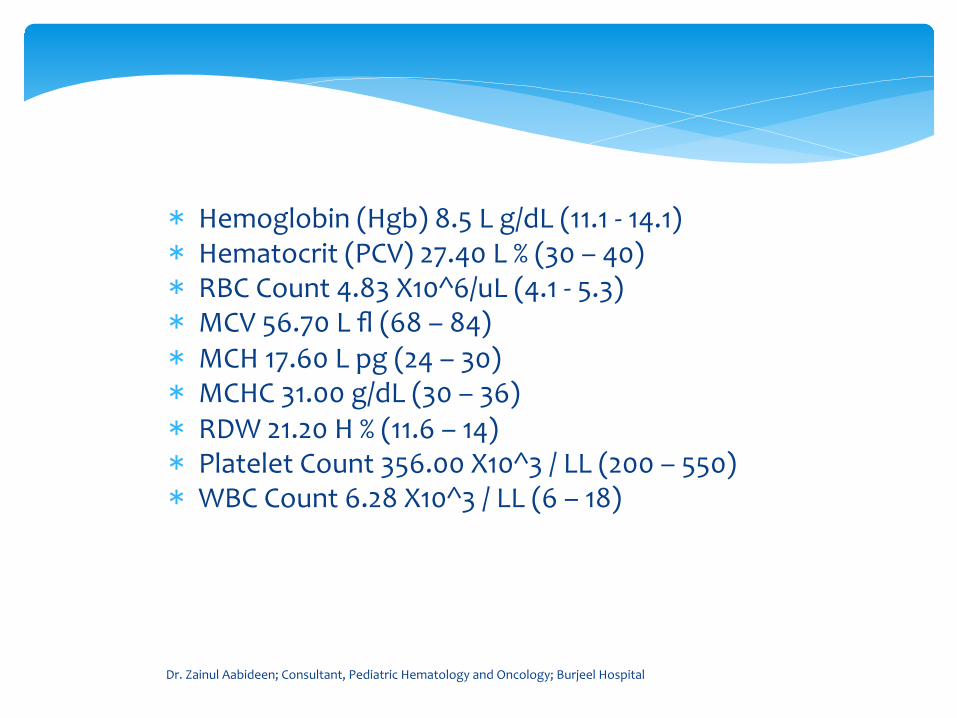
* Hemoglobin (Hgb) 8.5 L g/dL (11.1 -‐ 14.1) * Hematocrit (PCV) 27.40 L % (30 – 40) * RBC Count 4.83 X10^6/uL (4.1 -‐ 5.3) * MCV 56.70 L fl (68 – 84) * MCH 17.60 L pg (24 – 30) * MCHC 31.00 g/dL (30 – 36) * RDW 21.20 H % (11.6 – 14) * Platelet Count 356.00 X10^3 / LL (200 – 550) * WBC Count 6.28 X10^3 / LL (6 – 18)
Dr. Zainul Aabideen; Consultant, Pediatric Hematology and Oncology; Burjeel Hospital
Iron treatment given.
Dr. Zainul Aabideen; Consultant, Pediatric Hematology and Oncology; Burjeel Hospital
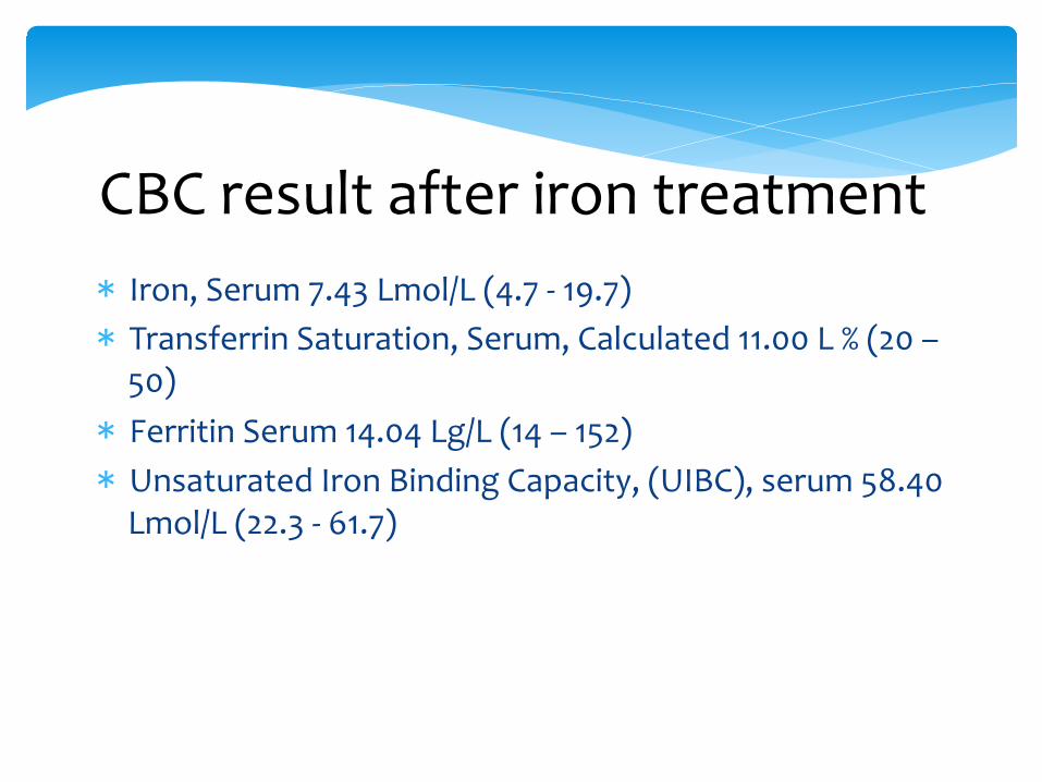
CBC result after iron treatment * Iron, Serum 7.43 Lmol/L (4.7 -‐ 19.7) * Transferrin Saturation, Serum, Calculated 11.00 L % (20 – 50) * Ferritin Serum 14.04 Lg/L (14 – 152) * Unsaturated Iron Binding Capacity, (UIBC), serum 58.40 Lmol/L (22.3 -‐ 61.7)
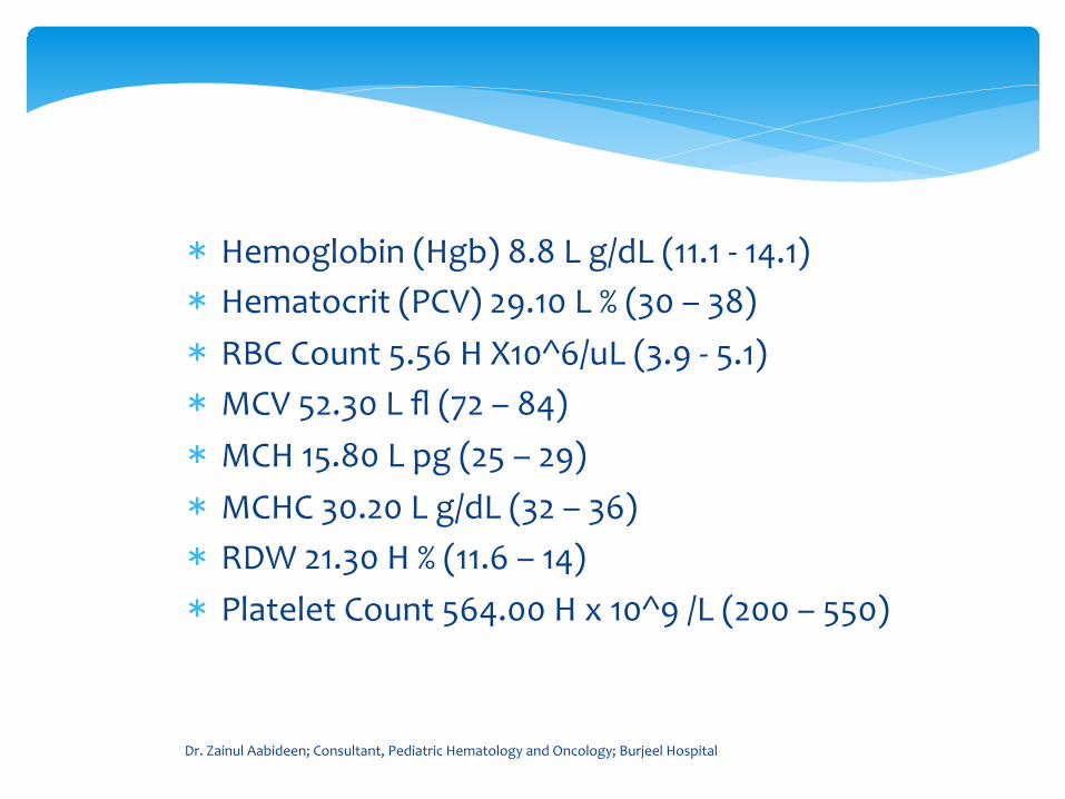
* Hemoglobin (Hgb) 8.8 L g/dL (11.1 -‐ 14.1) * Hematocrit (PCV) 29.10 L % (30 – 38) * RBC Count 5.56 H X10^6/uL (3.9 -‐ 5.1) * MCV 52.30 L fl (72 – 84) * MCH 15.80 L pg (25 – 29) * MCHC 30.20 L g/dL (32 – 36) * RDW 21.30 H % (11.6 – 14) * Platelet Count 564.00 H x 10^9 /L (200 – 550)
Dr. Zainul Aabideen; Consultant, Pediatric Hematology and Oncology; Burjeel Hospital
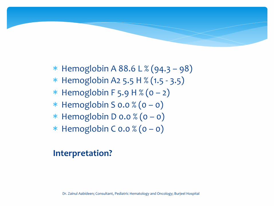
Case 2 * Hemoglobin A 88.6 L % (94.3 – 98) * Hemoglobin A2 5.5 H % (1.5 -‐ 3.5) * Hemoglobin F 5.9 H % (0 – 2) * Hemoglobin S 0.0 % (0 – 0) * Hemoglobin D 0.0 % (0 – 0) * Hemoglobin C 0.0 % (0 – 0) Interpretation?
Dr. Zainul Aabideen; Consultant, Pediatric Hematology and Oncology; Burjeel Hospital
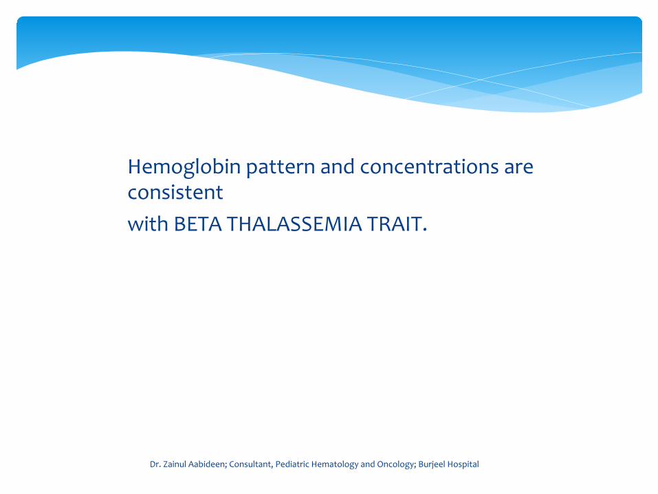
Case 2 Hemoglobin pattern and concentrations are consistent with BETA THALASSEMIA TRAIT.
Dr. Zainul Aabideen; Consultant, Pediatric Hematology and Oncology; Burjeel Hospital
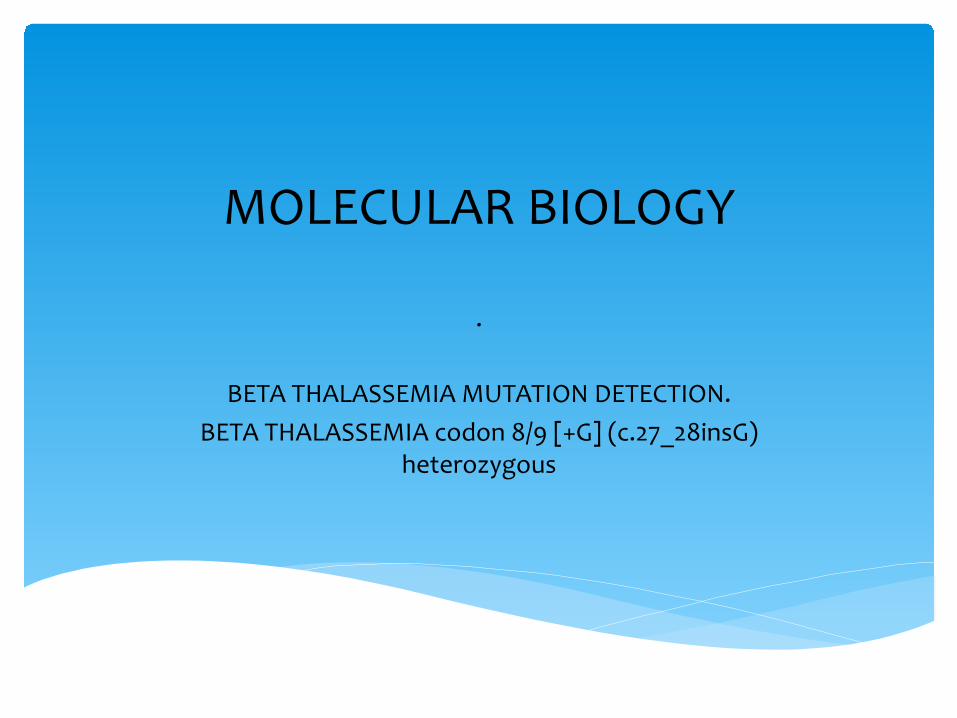
MOLECULAR BIOLOGY .
BETA THALASSEMIA MUTATION DETECTION. BETA THALASSEMIA codon 8/9 [+G] (c.27_28insG)
heterozygous

Case 3
2 years old Filipino boy 2 courses of Iron treatment and history of iron treatment with
mother
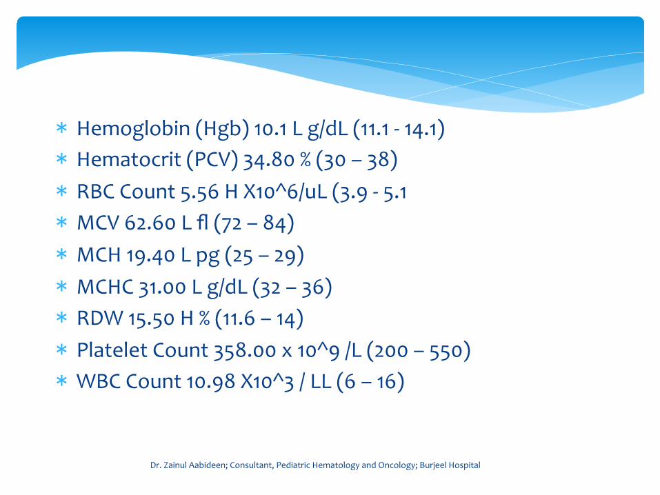
* Hemoglobin (Hgb) 10.1 L g/dL (11.1 -‐ 14.1) * Hematocrit (PCV) 34.80 % (30 – 38) * RBC Count 5.56 H X10^6/uL (3.9 -‐ 5.1 * MCV 62.60 L fl (72 – 84) * MCH 19.40 L pg (25 – 29) * MCHC 31.00 L g/dL (32 – 36) * RDW 15.50 H % (11.6 – 14) * Platelet Count 358.00 x 10^9 /L (200 – 550) * WBC Count 10.98 X10^3 / LL (6 – 16)
Dr. Zainul Aabideen; Consultant, Pediatric Hematology and Oncology; Burjeel Hospital
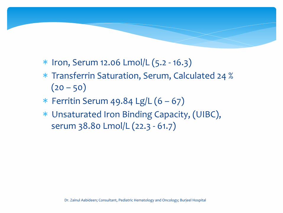
* Iron, Serum 12.06 Lmol/L (5.2 -‐ 16.3) * Transferrin Saturation, Serum, Calculated 24 % (20 – 50) * Ferritin Serum 49.84 Lg/L (6 – 67) * Unsaturated Iron Binding Capacity, (UIBC), serum 38.80 Lmol/L (22.3 -‐ 61.7)
Dr. Zainul Aabideen; Consultant, Pediatric Hematology and Oncology; Burjeel Hospital
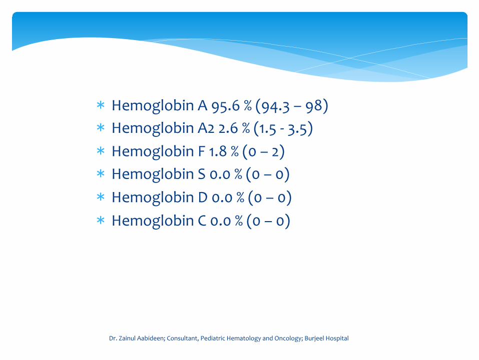
Electrophoresis Normal * Hemoglobin A 95.6 % (94.3 – 98) * Hemoglobin A2 2.6 % (1.5 -‐ 3.5) * Hemoglobin F 1.8 % (0 – 2) * Hemoglobin S 0.0 % (0 – 0) * Hemoglobin D 0.0 % (0 – 0) * Hemoglobin C 0.0 % (0 – 0)
Dr. Zainul Aabideen; Consultant, Pediatric Hematology and Oncology; Burjeel Hospital
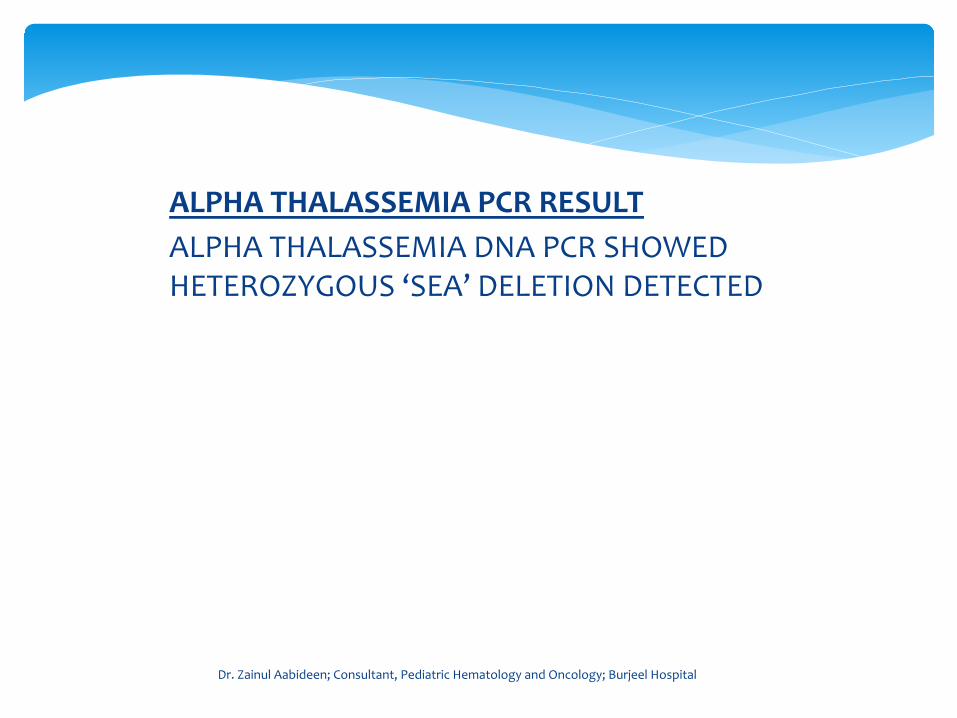
Electrophoresis Normal ALPHA THALASSEMIA PCR RESULT ALPHA THALASSEMIA DNA PCR SHOWED HETEROZYGOUS ‘SEA’ DELETION DETECTED
Dr. Zainul Aabideen; Consultant, Pediatric Hematology and Oncology; Burjeel Hospital
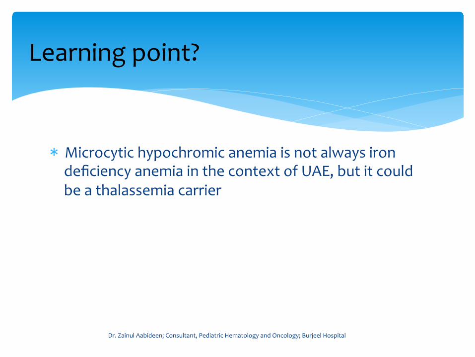
* Microcytic hypochromic anemia is not always iron deficiency anemia in the context of UAE, but it could be a thalassemia carrier
Learning point?
Dr. Zainul Aabideen; Consultant, Pediatric Hematology and Oncology; Burjeel Hospital
Hemoglobin. 2011;35(5-‐6):574-‐80. doi: 10.3109/03630269.2011.634698. * α-‐Thalassemia syndromes in the United Arab Emirates. Baysal E1. Author information Abstract * α-‐Thalassemia (α-‐thal) is usually due to deletions within the α-‐globin gene cluster, leading to
loss of function of one or both α-‐globin genes. α-‐Thalassemia is prevalent in the Arabian Peninsula, particularly in the United Arab Emirates (UAE) and Saudi Arabia. There are no large-‐scale reports regarding the prevalence of α-‐thal in the Arabian populations apart from sporadic surveys in the mid-‐1980s on red cell indices from Saudi Arabia and a more recent study from Kuwait. Several studies were conducted in an attempt to elucidate the frequency of α-‐thal in the UAE. Cord blood samples were collected from 419 consecutive newborns of UAE national mothers. The study involved polymerase chain reaction (PCR)-‐based analysis of the α-‐globin genes and sequencing using an ABI Genetic Analyser 3130. The findings demonstrated that 49% of the neonates had α-‐thal, one of the highest in the world.
Learning point?
Dr. Zainul Aabideen; Consultant, Pediatric Hematology and Oncology; Burjeel Hospital
Case 4
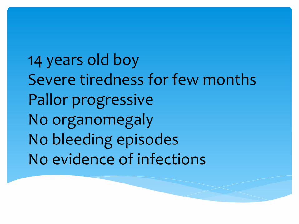
14 years old boy Severe tiredness for few months Pallor progressive No organomegaly No bleeding episodes No evidence of infections
*
What investigations you could do at this point?
Dr. Zainul Aabideen; Consultant, Pediatric Hematology and Oncology; Burjeel Hospital
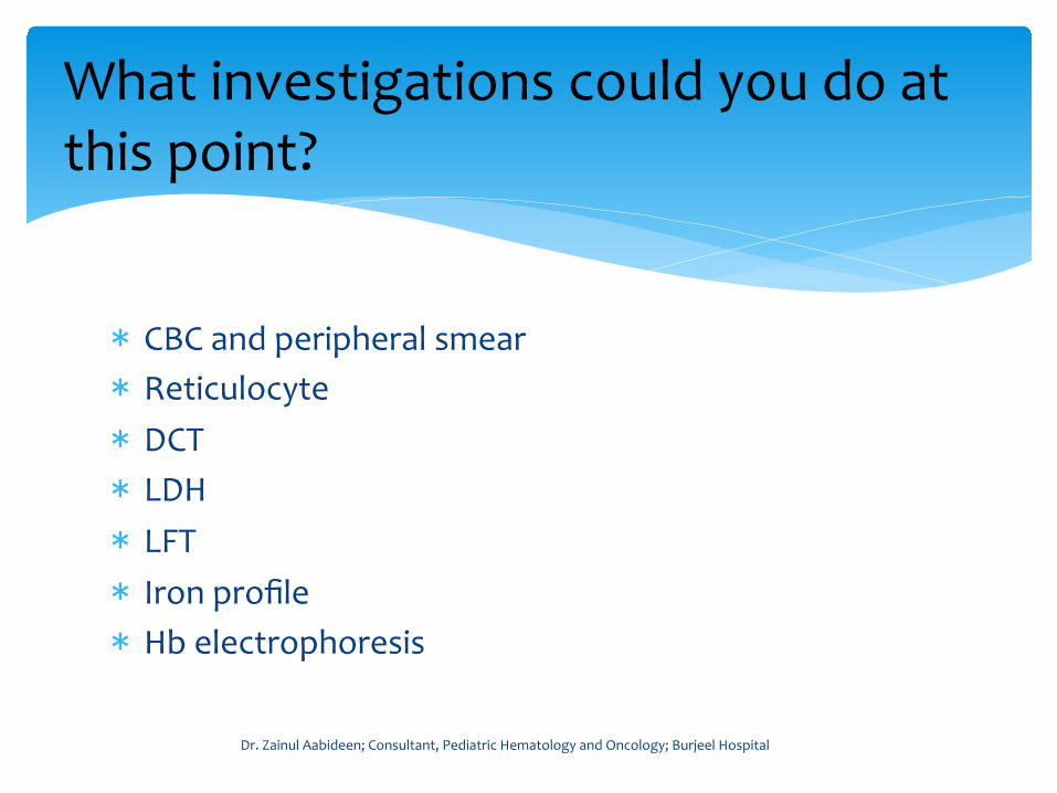
* CBC and peripheral smear * Reticulocyte * DCT * LDH * LFT * Iron profile * Hb electrophoresis
What investigations could you do at this point?
Dr. Zainul Aabideen; Consultant, Pediatric Hematology and Oncology; Burjeel Hospital
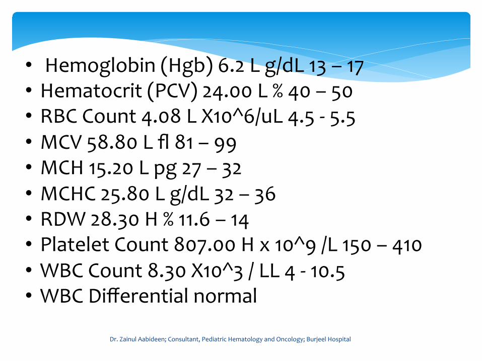
• Hemoglobin (Hgb) 6.2 L g/dL 13 – 17 • Hematocrit (PCV) 24.00 L % 40 – 50 • RBC Count 4.08 L X10^6/uL 4.5 -‐ 5.5 • MCV 58.80 L fl 81 – 99 • MCH 15.20 L pg 27 – 32 • MCHC 25.80 L g/dL 32 – 36 • RDW 28.30 H % 11.6 – 14 • Platelet Count 807.00 H x 10^9 /L 150 – 410 • WBC Count 8.30 X10^3 / LL 4 -‐ 10.5 • WBC Differential normal
Dr. Zainul Aabideen; Consultant, Pediatric Hematology and Oncology; Burjeel Hospital
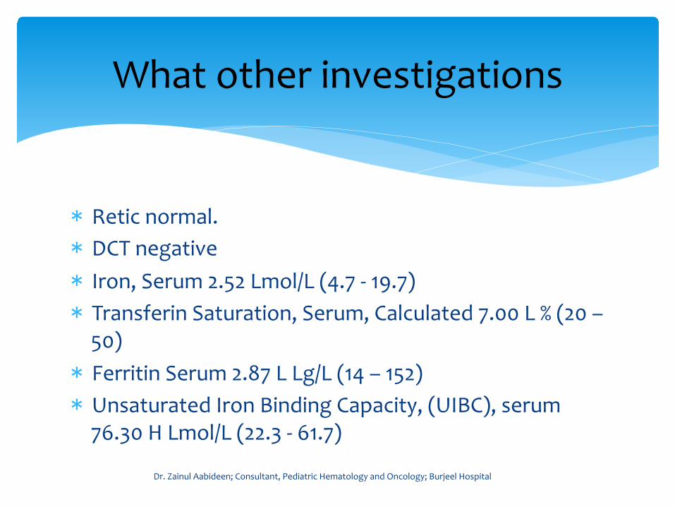
* Retic normal. * DCT negative * Iron, Serum 2.52 Lmol/L (4.7 -‐ 19.7) * Transferin Saturation, Serum, Calculated 7.00 L % (20 – 50) * Ferritin Serum 2.87 L Lg/L (14 – 152) * Unsaturated Iron Binding Capacity, (UIBC), serum 76.30 H Lmol/L (22.3 -‐ 61.7)
What other investigations
Dr. Zainul Aabideen; Consultant, Pediatric Hematology and Oncology; Burjeel Hospital
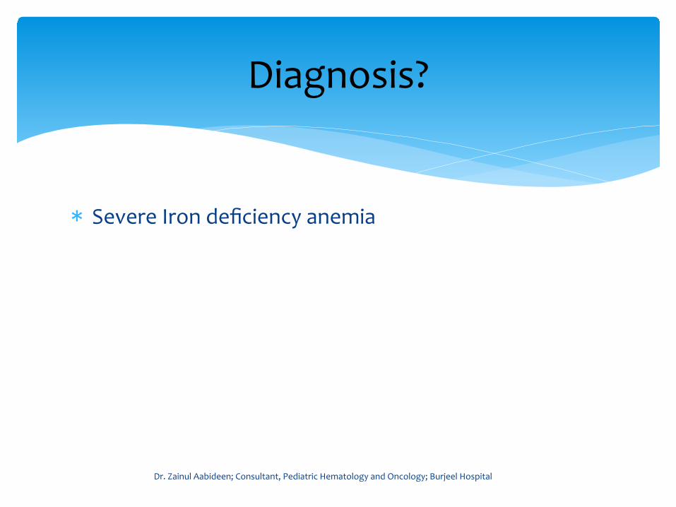
* Severe Iron deficiency anemia
Diagnosis?
Dr. Zainul Aabideen; Consultant, Pediatric Hematology and Oncology; Burjeel Hospital
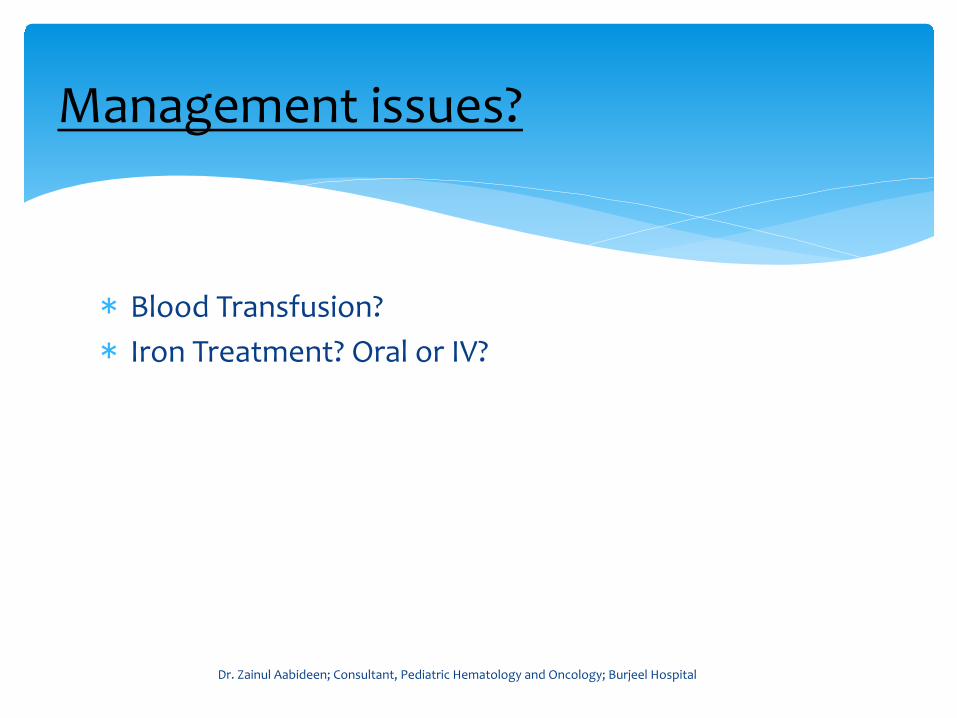
* Blood Transfusion? * Iron Treatment? Oral or IV?
Management issues?
Dr. Zainul Aabideen; Consultant, Pediatric Hematology and Oncology; Burjeel Hospital
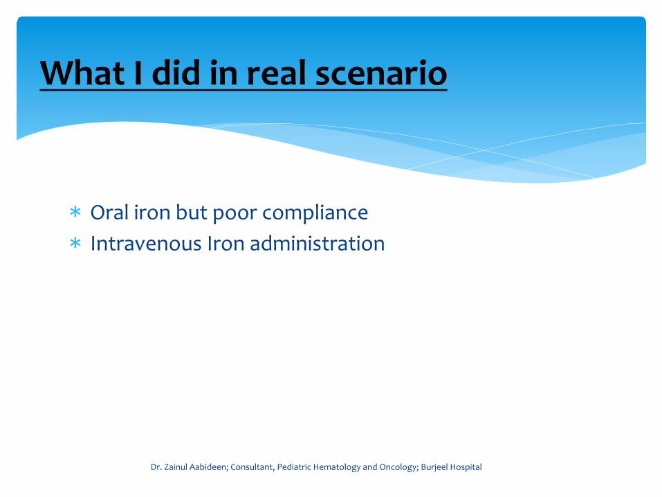
* Oral iron but poor compliance * Intravenous Iron administration
What I did in real scenario
Dr. Zainul Aabideen; Consultant, Pediatric Hematology and Oncology; Burjeel Hospital
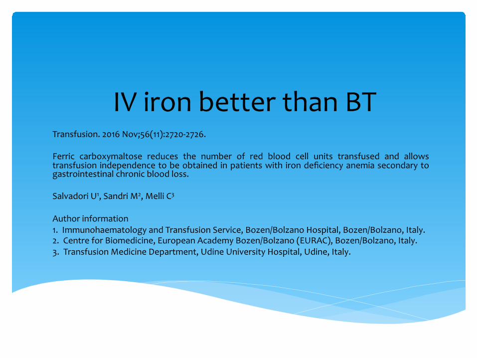
IV iron better than BT Transfusion. 2016 Nov;56(11):2720-‐2726. Ferric carboxymaltose reduces the number of red blood cell units transfused and allows transfusion independence to be obtained in patients with iron deficiency anemia secondary to gastrointestinal chronic blood loss. Salvadori U1, Sandri M2, Melli C3
Author information 1. Immunohaematology and Transfusion Service, Bozen/Bolzano Hospital, Bozen/Bolzano, Italy. 2. Centre for Biomedicine, European Academy Bozen/Bolzano (EURAC), Bozen/Bolzano, Italy. 3. Transfusion Medicine Department, Udine University Hospital, Udine, Italy.
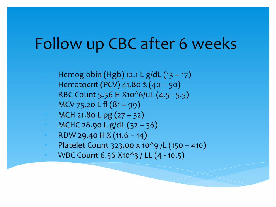
Follow up CBC after 6 weeks
• Hemoglobin (Hgb) 12.1 L g/dL (13 – 17) • Hematocrit (PCV) 41.80 % (40 – 50) • RBC Count 5.56 H X10^6/uL (4.5 -‐ 5.5) • MCV 75.20 L fl (81 – 99) • MCH 21.80 L pg (27 – 32) • MCHC 28.90 L g/dL (32 – 36) • RDW 29.40 H % (11.6 – 14) • Platelet Count 323.00 x 10^9 /L (150 – 410) • WBC Count 6.56 X10^3 / LL (4 -‐ 10.5)
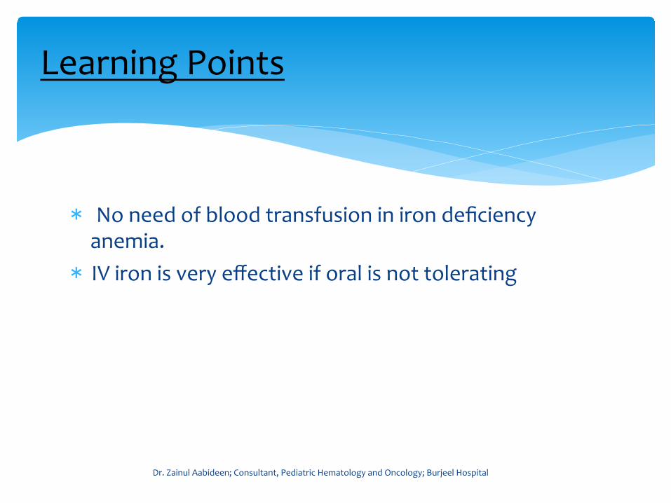
* No need of blood transfusion in iron deficiency anemia. * IV iron is very effective if oral is not tolerating
Learning Points
Dr. Zainul Aabideen; Consultant, Pediatric Hematology and Oncology; Burjeel Hospital

Case 4
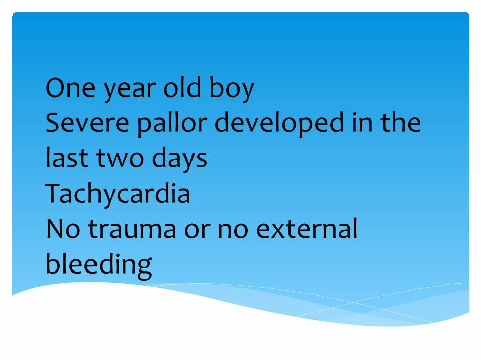
One year old boy Severe pallor developed in the last two days Tachycardia No trauma or no external bleeding
What pathological processes are happening here?
Acute hemolysis leading hemolytic anemia
What are the two differential diagnosis here?
1. G6PD deficiency anemia 2. Auto Immune Hemolytic Anemia (AIHA)
What are the two differential diagnosis here?
Dr. Zainul Aabideen; Consultant, Pediatric Hematology and Oncology; Burjeel Hospital
What questions you could ask in the history?
What investigations you could do?
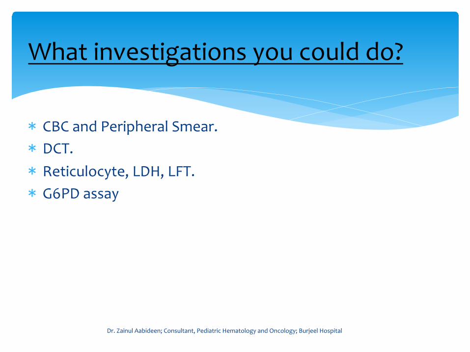
* CBC and Peripheral Smear. * DCT. * Reticulocyte, LDH, LFT. * G6PD assay
What investigations you could do?
Dr. Zainul Aabideen; Consultant, Pediatric Hematology and Oncology; Burjeel Hospital
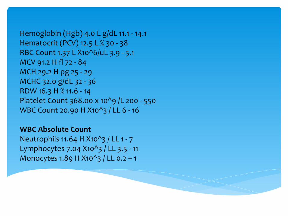
Hemoglobin (Hgb) 4.0 L g/dL 11.1 -‐ 14.1 Hematocrit (PCV) 12.5 L % 30 -‐ 38 RBC Count 1.37 L X10^6/uL 3.9 -‐ 5.1 MCV 91.2 H fl 72 -‐ 84 MCH 29.2 H pg 25 -‐ 29 MCHC 32.0 g/dL 32 -‐ 36 RDW 16.3 H % 11.6 -‐ 14 Platelet Count 368.00 x 10^9 /L 200 -‐ 550 WBC Count 20.90 H X10^3 / LL 6 -‐ 16 WBC Absolute Count Neutrophils 11.64 H X10^3 / LL 1 -‐ 7 Lymphocytes 7.04 X10^3 / LL 3.5 -‐ 11 Monocytes 1.89 H X10^3 / LL 0.2 – 1
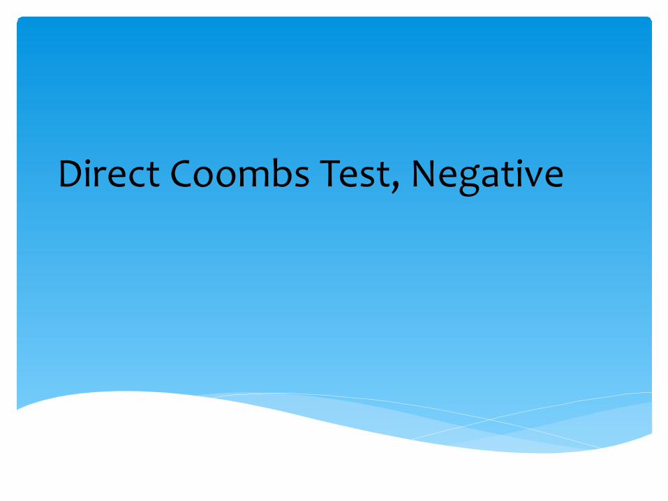
Direct Coombs Test, Negative
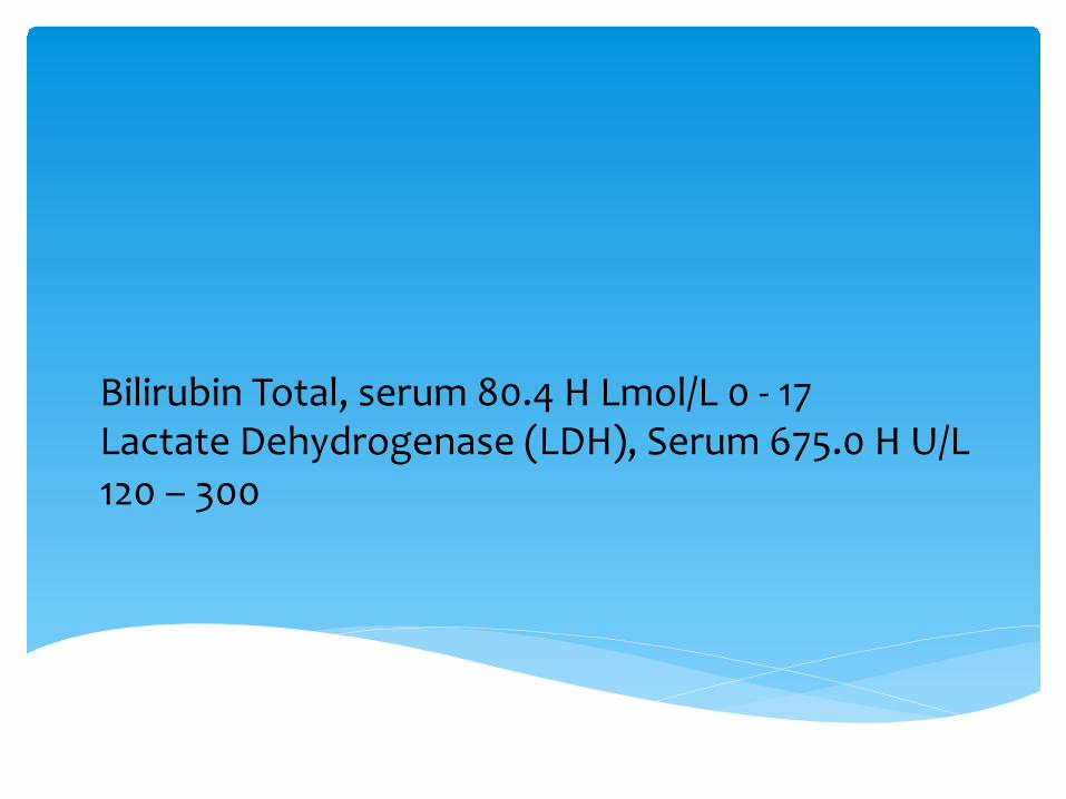
Bilirubin Total, serum 80.4 H Lmol/L 0 -‐ 17 Lactate Dehydrogenase (LDH), Serum 675.0 H U/L 120 – 300
*
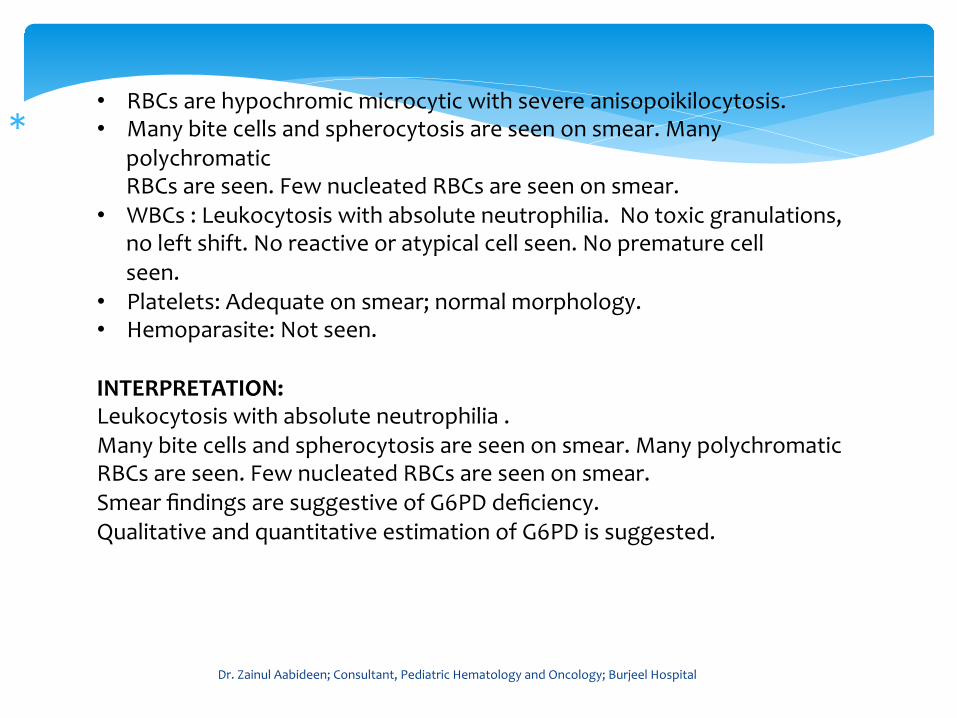
• RBCs are hypochromic microcytic with severe anisopoikilocytosis. • Many bite cells and spherocytosis are seen on smear. Many
polychromatic RBCs are seen. Few nucleated RBCs are seen on smear.
• WBCs : Leukocytosis with absolute neutrophilia. No toxic granulations, no left shift. No reactive or atypical cell seen. No premature cell seen.
• Platelets: Adequate on smear; normal morphology. • Hemoparasite: Not seen. INTERPRETATION: Leukocytosis with absolute neutrophilia . Many bite cells and spherocytosis are seen on smear. Many polychromatic RBCs are seen. Few nucleated RBCs are seen on smear. Smear findings are suggestive of G6PD deficiency. Qualitative and quantitative estimation of G6PD is suggested.
Dr. Zainul Aabideen; Consultant, Pediatric Hematology and Oncology; Burjeel Hospital
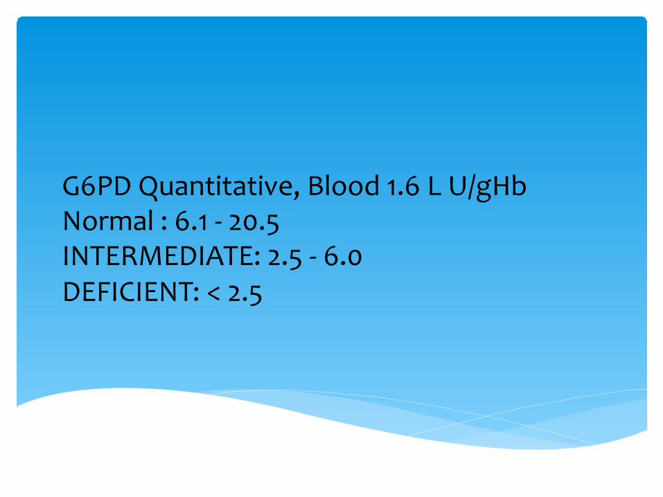
G6PD Quantitative, Blood 1.6 L U/gHb Normal : 6.1 -‐ 20.5 INTERMEDIATE: 2.5 -‐ 6.0 DEFICIENT: < 2.5
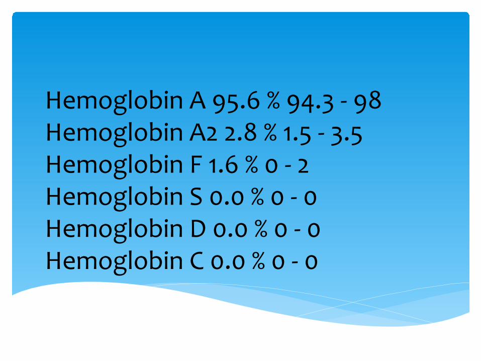
Hemoglobin A 95.6 % 94.3 -‐ 98 Hemoglobin A2 2.8 % 1.5 -‐ 3.5 Hemoglobin F 1.6 % 0 -‐ 2 Hemoglobin S 0.0 % 0 -‐ 0 Hemoglobin D 0.0 % 0 -‐ 0 Hemoglobin C 0.0 % 0 -‐ 0
Treatment
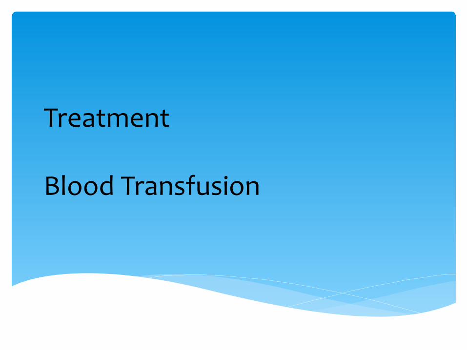
Treatment Blood Transfusion
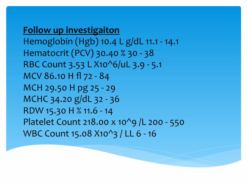
Follow up investigaiton Hemoglobin (Hgb) 10.4 L g/dL 11.1 -‐ 14.1 Hematocrit (PCV) 30.40 % 30 -‐ 38 RBC Count 3.53 L X10^6/uL 3.9 -‐ 5.1 MCV 86.10 H fl 72 -‐ 84 MCH 29.50 H pg 25 -‐ 29 MCHC 34.20 g/dL 32 -‐ 36 RDW 15.30 H % 11.6 -‐ 14 Platelet Count 218.00 x 10^9 /L 200 -‐ 550 WBC Count 15.08 X10^3 / LL 6 -‐ 16
Learning point? Differential diagnosis of severe anemia
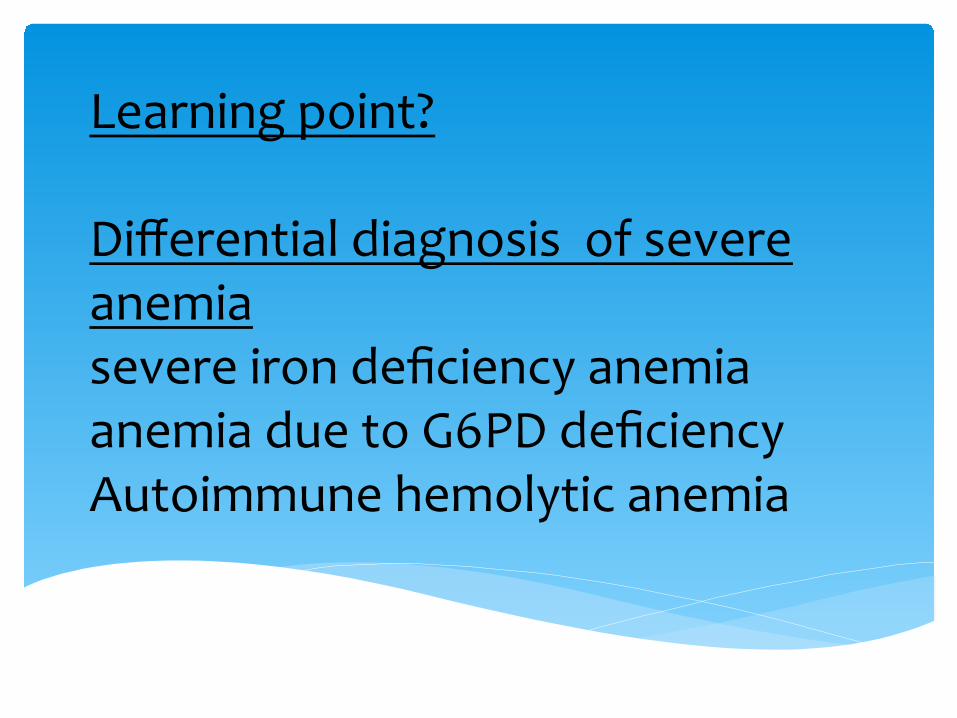
Learning point? Differential diagnosis of severe anemia severe iron deficiency anemia anemia due to G6PD deficiency Autoimmune hemolytic anemia
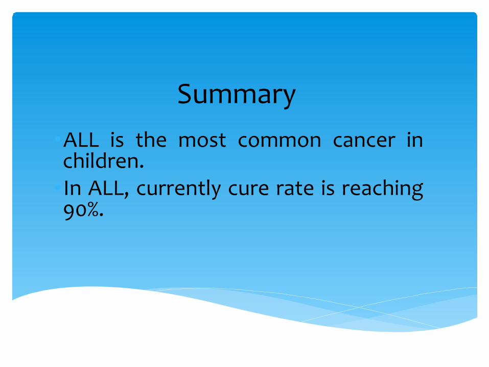
Summary • ALL is the most common cancer in children. • In ALL, currently cure rate is reaching 90%.
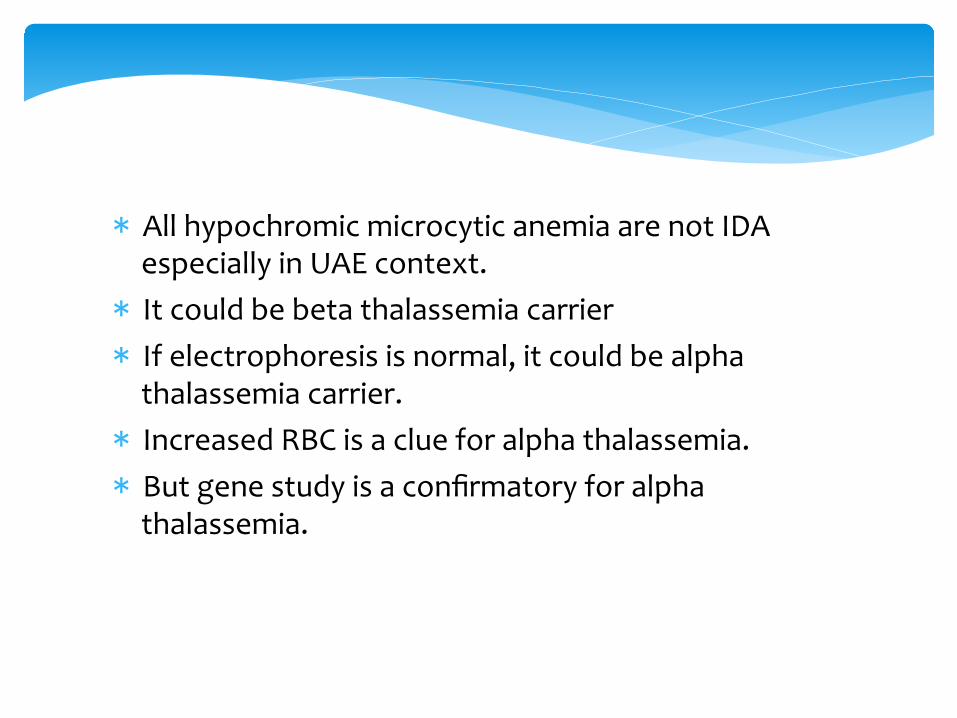
* All hypochromic microcytic anemia are not IDA especially in UAE context. * It could be beta thalassemia carrier * If electrophoresis is normal, it could be alpha thalassemia carrier. * Increased RBC is a clue for alpha thalassemia. * But gene study is a confirmatory for alpha thalassemia.
Thank you * In iron deficiency anemia, even if it is very severe, we should avoid blood transfusion. * IV iron injection is very effective.
Thank you * In acute, severe anemia, family history of G6PD and intake of fava beans need to ask. * Do Direct Coombs test, if positive, it could be AIHA. * It can be very difficult to manage. * Discuss with hematologist.
REFERENCES 1. Expert Rev Hemato. 2017 Nov;10(11):1023-‐1028 Anemia in children: prevalence, causes, and diagnostic work-‐up, and long-‐term consequences. Allali Set all 2. Pediatr Int. 2018 Jan; 60(1): 4-‐12 Treatment and biology of pediatric acute lymphoblastic leukemia. Kato M, Manabe A
Dr. Zainul Aabideen; Consultant, Pediatric Hematology and Oncology; Burjeel Hospital
Dr. Zainul Aabideen Kanakkande Consultant Pediatrics, Pediatric Hematology and Oncology
#: +971568267564 [email protected]
Thank you
Questions?