Embed Size (px)
Citation preview
1
ISSN 2286-4822
www.euacademic.org
EUROPEAN ACADEMIC RESEARCH
Vol. V, Issue 1/ April 2017
Impact Factor: 3.4546 (UIF)
DRJI Value: 5.9 (B+)
Helicobacter pylori and portal hypertensive
gastropathy diagnosed by narrow band imaging
(NBI): The role of its eradication
ESLAM SAFWAT
HANY ALY HUSSEIN
HANY SAMIR RASMY1
MD Internal Medicine, Internal Medicine Department
Faculty of Medicine, Ain Shams University, Cairo, Egypt
SARAH ADEL HAKIM
MD Pathology, Pathology Department
Faculty of Medicine, Ain Shams University, Cairo, Egypt
EHAB HAMED ELSAYED
MD Tropical Medicine
National Research Center, Cairo, Egypt
Abstract:
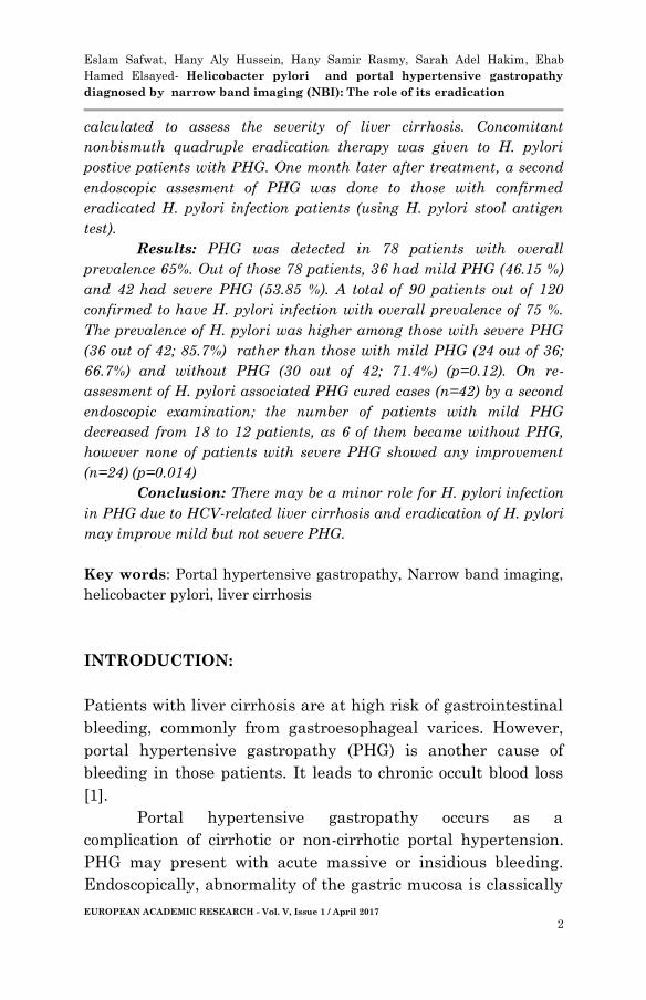
Background and Aim: Portal hypertensive gastropathy
(PHG) is a common endoscopic finding in patients with portal
hypertension. The pathophysiology of this condition is not obviously
understood. Although portal hypertension remains the crucial trigger
for the development of PHG, other factors could be responsible for the
progression of this condition. The aim of this study was to evaluate the
prevalence of helicobacter pylori (H. pylori) infection among cirrhotic
patients with PHG diagnosed by using narrow band imaging (NBI)
system and to asses the role of its eradication in improvement of PHG.
Patients and Methods: This study included 120 consecutive
patients with HCV-related liver cirrhosis. All patients were subjected
to an upper gastrointestinal endoscopy using NBI technique and
histopathologic testing of H. pylori. The diagnosis and the severity of
PHG were assesed on doing endoscopy. Child-Pugh score was
1 Corresponding author: [email protected]
Eslam Safwat, Hany Aly Hussein, Hany Samir Rasmy, Sarah Adel Hakim, Ehab
Hamed Elsayed- Helicobacter pylori and portal hypertensive gastropathy
diagnosed by narrow band imaging (NBI): The role of its eradication
EUROPEAN ACADEMIC RESEARCH - Vol. V, Issue 1 / April 2017
2
calculated to assess the severity of liver cirrhosis. Concomitant
nonbismuth quadruple eradication therapy was given to H. pylori
postive patients with PHG. One month later after treatment, a second
endoscopic assesment of PHG was done to those with confirmed
eradicated H. pylori infection patients (using H. pylori stool antigen
test).
Results: PHG was detected in 78 patients with overall
prevalence 65%. Out of those 78 patients, 36 had mild PHG (46.15 %)
and 42 had severe PHG (53.85 %). A total of 90 patients out of 120
confirmed to have H. pylori infection with overall prevalence of 75 %.
The prevalence of H. pylori was higher among those with severe PHG
(36 out of 42; 85.7%) rather than those with mild PHG (24 out of 36;
66.7%) and without PHG (30 out of 42; 71.4%) (p=0.12). On re-
assesment of H. pylori associated PHG cured cases (n=42) by a second
endoscopic examination; the number of patients with mild PHG
decreased from 18 to 12 patients, as 6 of them became without PHG,
however none of patients with severe PHG showed any improvement
(n=24) (p=0.014)
Conclusion: There may be a minor role for H. pylori infection
in PHG due to HCV-related liver cirrhosis and eradication of H. pylori
may improve mild but not severe PHG.
Key words: Portal hypertensive gastropathy, Narrow band imaging,
helicobacter pylori, liver cirrhosis
INTRODUCTION:
Patients with liver cirrhosis are at high risk of gastrointestinal
bleeding, commonly from gastroesophageal varices. However,
portal hypertensive gastropathy (PHG) is another cause of
bleeding in those patients. It leads to chronic occult blood loss
[1].
Portal hypertensive gastropathy occurs as a
complication of cirrhotic or non-cirrhotic portal hypertension.
PHG may present with acute massive or insidious bleeding.
Endoscopically, abnormality of the gastric mucosa is classically
Eslam Safwat, Hany Aly Hussein, Hany Samir Rasmy, Sarah Adel Hakim, Ehab
Hamed Elsayed- Helicobacter pylori and portal hypertensive gastropathy
diagnosed by narrow band imaging (NBI): The role of its eradication
EUROPEAN ACADEMIC RESEARCH - Vol. V, Issue 1 / April 2017
3
described as a mosaic-like pattern that resembles the skin of a
snake, with or without red spots [2]. Moreover, it seemed that
the color of the mucosa was due to the degree of capillary
dilatation, while the degree of red spots was due to the amount
of intramucosal hemorrhage [3].
The narrow band imaging (NBI) is an endoscopic
imaging technique for the enhanced visualization of mucosal
microscopic structure and capillaries of the superficial mucosal
layer, depending on changing the spectral features of the
illumination. NBI obtains its images by using narrower bands
of red, blue and green filters (R/B/G), which are different from
conventional red, blue and green filters [4]. The depth of
penetration into the mucosa depends on the wave length used
superficial for blue band and deep for red band and
intermediate for green band [5]. Thus, lesions could be
identified by changes in color and irregularity of mucosal
surface [6].
Combining NBI with magnifying system allow very clear
images of the capillaries of the mucosal surface and
microvascular architecture of the gastric mucosa in patients
with liver cirrhosis [7].
Infection by helicobacter pylori (H. pylori) is highly
prevalent, especially in low socioeconomic developing countries
[8]. It is responsible for lesions like gastroduodenal erosions
and ulcers. In patients with liver cirrhosis, their prevalence is
controversial, as well as the association with PHG [9].
The endoscopic findings of an H. pylori affected stomach
include erythema, erosions, antral nodularity, thickening of
gastric mucosal folds, and visible submucosal vessels. However,
these findings have low sensitivity and specificity for diagnosis
[10]. Recently, Taiwanese endoscopists performed a study using
close-up observation between the endoscopic tip and the gastric
mucosa and found the "mosaic pattern" in the corpus mucosa.
This method is a more sensitive and specific way to
Eslam Safwat, Hany Aly Hussein, Hany Samir Rasmy, Sarah Adel Hakim, Ehab
Hamed Elsayed- Helicobacter pylori and portal hypertensive gastropathy
diagnosed by narrow band imaging (NBI): The role of its eradication
EUROPEAN ACADEMIC RESEARCH - Vol. V, Issue 1 / April 2017
4
determine H. pylori infection status [11]. They classified gastric
mucosal patterns into two categories [normal regular
arrangement of collecting venules (RAC) and abnormal mosaic
pattern]. However, the classification was insufficient to predict
all H. pylori infections. Another used four categories: a normal
RAC and three abnormal patterns including mosaic-like
appearance (type A), diffuse homogenous redness (type B), and
untypical pattern (type C) to predict a H. pylori-infected
stomach [12].
Conventional white light endoscopy correlates poorly
with histopathological findings of H. pylori-induced gastritis
[13]. Some studies using magnifying endoscopy have shown
that endoscopic features are associated with histopathological
findings related to H. pylori infection [4]. Successful eradication
of H. pylori dramatically changes the histopathological findings
of gastritis. Recently, changes of magnifying endoscopic
features with NBI were investigated during H. pylori
eradication [14].
Thus, studying the association of H. pylori with PHG
could be useful for better understanding of the pathogenesis of
PHG, then eradication of H. pylori should be beneficial in the
management of PHG.
We performed this study to verify the prevalence of H.
pylori infection among HCV- cirrhotic patients with PHG
diagnosed by using the magnifying NBI system and evalute the
role of its eradication in improvement of PHG.
PATIENTS AND METHODS:
One hundred and twenty (120) consecutive patients with HCV
related liver cirrhosis were enrolled in the study, recruited from
those attending the Endoscopy unit of Ain Shams University
hospital. The study was performed in the period between June
Eslam Safwat, Hany Aly Hussein, Hany Samir Rasmy, Sarah Adel Hakim, Ehab
Hamed Elsayed- Helicobacter pylori and portal hypertensive gastropathy
diagnosed by narrow band imaging (NBI): The role of its eradication
EUROPEAN ACADEMIC RESEARCH - Vol. V, Issue 1 / April 2017
5
2015 and March 2016. An informed consent was obtained from
each patient.
Patients with hepatocellular carcinoma, gastric surgery,
peptic ulcer or gastric malignancy, recent variceal bleeding
(within 2 weeks), and patients using beta blockers, nitrates,
nonsteroidal anti-inflammatory drugs, proton pump inhibitors,
antibiotics (up to 1 month) or a prior H. pylori eradication
therapy were excluded from the study.
A thorough medical history and full clinical examination
were applied in all participants. A complete blood count, liver
and renal functions were performed for all. An abdominal
ultrasound (Toshiba real-time scanner instrument with a 3.5
MHz convex transducer) was done. The diagnosis of liver
cirrhosis was based on clinical, biochemichal and radiological
findings. HCV antibodies were detected using Microparticle
Enzyme Immunoassay (AxSYM, third generation assay, Abbott
Laboratories, IL, USA). The severity of liver disease was
assessed using Child-pugh classification.
All patients were endoscopically evaluated by the NBI
technique using Pentax EPK-i video processor which provides a
spectacular image with an unrivaled of 1.25 megapixels which
is approximately 50% higher than any other endoscopy using a
sophisticated software that enhances the image area providing
detailed imaging of mucosa topography and vascularity.
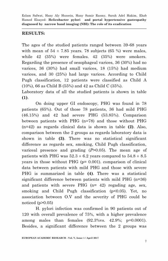
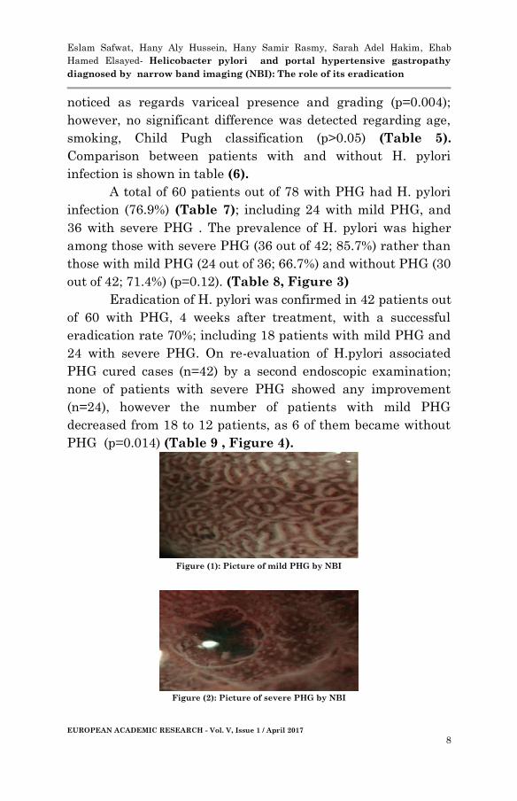
Regarding endoscopic NBI picture of PHG, Mosaic like pattern
and reddening mucosa (equivalent to mild PHG by Baveno
classification) [15] appeared as extended and swollen gastric
pits with various degrees of dilated and convoluted capillaries
surrounding the gastric pits (Figure 1) , while intra-mucosal
hemorrhage around capillaries (equivalent to severe PHG)
(Figure 2) [16]. OV were classified as small (small straight),
medium (tortuous occupying less than 1/3 the lumen) , and
large (coil shaped occupying more than 1/3 the lumen) [17].
Eslam Safwat, Hany Aly Hussein, Hany Samir Rasmy, Sarah Adel Hakim, Ehab
Hamed Elsayed- Helicobacter pylori and portal hypertensive gastropathy
diagnosed by narrow band imaging (NBI): The role of its eradication
EUROPEAN ACADEMIC RESEARCH - Vol. V, Issue 1 / April 2017
6
Biopsy specimens were taken from the gastric antrum, body
and incisura (2-1-2). Paraffin embedded sections were prepared
from the specimens. The specimens were then dewaxed and
taken to water and then incubated in 2% Giemsa solution in
distilled water for 30 minutes at room temperature. After
rinsing in tap water the sections were quickly dehydrated
through ethanol solutions before being cleared with xylene and
mounted in DPX (a mixture of distyrene, a plasticizer, dissolved
in toluene-xylene). Under light microscopy, curved, bent, pole-
like, spiral, and fusiform bacteria were accepted as H. pylori.
Patients with PHG who were positive for H. pylori
received concomitant nonbismuth quadruple eradication
therapy (esmoprazole 40 mg BID, amoxicillin 1000 mg BID,
metronidazole 500 mg BID, and clarithromycin 500 mg BID for
14 days). Results of successful eradication were diagnosed by H.
pylori stool antigen (HPSA) using rapid strip HPSA test (a
rapid immunoassay test) done 4 weeks after receiving
eradication therapy and they were off PPI and antibiotics .
A second endoscopic examination was carried out for those with
negative HPSA to re-assess PHG .
Statistical analysis: The collected data was revised, coded,
tabulated and introduced to a PC using Statistical package for
Social Science (IBM Corp. Released 2011. IBM SPSS Statistics
for Windows, Version 20.0. Armonk, NY: IBM Corp.).
Quantitative parametric variables are expressed as mean and
SD. Qualitative variables are expressed as frequencies and
percent. Student t Test was used to compare a continuous
variable between two study groups. Chi-square was used to
examine the relationship between Categorical variables.
Wilcoxon signed rank test was used to the statistical
significance of the difference of PHG before and after h.pylori
treatment. P-value < 0.05 was considered statistically
significant.
Eslam Safwat, Hany Aly Hussein, Hany Samir Rasmy, Sarah Adel Hakim, Ehab
Hamed Elsayed- Helicobacter pylori and portal hypertensive gastropathy
diagnosed by narrow band imaging (NBI): The role of its eradication
EUROPEAN ACADEMIC RESEARCH - Vol. V, Issue 1 / April 2017
7
RESULTS:
The ages of the studied patients ranged between 39-68 years
with mean of 54 ± 7.85 years. 78 subjects (65 %) were males,
while 42 (35%) were females. 42 (35%) were smokers.
Regarding the presence of oesophageal varices, 36 (30%) had no
varices, 36 (30%) had small varices, 18 (15%) had medium
varices, and 30 (25%) had large varices. According to Child
Pugh classification, 12 patients were classified as Child A
(10%), 66 as Child B (55%) and 42 as Child C (35%).
Laboratory data of all the studied patients is shown in table
(1).
On doing upper GI endoscopy, PHG was found in 78
patients (65%). Out of those 78 patients, 36 had mild PHG
(46.15%) and 42 had severe PHG (53.85%). Comparison
between patients with PHG (n=78) and those without PHG
(n=42) as regards clinical data is shown in table (2). Also,
comparison between the 2 groups as regards laboratory data is
shown in table (3). There was no statistical significant
difference as regards sex, smoking, Child Pugh classification,
variceal presence and grading (P>0.05). The mean age of
patients with PHG was 52.3 ± 6.2 years compared to 54.8 ± 8.5
years in those without PHG (p= 0.001). comparison of clinical
data between patients with mild PHG and those with severe
PHG is summarized in table (4). There was a statistical
significant difference between patients with mild PHG (n=36)
and patients with severe PHG (n= 42) regading age, sex,
smoking and Child Pugh classification (p<0.05). Yet, no
association between O.V and the severity of PHG could be
noticed (p>0.05)
H. pylori infection was confirmed in 90 patients out of
120 with overall prevalence of 75%, with a higher prevalence
among males than females (92.3%vs. 42.9%; p=0.0001).
Besides, a significant difference between the 2 groups was
Eslam Safwat, Hany Aly Hussein, Hany Samir Rasmy, Sarah Adel Hakim, Ehab
Hamed Elsayed- Helicobacter pylori and portal hypertensive gastropathy
diagnosed by narrow band imaging (NBI): The role of its eradication
EUROPEAN ACADEMIC RESEARCH - Vol. V, Issue 1 / April 2017
8
noticed as regards variceal presence and grading (p=0.004);
however, no significant difference was detected regarding age,
smoking, Child Pugh classification (p>0.05) (Table 5).
Comparison between patients with and without H. pylori
infection is shown in table (6).
A total of 60 patients out of 78 with PHG had H. pylori
infection (76.9%) (Table 7); including 24 with mild PHG, and
36 with severe PHG . The prevalence of H. pylori was higher
among those with severe PHG (36 out of 42; 85.7%) rather than
those with mild PHG (24 out of 36; 66.7%) and without PHG (30
out of 42; 71.4%) (p=0.12). (Table 8, Figure 3)
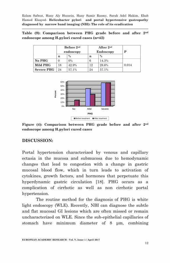
Eradication of H. pylori was confirmed in 42 patients out
of 60 with PHG, 4 weeks after treatment, with a successful
eradication rate 70%; including 18 patients with mild PHG and
24 with severe PHG. On re-evaluation of H.pylori associated
PHG cured cases (n=42) by a second endoscopic examination;
none of patients with severe PHG showed any improvement
(n=24), however the number of patients with mild PHG
decreased from 18 to 12 patients, as 6 of them became without
PHG (p=0.014) (Table 9 , Figure 4).
Figure (1): Picture of mild PHG by NBI
Figure (2): Picture of severe PHG by NBI
Eslam Safwat, Hany Aly Hussein, Hany Samir Rasmy, Sarah Adel Hakim, Ehab
Hamed Elsayed- Helicobacter pylori and portal hypertensive gastropathy
diagnosed by narrow band imaging (NBI): The role of its eradication
EUROPEAN ACADEMIC RESEARCH - Vol. V, Issue 1 / April 2017
9
Table (1): Laboratory data among all the studied patients
Laboratory parameter Mean ±SD Minimum Maximum
WBCS (x 103/mm³) 8.05 4.95 2.7 20
PLT(x 103/mm³) 117.1 58.33 44 230
HB (gm/dl) 9.95 1.36 8 12
ALT(IU/L) 31.75 14.37 12 81
AST(IU/L) 62.2 38.23 26 200
Bilirubin (mg/dl) 1.92 0.91 0.5 3.9
Albumin (gm/dl) 2.3 0.46 1.7 3.5
Protein (gm/dl) 6.29 0.64 5 8
INR 1.58 0.35 1.1 2.5
Creatinine (mg/dl) 1.55 1 0.5 4
Table (2): Comparison between patients with and without PHG
regarding clinical data
Parameter
PHG before ttt P
Patients
without PHG
(n= 42)
Patients with
PHG (n= 78)
Age 52.3 ± 6.2 54.8 ± 8.5 0.001
Sex
Male 24 30.8% 54 69.2% 0.185
Female 18 42.9% 24 57.1%
Smoking
Yes 18 42.9% 24 57.1% 0.185
No 24 30.8% 54 69.2%
Child
classification
A 6 50% 6 50% 0.131
B 18 27.3% 48 72.7%
C 18 42.9% 24 57.1%
Varices
No 18 50% 18 50% 0.086
Small 12 33.3% 24 66.7%
Medium 6 33.3% 12 66.7%
Large 6 20% 24 80%
Table (3): Comparison between patients with and without PHG
regarding laboratory data
PHG before ttt
P
Patients without
PHG
(n= 42)
Patients
with PHG
(n= 78)
Mean ±SD Mean ±SD
WBCs
(x 103/mm³)
9
5.2 6.2 3.8 0.001
PLT(x 103/mm³) 136.5 61.3 81 27.7 0.001
Hb (gm/dl) 10.2 1.2 9.6 1.5 0.036
ALT(IU/L) 34.4 10.4 30.3 16 0.135
AST(IU/L) 83.1 53.8 50.9 18.7 < 0.001
Bilirubin (mg/dl) 1.7 0.9 2.4 0.7 < 0.001
Albumin (gm/dl) 2.4 0.5 2.1 .40 0.001
Eslam Safwat, Hany Aly Hussein, Hany Samir Rasmy, Sarah Adel Hakim, Ehab
Hamed Elsayed- Helicobacter pylori and portal hypertensive gastropathy
diagnosed by narrow band imaging (NBI): The role of its eradication
EUROPEAN ACADEMIC RESEARCH - Vol. V, Issue 1 / April 2017
10
T. Proteins (gm/dl) 6.1 0.4 6.4 0.7 0.031
INR 1.7 0.4 1.5 0.3 0.001
Creatinine (mg/dl) 1.2 0.5 1.7 1.2 0.001
Table (4): Comparison between patients with mild and severe PHG as
regards clinical data
PHG before ttt
P
Mild
(n=36)
Severe
(n=42)
Age (years) 58.8 ± 8.5 51.4 ± 7 0.001
Sex
Male 18 23.1% 36 46.2% 0.001
Female 18 42.9% 6 14.3%
Smoking
Yes 6 14.3% 18 42.9% 0.012
No 30 38.5% 24 30.8%
Child
classification
A 6 50% 0 0%
0.001 B 12 18.2% 36 54.5%
C 18 42.9% 6 14.3%
Varices
No 6 16.7% 12 33.3%
0.671 Small 12 33.3% 12 33.3%
Medium 6 33.3% 6 33.3%
Large 12 40% 12 40%
Table (5): Relationship between H. pylori and clinical data
H. pylori
P Negative (n=30) Positive (n=90)
Age 55.4 ± 6.5 53.5 ± 8.2 0.244
Sex
Male 6 7.7% 72 92.3% 0.0001
Female 24 57.1% 18 42.9%
Smoking
Yes 12 28.6% 30 71.4% 0.507
No 18 23.1% 60 76.9%
DM
Yes 12 28.6% 30 71.4% 0.507
No 18 23.1% 60 76.9%
HTN
Yes 12 33.3% 24 66.7% 0.168
No 18 21.4% 66 78.6%
Child
classification
A 0 0% 12 100.0% 0.107
B 18 27.3% 48 72.7%
C 12 28.6% 30 71.4%
Varices
No 12 33.3% 24 66.7% 0.004
Small 12 33.3% 24 66.7%
Medium 6 33.3% 12 66.7%
Eslam Safwat, Hany Aly Hussein, Hany Samir Rasmy, Sarah Adel Hakim, Ehab
Hamed Elsayed- Helicobacter pylori and portal hypertensive gastropathy
diagnosed by narrow band imaging (NBI): The role of its eradication
EUROPEAN ACADEMIC RESEARCH - Vol. V, Issue 1 / April 2017
11
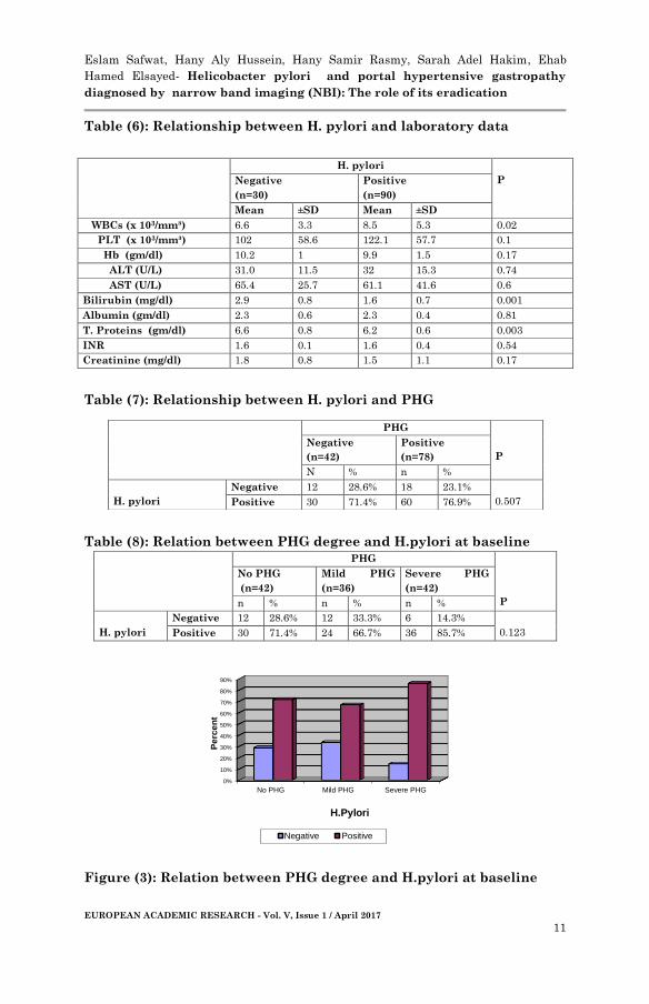
Table (6): Relationship between H. pylori and laboratory data
Table (7): Relationship between H. pylori and PHG
Table (8): Relation between PHG degree and H.pylori at baseline
PHG
P
No PHG
(n=42)
Mild PHG
(n=36)
Severe PHG
(n=42)
n % n % n %
H. pylori
Negative 12 28.6% 12 33.3% 6 14.3%
0.123 Positive 30 71.4% 24 66.7% 36 85.7%
Figure (3): Relation between PHG degree and H.pylori at baseline
0%
10%
20%
30%
40%
50%
60%
70%
80%
90%
No PHG Mild PHG Severe PHG
Pe
rce
nt
H.Pylori
Negative Positive
H. pylori
P Negative
(n=30)
Positive
(n=90)
Mean ±SD Mean ±SD
WBCs (x 103/mm³) 6.6 3.3 8.5 5.3 0.02
PLT (x 103/mm³) 102 58.6 122.1 57.7 0.1
Hb (gm/dl) 10.2 1 9.9 1.5 0.17
ALT (U/L) 31.0 11.5 32 15.3 0.74
AST (U/L) 65.4 25.7 61.1 41.6 0.6
Bilirubin (mg/dl) 2.9 0.8 1.6 0.7 0.001
Albumin (gm/dl) 2.3 0.6 2.3 0.4 0.81
T. Proteins (gm/dl) 6.6 0.8 6.2 0.6 0.003
INR 1.6 0.1 1.6 0.4 0.54
Creatinine (mg/dl) 1.8 0.8 1.5 1.1 0.17
PHG
P
Negative
(n=42)
Positive
(n=78)
N % n %
H. pylori
Negative 12 28.6% 18 23.1%
0.507 Positive 30 71.4% 60 76.9%
Eslam Safwat, Hany Aly Hussein, Hany Samir Rasmy, Sarah Adel Hakim, Ehab
Hamed Elsayed- Helicobacter pylori and portal hypertensive gastropathy
diagnosed by narrow band imaging (NBI): The role of its eradication
EUROPEAN ACADEMIC RESEARCH - Vol. V, Issue 1 / April 2017
12
Table (9): Comparison between PHG grade before and after 2nd
endoscope among H.pylori cured cases (n=42)
Before 2nd
endoscopy
After 2nd
Endoscopy
P
n % n %
No PHG 0 0% 6 14.3%
0.014
Mild PHG 18 42.9% 12 28.6%
Severe PHG 24 57.1% 24 57.1%
Figure (4): Comparison between PHG grade before and after 2nd
endoscope among H.pylori cured cases
DISCUSSION:
Portal hypertension characterized by venous and capillary
ectasia in the mucosa and submucosa due to hemodynamic
changes that lead to congestion with a change in gastric
mucosal blood flow, which in turn leads to activation of
cytokines, growth factors, and hormones that perpetuate this
hyperdynamic gastric circulation [18]. PHG occurs as a
complication of cirrhotic as well as non cirrhotic portal
hypertension.
The routine method for the diagnosis of PHG is white
light endoscopy (WLE). Recently, NBI can diagnose the subtle
and flat mucosal GI lesions which are often missed or remain
uncharacterized on WLE. Since the sub-epithelial capillaries of
stomach have minimum diameter of 8 μm, combining
0%
10%
20%
30%
40%
50%
60%
No Mild Severe
Pe
rce
nt
PHG
Before treatment After treatment
Eslam Safwat, Hany Aly Hussein, Hany Samir Rasmy, Sarah Adel Hakim, Ehab
Hamed Elsayed- Helicobacter pylori and portal hypertensive gastropathy
diagnosed by narrow band imaging (NBI): The role of its eradication
EUROPEAN ACADEMIC RESEARCH - Vol. V, Issue 1 / April 2017
13
magnification endoscopy with NBI has been studied for detailed
examination of capillary patterns in stomach [19].
PHG was found in 78 patients out of 120 patients with
liver cirrhosis included in the present study (65%). In previuos
studies, the prevalence of PHG in cirrhotic patients varies
between 20% and 98% [20]. This great variation may be
attributed to the study of different populations and variable
patient selection, in addition to different interpretation of
endoscopic lesions.
Only the age of the patients that showed a significant
difference regarding the presence of PHG (p=0.001); besides, a
relatively younger age was noticed in those with severe PHG
(p=0.001). Male gender dominanted in severe PHG (p=0.001).
Other clinical parameters, including Child Pugh class and the
presence or the severity of varices, showed insignificant
relation with the presence of PHG. Some previous studies have
demonstrated a higher prevalence of PHG in patients with
advanced liver disease, esophageal varices [21], however, these
results may be inconclusive and not reaching significance. Our
findings correlate well with those of Pan et al. [22] who found
that the presence of PHG is not affected either by the severity
of liver disease or by the presence or the grade of varices.
The pathogenesis of PHG is still not fully understood.
The pre-eminent pathogenetic factor seems to be an increase of
the portal pressure. Several investigators have evaluated the
effect of H. pylori on PHG with controversial results. To fill the
knowledge gap in this area, we aimed at exploring the
prevalence of H. pylori in patients with PHG that could help
identifying the pathogenesis of PHG.
In the present study, a high prevalence of H. pylori was
reported among all the studied patients (90 patients out of 120)
with overall prevalence of 75%, a figure that is higher than that
of Abbas et al. (62.1%) [23]. In a study from south India, 16 of
the 37 patients with cirrhosis were positive for H. pylori
Eslam Safwat, Hany Aly Hussein, Hany Samir Rasmy, Sarah Adel Hakim, Ehab
Hamed Elsayed- Helicobacter pylori and portal hypertensive gastropathy
diagnosed by narrow band imaging (NBI): The role of its eradication
EUROPEAN ACADEMIC RESEARCH - Vol. V, Issue 1 / April 2017
14
(43.24%) [24]. Also, we noticed male predominance among H.
pylori patients (92.3%vs. 42.9%; p=0.0001).
Furthermore, H. pylori infection was more prevalent
among patients with PHG but the difference did not reach
significance (76.9% vs. 71.4%; p=0.507); thus our study
concluded that the role of H. pylori in the pathogenesis of PHG
is minor. Numerous studies have demonstrated that H. pylori
infection is not associated with PHG [25-28]. Contrariwise,
Sathar et al. [29] reported an association between H. pylori
infection and PHG in cirrhotic patients, but some limitations
were noticed in their study, including low specificity and low
sensitivity of H. pylori serology in cirrhotic patients [30, 31],
potential selection bias, and underreporting of H. pylori
seroprevalence when considering that the study was performed
in India where H. pylori prevalence is extremely high in the
general population [32].
On discriminating PHG into mild and severe; the
prevalence of H. pylori was higher among patients with severe
PHG (36/42, 85.7%) rather than mild PHG (24/36, 66.7%); a
finding that goes with Sathar et al. [29] where seroprevalence
rate in cirrhotics with severe PHG (19/24, 79.2%) compared to
those with mild PHG (12/46, 26.1%). But, this finding was
claimed as its prevalence in literature was found to range
widely from 23% to 79% and from 22% to 81% in cirrhotics with
mild and severe PHG, respectively [33]. Consequently, this
finding is not reliable, and a selection bias cannot be ruled out.
In addition, in a previous study, the H. pylori status was 52%,
22%, and 0% in patients with mild, moderate, and severe
gastropathy, respectively, indicating an inverse relatioinship of
severity of PHG with H. pylori colonization and this explained
by severe congestive gastropathy make the gastric mucosa not
suitable for colonization of H. pylori and this may be related to
decreased synthesis of urea by the unhealthy gastric
mucosa[24].
Eslam Safwat, Hany Aly Hussein, Hany Samir Rasmy, Sarah Adel Hakim, Ehab
Hamed Elsayed- Helicobacter pylori and portal hypertensive gastropathy
diagnosed by narrow band imaging (NBI): The role of its eradication
EUROPEAN ACADEMIC RESEARCH - Vol. V, Issue 1 / April 2017
15
An explanation for increased prevalence of H. pylori among
severe PHG in our study that some factors like increased
inducible nitric oxide synthase expression resulting in high
reactive oxygen species, impairment of gastric mucosal defence
due to PHG in cirrhotic patients might increase virulence of H.
pylori to produce a synergistic effect between H. pylori and
PHG. Furthermore, colonization with H. pylori strains result in
gastric inflammatory response, including interleukin-8, tumor
necrosis factor-α, which may be associated with the sequence of
events leading to PHG [29].
Eradication of H. pylori was confirmed in 42 patients out
of 60 with PHG, 4 weeks after treatment, with a successful
eradication rate 70%. On re-evaluation of H. pylori associated
PHG cured cases by a second endoscopic examination; none of
patients with severe PHG showed any improvement, however
the number of patients with mild PHG decreased from 18 to 12
patients, as 6 of them became without PHG with significant
difference regarding mild PHG improvement before and after
H.pylori eradication (p= 0.014).
There was a diverse of contradiction about the role of H.
pylori eradication in cirrhotic patients. In past, many studies
reported that no need for its routine eradication in cirrhotic
patients [34-36]; while Sathar and co-worker [29] found that
there was significant association between H. pylori infection
and PHG in cirrhotic patients which is also related to severity
of PHG. Thus, H. pylori needs to be eradicated in cirrhotic
patients with PHG.
In conclusion, there may be a minor role for H. pylori in
the pathogenesis of PHG in cirrhotic patients. A significant
difference regarding mild PHG improvement before and after
H. pylori eradication was also noticed. Further prospective
studies on large number of patients are warranted to show
whether routine eradication of H. pylori may benefit the
Eslam Safwat, Hany Aly Hussein, Hany Samir Rasmy, Sarah Adel Hakim, Ehab
Hamed Elsayed- Helicobacter pylori and portal hypertensive gastropathy
diagnosed by narrow band imaging (NBI): The role of its eradication
EUROPEAN ACADEMIC RESEARCH - Vol. V, Issue 1 / April 2017
16
treatment of PHG especially severe portal gastropathy in
cirrhosis.
REFERENCES:
1. Garcia-Tsao G and Ripoll C. Management of Gastropathy
and Gastric Vascular Ectasia in Portal Hypertension. Clin
Liver Dis. 2010; 14(2): 281-295.
2. Cubillas R and Rockey C. Portal hypertensive gastropathy:
a review. Liver International. 2010; 1094-1102.
3. Hayashi S and Saeki S. Endoscopic microvascular
architecture of the portal hypertensive gastric mucosa on
narrow-band imaging. Digestive Endoscopy. 2007; 19: 116-
123.
4. Tahara T, Shibata T, Nakamura M, Yoshioka D,Okubo M,
Arisawa T, Hirata I. Gastric mucosal pattern by using
magnifying narrow band imaging endoscopy clearly
distinguishes histological and serological severity of
chronic gastritis. Gastrointestinal endoscopy J. 2009;
70(2): 246-253.
5. Sambongi M, Igarashi M, Obi T, Yamaguchi M, Oyama N,
Kobayashi M, Sano Y, Yoshida S, Gono K. Analysis of
spectral reflectance of mucous membrane for endoscopic
diagnosis. Med Phys. 2000; 27: 1396-1398.
6. Tajiri H, Matsuda K, Fujisaki J. What can we see with the
endoscope? Present status and future perspectives. Dig
Endosc. 2002; 14: 131-137.
7. Sano Y, Kobayashi M, Hamamoto Y. New diagnostic
method based on color imaging using narrow band imaging
system for gastrointestinal tract. Gastrointestinal
endoscopy J. 2001; 53: AB125.
Eslam Safwat, Hany Aly Hussein, Hany Samir Rasmy, Sarah Adel Hakim, Ehab
Hamed Elsayed- Helicobacter pylori and portal hypertensive gastropathy
diagnosed by narrow band imaging (NBI): The role of its eradication
EUROPEAN ACADEMIC RESEARCH - Vol. V, Issue 1 / April 2017
17
8. Malatya HM. Epidemiology of Helicobacter pylori
infection. Best Pract Res Clin Gastroenterol. 2007; 21:205-
214.
9. Pellicano R, Leone N, Berrutti M, Cutufia MA, Fiorentino
M, Rizzetto M. Helicobacter pylori seroprevalence in
hepatitis C virus positive patients with cirrhosis. J
Hepatol. 2000; 33:648-650.
10. Redéen S, Petersson F, Jönsson KA,Borch K. Relationship
of gastroscopic features to histological findings in gastritis
and Helicobacter pylori infection in a general population
sample. Endoscopy 2003; 35: 946-950.
11. Yan SL, Wu ST, Chen CH, Hung YH, Yang TH, Pang
VS, Yeh YH. Mucosal patterns of Helicobacterpylori-
related gastritis without atrophy in the gastric corpus
using standard endoscopy. World J Gastroenterol. 2010;
16: 496-500.
12. Cho JH, Chang YW, Jang JY, Shim JJ, Lee CK, Dong
SH, Kim HJ, Kim BH, Lee TH, Cho JY .Close Observation
of Gastric Mucosal Pattern by Standard Endoscopy Can
Predict Helicobacter pylori Infection Status .J
Gastroenterol Hepatol. 2013; 28(2):279-284.
13. Laine L, Cohen H, Sloane R, Marin-Sorensen M, Weinstein
WM. Interobserver agreement and predictive value of
endoscopic findings for H. pylori and gastritis in normal
volunteers. Gastrointest Endosc. 1995; 42: 420-423.
14. Okubo M, Tahara T, Shibata T, Nakamura M, Yoshioka
D, Maeda Y, Yonemura J, Ishizuka T, Arisawa T, Hirata I.
Changes in gastric mucosal patterns seen by magnifying
NBI during H. pylori eradication. J Gastroenterol. 2011;
46: 175-182.
15. De Franchis R. Evolving Consensus in Portal
Hypertension Report of the Baveno IV Consensus
Workshop on methodology of diagnosis and therapy in
portal hypertension. J Hepatol. 2005; 43(1): 167-176.
Eslam Safwat, Hany Aly Hussein, Hany Samir Rasmy, Sarah Adel Hakim, Ehab
Hamed Elsayed- Helicobacter pylori and portal hypertensive gastropathy
diagnosed by narrow band imaging (NBI): The role of its eradication
EUROPEAN ACADEMIC RESEARCH - Vol. V, Issue 1 / April 2017
18
16. Hayashi S ,Saeki S. Endoscopic microvascular architecture
of portal hypertensive gastric mucosa on narrow band
imaging. Digestive endoscopy. 2007; 19: 116-123.
17. Sanyal A, Grace N, Carey W, et al. Practice Guidelines
Committee of the American Association for the Study of
Liver Diseases; Practice Parameters Committee of the
American College of Gastroenterology. Prevention and
management of gastroesophageal varices and variceal
hemorrhage in cirrhosis. Hepatology 2007; 46: 922–38.
18. Ohta M, Yamaguchi S, Gotoh N, Tomikawa M.
Pathogenesis of portal hypertensive gastropathy: a clinical
and experimental review. Surgery. 2002; 131(1 Suppl):
S165-70.
19. Yao K, Oishi T, Matsui T, Yao T, Iwashita A. Novel
magnified endoscopic findings of microvascular
architecture in intramucosal gastric cancer. Gastrointest
Endosc. 2002; 56(2): 279-284.
20. Fontana RJ, Sanyal AJ, Mehta S, et al. Portal
hypertensive gastropathy in chronic hepatitis C patients
with bridging fibrosis and compensated cirrhosis: results
from the HALT-C trial. Am J Gastroenterol. 2006; 101:
983–992.
21. Merli M, Nicolini G, Angeloni S, Gentili F, Attili AF,
Riggio O. The natural history of portal hypertensive
gastropathy in patients with liver cirrhosis and mild portal
hypertensionAm J Gastroenterol. 2004; 99(10): 1959-1965.
22. Pan WD, Xun RY, Chen YM. Correlations of portal
hypertensive gastropathy of hepatitis B cirrhosis with
other factors. Hepatobiliary Pancreat Dis Int. 2002; 1(4):
527-531.
23. Abbas Z, Yakoob J, Usman MW, Shakir T, Hamid S, Jafri
W. Effect of helicobacter pylori and its virulence factors on
portal hypertensive gastropathy and interleukin (IL)-8, IL-
Eslam Safwat, Hany Aly Hussein, Hany Samir Rasmy, Sarah Adel Hakim, Ehab
Hamed Elsayed- Helicobacter pylori and portal hypertensive gastropathy
diagnosed by narrow band imaging (NBI): The role of its eradication
EUROPEAN ACADEMIC RESEARCH - Vol. V, Issue 1 / April 2017
19
10, and tumor necrosis factor-alpha levels. Saudi J
Gastroenterol. 2014; 20(2): 120-127.
24. Batmanabane V, Kate V, Ananthakrishnan N. Prevalence
of Helicobacter pylori in patients with portal hypertensive
gastropathy--a study from south India. Med Sci Monit.
2004; 10(4): CR133-136.
25. Parikh SS, Desai SB, Prabhu SR, Trivedi MH, Shankaran
K, Bhukhanwala FA, Kalro RH, Desai HG. Congestive
gastropathy: factors influencing development, endoscopic
features, Helicobacter pylori infection, and microvessel
changes. Am J Gastroenterol. 1994; 89:1036-1042.
26. Urso G, Interlandi D, Puglisi M, Abate G, Bertino G, Raciti
C, Sciacca C, Bruno M, Panarello A, Di Prima P, et al. Role
of Helicobacter pylori in patients with portal hypertensive
gastropathy by liver cirrhosis hepatitis C virus-related.
Minerva Gastroenterol Dietol. 2006; 52: 303-308.
27. Al Mofleh IA. Does Helicobacter pylori affect portal
hypertensive gastropathy? Saudi J Gastroenterol. 2007;
13: 95-97.
28. Arafa UA, Fujiwara Y, Higuchi K, Shiba M, Uchida T,
Watanabe T, Tominaga K, Oshitani N, Matsumoto T,
Arakawa T. No additive effect between Helicobacter pylori
infection and portal hypertensive gastropathy on inducible
nitric oxide synthase expression in gastric mucosa of
cirrhotic patients. Dig Dis Sci. 2003; 48: 162-168.
29. Sathar SA, Kunnathuparambil SG, Sreesh S, Narayanan
P, Vinayakumar KR. Helicobacter pylori infection in
patients with liver cirrhosis: prevalence and association
with portal hypertensive gastropathy. Ann Gastroenterol.
2014; 27(1): 48-52.
30. Nardone G, Coscione P, D'Armiento FP, Del Pezzo M,
Pontillo M, Mossetti G, Lamberti C, Budillon G. Cirrhosis
negatively affects the efficiency of serologic diagnosis of
Eslam Safwat, Hany Aly Hussein, Hany Samir Rasmy, Sarah Adel Hakim, Ehab
Hamed Elsayed- Helicobacter pylori and portal hypertensive gastropathy
diagnosed by narrow band imaging (NBI): The role of its eradication
EUROPEAN ACADEMIC RESEARCH - Vol. V, Issue 1 / April 2017
20
Helicobacter pylori infection. Ital J Gastroenterol. 1996;
28(6): 332-336.
31. Sanchez-Mete L, Zullo A, Hassan C, Rinaldi V, Magno MS,
Festuccia F, Morini S, Attili AF. Helicobacter pylori
diagnosis in patients with liver cirrhosis. Dig Liver Dis.
2003; 35(8): 566-570.
32. Dikshit RP, Mathur G, Mhatre S, Yeole BB.
Epidemiological review of gastric cancer in India. Indian J
Med Paediatr Oncol. 2011; 32(1): 3-11.
33. Zullo A, Hassan C, Morini S. Helicobacter pylori infection
in patients with liver cirrhosis: facts and fictions. Dig
Liver Dis. 2003; 35(3): 197-205.
34. Balan KK, Jones AT, Roberts NB, Pearson JP, Critchley
M, Jenkins SA. The effects of Helicobacter pylori
colonization on gastric function and the incidence of portal
hypertensive gastropathy in patients with cirrhosis of the
liver. The American journal of
gastroenterology.1996; 91(7): 1400-1406.
35. Dai L, Wu X. Helicobacter pylori and congestive
gastropathy.Chinese journal of hepatology. 1999; 7(1): pg
22-23.
36. Bahnacy A, Kupcsulik P, Elés ZS, Jàray B, Flautner L.
Helicobacter pylori and congestive gastropathy. Z
Gastroenterol 1997; 35(2): 109-112.