Embed Size (px)
Citation preview
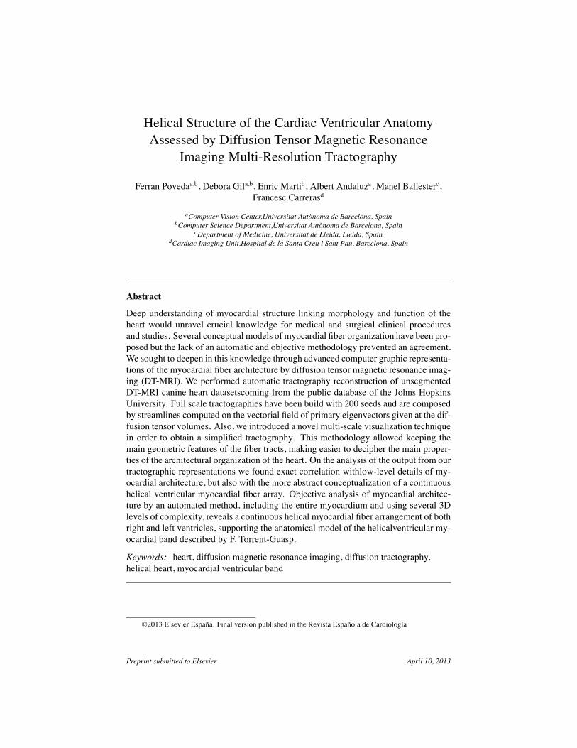
Helical Structure of the Cardiac Ventricular AnatomyAssessed by Diffusion Tensor Magnetic Resonance
Imaging Multi-Resolution TractographyFerran Povedaa,b, Debora Gila,b, Enric Martib, Albert Andaluza, Manel Ballesterc,
Francesc Carrerasd
aComputer Vision Center,Universitat Autònoma de Barcelona, SpainbComputer Science Department,Universitat Autònoma de Barcelona, Spain
cDepartment of Medicine, Universitat de Lleida, Lleida, SpaindCardiac Imaging Unit,Hospital de la Santa Creu i Sant Pau, Barcelona, Spain
AbstractDeep understanding of myocardial structure linking morphology and function of theheart would unravel crucial knowledge for medical and surgical clinical proceduresand studies. Several conceptual models of myocardial fiber organization have been pro-posed but the lack of an automatic and objective methodology prevented an agreement.We sought to deepen in this knowledge through advanced computer graphic representa-tions of the myocardial fiber architecture by diffusion tensor magnetic resonance imag-ing (DT-MRI). We performed automatic tractography reconstruction of unsegmentedDT-MRI canine heart datasetscoming from the public database of the Johns HopkinsUniversity. Full scale tractographies have been build with 200 seeds and are composedby streamlines computed on the vectorial field of primary eigenvectors given at the dif-fusion tensor volumes. Also, we introduced a novel multi-scale visualization techniquein order to obtain a simplified tractography. This methodology allowed keeping themain geometric features of the fiber tracts, making easier to decipher the main proper-ties of the architectural organization of the heart. On the analysis of the output from ourtractographic representations we found exact correlation withlow-level details of my-ocardial architecture, but also with the more abstract conceptualization of a continuoushelical ventricular myocardial fiber array. Objective analysis of myocardial architec-ture by an automated method, including the entire myocardium and using several 3Dlevels of complexity, reveals a continuous helical myocardial fiber arrangement of bothright and left ventricles, supporting the anatomical model of the helicalventricular my-ocardial band described by F. Torrent-Guasp.Keywords: heart, diffusion magnetic resonance imaging, diffusion tractography,helical heart, myocardial ventricular band
©2013 Elsevier España. Final version published in the Revista Española de Cardiología
Preprint submitted to Elsevier April 10, 2013
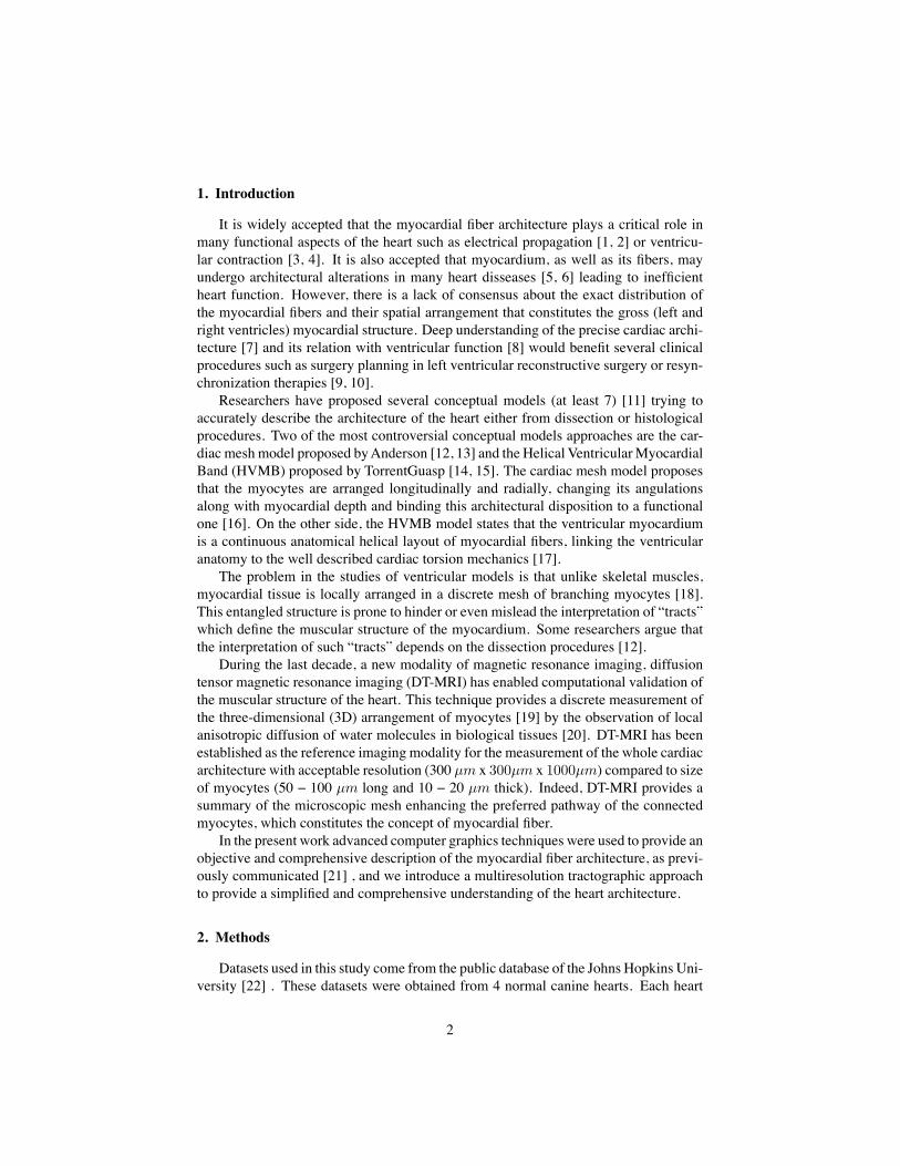
1. IntroductionIt is widely accepted that the myocardial fiber architecture plays a critical role in
many functional aspects of the heart such as electrical propagation [1, 2] or ventricu-lar contraction [3, 4]. It is also accepted that myocardium, as well as its fibers, mayundergo architectural alterations in many heart disseases [5, 6] leading to inefficientheart function. However, there is a lack of consensus about the exact distribution ofthe myocardial fibers and their spatial arrangement that constitutes the gross (left andright ventricles) myocardial structure. Deep understanding of the precise cardiac archi-tecture [7] and its relation with ventricular function [8] would benefit several clinicalprocedures such as surgery planning in left ventricular reconstructive surgery or resyn-chronization therapies [9, 10].
Researchers have proposed several conceptual models (at least 7) [11] trying toaccurately describe the architecture of the heart either from dissection or histologicalprocedures. Two of the most controversial conceptual models approaches are the car-diac mesh model proposed by Anderson [12, 13] and the Helical Ventricular MyocardialBand (HVMB) proposed by TorrentGuasp [14, 15]. The cardiac mesh model proposesthat the myocytes are arranged longitudinally and radially, changing its angulationsalong with myocardial depth and binding this architectural disposition to a functionalone [16]. On the other side, the HVMB model states that the ventricular myocardiumis a continuous anatomical helical layout of myocardial fibers, linking the ventricularanatomy to the well described cardiac torsion mechanics [17].
The problem in the studies of ventricular models is that unlike skeletal muscles,myocardial tissue is locally arranged in a discrete mesh of branching myocytes [18].This entangled structure is prone to hinder or even mislead the interpretation of “tracts”which define the muscular structure of the myocardium. Some researchers argue thatthe interpretation of such “tracts” depends on the dissection procedures [12].
During the last decade, a new modality of magnetic resonance imaging, diffusiontensor magnetic resonance imaging (DT-MRI) has enabled computational validation ofthe muscular structure of the heart. This technique provides a discrete measurement ofthe three-dimensional (3D) arrangement of myocytes [19] by the observation of localanisotropic diffusion of water molecules in biological tissues [20]. DT-MRI has beenestablished as the reference imaging modality for the measurement of the whole cardiacarchitecture with acceptable resolution (300 µm x 300µm x 1000µm) compared to sizeof myocytes (50 − 100 µm long and 10 − 20 µm thick). Indeed, DT-MRI provides asummary of the microscopic mesh enhancing the preferred pathway of the connectedmyocytes, which constitutes the concept of myocardial fiber.
In the present work advanced computer graphics techniques were used to provide anobjective and comprehensive description of the myocardial fiber architecture, as previ-ously communicated [21] , and we introduce a multiresolution tractographic approachto provide a simplified and comprehensive understanding of the heart architecture.
2. MethodsDatasets used in this study come from the public database of the Johns Hopkins Uni-
versity [22] . These datasets were obtained from 4 normal canine hearts. Each heart
2
was placed in an acrylic container filled with Fomblin, a perfluoropolyether (Ausimon,Thorofare, NJ). Fomblin has a low dielectric effect and minimal MR signal thereby in-creasing contrast and eliminating unwanted susceptibility artifacts near the boundariesof the heart. The long axis of the hearts was aligned with the z-axis of the scanner.
Images were acquired with a 4-element knee phased array coil on a 1.5 T GE CV/IMRI Scanner (GE, Medical System, Wausheka, WI) using an enhanced gradient systemwith 40 mT/m maximum gradient amplitude and a 150 T/m/s slew rate. Hearts wereplaced in the center of the coil and a 3D fast spin echo sequence was used to acquirediffusion images with a minimum of sixteen non-collinear gradient directions and amaximum b-value of 1500 s/mm2. The size of each voxel was about 312.5 µm x 312.5µm x 800 µm. Resolution resulting from a zero padding in fourier space to adaptoriginal image size of 192x192 to 256x256. The final dataset was arranged in about256 x 256 x 108 arrays (depending on the scanned heart) and contains two kinds ofdata: geometry/scalar data and diffusion tensor data. For diffusion tensor data, eachvoxel in the array consisted of 3 eigenvalues and 3 eigenvectors. The size of eachvoxel was about 312.5 µm x 312.5 µm x 800 µm.
Full scale tractographies presented in this study have been built with 200 seeds.These seeds have been randomly chosen over the entire anatomy only taking out a verysmall range of points related to the lowest eigenvalues that are likely to be bad startingpoints for the reconstruction. The strategy for the seed selection in the reconstructionsof lower resolution in the scale-space has been to scale proportionally these values tothe downscaling magnitude.
3. Key Points for Ventricular Tractography Reconstruction3.1. Data completeness
It is undisputed that the basal ring is crucial to fully understand heart anatomy andfunction. However, in some publications [23, 24, 25] the myocardial volume is cut justbelow the mitral valve to avoid noisy tractography in the auricular cavities. Given thatsuch plane cut discards, the basal ring reconstructions are not complete for a reliableinterpretation of the cardiac architecture.
3.2. DT-MRI Vector field orientationTractography is a technique inherited from the study of fluids. In this field orienta-
tion of vector fields stands for fluid stream directions and, thus, reconstructions presentno ambiguity. However, in the case of anatomical structures DT-MRI vector fields havean orientation that does not correspond to any physiological property. For a successfultractography reconstruction, DT-MRI vector fields should be reoriented. The few exist-ing approaches are based on either local properties of the flux or parametric models ofthe heart. By their local nature, local approaches[24] might introduce suboptimal fibersnot consistent with the global structure. Although parametric models of the ventricles[26, 27] provide a good solution to solve fiber orientation, because of their complexity,they are usually restricted to the left ventricle. We propose a geometrical organizationcoherent to gross heart anatomy.
3
3.3. VisualizationComprehensive visualization of fiber tracts should involve a proper assignment of
colors providing information about the orientation of the myocardial fibers. Often colormaps are defined using a global coordinate system which might mislead the global struc-ture. In order to properly encode the anatomical structure, color maps based on localinformation should be considered.
3.4. Heart architecture interpretationFully detailed tractographic reconstructions fit perfectly to make low level descrip-
tions, but might fail on a higher level of analysis as a result of their complexity. In orderto obtain more comprehensive descriptions of global myocardial structure we proposethe use of a multiresolution approach applied to the standard tractographic algorithms.This may help to generate simpler visualizations which in turn may help to better un-derstand the detailed myocardial architecture.
4. Fullscale TractographyHeart tractography is seen as a reconstruction composed by several streamlines [28]
(also known as fiber tracks on this field). The main property that clearly defines astreamline is that it is a curve tangential to the vector field at any point of such curve.
In this work, tractographies will be composed by streamlines computed on the vec-torial field of primary eigenvectors given at the diffusion tensor volumes. We computedthose streamlines using a fifth order Runge-Kutta-Fehlbert [29] integration method
4.1. Data completenessIn order to get complete reconstructions of the myocardial anatomy we have con-
sidered the whole DT-MRI volumes including the atrial cavities and the basal ring.Noise on the streamline reconstruction is mainly caused by thin atrial tissue which in-troduces significant clutter on the visualization. In order to minimize such artifact, ourstreamlining method stops integration of streams with large Runge-Kutta estimated re-construction error.
4.2. DT-MRI Vector field orientationTractography is a graphical representation inherited from fluid mechanics where
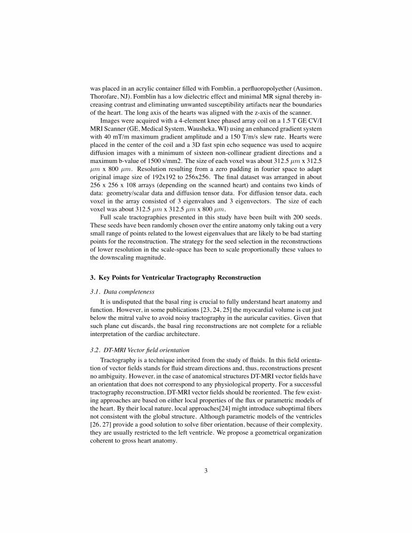
both direction and orientation of the vector fields are a meaningful part of the repre-sented information. However, on DT-MRI data vectors can be considered bidirectionalseeing that the water diffusion represented by this eigenvector occurs in one dimensionbut it does on the two possible orientations at the same time. Sometimes the datasetswill have a nearly organized structure, but we can also get opposed orientations (Fig.1a) at some points of the vectorial field that hinder its reconstruction.
We apply a geometrical reorganization of the vector field using local coordinatesystems coherent to ventricular anatomy and fluid mechanics. Ventricular anatomy canbe described by means of a longitudinal axis and angular coordinates with respect to thisaxis on axial cuts. In order to properly reorient both ventricles, our longitudinal axishas been set across the left ventricle, near the septum, ensuring that it never crosses
4
(a) Opposed orientation (b) reorganization on a radial basis
Figure 1: Orientation of the vectorial field datasets.
any myocardial wall. In order to make valid the vector field for streamlining this axisshould define for each axial cut a center of rotation. Therefore at every axial cut of theDT-MRI we reorganize vector orientations on a stream-like fashion (Fig. 1b) aroundthe point where the coordinate axis intersects the same axial cut. This implementationallows fast reorientation avoiding any smoothing of the vectorial field.
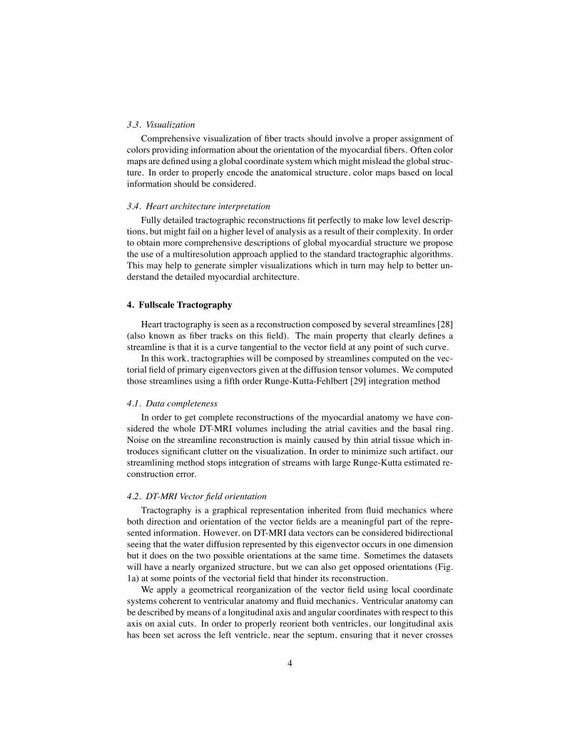
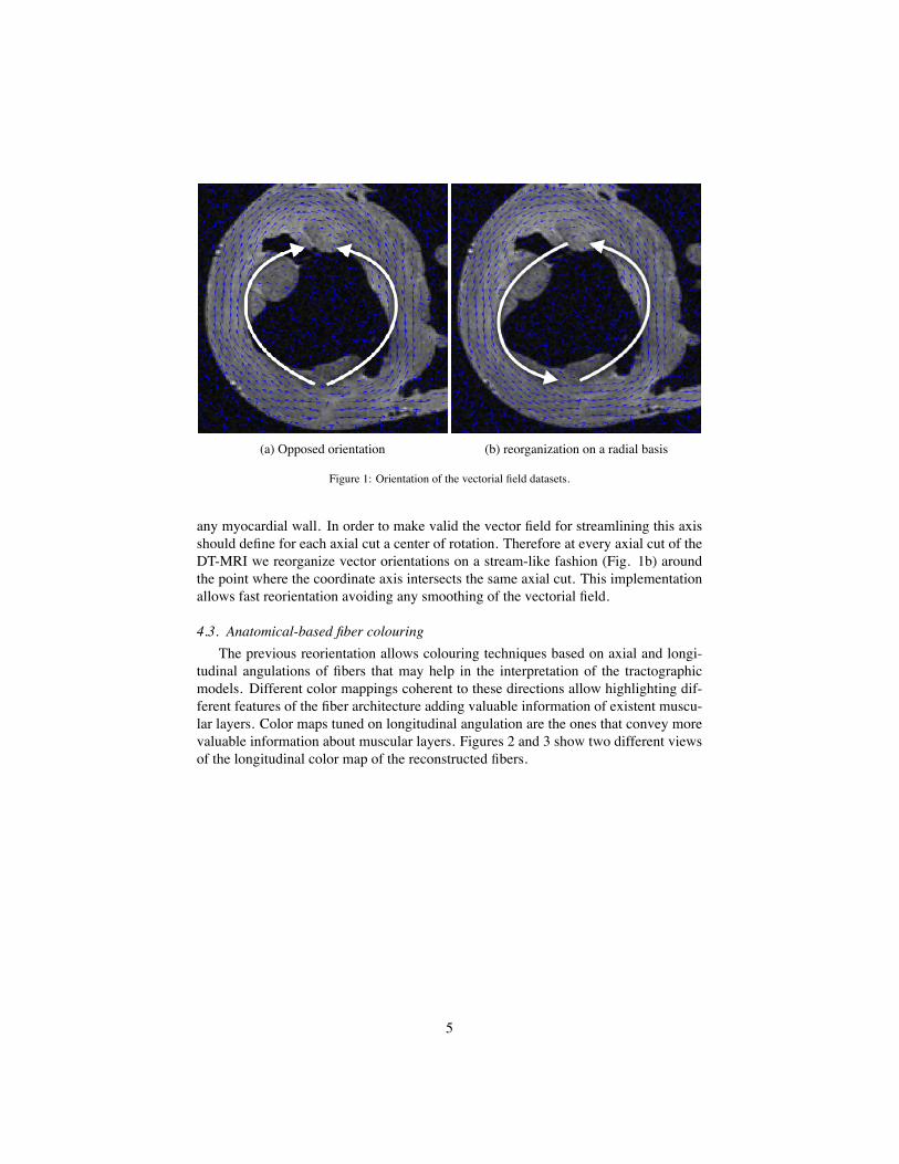
4.3. Anatomical-based fiber colouringThe previous reorientation allows colouring techniques based on axial and longi-
tudinal angulations of fibers that may help in the interpretation of the tractographicmodels. Different color mappings coherent to these directions allow highlighting dif-ferent features of the fiber architecture adding valuable information of existent muscu-lar layers. Color maps tuned on longitudinal angulation are the ones that convey morevaluable information about muscular layers. Figures 2 and 3 show two different viewsof the longitudinal color map of the reconstructed fibers.
5
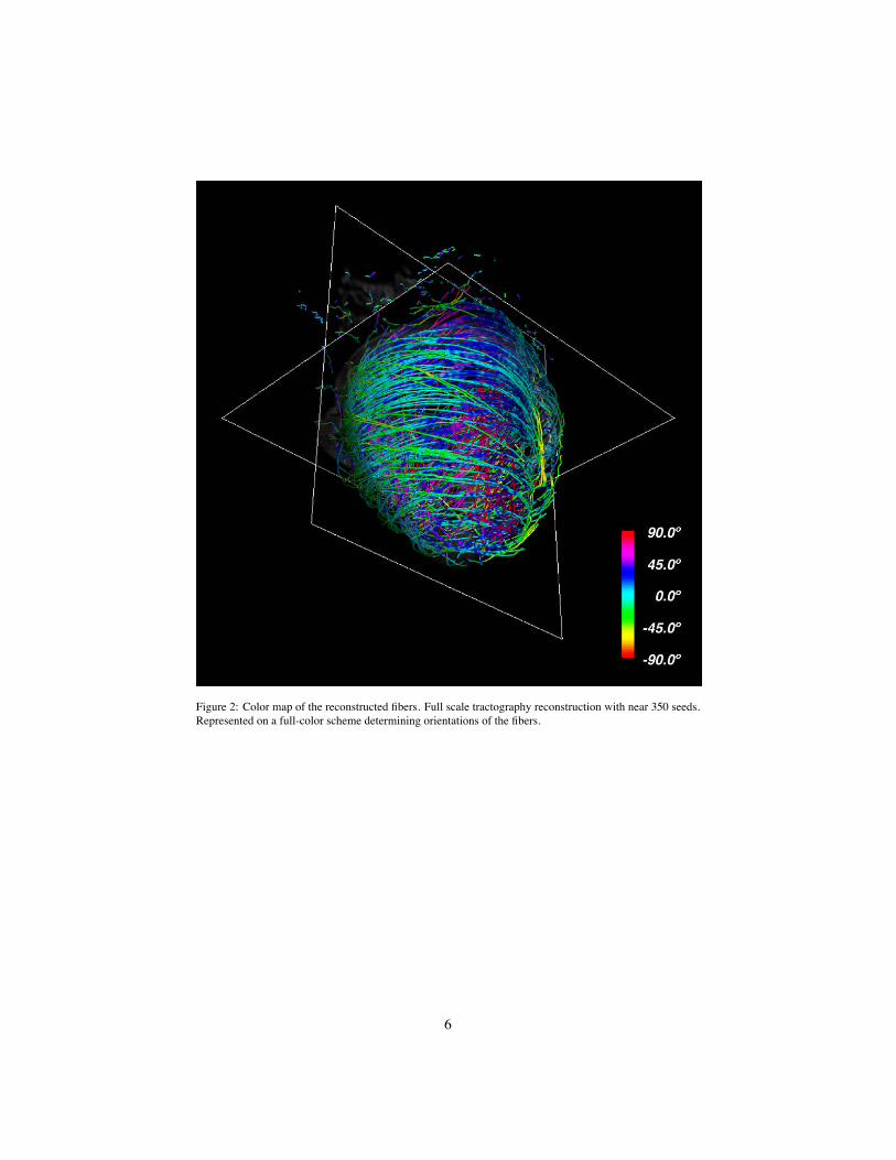
Figure 2: Color map of the reconstructed fibers. Full scale tractography reconstruction with near 350 seeds.Represented on a full-color scheme determining orientations of the fibers.
6
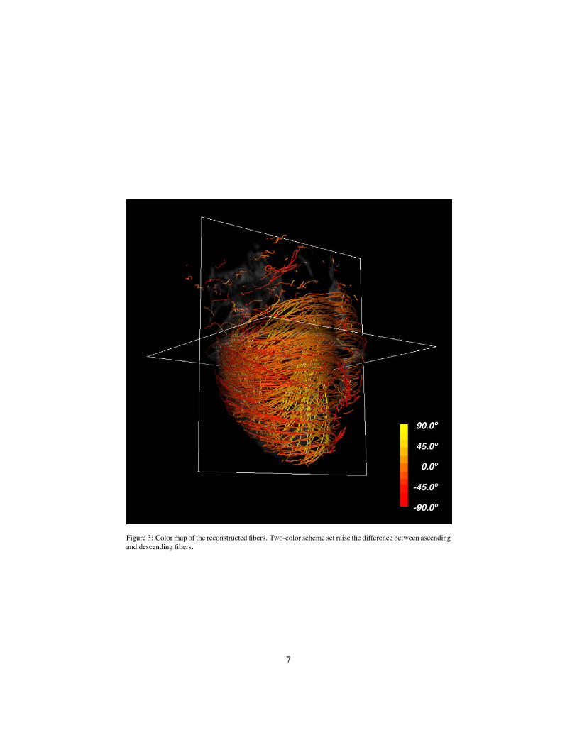
Figure 3: Color map of the reconstructed fibers. Two-color scheme set raise the difference between ascendingand descending fibers.
7
5. Multi-Resolution TractographyThe representation of a fully detailed tractography has been the state of the art
methodology to work out the comprehension of the heart. On this task, tractographicmodels have achieved interesting results but have also demonstrated weakness not help-ing to clarify a widely accepted unique myocardial anatomy description.
Intuitively, on a real world context, when an observer tries to make a gross analysishe can step away a few meters from the object of analysis and get a more contextualview. We will extrapolate this everyday behaviour to our problem.
In order to resolve this in a computer graphic representation it is common to usemultiresolution models which try to find solutions to build different models of the samedata with different levels of detail but without a loss of fidelity. It is usually applied totexture mapping and it is known as mipmapping [30] based on the well known pyramidrepresentation [31] . This technique applies a Gaussian filtering and later an exponentialreduction via a subsampling of the full-scale texture. Reduced textures are somehow“summaries” of the original texture and would be used to represent this texture at dif-ferent scales. These “summaries” are statistically complete in such a way the Gaussiansmoothing keeps the contextual information before applying downsampling. The use ofthese downscaled images is also common in other fields like in computer vision wherethis operation can be seen as a computation on the scale space.
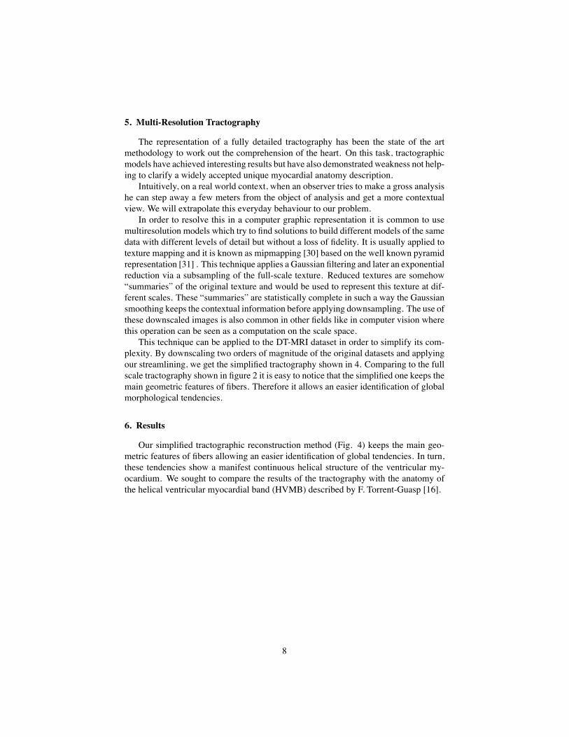
This technique can be applied to the DT-MRI dataset in order to simplify its com-plexity. By downscaling two orders of magnitude of the original datasets and applyingour streamlining, we get the simplified tractography shown in 4. Comparing to the fullscale tractography shown in figure 2 it is easy to notice that the simplified one keeps themain geometric features of fibers. Therefore it allows an easier identification of globalmorphological tendencies.
6. ResultsOur simplified tractographic reconstruction method (Fig. 4) keeps the main geo-
metric features of fibers allowing an easier identification of global tendencies. In turn,these tendencies show a manifest continuous helical structure of the ventricular my-ocardium. We sought to compare the results of the tractography with the anatomy ofthe helical ventricular myocardial band (HVMB) described by F. Torrent-Guasp [16].
8
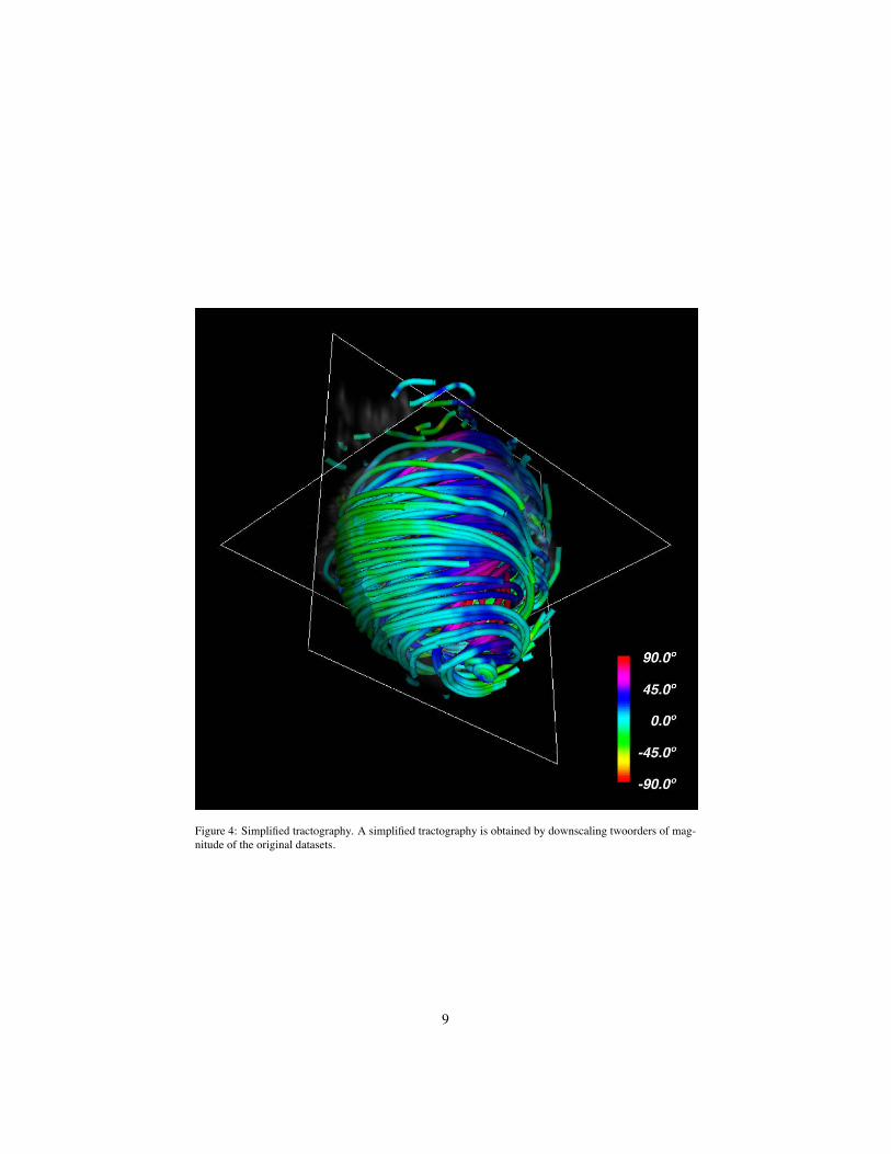
Figure 4: Simplified tractography. A simplified tractography is obtained by downscaling twoorders of mag-nitude of the original datasets.
9
HVMB describes a longitudinal arrangement of ventricular myocardial fibers form-ing a unique functional muscular band (Fig. 5) starting at the pulmonary artery (PA)and finishing at the aorta (Ao). This muscle wraps the left ventricle and part of theright ventricle (right and left segments) connecting to an helicoidal structure starting atthe basal ring going inside the left ventricle towards the apex and returning to connectwith the aorta (descending and ascending segments) wrapping with this turn the entireanatomy of the heart.
Figure 5: Ventricular myocardial band. Schematic presentation of the ventricular myocardial band dissection.Ao, aorta; PA, pulmonary artery; ptc, pulmonary-tricuspid cord; af, aberrant fibers; rf, right septal fibers; if,intraseptal fibers; AS ascending segment; DS, descending segment; LS left segment; RS right segment; lt,left trigone; rt, right trigone.
6.1. Fullscale TractographyFor the purpose of comparing tractographic results with the band model, step by step
tractographic reconstructions were compared with the myocardial fiber tracts depictedat the Torrent-Guasp’s rubber-silicone mould of the HVMB [32] (Figs. 6,7,8,9).
10
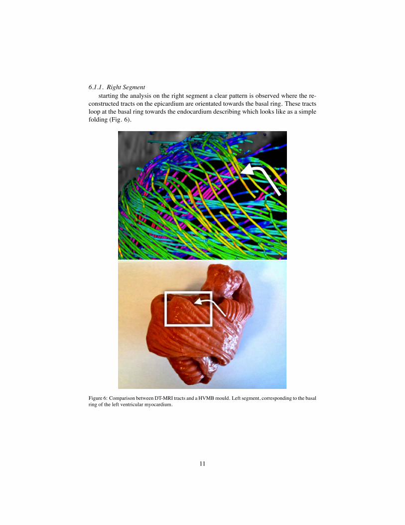
6.1.1. Right Segmentstarting the analysis on the right segment a clear pattern is observed where the re-
constructed tracts on the epicardium are orientated towards the basal ring. These tractsloop at the basal ring towards the endocardium describing which looks like as a simplefolding (Fig. 6).
Figure 6: Comparison between DT-MRI tracts and a HVMB mould. Left segment, corresponding to the basalring of the left ventricular myocardium.
11
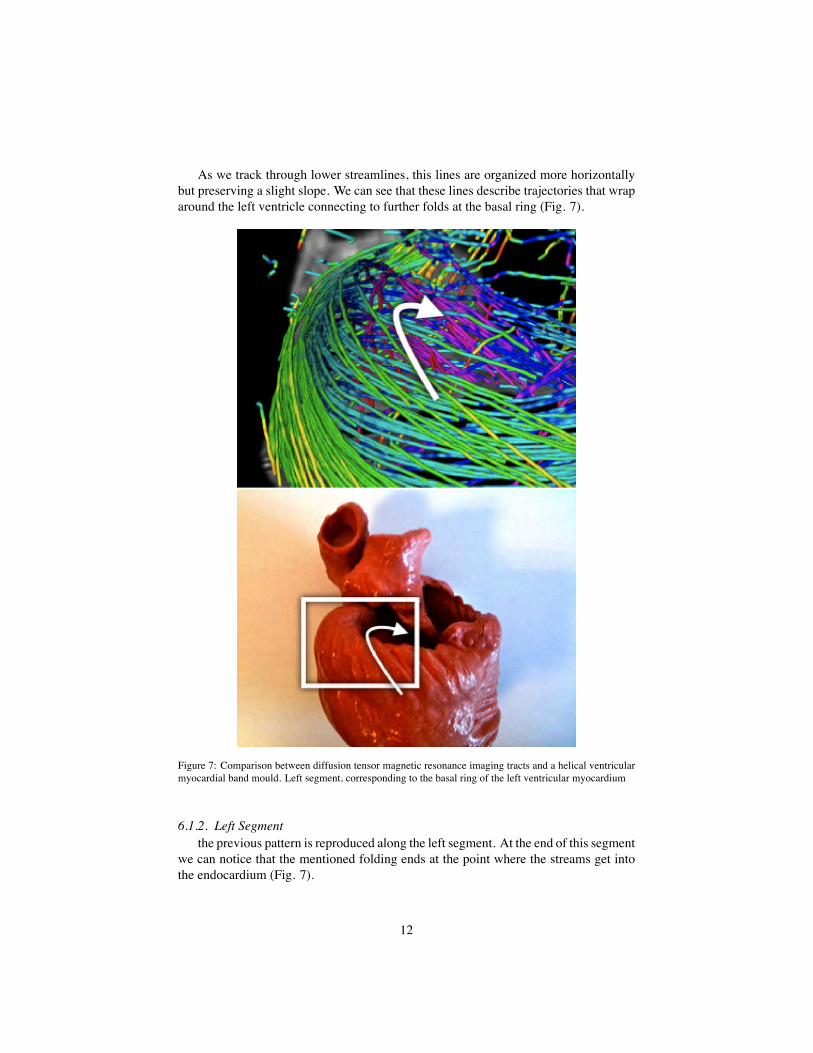
As we track through lower streamlines, this lines are organized more horizontallybut preserving a slight slope. We can see that these lines describe trajectories that wraparound the left ventricle connecting to further folds at the basal ring (Fig. 7).
Figure 7: Comparison between diffusion tensor magnetic resonance imaging tracts and a helical ventricularmyocardial band mould. Left segment, corresponding to the basal ring of the left ventricular myocardium
6.1.2. Left Segmentthe previous pattern is reproduced along the left segment. At the end of this segment
we can notice that the mentioned folding ends at the point where the streams get intothe endocardium (Fig. 7).
12

Figure 8: Comparison between diffusion tensor magnetic resonance imaging tracts and a helical ventric-ular myocardial band mould. Descending segment, corresponding to the inner wall of the left ventricularmyocardium.
Descending Segment: from an anterior view (Fig. 8) we can clearly distinguisha spiral-descending organization of the endocardium population of streams across theseptum. This structure continues to the apex and most of these streams continue on theright segment. Behind this endocardial structure it is also easy to notice an ascendingstructure that we will analyze in the following section from another visualization pointof view.
13
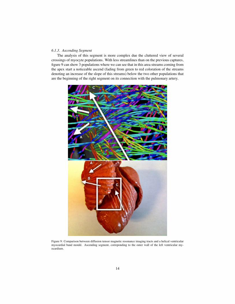
6.1.3. Ascending SegmentThe analysis of this segment is more complex due the cluttered view of several
crossings of myocyte populations. With less streamlines than on the previous captures,figure 9 can show 3 populations where we can see that in this area streams coming fromthe apex start a noticeable ascend (fading from green to red coloration of the streamsdenoting an increase of the slope of this streams) below the two other populations thatare the beginning of the right segment on its connection with the pulmonary artery.
Figure 9: Comparison between diffusion tensor magnetic resonance imaging tracts and a helical ventricularmyocardial band mould. Ascending segment, correponding to the outer wall of the left ventricular my-ocardium.
14
6.2. Simplified TractographyAlthough our simplified models provide easier interpretation of global tendencies,
they are still too complex for summarizing complex structure such as the Torrent-Guasp’s HVMB.
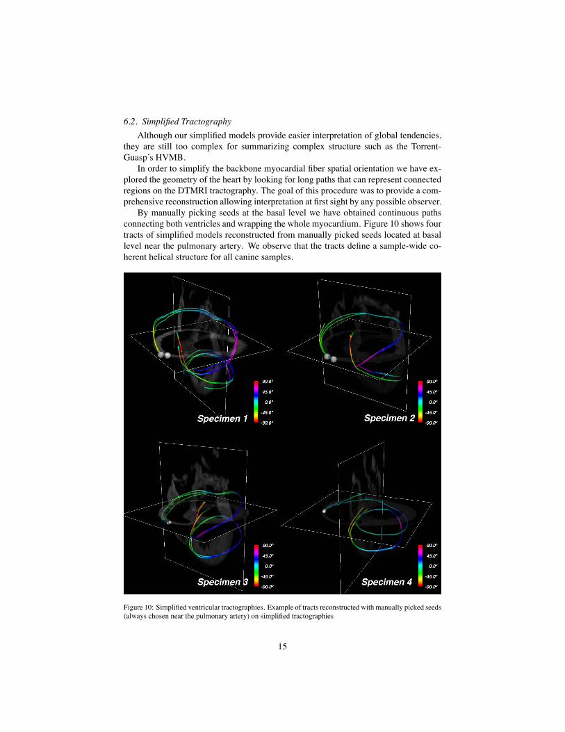
In order to simplify the backbone myocardial fiber spatial orientation we have ex-plored the geometry of the heart by looking for long paths that can represent connectedregions on the DTMRI tractography. The goal of this procedure was to provide a com-prehensive reconstruction allowing interpretation at first sight by any possible observer.
By manually picking seeds at the basal level we have obtained continuous pathsconnecting both ventricles and wrapping the whole myocardium. Figure 10 shows fourtracts of simplified models reconstructed from manually picked seeds located at basallevel near the pulmonary artery. We observe that the tracts define a sample-wide co-herent helical structure for all canine samples.
Figure 10: Simplified ventricular tractographies. Example of tracts reconstructed with manually picked seeds(always chosen near the pulmonary artery) on simplified tractographies
15
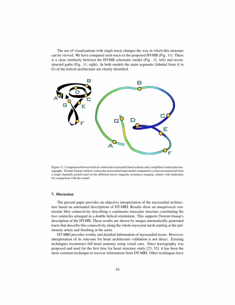
The use of visualizations with single tracts changes the way in which this structurecan be viewed. We have compared such tracts to the proposed HVMB (Fig. 11). Thereis a clear similarity between the HVMB schematic model (Fig. 11, left) and recon-structed paths (Fig. 11, right). In both models the main segments (labeled from A toG) of the helical architecture are clearly identified.
Figure 11: Comparison between helical ventricular myocardial band scheme and a simplified ventricular trac-tography. Torrent-Guasp’s helical ventricular myocardial band model compared to a tract reconstructed froma single manually picked seed on the diffusion tensor magnetic resonance imaging volume with landmarksfor comparison with the model.
7. DiscussionThe present paper provides an objective interpretation of the myocardial architec-
ture based on automated descriptions of DT-MRI. Results show an unequivocal ven-tricular fiber connectivity describing a continuous muscular structure constituting thetwo ventricles arranged in a double helical orientation. This supports Torrent-Guasp’sdescription of the HVMB. These results are shown by unique automatically generatedtracts that describe this connectivity along the whole myocytal mesh starting at the pul-monary artery and finishing at the aorta.
DT-MRI provides worthy and detailed information of myocardial tissue. However,interpretation of its outcome for heart architecture validation is not direct. Existingtechniques reconstruct full heart anatomy using visual cues. Since tractography wasproposed and used for the first time for heart structure study [23, 33], it has been themost common technique to recover information from DT-MRI. Other techniques have
16
been also explored as those in the work of Frindel [25] based on the optimization ofgraph models which promise future developments.
There are many factors that should be taken into account in order to obtain widelyacceptable reconstructions and interpretations. It follows that most of the existing ap-proaches [23, 24, 25, 26, 34, 35] do not provide enough evidence widely accepted bythe whole scientific community for either supporting or invalidating any particular ar-chitectural model. The only agreement is the existence of a layered structure of themyocardium through tractographic representations and visualization improvements incolor coding. Among these works, we want to remark the work of Helm [26] since, byits level of detail, it has been widely discussed on the literature hinting opposite read-ings. Such disagreement is direct consequence of a partial reconstruction of heart fiberanatomy.
In order to settle this disagreement we have used all the data in DT-MRI withoutsegmentation to avoid instrumentalization of the study, and we have demonstrated thatit is possible to reconstruct the whole myocardium including some complex structuresas the basal loop, unfortunately hidden or misinterpreted by other studies. For thisit has been also necessary to define a method to ensure a correct use of streamliningtechniques to the particularities of the DT-MRI vector fields.
Validation of the correctness of local structures is not enough to extend the interpre-tation to a global point of view. Hence, to deal with higher level interpretations of the ar-chitectural organization of the heart we have also looked for higher level representationsthat can ease its interpretation and validation. We have contributed a multi-resolutionmethod for tractography which using downsampling of the DT-MRI volumes can showglobal features of the heart structure. This work also includes colouring techniquesapplied to our solution which can ease the reading of the tractographic 3D models.
For studies requiring Q-ball analysis it is mandatory to use not less than 60 direc-tions per voxel. However, DTI tensors only provide an average description of waterdiffusion and, thus, a large number of diffusion directions do not significantly improvetheir quality. It follows that existing DTI cardiac studies (like the widespread used JHUdata set [36]) for DTI tensor analysis usually restrict values between 12 and 32 direc-tions [37] for the sake of a good compromise between acquisition time and quality.Furthermore, a recent study reports that DTI primary eigenvector is invariant under alarge variation of acquisition device parameters and, in particular, to a low number ofdiffusion directions [38]. Our own research suggests that heart preparation and volumespatial resolution are, indeed, one of the most influencing conditions on DTI quality.Acquisition field-of-view should be carefully adjusted to fit just the myocardial vol-ume, which should be in suspension inside a recipient in order to avoid distortions indiffusion near myocardial boundaries.
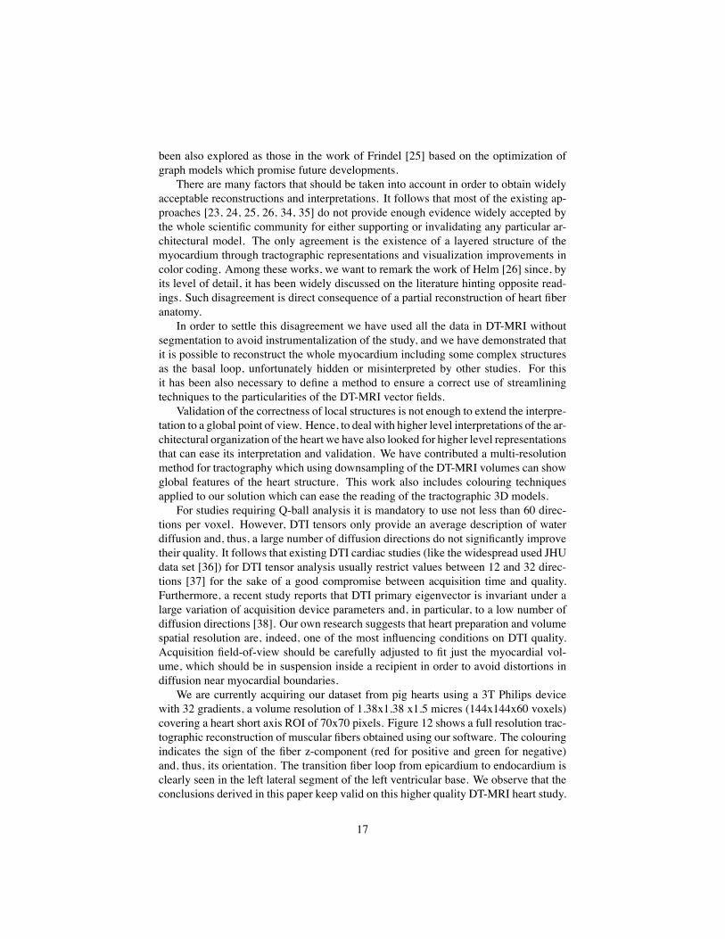
We are currently acquiring our dataset from pig hearts using a 3T Philips devicewith 32 gradients, a volume resolution of 1.38x1.38 x1.5 micres (144x144x60 voxels)covering a heart short axis ROI of 70x70 pixels. Figure 12 shows a full resolution trac-tographic reconstruction of muscular fibers obtained using our software. The colouringindicates the sign of the fiber z-component (red for positive and green for negative)and, thus, its orientation. The transition fiber loop from epicardium to endocardium isclearly seen in the left lateral segment of the left ventricular base. We observe that theconclusions derived in this paper keep valid on this higher quality DT-MRI heart study.
17
Figure 12: High resolution reconstruction of a diffusion tensor magnetic resonance imaging from a pig heartobtained with a 3T magnet (see text).
8. ConclusionsThe objective analysis of myocardial architecture by an automated method includ-
ing the entire myocardium and using several 3D levels of complexity reveals a continu-ous helical myocardial fiber arrangement of both right and left ventricles, thus support-ing the anatomical studies performed by F. Torrent-Guasp.
9. AcknowledgementsWe want to acknowledge Drs. Patrick A. Helm and Raimond L. Winslow at the
Center for Cardiovascular Bioinformatics and Modeling and Dr. Elliot McVeigh atthe National Institute of Health for provision of datasets of DT-MRI. This work wassupported by the Spanish TIN2009-13618 and TIN2012-33116.
18
References1. Roberts, D.E., Hersh, L.T., Scher, A.M.. Influence of cardiac fiber orientation
on wavefront voltage, conduction velocity, and tissue resistivity in the dog. CircRes 1979;44(5):701--12.
2. Taccardi, B., Punske, B.B., Macchi, E., Macleod, R.S., Ershler, P.R..Epicardial and intramural excitation during ventricular pacing: effect of my-ocardial structure. Am J Physiol Heart Circ Physiol 2008;294(4):H1753--66.doi:10.1152/ajpheart.01400.2007.
3. LeGrice, I.J., Takayama, Y., Covell, J.W.. Transverse shear along myocardialcleavage planes provides a mechanism for normal systolic wall thickening. CircRes 1995;77(1):182--93.
4. Zoalo Y Guevara E, B.D.e.a.. La reducción en el nivel y la velocidad de la torsiónventricular puede asociarse a incremento en la eficiencia ventricular izquierda:evaluación mediante ecografía speckle-tracking. Rev Esp Cardiología 2008;61:705--713.
5. Roberts, W.C., Siegel, R.J., McManus, B.M.. Idiopathic dilated cardiomyopa-thy: analysis of 152 necropsy patients. Am J Cardiol 1987;60(16):1340--55.
6. Wickline, S.A., Verdonk, E.D., Wong, A.K., Shepard, R.K., Miller, J.G..Structural remodeling of human myocardial tissue after infarction. quantificationwith ultrasonic backscatter. Circulation 1992;85(1):259--68.
7. Ballester M Ferreira A, C.F.. The myocardial band. Heart Fail Clin 2008;4:261--272. doi:10.1016/j.hfc.2008.02.011.
8. Aguilar, J.A.C., Martínez, A.H., Segarra, M.T.T., Ramón-Llin, J.A., Torrent-Guasp, F.. Estudio experimental de la llamada fase de relajación isovolumétricadel ventrículo izquierdo. Revista Espanola De Cardiologia 2009;62:392--399.doi:10.1016/S0300-8932(09)70896-6.
9. Torrent Guasp, F., JM, C.R.., Ballester Rodés, M.. Cuatro propuestas para laremodelación ventricular en el tratamiento de la miocardiopatía dilatada. Rev EspCardiología 1997;50:682--690.
10. Jung, B.A., Kreher, B.W., Markl, M., Hennig, J.. Visualization of tissue veloc-ity data from cardiac wall motion measurements with myocardial fiber tracking:principles and implications for cardiac fiber structures. Eur J Cardiothorac Surg2006;29 Suppl 1:S158--64. doi:10.1016/j.ejcts.2006.02.060.
11. Gilbert, S.H., Benson, A.P., Li, P., Holden, A.V.. Regional localisationof left ventricular sheet structure: integration with current models of car-diac fibre, sheet and band structure. European Journal of Cardio-ThoracicSurgery 2007;32(2):231--249. URL: http://ejcts.oxfordjournals.org/content/32/2/231.abstract. doi:10.1016/j.ejcts.2007.03.032.arXiv:http://ejcts.oxfordjournals.org/content/32/2/231.full.pdf+html.
19
12. Anderson, R.H., Ho, S.Y., Redmann, K., Sanchez-Quintana, D., Lunkenheimer,P.P.. The anatomical arrangement of the myocardial cells making up the ventricu-lar mass. Eur J Cardiothorac Surg 2005;28(4):517--25. doi:10.1016/j.ejcts.2005.06.043.
13. Anderson, R.H., Smerup, M., Sanchez-Quintana, D., Loukas, M., Lunken-heimer, P.P.. The three-dimensional arrangement of the myocytes in the ventric-ular walls. Clin Anat 2009;22(1):64--76. doi:10.1002/ca.20645.
14. F, T.G.. Estructura y función del corazón. Rev Esp Cardiol 1985;51:91--102.15. Torrent Guasp, F., Ballester, M., Buckberg, G.D., Carreras, F., Flotats, A., Car-
rió, I., et al. Spatial orientation of the ventricular muscle band: physiologic contri-bution and surgical implications. J Thorac Cardiovasc Surg 2001;122(2):389--92.doi:10.1067/mtc.2001.113745.
16. Torrent Guasp, F.. La mecánica agonista-antagonista de los segmentos descen-dente y ascendente de la banda miocárdica ventricular. Rev Esp Cardiología 2001;54:1091--102.
17. Carreras, F., Garcia-Barnes, J., Gil, D., Pujadas, S., Li, C.H., Suarez-Arias,R., et al. Left ventricular torsion and longitudinal shortening: two fundamen-tal components of myocardial mechanics assessed by tagged cine-mri in nor-mal subjects. Int J Cardiovasc Imaging 2012;28(2):273--84. doi:10.1007/s10554-011-9813-6.
18. LeGrice, I.J., Smaill, B.H., Chai, L.Z., Edgar, S.G., Gavin, J.B., Hunter, P.J..Laminar structure of the heart: ventricular myocyte arrangement and connectivetissue architecture in the dog. Am J Physiol 1995;269(2 Pt 2):H571--82.
19. Scollan, D.F., Holmes, A., Winslow, R., Forder, J.. Histological validationof myocardial microstructure obtained from diffusion tensor magnetic resonanceimaging. Am J Physiol 1998;275(6 Pt 2):H2308--18.
20. de Figueiredo, E.H.M.S.G., Borgonovi, A.F.N.G., Doring, T.M.. Basic conceptsof mr imaging, diffusion mr imaging, and diffusion tensor imaging. Magn ResonImaging Clin N Am 2011;19(1):1--22. doi:10.1016/j.mric.2010.10.005.
21. Poveda, F., Martí, E., Gil, D., Carreras, F., Ballester, M.. Helical structure ofventricular anatomy by diffusion tensor cardiac mr tractography. JACC Cardio-vasc Imaging 2012;5(7):754--5. doi:10.1016/j.jcmg.2012.04.005.
22. University, J.H.. Public dt-mri dataset. 2011. URL: http://gforge.icm.jhu.edu/gf/project/dtmri_data_sets/.
23. Zhukov, L., Barr, A.H.. Heart-muscle fiber reconstruction from diffusion tensormri. In: Proc. IEEE Visualization Conf. 2003, p. 79.
20
24. Rohmer, D., Sitek, A., Gullberg, G.T.. Reconstruction and visualization of fiberand laminar structure in the normal human heart from ex vivo diffusion tensormagnetic resonance imaging (dtmri) data. Invest Radiol 2007;42(11):777--89.doi:10.1097/RLI.0b013e3181238330.
25. Frindel, C., Schaerer, J., Gueth, P., Clarysse, P., Zhu, Y.M., Robini, M.. Aglobal approach to cardiac tractography. In: Biomedical Imaging: From Nano toMacro, 2008. ISBI 2008. 5th IEEE International Symposium on. 2008, p. 883--886. doi:10.1109/ISBI.2008.4541138.
26. Helm, P., Beg, M.F., Miller, M.I., Winslow, R.L.. Measuring and mappingcardiac fiber and laminar architecture using diffusion tensor mr imaging. Ann NY Acad Sci 2005;1047:296--307. doi:10.1196/annals.1341.026.
27. Gil, D., Garcia-Barnes, J., Hernández-Sabate, A., Marti, E.. Manifoldparametrization of the left ventricle for a statistical modelling of its completeanatomy. In: Society of Photo-Optical Instrumentation Engineers (SPIE) Con-ference Series; vol. 7623 of Society of Photo-Optical Instrumentation Engineers(SPIE) Conference Series. 2010, doi:10.1117/12.844480.
28. Granger, R.. Fluid mechanics. Mineola, NY: Courier Dover Publications; 1995.29. Fehlberg, E.. Klassische runge-kutta-formeln vierter und niedrigerer ordnung mit
schrittweiten-kontrolle und ihre anwendung auf w¨armeleitungsprobleme. Com-puting (Arch Elektron Rechnen) 1970;6:61--71.
30. Williams, L.. Pyramidal parametrics. SIGGRAPH Comput Graph 1983;17(3):1--11. URL: http://doi.acm.org/10.1145/964967.801126. doi:10.1145/964967.801126.
31. Burt, P.J.. Fast filter transform for image processing. Graphical Models /graph-ical Models and Image Processing /computer Vision, Graphics, and Image Pro-cessing 1981;16:20--51. doi:10.1016/0146-664X(81)90092-7.
32. Torrent Guasp, F., Whimster, W.F., Redmann, K.. A silicone rubber mould ofthe heart. Technol Health Care 1997;5(1-2):13--20.
33. Basser, P.J., Pajevic, S., Pierpaoli, C., Duda, J., Aldroubi, A.. In vivo fibertractography using dt-mri data. Magn Reson Med 2000;44(4):625--32.
34. Schmid, P., Jaermann, T., Boesiger, P., Niederer, P.F., Lunkenheimer, P.P.,Cryer, C.W., et al. Ventricular myocardial architecture as visualised in post-mortem swine hearts using magnetic resonance diffusion tensor imaging. Eur JCardiothorac Surg 2005;27(3):468--72. doi:10.1016/j.ejcts.2004.11.036.
35. Peeters, T., Bartroli, A.V., Strijkers, G., ter Haar Romeny, B.. Visualization ofthe fibrous structure of the heart. In: VMV 2006. 2006, p. 309--316.
21
36. Sermesant, M., Chabiniok, R., Chinchapatnam, P., Mansi, T., Billet, F.,Moireau, P., et al. Patient-specific electromechanical models of the heart for theprediction of pacing acute effects in crt: a preliminary clinical validation. MedImage Anal 2012;16(1):201--15. doi:10.1016/j.media.2011.07.003.
37. Krishnamurthy, A., Villongco, C.T., Chuang, J., Frank, L.R., Nigam,V., Belezzuoli, E., et al. Patient-specific models of cardiac biome-chanics. Journal of Computational Physics 2012;(0):--. URL: http://www.sciencedirect.com/science/article/pii/S0021999112005463.doi:10.1016/j.jcp.2012.09.015.
38. Gilbert, S., Trew, M., Smaill, B., Radjenovic, A., Bernus, O.. Measurementof myocardial structure: 3d structure tensor analysis of high resolution mri quan-titatively compared to dt-mri. In: Camara, O., Mansi, T., Pop, M., Rhode, K.,Sermesant, M., Young, A., editors. Statistical Atlases and Computational Modelsof the Heart. Imaging and Modelling Challenges; vol. 7746 of Lecture Notes inComputer Science. Springer Berlin Heidelberg. ISBN 978-3-642-36960-5; 2013,p. 207--214. URL: http://dx.doi.org/10.1007/978-3-642-36961-2_24.doi:10.1007/978-3-642-36961-2_24.
22