Embed Size (px)
Citation preview
Cynthia L. Webner, DNP, RN, CCNS, CCRN-CMC, CHFN
HEART SOUNDS:
DO YOU HEAR WHAT I HEAR?
1 2014 WWW.CARDIONURSING.COM

2
"THE MOST IMPORTANT PRACTICAL LESSON
THAN CAN BE GIVEN TO NURSES IS TO
TEACH THEM WHAT TO OBSERVE...“
~FLORENCE NIGHTINGALE, 1859
3
We Begin With The Cardiac Cycle
TO UNDERSTAND
HEART SOUNDS
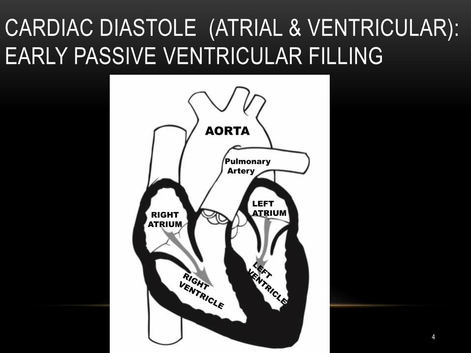
CARDIAC DIASTOLE (ATRIAL & VENTRICULAR):
EARLY PASSIVE VENTRICULAR FILLING
4
RIGHT
ATRIUM
LEFT
ATRIUM
AORTA
Pulmonary
Artery
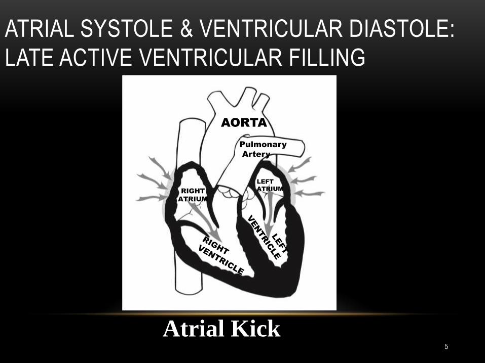
ATRIAL SYSTOLE & VENTRICULAR DIASTOLE:
LATE ACTIVE VENTRICULAR FILLING
5
Atrial Kick
RIGHT
ATRIUM
LEFT
ATRIUM
AORTA
Pulmonary
Artery
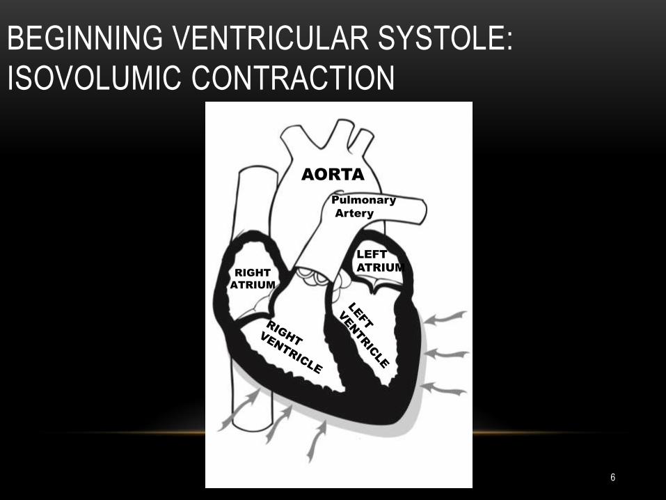
BEGINNING VENTRICULAR SYSTOLE:
ISOVOLUMIC CONTRACTION
6
RIGHT
ATRIUM
LEFT
ATRIUM
AORTA
Pulmonary
Artery
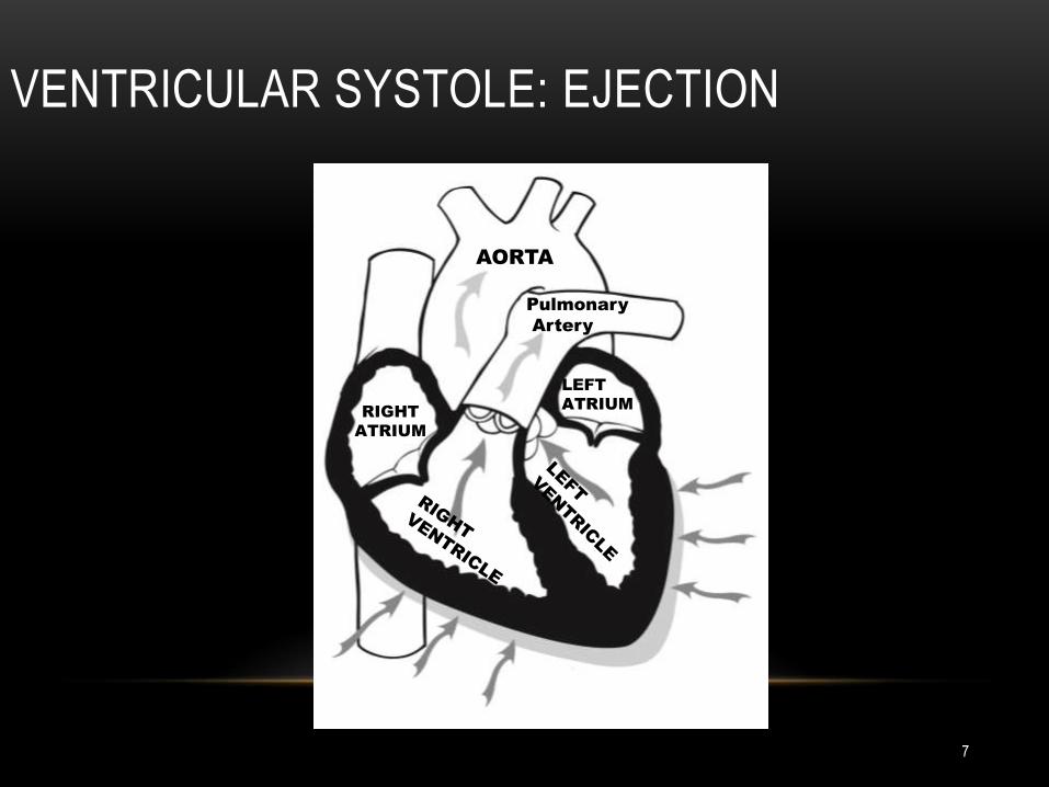
VENTRICULAR SYSTOLE: EJECTION
7
RIGHT
ATRIUM
LEFT
ATRIUM
AORTA
Pulmonary
Artery
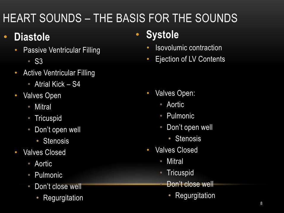
• Systole • Isovolumic contraction
• Ejection of LV Contents
• Valves Open:
• Aortic
• Pulmonic
• Don’t open well
• Stenosis
• Valves Closed
• Mitral
• Tricuspid
• Don’t close well
• Regurgitation
• Diastole • Passive Ventricular Filling
• S3
• Active Ventricular Filling
• Atrial Kick – S4
• Valves Open
• Mitral
• Tricuspid
• Don’t open well
• Stenosis
• Valves Closed
• Aortic
• Pulmonic
• Don’t close well
• Regurgitation
HEART SOUNDS – THE BASIS FOR THE SOUNDS
8
9
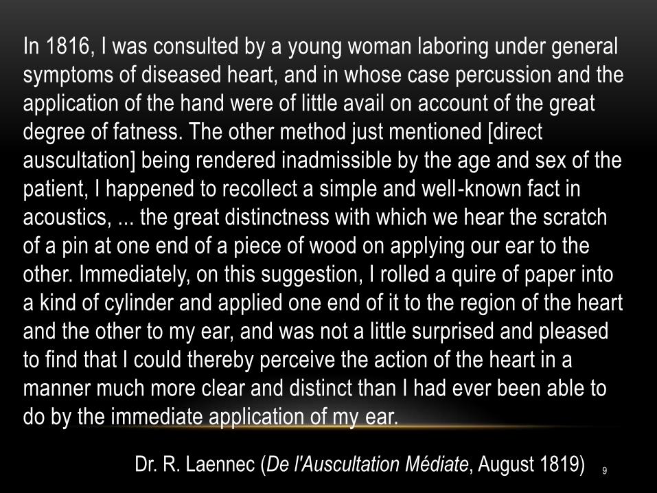
In 1816, I was consulted by a young woman laboring under general
symptoms of diseased heart, and in whose case percussion and the
application of the hand were of little avail on account of the great
degree of fatness. The other method just mentioned [direct
auscultation] being rendered inadmissible by the age and sex of the
patient, I happened to recollect a simple and well-known fact in
acoustics, ... the great distinctness with which we hear the scratch
of a pin at one end of a piece of wood on applying our ear to the
other. Immediately, on this suggestion, I rolled a quire of paper into
a kind of cylinder and applied one end of it to the region of the heart
and the other to my ear, and was not a little surprised and pleased
to find that I could thereby perceive the action of the heart in a
manner much more clear and distinct than I had ever been able to
do by the immediate application of my ear.
Dr. R. Laennec (De l'Auscultation Médiate, August 1819)
10
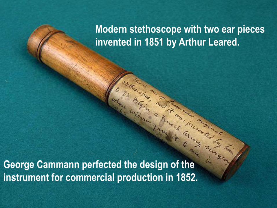
George Cammann perfected the design of the
instrument for commercial production in 1852.
Modern stethoscope with two ear pieces
invented in 1851 by Arthur Leared.
11
Dr. Terry Tegtmeier 1999
“THE MOST IMPORTANT PART OF THE
STETHOSCOPE IS THE PART BETWEEN THE
EAR PIECES”
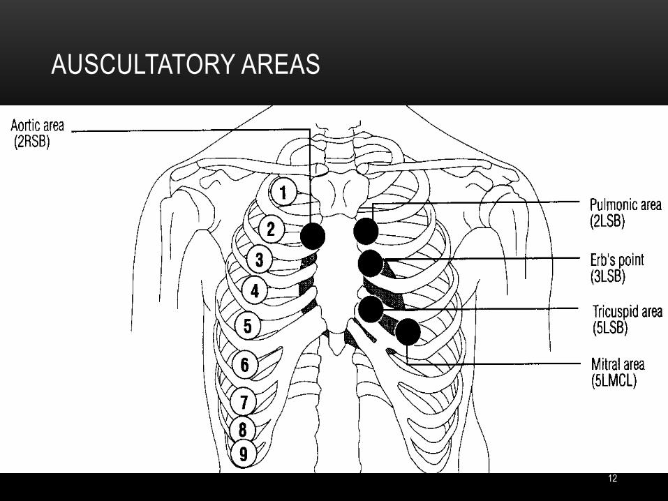
AUSCULTATORY AREAS
12
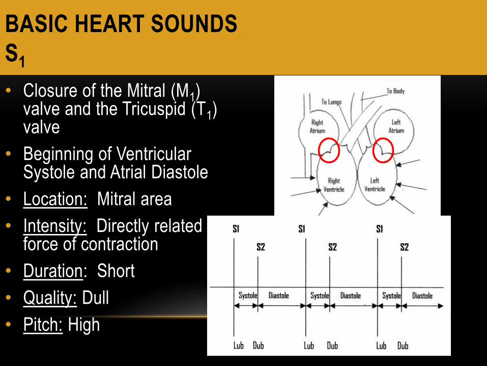
BASIC HEART SOUNDS
S1
• Closure of the Mitral (M1) valve and the Tricuspid (T1) valve
• Beginning of Ventricular Systole and Atrial Diastole
• Location: Mitral area
• Intensity: Directly related to force of contraction
• Duration: Short
• Quality: Dull
• Pitch: High
13
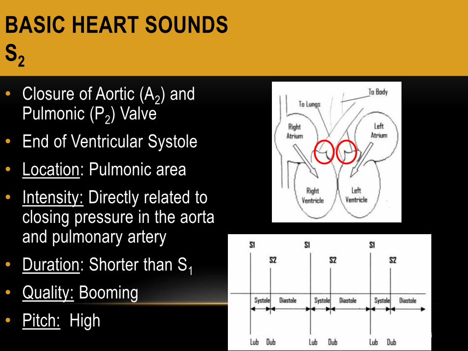
BASIC HEART SOUNDS
S2
• Closure of Aortic (A2) and Pulmonic (P2) Valve
• End of Ventricular Systole
• Location: Pulmonic area
• Intensity: Directly related to closing pressure in the aorta and pulmonary artery
• Duration: Shorter than S1
• Quality: Booming
• Pitch: High 14
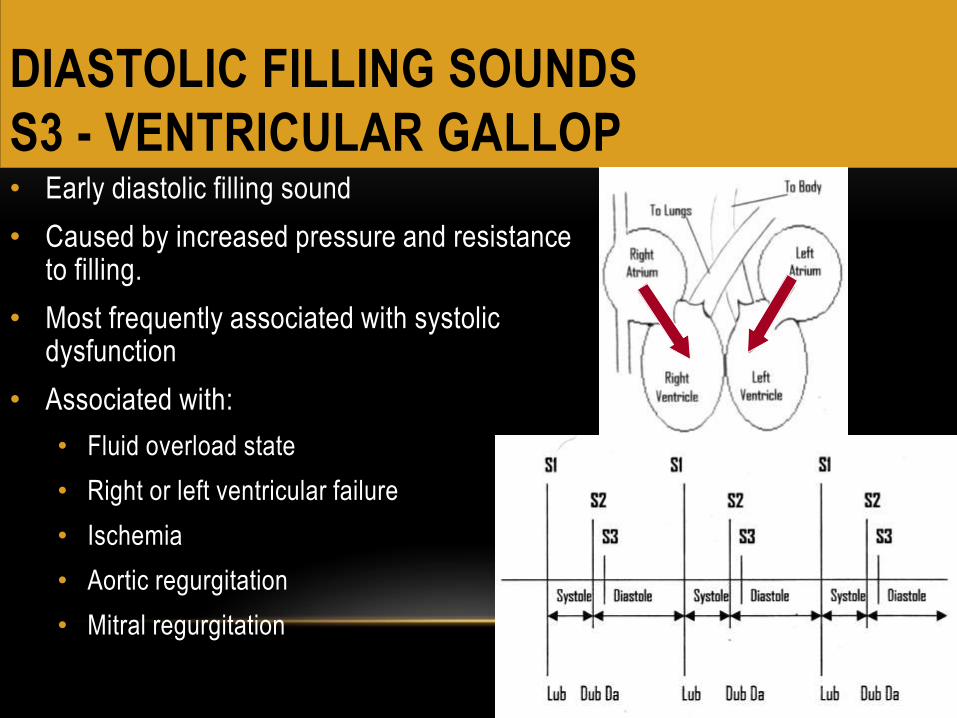
DIASTOLIC FILLING SOUNDS
S3 - VENTRICULAR GALLOP • Early diastolic filling sound
• Caused by increased pressure and resistance to filling.
• Most frequently associated with systolic dysfunction
• Associated with:
• Fluid overload state
• Right or left ventricular failure
• Ischemia
• Aortic regurgitation
• Mitral regurgitation
15
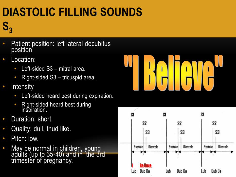
DIASTOLIC FILLING SOUNDS
S3
• Patient position: left lateral decubitus position
• Location:
• Left-sided S3 – mitral area.
• Right-sided S3 – tricuspid area.
• Intensity
• Left-sided heard best during expiration.
• Right-sided heard best during inspiration.
• Duration: short.
• Quality: dull, thud like.
• Pitch: low.
• May be normal in children, young adults (up to 35-40) and in the 3rd trimester of pregnancy.
16
DIASTOLIC FILLING SOUNDS
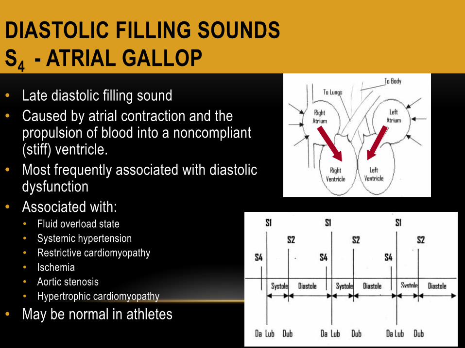
S4 - ATRIAL GALLOP
• Late diastolic filling sound
• Caused by atrial contraction and the propulsion of blood into a noncompliant (stiff) ventricle.
• Most frequently associated with diastolic dysfunction
• Associated with: • Fluid overload state
• Systemic hypertension
• Restrictive cardiomyopathy
• Ischemia
• Aortic stenosis
• Hypertrophic cardiomyopathy
• May be normal in athletes 17
DIASTOLIC FILLING SOUNDS
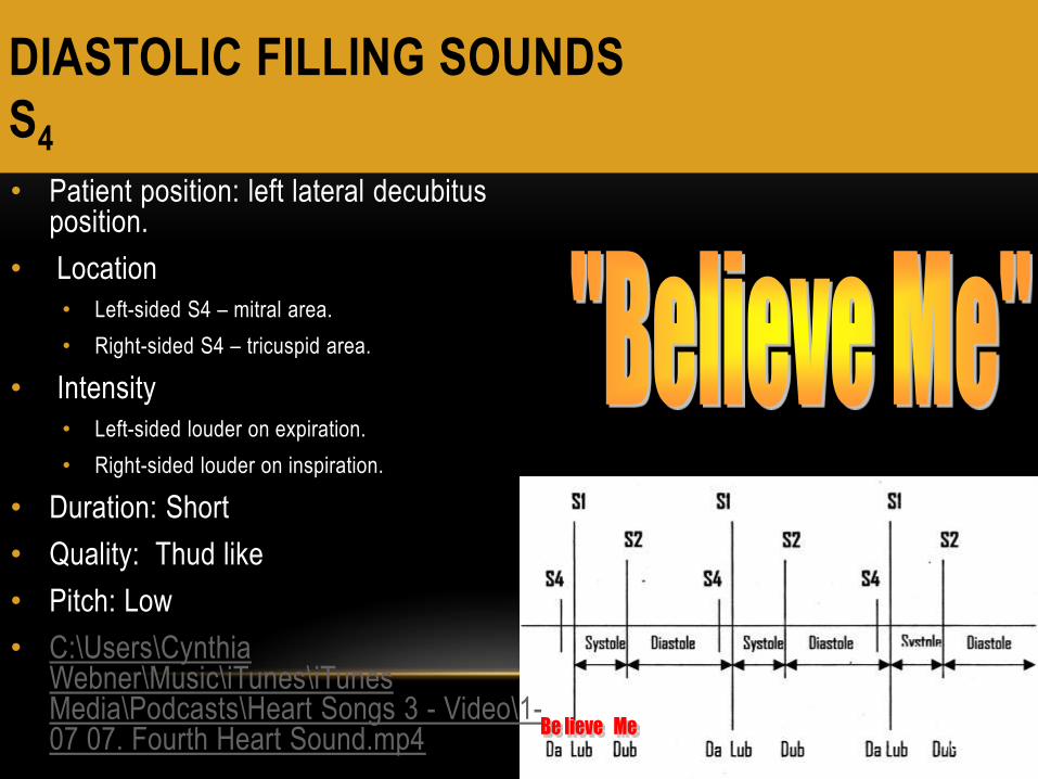
S4
• Patient position: left lateral decubitus position.
• Location
• Left-sided S4 – mitral area.
• Right-sided S4 – tricuspid area.
• Intensity
• Left-sided louder on expiration.
• Right-sided louder on inspiration.
• Duration: Short
• Quality: Thud like
• Pitch: Low
• C:\Users\Cynthia Webner\Music\iTunes\iTunes Media\Podcasts\Heart Songs 3 - Video\1-07 07. Fourth Heart Sound.mp4 18
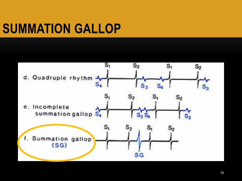
SUMMATION GALLOP
19
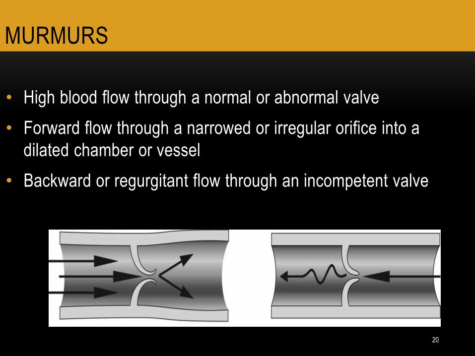
MURMURS
20
• High blood flow through a normal or abnormal valve
• Forward flow through a narrowed or irregular orifice into a
dilated chamber or vessel
• Backward or regurgitant flow through an incompetent valve
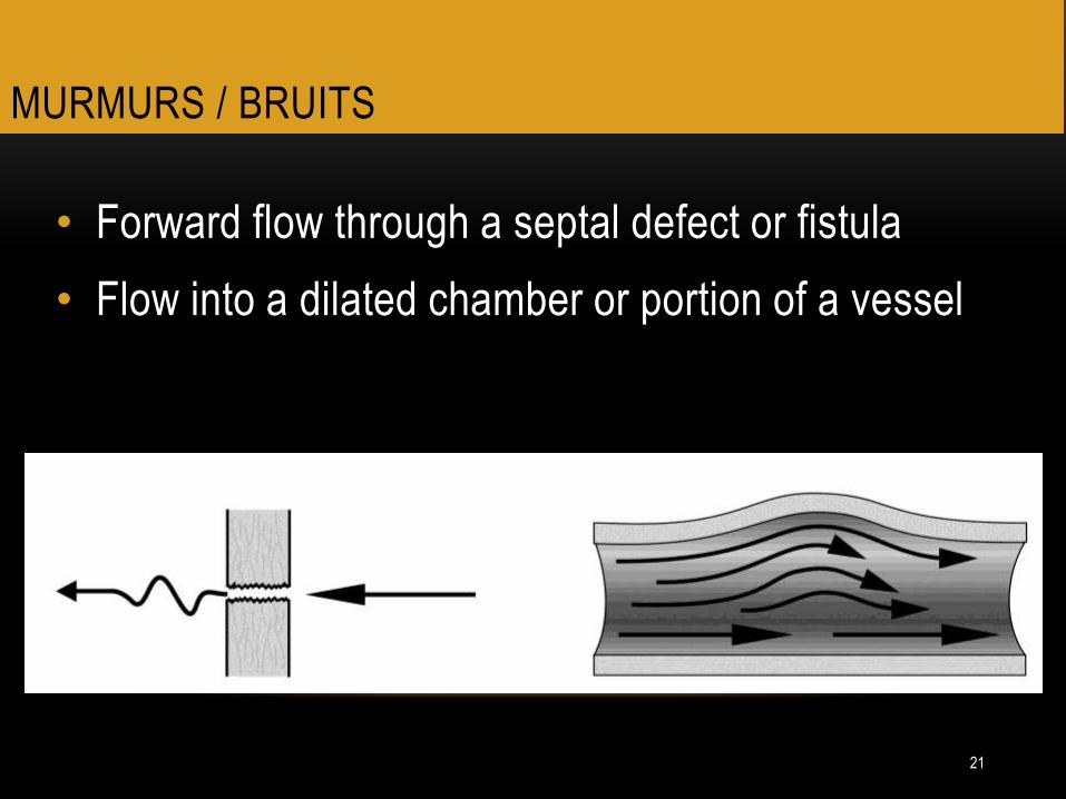
MURMURS / BRUITS
21
• Forward flow through a septal defect or fistula
• Flow into a dilated chamber or portion of a vessel
22
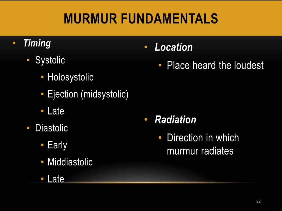
MURMUR FUNDAMENTALS
• Timing
• Systolic
• Holosystolic
• Ejection (midsystolic)
• Late
• Diastolic
• Early
• Middiastolic
• Late
• Location
• Place heard the loudest
• Radiation
• Direction in which
murmur radiates
23
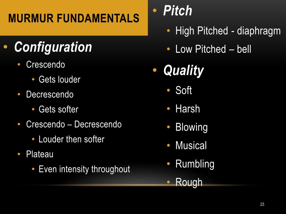
MURMUR FUNDAMENTALS
• Configuration • Crescendo
• Gets louder
• Decrescendo
• Gets softer
• Crescendo – Decrescendo
• Louder then softer
• Plateau
• Even intensity throughout
• Pitch
• High Pitched - diaphragm
• Low Pitched – bell
• Quality
• Soft
• Harsh
• Blowing
• Musical
• Rumbling
• Rough
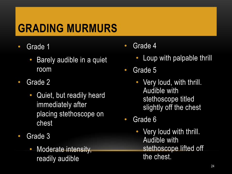
• Grade 1
• Barely audible in a quiet
room
• Grade 2
• Quiet, but readily heard
immediately after
placing stethoscope on
chest
• Grade 3
• Moderate intensity,
readily audible
• Grade 4
• Loup with palpable thrill
• Grade 5
• Very loud, with thrill. Audible with stethoscope titled slightly off the chest
• Grade 6
• Very loud with thrill. Audible with stethoscope lifted off the chest.
GRADING MURMURS
24
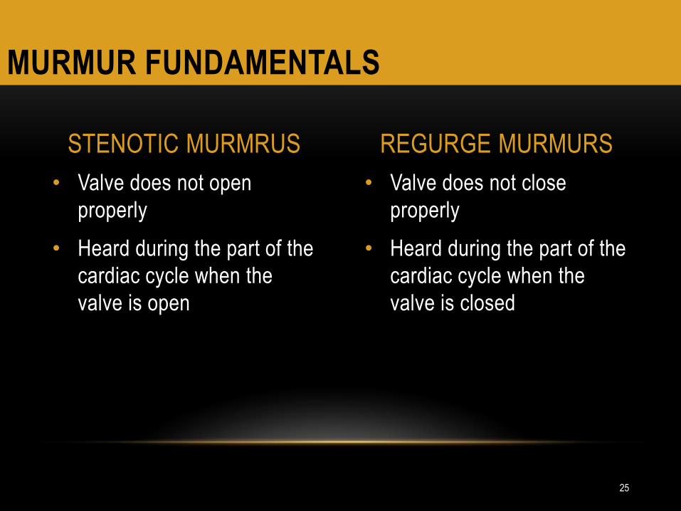
• Valve does not close
properly
• Heard during the part of the
cardiac cycle when the
valve is closed
• Valve does not open
properly
• Heard during the part of the
cardiac cycle when the
valve is open
MURMUR FUNDAMENTALS
STENOTIC MURMRUS REGURGE MURMURS
25
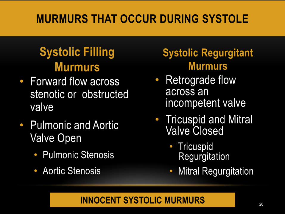
• Retrograde flow across an incompetent valve
• Tricuspid and Mitral Valve Closed
• Tricuspid Regurgitation
• Mitral Regurgitation
• Forward flow across stenotic or obstructed valve
• Pulmonic and Aortic Valve Open
• Pulmonic Stenosis
• Aortic Stenosis
MURMURS THAT OCCUR DURING SYSTOLE
Systolic Filling
Murmurs Systolic Regurgitant
Murmurs
26 INNOCENT SYSTOLIC MURMURS
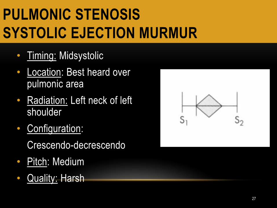
PULMONIC STENOSIS
SYSTOLIC EJECTION MURMUR
• Timing: Midsystolic
• Location: Best heard over pulmonic area
• Radiation: Left neck of left shoulder
• Configuration:
Crescendo-decrescendo
• Pitch: Medium
• Quality: Harsh
27
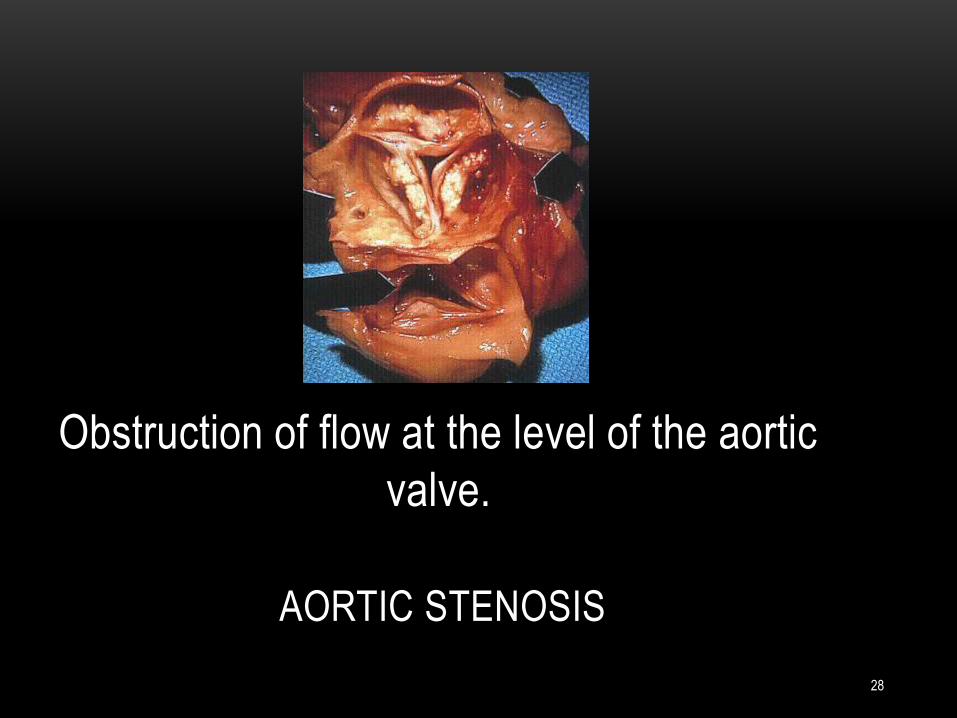
AORTIC STENOSIS
Obstruction of flow at the level of the aortic
valve.
28
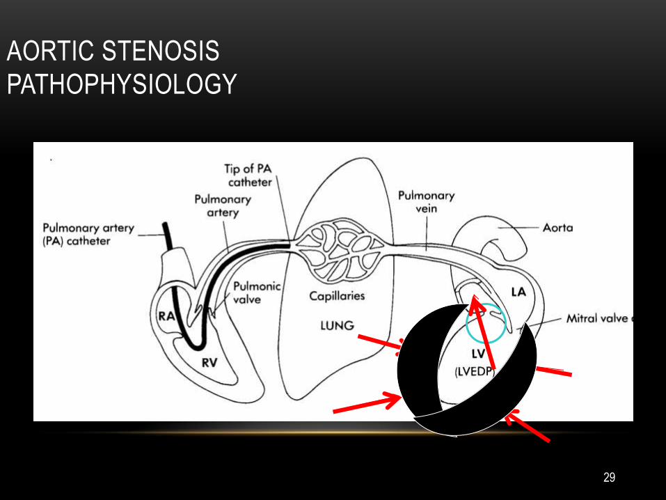
AORTIC STENOSIS
PATHOPHYSIOLOGY
29
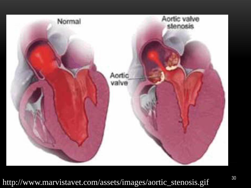
30 http://www.marvistavet.com/assets/images/aortic_stenosis.gif
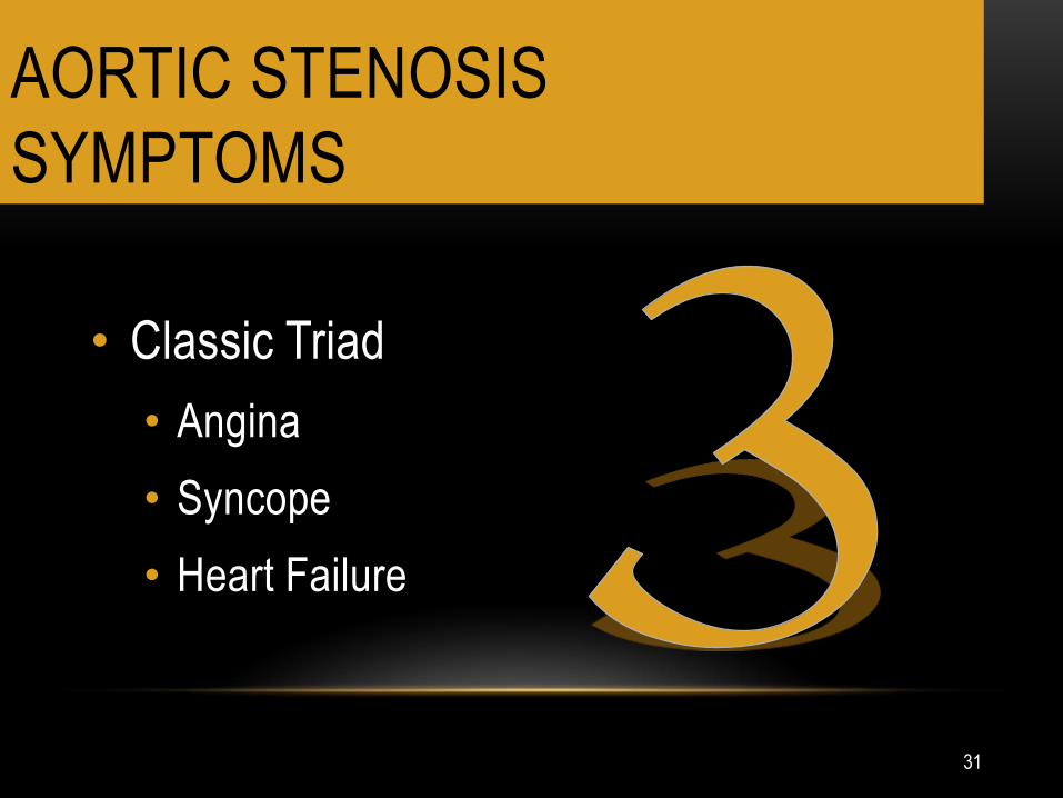
AORTIC STENOSIS
SYMPTOMS
• Classic Triad
• Angina
• Syncope
• Heart Failure
31
AORTIC STENOSIS
SIGNS (EXAMINATION)
• In addition to classic triad:
• Decreased pulse sharpness
• Systolic Ejection Murmur
• S4
32
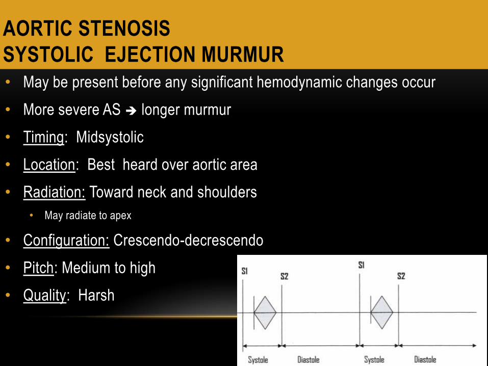
AORTIC STENOSIS
SYSTOLIC EJECTION MURMUR
• May be present before any significant hemodynamic changes occur
• More severe AS longer murmur
• Timing: Midsystolic
• Location: Best heard over aortic area
• Radiation: Toward neck and shoulders
• May radiate to apex
• Configuration: Crescendo-decrescendo
• Pitch: Medium to high
• Quality: Harsh
33
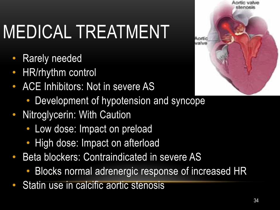
MEDICAL TREATMENT
• Rarely needed
• HR/rhythm control
• ACE Inhibitors: Not in severe AS
• Development of hypotension and syncope
• Nitroglycerin: With Caution
• Low dose: Impact on preload
• High dose: Impact on afterload
• Beta blockers: Contraindicated in severe AS
• Blocks normal adrenergic response of increased HR
• Statin use in calcific aortic stenosis
34
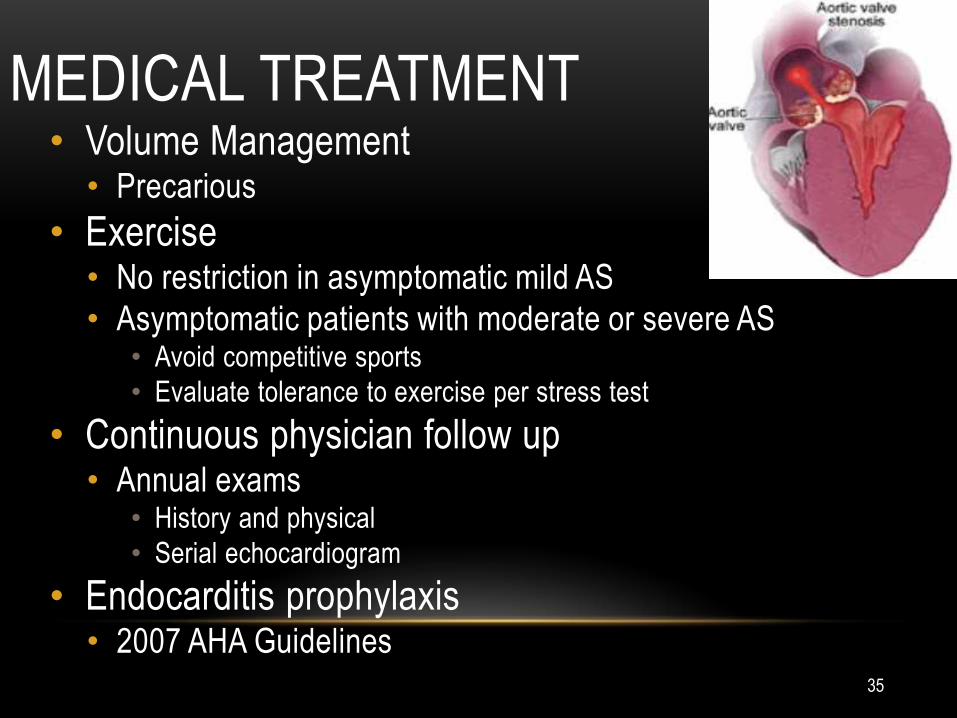
MEDICAL TREATMENT • Volume Management • Precarious
• Exercise • No restriction in asymptomatic mild AS
• Asymptomatic patients with moderate or severe AS • Avoid competitive sports
• Evaluate tolerance to exercise per stress test
• Continuous physician follow up • Annual exams
• History and physical
• Serial echocardiogram
• Endocarditis prophylaxis • 2007 AHA Guidelines
35
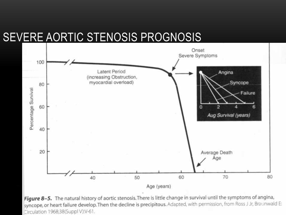
SEVERE AORTIC STENOSIS PROGNOSIS
36
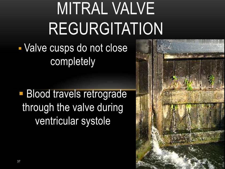
MITRAL VALVE
REGURGITATION Valve cusps do not close
completely
Blood travels retrograde
through the valve during
ventricular systole
37
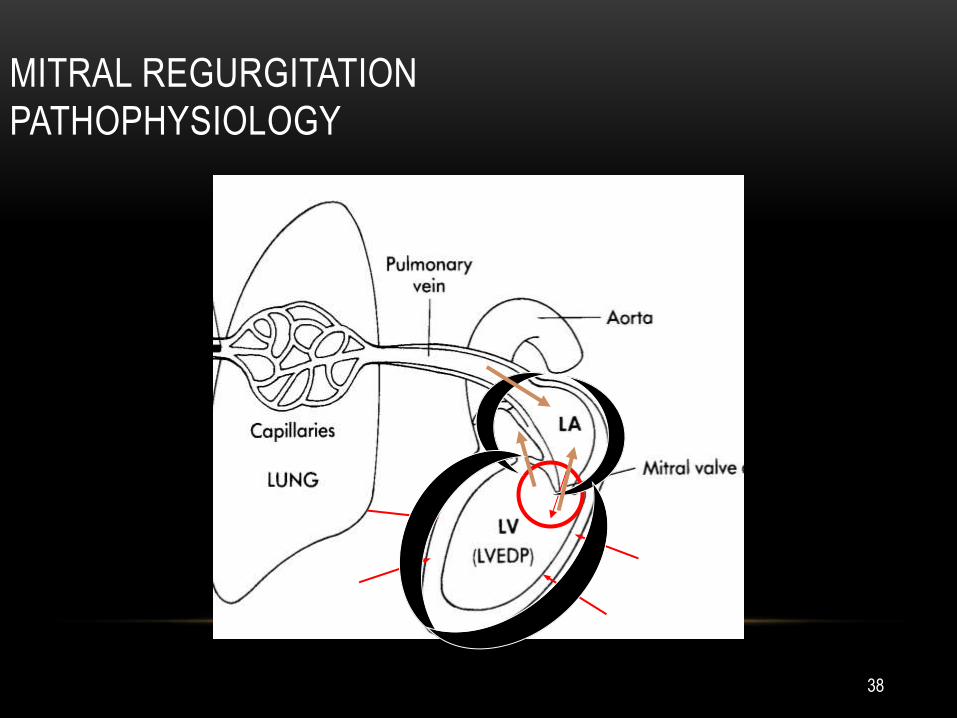
MITRAL REGURGITATION
PATHOPHYSIOLOGY
38
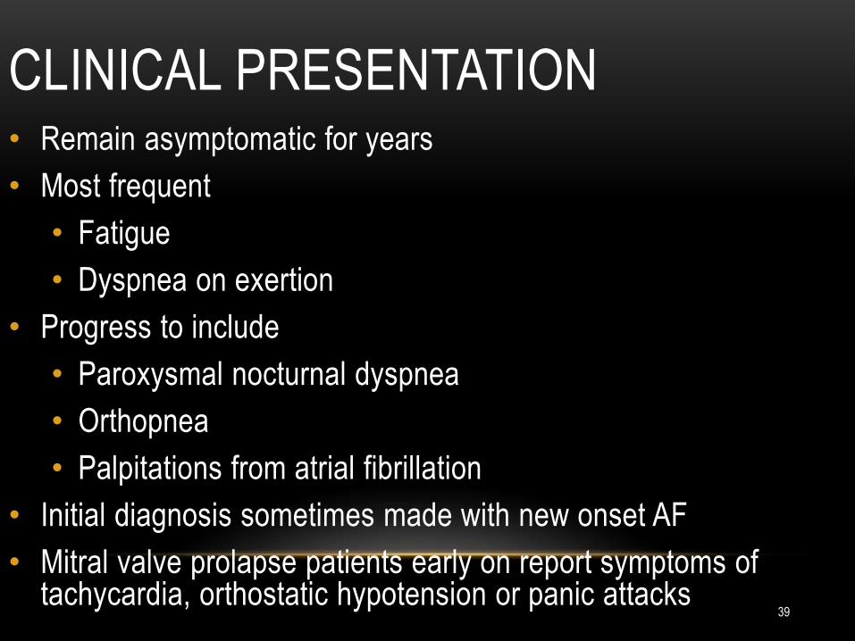
CLINICAL PRESENTATION • Remain asymptomatic for years
• Most frequent
• Fatigue
• Dyspnea on exertion
• Progress to include
• Paroxysmal nocturnal dyspnea
• Orthopnea
• Palpitations from atrial fibrillation
• Initial diagnosis sometimes made with new onset AF
• Mitral valve prolapse patients early on report symptoms of tachycardia, orthostatic hypotension or panic attacks
39
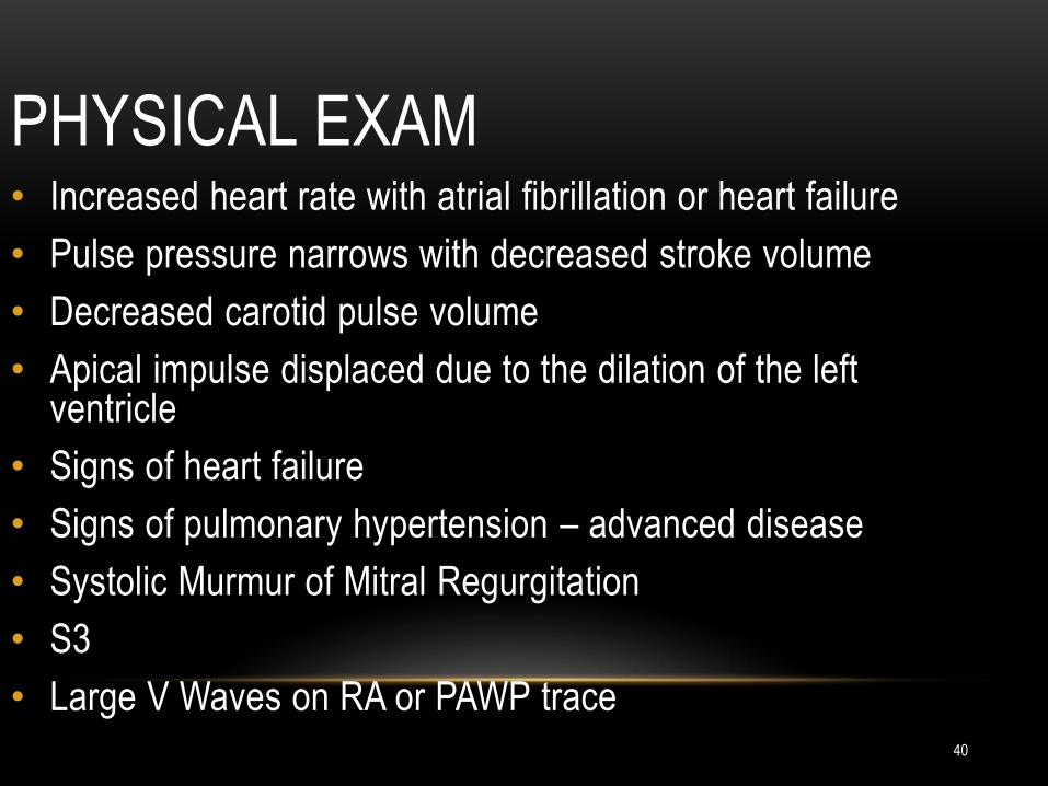
PHYSICAL EXAM • Increased heart rate with atrial fibrillation or heart failure
• Pulse pressure narrows with decreased stroke volume
• Decreased carotid pulse volume
• Apical impulse displaced due to the dilation of the left ventricle
• Signs of heart failure
• Signs of pulmonary hypertension – advanced disease
• Systolic Murmur of Mitral Regurgitation
• S3
• Large V Waves on RA or PAWP trace 40
41
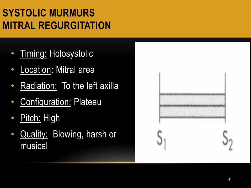
SYSTOLIC MURMURS
MITRAL REGURGITATION
• Timing: Holosystolic
• Location: Mitral area
• Radiation: To the left axilla
• Configuration: Plateau
• Pitch: High
• Quality: Blowing, harsh or
musical
ACUTE MITRAL REGURGITATION
PATHOPHYSIOLOGY
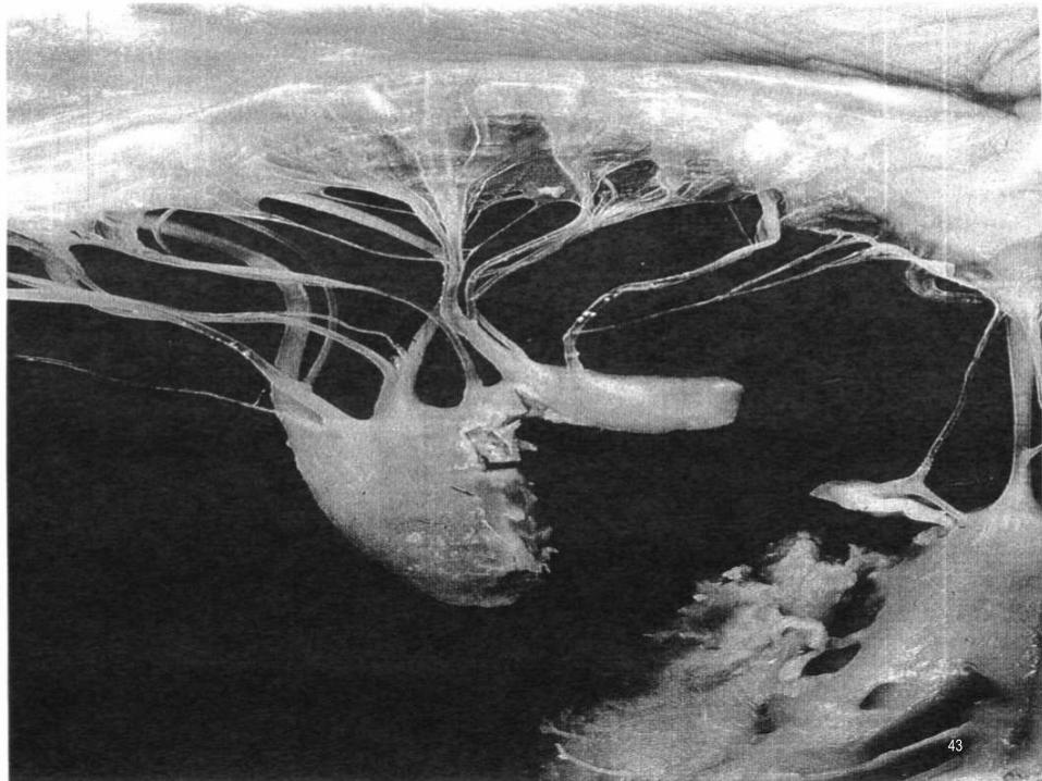
• Acute MI • Impairment or rupture of a papillary muscle
• Damaged to myocardial wall → damage to attachment of the papillary muscle to that ventricular wall
• Papillary muscle continues to contract with each cardiac cycle
• Attachment of papillary muscle to ventricular wall becomes weaker with each contraction
• With enough damage to the myocardial wall or papillary muscle the papillary muscle will actually disconnect from the ventricular wall
• Acute mitral regurgitation state
• Emergency measures are necessary to preserve the patient’s life 42
43
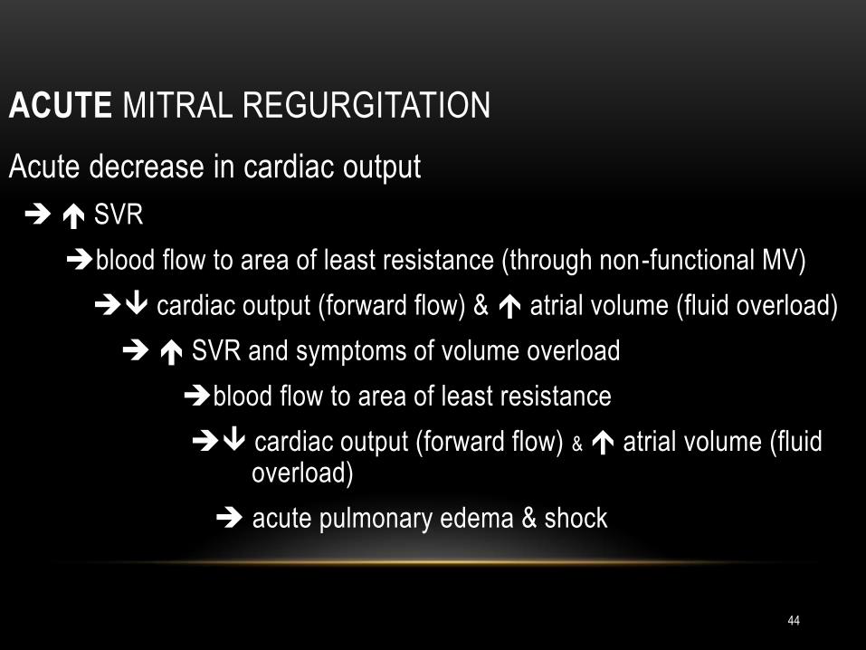
ACUTE MITRAL REGURGITATION
Acute decrease in cardiac output
SVR
blood flow to area of least resistance (through non-functional MV)
cardiac output (forward flow) & atrial volume (fluid overload)
SVR and symptoms of volume overload
blood flow to area of least resistance
cardiac output (forward flow) & atrial volume (fluid overload)
acute pulmonary edema & shock
44
MEDICAL TREATMENT • No treatment for asymptomatic patient with normal ventricular
function
• Continuous physician follow up
• Annual exams
• History and physical
• Serial echocardiogram
• Rhythm Control
• Atrial fibrillation
• Anticoagulation in patients
• ACE Inhibitors
• Useful in non-surgical candidates
• No benefit in asymptomatic patients 45
TREATMENT FOR ACUTE MR
• STAT Echo
• Surgery emergently
• IABP
• Afterload Reduction
• Nitroprusside
• Antibiotics
46
SURGICAL TREATMENT
• EF < 60% considered abnormal
• Surgical options include:
• Mitral valve repair
• Mitral valve replacement with preservation of mitral apparatus
• Mitral valve replacement with removal of mitral apparatus
• Mortality rates in those >75 higher with mitral valve surgery than aortic valve
• Mortality rates less with repair than replacement
47
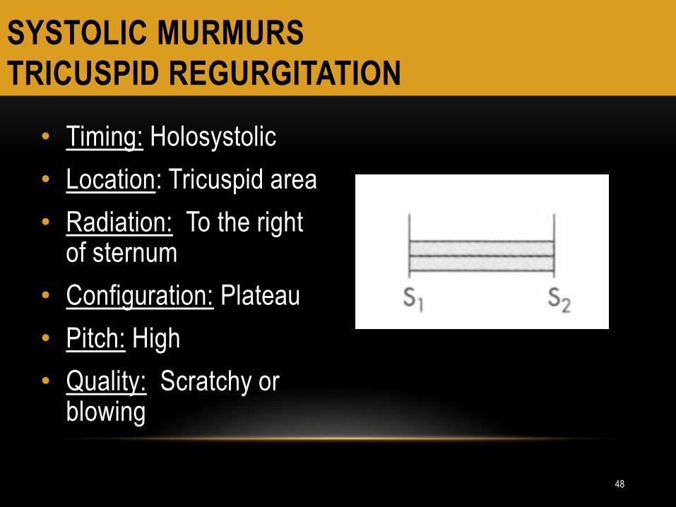
SYSTOLIC MURMURS
TRICUSPID REGURGITATION
• Timing: Holosystolic
• Location: Tricuspid area
• Radiation: To the right of sternum
• Configuration: Plateau
• Pitch: High
• Quality: Scratchy or blowing
48
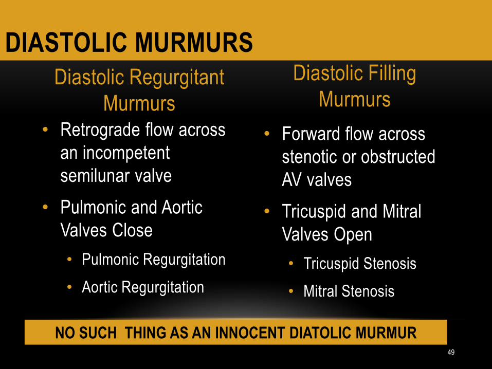
• Forward flow across
stenotic or obstructed
AV valves
• Tricuspid and Mitral
Valves Open
• Tricuspid Stenosis
• Mitral Stenosis
• Retrograde flow across
an incompetent
semilunar valve
• Pulmonic and Aortic
Valves Close
• Pulmonic Regurgitation
• Aortic Regurgitation
DIASTOLIC MURMURS
Diastolic Regurgitant
Murmurs
Diastolic Filling
Murmurs
49
NO SUCH THING AS AN INNOCENT DIATOLIC MURMUR
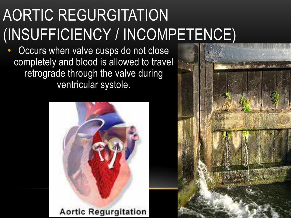
AORTIC REGURGITATION
(INSUFFICIENCY / INCOMPETENCE) • Occurs when valve cusps do not close
completely and blood is allowed to travel retrograde through the valve during
ventricular systole.
50
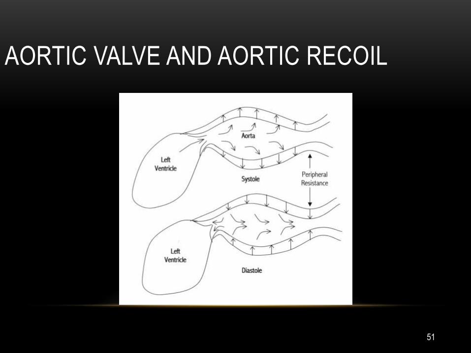
AORTIC VALVE AND AORTIC RECOIL
51
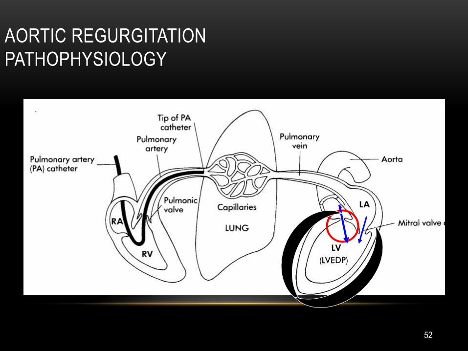
AORTIC REGURGITATION
PATHOPHYSIOLOGY
52
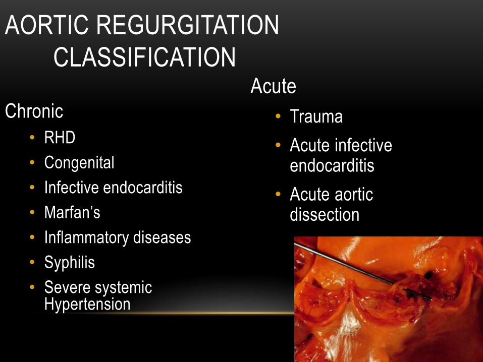
AORTIC REGURGITATION
CLASSIFICATION
Chronic
• RHD
• Congenital
• Infective endocarditis
• Marfan’s
• Inflammatory diseases
• Syphilis
• Severe systemic Hypertension
53
Acute
• Trauma
• Acute infective endocarditis
• Acute aortic dissection
CHRONIC AORTIC REGURGITATION
SYMPTOMS
• Exertional dyspnea
• PND
• Orthopnea
• Angina
• Aware of heart beat – especially when lying
• Pulsatile sensation in head
54
SIGNS OF HYPERDYNAMIC PERFUSION • Warm, flushed, reddish mucous membranes
• Wide pulse pressure (>100mmHg)
• De-Musset Sign
• Head bobbing with each heart beat
• Water-Hammer pulse
• Rapid rise and collapse of the pulse upon palpitation
• Corrigan’s Pulse
• Large carotid pulsation in the neck
• Traube’s Sign
• Loud, sharp “pistol-shot-like” sound heard over the femoral pulse
• Duroziez’s Sign
• Murmur heard over the femoral artery when compressed
• Quinke’s Sign
• Pulsitile blanching and reddening of the fingernails when light pressure is applied 55
PHYSICAL EXAMINATION
• Apical Impulse
• Diastolic Murmur of AR
• Systolic Flow Murmur
• Austin Flint Murmur
• Signs of Hyperdynamic Perfusion
56
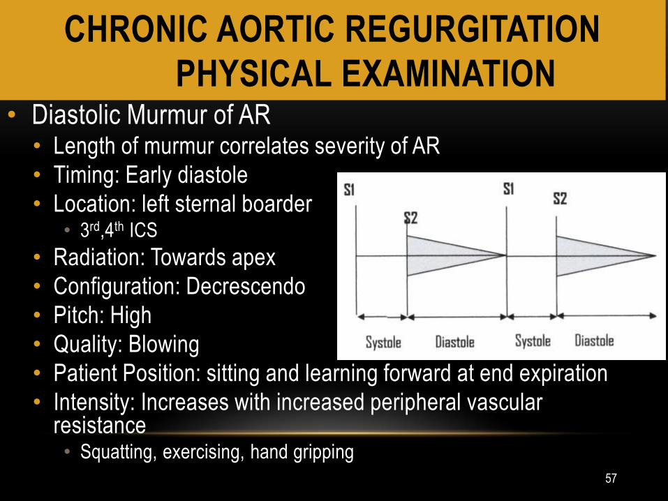
CHRONIC AORTIC REGURGITATION
PHYSICAL EXAMINATION • Diastolic Murmur of AR • Length of murmur correlates severity of AR
• Timing: Early diastole
• Location: left sternal boarder • 3rd,4th ICS
• Radiation: Towards apex
• Configuration: Decrescendo
• Pitch: High
• Quality: Blowing
• Patient Position: sitting and learning forward at end expiration
• Intensity: Increases with increased peripheral vascular resistance • Squatting, exercising, hand gripping
57
SYSTOLIC FLOW MURMUR WITH CHRONIC AR
• Result of turbulent flow across valve during systolic
• Large volumes of blood from hyperdynamic perfusion causes turbulence
• Timing: Mid systolic
• Location: Along left sternal boarder
• Configuration: Crescendo-decrescendo
• Pitch: Medium (best with diaphragm)
• Quality: Soft
• Intensity: May increase after coughing or when elevating legs while in lying position
58
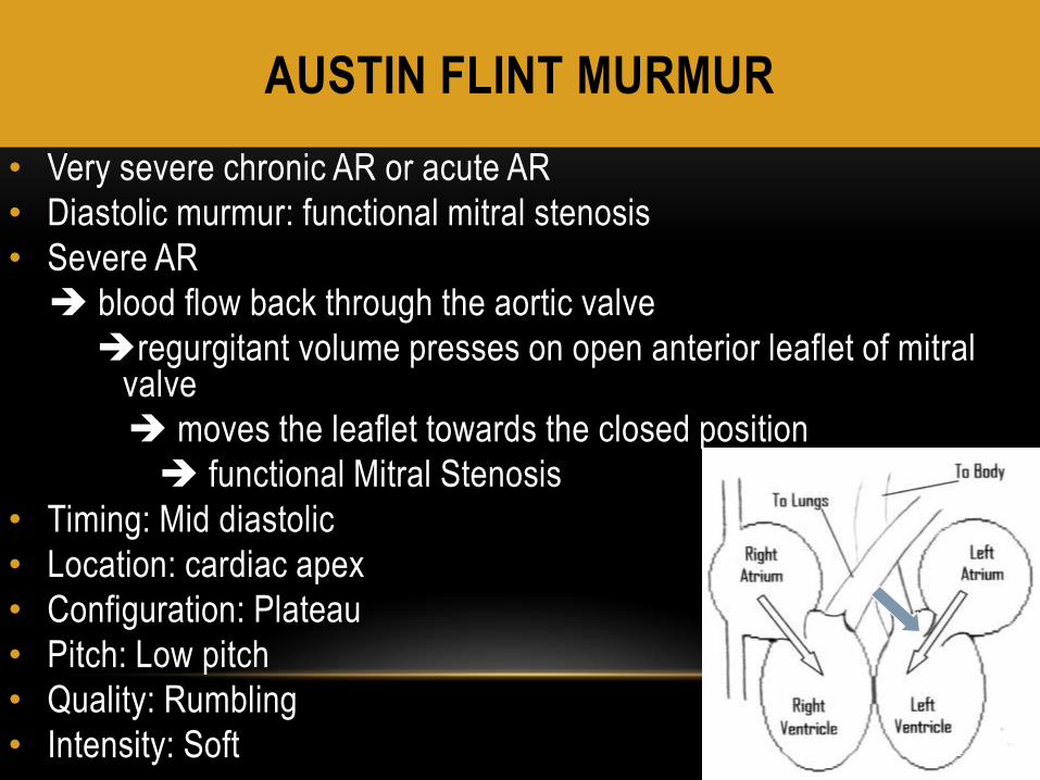
AUSTIN FLINT MURMUR
• Very severe chronic AR or acute AR
• Diastolic murmur: functional mitral stenosis
• Severe AR
blood flow back through the aortic valve
regurgitant volume presses on open anterior leaflet of mitral valve
moves the leaflet towards the closed position
functional Mitral Stenosis
• Timing: Mid diastolic
• Location: cardiac apex
• Configuration: Plateau
• Pitch: Low pitch
• Quality: Rumbling
• Intensity: Soft
59
CHRONIC AORTIC REGURGITATION
MEDICAL TREATMENT • If normal LV function no treatment
• Arterial Vasodilators in symptomatic patients with severe AR and symptoms of LV dysfunction and not a surgical candidate (Class IB)
• Symptom relief preop (Class IIB)
• Decrease afterload decrease regurgitation
• Not indicated in asymptomatic patients (Class III)
• Digoxin and diuretics helpful with HF symptoms
• Avoid arterial vasoconstrictors
• Intra-aortic balloon pump
• Contraindicated in all patients with AR
• Continuous physician follow up
• Annual exams
• History and physical
• Serial echocardiogram
60
SURGICAL TREATMENT • Mortality rates increase as EF decreases
• Once symptomatic 50% will not survive > 3-5 years without surgery
• Valve repair reasonable alternative to replacement in this population
• Valve replacement options the same as with AS
• Goal should be quality of life not longevity
• Looking for symptom relief
• Acute AR requires acute intervention 61
ACUTE AORTIC REGURGITATION
TREATMENT • Urgent Surgical Intervention
• STAT ECHO
• Reduce afterload
• Nitroprusside
• Reduce preload
• Help reduce fluid overload
• Beta blockers
• With caution
• Block sympathetic response of increased HR
• Inotropes
• Increase contractility for forward flow
62
DIASTOLIC MURMURS
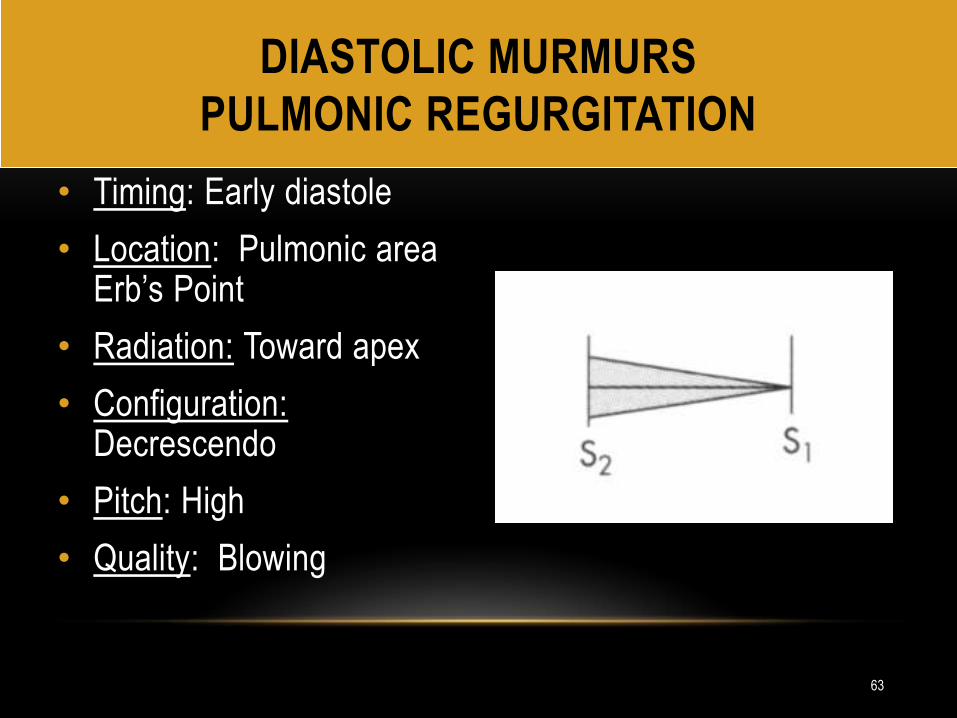
PULMONIC REGURGITATION
• Timing: Early diastole
• Location: Pulmonic area Erb’s Point
• Radiation: Toward apex
• Configuration: Decrescendo
• Pitch: High
• Quality: Blowing
63
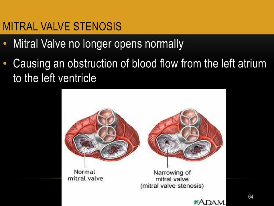
MITRAL VALVE STENOSIS
• Mitral Valve no longer opens normally
• Causing an obstruction of blood flow from the left atrium
to the left ventricle
64
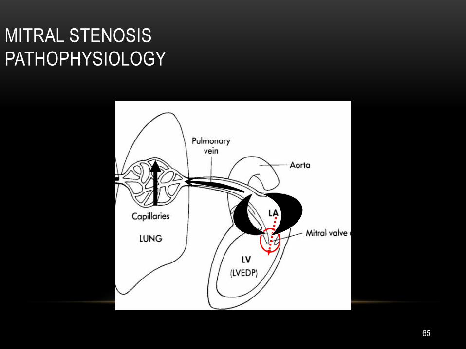
MITRAL STENOSIS
PATHOPHYSIOLOGY
65
SYMPTOMS
• Dyspnea with exertion
• Pulmonary symptoms increase
• Development of orthopnea and paroxysmal nocturnal
dyspnea
• Valve orifice less than 1.0 cm2
• dyspnea at rest
• confined to the bed or chair
• Develop cough and hemoptysis
• Ultimately RV Failure
66
SYMPTOMS
• Often discovered with conditions that increase heart rate • Pregnancy
• New onset atrial fibrillation
• Hyperthyroidism
• Fever
• Stroke • Enlarged atrium
• High risk for development of thrombi
• Atrial Fibrillation • 50% of patients with MS
• Enlarged atrium
67
PHYSICAL EXAM
• Signs of right ventricular failure if disease process is severe
• Jugular venous distension
• Hepatomegaly
• Peripheral edema
• Ascites
• Mitral Facies
• Pinkish-purple discoloration of the cheeks
• Common with severe mitral stenosis
68
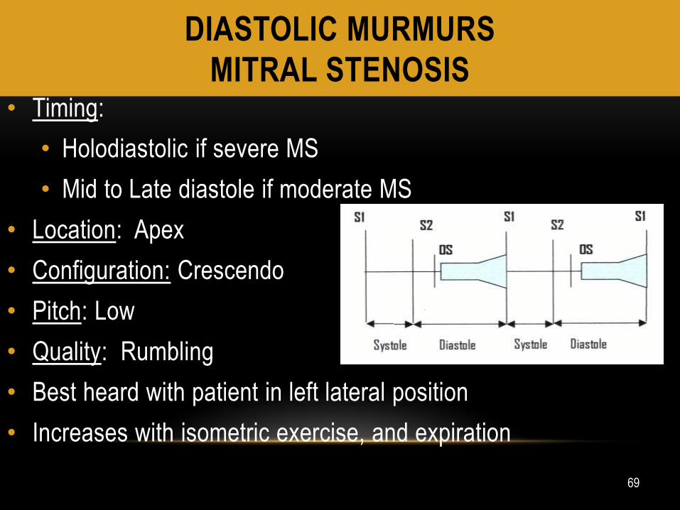
DIASTOLIC MURMURS
MITRAL STENOSIS • Timing:
• Holodiastolic if severe MS
• Mid to Late diastole if moderate MS
• Location: Apex
• Configuration: Crescendo
• Pitch: Low
• Quality: Rumbling
• Best heard with patient in left lateral position
• Increases with isometric exercise, and expiration
69
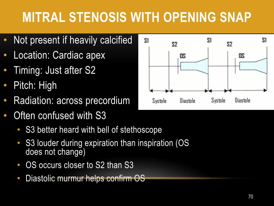
MITRAL STENOSIS WITH OPENING SNAP
• Not present if heavily calcified
• Location: Cardiac apex
• Timing: Just after S2
• Pitch: High
• Radiation: across precordium
• Often confused with S3
• S3 better heard with bell of stethoscope
• S3 louder during expiration than inspiration (OS does not change)
• OS occurs closer to S2 than S3
• Diastolic murmur helps confirm OS
70
MEDICAL TREATMENT
• Is of limited use in asymptomatic patients in NSR
• Atrial Fibrillation Treatment
• Beta blockers or calcium channel blockers
• Maintain a ventricular rate of less than 100 beats per minute
• Since atrial fibrillation is poorly tolerated it is reasonable to attempt to return the patient to normal sinus rhythm with cardioversion
• Heart Rate Control
• Calcium channel blockers, beta-blockers helpful if experiencing exercise intolerance
• Other Benefits of Beta-blockers and Calcium Channel Blockers
• Decrease ventricular wall tension
• Improve filling from the atria
71
MEDICAL TREATMENT
• Preload Reduction
• Diuretics and sodium restriction if fluid overloaded
• Anticoagulation
• High risk due to LA enlargement
• Class I ACC/AHA Recommendations • MS with atrial fibrillation
• MS and prior embolic event
• MS and left atrial thromus
• Class IIb ACC/AHA Recommendations • Consider in asymptomatic patients with severe MS and LA dimension > 55
mm by echocardiogram
• Continuous Follow Up for Asymptomatic Patients 72
SURGICAL TREATMENT
• Once symptoms occur surgery should occur
• Valve area <1.5 cm2
• Symptoms at rest
• Lifestyle affected
• Surgical Options
• Percutaneous mitral balloon valvotomy
• Closed surgical commissurotomy
• Open surgical commissurotomy
• Mitral valve replacement
73
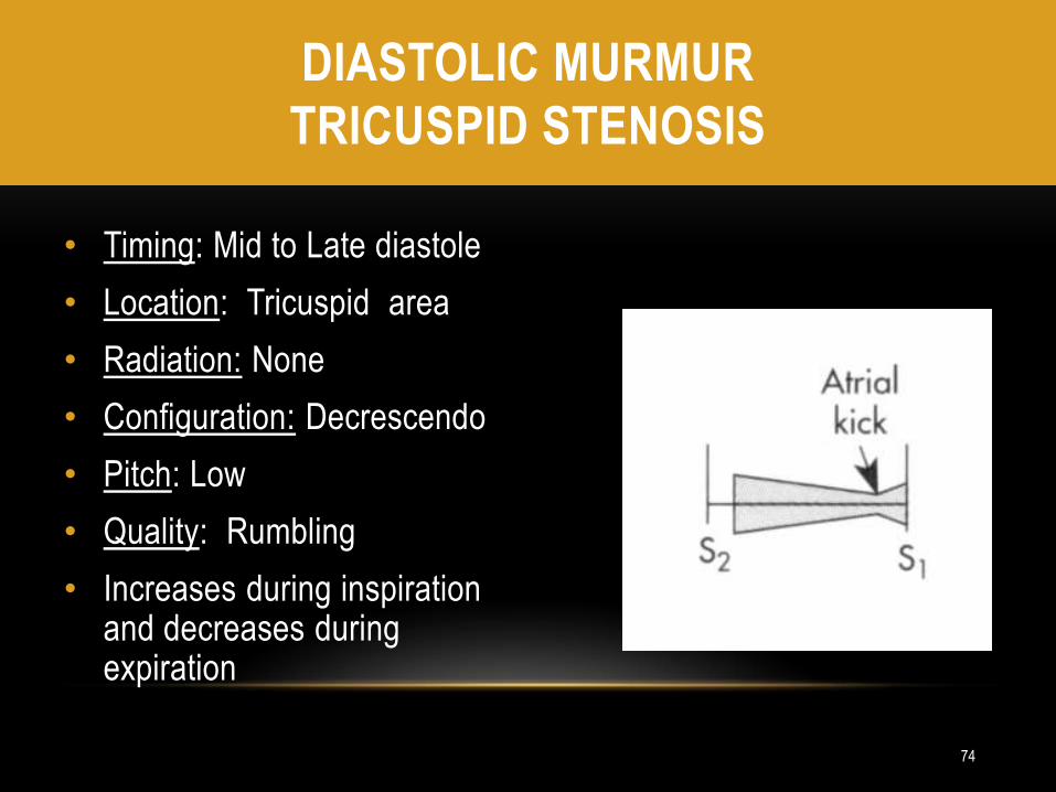
DIASTOLIC MURMUR
TRICUSPID STENOSIS
• Timing: Mid to Late diastole
• Location: Tricuspid area
• Radiation: None
• Configuration: Decrescendo
• Pitch: Low
• Quality: Rumbling
• Increases during inspiration and decreases during expiration
74
OTHER SOUNDS
PERICARDIAL FRICTION RUB
• Timing: Systolic, Early diastolic and late diastolic
• Location: Tricuspid area and Xyphoid area
• Radiation: None
• Configuration: Plateau
• May get louder during inspiration
• Pitch: High
• Quality: Grating, scratching
75
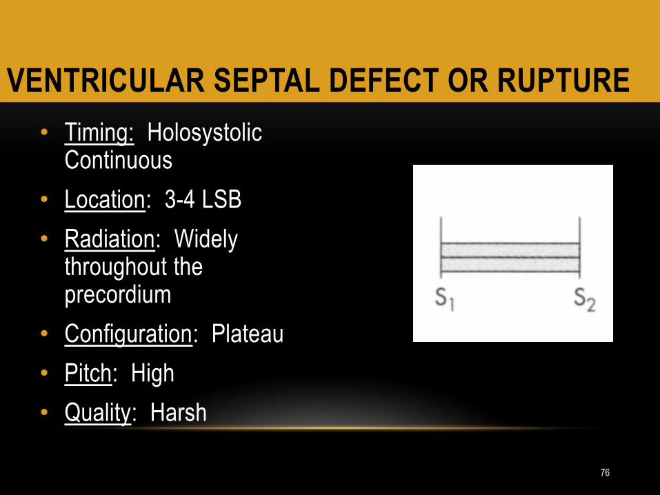
VENTRICULAR SEPTAL DEFECT OR RUPTURE
• Timing: Holosystolic Continuous
• Location: 3-4 LSB
• Radiation: Widely throughout the precordium
• Configuration: Plateau
• Pitch: High
• Quality: Harsh
76
77
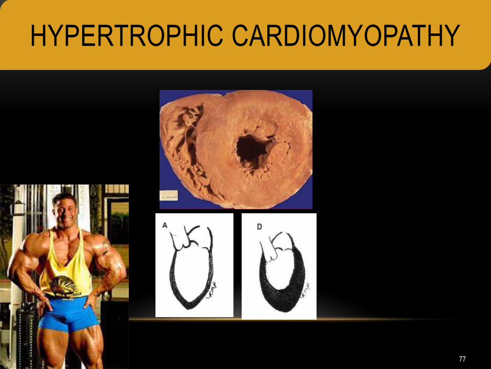
HYPERTROPHIC CARDIOMYOPATHY
• 1 of every 500 (Maron et al, 2003)
• Primary genetic cardiomyopathy
• Effects men and women equally
• Hypertrophy of myocardial muscle mass in the absence of increased ventricular afterload
• Associated with decreased ventricular filling (diastolic dysfunction) and decreased cardiac output
• Most common cause of sudden death in young adults
• Cause unknown
• 50% transmitted genetically
78
HYPERTROPHIC CARDIOMYOPATHY
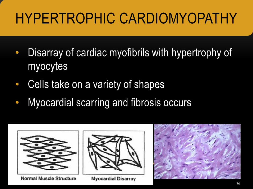
• Disarray of cardiac myofibrils with hypertrophy of
myocytes
• Cells take on a variety of shapes
• Myocardial scarring and fibrosis occurs
79
HYPERTROPHIC CARDIOMYOPATHY
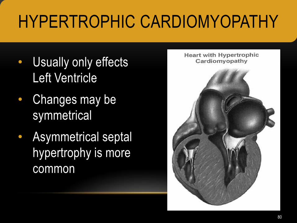
• Usually only effects
Left Ventricle
• Changes may be
symmetrical
• Asymmetrical septal
hypertrophy is more
common
80
HYPERTROPHIC CARDIOMYOPATHY
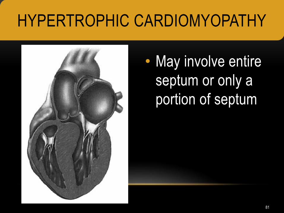
• May involve entire
septum or only a
portion of septum
81
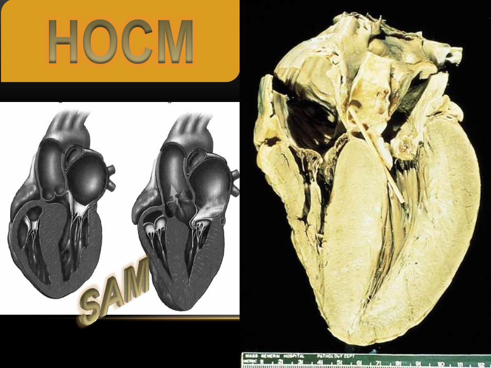
HYPERTROPHIC CARDIOMYOPATHY
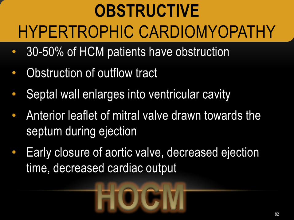
• 30-50% of HCM patients have obstruction
• Obstruction of outflow tract
• Septal wall enlarges into ventricular cavity
• Anterior leaflet of mitral valve drawn towards the
septum during ejection
• Early closure of aortic valve, decreased ejection
time, decreased cardiac output
82
OBSTRUCTIVE
HYPERTROPHIC CARDIOMYOPATHY
83
• Many asymptomatic for years
• Incidence of sudden death often first presentation
• Or identified during screening of relative of patient with HCM
• Symptoms related to severity of diastolic dysfunction
• Heart failure
• Dyspnea #1 sign
• Syncope / palpitations with activity
• Chest pain
• Supraventricular arrhythmias
• Development of mitral regurgitation
84
HYPERTROPHIC CARDIOMYOPATHY
PRESENTATION
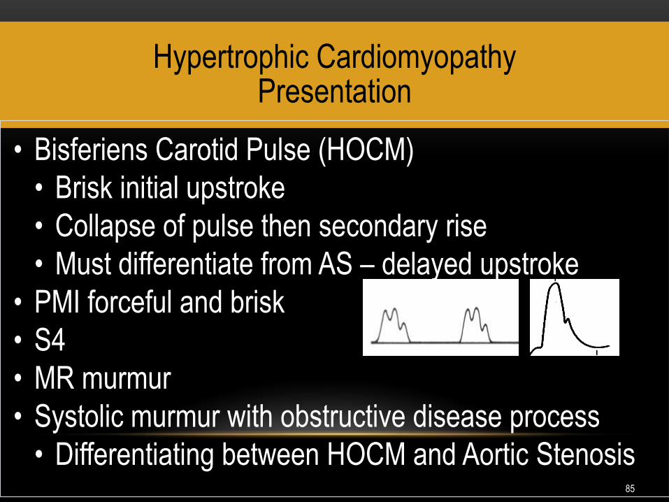
Hypertrophic Cardiomyopathy Presentation
• Bisferiens Carotid Pulse (HOCM)
• Brisk initial upstroke
• Collapse of pulse then secondary rise
• Must differentiate from AS – delayed upstroke
• PMI forceful and brisk
• S4
• MR murmur
• Systolic murmur with obstructive disease process
• Differentiating between HOCM and Aortic Stenosis 85
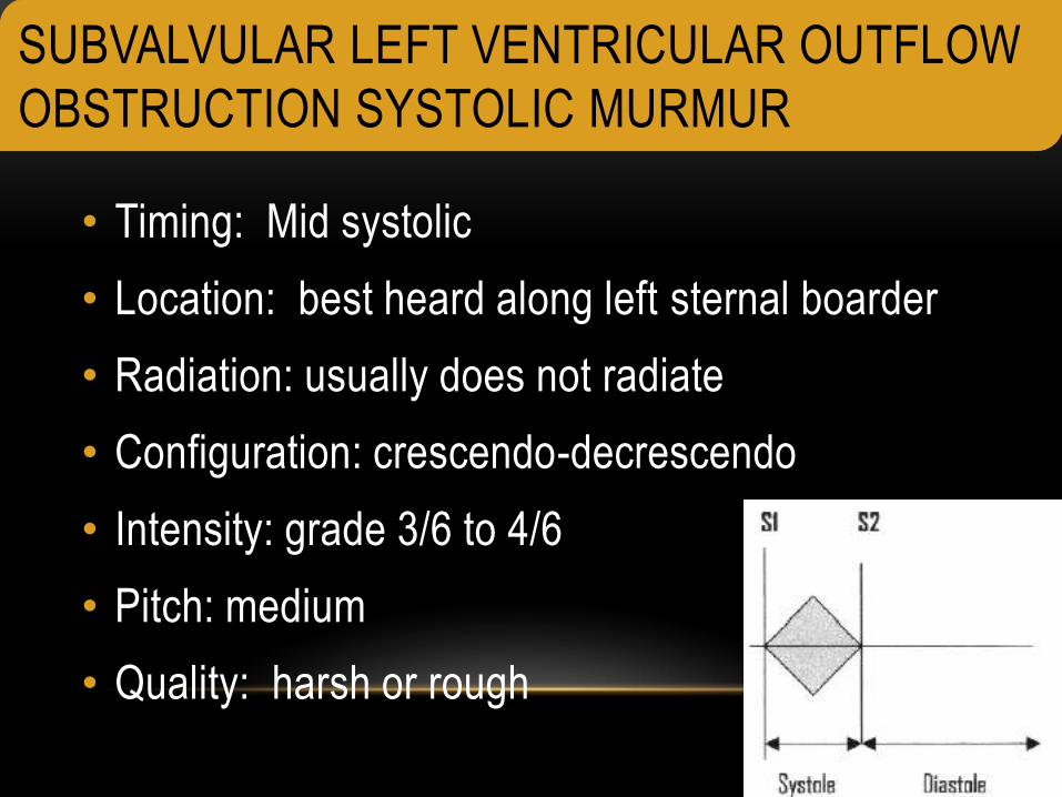
• Timing: Mid systolic
• Location: best heard along left sternal boarder
• Radiation: usually does not radiate
• Configuration: crescendo-decrescendo
• Intensity: grade 3/6 to 4/6
• Pitch: medium
• Quality: harsh or rough
86
SUBVALVULAR LEFT VENTRICULAR OUTFLOW
OBSTRUCTION SYSTOLIC MURMUR
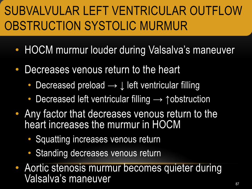
• HOCM murmur louder during Valsalva’s maneuver
• Decreases venous return to the heart • Decreased preload → ↓ left ventricular filling
• Decreased left ventricular filling → ↑obstruction
• Any factor that decreases venous return to the heart increases the murmur in HOCM
• Squatting increases venous return
• Standing decreases venous return
• Aortic stenosis murmur becomes quieter during Valsalva’s maneuver
87
SUBVALVULAR LEFT VENTRICULAR OUTFLOW
OBSTRUCTION SYSTOLIC MURMUR
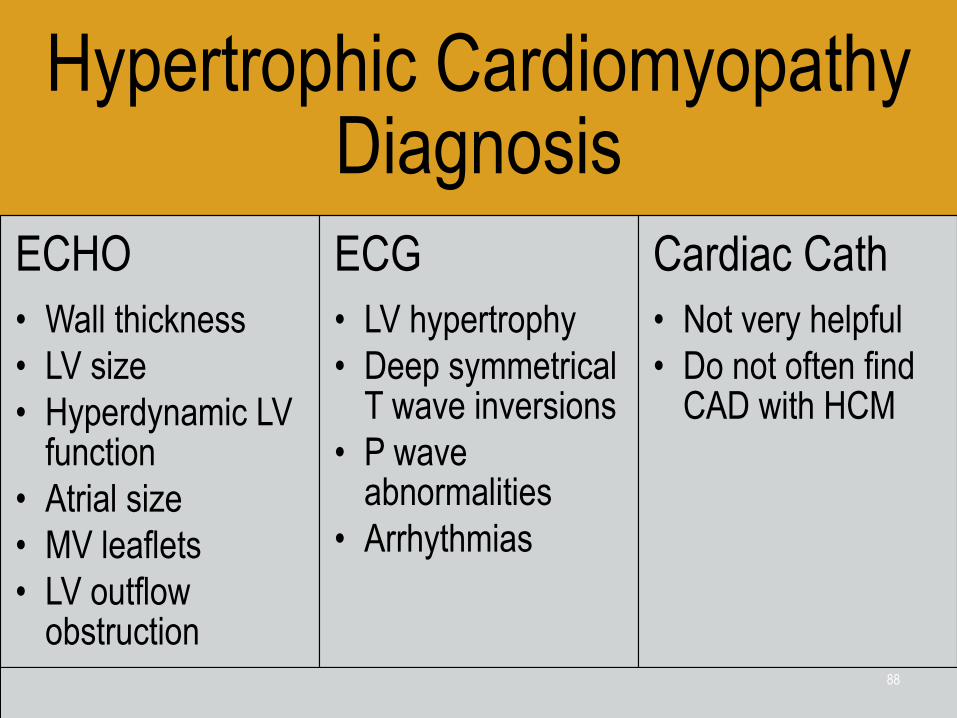
Hypertrophic Cardiomyopathy Diagnosis
ECHO
• Wall thickness
• LV size
• Hyperdynamic LV function
• Atrial size
• MV leaflets
• LV outflow obstruction
ECG
• LV hypertrophy
• Deep symmetrical T wave inversions
• P wave abnormalities
• Arrhythmias
Cardiac Cath
• Not very helpful
• Do not often find CAD with HCM
88
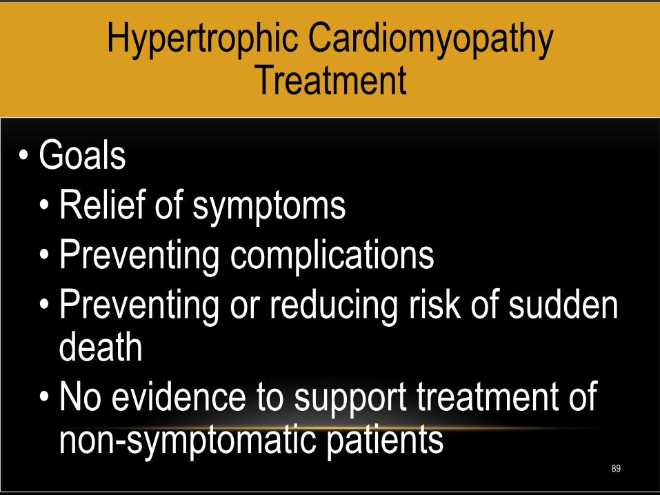
Hypertrophic Cardiomyopathy Treatment
• Goals
• Relief of symptoms
• Preventing complications
• Preventing or reducing risk of sudden death
• No evidence to support treatment of non-symptomatic patients
89
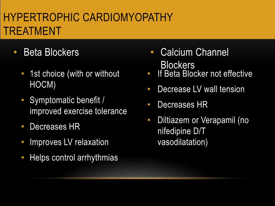
• Beta Blockers • Calcium Channel
Blockers • If Beta Blocker not effective
• Decrease LV wall tension
• Decreases HR
• Diltiazem or Verapamil (no
nifedipine D/T
vasodilatation)
• 1st choice (with or without
HOCM)
• Symptomatic benefit /
improved exercise tolerance
• Decreases HR
• Improves LV relaxation
• Helps control arrhythmias
HYPERTROPHIC CARDIOMYOPATHY
TREATMENT
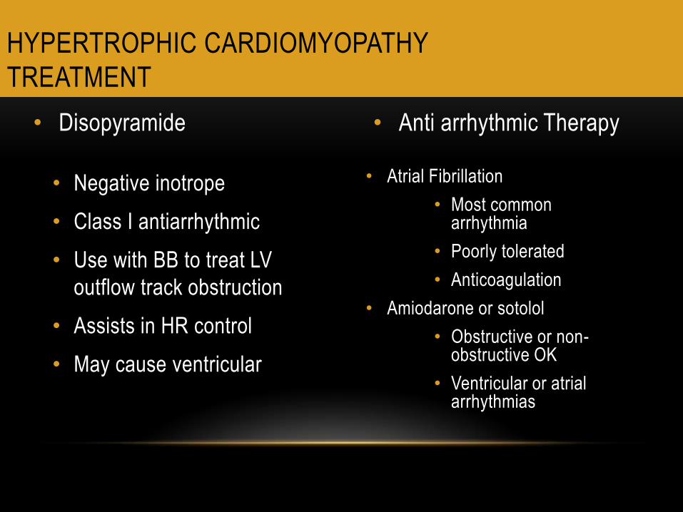
• Disopyramide
• Anti arrhythmic Therapy
• Atrial Fibrillation
• Most common arrhythmia
• Poorly tolerated
• Anticoagulation
• Amiodarone or sotolol
• Obstructive or non-obstructive OK
• Ventricular or atrial arrhythmias
• Negative inotrope
• Class I antiarrhythmic
• Use with BB to treat LV
outflow track obstruction
• Assists in HR control
• May cause ventricular arrhythmias – monitor QT
HYPERTROPHIC CARDIOMYOPATHY
TREATMENT
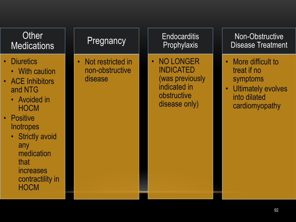
Other Medications
• Diuretics
• With caution
• ACE Inhibitors and NTG
• Avoided in HOCM
• Positive Inotropes
• Strictly avoid any medication that increases contractility in HOCM
Pregnancy
• Not restricted in non-obstructive disease
Endocarditis Prophylaxis
• NO LONGER INDICATED (was previously indicated in obstructive disease only)
Non-Obstructive Disease Treatment
• More difficult to treat if no symptoms
• Ultimately evolves into dilated cardiomyopathy
92
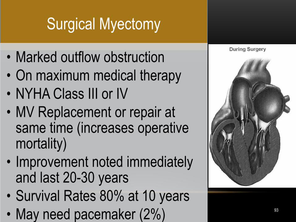
Surgical Myectomy
• Marked outflow obstruction
• On maximum medical therapy
• NYHA Class III or IV
• MV Replacement or repair at same time (increases operative mortality)
• Improvement noted immediately and last 20-30 years
• Survival Rates 80% at 10 years
• May need pacemaker (2%) 93
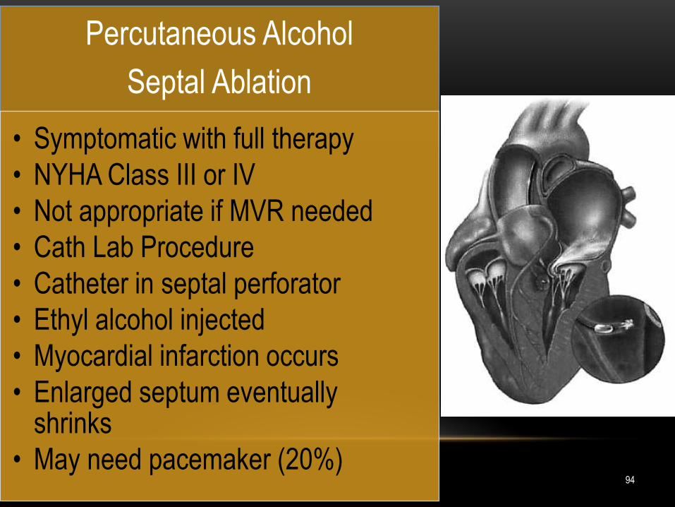
Percutaneous Alcohol
Septal Ablation
• Symptomatic with full therapy
• NYHA Class III or IV
• Not appropriate if MVR needed
• Cath Lab Procedure
• Catheter in septal perforator
• Ethyl alcohol injected
• Myocardial infarction occurs
• Enlarged septum eventually shrinks
• May need pacemaker (20%) 94
SOUNDS ASSOCIATED WITH GROIN
COMPLICATIONS
95
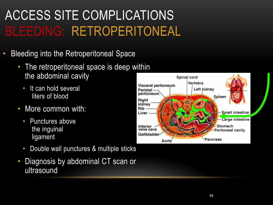
ACCESS SITE COMPLICATIONS
BLEEDING: RETROPERITONEAL
• Bleeding into the Retroperitoneal Space
• The retroperitoneal space is deep within the abdominal cavity
• It can hold several liters of blood
• More common with:
• Punctures above the inguinal ligament
• Double wall punctures & multiple sticks
• Diagnosis by abdominal CT scan or ultrasound
96
ACCESS SITE COMPLICATIONS
BLEEDING: RETROPERITONEAL • Manifestations
• Tachycardia (? beta blockers)
• Due to hypovolemia
• Pain
• Flank, leg, back or lower abdominal pain
• Referred pain due to nerve compression
• Ipsilateral shoulder
• Feeling to have a bowel movement
• Foot or leg palsy
• Distension of the abdomen
• Hypotension (late sign)
• Body is no longer compensating
97
ACCESS SITE COMPLICATIONS
BLEEDING: PSEUDOANEURYSM
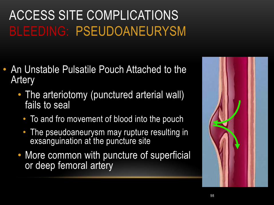
• An Unstable Pulsatile Pouch Attached to the Artery
• The arteriotomy (punctured arterial wall) fails to seal
• To and fro movement of blood into the pouch
• The pseudoaneurysm may rupture resulting in exsanguination at the puncture site
• More common with puncture of superficial or deep femoral artery
98
99
ACCESS SITE COMPLICATIONS
BLEEDING: PSEUDOANEURYSM
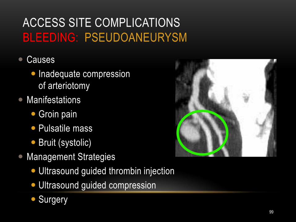
Causes
Inadequate compression
of arteriotomy
Manifestations
Groin pain
Pulsatile mass
Bruit (systolic)
Management Strategies
Ultrasound guided thrombin injection
Ultrasound guided compression
Surgery
100
ACCESS SITE COMPLICATIONS
ARTERIOVENOUS FISTULA
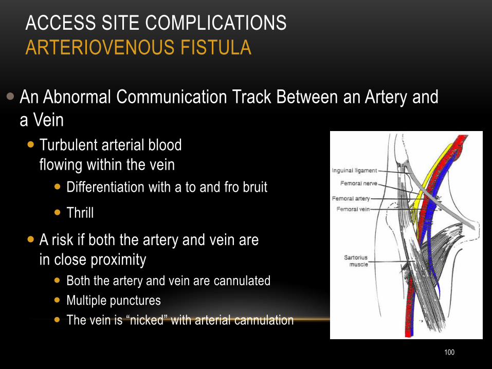
An Abnormal Communication Track Between an Artery and
a Vein
Turbulent arterial blood
flowing within the vein
Differentiation with a to and fro bruit
Thrill
A risk if both the artery and vein are punctured
in close proximity
Both the artery and vein are cannulated
Multiple punctures
The vein is “nicked” with arterial cannulation
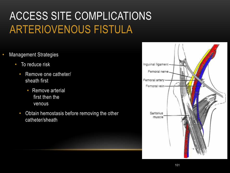
ACCESS SITE COMPLICATIONS
ARTERIOVENOUS FISTULA
• Management Strategies
• To reduce risk
• Remove one catheter/
sheath first
• Remove arterial
first then the
venous
• Obtain hemostasis before removing the other
catheter/sheath
101

QUESTIONS
102
103
LISTEN CAREFULLY!!!!
A DILIGENT ASSESSMENT BY A NURSE
MAYUNCOVER A FINDING THAT WILL MAKE A
DIFFERENCE IN A LIFE.

BE THE BEST THAT YOU CAN BE EVERY DAY. YOUR PATIENTS ARE COUNTING
ON IT!
104 www.cardionursing.com