Embed Size (px)
Citation preview

Gen Physiol. Biophys. (1990). 9, 219 -244 219
Heart Muscle: Mathematical Modelling of the Mechanical Activity and Modelling of Mechanochemical Uncoupling
L. B. KATSNELSON, V. Y A . IZÁKOV and V. S. MARKHASIN
Laboratory of Biophysics, Institute of Industrial Hygiene and Occupational Diseases,
Sverdlovsk 620014, USSR
Abstract. A mechanical model of heart muscle is proposed which includes rheological equations and equations for Ca-troponin interaction, for the dependences of the number of myosin cross-bridges on the length of sarcomere and on the speed of motion. The main assumption of the model is the dependence of the troponin affinity to calcium ions on the number of myosin cross-bridges attached. The model successfully imitates isometric and isotonic contractions, the "length-force" relationships, load-dependent relaxation, and the group of mechanical phenomena known as mechanochemical uncoupling.
Key words: Myocardial tension — Muscle mechanics — Modelling of muscle — Troponin-Ca relationship
Introduction
Several models are available at present which describe different aspects of the mechanical activity of skeletal and cardiac muscles (Fung 1970; Morel 1985; Simmons and Jewell 1973). Depending on the specific objective, these models describe the time course of single isometric and isotonic contractions, the dependence of the speed of shortening on the load, the relation between length and force, etc.
A common disadvantage of all these models is that they reproduce but a limited number of phenomena in the mechanical activity of muscle. Thus, while simulating successfully the dynamics of the isometric contractions, the models fail to describe isotonic contractions or responses to rapid changes in the length or load.
The models based on the mechanochemical cycle of myosin cross-bridges are very complex and require application of partial differential equations (Hill

220 Katsnelson and Izákov
1975; Huxley 1957; Eisenbergand Hill 1978). Moreover, the dependences of the cross-bridge attachment and detachment rate constants on the mechanical distortion do not follow from experiment, but are usually fitted in such models (Huxley and Simmons 1971; Julian et al. 1974).
Many important experimental data concerning cardiac muscle mechanics go beyond the scope of these models, e.g.:
1. The models do not simulate the process of muscle relaxation and, in particular, the dependence of the speed of isotonic relaxation on the length and load (Strauer 1973) and the so-called "load-dependent" relaxation (Brutsaert et al. 1980).
2. The models do not simulate the events which come under a general term "mechanochemical uncoupling" (Kaufmann et al. 1972). These include the inactivating effect of short-term muscle deformations, earlier relaxation of the muscle after preliminary shortening, differences in the "length-force" relationship slopes under isometric and isotonic conditions.
3. The models fail to simulate the experimentally established relations between the concentration of free calcium, the kinetics of the "calcium-troponin complex" and the mechanical activity of the muscle under isometric and isotonic conditions (Allen et al. 1983: Allen and Blinks 1978; Moss 1986).
The above list might be continued. The reason for developing a new model is our belief that many phenomena known under their proper names have a common mechanism at the basis and that many phenomena may be understood by introducing laws describing the effects of the mechanical conditions on the time course of activation of the contractile proteins.
The purpose of this report is to present a new and. at the same time, relatively simple model for the contraction-relaxation cycle of heart muscle, which would simulate the main mechanical events pertaining to the functioning of this muscle. In constructing the model, we considered new experimental data, specifically, those concerning the kinetics of ionized calcium and of that bound to troponin (Robertson et al. 1981), and the dependence of the number of attached cross-bridges on the speed of motion of the sarcomeres (Ferennezi et al. 1984). Since there is no detailed information available about certain molecular processes during muscle relaxation, some assumptions were unavoidable. The plausibility of the assumptions made was verified by successful modelling of a wide range of experimental data.
General Structure of the Model
The majority of the contemporary models of muscle contraction comprise three main parts corresponding to rheological, activation and mechanochemical ev-

Heart Muscle Modelling 221
ents (Izákov et al. 1979). In general, we follow this tradition. The rheological unit accounts for the presence of elastic elements which are
external with respect to the sarcomeres. The muscle is represented by a three-element model comprising the active contractile element (CE) and two passive non-linear elastic elements, the parallel (PE) and the serial (SE) ones.
The parallel elastic element (PE) determines the elastic properties of resting cardiac muscle. The viscoelastic behaviour of a passive muscle is not considered, since, over the dynamic range of interest, it is unlikely to be due to the properties of the cell; rather, it reflects the characteristics of the myocardium as a composite material consisting of a connective tissue framework filled with extracellular liquid (Tsaturian et al. 1984).
The force generator is represented by the contractile element. Functionally, the latter may be assumed to consist of two units representing the activation process and mechanochemical events. Activation is triggered by the binding of calcium by troponin. It determines the number and the dynamics of the possible sites of force generation. The mechanochemical unit of the model simulates the rules governing the attachment and detachment of the cross-bridges.
In different models, the mechanism of activation has been described with different degrees of detail (Julian 1969; Cannel and Allen 1984; Robertson et al. 1981). Depending on the objective of the study, the concentration of free calcium has either been described as a function of time (Julian. 1969) or obtained by solving a material balance equation for calcium taking into account its release from different sources, its uptake by the sarcoplasmic reticulum and binding with troponin and with some of the buffer systems (Cannel and Allen 1984). Since the precise kinetics of calcium interactions with all these components are not known for myocardium, we have chosen a simpler variant: the dynamics of calcium concentration is assumed to be a known function of time.
In modelling the muscle, a correct description of the behaviour of a mechanochemical system is the most intricate problem, because many aspects in the molecular behaviour of the myosin cross-bridges remain unclear. In any case. it is essential "to guess" the basic rules of motion.
The choice is between two strategies. The first strategy consists in the use of the data on the biochemical cycle of the actomyosin ATPase and the data on the mechanochemical states of the attached cross-bridges (Comincioli et al. 1984; Eisenberg et al. 1980). The other strategy is based on the fact that the behaviour of a mechanochemical system and the equation of motion may be assumed phenomenologically by describing the relation between the force of cross-bridges and the probability of their attachment as well as the velocity of motion. We use the second approach.
The majority of the models, with the exception of that developed by Panerai (1980) have assumed the activation to be independent of mechanical
224 Katsnelson and I/ako\
Fig. 2. Dynamics of the free calcium concentration.
stiffness of the sarcomere at time t are equal to A(t).n(t) multiplied by a constant factor, i.e.
N(t) = n.A(t).n{t). (6)
The velocity of the sarcomere shortening is assumed to be negative /,(/) = = V(t) < 0. while V(i) > 0 during stretching (relaxation).
The model also uses parameter Vm.M, which is the maximum shortening velocity in the absence of external load.
Activation
The number of calcium-troponin complexes and their kinetics are the major regulators of the contractile element force (Descherevsky 1977). Probably, the mechanisms of many biomechanical processes in muscle, including mechanochemical uncoupling (Tregear and Marston 1979) should be sought in the process of activation.
The concentration of free calcium ions in the reaction /one during the contraction-relaxation cycle. Ca(/). is assumed to be a function of lime. The time course of function Ca(?) is shown in Fig. 2. The concentration of calcium is expressed as a molar fraction of the total amount of troponin in the myocyte.
Curve Ca(r) is approximated by the following equation:
Ca(f) Cam . [1 - e x p ( - < v : ) ] : . t < i,i
Ca„ e x p ( r / , r ) ] . e x p [ - ^ ( / - td):
(7) t > l.

Heart Muscle Modelling 225
where Cam is the maximum concentration of free Ca; aL and bc are the constants which determine the rise and the decay of the calcium curve; td is the time at which the supply of calcium in the reaction zone is cut off.
Function (7) is a convenient form to describe the time-dependence of the free calcium concentration in myocytes, and is based on the formula proposed by Panerai (1980) with a small modification.
Strictly speaking, assumption (7) for function Ca(r) is a considerable simplification of the problem. The actual kinetics of free calcium is a result of complex processes of calcium transport into the cell, its uptake into the sarcoplasmic reticulum and the exchange of calcium with other Ca-buffer systems (Robertson elal. 1981; Baylor et al. 1982; Miledi et al. 1982). It is quite probable that, under specific conditions of muscle functioning, function Ca(?) has another shape. Nevertheless, in many cases, the assumption (7) is quite sufficient for the above function Ca(r). To simplify the model, we have not included the calcium balance equations. For the purposes of the study though, /d, Cam, ac, bc
are parameters and, depending on the objective, they may be assigned different values.
The kinetics of the calcium-troponin complexes is determined by the reaction
Ca2+ + Tn'*±CaTn.
Circumstantial evidence obtained from the experiments studying the effects of mechanical conditions on the electromechanical coupling suggests that the length and'or the load may have an effect on this process. Panerai (1980) claims that c2, i.e. the decay rate constant for calcium-troponin complexes, is the function of the sarcomere length. The theoretical and experimental grounds of this statement are not clear, however.
Our model is based on the results of the experiments, which demonstrate that the troponin-calcium binding equilibrium constant (taking into account either the pure troponin or the whole complex of the regulating proteins in the thin filament) is by an order of magnitude greater in the presence of S,-myosin subfragments (Hill et al. 1983). More elaborate experiments showed that in the presence of cross-bridges attached to the thin filament, the Ca-troponin decay rate constant reduces at least by a factor of 10 in comparison with a system without myosin (Rosenfeld and Taylor 1985).
The latter experiment implies two conclusions as to the behaviour of calcium-troponin complexes in intact muscle.
First, the decay of Ca-troponin complexes outside the zone where the thin
228 Katsnelson and Izákov
filament which is accessible to "scanning" for a free cross-bridge. The less the distance between the thin and the thick filaments, the longer this section. Since in intact muscle the volume of the sarcomere, remains constant even at changing length, the distance between the filaments is inversely proportional to the length of the sarcomere (Goldman et al. 1979). Stated otherwise
n, = *(/,), (15)
«, increasing with the increase in length (/,). In the model, «,(/,) are defined as follows:
0, if H /(/ l)<0,
TO), ^ 0 ^ W(/ , )<1 , (16)
1, if W{lx)>\,
where W\(lx) is the linear function of /, having the following forms:
TO ) = £.-/• + & , (17)
where coefficients g, and g2 determine the effect of the length on the probability that the cross-bridges attach to the actin filament.
Now we proceed to consider function n2 = n2(t) in more detail. The experiments studying stiffness of a muscle in constant activation, carried out under conditions when changes in the sarcomere length can be neglected (the amount of deformation is 0.5% or less), demonstrate that the number of attached cross-bridges and, hence, probability «2, depend only on the speed of motion (15). It is also known that, under these conditions, the setting of a constant speed of motion with the help of an ergometer leads to the establishment of a steady-state value of stiffness only upon the completion of the transient (Saeki et al. 1980). These results suggest that for an arbitrary contraction-relaxation cycle probability n2{t) is determined by the equation:
dn2(t)
át = K^[V{t)\.[\ -n2(t)] - K_[V{t)]n2{t). (18)
Let us denote probability n2 as m(V) in the steady-state contractile (relaxation) process at speed V under constant activation during the plateau of the length-force relationship. It is evident that
* í ľ ) = £ Í D , G W , (19) m(0) G,(0)
where G^{V) is the stiffness of the sarcomere under the steady-state conditions at speed V, and G* is the same stiffness normalized to its value in ideal isometry ( | /=0 ) .

Heart Muscle Modelling 229
Taking into account that m{V) is the solution of equation (18) and dm(V) n „ , , ¥. ,
= 0 tor a given steady-state speed V, we obtain df
0 = Ä : + ( F ) . [ 1 -m{V)]-K.{V).m{V).
Subtracting equation (18) from this relation and assuming q(V) = K+(V) + K_(V), we obtain the equation for n2(t) used in the model
^ ^ = q[V(t)\. {m[V(t)] - n2(t)}. (20) d?
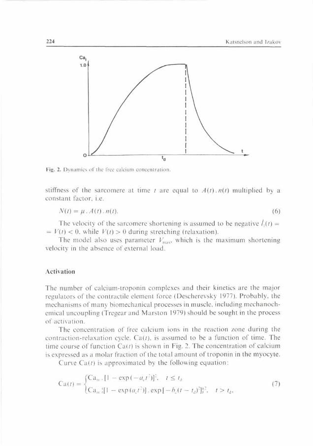
The available experimental data (Ferennezi et al. 1984) suggest the following dependence of q(V) on the speed of motion:
Stated otherwise, it is assumed for simplicity that at V > 0 (relaxation, stretching) q(V) is constant. For F < 0 (contraction) g(K) is a linear function of the speed of shortening. The shape of function q{V) is shown in Fig. 4.
Thus, probability n = nln2 together with the decay rate constant for calcium-troponin complexes depends directly on the sarcomere length and indirectly, through (20), on the speed of the contractile element.
Average Force of a Cross-Bridge
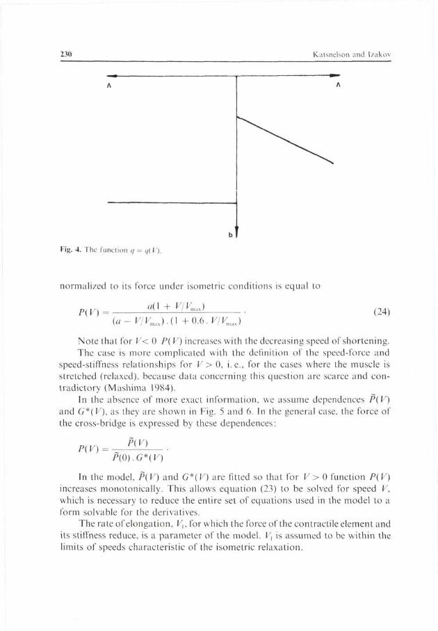
Under conditions of total activation and length-independence (the length-force relationship plateau) for a given constant rate of motion, certain steady-state values of force and stiffness are reached after a short-time transient. The force-speed relationship is defined, under these conditions, by the known Hill equation (Hill 1970)
HV) = a{\ + F/-Kmax) F(0) a- V/Vm„
where P(0) is the force of an isometric contraction. This equation is valid only for shortening, i.e., when V < 0. Under the same conditions, the relation between steady-state stiffness G* and the speed of shortening (V) has the following form (Ferennezi et al. 1984):
G*(K) = 1+0.6.K/Fmax . (23)
Accounting for equations (20) and (21) the average force of the cross-bridge
230 Katsnelson and Izákov
Fig. 4. The function q = </( V).
normalized to its force under isometric conditions is equal to
P(V) a{\ + V/K,J
(a- F/Fmilx).(l +0.6.F/K,,1X) (24)
Note that for V< 0 P( V) increases with the decreasing speed of shortening. The case is more complicated with the definition of the speed-force and
speed-stiffness relationships for V > 0, i.e., for the cases where the muscle is stretched (relaxed), because data concerning this question are scarce and contradictory (Mashima 1984).
In the absence of more exact information, we assume dependences P{V) and G*(V), as they are shown in Fig. 5 and 6. In the general case, the force of the cross-bridge is expressed by these dependences:
P(V) = -P(V)
P(0).G*(V)
In the model, P(V) and G*(V) are fitted so that for V > 0 function P(V) increases monotonically. This allows equation (23) to be solved for speed F, which is necessary to reduce the entire set of equations used in the model to a form solvable for the derivatives.
The rate of elongation, Vu for which the force of the contractile element and its stiffness reduce, is a parameter of the model. Vx is assumed to be within the limits of speeds characteristic of the isometric relaxation.
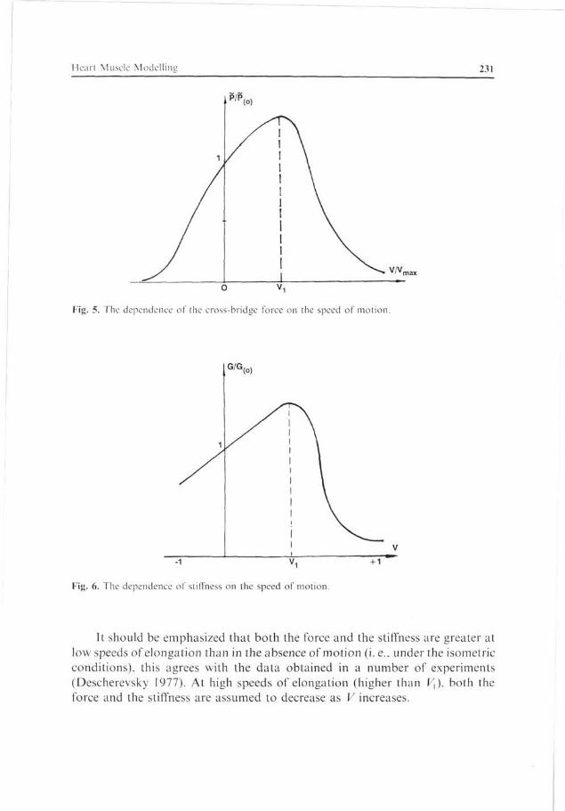
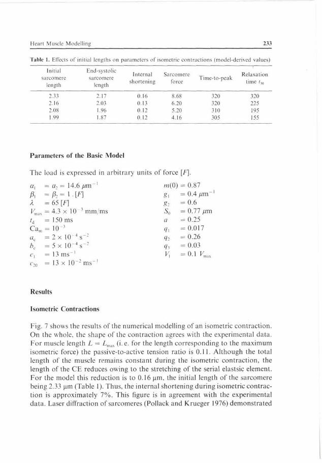
Heart Muscle Modelling 231
P/P
Fig. 5. The dependence of the cross-bridge force on the speed of motion.
G/G
Fig. 6. The dependence of stiffness on the speed of motion.
It should be emphasized that both the force and the stiffness are greater at low speeds of elongation than in the absence of motion (i.e.. under the isometric conditions), this agrees with the data obtained in a number of experiments (Descherevsky 1977). At high speeds of elongation (higher than F,), both the force and the stiffness are assumed to decrease as V increases.

232 Katsnelson and Izákov
1.0
0.8
0.6
0.4
02
0.1 0.2 0.3 0.4 0.5 0.6 0.7 0.8
Fig. 7. Relationship between time-courses of activation (A) and of force (P); A and P are related to the respective maximal values ("relative units").
Isometric and isotonic Conditions
For the mechanical activity of heart muscle under isometric conditions, the following set of equations may be written:
l2 = const
A. P(ll).Al. n2.»,(/,). [0.5 . /, + S0] - ft exp[(/2 - /,) - l] = 0
A, =c , .Ca(0 . ( l -Al)-C20.a[nl(ll).n2].Al
/i2 = tf(/,)|m(0). ( /*(/ ,)-«2] .
The explicit expressions for P(l]), «,(/,), 7t(n] .n2), Ca(/), q(lt), G*(/,) have been given above.
To describe the contraction of a muscle under the isotonic conditions for a constant load D, the identity l2 = const in the earlier set should be replaced by the relation
D = ft .exp[a,(/2 - /,) - 1] + A[expa2/2 - 1].
Differentiating it with respect to dt, we obtain the following equation
ax.ft. (l2 - / ,) . exp[a,(/2 - / , ) ] + a2. ft2. l2 exp(a2/2) = 0
Both sets of equations are soluble for the derivatives of unknown functions.
Heart Muscle Modelling 233
Table 1. Effects of initial lengths on parameters of isometric contractions (model-derived values)
Initial sarcomere
length
End-systolic sarcomere
length
Internal Sarcomere shortening force
Time-to-peak Relaxation
time /,„
2.33 2.16 2.08 1.99
2.17 2.03 1.96 1.87
0.16 0.13 0.12 0.12
8.68 6.20 5.20 4.16
320 320 310 305
320 225 195 155
Parameters of the Basic Model
The load is expressed in arbitrary units of force [F].
cr, = a2 = 14.6 /mi"' ft =ft=\-[f] X = 65 [F] f/max = 4.3 x lO-'mm/ms td = 150 ms Cam = 10-3
ac = 2 x l 0 - 4 s " 2
bc = 5 x l 0 " 4 s " 2
c, = 1 3 ms"1
c-,n = 13 x 10~2 ms"1
w(0) g\ g-i
s0 a Q i
9? q 2 F,
= 0.87 = 0.4//m"' = 0.6 = 0.77 /mi = 0.25 = 0.017 = 0.26 = 0.03 - 0 . 1 Vmx
Results
Isometric Contractions
Fig. 7 shows the results of the numerical modelling of an isometric contraction. On the whole, the shape of the contraction agrees with the experimental data. For muscle length L = LmdX (i. e. for the length corresponding to the maximum isometric force) the passive-to-active tension ratio is 0.11. Although the total length of the muscle remains constant during the isometric contraction, the length of the CE reduces owing to the stretching of the serial elastsic element. For the model this reduction is to 0.16 um, the initial length of the sarcomere being 2.33 um (Table 1). Thus, the internal shortening during isometric contraction is approximately 7%. This figure is in agreement with the experimental data. Laser diffraction of sarcomeres (Pollack and Krueger 1976) demonstrated

234 Kalsnelson and Izako\
n q
0.7
0.5
on
0.1
P
0.1
1
/ 2 \
3
4 I 0.2 0.3 0.4 0.5 0.6 0.7
— t
Fig. 8. Modelling of the time-course oi' postload isotonic contractions. I - isometric mode 2. 3, 4 - postload at 0.2. 0.4. and 0.8 P„, respectively. Force in relative units, time in seconds.
Fig. 9. Modelling of length changes in a series of post-load contractions. I, 2. 3. 4 post-load at 0.2. 0.4. 0.6. and 0.8 P„, respectively.

Heart Muscle Modclline 235
1.0
0.5
P/P0
"Ôľí 0 3 ČTŠ Ô7Ž ' 0^9 '
Fig. 10. The (P/P„) force — shortening velocity {9} relationship in postload contractions.
that during the isometric contraction of a cardiac muscle the sarcomeres are shortened by 5—12%.
A comparison of the time courses of force {P) and activation (A) shows that activation reaches a maximum 80—90 ms earlier than does force. The time course of isometric relaxation generally coincides with the decay of activation. Activation, however, decreases faster than does force. Thus, at 300 ms the activation is 30% of the maximum value, whereas the force is 80%, and the corresponding figures at 500 ms are 7% and 30%. In accordance with the experimental data (Chapman 1979) the electromechanical delay (i.e. the lag-time from the moment when calcium begins to enter the cell to the onset of tension) in the model is 10—20 ms.
It should be noted also that the peak of free calcium concentration coincides with the moment when P reaches its maximum value, and near the contraction peak, the concentration of free calcium is close to zero (in Fig. 7, these moments are marked with arrows). These results of modelling also agree with the experimental data (Yue 1987).
Isotonic Contractions
Figs. 8 and 9 show the results of the numerical modelling of isotonic contrac-

236 K.atsnelson and I/akov
P
1.0
0.8
0.6
0.4
0.2
0.1 0.3 0.5 0.7 0.9
Fig. 11. Modelling of the effect of the initial length on isometric contractions. 1, 2, 3, 4 — length of sarcomere 2.33, 2.16, 2.08, and 1.99 /an, respectively.
tions at different values of the afterload (0.2; 0.4 and 0.8 P0). The basic observation is that after shortening the isometric phase of relaxation in afterloading contractions begins earlier and proceeds faster than relaxation in the isometric mode. This is the so-called "load-dependent relaxation" according to Poggessi et al. (1982). The model reproduces this particular feature fairly well. Moreover, in the experiment, with loads close to P0, the weight after shortening has been observed to reset with a little delay. The model reproduces also this feature.
In accordance with the experimental data, in the model, a decrease in load results in an increase in the speed of both the shortening and the relaxation. Our model also successfully reproduces the relation between the durations of isometric and isotonic contractions. From the literature it follows that the peak of the isotonic shortening is achieved later than the peak of the isometric force (Izákov et al. 1981). In the model, this lag is 100—150 ms.
The force may be plotted against the initial values of the speed of shortening (Fig. 10). Again, the results of the model relationship are close to the experimentally obtained data (Izákov et al. 1981).
"Length-Force" Relationship
The characteristic feature of muscle is the dependence of the tension developed

Heart Muscle Modelling 237
P/P,
1.0
0.9
0.8
0.7
0.6
0.5
0.4
0.3
0.2
0.1
max
4 ^ - — ^ ^
l ^ - ^ ^
* S ^
2
— ^ ^ '/'max 0.85 0.90 0.95 1.00
Fig. 12. Modelling of the "length-force" relationship. Abscissa: /'/max: ordinata: P/Pm.,y(Pm^ is the force at / = /mas). 1 - the "initial length-force" relationship for isometric contractions at basic values of the model parameters. 2 — the "length-force" relationship for passive muscle. 3 — the "end-sytolic length — end-systolic force" relationship at basic values of the model's parameters 4
- the "initial length-force" relationship for isometric contractions when the dependence of troponin affinity to calcium ions on the number of myosin crossbridges attaches is switched off.
Table 2. Modelling of the "length-force" relationship under isotonic conditions
Total end-systolic length, /an
2.31 2.27 2.19 2.06
End sytolic sarcomere length.
fim
2.16 2.14 2.09 1.99
Relative change in length
(from /„,.,„)
0.99 0.97 0.94 0.88
End-systolic sarcomere
force
7 3 5.64 3.87 2.0
Time to peak of shortening.
m s
350 370 370 370
on the initial muscle length. The results of modelling of this relationship for isometric contractions are shown in Fig. 11. This figure shows a superposition of contractions at initial lengths of the sarcomeres from 2.33 to 1.99 um. A larger initial length is observed to give stronger force of contraction, maximum rate of change in force, and maximum rate of relaxation. The data for the other parameters are presented in Table 1. It is important that the model correctly
238 Katsnelson and l /ako\
L
P
0.9
0.7
0.5
0.3
0.1
0.1 0.2 0.3 0.4 0.5 0.6 0.7 0.1 0.3 0.5 0 7
L P
0.9
0.7
05
0.3
0.1
n c i
2 \
t
0.1 0.3 0 5 0.7
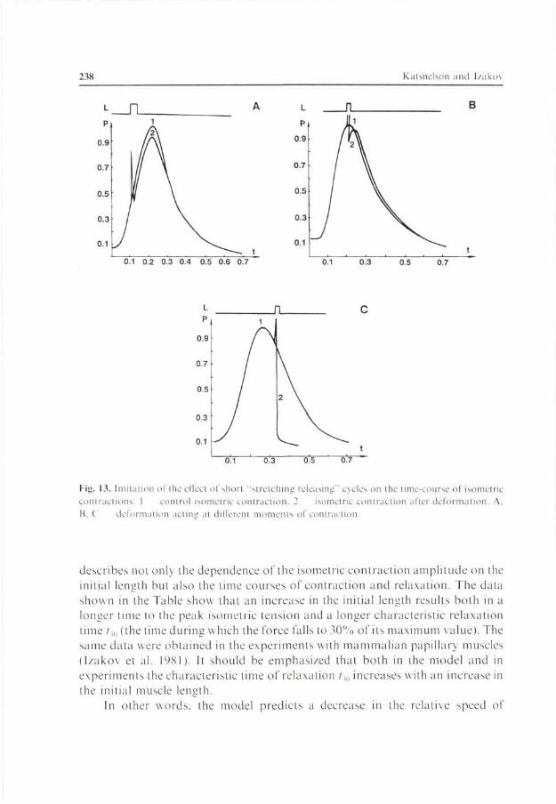
Fig. 13. Imitation of the effect of short "stretching releasing" cycles on the time-course of isometric contractions. 1 control isometric contraction. 2 isometric contraction after deformation A. B. C deformation actum at different moments of contraction.
describes not only the dependence of the isometric contraction amplitude on the initial length but also the time courses of contraction and relaxation. The data shown in the Table show that an increase in the initial length results both in a longer time to the peak isometric tension and a longer characteristic relaxation time tMl (the time during which the force falls to 30% of its maximum value). The same data were obtained in the experiments with mammalian papillary muscles (Izákov et al. 1981). It should be emphasized that both in the model and in experiments the characteristic time of relaxation /,„ increases with an increase in the initial muscle length.
In other words, the model predicts a decrease in the relative speed of

Heart Muscle Modelling 239
P
0.9
0.7
0.5
0.3
0 1
—[T
0.1 0.3
L
P
0.9
0.7
0.5
0.3
0.1
J
í\2
t
0.5 0.7 0.1 0.3 0.5 0.7
0.1
P
0.9
0.7
0.5
0.3
0.1
U
2
^y
\ 1
0.3 0.5 0.7
P
0.9
0.7
05
0.3
n 1
L
í
/
/ *
/
J 0.1 c
v 2 \
— -.3 0.5
t 0.7
Fig. 14. Imitation of the effect of short "last releasing-strelching" cycles. 1 control isometric contraction. 2 isometric contraction after deformation. A. B. C different lag-times between the onset of the contraction and the deformation. D the deformation strength doubled as compared with C
relaxation with the increasing pre-load. In accordance with the model representation, this result is due to the effect of the sarcomere length on the affinity of troponin for calcium ions.
The "length-force" relationship can be obtained experimentally also in another way (Izákov et al. 1981; Bodem and Sonnenblick 1974), by comparing, under isotonic conditions, the end-systolic length of the muscle with the end-systolic force, i.e. the total load lifted by the mucle. This procedure is carried out for isotonic contractions at different loads.
Simulation of this procedure is illustrated in Fig. 12 and in Table 2. The
240 Katsnelson and Izákov
Figure compares the length-force relationship under isometric and isotonic conditions (curves 1 and 3). The isotonic curve is below the isometric one. Moreover, the greater the shortening (the end-systolic length is smaller), the larger the difference between the "isometric" and "isotonic" length-force relationships. This difference results from mechanochemical uncoupling. The model simulates the experimental data (Allen and Kentish 1985; Bodem and Sonnen-blick 1974) fairly well. Unlike under isometric conditions, changes in the initial length have an opposite effect on the time to the peak of isotonic shortening.
Simulation of the Effect of Short-Time Changes in Length
One of the manifestations of mechanochemical uncoupling in muscle is the effect of short-time deformations (Hill 1970; Izákov et al. 1981). The effects of muscle deformation depend on its magnitude, on the moment of onset in the contraction-relaxation cycle, on the direction of the deformation (whether the muscle is stretched or released), and on the inotropic state of the muscle. The rate of deformation is also of some importance (Bodem and Sonnenblick 1974; Izákov et al. 1981). It has been found that the later the onset of deformation in the contraction-relaxation cycle the more pronounced its inactivating effect. On the whole, all these phenomena are well simulated by our model. Fig. 13 shows the action of a short-time "stretch-release" cycle. It is seen that the inactivating effect of such deformations is small if they fall within the early phases of the contraction-relaxation cycle (Fig. \3A and 13Ä). However even small stretching during the relaxation phase can result in a fast termination of relaxation (Fig. 13C). The "release-stretch" cycles show the same effect (Fig. 14). Deformations of different type differ in their effects on the time course of contraction and relaxation.
Conclusions
The results presented herein show that the mathematical model developed by us simulates a sufficiently large number of experimental data concerning the mechanics of muscle contraction. They include:
1) Time relations between the dynamics of free calcium, activation and mechanical tension;
2) Shape of isotonic and isometric contractions; 3) Dependence of the characteristic time of relaxation on the initial and
end-systolic muscle length and on the load; 4) Length-force and force-speed relationships.

Heart Muscle Modelling 241
The above relationships, however, are simulated by the other known models as well. The principal contribution of our model is that we succeeded in modelling a number of mechanical phenomena reflecting mechanochemical uncoupling. These phenomena, to our knowledge, are not simulated by the other models of muscle contraction. The model suggested simulates "load-dependent relaxation" according to Brutsaert et al. (1984), the effects of short-time deformations, changes in the slope of the "length-force" relationship under the action of inotropic agents, etc. The central idea of the model is the dependence of the affinity of troponin for calcium on the number of attached cross-bridges. The probability that a myosin cross-bridge attaches to the actin was, in turn, defined as the function of the rate of motion and of the mechanical cooidinate.
References
Allen D. G., Blinks J. R. (1978) Calcium transients in aeqorin injected fiog cardiac muscle Nature 273. 509—512
Allen D. G., Cannell M. B., l.ab M J , Orchard C. H. (1983). Shortening during contractions slows the calcium transient in cat papillai- muscle. .1. Physiol. (London) 334, 108 —109
Allen D. G.. Kentish J. C. (1985): The cellulai basis of the length-tension relation in cardiac muscle. J. Mol. Cell. Cardiol. 17, 821 840
Baylor S. M., Chandler W. K , Marshall M W (1982): Use ol metalloehroimc dyes to measure changes in myoplasmic calcium during activity in frog skeletal fibres J. Physiol. (London) 331. 139- 177
Bodem R , Sonnenblick E. II (1974): Dcat [nation ot contraction by quick release in the isolated papillary muscle of the cat: effects of levei damping, caffeine and tetanization. Circ. Res. 34. 214 -225
Brutsaert D. L., Housmans P. R., Goethals M. A. (1980). Dual control of relaxation: its role m the ventricular function in mammalian heait. Circ. Res. 47. 63/ -653.
Brutsaert D. I.., Rademakeis F. E . Sys S. I '. (1984): Tripple control of relaxation: implications in cardiac disease Circulation 69, 190 196
Cannel M. B.. Allen D. G. (1984). Model of calcium movements during activation in the sarcomere of frog skeletal muscle Biophys J 45,913 925
Chapmann R. A. (1979): Excitation-contraction coupling in cardiac muscle. Progr. Biophys. Mol. Biol 35, I 52
Comincioli V., Torelli A., Pogessi C , Keggiani C (1984): A four state cross-bridge model for muscle contraction Mathematical study and validation J. Math. Biol 20. 277 304
Descherevsky V. I. (1977): Mathematical Models ol Muscle Contraction. Moskva, Nauka (in Russian)
Eisenberg E., Hill T. L. (1978): A cross-bridge model of muscle contraction. Progr. Biophys. Mol. Biol. 33. 55 82
Eisenberg E., Hill ľ. I ., Chen Y. (1980): Cross-bridge model of muscle contraction. Quant. Anal. 29, 195 227
Ferennezi M. A., Goldman Y E., Simmons R. M. (1984) The dependence of force and shortening

242 Katsnelson and Izákov
velocity on substrate concentration in skinned muscle libers from Rana temporaria. J. Physiol. (London) 350. 519 543
Fung Y. C. (1970): Mathematical representation of mechanical properties of the heart muscle. J. Biomech. 3. 381 404
Goldman Y. F... Matsubara L. Simmons R. M. (1979): Lateral filamentary spacing in frog skinned muscle fibres in relaxed and rigor slates. J. Physiol. (London) 295. 80 81
Gordon A. M.. Ridgway E. B.. Martyn D. A. (1983): Calcium sensitivity is modified by contraction. In: Contractile Mechanisms in Muscle. Vol II. Mechanics. Energetics and Molecular Models. (Eds. H. Sugi and G. Pollack), pp. 553 564. Plenum Publ. Corp.. New York
Hill A. (1970): First and Last Experiments m Muscle Mechanics. Univ. Press. Cambridge Hill T. L. (1975): Theoretical formation for sliding filament model of contraction of striated muscle.
Prog. Biophys. Mol. Biol. 29. 105 159 Hill T. L.. Eisenberg E.. Green L. (1983): Alternate model for the cooperative equilibrium binding
of myosin-S-1-nucleotide complex to act in troponin tropomyosin. Proc. Nat. Acad. Sci. USA 80. 60 65
Huxley A. E. (1957): Muscle structure and theories of contraction. Prog. Biophys. Chem. 7. 255 318
Huxley A. F.. Simmons R. M. (197 I): Proposed mechanisms of force generation in striated muscle. Nature 233. 533 538
Izákov V. I.. Itkm G. P.. Markhasin V. S. (1981): Biomechanics of Cardiac Muscle. Moscow. Nauka (in Russian)
Izakev V. I.. Markhasin V. S.. Protsenko J. L.. Berschidskaya O. N. (1982): Cellular mechanisms of Frank-Starling phenomenon. Ad\. Physiol. Sci. 13. 89 108 (in Russian)
Izákov V. I.. Jasnikov G. P.. Cramarenko V. N.. Markhasin V, S. (1979): Mathematical model of myocardium contraction. In: Mathematical Biology, pp. 108 133. Nauka. Moscow
Julian E. J. (1969): Activation in a skeletal muscle contraction model with modification for insect fibrillar muscle. Biophys. J. 9. 547 570
Julian E. J.. Sollins K. R.. Sollins M. R. (1974): A model for transient and steady-state mechanical behaviour of contracting muscle. Biophys. J. 14. 546-562
Kaufmann R L.. Hennekcs R.. Lab M. J. (1971): Demonstration of an excitation-contraction uncoupling mechanism in mammalian ventricular myocardium. Nature 230. 150 151
Kaufmann R. L.. Bayer R. M.. Harnasch C. (1972): Autoregulation of contractility in the myocardial cell. Pfliigers Arch. 332. 96 I 16
Lab M. J. (1982): Contraction-excitation feedback in myocardium. Physiological basis and clinical relevance. Circ. Res. 50. 757 766
Mashima H. (1984): Force-velocity relation and contractility in striated muscles. Jpn. J. Physiol. 34. 1 17
Miledi R . Parker I . Zh u P. 11. (1982): Calcium transients evoked by action potential in frog twilch muscle fibres J. Physiol. (London) 333. 655 679
Morel J. E. (1985): Models ol muscle contraction and cell motility: a comparative study o\ usual concepts and the swelling theories. Prog. Biophys. Mol. Biol. 46. 97 126
Moss R ľ. (1986): Effects on shortening velocity ol' rabbit skeletal muscle due to variations m the level ol thm filament activation. J. Physiol. (London) 377. 487 505
Panerai R B. (1980): A model ol cardiac muscle mechanics and energetics. J. Biomech. 13. 929 940
Poggessi C . Reggiani C . Ricciardi L.. Mmelli R. ( 1982): Factors modulating the sensitivity of the relaxation lo loading conditions in rat cardiac muscle. Pfliigers Arch 394. 338 346
Pollack Ci. IE. Krueger J. W. (1976)sarcomere dynamics m intact cardiac muscle. Eur. J. Cardiol. 4 Suppf. 53 65

Heart Muscle Modelling 243
Robertson S. P.. Johnson J. D.. Potlei .1. D. (1981): Flic time course ol Ca exchange with calmodulin, troponin, parvalbumin and myosin in response to transient increase in Ca ' . Biophys. J. 34. 559 569
Rosenfeld S. S.. Taylor E. N. (198s) Kinetic studies of calcium binding to regulatory complexes from skeletal muscle J. Biol Chein. 260. 252 261
Saeki Y.. Sagawa K . Suga II (1980). transient tension responses of heart muscle in Ba-contracture to step length changes. Amei J. Physiol 238.11340 11347
Simmons R. M.. Jewell B R. (1973): Mechanics and model of muscle contraction. Rec. Adv. Physiol. 9. 87 147
Stniuer B. F (1973) Force velocity relations of isotonic relaxation in mammalian heart muscle. Amer. J. Physiol 224. 4.31 434
Tregear R. T.. Maiston S B (1979): Ihe cross budge theory. Annu. Rev. Physiol. 41. 723 736 Tsalurian A. K . Izákov V. E. Zhelamsky S. V.. Býkov B. L. (1984). Extracellular fluid filtration
as the reason for the viseoelastic behaviotu of ihe passive myocardium .1. Biomech. 17. 749 755
Yue D. T. (1987): Intracellular Ca"' related to rate of lone development in Iwitch contraction of heart. Amer. J. Physiol 252. 11760 11770
Final version accepted Decembei 20, 1989

![A Mathematical Model of the Neuromuscular Junction and Muscle … · 2018-08-13 · in the muscle ber [7]. The action potential propagates across the muscle ber membrane and down](https://img.pdfslide.us/doc/110x75/5e79e3bfefb160550272d2fa/a-mathematical-model-of-the-neuromuscular-junction-and-muscle-2018-08-13-in-the.jpg)