Embed Size (px)
Citation preview
150 VOLUME 20 NUMBER 2 FEBRUARY 2013 NATURE STRUCTURAL & MOLECULAR BIOLOGY
A R T I C L E S
HCV is the most common blood-borne virus in the United States and is estimated to infect ~3% of the world’s population. HCV’s genome is a single-stranded, positive-sense RNA molecule; thus, after release of the genome into the cytoplasm, the first step of viral replication is translation of a single open reading frame to yield the viral proteins. The HCV genomic RNA is therefore similar to cellular mRNA, serv-ing as the template for protein synthesis by translation machinery. However, unlike cellular mRNAs, which originate in the nucleus and are capped on their 5 ends by 7-methylguanosine and polyadenylated on their 3 ends (both important translation initiation signals), HCV genomic RNA is delivered directly to the cytoplasm lacking both a cap and a poly(A) tail (Fig. 1a). As a result, translation of HCV RNA is initiated by a mechanism that differs substantially from the one used by the cell to translate its own mRNA. Specifically, HCV uses an IRES RNA at the 5 end of the viral genome1 to hijack the cellular translation machinery. The IRES RNA sequence is highly conserved among HCV isolates and genotypes2,3. This conservation underscores the importance of the HCV IRES RNA to the viral replication cycle and reflects the specificity of the interactions between the IRES RNA and the translation machinery.
Mechanistic studies of the HCV IRES have focused on how it assem-bles an 80S ribosome at the AUG start codon, and they have revealed a mechanism different from canonical translation initiation4. In the canonical pathway, the 5 cap is recognized by the eukaryotic initia-tion factor 4F (eIF4F) complex, followed by binding of the 43S particle (which includes the 40S subunit; the ternary complex formed by eIF2, GTP and initiator methionyl-tRNA (eIF2–GTP–Met-tRNAiMet); eIF3 and other factors). The subunit then scans the mRNA to locate the start codon, at which time factor release and 60S subunit associa-tion yield an 80S ribosome5. In contrast, the HCV IRES first directly
binds the 40S subunit6–9, then eIF3 and the ternary complex bind10–12 (Fig. 1b). Subsequent GTP hydrolysis, eIF release and binding of a 60S subunit yield an 80S ribosome placed directly at the start codon13,14. In addition, the HCV IRES can, under conditions of cellular stress, use eIF2-independent pathways to generate 80S ribosomes15,16.
The function of HCV IRES is conferred by its structure. The IRES adopts an extended global architecture17, within which specific RNA structural domains drive different steps of 80S ribosome formation18 (Fig. 1c). Domain III binds the 40S subunit and eIF3 (refs. 6,7,19); domain IV provides the AUG initiation codon for interaction with the ternary complex11,12,20; the pseudoknot is important for placement of the AUG in the 40S subunit decoding groove21; and domain II (dII) is involved in eIF2-catalyzed GTP hydrolysis14, removal of eIF3j13, 60S subunit joining11 and the configuration of RNA in the decoding groove22. These findings, combined with the observation that HCV IRES binding alters the conformation of the 40S subunit23, indicate that the IRES RNA is an active manipulator of the translation machin-ery, not just a binding site for the ribosome and factors.
Among the aforementioned IRES domains, dII is particularly notable: it induces changes in the conformation of the 40S subunit23, and it docks within the ribosome’s decoding groove to interact with ribosomal protein rpS5 (refs. 24,25), which is known to contact E-site tRNA26–28. We therefore set out to study the role of dII sub-domain dIIb, the part of the IRES that penetrates deep into the ribosome’s decoding groove. We have discovered that dIIb muta-tions alter the conformation of the IRES–40S subunit complex and inhibit the first round of ribosomal translocation. The modeled position of this domain (adjacent to rpS5) suggests this effect may be due to altered contact with rpS5. This is, to our knowledge, the first evidence that a step after 80S ribosome assembly is directly
1Department of Biochemistry and Molecular Genetics, University of Colorado Denver School of Medicine, Aurora, Colorado, USA. 2Janelia Farm Research Campus, Howard Hughes Medical Institute, Ashburn, Virginia, USA. 3Howard Hughes Medical Institute, University of Colorado Denver School of Medicine, Aurora, Colorado, USA. Correspondence should be addressed to J.S.K. ([email protected]) or T.G. ([email protected]).
Received 31 July; accepted 12 November; published online 23 December 2012; doi:10.1038/nsmb.2465
HCV IRES manipulates the ribosome to promote the switch from translation initiation to elongationMegan E Filbin1, Breanna S Vollmar2, Dan Shi2, Tamir Gonen2 & Jeffrey S Kieft1,3
The internal ribosome entry site (IRES) of the hepatitis C virus (HCV) drives noncanonical initiation of protein synthesis necessary for viral replication. Functional studies of the HCV IRES have focused on 80S ribosome formation but have not explored its role after the 80S ribosome is poised at the start codon. Here, we report that mutations of an IRES domain that docks in the 40S subunit’s decoding groove cause only a local perturbation in IRES structure and result in conformational changes in the IRES–rabbit 40S subunit complex. Functionally, the mutations decrease IRES activity by inhibiting the first ribosomal translocation event, and modeling results suggest that this effect occurs through an interaction with a single ribosomal protein. The ability of the HCV IRES to manipulate the ribosome provides insight into how the ribosome’s structure and function can be altered by bound RNAs, including those derived from cellular invaders.
npg
© 2
013
Nat
ure
Am
eric
a, In
c. A
ll rig
hts
rese
rved
.

NATURE STRUCTURAL & MOLECULAR BIOLOGY VOLUME 20 NUMBER 2 FEBRUARY 2013 151
A R T I C L E S
influenced by the HCV IRES, and it may have implications for the understanding of translation initiation in general.
RESULTSIRES dIIb affects the rate of protein synthesisWe have previously reported that mutation of the dIIb apical loop (Fig. 1d) changes the configuration of the HCV RNA in the decod-ing groove of 40S subunit–HCV IRES RNA complexes and reduces IRES-driven protein synthesis22. This is consistent with other stud-ies showing that dIIb is important for HCV IRES–mediated transla-tion initiation11,29,30. To monitor protein production as a function of time, we measured the ability of three IRES RNAs in which dIIb was mutated to translate a downstream luciferase (LUC) reporter sequence (uncapped and not polyadenylated) in rabbit reticulocyte lysate (RRL) over a 90-min time course (Fig. 1d,e). All three dIIb mutants produced LUC at a rate that was slower than that of wild-type (WT) IRES RNA but faster than that of an IRES mutant lacking dII ( dII). We observed differences in total LUC production after only 15 min (Fig. 1f), and the mutants continued to make protein at a roughly constant rate but more slowly than WT. These results indi-cate that the activity of the IRES with dIIb mutations is inhibited but not abolished. Assuming identical rates of elongation and ribosome termination on each RNA sample, we conclude that the dIIb muta-tions slow, but do not halt, translation initiation.
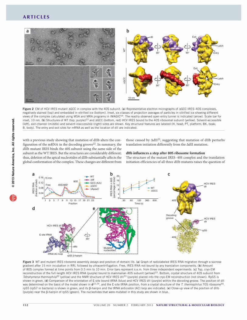
IRES–40S subunit complex conformation is influenced by dIIbCryo-EM reconstructions of HCV IRES–40S subunit complexes show that binding of full-length WT HCV IRES induces structural changes in the subunit, but the dII mutant does not cause this conformational change23. This raised the question of what the conformation of the 40S subunit would be when bound to the dIIb mutant RNAs gener-ated for this study. To answer this, we visualized IRES RNAs with the
apical GCC (nucleotides 82–84) in dIIb deleted ( GCC) in complex with the 40S subunit by EM, using both negative staining (to assess sample purity, homogeneity and concentration) and cryo-EM (to gen-erate a three-dimensional (3D) structure) (Fig. 2a). We obtained the reconstruction of the complex at a resolution comparable to previous HCV IRES–40S subunit reconstructions (17–20 Å)23. Comparison of the GCC IRES–40S subunit and WT IRES–40S subunit struc-tures revealed notable differences in the position and orientation of IRES domains and in the conformation of the 40S subunit (Fig. 2b). Specifically, in the mutant, dII did not loop away from the subunit’s surface to contact the side of the head and enter the decoding groove; rather, it lay across the platform. This change in the position of dII was accompanied by a rotation of the overall IRES orientation rela-tive to the body of the 40S subunit. This cryo-EM structure does not eliminate the possibility that the IRES location and conformation is an average of several similar structures. In other words, the GCC dele-tion might cause the conformation of the IRES and its position on the 40S subunit to be more dynamic, which could explain why structural features visible in the WT IRES are not seen in the GCC IRES and why the IRES appears to be rotated relative to the subunit. However, this possibility does not affect the conclusion that this relatively small deletion mutation at the tip of dIIb alters the IRES–40S subunit interaction, which is consistent with our observation that the GCC mutation lowers, but does not eliminate, IRES activity. When the 40S subunit was bound to the GCC mutant IRES, some features of its conformation were similar to the WT IRES–bound state, but some were markedly different. In both states, the latch formed between ribosomal RNA (rRNA) helices 18 and 34 was closed (this latch is open in apo-40S)23,31. However, the entry site (where mRNA enters the decoding groove) was much more open in the GCC mutant IRES complex; this feature was clearly visible in class averages assembled from individual particle images (Fig. 2a). This result is consistent
b
48S* 80S
GDP, factors
60S
IRES + 40SIRES
40S2
3
tRNA
5—Photinus luciferase —3
•
d e WTdllGCC
ApexCGCC-AUU
Luci
fera
se(m
illio
n R
LU /
s)
Time (min)
0
1.0
2.0
3.0
1.5
0.5
2.5
0 20 40 60 80 100
a
Cap AAA(n)
IRES 3 UTR
Translation
HCVViral entryUnpackaging
Transcription, processing, export protein
mRNA
viral RNA
f
Fra
ctio
n of
WT
tran
slat
ion
RNA species
WT dll
GCC
ApexC
GCC-AUU
0
0.2
0.4
0.6
0.8
1.015 min
c
IIa
IIb
IIIf
tRNAinit + eIF2 binding
eIF3 binding
60S association
AUG
IV
IIIb
IIIe
IIIcIIIa
IIId
Pseudoknot
40S binding
G CA A U U C G U G G C
C
80
A UA A U U C G U G G C
U G CA A U U C G U G G C
–– –
A A U U C G U G G C
–
WT GCC GCC-AUU ApexC
Figure 1 In vitro translation analysis of dIIb mutations. (a) HCV viral RNA and cellular mRNA differ in their origin and features. HCV viral RNA is delivered directly to the cytoplasm lacking a 7-methylguanosine cap and poly(A) tail, whereas cellular mRNA is produced and processed in the nucleus before exportation to the cytoplasm with a cap and tail. However, both are translated by the same cellular machinery, mandating different mechanisms of initiation. (b) Simplified diagram of HCV IRES 80S ribosome assembly mechanism. The IRES binds the 40S subunit (yellow), then eIF3 (green) and the ternary complex (eIF2–GTP– Met-tRNAi
Met; orange), and finally, after GTP hydrolysis and eIF release, the 60S subunit (blue) joins to form an 80S ribosome. The noncanonical 48S* complex requires fewer initiation factors compared to canonical 48S complexes. (c) Cartoon representation of the secondary structure of the HCV IRES. The location of the start AUG is shown. Boxed areas indicate the parts of the IRES involved in different steps of the mechanism shown in b. Ternary complex (eIF2–GTP– Met-tRNAi
Met) indicated by orange. (d) Schematic of mutations (red) made to dIIb in the context of the uncapped and unpolyadenylated LUC reporter. WT RNA is shown at left. (e) Time course of a translation assay from 0 to 90 min as measured by produced LUC relative light units (RLUs). (f) Fifteen-minute translation assay with RLUs calculated as a fraction of the WT IRES. Error bars represent s.e.m. for three biological replicates performed in triplicate.
npg
© 2
013
Nat
ure
Am
eric
a, In
c. A
ll rig
hts
rese
rved
.
152 VOLUME 20 NUMBER 2 FEBRUARY 2013 NATURE STRUCTURAL & MOLECULAR BIOLOGY
A R T I C L E S
with a previous study showing that mutation of dIIb alters the con-figuration of the mRNA in the decoding groove22. In summary, the dIIb mutant IRES binds the 40S subunit using the same side of the subunit as the WT IRES. But the structures are considerably different; thus, deletion of the apical nucleotides of dIIb substantially affects the global conformation of the complex. These changes are different from
those caused by dII23, suggesting that mutation of dIIb perturbs translation initiation differently from the dII mutation.
dIIb influences a step after 80S ribosome formationThe structure of the mutant IRES–40S complex and the translation initiation efficiencies of all three dIIb mutants raises the question of
Latch
dII
Exit
Entry
Latch
WT
Entry
ExitH
BK PT
H
B
GC
C
B
PT
90 90
90
10 nm
90
H
BK
B
b
Entry
a
50 nm
50 nm
Figure 2 EM of HCV IRES mutant GCC in complex with the 40S subunit. (a) Representative electron micrographs of GCC IRES–40S complexes, negatively stained (top) and embedded in vitrified ice (bottom). Inset, six classes of projection averages of particles in vitrified ice showing different views of the complex calculated using MSA and MRA programs in IMAGIC54. The readily observed open entry tunnel is indicated (arrow). Scale bar for inset, 10 nm. (b) Structures of WT (top, purple)23 and GCC (bottom, red) HCV IRES bound to the 40S ribosomal subunit (yellow). Solvent-accessible (left), exit-channel (middle) and solvent-inaccessible (right) sides are shown. Key structural features are labeled (H, head; PT, platform; BK, beak; B, body). The entry and exit sites for mRNA as well as the location of dII are indicated.
a b c
HCV IRES dII
E-site tRNA
dIIb
tRNA AC looprpS5 -hairpin
HCV IRESdII
40S
rpS5
Decodinggroove
d
1 3 5 7 9Fraction
11 13 15 17 19 21 23
IRE
S R
NA
(fra
ctio
n of
tota
l)
0.16
0.12
0.08
0.04
0
Free 40S/48S*
15 min WT
WT
dllGCC-AUU
80S
30 60 120 360 600Time (s)
IRE
S R
NA
in 8
0S(p
erce
ntag
e of
tota
l)
0
4
8
12
16
20
e
Mutatednucleotides
9040S
HCV IRESdII
GCC
GCCApexC
Figure 3 WT and mutant IRES ribosome assembly assays and position of domain IIb. (a) Graph of radiolabeled IRES RNA migration through a sucrose gradient after 15 min incubation in RRL followed by ultracentrifugation. Free, IRES RNA not bound by any translation components. (b) Amount of 80S complex formed at time points from 0.5 min to 10 min. Error bars represent s.e.m. from three independent experiments. (c) Top, cryo-EM reconstruction of the full-length HCV IRES RNA (purple) bound to mammalian 40S subunit (yellow)23. Bottom, crystal structure of 40S subunit from Tetrahymena thermophila32 (yellow) and the NMR structure of HCV IRES dII33 (purple) placed into the cryo-EM reconstruction (not shown). RpS5 is shown in green. (d) Comparison of the orientation of E-site bound tRNA (blue) and HCV IRES dII (purple) within the decoding groove. The position of dII was determined on the basis of the model shown in d22,24, and the E-site tRNA position, from a crystal structure of the T. thermophilus 70S ribosome55. rpS5 (rpS7 in bacteria) is shown in green, and its -hairpin and the tRNA anticodon (AC) loop are indicated. (e) Close-up view of the position of dIIb (purple) near the -hairpin of rpS5 (green). The nucleotides that were mutated in this study are shown in blue.
npg
© 2
013
Nat
ure
Am
eric
a, In
c. A
ll rig
hts
rese
rved
.

NATURE STRUCTURAL & MOLECULAR BIOLOGY VOLUME 20 NUMBER 2 FEBRUARY 2013 153
A R T I C L E S
which step in initiation is affected by these mutations. Removal of domain II or replacement of the entire dIIb apical loop with an ultra-stable UUCG tetraloop has been reported to inhibit 80S ribosome formation11,12,14, which suggested that our targeted dIIb mutations would do the same. To test this, we assayed ribosome assembly in RRL, separating the resultant ribosomal complexes by ultracentrifugation in a sucrose density gradient. Consistent with previous reports11, we observed robust 80S formation with WT IRES and minimal 80S for-mation in the dII mutant after 15 min (Fig. 3a). However, contrary to our expectation, all of the targeted dIIb mutants were as efficient as WT IRES RNA at forming IRES–40S complexes, IRES–48S* com-plexes (asterisk denotes complex formed by a noncanonical pathway) and IRES–80S ribosomes (Fig. 3a). This result indicated that these mutations do not inhibit the formation of 80S ribosomes on the HCV IRES RNA. Furthermore, we observed no difference in the rate of 80S formation between dIIb mutant and WT IRES during a 10-min time course (Fig. 3b). These data, combined with the translation initia-tion data (Fig. 1e), confirm that mutation of dIIb does not inhibit 80S ribosome formation on the IRES. Thus, we conclude that the altered position of the GCC mutant IRES RNA on the 40S subunit and the different conformation of the 40S subunit (relative to the WT IRES–40S complex) can support 80S ribosome formation, but some subsequent event is slowed by mutation of dIIb.
Modeled local interaction between dIIb and rpS5To determine which interactions of dIIb with the ribosome are dis-rupted by mutation, we modeled the placement of dII on the ribosome by docking a 40S subunit structure32 and the structure of dII33 into the cryo-EM density of the complete WT HCV IRES–40S subunit com-plex22,23 (Fig. 3c). Our placement of dII is consistent with a previously
published model based on an IRES–80S complex24, with additional detail provided by the crystal structure of a 40S subunit. We compared our model with the crystal structure of a bacterial ribosome bound to tRNAs to determine how similar the contacts with the 40S subunit are between our modeled dII and a bound E-site tRNA (Fig. 3d). We observed only one instance of overlap, at the anticodon loop of the tRNA and the modeled position of the HCV IRES dIIb, which are both positioned directly against the -hairpin structure of rpS5 (or its bacterial homolog rpS7) (Fig. 3c). Specifically, we observed that the mutated nucleotides in dIIb are directly adjacent to the rpS5
-hairpin (Fig. 3e). The modeled position of dIIb is consistent with observed cross-linking of dII to rpS5 (ref. 25), with the previous IRES–80S ribosome model24 and with the role of the -hairpin in contacting tRNA26–28.
The putative location of our dIIb mutations adjacent to the -hairpin of rpS5 suggests that they disrupt a specific contact between
dIIb and rpS5. Previous chemical probing of these mutant RNAs showed no global change in IRES secondary structure, but it remains possible that the mutations change the overall structure of dII. To assess this, we characterized the structures of the dIIb mutant RNAs using NMR. We generated WT and mutant samples comprising nucleo-tides 76–100 of the HCV IRES (Fig. 4a), which contain dIIb and are identical to RNAs used to solve the structure of dII33. Comparison of the one-dimensional 1H spectra obtained in water showed very little difference in the chemical shifts or relative intensity of the peaks obtained for WT and mutant RNAs (Fig. 4b). We observed the larg-est chemical shift in the imino protons of G87 and G88, which are adjacent to the apical loop of dIIb. The visibility of these imino proton peaks in all spectra indicates that base-pair formation is unaltered by dIIb mutation.
1H (p.p.m.)
10.0
GCC-AUU
10.511.011.512.012.513.0
U97G90G68
G67
G77G87U95
G98U78
U91G71
G88G75
U92G82
G94
WT
Apex C
GCC
C - GU GG - CC - GG UA UA AA UG AA - UC - GG - CG - C
U UA AGCC
5 3
Loop Emotif
IIb
G
70
75
8085
90
95
100
a b c d
SHAPE
C83
11.0
12.0
13.0
11.012.013.0
11.5
12.5
13.5
11.512.513.5
U78-G87cross-peak
WTApexC
U78-G87cross-peak
1H (p.p.m.)
1 H (
p.p.
m.)
U97G90 G98G77 G87U95
5.5
6.0
6.5
7.0
7.5
8.0
8.5
12.013.0 12.513.51H (p.p.m.)
1H (p.p.m
.)
C83
NMR
Base pairC79-G87
e fFigure 4 Characterization of the structural changes induced by dIIb mutation. (a) Secondary structure of the RNA sequence (previously solved)33 used to characterize the structural changes induced by mutation of dIIb. Elements are color-coded to match b–f. (b) One-dimensional 1H-NMR spectra (in water) of the WT and dIIb mutant RNAs. This part of the spectrum contains resonances from the base imino protons. Assignments for WT are labeled. The gray boxes indicate the most shifted resonances in all three mutant spectra. (c) Overlaid 2D 1H-NOESY NMR spectra (in water) for WT (black) and apexC (red). The portion containing the cross-peaks between imino protons is shown, and the cross-peak for G87 and U78 imino protons is indicated. (d) Overlaid spectra and color scheme as in c, showing the cross-peaks between imino and other protons. Dashed boxes indicate locations of the cross-peaks between the G87 imino and the C79 amino protons. Imino proton resonances are indicated. (e) Close-up view of the tip of dIIb against the -hairpin of rpS5 (green). The C79-G87 base pair (orange) and the location of the single base deletion in apexC (C83, red) are indicated. (f) View as in e, with the locations of previously reported increases in chemical modification in the apexC mutant22 shown (yellow). SHAPE, selective 2-OH acylation analyzed by primer extension.
npg
© 2
013
Nat
ure
Am
eric
a, In
c. A
ll rig
hts
rese
rved
.

154 VOLUME 20 NUMBER 2 FEBRUARY 2013 NATURE STRUCTURAL & MOLECULAR BIOLOGY
A R T I C L E S
The spectrum of mutant apexC (dIIb mutant with only apical C83 deleted) was most similar to that of the WT, suggesting that the apexC mutation elicits the post–80S ribosome functional effect with the smallest change in loop structure. On the basis of our pre-diction that the single deleted nucleotide (C83) would contact the
-hairpin of rpS5 (Fig. 3e), we selected this mutant for additional NMR experiments. When we overlaid the WT and apexC spectra obtained by 2D 1H-NOESY in water, the portions that contained the imino-imino cross-peaks overlapped almost perfectly, with the larg-est shift occurring at the G87 imino (Fig. 4c). Likewise, we observed only small chemical-shift changes in spectra showing the cross-peaks between the imino and other protons (Fig. 4d). For example, cross-peaks between the G87 imino and C79 amino protons were shifted but intense (Fig. 4d), again confirming that this base pair at the base of dIIb still forms in the mutant RNA (Fig. 4e). Changes to the spec-tra were limited to nucleotides adjacent to the deletion, showing the localization of structural changes to the dIIb apical loop. This result is consistent with published chemical-probing data, based on selective 2 -OH acylation analyzed by primer extension, for the apexC and other mutant RNAs, in that changes in the chemical probing pattern were limited to the apical loop in the IRES–40S subunit complex22 (Fig. 4f). Taken together, our data show that C83 deletion—and, we would predict, the other dIIb mutations shown in Figure 1—induces a local structural perturbation that, we propose, disrupts dIIb interac-tion with the -hairpin of rpS5 and is accompanied by a global change in the structure of the IRES–40S complex and the inhibition of a step after 80S ribosome formation.
AUG start codon placement is unaffected by dIIb mutationsTo determine which step after 80S ribosome formation is affected by dIIb mutation, we first explored the possibility that the mutations cause incorrect placement of the AUG start codon. To test this, we used primer extension inhibition (toeprinting) analysis10,34–36 on WT and dIIb mutant RNA in the unbound and 80S ribosome–bound states (the latter is formed in RRL with cycloheximide) (Fig. 5a, compare lanes 5, 7, 9 and 11 with 6, 8, 10 and 12). As expected, we observed no toeprint for unbound RNAs; however, when bound to the 80S ribo-some, all mutants produced similar toeprints at nucleotide positions +15 and +16 downstream of the AUG initiation codon (where A is in position +1) (Fig. 5a,b). This indicates that the AUG is properly positioned in all of these complexes and that the mutations probably affect a step after 80S ribosome assembly on the IRES.
dIIb mutants initiate in the correct reading frameMutations to the -hairpin of rpS7 have been shown to increase frameshift rate37. We therefore considered the possibility that the dIIb mutants, by disrupting contact with rpS5, could cause frameshift during or after the first translocation event and, thus, an apparent decrease in translation. To test this, we conducted translation assays using reporters to which one or two nucleotides were added after the IRES start codon but before the start codon of the LUC open reading frame. If the ribosome sometimes slips out of frame on the dIIb mutant RNAs, the addition of these nucleotides would rescue a 1- or 2-nucleotide frameshift (Fig. 5c). We did not observe a partial rescue (Fig. 5d); in fact, introduction of these nucleotides decreased
1 2 3 4 5 6 7 8 9 10 11 12
+15,+16
U C A G + + + +WT !GCC
GCC-AUU+ + + +
1 2 3 4 5 6 7 8 9 10 11 12
+1
a c
Luci
fera
se (
mill
ion
RLU
/ s)
5.0
4.0
3.0
2.0
1.0
0
Frameshift mutant
d
U C A GWT !GCC !ApexC
WT
GCC-AUU
+15,+16+1
pknot dIV
Position in gelTop Bottomb
GCC
0.5
1.5
Reverse transcriptase stop intensity
(10,000 c.p.m
.)
0.5
1.5
0.5
1.5
0.5
1.5ApexC
ApexC
GCC-AUU
320
342
356
CHX + RRL
WT
GCC
GCC + 1
GCC + 2
WT +
1
WT +
2
90 min
5—— 3
A (+1)AA (+2)
AUGAGCACAAAUCCUAAACCUCAAAGAAAAACC AUG
Domain IV
Photinus luciferase
Photinusluciferase
CHX + RRL
Figure 5 Biochemical analysis of AUG docking and potential frameshift. (a) Denaturing sequencing gel of the reverse transcription and toeprinting of WT and mutant IRES RNAs; the relevant part of the gel is magnified at right. Lanes 1–4, dideoxy sequencing reaction; lanes 5, 7, 9 and 11, free IRES; lanes 6, 8, 10 and 12, IRES–80S complexes (formed by incubation in RRL with cycloheximide (CHX)). Nucleotide numbers (bullets; left), the A of the AUG (black arrow; +1) and the toeprint (blue arrow; +15 and +16) are indicated. (b) Graph of quantified, normalized and background-corrected IRES–80S toeprints as in a. Positions +1 (black) and +15 and +16 (blue) are indicated. pknot, pseudoknot; dIV, domain IV. (c) Cartoon representation of the uncapped, unpolyadenylated monocistronic LUC reporter; the RNA region between the viral AUG and the LUC AUG (both shown in red) is magnified. One or two adenosines (blue box) were added for frameshift analysis. (d) LUC activity detected during a 90-min translation assay of WT and
GCC reporters, unmodified or with one (+1) or two (+2) additional adenosine residues. RLUs, relative light units. Error bars represent s.e.m. for three biological replicates performed in triplicate.
npg
© 2
013
Nat
ure
Am
eric
a, In
c. A
ll rig
hts
rese
rved
.
NATURE STRUCTURAL & MOLECULAR BIOLOGY VOLUME 20 NUMBER 2 FEBRUARY 2013 155
A R T I C L E S
translation efficiency even more than the dIIb mutations. Hence, the decreased translation initiation efficiency observed for the dIIb mutants is not caused by frameshifting.
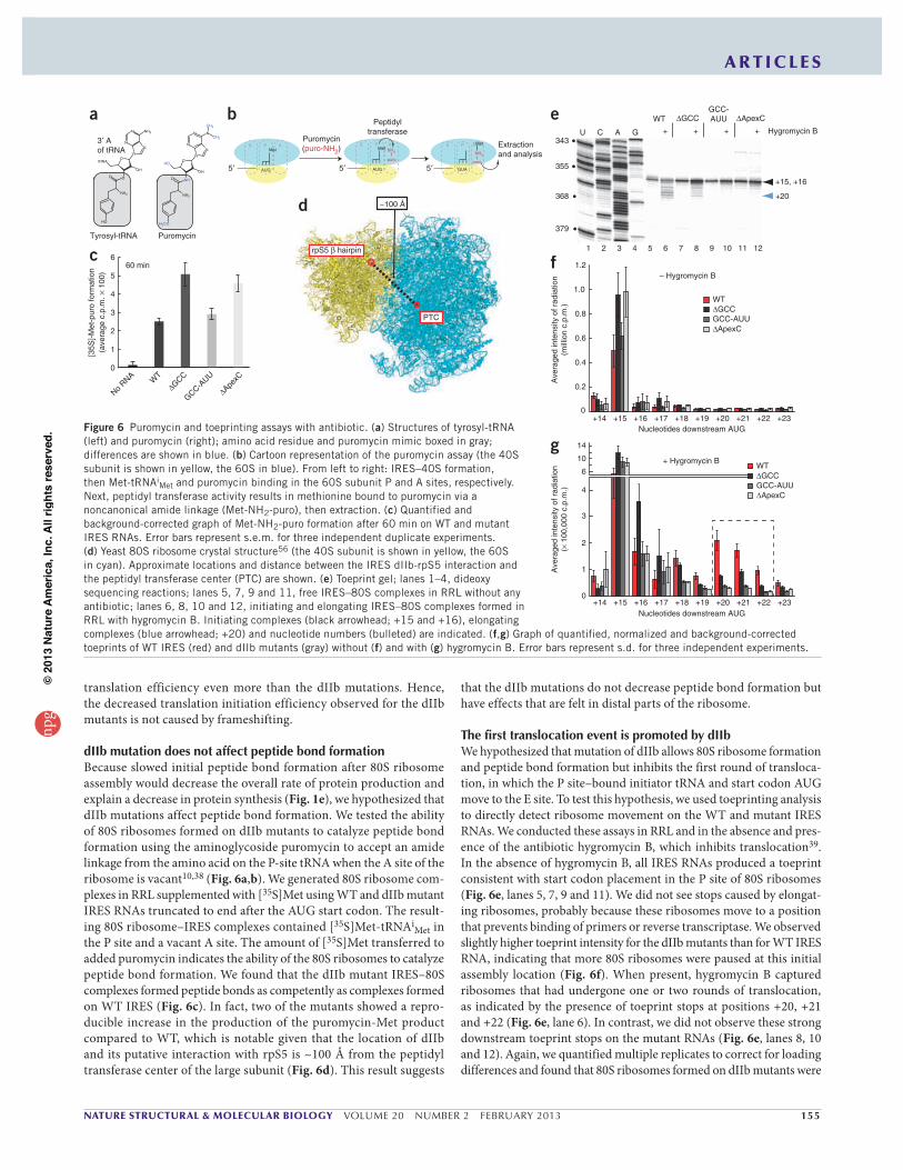
dIIb mutation does not affect peptide bond formationBecause slowed initial peptide bond formation after 80S ribosome assembly would decrease the overall rate of protein production and explain a decrease in protein synthesis (Fig. 1e), we hypothesized that dIIb mutations affect peptide bond formation. We tested the ability of 80S ribosomes formed on dIIb mutants to catalyze peptide bond formation using the aminoglycoside puromycin to accept an amide linkage from the amino acid on the P-site tRNA when the A site of the ribosome is vacant10,38 (Fig. 6a,b). We generated 80S ribosome com-plexes in RRL supplemented with [35S]Met using WT and dIIb mutant IRES RNAs truncated to end after the AUG start codon. The result-ing 80S ribosome–IRES complexes contained [35S]Met-tRNAiMet in the P site and a vacant A site. The amount of [35S]Met transferred to added puromycin indicates the ability of the 80S ribosomes to catalyze peptide bond formation. We found that the dIIb mutant IRES–80S complexes formed peptide bonds as competently as complexes formed on WT IRES (Fig. 6c). In fact, two of the mutants showed a repro-ducible increase in the production of the puromycin-Met product compared to WT, which is notable given that the location of dIIb and its putative interaction with rpS5 is ~100 Å from the peptidyl transferase center of the large subunit (Fig. 6d). This result suggests
that the dIIb mutations do not decrease peptide bond formation but have effects that are felt in distal parts of the ribosome.
The first translocation event is promoted by dIIbWe hypothesized that mutation of dIIb allows 80S ribosome formation and peptide bond formation but inhibits the first round of transloca-tion, in which the P site–bound initiator tRNA and start codon AUG move to the E site. To test this hypothesis, we used toeprinting analysis to directly detect ribosome movement on the WT and mutant IRES RNAs. We conducted these assays in RRL and in the absence and pres-ence of the antibiotic hygromycin B, which inhibits translocation39. In the absence of hygromycin B, all IRES RNAs produced a toeprint consistent with start codon placement in the P site of 80S ribosomes (Fig. 6e, lanes 5, 7, 9 and 11). We did not see stops caused by elongat-ing ribosomes, probably because these ribosomes move to a position that prevents binding of primers or reverse transcriptase. We observed slightly higher toeprint intensity for the dIIb mutants than for WT IRES RNA, indicating that more 80S ribosomes were paused at this initial assembly location (Fig. 6f). When present, hygromycin B captured ribosomes that had undergone one or two rounds of translocation, as indicated by the presence of toeprint stops at positions +20, +21 and +22 (Fig. 6e, lane 6). In contrast, we did not observe these strong downstream toeprint stops on the mutant RNAs (Fig. 6e, lanes 8, 10 and 12). Again, we quantified multiple replicates to correct for loading differences and found that 80S ribosomes formed on dIIb mutants were
a
NH2
NO
NHO
N
N
N
N
OH
H3CO
CH3
CH3
OHtRNA
NH2
NO
OO
N
N
N
NH2
OH
OH
Tyrosyl-tRNA Puromycin
3 Aof tRNA
b
AUG
E P A
5 5 5puro
NH2
puro
GUA
Puromycin(puro-NH2)
Peptidyltransferase
MetE P A E P Extraction
and analysisMet
AUG
Met
NH2
A
0
1
2
3
4
5
6
No RNA W
TGCC
GCC-AUU
ApexC
60 min
[35S
]-M
et-p
uro
form
atio
n(a
vera
ge c
.p.m
. 1
00)
c
~100 Åd
PTC
rpS5 hairpin
U C A G + + + + Hygromycin B
WT GCC ApexCGCC-AUU
1 2 3 4 5 6 7 8 9 10 11 12
343
355
368
379
+15, +16
+20
+14 +17 +18 +19 +20 +21 +22 +23Nucleotides downstream AUG
0
0.2
0.4
0.6
0.8
1.0
1.2
Ave
rage
d in
tens
ity o
f rad
iatio
n(m
illio
n c.
p.m
.)
– Hygromycin B
WT
GCC-AUUGCC
ApexC
+15 +16
0
1
2
3
4
6
10
14
+ Hygromycin B
+14 +17 +18 +19 +20 +21 +22 +23Nucleotides downstream AUG
+15 +16
Ave
rage
d in
tens
ity o
f rad
iatio
n(
100
,000
c.p
.m.)
e
f
gWT
GCC-AUUGCC
ApexC
Figure 6 Puromycin and toeprinting assays with antibiotic. (a) Structures of tyrosyl-tRNA (left) and puromycin (right); amino acid residue and puromycin mimic boxed in gray; differences are shown in blue. (b) Cartoon representation of the puromycin assay (the 40S subunit is shown in yellow, the 60S in blue). From left to right: IRES–40S formation, then Met-tRNAi
Met and puromycin binding in the 60S subunit P and A sites, respectively. Next, peptidyl transferase activity results in methionine bound to puromycin via a noncanonical amide linkage (Met-NH2-puro), then extraction. (c) Quantified and background-corrected graph of Met-NH2-puro formation after 60 min on WT and mutant IRES RNAs. Error bars represent s.e.m. for three independent duplicate experiments. (d) Yeast 80S ribosome crystal structure56 (the 40S subunit is shown in yellow, the 60S in cyan). Approximate locations and distance between the IRES dIIb-rpS5 interaction and the peptidyl transferase center (PTC) are shown. (e) Toeprint gel; lanes 1–4, dideoxy sequencing reactions; lanes 5, 7, 9 and 11, free IRES–80S complexes in RRL without any antibiotic; lanes 6, 8, 10 and 12, initiating and elongating IRES–80S complexes formed in RRL with hygromycin B. Initiating complexes (black arrowhead; +15 and +16), elongating complexes (blue arrowhead; +20) and nucleotide numbers (bulleted) are indicated. (f,g) Graph of quantified, normalized and background-corrected toeprints of WT IRES (red) and dIIb mutants (gray) without (f) and with (g) hygromycin B. Error bars represent s.d. for three independent experiments.
npg
© 2
013
Nat
ure
Am
eric
a, In
c. A
ll rig
hts
rese
rved
.
156 VOLUME 20 NUMBER 2 FEBRUARY 2013 NATURE STRUCTURAL & MOLECULAR BIOLOGY
A R T I C L E S
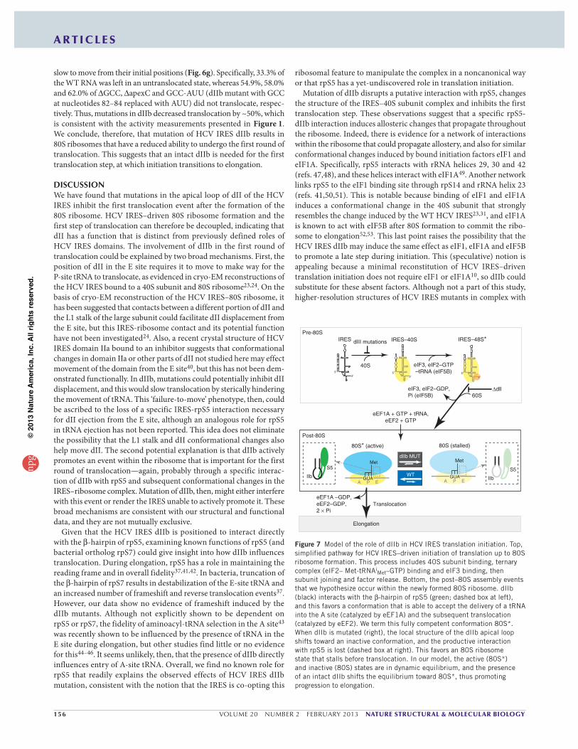
slow to move from their initial positions (Fig. 6g). Specifically, 33.3% of the WT RNA was left in an untranslocated state, whereas 54.9%, 58.0% and 62.0% of GCC, apexC and GCC-AUU (dIIb mutant with GCC at nucleotides 82–84 replaced with AUU) did not translocate, respec-tively. Thus, mutations in dIIb decreased translocation by ~50%, which is consistent with the activity measurements presented in Figure 1. We conclude, therefore, that mutation of HCV IRES dIIb results in 80S ribosomes that have a reduced ability to undergo the first round of translocation. This suggests that an intact dIIb is needed for the first translocation step, at which initiation transitions to elongation.
DISCUSSIONWe have found that mutations in the apical loop of dII of the HCV IRES inhibit the first translocation event after the formation of the 80S ribosome. HCV IRES–driven 80S ribosome formation and the first step of translocation can therefore be decoupled, indicating that dII has a function that is distinct from previously defined roles of HCV IRES domains. The involvement of dIIb in the first round of translocation could be explained by two broad mechanisms. First, the position of dII in the E site requires it to move to make way for the P-site tRNA to translocate, as evidenced in cryo-EM reconstructions of the HCV IRES bound to a 40S subunit and 80S ribosome23,24. On the basis of cryo-EM reconstruction of the HCV IRES–80S ribosome, it has been suggested that contacts between a different portion of dII and the L1 stalk of the large subunit could facilitate dII displacement from the E site, but this IRES-ribosome contact and its potential function have not been investigated24. Also, a recent crystal structure of HCV IRES domain IIa bound to an inhibitor suggests that conformational changes in domain IIa or other parts of dII not studied here may effect movement of the domain from the E site40, but this has not been dem-onstrated functionally. In dIIb, mutations could potentially inhibit dII displacement, and this would slow translocation by sterically hindering the movement of tRNA. This ‘failure-to-move’ phenotype, then, could be ascribed to the loss of a specific IRES-rpS5 interaction necessary for dII ejection from the E site, although an analogous role for rpS5 in tRNA ejection has not been reported. This idea does not eliminate the possibility that the L1 stalk and dII conformational changes also help move dII. The second potential explanation is that dIIb actively promotes an event within the ribosome that is important for the first round of translocation—again, probably through a specific interac-tion of dIIb with rpS5 and subsequent conformational changes in the IRES–ribosome complex. Mutation of dIIb, then, might either interfere with this event or render the IRES unable to actively promote it. These broad mechanisms are consistent with our structural and functional data, and they are not mutually exclusive.
Given that the HCV IRES dIIb is positioned to interact directly with the -hairpin of rpS5, examining known functions of rpS5 (and bacterial ortholog rpS7) could give insight into how dIIb influences translocation. During elongation, rpS5 has a role in maintaining the reading frame and in overall fidelity37,41,42. In bacteria, truncation of the -hairpin of rpS7 results in destabilization of the E-site tRNA and an increased number of frameshift and reverse translocation events37. However, our data show no evidence of frameshift induced by the dIIb mutants. Although not explicitly shown to be dependent on rpS5 or rpS7, the fidelity of aminoacyl-tRNA selection in the A site43 was recently shown to be influenced by the presence of tRNA in the E site during elongation, but other studies find little or no evidence for this44–46. It seems unlikely, then, that the presence of dIIb directly influences entry of A-site tRNA. Overall, we find no known role for rpS5 that readily explains the observed effects of HCV IRES dIIb mutation, consistent with the notion that the IRES is co-opting this
ribosomal feature to manipulate the complex in a noncanonical way or that rpS5 has a yet-undiscovered role in translation initiation.
Mutation of dIIb disrupts a putative interaction with rpS5, changes the structure of the IRES–40S subunit complex and inhibits the first translocation step. These observations suggest that a specific rpS5-dIIb interaction induces allosteric changes that propagate throughout the ribosome. Indeed, there is evidence for a network of interactions within the ribosome that could propagate allostery, and also for similar conformational changes induced by bound initiation factors eIF1 and eIF1A. Specifically, rpS5 interacts with rRNA helices 29, 30 and 42 (refs. 47,48), and these helices interact with eIF1A49. Another network links rpS5 to the eIF1 binding site through rpS14 and rRNA helix 23 (refs. 41,50,51). This is notable because binding of eIF1 and eIF1A induces a conformational change in the 40S subunit that strongly resembles the change induced by the WT HCV IRES23,31, and eIF1A is known to act with eIF5B after 80S formation to commit the ribo-some to elongation52,53. This last point raises the possibility that the HCV IRES dIIb may induce the same effect as eIF1, eIF1A and eIF5B to promote a late step during initiation. This (speculative) notion is appealing because a minimal reconstitution of HCV IRES–driven translation initiation does not require eIF1 or eIF1A10, so dIIb could substitute for these absent factors. Although not a part of this study, higher-resolution structures of HCV IRES mutants in complex with
40S eIF3, eIF2–GTP–tRNA (eIF5B)
60S!dll
IRES IRES–40S IRES–48S*
Elongation
80S* (active) 80S (stalled)
dIIb MUT
WT
eIF3, eIF2–GDP,Pi (eIF5B)
eEF1A + GTP + tRNA,eEF2 + GTP
IIb GUA GUA IIb
S5 S5
eEF1A –GDP,eEF2–GDP,2 Pi
Met Met
Pre-80S
Post-80S
Translocation
dIII mutations
355
3
53
EPA EPA
Figure 7 Model of the role of dIIb in HCV IRES translation initiation. Top, simplified pathway for HCV IRES–driven initiation of translation up to 80S ribosome formation. This process includes 40S subunit binding, ternary complex (eIF2– Met-tRNAi
Met–GTP) binding and eIF3 binding, then subunit joining and factor release. Bottom, the post–80S assembly events that we hypothesize occur within the newly formed 80S ribosome. dIIb (black) interacts with the -hairpin of rpS5 (green; dashed box at left), and this favors a conformation that is able to accept the delivery of a tRNA into the A site (catalyzed by eEF1A) and the subsequent translocation (catalyzed by eEF2). We term this fully competent conformation 80S*. When dIIb is mutated (right), the local structure of the dIIb apical loop shifts toward an inactive conformation, and the productive interaction with rpS5 is lost (dashed box at right). This favors an 80S ribosome state that stalls before translocation. In our model, the active (80S*) and inactive (80S) states are in dynamic equilibrium, and the presence of an intact dIIb shifts the equilibrium toward 80S*, thus promoting progression to elongation.
npg
© 2
013
Nat
ure
Am
eric
a, In
c. A
ll rig
hts
rese
rved
.

NATURE STRUCTURAL & MOLECULAR BIOLOGY VOLUME 20 NUMBER 2 FEBRUARY 2013 157
A R T I C L E S
80S ribosomes and chemical probing of the rRNA in these complexes before and after translocation could provide insight into the putative allos-teric changes associated with this translocation-slowing phenotype.
We would like to propose the following model to explain the role of HCV IRES dIIb in events that occur within the IRES–ribosome complex before and during the first translocation step (Fig. 7): first, the IRES assembles an 80S ribosome such that the ribosome is poised at the start codon with an initiator tRNA in the P site. Within this ribosome, dIIb contacts the -hairpin of rpS5, thereby stabilizing the ribosome in a conformation that is conducive to translocation. Delivery of aminoacyl-tRNA to the ribosome by eukaryotic elonga-tion factor 1A (eEF1A) and subsequent peptide bond formation are then followed by rapid and efficient eEF2-catalyzed translocation. In the dIIb mutants studied here, the mutation induces a local change in the apical loop structure that perturbs the interaction with rpS5, affecting 40S subunit conformation. The dIIb mutant IRES–40S com-plex is still capable of forming an 80S ribosome, but one whose con-formational equilibrium is shifted such that its ability to translocate is inhibited. Although aminoacyl-tRNA may still be delivered to the A site and a peptide bond formed, the mutant-bound ribosome stalls at the start site. However, the ribosome samples conformations, so these ribosomes are not permanently stalled; rather, they occasion-ally sample a productive state in which they are able to translocate. In summary, our data support a model in which dIIb selects a produc-tive state from the conformational ensemble, and ribosomes bound to IRESs with mutated dIIb spend more time in an unproductive state and transition to elongation less efficiently.
Our data open another door to understanding the intricacies of translation initiation and ribosome function. The ribosome is fundamentally a Brownian machine that samples many conforma-tions—protein factors and tRNA binding shift the conformational equilibrium, providing efficiency and directionality. Thus, the ribo-some is programmed to be manipulated by its binding partners. This inherent characteristic of the ribosome is crucial for canonical transla-tion processes and allows subtle and robust regulation of ribosome function. Our results reveal that these principles are exploited by a single loop of the HCV IRES, supporting the view of the HCV IRES as a dynamic manipulator of the translation machinery and lending insight into how the translation machinery works in cap-dependent and cap-independent pathways.
METHODSMethods and any associated references are available in the online version of the paper.
Accession codes. The cryo-EM map of the mutant HCV IRES–rabbit 40S subunit complex has been deposited in the Electron Microscopy Data Bank with accession number 5527.
ACKNOWLEDGMENTSThe authors thank the members of J.S.K.’s lab and R. Davis, D. Bentley, D. Barton and T. Evans for useful suggestions and discussions, and M. Ruehle, T. Blumenthal, T. Cech and M. Johnston for critical reading of this manuscript. We also thank C. Spahn (Institut für Medizinische Physik und Biophysik, Charite–Universitätsmedizin Berlin) for data files and advice with structural modeling; P. Lukavsky (Central European Institute of Technology, Masaryk University) for NMR resonance assignments and the pUC18 plasmid for toeprinting experiments; A.Willis (Medical Research Council Toxicology Unit) for the pRL plasmid for LUC experiments and G. Armstrong and E. Eisenmesser for assistance in NMR data collection and processing. This work was supported by US National Institutes of Health grant GM081346 to J.S.K. M.E.F. was supported as an American Heart Association predoctoral fellow (grant no. 0815655G). J.S.K. is an Early Career Scientist of the Howard Hughes Medical Institute. T.G.’s laboratory is supported by the Howard Hughes Medical Institute.
AUTHOR CONTRIBUTIONSM.E.F. conducted all biochemical experiments. J.S.K. and M.E.F. conducted and analyzed the NMR experiments. B.S.V., D.S., T.G. and J.S.K. conducted the cryo-EM experiments, with structure calculation by B.S.V. Results were interpreted by M.E.F., B.S.V., J.S.K. and T.G. M.E.F. and J.S.K. designed the overall study and wrote the manuscript. All authors contributed to figure construction.
COMPETING FINANCIAL INTERESTSThe authors declare no competing financial interests.
Published online at http://www.nature.com/doifinder/10.1038/nsmb.2465. Reprints and permissions information is available online at http://www.nature.com/reprints/index.html.
1. Tsukiyama-Kohara, K., Iizuka, N., Kohara, M. & Nomoto, A. Internal ribosome entry site within hepatitis C virus RNA. J. Virol. 66, 1476–1483 (1992).
2. Bukh, J., Purcell, R.H. & Miller, R.H. Sequence analysis of the 5 noncoding region of hepatitis C virus. Proc. Natl. Acad. Sci. USA 89, 4942–4946 (1992).
3. Simmonds, P. et al. Sequence variability in the 5 non-coding region of hepatitis C virus: identification of a new virus type and restrictions on sequence diversity. J. Gen. Virol. 74, 661–668 (1993).
4. Fraser, C.S. & Doudna, J.A. Structural and mechanistic insights into hepatitis C viral translation initiation. Nat. Rev. Microbiol. 5, 29–38 (2007).
5. Jackson, R.J., Hellen, C.U. & Pestova, T.V. The mechanism of eukaryotic translation initiation and principles of its regulation. Nat. Rev. Mol. Cell Biol. 11, 113–127 (2010).
6. Kieft, J.S., Zhou, K., Jubin, R. & Doudna, J.A. Mechanism of ribosome recruitment by hepatitis C IRES RNA. RNA 7, 194–206 (2001).
7. Kolupaeva, V.G., Pestova, T.V. & Hellen, C.U. An enzymatic footprinting analysis of the interaction of 40S ribosomal subunits with the internal ribosomal entry site of hepatitis C virus. J. Virol. 74, 6242–6250 (2000).
8. Lytle, J.R., Wu, L. & Robertson, H.D. The ribosome binding site of hepatitis C virus mRNA. J. Virol. 75, 7629–7636 (2001).
9. Lytle, J.R., Wu, L. & Robertson, H.D. Domains on the hepatitis C virus internal ribosome entry site for 40s subunit binding. RNA 8, 1045–1055 (2002).
10. Pestova, T.V., Shatsky, I.N., Fletcher, S.P., Jackson, R.J. & Hellen, C.U. A prokaryotic-like mode of cytoplasmic eukaryotic ribosome binding to the initiation codon during internal translation initiation of hepatitis C and classical swine fever virus RNAs. Genes Dev. 12, 67–83 (1998).
11. Otto, G.A. & Puglisi, J.D. The pathway of HCV IRES-mediated translation initiation. Cell 119, 369–380 (2004).
12. Ji, H., Fraser, C.S., Yu, Y., Leary, J. & Doudna, J.A. Coordinated assembly of human translation initiation complexes by the hepatitis C virus internal ribosome entry site RNA. Proc. Natl. Acad. Sci. USA 101, 16990–16995 (2004).
13. Fraser, C.S., Hershey, J.W. & Doudna, J.A. The pathway of hepatitis C virus mRNA recruitment to the human ribosome. Nat. Struct. Mol. Biol. 16, 397–404 (2009).
14. Locker, N., Easton, L.E. & Lukavsky, P.J. HCV and CSFV IRES domain II mediate eIF2 release during 80S ribosome assembly. EMBO J. 26, 795–805 (2007).
15. Terenin, I.M., Dmitriev, S.E., Andreev, D.E. & Shatsky, I.N. Eukaryotic translation initiation machinery can operate in a bacterial-like mode without eIF2. Nat. Struct. Mol. Biol. 15, 836–841 (2008).
16. Kim, J.H., Park, S.M., Park, J.H., Keum, S.J. & Jang, S.K. eIF2A mediates translation of hepatitis C viral mRNA under stress conditions. EMBO J. 30, 2454–2464 (2011).
17. Kieft, J.S. et al. The hepatitis C virus internal ribosome entry site adopts an ion-dependent tertiary fold. J. Mol. Biol. 292, 513–529 (1999).
18. Lukavsky, P.J. Structure and function of HCV IRES domains. Virus Res. 139, 166–171 (2009).
19. Sizova, D.V., Kolupaeva, V.G., Pestova, T.V., Shatsky, I.N. & Hellen, C.U. Specific interaction of eukaryotic translation initiation factor 3 with the 5 nontranslated regions of hepatitis C virus and classical swine fever virus RNAs. J. Virol. 72, 4775–4782 (1998).
20. Honda, M., Brown, E.A. & Lemon, S.M. Stability of a stem-loop involving the initiator AUG controls the efficiency of internal initiation of translation on hepatitis C virus RNA. RNA 2, 955–968 (1996).
21. Berry, K.E., Waghray, S. & Doudna, J.A. The HCV IRES pseudoknot positions the initiation codon on the 40S ribosomal subunit. RNA 16, 1559–1569 (2010).
22. Filbin, M.E. & Kieft, J.S. HCV IRES domain IIb affects the configuration of coding RNA in the 40S subunit’s decoding groove. RNA 17, 1258–1273 (2011).
23. Spahn, C.M. et al. Hepatitis C virus IRES RNA-induced changes in the conformation of the 40s ribosomal subunit. Science 291, 1959–1962 (2001).
24. Boehringer, D., Thermann, R., Ostareck-Lederer, A., Lewis, J.D. & Stark, H. Structure of the hepatitis C virus IRES bound to the human 80S ribosome: remodeling of the HCV IRES. Structure 13, 1695–1706 (2005).
25. Fukushi, S. et al. Ribosomal protein S5 interacts with the internal ribosomal entry site of hepatitis C virus. J. Biol. Chem. 276, 20824–20826 (2001).
26. Wower, J., Scheffer, P., Sylvers, L.A., Wintermeyer, W. & Zimmermann, R.A. Topography of the E site on the Escherichia coli ribosome. EMBO J. 12, 617–623 (1993).
npg
© 2
013
Nat
ure
Am
eric
a, In
c. A
ll rig
hts
rese
rved
.
158 VOLUME 20 NUMBER 2 FEBRUARY 2013 NATURE STRUCTURAL & MOLECULAR BIOLOGY
A R T I C L E S
27. Yusupov, M.M. et al. Crystal structure of the ribosome at 5.5-Å resolution. Science 292, 883–896 (2001).
28. Döring, T., Mitchell, P., Osswald, M., Bochkariov, D. & Brimacombe, R. The decoding region of 16S RNA; a cross-linking study of the ribosomal A, P and E sites using tRNA derivatized at position 32 in the anticodon loop. EMBO J. 13, 2677–2685 (1994).
29. Odreman-Macchioli, F., Baralle, F.E. & Buratti, E. Mutational analysis of the different bulge regions of hepatitis C virus domain II and their influence on internal ribosome entry site translational ability. J. Biol. Chem. 276, 41648–41655 (2001).
30. Kalliampakou, K.I., Psaridi-Linardaki, L. & Mavromara, P. Mutational analysis of the apical region of domain II of the HCV IRES. FEBS Lett. 511, 79–84 (2002).
31. Passmore, L.A. et al. The eukaryotic translation initiation factors eIF1 and eIF1A induce an open conformation of the 40S ribosome. Mol. Cell 26, 41–50 (2007).
32. Rabl, J., Leibundgut, M., Ataide, S.F., Haag, A. & Ban, N. Crystal structure of the eukaryotic 40S ribosomal subunit in complex with initiation factor 1. Science 331, 730–736 (2011).
33. Lukavsky, P.J., Kim, I., Otto, G.A. & Puglisi, J.D. Structure of HCV IRES domain II determined by NMR. Nat. Struct. Biol. 10, 1033–1038 (2003).
34. Pestova, T.V., Hellen, C.U. & Shatsky, I.N. Canonical eukaryotic initiation factors determine initiation of translation by internal ribosomal entry. Mol. Cell Biol. 16, 6859–6869 (1996).
35. Wilson, J.E., Pestova, T.V., Hellen, C.U. & Sarnow, P. Initiation of protein synthesis from the A site of the ribosome. Cell 102, 511–520 (2000).
36. Hartz, D., McPheeters, D.S., Traut, R. & Gold, L. Extension inhibition analysis of translation initiation complexes. Methods Enzymol. 164, 419–425 (1988).
37. Devaraj, A., Shoji, S., Holbrook, E.D. & Fredrick, K. A role for the 30S subunit E site in maintenance of the translational reading frame. RNA 15, 255–265 (2009).
38. Monro, R.E. & Marcker, K.A. Ribosome-catalysed reaction of puromycin with a formylmethionine-containing oligonucleotide. J. Mol. Biol. 25, 347–350 (1967).
39. Peske, F., Savelsbergh, A., Katunin, V.I., Rodnina, M.V. & Wintermeyer, W. Conformational changes of the small ribosomal subunit during elongation factor G-dependent tRNA-mRNA translocation. J. Mol. Biol. 343, 1183–1194 (2004).
40. Dibrov, S.M. et al. Structure of a hepatitis C virus RNA domain in complex with a translation inhibitor reveals a binding mode reminiscent of riboswitches. Proc. Natl. Acad. Sci. USA 109, 5223–5228 (2012).
41. Robert, F. & Brakier-Gingras, L. A functional interaction between ribosomal proteins S7 and S11 within the bacterial ribosome. J. Biol. Chem. 278, 44913–44920 (2003).
42. Galkin, O. et al. Roles of the negatively charged N-terminal extension of Saccharomyces cerevisiae ribosomal protein S5 revealed by characterization of a yeast strain containing human ribosomal protein S5. RNA 13, 2116–2128 (2007).
43. Geigenmüller, U. & Nierhaus, K.H. Significance of the third tRNA binding site, the E site, on E. coli ribosomes for the accuracy of translation: an occupied E site prevents the binding of non-cognate aminoacyl-tRNA to the A site. EMBO J. 9, 4527–4533 (1990).
44. Petropoulos, A.D. & Green, R. Further in vitro exploration fails to support the allosteric three-site model. J. Biol. Chem. 287, 11642–11648 (2012).
45. Uemura, S. et al. Real-time tRNA transit on single translating ribosomes at codon resolution. Nature 464, 1012–1017 (2010).
46. Chen, C. et al. Allosteric vs. spontaneous exit-site (E-site) tRNA dissociation early in protein synthesis. Proc. Natl. Acad. Sci. USA 108, 16980–16985 (2011).
47. Malygin, A.A., Yanshina, D.D. & Karpova, G.G. Interactions of human ribosomal proteins S16 and S5 with an 18S rRNA fragment containing their binding sites. Biochimie 91, 1180–1186 (2009).
48. Ian’shina, D.D., Malygin, A.A. & Karpova, G.G. Binding of human ribosomal protein S5 with the 18S rRNA fragment 1203–1236/1521–1698 [in Russian]. Mol. Biol. (Mosk.) 40, 460–467 (2006).
49. Yu, Y. et al. Position of eukaryotic translation initiation factor eIF1A on the 40S ribosomal subunit mapped by directed hydroxyl radical probing. Nucleic Acids Res. 37, 5167–5182 (2009).
50. Antúnez de Mayolo, P. & Woolford, J.L. Jr. Interactions of yeast ribosomal protein rpS14 with RNA. J. Mol. Biol. 333, 697–709 (2003).
51. Lomakin, I.B., Kolupaeva, V.G., Marintchev, A., Wagner, G. & Pestova, T.V. Position of eukaryotic initiation factor eIF1 on the 40S ribosomal subunit determined by directed hydroxyl radical probing. Genes Dev. 17, 2786–2797 (2003).
52. Acker, M.G. et al. Kinetic analysis of late steps of eukaryotic translation initiation. J. Mol. Biol. 385, 491–506 (2009).
53. Fringer, J.M., Acker, M.G., Fekete, C.A., Lorsch, J.R. & Dever, T.E. Coupled release of eukaryotic translation initiation factors 5B and 1A from 80S ribosomes following subunit joining. Mol. Cell. Biol. 27, 2384–2397 (2007).
54. van Heel, M., Harauz, G., Orlova, E.V., Schmidt, R. & Schatz, M. A new generation of the IMAGIC image processing system. J. Struct. Biol. 116, 17–24 (1996).
55. Selmer, M. et al. Structure of the 70S ribosome complexed with mRNA and tRNA. Science 313, 1935–1942 (2006).
56. Ben-Shem, A. et al. The structure of the eukaryotic ribosome at 3.0-Å resolution. Science 334, 1524–1529 (2011).
npg
© 2
013
Nat
ure
Am
eric
a, In
c. A
ll rig
hts
rese
rved
.
NATURE STRUCTURAL & MOLECULAR BIOLOGYdoi:10.1038/nsmb.2465
ONLINE METHODSPlasmid construction and cloning. We constructed pUC19-based plasmids containing the HCV genotype Ib wild-type (WT; nucleotides 40–372) and dII mutant (nucleotides 119–372) sequences flanked by a 5 hammerhead and 3 hepatitis delta ribozyme as previously described6,17,22. Plasmids with the WT or mutant HCV IRES between two luciferase-encoding genes were made by PCR amplification of the desired sequence and ligation into the EcoRI and NcoI sites of plasmid pRL57 (gift of A. Willis). The plasmid used to generate RNA for toeprinting analysis (containing 85 additional 3 nucleotides on the wild-type HCV genotype 1a as well as the primer binding site) was a kind gift of P. Lukavsky14. We generated the genotype 1b dII mutant used in toeprinting by PCR amplification of the desired sequence and ligation into the HindIII and XbaI sites of this plasmid. All mutants were made using the QuikChange mutagenesis kit (Stratagene).
RNA preparation. We made RNAs for assembly assays and puromycin experi-ments using DNA generated by PCR using M13 (-41) forward and reverse primers (5 -GGTTTTCCCAGTCACGAC-3 and 5 -GGAAACAGCTATGACCATG-3 , respectively) and the relevant plasmid template. The PCR products were used for in vitro transcription reactions as described58. We purified and concentrated RNA as described22. Monocistronic Photinus luciferase RNAs were made from PCR templates using forward T7-HCV and reverse photinus primers (5 -TAATACGACTCACTATAGGGCTCCCCTGTGAGGAACTACTGTCTT-3 and 5 -TTACACGGCGATCTTTCCGCCCTTCTT-3 , respectively) using the T7 MegaScript kit (Ambion). RNAs were DNase treated, then purified with TRI Reagent (Sigma) and chloroform followed by 100% isopropanol 75% ethanol precipitations, respectively. We made RNAs for toeprinting from EcoRI-linearized plasmids in the same manner as the luciferase RNAs.
Radiolabeling RNA and primers. For assembly assays, we 5 radiolabeled RNA as described22 and diluted it to approximately 1,000 c.p.m. l!1. DNA primers were 5 radiolabeled in a reaction containing 800 pmol primer in the same conditions as the RNA, mixed with 20 l 9 M urea loading buffer, loaded directly onto a 10% urea denaturing gel, purified and diluted to approximately 25,000 c.p.m. l!1.
Ribosome assembly assays. We diluted 5 -radiolabeled HCV IRES RNAs to ~1,000 c.p.m. l!1, heated to 85 °C for 30 s, then cooled on the desktop. 1 l of this RNA was then added to a mixture containing 30 l rabbit reticulocyte lysate (RRL), 0.5 l amino acid mixture minus leucine, 0.5 l amino acid mixture minus methionine (all provided in RRL translation kit, micrococcal-nuclease treated; Promega) and 18 l RNase-free water. Reactions then were incubated at 30 °C for the desired time, halted by the addition of ribosome association dilution buffer (50 mM Tris (pH 7.5), 50 mM NaCl, 5 mM MgCl2 and 1 mM DTT) and placed on ice. The reactions were analyzed with 10–35% sucrose gra-dients in ribosome association dilution buffer by ultracentrifugation in a SW41 Ti swinging bucket rotor at 36,000 r.p.m. (~222,000g) for 3 h. We fractionated the gradients into ~0.5 ml fractions using a BIOCOMP Gradient Station and Gilson FC203B fraction collector. Two hundred microliters of each fraction were blotted onto membranes, air dried and analyzed using a PhosphorImager. We quantified the spots using ImageQuant software and reported each as a fraction of the total radiation. Note that 40S and 48S* were indistinguishable in a 10–35% sucrose gradient.
Luciferase assays. We conducted translation assays as described22 with the fol-lowing exceptions: for the time-point experiment, reactions were brought up to 125 l volume so that 25 l could be removed at each time point, halted with 200 l cold 1" Passive Lysis Buffer (Promega) and placed at –80 °C to ensure no further activity.
Toeprinting assays. We completed toeprinting assays essentially as described22. For 48S-bound IRES, 10.75 l RRL was mixed with 0.5 l RNasin Plus and 0.5 mM guanylyl imidodiphosphate and incubated at 30 °C for 5 min, after which 0.5 g toeprint RNA was added to a final volume of 15 l. For 80S-bound IRES, 10.8 l RRL was mixed with 0.5 l RNasin Plus (Promega) alone or with 3 mg ml!1 cycloheximide or 2 mg ml!1 hygromycin b and incubated at 30 °C for 5 min. This incubation was followed by addition of 0.5 g toeprint RNA to a final volume of
15 l. We made the ladder used for analysis with wild-type toeprint RNA reverse transcribed with SuperScript III Reverse Transcriptase (Life Technologies) with annealing and extension temperatures at 45 °C.
Puromycin assays. We first biotinylated 3 -truncated IRES RNAs (nucleotides 40–344, which stop after the AUG codon) using the 5 EndTag Nucleic Acid Labeling System (Vector Laboratories). Briefly, 65 g of RNA was phosphor-ylated with ATP S using T4 polynucleotide kinase in a 20- l reaction, at 37 °C for 1 h. Biotinylation then was carried out upon the addition of ~385 g biotin (long arm) maleimide for 1 h at 65 °C. Reactions then were extracted with equal volume phenol-chloroform-isoamyl alcohol, pH 6.7 (25:24:1) (Fisher), pre-cipitated with 1/10 volume 3 M acetic acid (pH 5.2) and three volumes 100% cold ethanol overnight. RNA was pelleted and washed with 70% ethanol, dried and resuspended in 10 l RNase-free water. Concentration was determined by absorbance at 260 nm. To conduct the assay, we mixed 30 l RRL with 16.5 l RNase-free water and 1 l #-[35S]methionine (>1,000 Ci mmol!1) and incubated the mixture at 30 °C for 15 min to allow aminoacylation of initiator methionine tRNA. Biotinylated RNA (2.25 g) was added to the reaction and incubated at 30 °C for 25 min for 80S ribosome formation. Reactions were mixed with one tube (0.6 ml) streptavidin paramagnetic beads (MagneSphere, Promega), prewashed three times with 0.5" saline sodium citrate (SSC) buffer (7.5 mM trisodium cit-rate dehydrate (pH 7.2), 75 mM NaCl) and once with 300 l ribosome associa-tion dilution buffer), resuspended in 50 l ribosome association dilution buffer and incubated at 30 °C for 10 min. Complexes were then washed six times with 500 l ribosome association dilution and resuspended such that reactions were split into duplicates with and without 1 mM puromycin. The assay was then carried out at 35 °C for 60 min. Puromycin was extracted with 500 l 200 mM potassium phosphate buffer (pH 8) and 500 l ethyl acetate with continuous shaking for 10 min at 35 °C. The upper ethyl acetate layer was removed and mixed with 7 ml ScintiSafe (Fisher) liquid scintillation fluid, and counts per minute were averaged between two 10-min count times. This method of immobiliz-ing the IRES-80S ribosomes greatly reduced background levels of puromycin-[35S]methionine formation compared to results for 80S ribosomes purified by ultracentrifugation in sucrose gradients.
Nuclear magnetic resonance. We collected NMR spectra at 25 °C on a Varian 900 MHz spectrometer using Standard Varian Biopack pulse sequences for all experiments. This included both one-dimensional spectra as well as all homo-nuclear two-dimensional spectra employing a 3919 WATERGATE for water sup-pression. NOESY (Biopack pulse sequence, WBNOESY) spectrum was collected with 256 indirect points. Two-dimensional data were processed using a Gaussian weighting function in the direct dimension and a sine-bell weighting function in the indirect dimension.
Negative-stain electron microscopy. We prepared the GCC mutant IRES RNA as described above and the 40S ribosomal subunits from RRL as previously described6. We assembled IRES–40S subunit complex as previously described23. We applied the complex to freshly glow-discharged carbon coated 400 mesh copper grids and stained with 0.75% uranyl formate as described59. Samples were viewed on a 120-kV transmission electron microscope (FEI, Hillsboro, OR). Images were recorded at a nominal magnification of "40,000 using a bottom mount 4k " 4k Gatan slow-scan charge coupled device (CCD) camera.
Electron cryomicroscopy. Purified GCC mutant IRES RNA in RNase-free water was heated to 70 °C for 2 min then cooled to room temperature. Buffer solution was added to a final concentration of 20 mM Tris-HCl (pH 7.4), 100 mM acetic acid, 2.5 mM MgCl2, 1 mM DTT and 40 mM KCl. Purified 40S subunits were added at a 1:1 molar ratio with the IRES RNA to a final concentration of 500 nM complex. Complex was stored on ice until diluted (generally to 100 nM) and used in microscopy.
We prepared vitrified samples of the GCC mutant IRES-40S subunit com-plexes at 100 nM using an FEI Vitrobot. Briefly, 3.5 l was applied to a Quantifoil Holey Carbon grid (Vitrobot chamber was at 4 °C and 100% humidity). After a 20-s pause, the grid was blotted with filter paper (force = 0, blot time = 2 s) and plunged into liquid ethane. Frozen samples were loaded onto a Gatan cryo-holder and inserted into an FEI Tecnai F20 operating at 200 kV equipped with a field emission gun. Images were collected at a nominal magnification of "62,000 using
npg
© 2
013
Nat
ure
Am
eric
a, In
c. A
ll rig
hts
rese
rved
.
NATURE STRUCTURAL & MOLECULAR BIOLOGY doi:10.1038/nsmb.2465
a 4k " 4k Tietz CMOS detector. Images were binned two times yielding a pixel size of 2.66 Å per pixel. Approximately 29,000 particles were selected from 1,790 images using Electron Micrograph Utility (http://cryoem.ucsf.edu/). Class aver-ages were determined using five consecutive rounds of MSA (multivariate statisti-cal analysis) and MRA (multireference alignment) in IMAGIC54. Contrast transfer function parameters for each image were determined using CTFFIND3 (ref. 60). An initial model for refinement and three-dimensional reconstruction was gen-erated by filtering a previously published apo-40S reconstruction (EMD-1346) model to 40 Å31. Initial parameters were generated during cycles of randomized search and refinement using FREALIGN v. 8.08 (ref. 61). After initial parameters were determined, consecutive cycles of local refinement and reconstruction were carried out until there was no apparent improvement in the alignment. Resolution of the three-dimensional model was calculated with the program RMEASURE62 and determined to be ~17.5 Å. The density was normalized using MAPMAN63 and filtered to 20 Å using BFACTOR. Difference maps presented in the figures were calculated using MAPMAN63. We calculated the density corresponding to the WT IRES from a difference map between the WT IRES–40S and apo-40S23, and the mutant IRES was calculated from a difference map between the GCC
IRES–40S reconstruction and apo-40S23. Reconstructions and difference maps were assembled as displayed in Figure 2b using UCSF Chimera64.
57. Stoneley, M., Paulin, F.E., Le Quesne, J.P., Chappell, S.A. & Willis, A.E. C-Myc 5 untranslated region contains an internal ribosome entry segment. Oncogene 16, 423–428 (1998).
58. Keel, A.Y., Easton, L.E., Lukavsky, P.J. & Kieft, J.S. Large-scale native preparation of in vitro transcribed RNA. Methods Enzymol. 469, 3–25 (2009).
59. Ohi, M., Li, Y., Cheng, Y. & Walz, T. Negative Staining and Image Classification - Powerful Tools in Modern Electron Microscopy. Biol. Proced. Online 6, 23–34 (2004).
60. Mindell, J.A. & Grigorieff, N. Accurate determination of local defocus and specimen tilt in electron microscopy. J. Struct. Biol. 142, 334–347 (2003).
61. Grigorieff, N. FREALIGN: high-resolution refinement of single particle structures. J. Struct. Biol. 157, 117–125 (2007).
62. Sousa, D. & Grigorieff, N. Ab initio resolution measurement for single particle structures. J. Struct. Biol. 157, 201–210 (2007).
63. Kleywegt, G.J. & Jones, T.A. xdlMAPMAN and xdlDATAMAN - programs for reformatting, analysis and manipulation of biomacromolecular electron-density maps and reflection data sets. Acta Crystallogr. D Biol. Crystallogr. 52, 826–828 (1996).
64. Pettersen, E.F. et al. UCSF Chimera–a visualization system for exploratory research and analysis. J. Comput. Chem. 25, 1605–1612 (2004).
npg
© 2
013
Nat
ure
Am
eric
a, In
c. A
ll rig
hts
rese
rved
.