Embed Size (px)
Citation preview

JOURNAL OF VIROLOGY, May 2002, p. 5024–5033 Vol. 76, No. 100022-538X/02/$04.00�0 DOI: 10.1128/JVI.76.10.5024–5033.2002Copyright © 2002, American Society for Microbiology. All Rights Reserved.
Pestivirus Internal Ribosome Entry Site (IRES) Structure andFunction: Elements in the 5� Untranslated Region
Important for IRES FunctionSimon P. Fletcher and Richard J. Jackson*
Department of Biochemistry, University of Cambridge, Cambridge CB2 1GA, United Kingdom
Received 27 November 2001/Accepted 15 February 2002
The importance of certain structural features of the 5� untranslated region of classical swine fever virus(CSFV) RNA for the function of the internal ribosome entry site (IRES) was investigated by mutagenesisfollowed by in vitro transcription and translation. Deletions made from the 5� end of the CSFV genomesequence showed that the IRES boundary was close to nucleotide 65: thus, the IRES includes the whole ofdomain II but no sequences upstream of this domain. Deletions which invaded domain II even to a small extentreduced activity to about 20% that of the full-length structure, and this 20% residual activity persisted withmore extensive deletions until the whole of domain II had been removed and the deletions invaded thepseudoknot, whereupon IRES activity fell to zero. The importance of both stems of the pseudoknot was verifiedby making mutations in both sides of each stem; this severely reduced IRES activity, but the compensatingmutations which restored base pairing caused almost full IRES function to be regained. The importance of thelength of the loop linking the two stems of the pseudoknot was demonstrated by the finding that a reductionin length from the wild-type AUAAAAUU to AUU almost completely abrogated IRES activity. Random A3Usubstitutions in the wild-type sequence showed that IRES activity was fairly proportional to the number of Aresidues retained in this pseudoknot loop, with a preference for clustered neighboring A residues rather thandispersed As. Finally, it was found that the sequence of the highly conserved domain IIIa loop is, rathersurprisingly, not important for the maintenance of full IRES activity, although amputation of the entiredomain IIIa stem and loop was highly debilitating. These results are interpreted in the light of recent models,derived from cryo-electron microscopy, of the interaction of the closely related hepatitis C virus IRES with 40Sribosomal subunits.
Hepatitis C virus (HCV) RNA is translated by a mechanismof internal ribosome entry that is distinctly different from thepicornavirus precedent. Small (40S) ribosomal subunits canbind directly to the HCV internal ribosome entry site (IRES)at the correct site even in the absence of any translation initi-ation factors (19, 25), whereas the presence of factors isneeded for small ribosomal subunit binding to picornavirusIRESs (23, 24). Consequently, initiation on the HCV IRESdoes not require eukaryotic initiation factor 4A (eIF4A), -4B,-4E, or -4G or ATP hydrolysis (25), in contrast to the picor-navirus IRESs, which require eIF4A and -4B and at least thecentral domain of eIF4G as well as ATP (23, 24). Thus, at thesuperficial operational level, initiation on the HCV IRES issimilar to initiation of translation of prokaryotic mRNAs, butwith a complex 330-nucleotide (nt) IRES apparently playing arole analogous to the prokaryotic Shine-Dalgarno sequence.Recent results obtained with cryo-electron microscopy ofIRES-40S subunit complexes, however, show that the twomechanisms are very different in detail. In the prokaryoticsystem the Shine-Dalgarno motif pairs with the 16S rRNA inthe platform region of the small subunit, which places themRNA in the appropriate channel, with the initiation codon inthe active site (36). In contrast, the bulk of the HCV IRES
binds to the back, or solvent side, of the 40S subunit, behindthe platform region (33). Only a small part of the IRES lies inthe vicinity of the subunit interface and the active sites of thesubunit.
Among other viral IRESs, the HCV IRES is most closelysimilar to the pestivirus IRESs. Originally there were threerecognized species of pestivirus, differing in their host animal:border disease virus (BDV), which infects sheep; bovine viraldiarrhea virus (BVDV); and classical swine fever virus(CSFV), formerly known as hog cholera virus. Subsequently, ithas been determined that there are two types of BVDV(BVDV-1 and -2), which are classified as two different speciesrather than different strains (14). In addition, it has recentlybeen shown that the pestiviruses isolated from reindeer andgiraffes are each a novel and distinct species (1), and a varietyof yet to be classified viruses have been isolated from othermammalian species (2).
Although a great deal of work has been devoted to the HCVIRES, relatively little attention has been given to those of thepestiviruses (6, 21, 25, 27, 30), yet the picornavirus precedenthas shown that a comparative study of closely similar IRESscan be particularly informative. There are strong similaritiesbetween the structure of the pestivirus IRES (Fig. 1) and therevised HCV IRES structure of Honda et al. (16). In terms ofgross features, the main differences are that domain II isslightly longer in HCV than in the pestiviruses; stem 1 of thepseudoknot is bipartite in the pestiviruses and is longer than inthe HCV IRES; the pseudoknot loop linking stem 1 to stem 2
* Corresponding author. Mailing address: Department of Biochem-istry, University of Cambridge, Old Addenbrooke’s Site, 80 TennisCourt Rd., Cambridge CB2 1GA, United Kingdom. Phone: (44) 1223-333682. Fax: (44) 1223-766002. E-mail: [email protected].
5024
on April 10, 2019 by guest
http://jvi.asm.org/
Dow
nloaded from

is longer in pestiviruses; and whereas there are two side stem-loops, IIId1 and IIId2, in the pestivirus IRES, there is just asingle IIId domain in the HCV IRES. On the other hand, thereare remarkable similarities between the HCV and pestivirusIRESs in the sequence of the unpaired regions at the top ofdomain II and in the loops of domains IIIa, IIIc, and IIIe (incontrast to considerable differences in the sequences at the topof domain IIIb).
Here we describe the results of a mutagenesis approach to
explore the significance of some features of the CSFV IRES asfollows: the position of the 5� boundary relative to domain II,the importance of domain II, the importance of base pairing inboth stems of the pseudoknot, the significance of the lengthand composition of the A-rich pseudoknot loop, and the roleof the highly conserved domain IIIa loop. While our work wasin progress, some (but not all) of these questions have beenaddressed by others (19, 20, 21), but as these published resultsare rather contradictory, a third opinion is called for, which is
FIG. 1. Secondary structure of the CSFV IRES. The structure shown is based on previously published structures (4, 9) plus our own structureprobing data and phylogenetic comparisons, with the various domains designated according to the nomenclature in current usage. Arrows showthe end points of the various 5� deletions that were obtained for the experiments of Fig. 7 and 8; the designated residue is the extreme 5� end ofthe CSFV sequences retained in the deletion mutant.
VOL. 76, 2002 PESTIVIRUS IRES STRUCTURE AND FUNCTION 5025
on April 10, 2019 by guest
http://jvi.asm.org/
Dow
nloaded from
why we have presented our data relating to these questions inaddition to our results on completely novel aspects.
MATERIALS AND METHODS
Plasmid constructs. The control dicistronic construct, pXLJ�OS, used in thiswork was identical to pXLJO described by Reynolds et al. (28, 29) except that theAsp718 site in the intercistronic cloning cassette was filled in to create a SnaBIsite. Downstream of a bacteriophage T7 promoter, it has a Xenopus laevis 5�untranslated region (UTR), coding region, and 3� UTR and then a short cloningcassette with sites for SalI, SacI, SnaBI, and NcoI (in that order), followed bysequences coding for what is known as NS�, a slightly truncated form of theinfluenza virus (strain A/PR/34) NS1 protein described previously (3), and finallythe complete NS1 3� UTR terminating in an EcoRI site. The CSFV sequencesused in this study were originally obtained as construct pCSFV/5B (generouslydonated by G. Meyers and H.-J. Thiel), which has the first 1,248 nt of the CSFVAlfort Tübingen genome (accession number JO4358). PCR was used to amplifynt 1 to 826 of this fragment, with the forward primer designed to change theextreme 5�-terminal sequence from GTATAG to GTCGAC in order to intro-duce a SalI site. Following restriction digestion with SalI and HindIII, the re-sulting product, which spanned nt 1 to 755 of the CSFV genome, was insertedinto the corresponding sites of pGEM-1 (Promega). This was cut with AgeI(between nt 438 and 439 of the CSFV sequence) and the overhangs were filledin. The CSFV sequences were released by cutting with SalI and were clonedbetween the SalI and SnaBI sites of pXLJ�OS. The resulting dicistronic con-struct, pXLCSFV 1-442.NS�, has the complete CSFV 5� UTR as an intercistronicspacer, and the downstream cistron consists of the first 67 nt of the CSFV codingsequences downstream of the AUG fused to the NS� reading frame via aGUACC linker. For some experiments, pXLCSFV 1-423.NS� was used. This wasgenerated by making deletions from the AgeI site by the method of Henikoff (15):it has the first 48 nt of CSFV coding sequences fused to the NS� reading framevia an ACC linker.
To prepare a nested set of 5� deletions, pXLCSFV 1-423.NS� was cut at theSalI site at the start of the CSFV 5� UTR sequences, and the DNA was digestedwith exonuclease III for various times, followed by exonuclease VII, as describedby Henikoff (15). The truncated CSFV sequences plus the NS� coding sequencesand 3� UTR were released by cutting with EcoRI, and the fragments were clonedbetween the SnaBI and EcoRI sites of pXLJ�OS.
Point mutations and small internal deletions were generated by the PCRmethod of Picard et al. (26), incorporating modifications to the protocol de-scribed previously (17). All constructs were verified by dideoxynucleotide se-quencing, and all plasmids were propagated by standard methods in Escherichiacoli TG1 by using ampicillin selection (31).
In vitro transcription and translation assays. All plasmids were linearized bydigestion with EcoRI prior to transcription. The generation of capped or un-capped RNAs by transcription with bacteriophage T7 RNA polymerase wasperformed as described previously (18, 28). Translation assays were carried outas described previously (12), with added KCl at 100 mM and Mg2� at 0.5 mMand with [35S]methionine as the radiolabeled amino acid. Aliquots of the trans-lation assay samples were analyzed by gel electrophoresis on 20% polyacrylamidegels as described previously (8). Stained, dried gels were exposed to Hyperfilm�-Max (Amersham International). Quantitation was done by scanning densitom-etry of the dried films using Phoretix software; a range of different exposures wasused in order to ensure that the determinations were made under conditions inwhich the response of the film was linear.
The coupled transcription-translation assays were carried out using the in-house system described by Craig et al. (7), but with added KCl at a finalconcentration of 100 mM, the optimum for translation dependent on the CSFVIRES, instead of the 40 to 60 mM concentration generally used for uncappedRNA products that would be translated by the conventional scanning mechanism(7).
Transfection assays. BHK-21 cells were prepared at 50 to 80% confluence insix-well Costar tissue culture dishes and were then infected with recombinantvaccinia virus vTF7-3 (11). After 2.5 h at 37°C, the medium was removed and thecell monolayer was washed with Glasgow minimum essential medium and thenOPTIMEM (Gibco-BRL). DNA transfection was carried out using 5 �g ofplasmid DNA mixed with OPTIMEM and LIPOFECTIN (Gibco-BRL) as de-scribed in the supplier’s protocol. At 20 h posttransfection the medium wasreplaced with methionine-free Eagle’s medium. After a further 60 min, 30 �Ci of[35S]methionine (Amersham International) was added per well and the plateswere incubated for another 2 h. Cells were subsequently scraped from the plates,pelleted by centrifugation, and processed for gel electrophoresis. Following au-
toradiography of the dried gel, NS� expression was quantitated by scanningdensitometry of the autoradiograph.
To detect and quantify the yield of cyclin, samples of cell extracts weresubjected to Western blotting essentially as described by Harlow and Lane (13),using a 1:100 dilution of a mouse monoclonal antibody raised against X. laeviscyclin B2 (a gift of J. Gannon and T. Hunt) and a 1:5,000 dilution of alkalinephosphatase-conjugated goat anti-mouse secondary antibody. Alkaline phospha-tase activity was detected using 5-bromo-4-chloro-3-indolyl phosphate and NitroBlue Tetrazolium as described by Harlow and Lane (13), and the staining wasquantitated by scanning densitometry in reflectance mode.
RESULTS
Importance of both stems of the pseudoknot structure. InHCV, stem 1 of the pseudoknot consists of nine contiguousbase pairs (eight of them are G-C pairs) which are followed byjust a single unpaired U residue before the 3� part of stem 2(16). In all the pestiviruses, in contrast, stem 1 is bipartite,consisting of two base-paired stems, referred to here as stem1A and 1B (Fig. 1), with an intervening loop, or “bubble.” Inthe 5� UTR of all pestiviruses, the top strand of stem 1 (asshown in Fig. 1) comprises 14 nt and the bottom strand com-prises 16 residues, with the excess of 2 nt being absorbed in theunpaired bubble. There are differences between the three mainspecies with respect to the lengths of the two parts of stem 1,with corresponding differences in the number of residues in theunpaired central bubble: in BDV, stem 1A is 7 bp and stem 1Bis 5 bp; in BVDV the corresponding numbers are 8 and 6 bp,respectively; and in CSFV the corresponding numbers are 7and 6 bp, respectively (Fig. 1).
The importance of both paired stems of the pseudoknot forHCV IRES function, and of just stem 2 in the CSFV IRES, hasbeen well documented through mutational analysis (30, 34,35). However, in a more recent publication doubts have beenraised as to whether stem 2 of the HCV pseudoknot reallyforms as drawn or whether what is more important is theprimary sequence of stem 2 rather than just base pairing (19).In addition, although these stems have been found to be sus-ceptible to RNase V1, cleavage by single-strand specific nucle-ases has been reported to occur in stem 1B of the CSFVpseudoknot and in the 5� side of stem 2 of both the CSFV andHCV IRESs (20, 21). We have also observed hits in the 5� sideof stem 2 of the CSFV IRES pseudoknot by reagents specificfor single-stranded residues (kethoxal and RNases A and T1),as well as at the extremities of the bottom strand of stem 1A(data not shown). Given the doubts raised by Kieft et al. (19),the somewhat ambiguous structure probing data, and the factthat the CSFV pseudoknot is a much less formidable barrier toreverse transcription than the HCV pseudoknot (25), and thusis possibly less “tight,” the existence of the base-paired struc-ture in the CSFV IRES and its significance for IRES activitycannot be taken for granted.
We therefore used a mutagenesis approach to address theissue of whether base pairing in stem 1A is necessary for IRESactivity. Unless otherwise stated, the parent construct usedthroughout this work was pXLCSFV 1-442.NS�, a dicistronicconstruct with X. laevis cyclin B2 cDNA as the upstream cis-tron, followed by nt 1 to 442 of the CSFV genome, joined viaa GUACC linker to what we refer to as the NS� cistron, whichconsists of the coding sequences of a slightly truncated form ofthe influenza virus NS1 gene, plus the complete 3� UTR of theNS1 cDNA (3, 28). Since the CSFV initiation codon is at nt 373
5026 FLETCHER AND JACKSON J. VIROL.
on April 10, 2019 by guest
http://jvi.asm.org/
Dow
nloaded from

to 375, the first 67 nt of viral coding sequences following theAUG initiation codon are retained in this construct, and thedownstream reading frame codes for a fusion protein consist-ing of the first part of viral Npro linked to NS�. The reason foradopting this approach is that we find that, as in the case of theHCV IRES (28, 29), maximum IRES activity with NS� as areporter requires retention of the 5�-proximal viral coding se-quences (S. P. Fletcher and R. J. Jackson, unpublished obser-vations).
Three contiguous residues in the top strand of stem 1A weremutated to generate pXLCSFV 1-442.NS� 131GUC, in whichthe disruption of 3 bp of stem 1A would probably be sufficientto destabilize the whole of stem 1A (Fig. 1). In a separatemutagenesis, three contiguous residues of the bottom strand ofstem 1A were mutated to generate pXLCSFV 1-442.NS�342GAC, with the same likely outcome. Each of these muta-tions effectively inactivated internal initiation completely (Fig.2). However, when the two mutations were incorporated intothe same construct, which would allow the (re)formation of a7-bp stem 1A, a high level of IRES activity was recovered (Fig.2). For quantitation throughout this work we have translatedall RNAs at four (occasionally three) different RNA concen-trations and then used densitometry of the autoradiographs todetermine the ratio of the yields of NS� to cyclin, which is takenas a measure of IRES activity, for each assay. At each RNAconcentration this yield ratio determined for the mutant wasexpressed as a percentage of the same ratio determined for thewild-type control construct, and then the four (or three) valuesso calculated were averaged. (In general, the defectiveness ofdebilitating mutations was actually greatest at low RNA con-
centrations and lowest at high concentrations.) By these crite-ria the double 131GUC/342GAC mutant recovered 75% (aver-age value) of the wild-type activity, with individual valuesranging from 56% at the lowest RNA concentration to 90% atthe highest. This result provides compelling evidence that thebase-paired stem 1A really does exist and is critical for IRESactivity.
We have also carried out similar mutagenesis on stem 2,which we consider to be 6 bp in length. It is true that there isa potential seventh base pair (Fig. 1), but we doubt whetherthis would exist in reality since it would be a terminal G-U pairand because a potential base pair in this position is not abso-lutely conserved in all pestiviruses. Evidence for the impor-tance of stem 2 has been provided by Rijnbrand et al. (30), whogenerated a pair of mutants, each of which abolished thecomplementarity throughout the whole of stem 2 apart fromone central position. Our mutations of stem 2 have been lessdrastic, being limited to two contiguous residues. Two pairs ofsuch mutants were generated. In one of these pairs, the 325UCand 357GA mutants, each individual mutation would disrupt 2bp in the center of stem 2, thus generating a bubble with, intheory, just 2 bp on either side (Fig. 1), which would probablydestabilize the whole of stem 2. Each of these mutations se-verely compromised IRES activity (Table 1), but when bothmutations were incorporated in the same construct, thus re-storing six contiguous base pairs in stem 2, the recovery ofIRES activity was complete (Table 1).
The other pair of mutations, 327UC and 355GA, would each,individually, disrupt 2 bp of stem 2 but would leave four con-tiguous base pairs intact. Thus, these mutations might have less
FIG. 2. CSFV IRES activity requires base pairing in stem 1a of the pseudoknot. Capped dicistronic transcripts of the parent constructpXLCSFV 1-442.NS�, the negative control pXLJ�OS, the two individual mutants pXLCSFV 1-442.NS� 131GUC and pXLCSFV 1-442.NS� 342GAC,and the double (compensatory) mutant, pXLCSFV 1-442.NS� 131GUC/342GAC, were translated at final concentrations of 50 (a), 25 (b), 12.5 (c),and 6.25 (d) �g/ml. The translation products were resolved on a 20% acrylamide gel, and the resulting autoradiograph is shown. XL, the cyclinB2 cistron translation product; NS�, the product derived from the downstream cistron.
VOL. 76, 2002 PESTIVIRUS IRES STRUCTURE AND FUNCTION 5027
on April 10, 2019 by guest
http://jvi.asm.org/
Dow
nloaded from

of a destabilizing effect on stem 2 than the pair discussedpreviously. In fact, the effect of these mutations, when testedindividually, was asymmetric (Table 1). The 355GA mutationwas almost as detrimental to IRES activity as the mutationsdescribed above which were located in the center of stem 2.However, an IRES with the 327UC mutation retained abouthalf the activity of the wild-type control (Table 1). In the 327UCmutant there is a possibility of U-U pairing between positions327 and 356, adjacent to four canonical base pairs, whereas inthe 355GA mutant the equivalent positions are both occupiedby A residues. It is possible that this provides an explanationfor the much higher activity of the 327UC mutant than of the355GA mutant, since U-U pairing adjacent to two (or more)canonical base pairs is found in other situations, notably in thewobble position of codon-anticodon interactions involvingsome mitochondrial tRNAs.
Nevertheless, despite the unexpectedly high activity of theIRES with the 327UC mutation, what is noteworthy is thatincorporation of the two mutations (327UC and 355GA) intothe same construct restored full IRES activity. Thus, the exis-tence of stem 2 and its importance for CSFV IRES functionare supported by direct experimentation.
Mutational analysis of the pseudoknot loop. We next exam-ined the loop joining stem 1A and stem 2. In BDV and CSFVstrains this almost invariably has the sequence AUAAAAAU(on the assumption discussed above that the 3�-terminal U ofthis motif is not involved as a seventh base pair in stem 2 of thepseudoknot). In most BVDV strains it is simply AAAAA. Incontrast, in HCV there is just a single invariant U residuebetween the two stems, which may correlate with the fact thatstem 1 is shorter in HCV than in the pestiviruses. The ques-tions we wished to ask were whether the length of the loop inthe CSFV pseudoknot is critical for IRES activity, and if so,whether the nucleotide composition of the loop segment isimportant. Accordingly, the five consecutive A residues of thisloop were either deleted or replaced by five U residues. Figure3 shows the results of in vitro translation assays of cappeddicistronic transcripts of these mutants over a range of RNAconcentrations. The relative IRES efficiency, averaged over therange of RNA concentrations as described previously, wascalculated to be about 6% in the case of the deletion mutantand 25% for the substitution mutant (Table 2). As in manyassays of IRESs with debilitating mutations, the efficiency rel-
ative to the wild type was lower at low RNA concentrationsthan at high RNA input.
Clearly, the length of the loop is quite critical for IRESactivity, but the nucleotide composition is not unimportant,which suggests that this segment serves as more than just aspacer between the two stems of the pseudoknot. In a furtherexamination of this aspect, we tested the IRES activity of avariety of mutants which retained a loop of wild-type lengthbut of a varying ratio of A and U residues. The quantitativeresults are summarized in Table 2. In general, it appeared thatthe higher the number of A residues, the more efficient was theIRES. However, it also seemed that contiguous A residuesallowed for higher IRES activity than dispersed As. Interest-ingly, sequences with potentially self-complementary ends did
FIG. 3. CSFV IRES activity is strongly influenced by the size andcomposition of the pseudoknot loop. Capped dicistronic transcripts ofthe parent construct pXLCSFV 1-442.NS�, the negative controlpXLJ�OS, and the pseudoknot loop mutants pXLCSFV 1-442.NS��349-353 and pXLCSFV 1-442.NS� 349A53U5 were translated at finalconcentrations of 50 (a), 25 (b), 12.5 (c), and 6.25 (d) �g/ml. Thetranslation products were resolved on a 20% acrylamide gel, and theresulting autoradiograph is shown. XL, the cyclin B2 cistron transla-tion product; NS�, the product derived from the downstream cistron.
TABLE 1. IRES activity of pseudoknot stem 2 mutantsa
Mutated residues of wild type(XLCSFV 1-442.NS�)b
% IRES activity(relative to wild type)
325AG/357CU; 327AG/355CU ......................................................100325UC/357CU.............................................................................. 16325AG/357GA.............................................................................. 15325UC/357GA ............................................................................. 99327UC/355CU.............................................................................. 52327AG/355GA.............................................................................. 15327UC/355GA ............................................................................. 91
a Capped transcripts of the designated mutants were translated at RNA con-centrations of 50, 25, 12.5, and 6.25 �g/ml. Quantitative densitometry of theresulting autoradiographs was used to calculate the ratio of the yields of NS� andcyclin at each RNA concentration, and this ratio was expressed as a percentageof the ratio observed with the wild-type construct. The averages of the fourvalues of relative efficiency thus obtained for each mutant are shown.
b Wild-type sequence is shown in bold.
TABLE 2. IRES activity of pseudoknot loop mutations
Sequence of mutation % IRES activity(relative to wild type)a
AU AAAAA U (wild type [XLCSFV 1-442.NS�]) ..................100AU AAAAU U .............................................................................113AU AAAUU U ............................................................................. 94AU AAAUA U ............................................................................. 92AU AAUUU U ............................................................................. 53AU UUUAA U ............................................................................. 52AU UAAUA U ............................................................................. 41AU UUAAU U ............................................................................. 37AU UAUUA U ............................................................................. 29AU AUUUU U ............................................................................. 28AU UUUUU U ............................................................................. 25AU U (5-nt deletion)................................................................ 6
a IRES activity relative to the wild-type control was calculated as described inTable 1.
5028 FLETCHER AND JACKSON J. VIROL.
on April 10, 2019 by guest
http://jvi.asm.org/
Dow
nloaded from

not give unusually high or unexpectedly low IRES activity,though it is of course possible that topological constraintsimposed by the two stems of the pseudoknot might have pre-vented any base pairing between the two complementary ends.
As mentioned above, the equivalent loop in the HCV IRESconsists of just a single U residue, which may be explained bythe fact that stem 1 is considerably shorter in the HCV IRESthan in the CSFV IRES and thus a single residue in the loopis sufficient to span between stem 1 and stem 2. In order to testwhether there were any constraints on maintaining a smallloop, we inserted five A residues into this site to generate aloop with the sequence UAAAAA. The results of translationassays of dicistronic mRNAs with this insertion are shown inFig. 4. Visual inspection suggests that this insertion had nosignificant influence on IRES activity, and this was confirmedby densitometry and quantitation. The efficiency relative tothat of the wild type was determined at each of the four RNAconcentrations, and the average value was calculated to be84%.
The highly conserved IIIa loop sequence may not be impor-tant for IRES function. We next turned our attention to stem-loop IIIa, since this is not only very highly conserved among thepestiviruses but is also strongly conserved between pestivirusesand HCV. The stem is invariably 6 bp long, though in somespecies this includes a single G-U pair at or near the base ofthe stem. Among the �150 different pestivirus 5� UTR se-quences in the database at the time of writing, the AGUA loopsequence is absolutely invariant. Among the 314 differentstrains and isolates of HCV examined by Smith et al. (32), only5 isolates (all of them type 4 strains) showed any variation fromAGUA, and in all five cases it was a substitution of a pyrimi-dine for the last A of the loop sequence. Thus, on phylogeneticgrounds there is good reason to believe that the loop sequencemust be important. Accordingly, we introduced random muta-tions into either the central two positions of this loop sequenceor the two flanking positions and recovered 11 different se-quences, which taken together have all four possible nucleo-tides at each position (Fig. 5). Surprisingly, when these weretested in the coupled transcription-translation system, theywere all highly (and equally) active, with no significant differ-ence from the wild type.
In view of this surprisingly high level of activity, no furtherexperiments were conducted with these mutants, and insteadwe took the more drastic step of amputating all the base-pairedresidues and the loop of domain IIIa and replacing these 16 ntwith a single C residue. Capped transcripts of this deletionmutant construct were translated (in the uncoupled system)over a range of RNA concentrations, and the outcome isshown in Fig. 6. Clearly, the complete amputation of the IIIAstem-loop seriously compromised IRES activity, althoughthere was a small residual activity which, averaged over thefour RNA concentrations, was 6% that of the wild type.
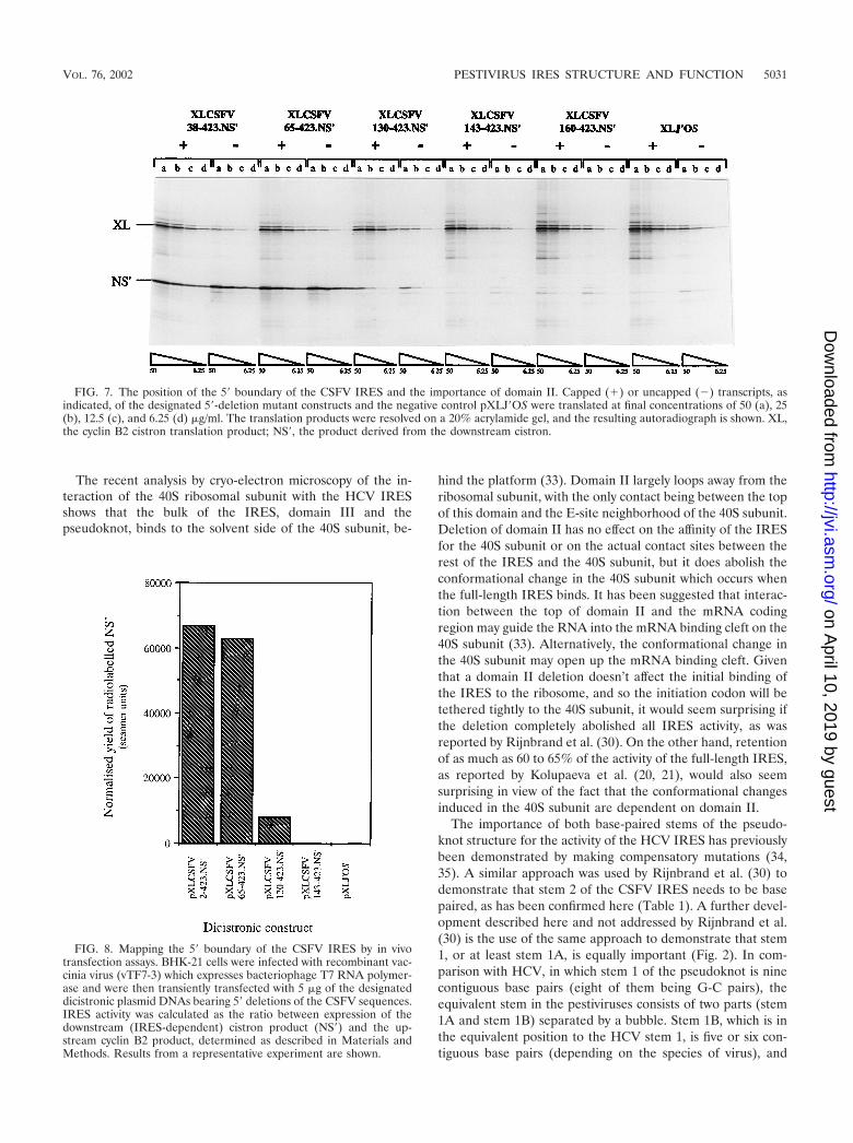
The significance of domain II for IRES activity. We made aseries of deletions from the 5� end of the CSFV sequence inorder to map the 5� boundary of the IRES and to assess thecontribution of domain II to IRES activity. These deletionswere made in the background of a construct (pXLCSFV1-423.NS�) which retains fewer viral coding sequences thanpXLCSFV 1-442.NS� but nevertheless exhibits comparableIRES activity (S. P. Fletcher and R. J. Jackson, unpublished
observations). The results obtained with a representative sub-set of these deletion mutants are shown in Fig. 7. The mutantswere found to fall into three groups with respect to IRESactivity. Those which included an intact domain II (Fig. 1),such as XLCSFV 38-423.NS� and XLCSFV 65-423.NS�, re-tained a high level of IRES activity (Fig. 7). We also obtaineddeletion mutants with end points at nt 2 and 14, both of whichshowed similar activity to XLCSFV 38-423.NS� and XLCSFV65-423.NS�, with no perceptible gradient of IRES efficiencythrough this series (data not shown). Deletions which extendinto domain II or remove it in its entirety fall into a second
FIG. 4. Expansion of the pseudoknot loop of the HCV IRES hasno effect on IRES activity. (A) Structure and primary sequence of theHCV IRES pseudoknot, showing the position of insertion of five Aresidues in pXLHCV 40-372.NS� 324insA5. (B) Capped dicistronictranscripts of the parent pXLHCV 40-372.NS� and a low activitydeletion mutant pXLHCV 40-339.NS� (29), the negative controlpXLJ�OS, and the insertion mutant pXLHCV 40-372.NS� 324insA5were translated at final concentrations of 50 (a), 25 (b), 12.5 (c), and6.25 (d) �g/ml. The translation products were resolved on a 20%acrylamide gel, and the resulting autoradiograph is shown. XL, thecyclin B2 cistron translation product; NS�, the product derived fromthe downstream cistron. The smaller size of the downstream cistronproduct encoded by pXLHCV 40-339.NS� is due to the fact that thisconstruct has no viral coding sequences, whereas in pXLHCV 40-372.NS� and its derivatives, the NS� coding sequence is fused to thefirst 30 nt of viral coding sequences, and thus the product is NS� withan N-terminal extension.
VOL. 76, 2002 PESTIVIRUS IRES STRUCTURE AND FUNCTION 5029
on April 10, 2019 by guest
http://jvi.asm.org/
Dow
nloaded from
group, represented in Fig. 7 by pXLCSFV 130-423.NS�, whichis missing the whole of domain II and also 1 bp of thepseudoknot stem 1A. The IRES activity of this mutant was veryseverely compromised but it was not zero (Fig. 7); the relativeefficiency averaged over the four RNA concentrations was19%. The deletions we obtained with end points at nt 79, 97,and 113, removing just part of domain II, were very similar inactivity to pXLCSFV 130-423.NS�, which is missing the wholeof this structural domain (data not shown). Finally, deletionswhich remove not only the whole of domain II but also invadestems 1A and 1B of the pseudoknot (e.g., pXLCSFV 143-423.NS� and pXLCSFV 160-423.NS�) constitute a third groupin which the IRES activity was effectively zero (Fig. 7). Theseresults place the 5� boundary such that the whole domain II,but very little sequence upstream of it, lies within the IRES.Deletions which enter and partially disrupt domain II or totallyremove it (pXLCSFV130-423.NS�) reduce IRES activity toabout 20% that of the control but do not totally abrogate it.
These conclusions were confirmed in transfection assays us-ing selected deletion mutants from this series. The yield of NS�from the IRES-dependent cistron was quantitated by meta-bolic labeling using [35S]methionine, followed by gel electro-phoresis, autoradiography, and densitometry. The yield of cy-clin from the upstream cistron was determined by Westernblotting using an antibody that is specific for X. laevis cyclin B2and does not cross-react with hamster cyclin. Data from arepresentative experiment are presented in Fig. 8, which con-firms the results of the in vitro translation assays: the deletionmutant pXLCSFV 65-423.NS�, which has a complete domainII, retained full IRES activity; pXLCSFV130-423.NS�, whichhas lost the whole of domain II, was seriously debilitated butnevertheless retained about 22% of wild-type IRES activity;and more extensive deletions resulted in complete loss of ac-tivity.
DISCUSSION
There is some minor controversy in the literature with re-spect to where the 5� boundary of the pestivirus IRES lies andmore serious controversy over the significance of domain II forIRES activity. Rijnbrand et al. (30) found that the deletion of26 nt from the 5� end actually increased IRES activity by about60%; a deletion to nt 66 reduced the activity to about two-thirds that of the full-length construct; and an internal deletionof nt 28 to 66 decreased activity still further. Consequently,
they placed the 5� boundary as lying within what was thencalled stem-loop B, between nt 28 and 66, whereas we wouldargue that all sequences up to the 5� side of domain II do notparticipate directly in IRES function. In our experiments wedid not observe any stimulation of IRES activity with short 5�deletions (though admittedly our deletion series did not in-clude any mutants with end points very near nt 28), and wefound that a deletion to nt 65 retained full activity. We wouldargue that our position for the 5� boundary is more consistentwith the phylogenetic data: our analysis of covariances betweenBDV, BVDV, and CSFV 5� UTR sequences shows strongconservation of structure downstream of the 5� end of domainII (Fig. 1), but there is much less conservation upstream of thispoint. Moreover, a boundary for the CSFV IRES at the 5�residue of domain II would be absolutely consistent with theposition of the 5� boundary of the HCV IRES (29) in therecently revised secondary structure model of the HCV 5�UTR (16) and is also consistent with the position of the 5�boundary of the BVDV IRES mapped by Chon et al. (6).
As for the significance of domain II in IRES function, Ri-jnbrand et al. (30) found that deletions which invaded domainII were essentially inactive in transfected Hep2 cells, whereasin our hands they retain about 20% of the activity of thefull-length IRES both in vitro and in transfected BHK cells.This is very similar to the results we obtained with 5� deletionsof the HCV IRES (29). It is only when the deletions not onlyremove the whole of domain II but invade stem 1 of thepseudoknot that IRES activity falls to zero in our hands. Incontrast, Kolupaeva et al. (20, 21) found that deletion of do-main II of either the CSFV or HCV IRES reduced translationefficiency only mildly, to 60 to 65% of the control, although itdid subtly alter the characteristics of 40S subunit binding to theIRES and was highly debilitating when combined with othermutations which on their own had only a mild phenotype.
FIG. 5. Silent mutations in the domain IIIa loop. The figure lists allthe mutants of the IIIa loop that were isolated, all of which were foundto have high IRES activity that was comparable to that of the wild type.Mutated residues are indicated with lowercase letters.
FIG. 6. Amputation of domain IIIa stem and loop abolishes CSFVIRES activity. Capped dicistronic transcripts of the parent constructpXLCSFV 1-442.NS�, the negative control pXLJ�OS, and the domainIIIa deletion mutant pXLCSFV 1-442.NS� �IIIa were translated atfinal concentrations of 50 (a), 25 (b), 12.5 (c), and 6.25 (d) �g/ml. Thetranslation products were resolved on a 20% acrylamide gel, and theresulting autoradiograph is shown. XL, the cyclin B2 cistron transla-tion product; NS�, the product derived from the downstream cistron.
5030 FLETCHER AND JACKSON J. VIROL.
on April 10, 2019 by guest
http://jvi.asm.org/
Dow
nloaded from
The recent analysis by cryo-electron microscopy of the in-teraction of the 40S ribosomal subunit with the HCV IRESshows that the bulk of the IRES, domain III and thepseudoknot, binds to the solvent side of the 40S subunit, be-
hind the platform (33). Domain II largely loops away from theribosomal subunit, with the only contact being between the topof this domain and the E-site neighborhood of the 40S subunit.Deletion of domain II has no effect on the affinity of the IRESfor the 40S subunit or on the actual contact sites between therest of the IRES and the 40S subunit, but it does abolish theconformational change in the 40S subunit which occurs whenthe full-length IRES binds. It has been suggested that interac-tion between the top of domain II and the mRNA codingregion may guide the RNA into the mRNA binding cleft on the40S subunit (33). Alternatively, the conformational change inthe 40S subunit may open up the mRNA binding cleft. Giventhat a domain II deletion doesn’t affect the initial binding ofthe IRES to the ribosome, and so the initiation codon will betethered tightly to the 40S subunit, it would seem surprising ifthe deletion completely abolished all IRES activity, as wasreported by Rijnbrand et al. (30). On the other hand, retentionof as much as 60 to 65% of the activity of the full-length IRES,as reported by Kolupaeva et al. (20, 21), would also seemsurprising in view of the fact that the conformational changesinduced in the 40S subunit are dependent on domain II.
The importance of both base-paired stems of the pseudo-knot structure for the activity of the HCV IRES has previouslybeen demonstrated by making compensatory mutations (34,35). A similar approach was used by Rijnbrand et al. (30) todemonstrate that stem 2 of the CSFV IRES needs to be basepaired, as has been confirmed here (Table 1). A further devel-opment described here and not addressed by Rijnbrand et al.(30) is the use of the same approach to demonstrate that stem1, or at least stem 1A, is equally important (Fig. 2). In com-parison with HCV, in which stem 1 of the pseudoknot is ninecontiguous base pairs (eight of them being G-C pairs), theequivalent stem in the pestiviruses consists of two parts (stem1A and stem 1B) separated by a bubble. Stem 1B, which is inthe equivalent position to the HCV stem 1, is five or six con-tiguous base pairs (depending on the species of virus), and
FIG. 7. The position of the 5� boundary of the CSFV IRES and the importance of domain II. Capped (�) or uncapped (�) transcripts, asindicated, of the designated 5�-deletion mutant constructs and the negative control pXLJ�OS were translated at final concentrations of 50 (a), 25(b), 12.5 (c), and 6.25 (d) �g/ml. The translation products were resolved on a 20% acrylamide gel, and the resulting autoradiograph is shown. XL,the cyclin B2 cistron translation product; NS�, the product derived from the downstream cistron.
FIG. 8. Mapping the 5� boundary of the CSFV IRES by in vivotransfection assays. BHK-21 cells were infected with recombinant vac-cinia virus (vTF7-3) which expresses bacteriophage T7 RNA polymer-ase and were then transiently transfected with 5 �g of the designateddicistronic plasmid DNAs bearing 5� deletions of the CSFV sequences.IRES activity was calculated as the ratio between expression of thedownstream (IRES-dependent) cistron product (NS�) and the up-stream cyclin B2 product, determined as described in Materials andMethods. Results from a representative experiment are shown.
VOL. 76, 2002 PESTIVIRUS IRES STRUCTURE AND FUNCTION 5031
on April 10, 2019 by guest
http://jvi.asm.org/
Dow
nloaded from

stem 1A is seven or eight contiguous base pairs. We haveshown that base pairing in stem 1A is essential for CSFV IRESactivity (Fig. 2), and although we have not directly examinedstem 1B, topological constraints dictate that this, too, mustsurely be base paired as shown in Fig. 1. That base pairing instem 1B is most likely to be critical for IRES activity is shownby the fact that disruption of 3 bp in this stem severely im-paired IRES activity (21), although compensatory mutations torestore base pairing were not tested. In view of this overwhelm-ing evidence in favor of the base pairing, but not the primarysequence, of the pseudoknot being critical for IRES activity, itis very puzzling that Kieft et al. (19) could not rescue HCVIRES activity by making compensating mutations in stem 2.The explanation for this unique negative result is not immedi-ately self-evident, though there is one obvious difference in thetechnical details of these experiments. Kieft et al. (19) assayedIRES activity using the coupled transcription-translation sys-tem. Because this system generates uncapped transcripts andso has been optimized for translation (via the scanning mech-anism) of uncapped mRNA, it is usually operated at a mono-valent cation concentration that is considerably lower than thatwhich is optimal for translation of capped mRNAs or transla-tion dependent on the HCV and pestivirus IRESs.
Whereas there is just a single U residue between stem 1 andstem 2 in the HCV pseudoknot, in pestiviruses there is a longerloop varying from five consecutive A residues in BVDV strainsto 8 nt (AUAAAAAU) in other species, including CSFV. Thislonger loop in CSFV than in HCV may be necessary for topo-logical reasons related to the greater length of stem 1A plus 1Bof the pestivirus pseudoknot. Certainly, reducing the length ofthe pseudoknot loop in the CSFV IRES by 5 nt very severelydecreased IRES activity (Fig. 3). If our suggestion of a topo-logical constraint is correct, this shortening would either distortthe relative positions of the two stems of the pseudoknot orpossibly cause the two stems to become partly unpaired.
However, our mutational analysis showed that for maximumIRES activity it is not just the length of the pseudoknot loopthat is important; it also has to be A-rich. A-rich bulges like thisare found in the group I intron ribozyme, where they arelikewise essential for activity (5). The IRESs of cardiovirusesand aphthoviruses also have an A-rich bulge, and mutations inthis bulge influence not so much the activity of the IRES as itsdependency on polypyrimidine tract binding protein for activ-ity (17). X-ray crystallography of the group I intron ribozymehas shown that most of the residues of the A-rich bulge areinvolved in tertiary interactions which depend on such residuesbeing specifically adenosines, a conclusion consistent with theinfluence of mutations of these residues on ribozyme activity(5). A common type of tertiary structure interaction, found inboth the group I intron and in 23S rRNA, is the insertion ofthese adenosine residues into the minor groove of neighboringhelices (5, 22). It would be intriguing if the A-rich pseudoknotbulge in the CSFV IRES were involved in similar tertiaryinteractions. On the whole, the influence of mutations of the Aresidues in the CSFV IRES pseudoknot loop is much lessdeleterious than in the case of the group I intron A-rich bulge,which would argue against the idea that they are involved incritical tertiary interactions. However, in structure probing ex-periments we find that even though this A-tract is not cleavedby cobra venom nuclease, the A residues are only moderately
susceptible to reaction with dimethyl sulfate, indeed rather lessreactive than nearby A residues such as the two between stem2 and the initiation codon or the A residue in the bubblebetween stem 1A and stem 1B (S. P. Fletcher and R. J. Jack-son, unpublished results).
Finally, the lack of any effect of mutation of the very highlyconserved loop IIIa sequence was surprising. However, ourresults closely parallel those of Kolupaeva et al. (21), whofound that mutation of the loop sequence from the wild-typeAGUA to AAAA had very little effect on translation efficiency,but mutations which would have altered the length of the stemand/or the size of the loop were deleterious. In addition, theseauthors observed that the binding of 40S ribosomal subunits tothe IRES afforded no protection of domain IIIa of either theCSFV or HCV IRESs (20, 21).
However, almost diametrically opposite results have beenreported for the HCV IRES (19). Not only were residues in theIIIa loop protected when 40S subunits bound to the HCVIRES, but modification interference experiments strongly im-plicated the two A residues as critical for 40S subunit binding.In addition, mutation of the loop sequence to UCAU reducedtranslation efficiency and 40S subunit binding affinity about10-fold. Nevertheless, the cryo-electron microscopy analysisactually suggests that domain IIIa, together with domains IIIband IIIc, extends away from the surface of the 40S subunit (33),which is hard to reconcile with the mutagenesis and structureprobing data published by the same group (19).
Since only two positions of this tetraloop were changed inany one of the many mutants we generated, it could conceiv-ably be argued that the sequence of the loop is necessary buthas very considerable inherent redundancy, with either just thetwo central residues or the two flanking A residues beingsufficient. Although this type of explanation would be formallyconsistent with all the results of the point mutations made inthe IIIa loop by Kolupaeva et al. (21), Kieft et al. (19), andourselves, it seems highly implausible and without precedent inany other system. Another explanation that is formally consis-tent with all the data is that the CSFV and HCV IRESs differwith respect to the importance of the loop IIIa sequence, butthis too seems highly improbable. It thus seems possible thatdespite its high degree of conservation, the sequence of theloop is not important for IRES activity, even though the lengthof the stem and the existence of the four-way junction probablyis critical. This in turn raises the question of whether there maynot be signals embedded within the IRES for other functionsnecessary for the viral life cycle apart from internal initiation oftranslation. Indeed, the results of recent experiments with thenewly developed HCV replicon system indicate that there arecis-acting signals within the IRES which are critical for efficientRNA replication (10).
ACKNOWLEDGMENTS
We thank G. Meyers and H.-J. Thiel for the original gift of theCSFV 5� UTR plasmid and for advice, Tim Hunt and Julian Gannonfor the gift of a highly specific antiserum raised against X. laevis cyclinB2, Paul Digard and Konrad Bishop for help with the transfectionassays, and Catherine Gibbs for technical support.
The costs of this work were supported mainly by a grant from theWellcome Trust, with additional support from the European Commis-sion Biotechnology Program (BIO4-CT95-0045). S.P.F. gratefully ac-
5032 FLETCHER AND JACKSON J. VIROL.
on April 10, 2019 by guest
http://jvi.asm.org/
Dow
nloaded from

knowledges the support of a research studentship awarded by theMedical Research Council.
REFERENCES
1. Avalos-Ramirez, R., M. Orlich, H.-J. Thiel, and P. Becher. 2001. Evidencefor the presence of two novel pestivirus species. Virology 286:456–465.
2. Becher, P., M. Orlich, A. D. Shannon, G. Horner, M. Konig, and H.-J. Thiel.1997. Phylogenetic analysis of pestiviruses from domestic and wild rumi-nants. J. Gen. Virol. 78:1357–1366.
3. Borman, A., and R. J. Jackson. 1992. Initiation of translation of humanrhinovirus RNA: mapping the internal ribosome entry site. Virology 188:685–696.
4. Brown, E. A., H. Zhang, L. Ping, and S. M. Lemon. 1992. Secondary structureof the 5� nontranslated regions of hepatitis C virus and pestivirus genomicRNAs. Nucleic Acids Res. 20:5041–5045.
5. Cate, J. H., A. R. Gooding, E. Podell, K. Zhou, B. L. Golden, C. E. Kundrot,T. R. Cech, and J. A. Doudna. 1996. Crystal structure of a group I ribozymedomain: principles of RNA packing. Science 273:1678–1685.
6. Chon, S. K., D. R. Perez, and R. O. Donis. 1998. Genetic analysis of theinternal ribosome entry segment of bovine viral diarrhoea virus. Virology251:370–382.
7. Craig, D., M. T. Howell, C. L. Gibbs, T. Hunt, and R. J. Jackson. 1992.Plasmid cDNA-directed protein synthesis in a coupled eukaryotic in vitrotranscription-translation system. Nucleic Acids Res. 20:4987–4995.
8. Dasso, M. C., and R. J. Jackson. 1989. On the fidelity of mRNA translationin the nuclease-treated rabbit reticulocyte lysate system. Nucleic Acids Res.17:3129–3144.
9. Deng, R., and K. V. Brock. 1993. 5� and 3� untranslated regions of pestivirusgenome: primary and secondary structure analysis. Nucleic Acids Res. 21:1949–1957.
10. Friebe, P., V. Lohmann, N. Krieger, and R. Bartenschlager. 2001. Sequencesin the 5� nontranslated region of hepatitis C virus required for RNA repli-cation. J. Virol. 75:12047–12057.
11. Fuerst, T. R., E. G. Niles, F. W. Studier, and B. Moss. 1986. Eukaryotictransient expression system based on recombinant vaccinia virus that syn-thesises bacteriophage T7 RNA polymerase. Proc. Natl. Acad. Sci. USA83:8122–8126.
12. Grünert, S., and R. J. Jackson. 1994. The immediate downstream codonstrongly influences the efficiency of utilization of eukaryotic translation ini-tiation sites. EMBO J. 13:3618–3630.
13. Harlow, E., and D. Lane. 1988. Antibodies: a laboratory manual. Cold SpringHarbor Laboratory Press, Cold Spring Harbor, N.Y.
14. Heinz, F. X., M. S. Collett, R. H. Purcell, E. A. Gould, C. R. Howard, M.Houghton, R. J. M. Moorman, C. M. Rice, and H.-J. Thiel. 2000. FamilyFlaviviridae, p. 859–878. In M. H. V. van Regenmortel, C. M. Fauquet,D. H. L. Bishop, E. B. Carstens, M. K. Estes, S. M. Lemon, J. Maniloff, M. A.Mayo, D. J. McGeoch, C. R. Pringle, and R. B. Wickner (ed.), Virus taxon-omy. Seventh Report of the International Committee on Taxonomy of Vi-ruses. Academic Press, San Diego, Calif.
15. Henikoff, S. 1987. Unidirectional digestion with exonuclease III in DNAsequence analysis. Methods Enzymol. 155:156–165.
16. Honda, M., M. R. Beard, L.-H. Ping, and S. M. Lemon. 1999. A phyloge-netically conserved stem-loop structure at the 5� border of the internalribosome entry site of hepatitis C virus is required for cap-independent viraltranslation. J. Virol. 73:1165–1174.
17. Kaminski, A., and R. J. Jackson. 1998. The polypyrimidine tract bindingprotein (PTB) requirement for internal initiation of translation of cardiovi-rus RNAs is conditional rather than absolute. RNA 4:626–638.
18. Kaminski, A., G. J. Belsham, and R. J. Jackson. 1994. Translation of en-cephalomyocarditis virus RNA: parameters influencing the selection of theinternal initiation site. EMBO J. 13:1673–1681.
19. Kieft, J. S., K. Zhou, R. Jubin, and J. A. Doudna. 2001. Mechanism ofribosome recruitment by hepatitis C IRES RNA. RNA 7:194–206.
20. Kolupaeva, V. G., T. V. Pestova, and C. U. T. Hellen. 2000. An enzymaticfootprinting analysis of the interaction of 40S ribosomal subunits with theinternal ribosomal entry site of hepatitis C virus. J. Virol. 74:6242–6250.
21. Kolupaeva, V. G., T. V. Pestova, and C. U. T. Hellen. 2000. Ribosomalbinding to the internal ribosome entry site of classical swine fever virus. RNA6:1791–1807.
22. Nissen, P., J. A. Ippolito, N. Ban, P. B. Moore, and T. A. Steitz. 2001. RNAtertiary interactions in the large ribosomal subunit: the A-minor motif. Proc.Natl. Acad. Sci. USA 98:4899–4903.
23. Pestova, T. V., C. U. T. Hellen, and I. N. Shatsky. 1996. Canonical eukaryoticinitiation factors determine initiation of translation by internal ribosomalentry. Mol. Cell. Biol. 16:6859–6869.
24. Pestova, T. V., I. N. Shatsky, and C. U. T. Hellen. 1996. Functional dissectionof eukaryotic initiation factor eIF4F: the 4A subunit and the central domainof the 4G subunit are sufficient to mediate internal entry of 43S preinitiationcomplexes. Mol. Cell. Biol. 16:6870–6878.
25. Pestova, T. V., I. N. Shatsky, S. P. Fletcher, R. J. Jackson, and C. U. T.Hellen. 1998. A prokaryotic-like mode of cytoplasmic eukaryotic ribosomebinding to the initiation codon during internal translation initiation of hep-atitis C and classical swine fever virus RNAs. Genes Dev. 12:67–83.
26. Picard, V., E. Ersdalbadju, A. Q. Lu, and S. C. Bock. 1994. A rapid andefficient one-tube PCR-based mutagenesis technique using Pfu DNA poly-merase. Nucleic Acids Res. 22:2587–2591.
27. Poole, T. L., C. Wang, R. A. Popp, L. N. D. Potgieter, A. Siddiqui, and M. S.Collett. 1995. Pestivirus translation initiation is by internal ribosome entry.Virology 206:750–754.
28. Reynolds, J. E., A. Kaminski, H. J. Kettinen, K. Grace, B. E. Clarke, A. R.Carroll, D. J. Rowlands, and R. J. Jackson. 1995. Unique features of internalinitiation of hepatitis C virus RNA translation. EMBO J. 14:6010–6020.
29. Reynolds, J. E., A. Kaminski, A. R. Carroll, B. E. Clarke, D. J. Rowlands,and R. J. Jackson. 1996. Internal initiation of translation of hepatitis C virusRNA: the ribosome entry site is at the authentic initiation codon. RNA2:867–878.
30. Rijnbrand, R., T. van der Straten, P. A. van Rijn, W. J. M. Spaan, and P. J.Bredenbeek. 1997. Internal entry of ribosomes is directed by the 5� noncod-ing region of classical swine fever virus and is dependent on the presence ofan RNA pseudoknot upstream of the initiation codon. J. Virol. 71:451–457.
31. Sambrook, J., E. F. Fritsch, and T. Maniatis. 1989. Molecular cloning: alaboratory manual, 2nd ed. Cold Spring Harbor Laboratory Press, ColdSpring Harbor, N.Y.
32. Smith, D. B., J. Mellor, L. M. Jarvis, F. Davidson, J. Kolberg, M. Urdea,P. L.Yap, P. Simmonds, J. D. Conradie, A. G. S. Neill, G. M. Dusheiko, M. C.Kew, R. Crookes, A. Koshy, C. K. Lin, C. Lai, I. M. Murray-Lyon, A.Elguneid, A. A. Gunaid, T. Yemen, S. Yemen, D. Mutimer, M. Ahmed, C.Nuchprayoon, S. Tanprasert, F. E. Preston, M. Makris, A. Chuansumrit, C.Mahasandana, D. Pritchard, E. Riley, B. M. Greenwood, A. A. Saeed, A. M.Alrasheed, M. G. Saleh, I. McFarlane, C. Tibbs, R. Williams, J. Power, E.Lawlor, and H. Kiyokawa. 1995. Variations in the hepatitis C virus noncod-ing region—implications for secondary structure, virus detection and typing.J. Gen. Virol. 76:1749–1761.
33. Spahn, C. M., J. S. Kieft, R. A. Grassucci, P. A. Penczek, K. Zhou, J. A.Doudna, and J. Frank. 2001. Hepatitis C virus IRES RNA-induced changesin the conformation of the 40S ribosomal subunit. Science 291:1959–1962.
34. Wang, C., P. Sarnow, and A. Siddiqui. 1994. A conserved helical element isessential for internal initiation of translation of hepatitis C virus RNA.J. Virol. 68:7301–7307.
35. Wang, C., S.-Y. Le, N. Ali, and A. Siddiqui. 1995. An RNA pseudoknot is anessential structural element of the internal ribosome entry site located withinthe hepatitis C virus 5� noncoding region. RNA 1:526–537.
36. Yusupova, G. Z., M. M. Yusupov, J. H. D. Cate, and H. F. Noller. 2001. Thepath of messenger RNA through the ribosome. Cell 106:233–241.
VOL. 76, 2002 PESTIVIRUS IRES STRUCTURE AND FUNCTION 5033
on April 10, 2019 by guest
http://jvi.asm.org/
Dow
nloaded from


![Ribosome Stoichiometry: From Form to Function · Ribosome abundance: A major model, also termed the ribosome concentration hypothesis [3], that explains how ribosomes could exert](https://img.pdfslide.us/doc/110x75/60de31e56d30fc4fb30719b8/ribosome-stoichiometry-from-form-to-function-ribosome-abundance-a-major-model.jpg)