Embed Size (px)
Citation preview
1
Molecular Cell BiologyFifth Edition
Chapter 19:Cytoskeleton I: Microfilaments and
Intermediate Filaments
Copyright © 2004 by W. H. Freeman & Company
Harvey Lodish • Arnold Berk • Paul Matsudaira •Chris A. Kaiser • Monty Krieger • Matthew P. Scott •
Lawrence Zipursky • James DarnellActin structureActin structure
Forms very large structuresForms very large structures-- assembly or assembly or disassembly alters cell morphology or the disassembly alters cell morphology or the morphology of specific compartments.morphology of specific compartments.Flexible cytoskeletonFlexible cytoskeletonMonomers, filament, cross linked filaments, Monomers, filament, cross linked filaments, imperfect bundles and networks.imperfect bundles and networks.An ancient highly conserved geneAn ancient highly conserved gene
Overview actin and intermediate filaments -functions
Actin is the most abundant intracellular Actin is the most abundant intracellular proteinproteinThe cytosolic concentration of non muscle The cytosolic concentration of non muscle actin is 0.1actin is 0.1--0.5 0.5 mMmM..In In microvillimicrovilli 5mM5mMModerate size of MW 42.000.Moderate size of MW 42.000.
Actin a highly conserved geneActin a highly conserved gene
In vertebrates four aIn vertebrates four a--actin isoform in actin isoform in muscles .muscles .ββ and and γγ actin non muscle cells.actin non muscle cells.ββ--actin in the leading edge of moving cells actin in the leading edge of moving cells γγ−− actin form filamentsactin form filamentsthe different isoforms differ in four of five the different isoforms differ in four of five
positions positions
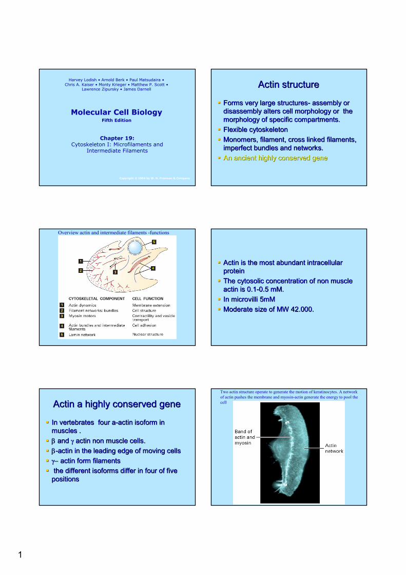
Two actin structure operate to generate the motion of keratinocytes. A network of actin pushes the membrane and myosin-actin generate the energy to pool the cell
2
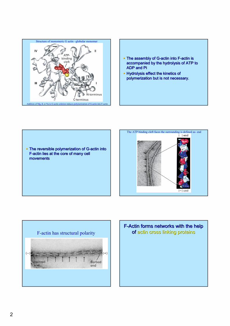
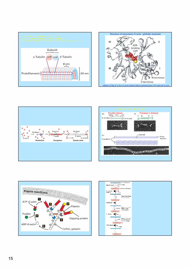
Structure of monomeric G actin –globular monomer
Addition of Mg, K or Na to G-actin solution induces polymerization of G-actin into F-actin
The assembly of GThe assembly of G--actin into Factin into F--actin is actin is accompanied by the hydrolysis of ATP to accompanied by the hydrolysis of ATP to ADP and PiADP and PiHydrolysis effect the kinetics of Hydrolysis effect the kinetics of polymerization but is not necessary.polymerization but is not necessary.
The reversible polymerization of GThe reversible polymerization of G--actin into actin into FF--actin lies at the core of many cell actin lies at the core of many cell movementsmovements
The ATP binding cleft faces the surrounding is defined as- end
F-actin has structural polarityFF--Actin forms networks with the help Actin forms networks with the help
of of actin cross linking proteinsactin cross linking proteins
3
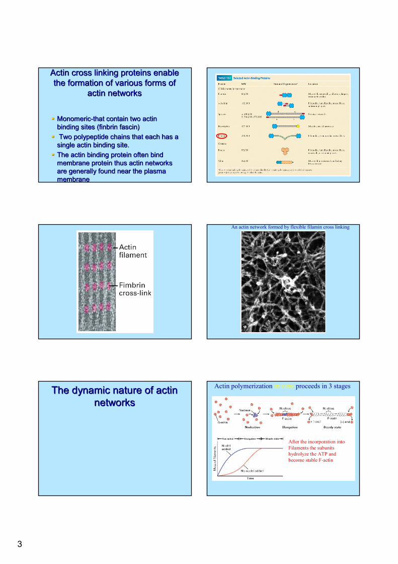
Actin cross linking proteins enable Actin cross linking proteins enable the formation of various forms of the formation of various forms of
actin networksactin networks
MonomericMonomeric--that contain two actin that contain two actin binding sites (binding sites (finbrinfinbrin fascinfascin))Two polypeptide chains that each has a Two polypeptide chains that each has a
single actin binding site.single actin binding site.The actin binding protein often bind The actin binding protein often bind membrane protein thus actin networks membrane protein thus actin networks are generally found near the plasma are generally found near the plasma membranemembrane
An actin network formed by flexible filamin cross linking
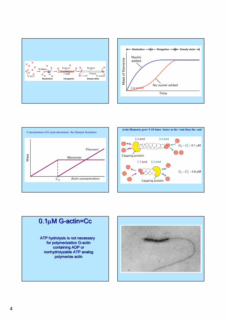
The dynamic nature of actin The dynamic nature of actin networksnetworks
After the incorporation intoFilaments the subunits hydrolyze the ATP and become stable F-actin (from red to white)
Actin polymerization in vitro proceeds in 3 stages
1- 2- 3-
4
Leg period
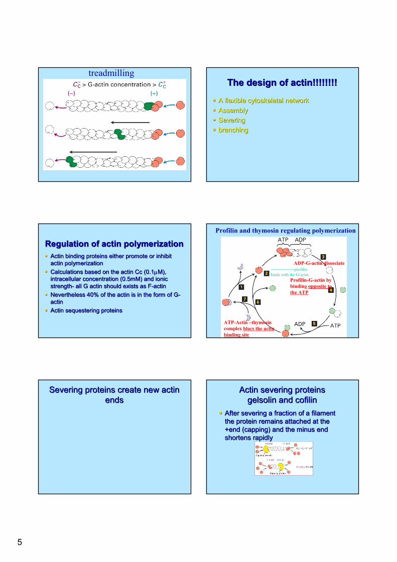
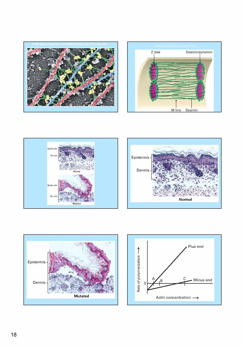
Concentration of G actin determines the filament formationActin filaments grow 5-10 times faster at the +end than the -end
0.10.1µµM GM G--actin=Ccactin=Cc
ATP hydrolysis is not necessary ATP hydrolysis is not necessary for polymerization Gfor polymerization G--actin actin
containing ADP or containing ADP or nonhydrolyzablenonhydrolyzable ATP analog ATP analog
polymerize actinpolymerize actin
5
treadmillingThe design of actin!!!!!!!!The design of actin!!!!!!!!
A flexible cytoskeletal networkA flexible cytoskeletal networkAssemblyAssemblySeveringSeveringbranchingbranching
Regulation of actin polymerizationRegulation of actin polymerizationActin binding proteins either promote or inhibit Actin binding proteins either promote or inhibit actin polymerizationactin polymerizationCalculations based on the actin Cc (0.1Calculations based on the actin Cc (0.1µµM), M), intracellular concentration (0.5mM) and ionic intracellular concentration (0.5mM) and ionic strengthstrength-- all G actin should exists as Fall G actin should exists as F--actin actin Nevertheless 40% of the actin is in the form of GNevertheless 40% of the actin is in the form of G--actinactinActin sequestering proteinsActin sequestering proteins
ATP-Actin –thymosincomplex blocs the actin binding site
G-actin ADP-G-actin dissociate
Profilin-G-actin by binding opposite to the ATP
Profilin and thymosin regulating polymerization
--------------profilinbinds with the G-actin
Severing proteins create new actin Severing proteins create new actin endsends
Actin severing proteinsActin severing proteinsgelsolingelsolin and and cofilincofilin
After severing a fraction of a filament After severing a fraction of a filament the protein remains attached at the the protein remains attached at the +end (capping) and the minus end +end (capping) and the minus end shortens rapidlyshortens rapidly
6
Signaling pathways that regulate Signaling pathways that regulate actin polymerizationactin polymerization
CofilinCofilin and and GelsolinGelsolin bind PIP2 that inhibits bind PIP2 that inhibits their binding to Ftheir binding to F--actin. Hydrolysis of PIP2 actin. Hydrolysis of PIP2 by phospholipase C release the proteins by phospholipase C release the proteins and induce severing of Fand induce severing of F--actin.actin.Phosphorylation of Phosphorylation of cofilincofilin regulate its activityregulate its activity1 1 µµMM Ca activate Ca activate gelsolingelsolin..
Capping proteins stabilize F-actinCapZ binds to F-actin +end and stabilizes itTropomodulin caps the minus end of F-actin and stabilizes it F-actin that is caped on both sides is stable
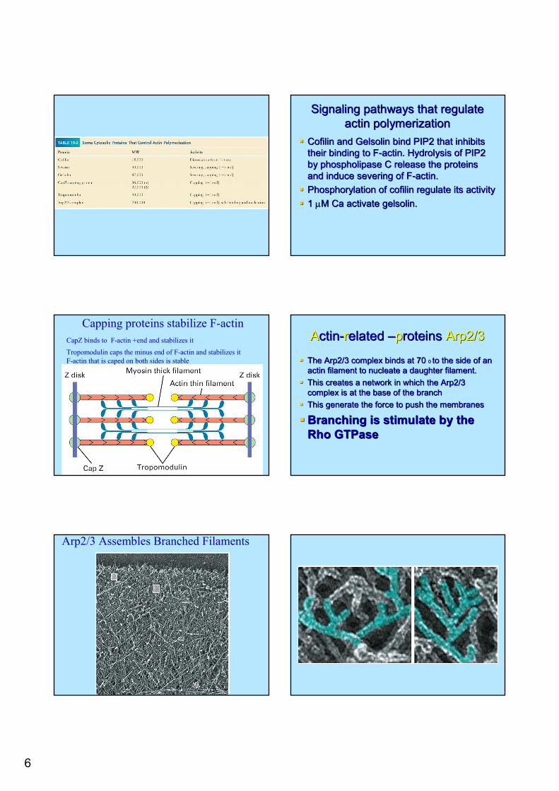
AActinctin--rrelated elated ––pproteins roteins Arp2/3Arp2/3
The Arp2/3 complex binds at 70 The Arp2/3 complex binds at 70 0 0 to the side of an to the side of an actin filament to nucleate a daughter filament.actin filament to nucleate a daughter filament.This creates a network in which the Arp2/3 This creates a network in which the Arp2/3 complex is at the base of the branchcomplex is at the base of the branchThis generate the force to push the membranesThis generate the force to push the membranes
Branching is stimulate by the Branching is stimulate by the RhoRho GTPaseGTPase
Arp2/3 Assembles Branched Filaments
7
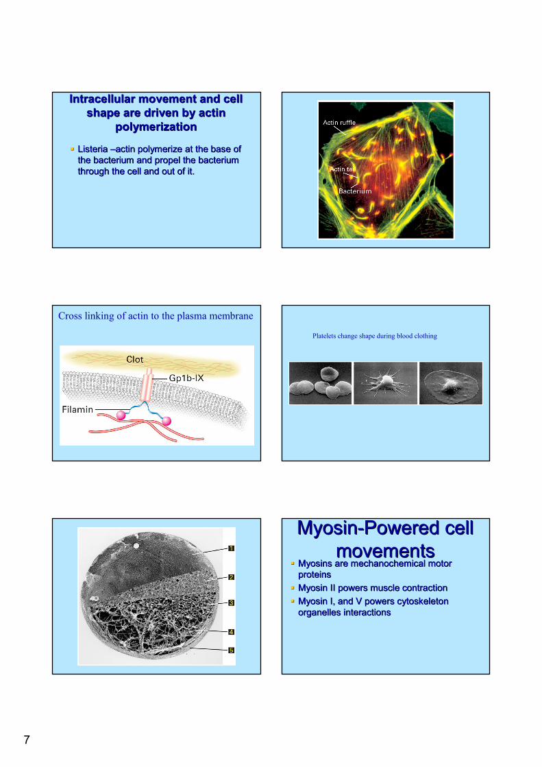
Intracellular movement and cell Intracellular movement and cell shape are driven by actin shape are driven by actin
polymerizationpolymerization
Listeria Listeria ––actin polymerize at the base of actin polymerize at the base of the bacterium and propel the bacterium the bacterium and propel the bacterium through the cell and out of it.through the cell and out of it.
Cross linking of actin to the plasma membrane
Platelets change shape during blood clothing
MyosinMyosin--Powered cell Powered cell movementsmovements
Myosins are mechanochemical motor Myosins are mechanochemical motor proteinsproteinsMyosin II powers muscle contractionMyosin II powers muscle contractionMyosin I, and V powers cytoskeleton Myosin I, and V powers cytoskeleton organelles interactionsorganelles interactions
8
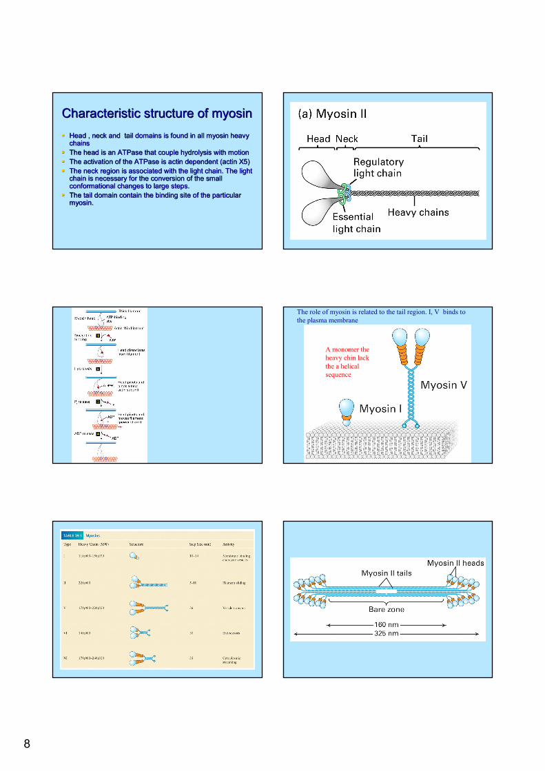
Characteristic structure of myosinCharacteristic structure of myosinHead , neck and tail domains is found in all myosin heavy Head , neck and tail domains is found in all myosin heavy chainschainsThe head is an ATPase that couple hydrolysis with motionThe head is an ATPase that couple hydrolysis with motionThe activation of the ATPase is actin dependent (actin X5)The activation of the ATPase is actin dependent (actin X5)The neck region is associated with the light chain. The light The neck region is associated with the light chain. The light chain is necessary for the conversion of the small chain is necessary for the conversion of the small conformational changes to large steps.conformational changes to large steps.The tail domain contain the binding site of the particular The tail domain contain the binding site of the particular myosin.myosin.
A monomer the heavy chin lack the a helical sequence
The role of myosin is related to the tail region. I, V binds tothe plasma membrane
9
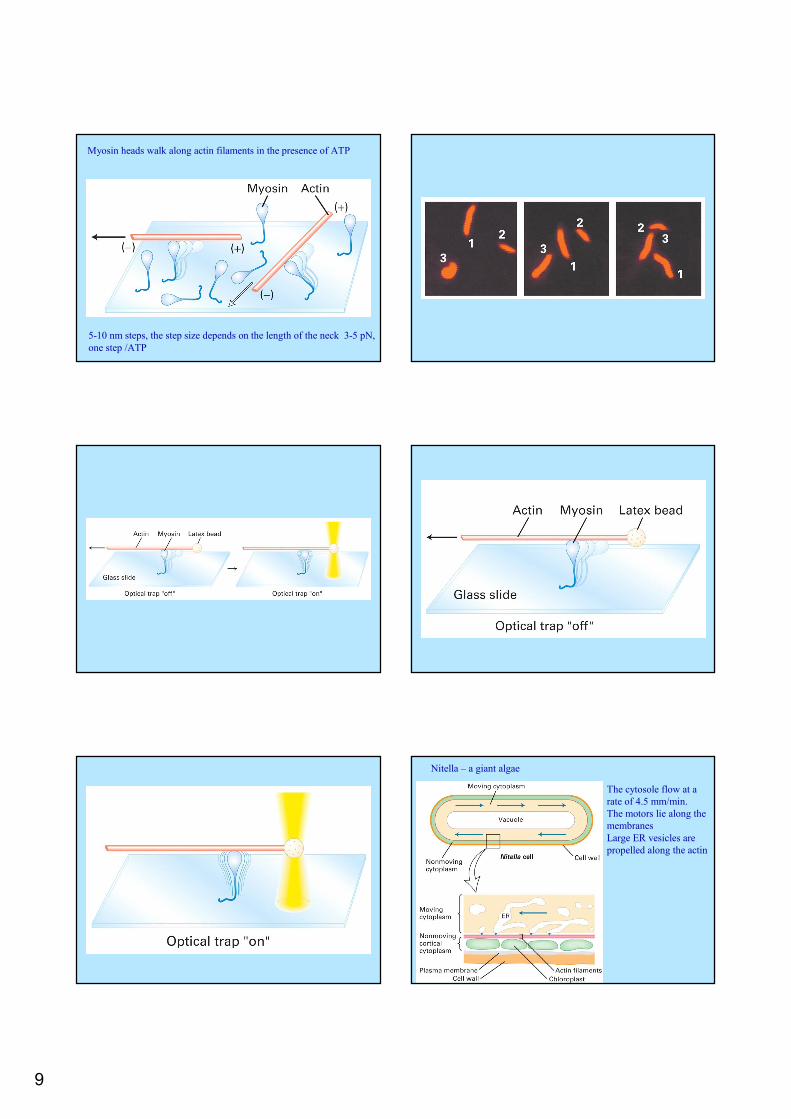
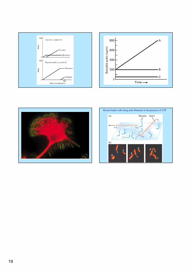
Myosin heads walk along actin filaments in the presence of ATP
5-10 nm steps, the step size depends on the length of the neck 3-5 pN, one step /ATP
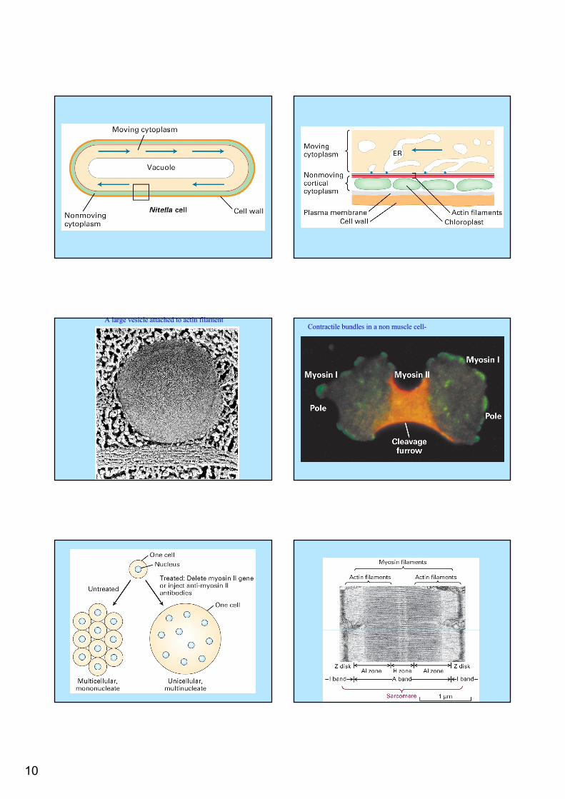
Nitella – a giant algae
The cytosole flow at a rate of 4.5 mm/min.The motors lie along the membranesLarge ER vesicles are propelled along the actin
10
A large vesicle attached to actin filamentContractile bundles in a non muscle cell-
11
12
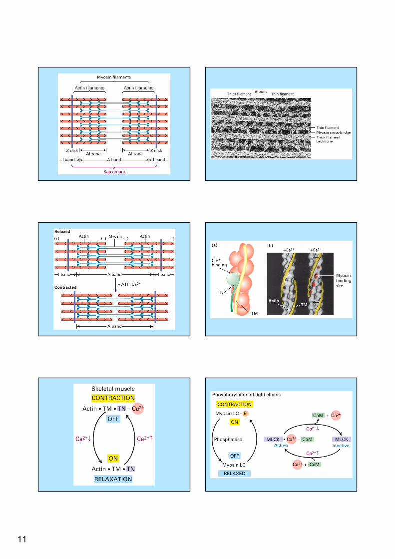
Cell locomotionCell locomotion
Cells in motion are characterized by being polar.Cells in motion are characterized by being polar.Cells extend a large membrane protrusion at the Cells extend a large membrane protrusion at the leading edge leading edge ––the lamellipodiumthe lamellipodiumThe lamellipodium the actin are crossed linked The lamellipodium the actin are crossed linked Finger like protrusions are formed the filopodiaFinger like protrusions are formed the filopodiaThese structures form stable contact with the These structures form stable contact with the substrate to prevent the membrane from retractingsubstrate to prevent the membrane from retracting
Force generation and cell Force generation and cell adhesionadhesion
Membrane extensionMembrane extension
Actin polymerization pushes the membrane Actin polymerization pushes the membrane forwardforward-- addition of Gaddition of G--actin to Factin to F--actinactinThe elastic Brownian ratchet model The elastic Brownian ratchet model ––explains the mechanism of G actin addition explains the mechanism of G actin addition and force generationand force generation
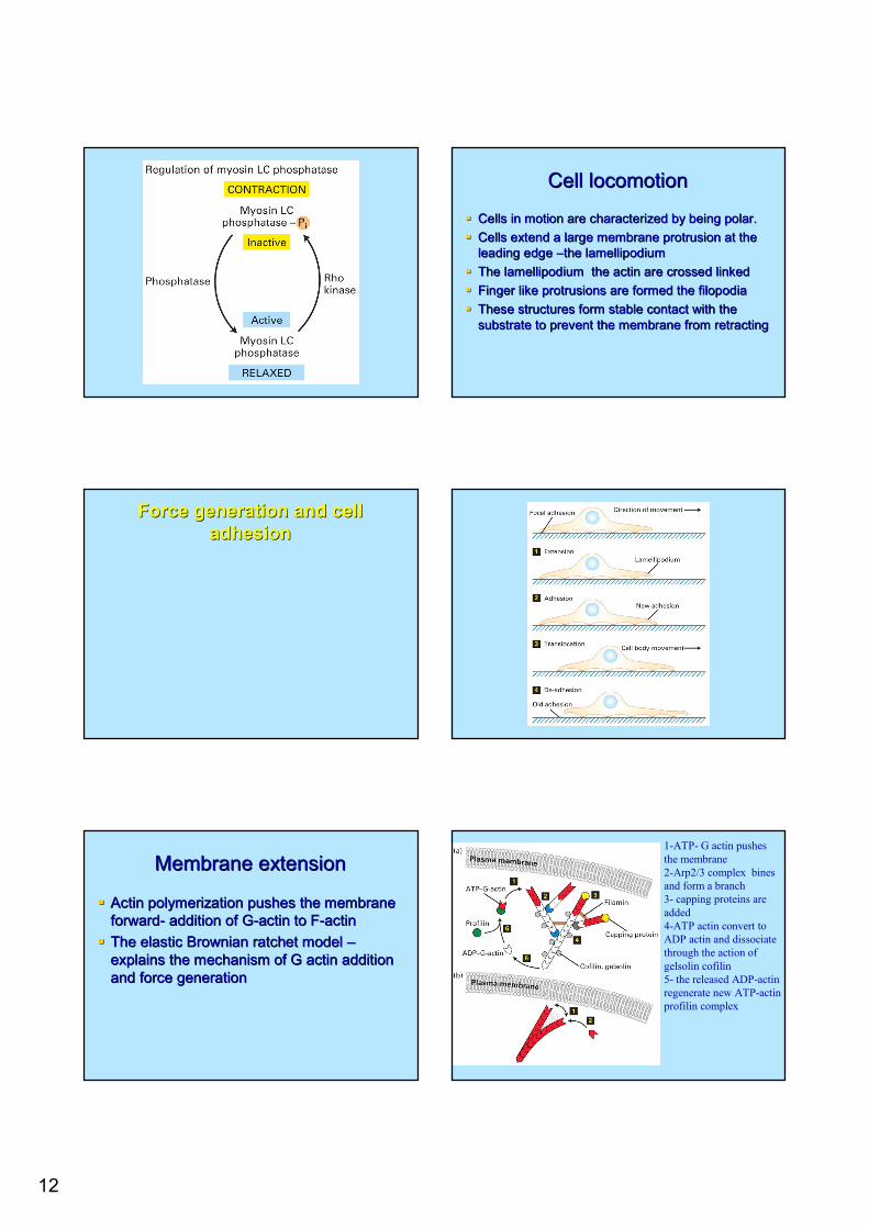
1-ATP- G actin pushes the membrane2-Arp2/3 complex bines and form a branch3- capping proteins are added4-ATP actin convert to ADP actin and dissociate through the action of gelsolin cofilin5- the released ADP-actin regenerate new ATP-actin profilin complex
13
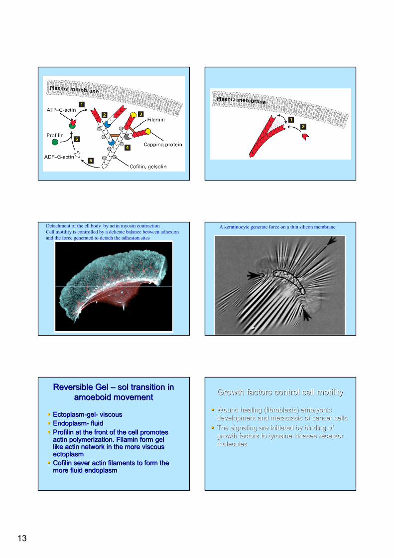
Detachment of the ell body by actin myosin contractionCell motility is controlled by a delicate balance between adhesion and the force generated to detach the adhesion sites
A keratinocyte generate force on a thin silicon membrane
Reversible Gel Reversible Gel –– sol transition in sol transition in amoeboid movementamoeboid movement
EctoplasmEctoplasm--gelgel-- viscousviscousEndoplasmEndoplasm-- fluidfluidProfilinProfilin at the front of the cell promotes at the front of the cell promotes actin polymerization. actin polymerization. FilaminFilamin form gel form gel like actin network in the more viscous like actin network in the more viscous ectoplasmectoplasmCofilinCofilin sever actin filaments to form the sever actin filaments to form the more fluid endoplasmmore fluid endoplasm
Growth factors control cell motilityGrowth factors control cell motility
Wound healing (fibroblasts) embryonic Wound healing (fibroblasts) embryonic development and metastasis of cancer cellsdevelopment and metastasis of cancer cellsThe signaling are initiated by binding of The signaling are initiated by binding of growth factors to tyrosine kinases receptor growth factors to tyrosine kinases receptor moleculesmolecules
14
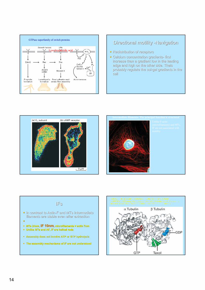
GTPase superfamily of switch proteins
Lysophosphatidic acid
Directional motility Directional motility --NavigationNavigation
Redistribution of receptorsRedistribution of receptorsCalcium concentration gradientsCalcium concentration gradients-- first first increase than a gradient low in the leading increase than a gradient low in the leading edge and high on the other side. edge and high on the other side. TheisTheisprobably regulate the solprobably regulate the sol--gel gradients in the gel gradients in the cellcell
Intermediate filaments- The principal function is structuralUnlike F-actin (microfilaments) and MTs, IF are not associated with motility
IFsIFs
In contrast to ActinIn contrast to Actin--F and MTs Intermediate F and MTs Intermediate filaments are stable even after extractionfilaments are stable even after extraction
MTs 24nm, MTs 24nm, IF 10nmIF 10nm,, microfilaments = actin 7nmmicrofilaments = actin 7nmUnlike MTs and AF, IF are helical rodsUnlike MTs and AF, IF are helical rods
Assembly does not involve ATP or GTP hydrolysisAssembly does not involve ATP or GTP hydrolysis
The assembly mechanisms of IF are not understoodThe assembly mechanisms of IF are not understood
Building blocks are α and β-tubulin bound together by noncovalent bonds55.000 MW. GTP-binding sites; α tubulin irreversibly, β tubulin binds GTP reversibly and serves as a GTPase. The GDP modulate the addition of tubulin subunits.
15
Protofilaments longitudinal interactions α- α; β- βCylindrical –tube, lateral interactions of 13 protofilamentsBecause of the structure addition of new dimers takes place only at the +or – ends.
Structure of monomeric G actin –globular monomer
Addition of Mg, K or Na to G-actin solution induces polymerization of G-actin into F-actin
Parallel dimers Tetramer is formedBasic structure – No polarity
16
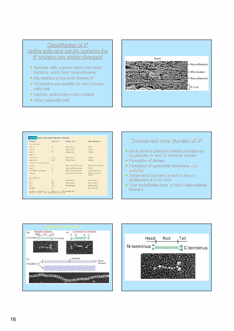
Classification of IFClassification of IFUnlike actin and tubulin isoforms the Unlike actin and tubulin isoforms the
IF proteins are widely divergentIF proteins are widely divergent
EpitheleEpithele cells express acidic and basic cells express acidic and basic keratins which form keratins which form heterodimeresheterodimeresthe keratins is the most diverse IF the keratins is the most diverse IF 10 keratins are specific for hard tissues10 keratins are specific for hard tissues--nails hairnails hairLaminsLamins-- exclusively in the nucleusexclusively in the nucleusOther epithelial cellsOther epithelial cells
Conserved core domain of IFConserved core domain of IF
All Ifs have a central All Ifs have a central αα helical core flanked helical core flanked by globular Nby globular N-- and Cand C-- terminal domainterminal domainFormation of dimersFormation of dimersFormation of symmetric tetramersFormation of symmetric tetramers-- No No polaritypolarityTetrameresTetrameres bind end to end to form a bind end to end to form a profilamentprofilament 22--3 nm thick3 nm thickFour Four protofibrilesprotofibriles form a 10nm intermediate form a 10nm intermediate filament filament
Parallel dimers Tetramer is formedBasic structure – No polarity
17
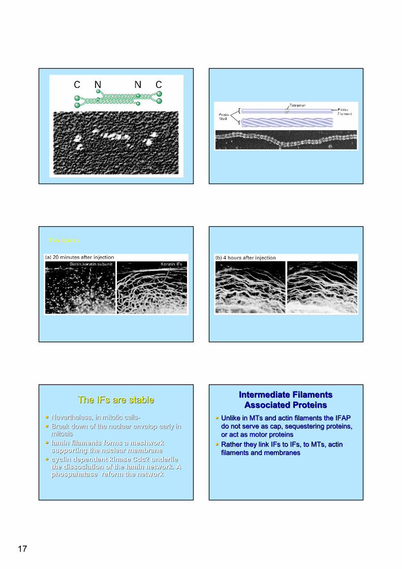
IF are dynamic
The The IFsIFs are stableare stable
Nevertheless, in mitotic cellsNevertheless, in mitotic cells--Break down of the nuclear envelop early in Break down of the nuclear envelop early in mitosis mitosis laminlamin filaments forms a meshwork filaments forms a meshwork supporting the nuclear membranesupporting the nuclear membranecyclin dependent kinase Cdc2 underlie cyclin dependent kinase Cdc2 underlie the dissociation of the the dissociation of the laminlamin network. A network. A phospahatasephospahatase reform the networkreform the network
Intermediate Filaments Intermediate Filaments Associated ProteinsAssociated Proteins
Unlike in MTs and actin filaments the IFAP Unlike in MTs and actin filaments the IFAP do not serve as cap, sequestering proteins, do not serve as cap, sequestering proteins, or act as motor proteinsor act as motor proteinsRather they link Rather they link IFsIFs to to IFsIFs, to MTs, actin , to MTs, actin filaments and membranesfilaments and membranes
18
Gold label of plectin cross linking between MTs and IFs Desmin filaments in muscle cell
19
Myosin heads walk along actin filaments in the presence of ATP