Embed Size (px)
Citation preview

CIBA TM Series 2021 No. 23
TRAINING MANUALOCTOBER 2021
HANDS-ON TRAINING ON DIAGNOSTICS AND MANAGEMENT OF Enterocytozoon hepatopenaei (EHP) IN SHRIMP
Aquatic Animal Health and Environment DivisionICAR-Central Institute of Brackishwater Aquaculture
75, Santhome High Road, MRC Nagar, Chennai 600 028(ISO 9001:2015 certified)
civer.indd 1 10/23/2021 9:32:18 AM


CIBA TM Series 2021 No. 23
Hands-on Training on Diagnostics and Management of Enterocytozoon
hepatopenaei (EHP) in shrimp
Publisher & Course Director
Dr. K.P. Jithendran Director, ICAR – CIBA
Chennai
Organising secretaries
M. Poornima, P.K. Patil, S.K. Otta, P. Ezhil Praveena, R. Ananda Raja, Sujeet Kumar,
T. Bhuvaneswari, Vidya Rajendran and T. Sathish Kumar
Prepared and edited by K.P. Jithendran, M. Poornima, P.K. Patil, S.K. Otta, P. Ezhil Praveena, R. Ananda Raja,
Sujeet Kumar, T. Bhuvaneswari, Vidya Rajendran and T. Sathish Kumar
Cite as: Jithendran K.P., Poornima M., Patil P.K., Otta S.K., Ezhil Praveena P et al.,
Hands on Training on Diagnostics and Management of Enterocytozoon hepatopenaei
(EHP) in shrimp, Training Manual Series, No-23, 2021, 102 pp.
DISCLAIMER
All rights reserved. No part of this publication may be reproduced, stored in a retrieval system or transmitted in any form or by any
means, electronic, mechanical, recording or otherwise, without the prior written permission of Director, CIBA.
The author and publisher are providing this book and its contents on an “as is” basis and make no representations or warranties of any
kind with respect to this book or its contents. In addition, the author and publisher do not represent or warrant that the information
accessible via this book is accurate, complete or current.
Except as specifically stated in this book, neither the author or publisher, nor any authors, contributors, or other representatives will be
liable for damages arising out of or in connection with the use of this book.

Sl.No Content Page No
Title
1 Overview of Hepatopancreatic microsporidiosis (HPM) in shrimp
K.P. Jithendran
1 - 2
2 Biology of Enterocytozoon hepatopenaei (EHP)
K.P. Jithendran, T. Sathish Kumar and M. Poornima
3 - 8
3 Enterocytozoon hepatopenaei (EHP): Clinical manifestation,
pathology, epidemiology and transmission
M. Poornima, P. Ezhil Praveena and K.P. Jithendran
9 - 15
4 Economic burden of Enterocytozoon hepatopenaei (EHP) to Indian
shrimp farming
P.K. Patil
16 - 24
5 Co-infection of Enterocytozoon hepatopenaei (EHP) with other
pathogens
S.K .Otta
25 - 29
6 Role of carriers, intermediate hosts and vectors in transmitting
hepatic microsporidiosis
P. Ezhil Praveena
30 - 32
7 Role of gut microbiome in white feces syndrome (WFS)
Sujeet Kumar
33 - 35
8 Microscopy including wet smear, special staining, SEM and TEM
P. Ezhil Praveena, T. Sathish Kumar and K.P. Jithendran
36 - 42
9 Polymerase Chain Reaction (PCR) for early diagnosis of
Enterocytozoon hepatopenaei (EHP)
R. Ananda Raja, T. Bhuvaneswari and J. J. S. Rajan
43 - 50
10 Quantitative detection of Enterocytozoon hepatopenaei using
Real Time Polymerase Chain Reaction (qPCR)
T. Bhuvaneswari, J.J.S. Rajan and M. Poornima
51 - 56
11 Loop-mediated isothermal amplification (LAMP) detection of
Enterocytozoon hepatopenaei (EHP)
T. Sathish Kumar
57 - 60
12 Management of Enterocytozoon hepatopenaei (EHP) in shrimp
hatcheries and farms
Vidya Rajendran, T. Sathish Kumar and K.P. Jithendran
61 - 66
13 Disinfectants and therapeutics in the treatment and control of
Enterocytozoon hepatopenaei (EHP)
T. Sathish Kumar
67 - 70
Practical
14 Collection and preservation of shrimp samples for diagnosis of
Enterocytozoon hepatopenaei (EHP)
Sujeet Kumar and P. Ezhil Praveena
71 -73
15 Detection of Enterocytozoon hepatopenaei (EHP) in shrimp pond soil
T. Sathish Kumar
74 - 76
16 PCR protocol for detection of Enterocytozoon hepatopenaei infection
in shrimp
T. Bhuvaneswari, R. Ananda Raja, J.J.S. Rajan and M. Poornima
77 - 80
17 TaqMan probe based qPCR assay protocol for diagnosis of
Enterocytozoon hepatopenaei (EHP)
J.J.S. Rajan, T. Bhuvaneswari and M. Poornima
81 - 83
18 Microscopic diagnosis of Enterocytozoon hepatopenaei (EHP)-
Histopathology
P. Ezhil Praveena, T. Sathish Kumar and N. Jagan Mohan Raj
84 - 86
19 Loop-mediated isothermal amplification (LAMP) protocol for
detection of Enterocytozoon hepatopenaei (EHP)
T. Sathish Kumar
87 - 89
20 Trouble shooting in PCR and qPCR
M. Poornima, S.K. Otta, T. Bhuvaneswari and J.J.S. Rajan
90 - 93
21 Artificial germination of EHP spore
T. Sathish Kumar
94 - 94
22 General laboratory biosafety 95 - 102

1
Hands-on Training on Diagnostics and management of Enterocytozoon hepatopenaei (EHP) in shrimp, 25-30 October, 2021
Overview of Hepatopancreatic microsporidiosis (HPM) in shrimp
K. P. Jithendran
Shrimp farming has emerged as one of the most important sectors in commercial aqua
farming and diseases are the major threats faced by the industry. Hepatopancreatic
microsporidiasis (HPM) in cultivated shrimp is an important emerging disease caused by
microsporidian Enterocytozoon hepatopenaei (EHP). Since the target organ of EHP is the
hepatopancreas, the disease was termed as hepatopancreatic microsporidiasis. E.
hepatopenaei was first reported as an unnamed microsporidian in growth retarded black tiger
shrimp, P. monodon from Thailand in 2004. Later this parasite was characterized in detail
and taxonomy was elucidated in 2009. The spore ultra structure of this parasite also
resembled another uncharacterized microsporidium previously reported in P. monodon from
Malaysia in 1989 and from the post larvae (PL) of diseased P. japonicus examined in
Australia in 2001. These early studies indicated potential for infection with EHP-like
microsporidians in several penaeid shrimp species, leading to the proposal that EHP-like
microsporidians were endemic within the Australia, Asia Pacific region prior to the
expansion of shrimp aquaculture. Subsequently, its spread was recorded in many shrimp
farming nations and now EHP outbreaks in shrimp farms have become common in Asian
countries including south-east Asian countries and Latin America.
HPM outbreaks were reported widely in China, Indonesia, Malaysia, Vietnam,
Thailand, India, Venezuela, Mexico, Brunei, Philippines, and Republic of Korea causing 20-
30% production loss. So far, EHP infection has been reported in three known cultured species
of penaeid shrimp i.e. P. monodon, P. vannamei, P. stylirostris and one suspected species (P.
japonicus). Though the susceptibility of different life cycle stages are not clear, post larvae
(PL-7 onwards), juveniles, sub adults and brood stock are found to be affected by the
parasite.
HPM is one of the major threats for the shrimp farming industry due to associated
growth retardation and significant losses in several shrimp farming countries. In India, the
disease emergence has been recorded in P. vannamei since 2014 by CIBA and RGCA as a
part of National Surveillance Programme on Aquatic Animal Diseases (NSPAAD), mainly in
Andhra Pradesh and Tamil Nadu. Since then, EHP epizootics and spread was severe in the
east coast of India. Recently, the disease has been recorded in the west coast of India and
inland saline areas in Haryana Punjab and Rajasthan. It is also unlikely that EHP has been a
2
Hands-on Training on Diagnostics and management of Enterocytozoon hepatopenaei (EHP) in shrimp, 25-30 October, 2021
pre-existing disease in Indian shrimp aquaculture, as national level data never indicated its
presence in other dominant species P. monodon. Initially EHP has not been taken seriously
due to low prevalence, and no substantial loss due to mortality as compared to WSSV.
However recently, the epizootics of EHP in India have been reported to be very high. The
EHP infection initially detected in the hatchery reported to be increased to 96.6% after
transferring shrimp to the pond. It appears that the disease entered India in the recent past
through infected brood stock and the geographical spread was attributed mainly through
transport of infected seeds to other parts of the country.
The emergence and spread of EHP in India will have a significant impact on the
shrimp production. In India, the probability of occurrence of this disease during the period
2018-19 in 7259 ha of shrimp farming area in 23 coastal districts was reported as 17%. The
production loss due to the disease during the period 2018-19 was also calculated as 0.77
million tonnes with a revenue loss of US$ 567.62 million.
Bibliography
Jithendran, K.P., Vengatesan, J., Praveena, E. and Bhuvaneswari, T., 2019. Epidemiology of
Hepatopancreatic Microsporidiosis caused by Enterocytozoon hepatopenaei in
India.https://www.researchgate.net/publication/336373768.
Kim, J.H., Lee, C., Jeon, H.J., Kim, B.K., Lee, N.K., Choi, S.K. and Han, J.E., 2021. First
report on Enterocytozoon hepatopenaei (EHP) infection in Pacific white shrimp
(Penaeusvannamei) cultured in Korea. Aquaculture, 737525.
Hudson, D.A., Hudson, N.B. and Pyecroft, S.B., 2001. Mortalities of Penaeus japonicus
prawns associated with microsporidian infection. Australian veterinary journal, 79,
504-505.
Rajendran, K.V., Shivam, S., Praveena, P.E., Rajan, J.J.S., Kumar, T.S., Avunje, S.,
Jagadeesan, V., Babu, S.P., Pande, A., Krishnan, A.N. and Alavandi, S.V., 2016.
Emergence of Enterocytozoon hepatopenaei (EHP) in farmed Penaeus (Litopenaeus)
vannamei in India. Aquaculture, 454, 272-280.
Thitamadee, S., Prachumwat, A., Srisala, J., Jaroenlak, P., Salachan, P.V., Sritunyalucksana,
K., Flegel, T.W. and Itsathitphaisarn, O., 2016. Review of current disease threats for
cultivated penaeid shrimp in Asia. Aquaculture, 452, 69–87.

3
Hands-on Training on Diagnostics and management of Enterocytozoon hepatopenaei (EHP) in shrimp, 25-30 October, 2021
Biology of Enterocytozoon hepatopenaei (EHP)
K. P. Jithendran, T. Sathish Kumar and M. Poornima
Microsporidians are obligate, unicellular, spore-forming eukaryotic organisms
belonging to the phylum Microsporidia. These organisms are ubiquitous, widely distributed
in nature, with more than 1200 identified species. Several of them are known to infect most
of the vertebrates, invertebrates, and some genera of protists. Almost half of the identified
microsporidian parasites are reported from aquatic hosts and some described as pathogens of
penaeid shrimp and finfish. A unique feature of the microsporidian is the spore‟s polar tubule
which pierces the host cell and through which the infective sporoplasm is transmitted. The
microsporidian infection in aquaculture species causes reduced growth rate, increased
morbidity and decreased productivity in culture farms.
Taxonomy of Microsporidia
Initially, these groups of organisms were considered to be Microspora and
Microsporidia. At present, they are classified under the phylum Microsporidia. Earlier the
phylum microsporidia were regarded as protozoans and ranked under the kingdom Protista
based on the conventional identification of the spore‟s morphological features, life cycle, and
interaction with the host. With advanced genetic studies (ribosomal RNA sequencing) carried
out recently have revealed that the phylum Microsporidia is related to the Kingdom Fungi. In
the phylum microsporidia, a total of 1200 species were reported under 187 genera. Many
microsporidians, except few, are host-specific and thus indicating their host specificity. Fifty
genera are pathogenic to crustacean hosts such as Malacostraca, Maxillipoda, Ostracoda, and
Branchiopoda. In Malacostraca, 20 genera are known to infect crab, shrimp, lobster,
amphipod, and isopod hosts. It is evident that Clade VI of the Terresporidia has hosts in
different trophic strata, especially in crustaceans and vertebrates. The family
Enterocytozoonidae was positioned under the Clade IV of Terresporidia.
Pathogens belonging to the family Enterocytozoonidae infect hosts inhabiting marine
and freshwater aquatic environments at different trophic levels including hyperparasitic
copepods, decapods, fish, and mammals. The marine shrimp microsporidian is named
Enterocytozoon hepatopenaei based on its distinctive ultrastructural characters and it was
placed under the family Enterocytozoonidae. E. hepatopenaei and E. beiniusi develop inside
the host cell cytoplasm, whereas the development of other species of the genera Enterospora
and Nucleospora is intranuclear. E. hepatopenaei shares a number of identical traits with E.

4
Hands-on Training on Diagnostics and management of Enterocytozoon hepatopenaei (EHP) in shrimp, 25-30 October, 2021
bieneusi such as localization of sporangial plasmodium, budding of sporoblasts, and
morphological features such as posterior vacuole, and two rows of polar filaments in 5-7
coils. Considering the spore size, marine habitat, and 16% difference in the 18 SSU (small
subunit) rRNA gene sequence, a new name E. hepatopenaei within the family
Enterocytozoonidae was accepted for this shrimp microsporidian
Phylogenetic classification of Enterocytozoon hepatopenaei
Phylum: Microspora (Sprague, 1977)
Class: Microsporea (Delphy, 1963)
Order: Microsporida (Balbiani, 1882)
Family: Enterocytozoonidae (Cali and Owen, 1990)
Genus: Enterocytozoon (Desportes et al., 1985)
Species: hepatopenaei (Tourtip et al. 2009)
Biology and Life Cycle of Microsporidians
The spore is the only infective stage of a microsporidian which lives outside the host.
The spore size ranges from 1-20 µm, but mostly in the range of 1-5 µm. The spores of EHP
are monokaryotic, oval-shaped, and measure about 1.1 ± 0.2 µm × 0.6 ± 0.2 µm. Mature EHP
spores contain a single nucleus, polar filament with 5-6 coils, a posterior vacuole, and an
anchoring disk at the anterior end of the polar filament. Spores are protected with a thick cell
wall composed of plasmalemma with an endospore (10 nm) and exospore (2 nm). The
endospore layer of the microsporidian spore is usually composed of complex proteins and α-
chitin layers and can withstand pressure during germination. In comparison, the exospore
layer comprises of proteins and mediates communication with the environmental stimuli. The
spore wall is also composed of spore wall proteins (SWP), which are reported to be involved
in host-parasite interaction. In EHP, the spore wall protein EhSWP1 is characterized by
heparin-binding motifs (HBM) at its N-terminus and a Bin-amphiphysin-Rvs-2 (BAR2)
domain at its c-terminus. EhSWP1 is localized and distributed in both the endospore and
exospore layers. It is also believed that EhSWP1 is a significant protein involved in the host-
parasite interaction by attaching spores to the host cell surface.
Microsporidians have a relatively simple life cycle and contain two major forms -
meronts and spores in three developmental phases such as infective, proliferative and
sporogonic phases. The two life cycle forms are unique to microsporidians with significant
morphological variations among different species. The infectious form of a microsporidian is
5
Hands-on Training on Diagnostics and management of Enterocytozoon hepatopenaei (EHP) in shrimp, 25-30 October, 2021
the resilient spore which can endure the extracellular environment for a long time. Typically,
the life cycle of a microsporidian starts once the spore enters the host cell. The spore fires a
polar tubule which pierces the host cell and through this tubule, the infective sporoplasm
heads straight into the host cytoplasm via spore germination. Often, the host cell endoplasmic
reticulum (ER), nuclei, and mitochondria are found surrounding the pathogen. In the
proliferation phase, the injected sporoplasm develops into meronts through merogony (binary
or multiple-fission) resulting in branched multinucleate plasmodial forms. In sporogony, the
cell membrane of meronts thickens and forms sporonts.
In the genus Enterocytozoon, during sporogony, precursors such as the polar filament
and anchoring disk develop before the cleavage of the sporangial plasmodium in the
cytoplasm. The sporonts develop into sporoblasts and form mature spores. Before the
maturation of sporoblasts, the extrusion apparatus including the polar tube, polaroblast,
posterior vacuole, and other spore organelles develop within the sporangial plasmodium and
are packed into the pre-sporoblast. After maturation, the shrimp hepatopancreas (HP) cell
swells, ruptures, releases mature spores into the aquatic pond surroundings through the
faeces, and transmits it to other shrimps. A mature spore is protected with a thick, chitinous
endospore. This protective layer makes the spore dormant and resistant to environmental
stress and maintains spore viability for a long duration in the aquatic habitat.
Metabolism
Hallmarks of microsporidians are their extreme streamlining of genomes and
organelles. Recent comparative genome analysis of the 3.26- Mbp genome of EHP and those
of their relatives in the family Enterocytozoonidae revealed that, like other microsporidia,
EHP cannot generate ATP by either glycolysis or oxidative phosphorylation. Of the 10
enzymes in the glycolytic pathway, EHP and E.bieneusi (the only other member in the genus
Enterocytozoon) possess only hexokinase and glyceraldehyde 3- phosphate dehydrogenase
genes but lack phosphofructokinase and pyruvate kinase, the two glycolytic enzymes capable
of generating ATP by substrate-level phosphorylation. Enzymes in alternative ATP-
generating pathways were also surveyed and found missing from the genomes of all species
in the family Enterocytozoonidae. Considering the coverage of every Enterocytozoonidae
genome under comparison, the absence of these enzymes reflects genuine loss during
evolution, rather than the failure to retrieve these genes during sequencing. On the other
hand, some ATP-consuming enzymes have been retained. These include a potentially

6
Hands-on Training on Diagnostics and management of Enterocytozoon hepatopenaei (EHP) in shrimp, 25-30 October, 2021
functional hexokinase as well as ATP-costly pyrimidine and purine biosynthesis pathways. It
has been postulated that the simultaneous loss of ATP-generating capacity and conservation
of ATP consuming features may serve to set up an ATP sink in the plasmodium that draws on
host ATP through ATP/ADP transporters abundant in the EHP genome, similar to a scenario
reported in Chlamydiae.
Comparative genomic investigation also revealed that glucose-6- phosphate
dehydrogenase (G6PD) is conserved in the family Enterocytozoonidae. It converts glucose-6-
phosphate produced by hexokinase into 6-phosphoglyconolate and in the process releases the
reducing equivalent, NADPH, that can scavenge harmful reactive oxygen species (ROS). It
has been conjectured that the conservation of G6PD might have been due, in part, to the
protection it might provide endo-parasites against oxidative damage from ROS that hosts
deploy as an immune strategy. In contrast to EHP that possesses only one copy of
hexokinase, other members of the family Enterocytozoonidae possess multiple hexokinase
genes for redundant isozymes that are purportedly secreted into the host cytosol to disturb
host metabolism in favor of parasite development. The prime reason why EHP is such a
threat to the shrimp industry is its potential for shrimp growth retardation. In part, this
probably occurs by generation of an ATP sink, but proteomic and metabolomic reports
suggest that at least two other mechanisms might also contribute to stunted growth (Ning et
al., 2019). One is an increase, after infection, in the level of a juvenile hormone called methyl
farnesoate (MF) due to the upregulation of farnesoic acid O-methyltransferase (FAMet), a
primary enzyme in the juvenile hormone biosynthesis pathway. This is accompanied by
downregulation of Juvenile hormone esterase-like carboxylesterase 1 (JHEC1), the enzyme
that degrades MF. The other possible mechanism is upregulation of ecdysteroid regulated-
like protein (ERP) which results in a decline in ecdysteroid that is necessary for stimulating
ecdysis.
Spore germination and infectivity
Spore germination is induced by different environmental triggers specific to species
and habitat. In vitro germination of spores is achieved by many physical and chemical
inducing factors such as changes in pH, dehydration followed by rehydration, hyperosmotic
settings, presence of anions or cations, and exposure to ultraviolet light or peroxides. Inside
the host cell, the spore initiates an exciting series of subcellular events called spore
germination. Spore germination is an osmotic event triggered by the calcium/calmodulin-

7
Hands-on Training on Diagnostics and management of Enterocytozoon hepatopenaei (EHP) in shrimp, 25-30 October, 2021
binding on the spore surface. It is believed that the next step is the inflow of water into the
spore, possibly with ionophore molecules/aquaporins of the spore membrane.
Spore germination starts with swelling of the polaroplast, and expansion of the posterior
vacuole resulting in swelling of the spore. Swelling of the spore ultimately increases the
osmotic pressure. Increasing pressure inside the spore ruptures the thinner apex of the spore
case and expels the polar tube/injection tube by eversion. Expansion of the posterior vacuole
forces the cytoplasm and nucleus through the tube. The ejected polar tube length may range
from 50–500 µm in length. The whole event of spore germination can be completed within a
few seconds. The ejected polar tube/injection tube can penetrate any obstacle, including other
microsporidian spores.
Spore germination is a rapid strategy carried out by spores to infect the host cell. A
unique feature of the microsporidia spore is the possession of an infiltrating organelle polar
tube/ polar tube filament for invading host cells. The development of microsporidia in host
cells is generally intra-cytoplasmic, although some genera are reported to develop inside the
host nucleoplasm. A microsporidian spore is a single cell compact pressure vessel like
structure which fires the sporoplasm into the host cell. The piercing apparatus of spores
includes three parts - polar tubule/injection tubule, polaroblast, and a posterior vacuole. The
polaroblast acts like an accordion-like membrane for storage, whereas the posterior vacuole
acts as a pressure-building organelle to initiate the firing.
Conclusion
Microspordia are obligate intracellular pathogens and spores are the infective material
of microsporidians for transmitting disease. In shrimp aquaculture, recently the emergence of
E. hepatopenaei (EHP) has caused major havoc in shrimp production. Studies on the biology,
life cycle, and transmission of the emerging microsporidian, Enterocytozoon hepatopenaei,
are limited. The pathogen-hostrelationship in microsporidians has illustrated several
instances of parasite-host coevolution.
Bibliography
Cali, A. and Takvorian, P.M. 2014. Developmental morphology and life cycles of the
microsporidia In: Weiss LM &Becnel JJ (ed.), Microsporidia-Pathogens of
Opportunity, Vol. 2 Wiley Blackwell Press, Hoboken, NJ p. 71–133.
8
Hands-on Training on Diagnostics and management of Enterocytozoon hepatopenaei (EHP) in shrimp, 25-30 October, 2021
Cali A., Becnel J.J. andTakvorian P.M., 2016. Microsporidia. In: Archibald J. et al. (Eds)
Handbook of the Protists. Springer, Cham. https://doi.org/10.1007/978-3-319-32669-6-
27-1
Chaijarasphong, T., Munkongwongsiri, N., Stentiford, G.D., Aldama-Cano, D.J., Thansa, K.,
Flegel, T.W., Sritunyalucksana, K. and Itsathitphaisarn, O., 2020. The shrimp
microsporidian Enterocytozoon hepatopenaei (EHP): Biology, pathology, diagnostics
and control. J. Inverte. Pathol, 107458.
Han, B. and Weiss, L. M. 2017. Microsporidia: Obligate Intracellular Pathogens Within the
Fungal Kingdom. Microbiology Spectrum, 5(2), 5-2.
Jaroenlak, P., Boakye, D. W., Vanichviriyakit, R., Williams, B., Sritunyalucksana, K., and
Itsathitphaisarn, O., 2018. Identification, characterization and heparin binding
capacity of a spore-wall, virulence protein from the shrimp microsporidian,
Enterocytozoon hepatopenaei (EHP). Paras. Vect., 11(1), 177.
https://doi.org/10.1186/s13071-018-2758-z
Stentiford, G.D., Feist, S.W., Stone, D.M., Bateman, K.S. and Dunn, A.M., 2013.
Microsporidia: diverse, dynamic, and emergent pathogens in aquatic systems. Trends in
Parasitology, 29, 567-578.
Texier, C., Vidau, C., Viguès, B., El Alaoui, H., Delbac, F., 2010. Microsporidia: a model
for minimal parasite–host interactions. Curr. Opinion. Microbiol., 13, 443-449.
Vavra, J. and Lukes, J. 2013. Microsporidia and “the art of living together”. Adv. Parasitol.
https://doi.org/10.1016/B978-0-12-407706-5.00004-6.
9
Hands-on Training on Diagnostics and management of Enterocytozoon hepatopenaei (EHP) in shrimp, 25-30 October, 2021
Enterocytozoon hepatopenaei (EHP): Clinical manifestation, pathology,
epidemiology and transmission
M. Poornima, P. Ezhil Praveena and K. P. Jithendran
Diseases usually are indicated by gross signs. A clinical sign is defined as an
objective manifestation of disease and observable phenomenon that can be identified. Each
disease entity has a constellation of signs more or less uniquely its own. Externally visible
clinical signs are often used for presumptive diagnosis of suspected infections in shrimp.
However in the case of EHP, there are no known distinctive visual clinical signs in shrimps
but known to cause severe unusual growth retardation over time which leads to an increased
variability in size. Size variation may not be apparent during early days of culture (DOC).
EHP is confined to the shrimp hepatopancreas, infects the hepatopancreatic tubules limiting
the assimilation of nutrients and results in growth retardation. In a more advanced stage,
EHP-infected shrimp typically display soft shells, lethargy, loss of appetite, reduced feed
intake, empty midguts and whitish hindgut. EHP infection in pond conditions may happen
continuously leading to progressive damage to the hepatopancreas with different degree of
infection. Hence the signs of EHP appear as size variation, overall slow growth and loss
during harvest. In hatcheries, EHP can be suspected if post larvae grow unusually slower and
show size variation. In addition, the coefficient for correlation between severity of EHP
infection and size is low. The coefficient of variation in shrimp size or weight is the best
indicator for its impact on production is in a culture pond. In a normal pond this would be in
the range of 10% or less but in a severely infected EHP pond it would be in the range of 30%
or more such that the harvest would be unprofitable. In shrimp farms, EHP does not cause
mass mortality. Severe infection by EHP in shrimp farms can increase the susceptibility to
other bacterial infections (Vibrio spp.) and result in low level daily mortality. The feed
conversion efficiency was drastically affected resulting in severe economic loss to the
farmers.
EHP is often associated with White Feces Syndrome (WFS). Farmers report EHP
infected shrimp exhibiting white faeces syndrome as early as 23 DOC, often with a phase of
recovery. A 100% prevalence of EHP in WFS affected shrimp farms has been recorded in the
states of Tamil Nadu and Andhra Pradesh in India. White faecal strands floating on surface of
an affected pond is one of the clinical signs of EHP infection. However, white faeces
syndrome is not a consistent feature as compared to slow growth and size variation and both

10
Hands-on Training on Diagnostics and management of Enterocytozoon hepatopenaei (EHP) in shrimp, 25-30 October, 2021
conditions are not always concurrent. For instance, a pond can still test positive with EHP
even after shrimp has stopped producing white faecal strings, suggesting that the association
between EHP and WFS is indirect. Conversely, there are EHP-free ponds that exhibit white,
floating mats of faecal strings containing vermiform structures called aggregated transformed
microvilli (ATM). Furthermore, it is known that shrimp may produce white faeces in
response to other causes such as heavy gregarine infections, vibriosis and hemocytic enteritis.
Since the relationship between EHP and WFS appears to be conditional and suggested that
WFS could be an indicator of EHP in shrimps.
Pathology
The impact of Microsporidian infection on hosts has been reported in several cases in
wild, culture and laboratory experiments of aquatic organisms. However, most
microsporidian diseases may result in chronic infection with low mortalities. There are four
contact methods that have been described in the way microsporidians avoid the lethal
response of the host. The methods are Type I: Direct contact e.g., Nosema and
Enterocytozoon, Type II: Indirect contactby parasite-produced isolation e.g., Anncalia,
Pleistophora, Brachiola, and Tubulinosema, Type Indirect contact by host-produced
isolation e.g., Endoreticulatus, and Type IV: Indirect contact by host- and parasite-produced
isolation e.g., Trachipleistophora, Encephalitozoonintestinalis. The shrimp microsporidian
EHP infect the host cell by direct contact method. With regard to EHP, the host interactions
are largely unknown and studies and research on host-parasite interactions are in the early
stages.
As EHP is an intracellular spore-forming parasite, it replicates within the cytoplasmic
area of the tubule epithelial cells in the hepatopancreas. When EHP enters the ponds it is
highly difficult to eradicate. Spores of EHP in fecal pellets or dried cadavers have shown to
be viable up to six months and retain infectivity for over a year under aqueous conditions.
The spores released in the environment are activated when external factors are suitable, and
infect host cells. Once the spores enter the gut of the shrimp they start infecting the hepatic
cells via extending a highly specialized polar tube. As the infection progresses there is an
increase in the number of spores since they replicate within the host cells. The new spores
thus formed then infect other healthy cells. EHP infection in pond conditions may happen
continuously leading to progressive damage to the hepatopancreas with different degree of
infection. Infections with the massive, production and release of spores by cell lysis, together

11
Hands-on Training on Diagnostics and management of Enterocytozoon hepatopenaei (EHP) in shrimp, 25-30 October, 2021
with sloughing of whole cells containing spores results in a loss of integrity of the HP tubule
epithelium and may be accompanied by some shrimp mortality. The spores, sloughed cells
and debris from lysed cells accumulate in the midgut making it the white fecal strings. In
addition, the cells that do lyse to release spores normally in a dispersed manner over time
allows for cell renewal and leaves the HP structure more-or-less intact. This allows for long
term infections that result in no external signs of disease but may cause retarded growth.
Epidemiology
An EHP infection can occur at a wide range of salinity (2 - 30 ppt). The prevalence
and the severity of the EHP infections were reported to be higher at 30 ppt than at 2 ppt and
15 ppt in Venezuela. It has been reported that calcium is an important second messenger that
activates many cell events and calcium influx might be, in part, responsible for the activation
of microsporidian spore discharge at higher salinities. In India there are some shrimp farming
areas in high and low salinity, and the prevalence of EHP seems to be lower at lower
salinities (below 5 ppt), as was observed during a shrimp disease survey in Andhra Pradesh in
2019. The difference in the severity of the EHP infection at the three different salinities was
probably due to the differential effect of salinity on spore germination. One of the critical
phases in the spore germination is the increase of intra-spore osmotic pressure. The difference
in salinities led to a hypotonic environment at 2 ppt and 15 ppt compared to hypertonic
environment at 30 ppt. It is possible that the hypertonic solution enhances the germination of
the spore by increasing the spore activation process.
Ammonia and nitrite concentration could influence the prevalence of EHP infection to
the shrimps in the super-intensive farms. A significant correlation between ammonia and
nitrite with the prevalence of EHP infections was reported. There was a high prevalence of
EHP infection with the increase in nitrite concentration and ammonia in shrimp ponds. The
concentration of a range 1 mg/l to 1.2 mg/l of ammonia and nitrite could influence EHP
infection prevalence in the shrimp farms. The accumulation of ammonia and nitrite might
impact shrimp production by reducing survival, growth performance, and damage of
hepatopancreas of P. vannamei, which provides the chance for the opportunistic pathogen
like EHP to infect the shrimp.
Transmission
Microsporidians are reported to be transmitted by both horizontal and vertical modes
of transmission. In general, the mode of transmission determines the virulence of a

12
Hands-on Training on Diagnostics and management of Enterocytozoon hepatopenaei (EHP) in shrimp, 25-30 October, 2021
microsporidian pathogen. Horizontal transmission is highly pathogenic as a large number of
spores are released into the water. Shrimp microsporidian EHP is transmitted horizontally by
ingestion of spores released from feces of infected shrimp, cannibalism and cohabitation. In
cohabitation, healthy shrimps ingest (fecal-oral route) spores released into the water by
infected shrimps. Transmission has been demonstrated in laboratory co-habitation
experiments using infected shrimp separated by mesh cages from naïve shrimp. In
cannibalism, healthy shrimps are infected by feeding (oral route) the infected moribund or
dead shrimps. Rapid and intense EHP infection established experimentally by HP injection
and reverse gauge of EHP inoculum in naïve shrimps and in shrimp post larvae (PL) by
adding the tissue homogenate infected with EHP. The oral challenges proved that EHP
spores alone are infectious via ingestion. Since reverse gavage is accepted as an unhindered
route that allows direct exposure of the shrimp HP to bacteria and toxins, it proved that
spores produced in the HP (similar to bacteria in size) could spread EHP internally to
uninfected HP tubule epithelial cells (autoinfection).
The possible ways of E. hepatopenaei vertical transmission route (trans-ovum) is
poorly understood. A study reported positive PCR test results for EHP in nauplii, zoeae 1 and
zoea 2 stages from EHP-infected female broodstock. But the study lacked clear histological
examination. Further in situ hybridization (ISH) test results with EHP-infected juvenile
shrimp and broodstock have shown positive signals for EHP in the HP tissue but none in the
ovaries or testes of the same specimens, making trans-gonadal transmission unlikely. At this
stage it is not possible to rule out potential for EHP transmission via vertical routes as
comprehensive in depth research work with larger sample numbers and all life stages are to
be studied. But there is possibility of vertical transmission of EHP from the infected female
brooders can still pass infections horizontally to their offspring in hatcheries via release of
feces containing spores into spawning tanks. Once some of the larvae become infected, they
will rapidly transmit the infection horizontally via spores in their feces or via cannibalism.
EHP can be transmitted orally by ingestion of infected live feeds such as brine shrimp
Artemia salina, polychaetes, and molluscs.
Recently, severe EHP infections in shrimp farms have become common, and these
infections are resulting in substantial economic losses to producers. The white feces are
composed, almost completely, of massive quantities of EHP spores, gut mucus, remnants of
sloughed tissues from hepatopancreas tubules infected with EHP. Massive numbers of EHP

13
Hands-on Training on Diagnostics and management of Enterocytozoon hepatopenaei (EHP) in shrimp, 25-30 October, 2021
spores in the „„white feces” of WFS-affected ponds, suggest that EHP is associated with
WFS. It is possible, that EHP rapidly multiplies in hepatopancreatic tubule epithelium cells,
then the degenerated epithelial cells and the large numbers of EHP spores release into the
feces. The white feces containing large quantities of EHP can break down and sink to the
pond bottom. The associated EHP can be ingested by shrimp, results in re-infection, and
ultimately will increase the severity of the infection.
Conclusion
The microsporidian parasite E. hepatopenaei, (EHP) causes hepatopancreatic
microsporidiosis (HPM) disease in shrimp. The disease does not cause mass mortality but
affects shrimp production due to growth retardation, and causes severe economic loss in
shrimp farming. Shrimp hepatopancreas is the target organ for EHP infection. There are no
specifically distinctive reported gross signs for this microsporidian infection in shrimp.
However, in a pond, shrimp with growth retardation, white gut, and pond with floating white
fecal threads can be an indicator for EHP infection. EHP infection can be transmitted
horizontally through the cohabitation and oral routes, and possibly by vertical transmission.
Bibliography
Caro, L.A., Alghamdi, F., De Belder, K., Lin, J., Mai, H.N., Millabas, J., Alrehaili, Y.,
Alazwari, A., Algetham, S. and Dhar, A.K., 2021. The effect of salinity on
Enterocytozoonhepatopenaei infection in Penaeusvannamei under experimental
conditions. BMC Veterinary Research, 17, 1-8.
Chaijarasphong, T., Munkongwongsiri, N., Stentiford, G.D., Aldama-Cano, D.J., Thansa, K.,
Flegel, T.W., Sritunyalucksana, K. and Itsathitphaisarn, O., 2020. The shrimp
microsporidian Enterocytozoon hepatopenaei (EHP): Biology, pathology, diagnostics
and control. Journal of Invertebrate Pathology, 107458.
Han, S., Wang, B., Wang, M., Liu, Q., Zhao, W. and Wang, L., 2017. Effects of ammonia
and nitrite accumulation on the survival and growth performance of white shrimp
Litopenaeus vannamei. Invertebrate Survival Journal, 14, 221-232.
Jithendran, K.P., Vengatesan, J., Praveena, E. and Bhuvaneswari, T., 2019. Epidemiology of
Hepatopancreatic Microsporidiosis caused by Enterocytozoonhepatopenaei in
India.https://www.researchgate.net/publication/336373768.

14
Hands-on Training on Diagnostics and management of Enterocytozoon hepatopenaei (EHP) in shrimp, 25-30 October, 2021
Karthikeyan, K. and Sudhakaran, R., 2019. Experimental horizontal transmission of
Enterocytozoonhepatopenaei in post‐larvae of whiteleg shrimp,
Litopenaeusvannamei. Journal of Fish Diseases, 42, 397-404.
Khac, H.V., Thanh, T.N.T., Thu, G.N.T., Le, C.H. and Nguyen, V.D., 2018. Vertical
transmission and early diagnosis of the microsporidian Enterocytozoon hepatopenaei in
whiteleg shrimp Penaeus vannamei. Journal of Pure and Applied
Microbiology,12.https://doi.org/10.22207/JPAM.12.3.11.
Krishnan, A.N., Kannappan, S., Aneesh, P.T., Praveena, P.E. and Jithendran, K.P., 2021.
Polychaete worm-A passive carrier for Enterocytozoon hepatopenaei in
shrimp. Aquaculture, 545, 737187.
Mai, H.N., Cruz-Flores, R., Caro, L.F.A., White, B.N. and Dhar, A.K., 2020. A comparative
study of Enterocytozoon hepatopenaei (EHP) challenge methods in
Penaeusvannamei. Journal of invertebrate pathology, 171, 107336.
Munkongwongsiri, N., Thepmanee, O., Lertsiri, K., Vanichviriyakit, R., Itsathitphaisarn, O.
and Sritunyalucksana, K., 2021. The false mussels (Mytilopsisleucophaeata) can be
mechanical carriers of the shrimp microsporidian Enterocytozoon hepatopenaei
(EHP).bioRxiv Preprint. https://doi.org/10.1101/2021.04.23.441221.
Nkuba, A.C., Mahasri, G., Lastuti, N.D.R. and Mwendolwa, A.A., 2021. Correlation of nitrite
and Ammonia with prevalence of Enterocytozoon hepatopenaei (EHP) in shrimp
(Litopenaeus vannamei) on several super-intensive ponds in East Java,
Indonesia. JurnalIlmiahPerikanandanKelautan, 13(1),58-67.
Otta, S.K., Patil, P.K., Jithendran, K.P., Rajendran, K.V., Alavandi, S.V. and Vijayan, K.K.,
2016. Managing Enterocytozoon hepatopenaei (EHP), microsporidial infections in
vannamei shrimp farming: An Advisory, CIBA e - publication No.29.
Salachan, P.V., Jaroenlak, P., Thitamadee, S., Itsathitphaisarn, O. and Sritunyalucksana, K.,
2016. Laboratory cohabitation challenge model for shrimp hepatopancreatic
microsporidiosis (HPM) caused by Enterocytozoon hepatopenaei (EHP). BMC
Veterinary Research, 13,1-7.
Stentiford, G.D., Becnel, J.J., Weiss, L.M., Keeling, P.J., Didier, E.S., Bjornson, S., Freeman,
M.A., Brown, M.J.F., Roesel, K., Sokolova, Y. and Snowden, K.F., 2016.
Microsporidia – emergent pathogens in the global food chain. Trends
inParasitology, 32(4), 336-348.

15
Hands-on Training on Diagnostics and management of Enterocytozoon hepatopenaei (EHP) in shrimp, 25-30 October, 2021
Tang, K.F., Han, J.E., Aranguren, L.F., White-Noble, B., Schmidt, M.M., Piamsomboon, P.,
Risdiana, E. and Hanggono, B., 2016. Dense populations of the microsporidian
Enterocytozoon hepatopenaei (EHP) in feces of Penaeusvannamei exhibiting white
feces syndrome and pathways of their transmission to healthy shrimp. Journal of
Invertebrate Pathology, 140, 1-7.
16
Hands-on Training on Diagnostics and management of Enterocytozoon hepatopenaei (EHP) in shrimp, 25-30 October, 2021
Economic burden of Enterocytozoon hepatopenaei (EHP) to Indian shrimp
farming
P. K. Patil
Global farmed shrimp trade worth US$ 17.7 B is dominated by India (24.9%) which
is the leading supplier of shrimp to the US (US$ 2.3 B) and the second-largest supplier to the
EU ($0.9 B) during 2018. Shrimp farming contributes to economic growth and provides
significant employment opportunities in developing countries. Shrimp farming is an
important economic activity earning a foreign exchange US$ 4.89 B in 2019-20 generating
14 million jobs in the country. Being the second-largest aquaculture producing country, India
produces 17.5% of the global farmed shrimp. Following the introduction of specific
pathogen-free (SPF) Pacific white shrimp, Penaeus vannamei in 2009, the establishment of
Aquaculture Quarantine Facility (AQF) practices and screening of imported broodstock for
OIE listed diseases have led to substantial improvement in shrimp health and production.
Scientific interventions for economic sustainability of the sector through the adoption of
biosecurity measures and better management practices were the corner stones of the sector.
The microsporidian, Enterocytozoon hepatopenaei (EHP) has become a significant
threat to shrimp aquaculture in recent years and has been widely reported from major shrimp
producing countries such as China, Thailand, Indonesia, Malaysia, Vietnam, India,
Bangladesh and Venezuela affecting the economic sustainability, production and supply of
shrimp in the global market. Since 2016, the disease is being reported from different shrimp
farming regions of India. The affected farms suffer significant growth retardation and low-
level mortality during the culture period. In the absence of apparent disease symptoms
farmers continue feed and apply other inputs, incurring substantial expenditure.
Following the introduction of P. vannamei to the Indian farming and the subsequent
practice of screening broodstock at the national quarantine and post larvae before stocking
have led to the substantial improvement in shrimp health and production. Additionally,
scientific interventions through the development of molecular diagnostic tools, adoption of
biosecurity measures, and better management practices helped in the sector's economic
sustainability. However, these financial gains were short-lived due to mounting losses due to
infectious diseases and stress due to deteriorating environmental conditions following farm
intensification. Economic effects of diseases are more evident to farmers, and their
management has a cost that has to be weighed against profits. A thorough understanding of
17
Hands-on Training on Diagnostics and management of Enterocytozoon hepatopenaei (EHP) in shrimp, 25-30 October, 2021
disease impact on shrimp productivity and comprehensive economic loss is of utmost
importance for formulating various health management efforts. The economic cost of disease
could be directly due to the loss of stock and the expenditures to control/manage the
infection. Indirect cost may include loss of employment in hatcheries, feed mills and
processing exports, etc. and the consequent loss to the exchequer.
To allocate the limited resources to control these infections, it is essential to
understand the risk factors involved and the economic loss quantification. The national drop
in shrimp production in Thailand (1996-97, 2013), China (1993) and in Brazil (2006) has
been attributed to the widespread occurrence of infectious diseases. Several studies have
reported the economic loss worth billions of US dollars and employment losses due to EHP
in different countries at the other period.
Estimating the national level economic cost of Enterocytozoon hepatopenaei (EHP)
Microsporidian infection, Enterocytozoon hepatopenaei (EHP) in addition to other
endemic diseases continue to cause significant economic losses in shrimp farming worldwide
including India. Since its first report in 2009, microsporidian pathogen Enterocytozoon
hepatopenaei (EHP) has emerged as a significant threat in all the shrimp farming countries
worldwide. Being an intracellular microsporidian parasite, EHP multiplies in the cytoplasm
of hepatopancreas and midgut leading to size variation, growth retardation loss of production.
Disease incidences due to microsporidian parasites have been reported worldwide, but there
are few studies on economic loss experienced by the world shrimp industry. As the resources
are scares and challenges are many, there is an urgent need to prioritize the research in
developing and implementing strategies and specific policies to reducing the losses due to
EHP and infectious diseases in general to Indian shrimp farming. A study in Thailand has
speculated economic loss due to EHP as US$ 76.4 M.
We have recently reported an economic loss due to infectious diseases in Indian
shrimp farming. Questioner based survey on the disease's occurrence and the associated loss
in terms of mortality and employment was conducted. Economic loss to India's aquaculture
sector due to infectious disease was estimated with particular reference to EHP based on the
difference between expected and actual production. Factors like mortality, FCR, stocking
density, culture period, average body weight and the survival rate were found to influence the
economic loss due to EHP. Our study estimated the economic loss due to infectious diseases

18
Hands-on Training on Diagnostics and management of Enterocytozoon hepatopenaei (EHP) in shrimp, 25-30 October, 2021
in shrimp with special reference to EHP and WSSV. The questionnaire-based survey
covering 23 shrimp farming coastal districts of India estimated the probability of EHP
occurrence as 17% with production loss of 1.80±0.24 ton per ha per crop. The total
production loss calculated per crop due to EHP was found to be 77,370 tons worth US$ 380
M. The overall annual economic loss due to infectious diseases to Indian shrimp farming was
estimated to be US$ 1,028.55 M which includes US$ 571.03 M due to EHP.
Fig. 1. Productivity loss due to shrimp diseases in India
Fig. 2. Production and economic loss due to shrimp diseases in India
Generally the studies conducted to estimate the economic loss in shrimp aquaculture
consider culture period of 120 days (average number of days usually needed for producing
market size shrimp) and the productivity obtained at the end of the culture period is used as
0
0.1
0.2
0.3
0.4
0
20000
40000
60000
80000
100000
EHP WSSV RMS Vibrio WSSV+EHP Other diseases*
Eco
no
mic
loss
(U
S $
Bill
ion
)
Pro
du
ctio
n lo
ss (
ton
ne
s)
Production loss Economic loss

19
Hands-on Training on Diagnostics and management of Enterocytozoon hepatopenaei (EHP) in shrimp, 25-30 October, 2021
the benchmark for economic loss estimation. Methodology to estimate PDO, production loss,
economic loss and employment loss are given below.
Estimation of the probability of disease occurrence (PDO)
The probability of disease occurrence (PDO) index is computed using the proportion
of disease occurrence at the farm level. The state-wise PDO index for each of the diseases is
worked out by using the total culture area in the state and the farm area affected by the
particular disease. The production loss due to particular disease at the national level is
calculated using the weighted average of PDO for the state based on the culture area and the
production data for each of the shrimp farming states.
Estimation of economic loss
Economic loss for each of the farms is calculated using the average farm gate price
(Rs.per kg) for different size of shrimp harvest for particular region. Loss of production in
monitory terms for each area is calculated using the PDO factor for each disease, loss of
production (t per ha per crop) and the total shrimp culture area (ha). The economic loss is
quantified by the production difference between healthy crop and diseased crop. The
economic burden of the disease is estimated in terms of loss in production which may be
attributed to poor quality, stunted growth and loss in employment in terms of man-days.
Estimation of employment loss
The loss of man-days in the farms is calculated based on the reduction in duration of
culture (no. days) and number of workers employed. The number of man-days was calculated
assuming one labour is employed per ha of farming area for the entire period of culture. The
number of man-days lost is valuated based on the average daily wages in the concerned
region.
Outbreaks of diseases in the surrounding area were the primary factor determining the
intensity of the disease while the bulk of the financial burden was attributed to expenditure
incurred on account of steps taken to contain the infectious agents from spreading. Further,
cessation of farming activity due to disease outbreaks leading to emergency harvest causes
loss of employment. Models for estimation of economic loss due to aquatic animal diseases
should include an estimate of loss undergone due to premature harvest and sale of small size
animals and the cost incurred to mitigate the disease which adds to the cost of production.

20
Hands-on Training on Diagnostics and management of Enterocytozoon hepatopenaei (EHP) in shrimp, 25-30 October, 2021
These economic losses could be reduced through effective implementation of better
management practices (BMP) which is mandatory for P. vannamei farms in the country.
The data suggested that the regular occurrence of EHP and WSSV led to the severe
reduction in production followed by reduced stocking densities in future crops. Steps taken to
reduce the severity and spread of diseases through the application of disinfectants and other
antimicrobial agents could further add to the cost of production. Distress harvest during the
disease outbreaks leading the discharge of large quantities of pond water containing the
pathogen loads further enhances the chances of spread of pathogen. While EHP spores are
highly resistant to harsh environmental exposures; introduction of the pathogen into the farm
would result in the continuous build-up in the system.
The study revealed EHP with a 17% probability of occurrence accounted for a
production loss of 0.77 M tons, with a corresponding revenue loss of Rs. 3977 crores (US$
567.62 M). The total employment loss due to major shrimp diseases was estimated to be 1.65
M man-days worth US$ 7.07 M. The overall probability of infectious disease occurrence in
the country was at 49% leading to an annual loss of 0.14 M ton shrimp worth US$ 1.02 B.
Economic loss due to shrimp diseases in Indian shrimp farming warrants prioritized
implementation of better management practices (BMP) and biosecurity protocols along with
policy interventions to reduce the direct and indirect losses.
Effective implementation of scientific farming could help reduce the impact of
diseases as estimated economic loss comprised mainly the direct loss due to mortality and
expenditures to control and manage EHP and other infectious diseases. The study of
economic impact assessment of diseases helps decide the proportionate investment in
national aquatic animal health management programs. The study revealed EHP is one of the
major threats for the Indian shrimp farming, leading to substantial economic losses, including
the consequent loss of jobs. In addition to disease surveillance, implementing better
management practices would reduce the loss and minimize financial losses. Further, region-
specific modifications in stocking density, culture period, and targeting size at harvest could
mitigate the losses. Prioritizing the research areas, including disease forecasting and policy
interventions would help the sector's economic sustainability.
Estimating the farm-level economic cost of Enterocytozoon hepatopenaei
Estimation of economic consequences of diseases in shrimp farming gives insight into
the risk factors addressed at local levels. The biological losses expressed in monitory terms at

21
Hands-on Training on Diagnostics and management of Enterocytozoon hepatopenaei (EHP) in shrimp, 25-30 October, 2021
farm level would help in the effective allocation of resources to develop suitable control and
prevention strategies to achieve the economic sustainability of the farming systems. The
direct cost of EHP or any disease affected farms is expenditure incurred on basic costs of
farming, expenditures towards prevention and treatment, the extraordinary cost to manage the
disease, production loss and the reduced unit price for small size harvest. In addition, indirect
costs of the disease include loss of employment, reduction in national production and foreign
exchange earnings. Analysis of farm-level economic losses will help to develop the cost-
effective strategies for managing the disease, assist the farmers to take an informed decision
on reducing the risks and ultimately determine the economic viability of the aquaculture
activity. Hence there is an urgent need to develop a stochastic model to estimate the
economic loss due to EHP and identify the critical risk factors associated at the farm level.
Recently we have developed a stochastic model to estimate the economic loss due to
EHP to Indian P. vannamei shrimp farms and identify the associated key risk factors at the
farm level. Using Monte Carlo simulation model in excel @ Risk was used to estimate the
cost of production and revenue of healthy and affected shrimp farms. The occurrence of the
EHP was found to be positively correlated with the stocking density and the higher FCR. We
estimated the farm level loss due to EHP at ` 61,778 (US$ 813) per ton shrimp production. In
the EHP affected farms, the significant factors negatively influencing the net returns were
expenditure on feed (0.51), seed (0.19) and labour (0.18). There was a significant regional
variation in the economic impact of EHP at the farm-level.
Cost of production
The production cost includes expenditure on pond preparation, seed, feed, labour,
consultation, health management, power and cost of disease and expressed as per ton
production of shrimp. Net returns are calculated as gross returns (biomass harvested*farm
gate price) - the cost of production. Lost profit (LP) for the EHP affected farms is estimated
as the difference between net returns of affected and healthy farms.
Direct cost of EHP
The direct cost in the stochastic model describing the cost of the disease in
commercial shrimp operations, include Biological Loss (BL), Treatment Cost (TC),
Extraordinary Cost (EC) and Prevention Cost (PC). The cost of biological loss is calculated
based on the difference in the biomass harvested in EHP affected and healthy shrimp farms.
The TC includes expenditure on chemicals, nutritional supplements and other healthcare

22
Hands-on Training on Diagnostics and management of Enterocytozoon hepatopenaei (EHP) in shrimp, 25-30 October, 2021
products to manage the disease while the EC includes expenditure to hire labour for treatment
and manage the disease and PC includes pond preparation and consultation expenditure.
We have used stochastic model to quantify the economic loss due to EHP at the farm-
level and variations in the determinant factors among different farming regions of the
country. The farm-level economic loss estimations are necessary to understand the associated
risk factors influencing the economic outcome of the disease, which can be addressed in a
targeted approach. Generally, it is observed that there is an association between the farms
with higher stocking density and loss due to EHP, which might be due to the challenges in
management of stress. The EHP affected farms show significant reduction in the harvest size
with higher FCR.
Our analysis using the model demonstrated that the EHP infected farms incur
considerable loss due to reduced average harvest weight, increased cost of feed in addition to
expenditure on treatment and prevention. The reduction in production is the strongest
stochastic variable in the EHP specific biological loss. In the absence of any insurance plan in
Indian shrimp farming presently, these losses have to be borne by the farmers. Recent
initiatives for covering the Indian shrimp farming under insurance could help partly recover
these losses in future.
The economic loss due to EHP was shown to be correlated with higher FCR.
Reduction in production due to EHP infection was shown to be partially compensated by
harvesting at a particular size and appropriate timing for reducing the cost of feed and for
realizing farm-gate price commensurate with the size of shrimp. In the absence of effective
therapeutic measures, treatment included expenditure on account of feed supplements to
reduce the stress and improve the growth. The increase in extraordinary cost might be due to
additional labour required for pond management during the disease. The EHP spores
accumulate in the pond soil and require the higher application of disinfectants. Hence the
expenditure on pond preparation and professional consultation was included in the cost of
prevention.
The influence of farm gate price on the economic impact suggests the importance of
shrimp size, which fetches a higher unit price. Reduction in the animal's size is the primary
determinant in reducing the profitability in EHP. Loss of production and expenditure on feed
were the significant risk factors responsible for influencing the economic loss, as indicated by
the sensitivity analysis confirming the effect of EHP on growth retardation and continuous
feeding by farmers in the absence of any observed mortality. As expected, loss of production
23
Hands-on Training on Diagnostics and management of Enterocytozoon hepatopenaei (EHP) in shrimp, 25-30 October, 2021
was the major component influencing the direct loss positively while higher farm gate price
was influencing negatively. Lower production leads to reduced supply of the commodity in
the market and price escalation in the endemic regions.
In addition to the cost of disease, farmers lose their net income due to lower sale
prices during disease incidence. In endemic areas, farmers are forced to harvest smaller
shrimp, further reducing the wider market accessibility. A significant difference in the state-
wise average farm gate prices of shrimp was not observed during the study period. The
reduced losses in farms of Gujarat might be due to premium harvest size, as the farm gate
prices per unit of shrimp are directly proportional to the size at harvest. Using the hard data
we have estimated the biological effect of EHP on reduced production, harvest size and their
correlation with the cost of prevention. The estimated loss of profit in EHP affected farms
was `1, 15, 200 per ton (US $ 1,576) of shrimp production.
Cost-benefit analysis of intervention
Using cost-benefit models, the information generated on biological losses could be
expressed in monetary figures. Measures to reduce the economic impact by identifying
components contributing to the loss would benefit the individual farmers and the sector in
general. Combining biological and economic models would help identify the most effective
cost-benefit analysis to address the issue at the national level. The losses due to EHP could be
overcome by practicing lower stocking density, scientific pond preparation and testing of
seeds before stocking. The short crop cycle is one of the critical factors helping in the
sustainability of the sector despite the threat of infectious diseases. Identification and
generating the appropriate information on the specific cost of the disease would help in the
efficient use of resources for economic control of the disease. The effect of measures like
reducing the expenditure on feed by harvesting the appropriate size of shrimp would help
reduce the losses.
Conclusion
Among the infectious diseases EHP has become the major disease in Indian shrimp
farming, causing substantial economic losses and the consequent loss of employment.
Revenue loss by EHP alone is more than two times than that of WSSV contributing more
than half of the national economic loss due to all other diseases combine. In addition to
disease surveillance implementation of better management practices would help in reducing
the loss of production and minimize economic losses. It is important to assess the economic

24
Hands-on Training on Diagnostics and management of Enterocytozoon hepatopenaei (EHP) in shrimp, 25-30 October, 2021
impact of important diseases especially EHP for appropriate investment in disease control
programs. Reduction in economic loss could be achieved through region-specific adjustments
in stocking density and culture period and targeting for appropriate average body weight at
harvest. Prioritizing the research, especially the application of artificial intelligence in disease
forecasting and implementation of required policy interventions are warranted for the
economic viability of the sector.
Bibliography
Patil, P.K., Geetha, R., Ravisankar, T., Avunje, S., Solanki, H.G., Abraham, T.J., Vinoth,
S.P., Jithendran, K.P., Alavandi, S.V. and Vijayan, K.K., 2021. Economic loss due to
diseases in Indian shrimp farming with special reference to Enterocytozoon
hepatopenaei (EHP) and white spot syndrome virus (WSSV). Aquaculture, 533,
736231.
25
Hands-on Training on Diagnostics and management of Enterocytozoon hepatopenaei (EHP) in shrimp, 25-30 October, 2021
Co-infection of Enterocytozoon hepatopenaei (EHP) with other pathogens
S. K. Otta
Co-infection is observed as a natural phenomenon in many of the organisms where
the host gets simultaneously infected with more than one pathogen. It is generally considered
that this event is not inconsequential to the host. Different pathogens can interact with the
host immune system differentially and thus changing the overall pathogenesis. Similarly, in
favoring their own survival and multiplication, certain pathogens can decide the virulence of
co-infecting pathogen. Therefore, this affects the overall treatment strategy and sometimes it
becomes difficult to provide appropriate treatment to the host.
The co-infection event has also been observed in shrimps and reported for many
pathogens. Particularly, simultaneous infections by a multiple number of viral pathogens are
considered to be very common in shrimp.
Enterocytozoon hepatopenaei (EHP), a hepatopancreatic microsporidian, has been an
emerging pathogen affecting cultured shrimps in many of the countries including India. As
this pathogen at present is considered as a major problem for the shrimp farmers, researchers
are putting considerable effort to know more about it. Some of these efforts have resulted to
provide knowledge regarding the co-infection of EHP with many other pathogens. In fact,
much before this pathogen got its present name, it has been affecting the cultured tiger
shrimps (Penaeus monodon) since as early as 2003 and was reported to cause monodon slow
growth syndrome where it was also found to be co-infected with many of the opportunistic
pathogens such as monodon baculovirus (MBV), hepatopancreatic parvo virus (HPV) and
Vibrio sp.
During the recent research developments, EHP has also been reported to be
simultaneously present with a number of other pathogens in cultured Penaeus vannamei. The
present chapter attempts to bring an update on such co-infection status.
Co-infection of EHP with shrimp viruses
Co-infection of EHP with Taura syndrome virus (TSV)
Co-infection of EHP with TSV was reported in one of the vannamei farms in
Venezuela. Presence of EHP in the ponds was confirmed by histopathology, in situ
hybridization and PCR. Sequence similarity of this EHP strain was assessed with south East
Asian strains through the amplification of 18s rRNA, β-tubulin and spore wall protein genes.
26
Hands-on Training on Diagnostics and management of Enterocytozoon hepatopenaei (EHP) in shrimp, 25-30 October, 2021
Interestingly, TSV was also detected from the same shrimps by RT-PCR and histopathology.
Considering the virulence of both the pathogens, continuous monitoring of ponds were
suggested.
Co-infection of EHP with white spot syndrome virus (WSSV)
During the routine surveillance of vannamei farms from Tamil Nadu and Andhra
Pradesh of India, simultaneous infection of shrimps with EHP and WSSV was recorded. In a
kind of first report, only about 2% of shrimps screened were found to be positive for both the
pathogens. During this study, a multiplex PCR assay was developed for simultaneous
detection of both the pathogens. The authors suggested using this multiplex PCR detection
method for quick and specific detection of both these pathogens in shrimp larvae before
stocking in ponds to avoid the spread of these virulent pathogens.
Similarly, a small percentage of shrimps (7.6%) from east coast of India were also
reported to have co-infection with both EHP and WSSV. A study conducted on screening of
pathogens from both vannamei and monodon firms along the east coast of India and reported
EHP to be dominant pathogen in P. vannamei compared to P. monodon, whereas MBV was
the dominant in P. monodon. Though EHP was also recorded in P. monodon, co-infection of
this pathogen with WSSV or MBV was not recorded.
Co-infection of EHP with infectious myonecrosis virus (IMNV)
Co-infection of EHP with IMNV in P. vannamei was recently reported from shrimp
farms of India. During a survey along the east coast of India, a major number of ponds (7 out
of 12) were reported to have co-infection of both IMNV and EHP. Presence of both the
pathogens in the same shrimp was detected both by histopathology and PCR. Interestingly,
shrimps with single infection of EHP didn‟t show any clinical symptoms where as co-
infected shrimps showed clinical symptoms for both the pathogens. Both the pathogens were
also found to have sequence similarity with the respective pathogens from India and other
parts of the country.
Co-infection of EHP with hepatopancreatic parvovirus (HPV)
This report involving simultaneous occurrence of both EHP and HPV in P. vannamei
samples was also from the east coast of India during the recent time. In this study, about 5.2%
of the farms were reported to have co-infection of both these pathogens. While doing electron
microscopy study, typical structures of microsporidian parasite and icosahedral virus like
27
Hands-on Training on Diagnostics and management of Enterocytozoon hepatopenaei (EHP) in shrimp, 25-30 October, 2021
particles were observed. Latter during PCR analysis, HPV was found to be present along with
EHP.
Co-infection of EHP with bacteria
Since EHP infections were often accompanied by opportunistic pathogens,
particularly Vibrios, the risk factor associated with EHP was verified through an
experimental challenge with the acute hepatopancreatic necrosis disease (AHPND) causing
Vibrio parahaemolyticus (AHPND-VP) and through a case study involving bacteria
associated septic hepatopancreatic necrosis (SHPN). In the experimental challenge when
EHP shrimp was infected with AHPND-VP, it caused significantly higher mortality than
AHPND-VP single infection. Similarly, 57% of shrimps with both EHP and AHPND-VP
infection showed severe hepatopancreas necrosis and sloughing whereas only 11% of
AHPND-VP single infection animals showed such severity. When the same authors
compared individual shrimp displaying histological signs of SHPN with the shrimp from the
same ponds without these signs, a strong association was found between SHPN and EHP,
indicating that shrimp with EHP have an increased susceptibility to SHPN. Thus the authors
came to a conclusion that EHP is a risk factor both for AHPND and SHPN. At a latter point
of time, it was reported detection of co-infection of both EHP and AHPND in naturally
infected samples and the authors developed a duplex recombinase polymerase amplification
for simultaneous visual diagnosis of both the pathogens.
In an investigation, it was observed that growth variation to cause large, medium and
small size of shrimps was directly related to the number of EHP spores present in the shrimp.
The authors also found that intestinal bacteria of small size shrimps were more similar to
medium size shrimps and different from large size shrimps indicating intestine microbiota to
be severely affected by EHP infection.
White feces syndrome (WFS) of shrimp is often associated with EHP. Through an
investigation, it was found that the mid-gut and hepatopancreas of WFS shrimps had more
EHP spores than the non-WFS shrimps. Further, HP microbiome study indicated dominance
of Vibrio spp, Propionigenium spp. and EHP in EHP-WFS shrimp and Propionigenium spp.
were uniquely high in EHP-WFS shrimp. This clearly indicated the association of specific
bacteria and EHP in bringing specific pathological features.
As has been mentioned earlier where EHP was found to be present along with WSSV
in the same farm, the authors also reported simultaneous presence of V. parahaemolyticus
28
Hands-on Training on Diagnostics and management of Enterocytozoon hepatopenaei (EHP) in shrimp, 25-30 October, 2021
and EHP in some of the shrimp farms. In this investigation, about 6.1% of samples were
found to have both EHP and VP. Application of probiotics was found to have inhibitory
effect against those isolated VP.
Conclusion
Like other pathogens, shrimp EHP can also occur as co-infection with other infectious
agents such as shrimp viruses and bacteria. Since this pathogen is comparatively new, many
more specific investigations and targeted surveillance are still necessary to find out the
association of this pathogen with other shrimp pathogens. However, with the existing
information, it appears that association of EHP with many of the other shrimp pathogens can
increase the severity of disease conditions and increase mortality. Therefore, specific
management protocols need to be developed to avoid many of the secondary infections and
keep the EHP specific infection under control.
Bibliography
Aranguren, L. F., Han, J. E. and Tang, K. F. J. 2017. Enterocytozoon hepatopenaei (EHP) is
a risk factor for acute hepatopancreatic necrosis disease (AHPND) and septic
hepatopancreatic necrosis (SHPN) in the Pacific white shrimp Penaeus vannamei.
Aquaculture, 471, 37-42.
Babu, B., Sathiyaraj, G., Mandal, A., Kandan, S., Biju, N., Palanisamy, S., You, S., Nisha,
R. G. and Prabhu, N. M. 2021. Surveillance of disease incidence in shrimp farms
located in the east coastal region of India and in vitro antibacterial efficacy of
probiotics against Vibrio parahaemolyticus. Journal of Invertebrate Pathology 179,
107536.
Chayaburakul, K., Nash, G., Pratanipipat, P., Sriurairatana, S. and Withyachumnarnkul, B.
2004. Multiple pathogens found in growth-retarded black tiger shrimp Penaeus
monodon cultivated in Thailand. Diseases of Aquatic Organisms, 60, 89-96.
Jithendran, K. P., Krishnan, A. N., Jagadeesan, V., Anandaraja, R., Praveena, P. E.,
Anushya, S., Amarnath, C. B. and Bhuvaneswari, T. 2021. Co-infection of infectious
myonecrosis virus and Enterocytozoon hepatopenaei in Penaeus vannamei farms in the
east coast of India. Aquaculture Research 52, 4701-4710.
Otta, S.K., Karunasagar, I. and Karunasagar, I., 2003. Detection of monodon baculovirus and
white spot syndrome virus in apparently healthy Penaeus monodon post-larvae from
India by polymerase chain reaction. Aquaculture, 220, 59-67.
29
Hands-on Training on Diagnostics and management of Enterocytozoon hepatopenaei (EHP) in shrimp, 25-30 October, 2021
Otta, S.K., Arulraj, R., Praveena, P.E., Manivel, R., Panigrahi, A., Bhuvaneswari, T.,
Ravichandran, P., Jithendran, K.P. and Ponniah, A.G., 2014. Association of dual viral
infection with mortality of Pacific white shrimp (Litopenaeus vannamei) in culture
ponds in India. Virus Disease, 25, 63-68.
Prachumwat, A., Munkongwongsiri, N., Eamsaard, W., Letsiri, K., Flegel, T. W.,
Stentiford, G. D. and Sritunyalucksana, K. 2021. A potential prokaryotic and
microsporidian pathobiome that may cause shrimp white feces syndrome (WFS).
bioRxiv preprint doi: https://doi.org/10.1101/2021.05.23.445355
Shen, H., Fan, X., Qiao, Y., Jiang, G., Wan, X., Cheng, J., Li, H., Dou, Y., Li, H., Wang, L.,
Shi, W., Qin, Y. and Shen, J. 2021. The links among Enterocytozoon hepatopenaei
infection, growth retardation and intestinal microbiota in different sized shrimp
Penaeus vannamei. AquacultureReports 21, 100888.
Singaravel,V. and Gopalakrishnan,A. and Martin, G. G. 2021. Multiple infections of
Enterocytozoon hepatopenaei and hepatopancreatic parvovirus in pond-reared Penaeus
vannamei in India. Aquaculture 545, 737232.
Tamizhvanan, S., Sivakumar, S., Kumar, S. S., Kumar, D. V., Suryakodi, S., Balaji, K.,
Rajkumar, T., Vimal, S., Majeed, S. A., Taju, G. and Hameed, A. S. S. 2019. Multiple
infections caused by white spot syndrome virus and Enterocytozoon hepatopenaei in
pond‐reared Penaeus vannamei in India and multiplex PCR for their simultaneous
detection. Journal of Fish Diseases 42, 447-454.
Tang, K. F. J., Aranguren, L. F., Piamsomboon, P., Han, J. E., Maskaykina, I.Y. and
Schmidt, M. M. 2017. Detection of the microsporidian Enterocytozoon hepatopenaei
(EHP) and Taura syndrome virus in Penaeus vannamei cultured in Venezuela.
Aquaculture 480, 17-21.
Wang, P., Ma, C., Liao, L., Yu, J., Yi, L., Qiao, Y., Liu, X., Gao, S., Shen, H. and Lu, Q.
2021. Simultaneous visual diagnosis of acute hepatopancreatic necrosis disease and
Enterocytozoon hepatopenaei infection in shrimp with duplex recombinase polymerase
amplification. Journal of Fish Diseases 44, 1753-1763.

30
Hands-on Training on Diagnostics and management of Enterocytozoon hepatopenaei (EHP) in shrimp, 25-30 October, 2021
Role of carriers, intermediate hosts and vectors in transmitting hepatic
microsporidiosis
P. Ezhil Praveena
Microsporidians are pervasive in nature and found to be affecting many hosts from
insects to humans. Microsporidians considered as protozoan parasites belonging to the
phylum Microspordia are eukaryotic obligate intracellular protozoans infecting insects, fish
and mammals. They are considered as opportunistic pathogen. The microsporidian spore is
highly resistant in nature and can remain viable for long. The infected shrimp shed the spore
and contaminates the water and soil. The aquatic animals like freshwater fish, marine
lobsters, crabs, copepods and shrimp have been found to be infected with various genera of
microsporidia but Enterocytozoon hepatopenaei infects Penaeid shrimps especially Penaeus
vannamei and in the recent years it has severely damaged P.vannamei culture because it
causes severe damage to the hepatopancreas and thereby affecting the growth of the shrimp.
In India incidence of this infectious agent has increased in the recent years and the farmers
are experiencing the brunt of this disease. In ponds, EHP can readily be transmitted among
individuals through cohabitation and as well as through healthy shrimp cannibalizing those
that were moribund or dead, ingesting spores from the water, from sediment, from eating
EHP-infected live feeds. Besides this some play the role as disease carriers and they aids in
disease progression in shrimp. They are;
Polychaetes
The EHP has the potential to persist in wild polychaetes as long as the worms inhabit
contaminated soil sediments and polychaetes were apparently capable of carrying infectious
spores for some period after ingestion of EHP-contaminated feed or soil sediments. The
results support the popular view that use of wild-caught polychaetes as live-feed to shrimp
broodstock can increase the risk of passive or mechanical transmission of EHP leading to
contamination risk in hatcheries. The EHP-positive gravid worms did not transmit the
microsporidian parasite to their progeny.
Mussel
Mytilopsisleucophaeata, false mussel was found to be capable of carrying infectious
spores for some period and can serve as a mechanical or passive carrier. It was reported that
M. leucophaeata can accumulate EHP spores released from EHP-infected shrimp. Further it
31
Hands-on Training on Diagnostics and management of Enterocytozoon hepatopenaei (EHP) in shrimp, 25-30 October, 2021
was found that spore amplification and infection are not seen in them. However, they can
serve as passive carriers and can transmit infective spores to naïve shrimp if removed from
EHP-infected pond and moved directly to an uninfected shrimp pond.
Crab
Crab were collected from shrimp farms in Nellore district of Andhra Pradesh and has
been screened for EHP by PCR and it was found to be positive for EHP indicating that they
could act as carriers of EHP
Acetes sp.
PCR screening of non-penaeids (Acetes sp.) collected from vannamei farms gave
positive amplification for EHP by PCR indicating their potential role in transmitting the
disease
Artemia
It was reported that sufficient copy numbers of EHP DNA could be detected from the
commercial Artemia and hence they can be suspected to be carriers for EHP. Another study
also reported that Artemia could be a vector transmission for EHP in shrimp. Histological
examination of infected adult Artemia revealed spores in them, which was confirmed by
PCR.
Other Potential carriers
Molluscs, squids, Frozen Artemia mass and aquatic insects
Bibliography
Karthikeyan, K. and Sudhakaran, R. 2020. Exploring the potentiality of Artemia salina to act
as a reservoir for microsporidian Enterocytozoon hepatopenaei of penaeid shrimp.
Biocatalysis and Agricultural Biotechnology 25, 101607.
Krishnan, A. N., Kannappan, S., Aneesh, P. T., Praveena, P. E. and Jithendran, K. P. 2021.
Polychaete worm - A passive carrier for Enterocytozoon hepatopenaei in shrimp.
Aquaculture 545, 737187.
Kummari, S., Divya, V., Haridas, Handique, S., Peter, S., Rakesh, C. G., Sneha, K. G.,
Manojkumar, B. and Pillai, D. 2018 Incidence of Hepatopancreatic Microsporidiasis,
by Enterocytozoon hepatopenaei (EHP) in Penaeus vannamei culture in Nellore

32
Hands-on Training on Diagnostics and management of Enterocytozoon hepatopenaei (EHP) in shrimp, 25-30 October, 2021
District, Andhra Pradesh, India and the role of management in its prevention and
transmission. Int. J. Curr. Microbiol. App. Sci. 7, 2125-2134.
Munkongwongsiri, N., Thepmanee, O., Lertsiri, K., Vanichviriyakit, R. Itsathitphaisarn, O.
and Sritunyalucksana, K. 2021. The false mussels (Mytilopsisleucophaeata) can be
mechanical carriers of the shrimp microsporidian Enterocytozoon hepatopenaei (EHP).
BioRxivdoi: https://doi.org/10.1101/2021.04.23.441221
Piamsomboon, P., Choi, S. K., Hanggono, B., Nuraini, Y. L., Wati, F., Tang, K. F. J., Park,
S., Kwak, D., Rhee, M. H., Han, J. E. and Kim, J. H. 2019 Quantification of
Enterocytozoon hepatopenaei (EHP) in penaeid shrimps from Southeast Asia and Latin
America using TaqMan probe-based Quantitative PCR. Pathogens 8:233.

33
Hands-on Training on Diagnostics and management of Enterocytozoon hepatopenaei (EHP) in shrimp, 25-30 October, 2021
Role of gut microbiome in white feces syndrome (WFS)
Sujeet Kumar
Introduction
Health management has become a serious issue in the rapidly growing shrimp culture.
There are growing evidences, suggesting that shrimp health is intimately linked to the gut
microbiome. These microbes colonise the shrimp gut and contributes in the digestion and
improvement of shrimp immunity. However, many a times change in gut microbes lead to
microbial imbalance causing manifestation of disease. Our knowledge in this field has started
unravelling with the arrival of high throughput sequencing (HTS) which enhances our
understanding in the beneficial and harmful role of microbes.
White feces syndrome
White feces syndrome (WFS), has emerged as serious problems in Indian shrimp
farming. The study conducted at ICAR-CIBA suggests that WFS alone is responsible for up
to 20-30% production loss of farmed shrimp. The WFS appears in shrimps from
approximately 2 months of culture onwards. This is characterised by formation of vermiform
bodies consisting of Aggregated Transformed Microvilli (ATM). The ATM has originated by
sloughing from epithelial cells of the shrimp hepatopancreatic tubules. When the occurrence
of ATM is severe, it leads to the formation of white faecal strings in shrimp and lead to
floating fecal strings.
Gut microbiome in healthy shrimp
Most studies carried out in shrimp suggest the wide presence of Proteobacteria in
aquatic environment and gut. The phylum Proteobacteria is highly diverse in terms of
physiology, morphology, and genetics. They are Gram-negative, and most are facultative or
obligate anaerobes. Among the proteobacteria, the Gammaproteobacteria, is the largest class
and have often been described as the most common bacteria in the gut of giant tiger shrimp
(Penaeus monodon) and Pacific white shrimp (Litopenaeus vannamei). This class, mainly
comprises Vibrio and Photobacterium spp. Many Vibrio spp. produce chitinolytic enzymes
explaining their dominance in a chitin-rich environment of crustacean gut. Several Vibrio
spp. are known to cause losses to the aquaculture industry. Despite that, Vibrio spp. are often
described as the dominant genus within the shrimp gut microbiota. Several other taxa such as
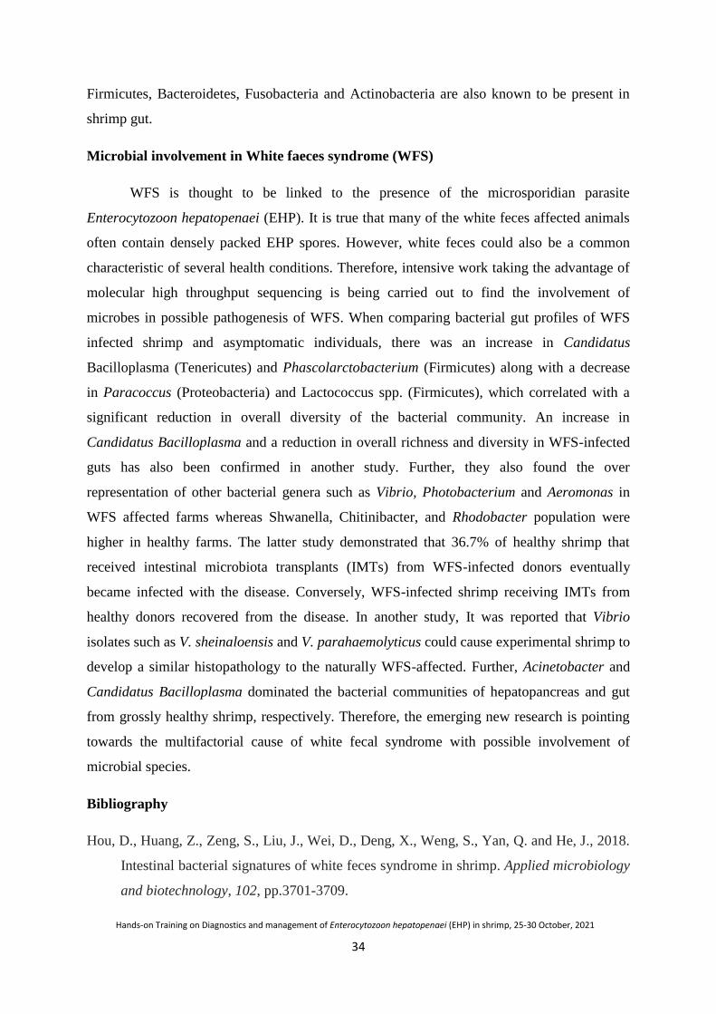
34
Hands-on Training on Diagnostics and management of Enterocytozoon hepatopenaei (EHP) in shrimp, 25-30 October, 2021
Firmicutes, Bacteroidetes, Fusobacteria and Actinobacteria are also known to be present in
shrimp gut.
Microbial involvement in White faeces syndrome (WFS)
WFS is thought to be linked to the presence of the microsporidian parasite
Enterocytozoon hepatopenaei (EHP). It is true that many of the white feces affected animals
often contain densely packed EHP spores. However, white feces could also be a common
characteristic of several health conditions. Therefore, intensive work taking the advantage of
molecular high throughput sequencing is being carried out to find the involvement of
microbes in possible pathogenesis of WFS. When comparing bacterial gut profiles of WFS
infected shrimp and asymptomatic individuals, there was an increase in Candidatus
Bacilloplasma (Tenericutes) and Phascolarctobacterium (Firmicutes) along with a decrease
in Paracoccus (Proteobacteria) and Lactococcus spp. (Firmicutes), which correlated with a
significant reduction in overall diversity of the bacterial community. An increase in
Candidatus Bacilloplasma and a reduction in overall richness and diversity in WFS-infected
guts has also been confirmed in another study. Further, they also found the over
representation of other bacterial genera such as Vibrio, Photobacterium and Aeromonas in
WFS affected farms whereas Shwanella, Chitinibacter, and Rhodobacter population were
higher in healthy farms. The latter study demonstrated that 36.7% of healthy shrimp that
received intestinal microbiota transplants (IMTs) from WFS-infected donors eventually
became infected with the disease. Conversely, WFS-infected shrimp receiving IMTs from
healthy donors recovered from the disease. In another study, It was reported that Vibrio
isolates such as V. sheinaloensis and V. parahaemolyticus could cause experimental shrimp to
develop a similar histopathology to the naturally WFS-affected. Further, Acinetobacter and
Candidatus Bacilloplasma dominated the bacterial communities of hepatopancreas and gut
from grossly healthy shrimp, respectively. Therefore, the emerging new research is pointing
towards the multifactorial cause of white fecal syndrome with possible involvement of
microbial species.
Bibliography
Hou, D., Huang, Z., Zeng, S., Liu, J., Wei, D., Deng, X., Weng, S., Yan, Q. and He, J., 2018.
Intestinal bacterial signatures of white feces syndrome in shrimp. Applied microbiology
and biotechnology, 102, pp.3701-3709.
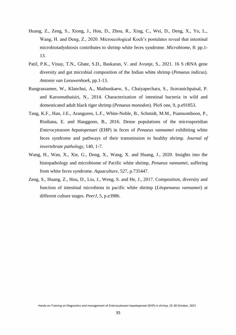
35
Hands-on Training on Diagnostics and management of Enterocytozoon hepatopenaei (EHP) in shrimp, 25-30 October, 2021
Huang, Z., Zeng, S., Xiong, J., Hou, D., Zhou, R., Xing, C., Wei, D., Deng, X., Yu, L.,
Wang, H. and Deng, Z., 2020. Microecological Koch‟s postulates reveal that intestinal
microbiotadysbiosis contributes to shrimp white feces syndrome. Microbiome, 8: pp.1-
13.
Patil, P.K., Vinay, T.N., Ghate, S.D., Baskaran, V. and Avunje, S., 2021. 16 S rRNA gene
diversity and gut microbial composition of the Indian white shrimp (Penaeus indicus).
Antonie van Leeuwenhoek, pp.1-13.
Rungrassamee, W., Klanchui, A., Maibunkaew, S., Chaiyapechara, S., Jiravanichpaisal, P.
and Karoonuthaisiri, N., 2014. Characterization of intestinal bacteria in wild and
domesticated adult black tiger shrimp (Penaeus monodon). PloS one, 9, p.e91853.
Tang, K.F., Han, J.E., Aranguren, L.F., White-Noble, B., Schmidt, M.M., Piamsomboon, P.,
Risdiana, E. and Hanggono, B., 2016. Dense populations of the microsporidian
Enterocytozoon hepatopenaei (EHP) in feces of Penaeus vannamei exhibiting white
feces syndrome and pathways of their transmission to healthy shrimp. Journal of
invertebrate pathology, 140, 1-7.
Wang, H., Wan, X., Xie, G., Dong, X., Wang, X. and Huang, J., 2020. Insights into the
histopathology and microbiome of Pacific white shrimp, Penaeus vannamei, suffering
from white feces syndrome. Aquaculture, 527, p.735447.
Zeng, S., Huang, Z., Hou, D., Liu, J., Weng, S. and He, J., 2017. Composition, diversity and
function of intestinal microbiota in pacific white shrimp (Litopenaeus vannamei) at
different culture stages. PeerJ, 5, p.e3986.

36
Hands-on Training on Diagnostics and management of Enterocytozoon hepatopenaei (EHP) in shrimp, 25-30 October, 2021
Microscopy including wet smear, special staining, SEM and TEM
P. Ezhil Praveena, T. Sathish Kumar and K. P. Jithendran
Microscopy
Wet mount/ Squash method
Microscopically, a typical microsporidian spores contain a single nucleus, coiled
polar filament, a posterior vacuole, an anchoring disk attached to polar filament, and a
thick electron-dense wall. Unique feature of this is very small spore size as compared to other
microsporidians and hence these features may not be discernible. Wet mounts of fresh
hepatopancreas (HP) tissue squash and/or faecal strands are examined by this method.
Diagnosis is made by the demonstration of characteristic spores under light microscope. The
spores are very small (1.1±0.2 by 0.6- 0.7±0.1 µm). This method may not be useful in detecting
the spores which are present in scant numbers. Trained or experienced personnel are required
for identification of the spores.
Microscopic detection of wet mount of spores of Enterocytozoon hepatopenaei from Penaeus
vannamei feces
Wet smear technique By using wet mount or squash method it will be difficult to identify the
microsporidians spore due very small size. Hence staining is advisable to differentiate the
spore by conventional stains such as Giemsa, Haematoxylin & Eosin (H&E) etc. The smear
preparations from the feacal sample / infected hepatopancreas were made in a glass slide and
allowed to dry at room temperature, fixed in methanol for 15 min and stained with Giemsa/
H&E. The stained smears can be observed under microscope using oil immersion.
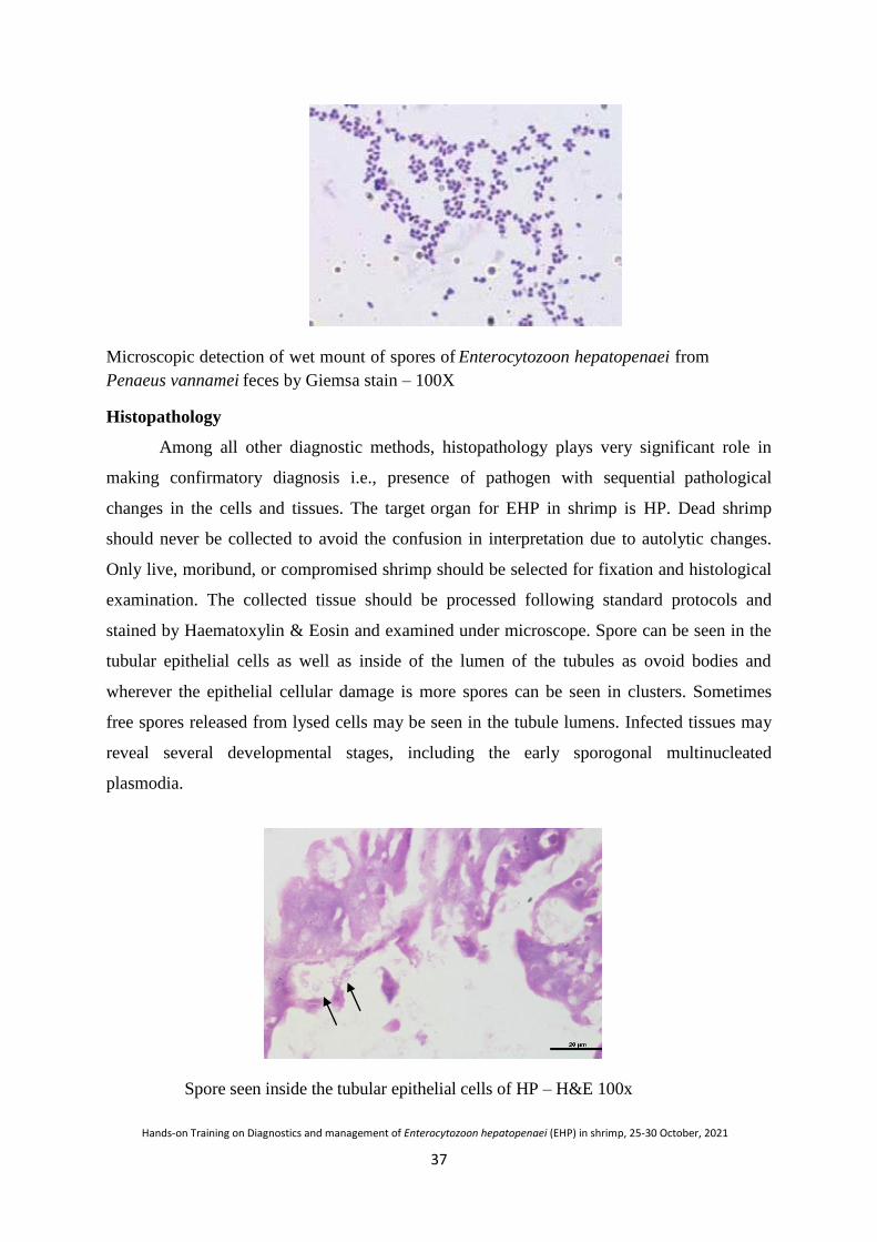
37
Hands-on Training on Diagnostics and management of Enterocytozoon hepatopenaei (EHP) in shrimp, 25-30 October, 2021
Microscopic detection of wet mount of spores of Enterocytozoon hepatopenaei from
Penaeus vannamei feces by Giemsa stain – 100X
Histopathology
Among all other diagnostic methods, histopathology plays very significant role in
making confirmatory diagnosis i.e., presence of pathogen with sequential pathological
changes in the cells and tissues. The target organ for EHP in shrimp is HP. Dead shrimp
should never be collected to avoid the confusion in interpretation due to autolytic changes.
Only live, moribund, or compromised shrimp should be selected for fixation and histological
examination. The collected tissue should be processed following standard protocols and
stained by Haematoxylin & Eosin and examined under microscope. Spore can be seen in the
tubular epithelial cells as well as inside of the lumen of the tubules as ovoid bodies and
wherever the epithelial cellular damage is more spores can be seen in clusters. Sometimes
free spores released from lysed cells may be seen in the tubule lumens. Infected tissues may
reveal several developmental stages, including the early sporogonal multinucleated
plasmodia.
Spore seen inside the tubular epithelial cells of HP – H&E 100x

38
Hands-on Training on Diagnostics and management of Enterocytozoon hepatopenaei (EHP) in shrimp, 25-30 October, 2021
Detection of EHP in shrimp feces
Purification of microsporidian spores from fecal samples
The efficiency of microscopic observation can be increased many-fold by
concentration of samples. The purification and concentration of microsporidian spores from
faecal samples by sedimentation by water-ether method is given here. Briefly, collect the
faecal strings and homogenize with distilled water and filtered through fine mesh to remove
the larger debris. The filtrate was then transferred to a 15 ml centrifuge tube. Add 3 ml of
diethyl ether to the centrifuge tube.
Thoroughly vortex the homogenate for 1 min and centrifuge at 5,000 rpm for 15 min.
Carefully remove the fat layer and supernatant and collect the pellet. Resuspend the pellet in
5 ml distilled water and centrifuge at 5,000 rpm for 5 min. Repeat the washing procedure
twice and finally collect the pellet and resuspend in 1 ml distilled water. Check for the
presence of spore using a light microscopy.
Special Stains – EHP spore detection
Besides the H& E stain EHP spore can be demonstrated from the tissue section by
using toluidine blue Ignatius et al., 1997; phloxine (Aldama-Cano et al., 2018), Giemsa
(Rajendran et al., 2016), or hematoxylin and eosin. A modified trichrome staining method
was developed, which improved the identification of EHP spores (Praveena et al., 2018).
Modified Trichrome Staining
A thin smear of purified spores can be stained by modified trichrome stain (Ryan-Blue
method) for microsporidians. To enhance the differentiation of spores, a microsporidia-
specific staining using modified trichrome stain may be useful. Briefly, trichrome stain
solution was prepared by mixing 6 g of chromotrope 2R (Sigma), 0.5 g of aniline blue
(Sigma), and 0.25 g of phosphortungstic acid (SRL, laboratories) with 2 ml glacial acetic
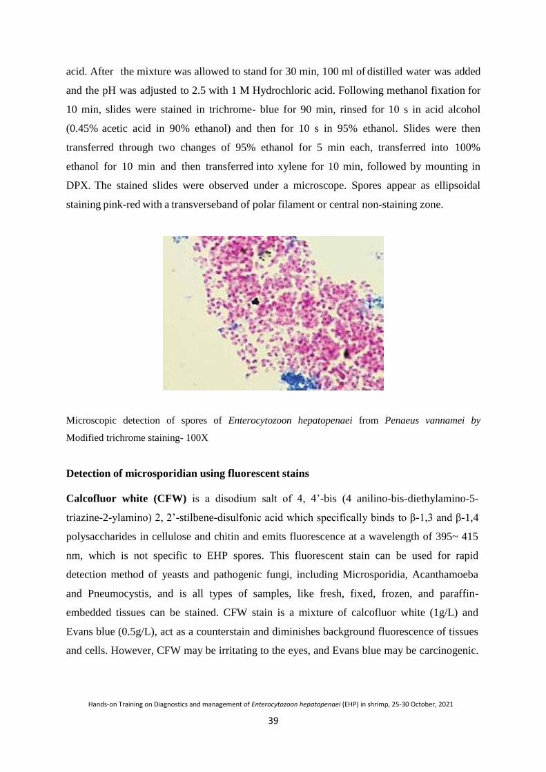
39
Hands-on Training on Diagnostics and management of Enterocytozoon hepatopenaei (EHP) in shrimp, 25-30 October, 2021
acid. After the mixture was allowed to stand for 30 min, 100 ml of distilled water was added
and the pH was adjusted to 2.5 with 1 M Hydrochloric acid. Following methanol fixation for
10 min, slides were stained in trichrome- blue for 90 min, rinsed for 10 s in acid alcohol
(0.45% acetic acid in 90% ethanol) and then for 10 s in 95% ethanol. Slides were then
transferred through two changes of 95% ethanol for 5 min each, transferred into 100%
ethanol for 10 min and then transferred into xylene for 10 min, followed by mounting in
DPX. The stained slides were observed under a microscope. Spores appear as ellipsoidal
staining pink-red with a transverse band of polar filament or central non-staining zone.
Microscopic detection of spores of Enterocytozoon hepatopenaei from Penaeus vannamei by
Modified trichrome staining- 100X
Detection of microsporidian using fluorescent stains
Calcofluor white (CFW) is a disodium salt of 4, 4‟-bis (4 anilino-bis-diethylamino-5-
triazine-2-ylamino) 2, 2‟-stilbene-disulfonic acid which specifically binds to β-1,3 and β-1,4
polysaccharides in cellulose and chitin and emits fluorescence at a wavelength of 395~ 415
nm, which is not specific to EHP spores. This fluorescent stain can be used for rapid
detection method of yeasts and pathogenic fungi, including Microsporidia, Acanthamoeba
and Pneumocystis, and is all types of samples, like fresh, fixed, frozen, and paraffin-
embedded tissues can be stained. CFW stain is a mixture of calcofluor white (1g/L) and
Evans blue (0.5g/L), act as a counterstain and diminishes background fluorescence of tissues
and cells. However, CFW may be irritating to the eyes, and Evans blue may be carcinogenic.

40
Hands-on Training on Diagnostics and management of Enterocytozoon hepatopenaei (EHP) in shrimp, 25-30 October, 2021
Utmost care is recommended. Calcofluor white stain can be mixed with a potassium
hydroxide mixture to clear up the specimen to facilitate visualization.
Procedure
Faecal sample/ hepatopancreas/purified spores to be examined placed onto a clean
glass slide. Add one drop of Calcofluor white stain (Sigma) and one drop of 10% Potassium
Hydroxide. Place a coverslip over the specimen. The slide was placed at room temperature
(25°C) for 5~10 min. Cover the slide with a paper towel and gently press to remove any
excess fluid. Examine the slide under a fluorescence microscope at the excitation wavelength
of 340~380 nm and emission wavelength of 400 nm with a 100× oil objective.
Interpretation of results
The microsporidian spore appears as bluish – white or turquoise oval halo. The
staining method is sensitive and fast, requires ~ 15 min to perform.
Microscopic detection of spores of Enterocytozoon hepatopenaei from Penaeus vannamei by
Calcoflour white Stain
In situ hybridization (ISH)
In situ hybridization (ISH) is another important tool to determine the location of
pathogen nucleic acid in tissue sections of PCR positive animals. It can be done using
digoxigenin-labeled (DIG- labeled) probes for the small subunit ribosomal RNA (SSU rRNA)
or for spore wall protein (SWP) gene. The gene probe was labelled with digoxigenin-11-
deoxyuridine triphosphate (dUTP) in a PCR reaction according to the manufacturer‟s
instructions. Following PCR, the digoxigenin-labelled DNA probe was prepared stored at
20°C. The Davidson‟s fixed shrimp tissue were processed, embedded in paraffin, and
sectioned (4 µm thick), deparaffinization, hydrated, proteinase K digestion, and post-fixation,
41
Hands-on Training on Diagnostics and management of Enterocytozoon hepatopenaei (EHP) in shrimp, 25-30 October, 2021
sections were overlaid with hybridization solution containing the EHP probe. Slides were
placed on a heated surface at 90⁰C for 10 min and hybridized overnight in a humid chamber
at 42⁰C. Final detection was performed with anti-digoxigenin antibody conjugated to alkaline
phosphatase. The slides were counterstained with bismark brown and examined using a light
microscope.
Transmission electron microscopy (TEM)
Small pieces (1-2 mm3) of HP tissue are rapidly fixed in 2.5% glutaraldehyde is ideal
for transmission electron microscopy (TEM). Fixed tissues are processed based on the tissue
type and post-fixed in osmium tetroxide. Semi-thin sections are made and the ideal area for
sectioning is made. Thin sections cut with diamond knives are placed on copper grids,
impregnated with uranyl acetate and lead citrate, and visualized for the following details.
Different stages of the parasite from early sporogonal plasmodia to mature spores in the
cytoplasm of host-cells. Electron-dense disks and precursors of the polar tubule develop in
the cytoplasm of the plasmodium prior to budding of early sporoblasts from the plasmodial
surface. Mature spores are oval, measuring 0.7-1.1 µm and contain a single nucleus, 5–6
coils of the polar filament, a posterior vacuole, an anchoring disk attached to the polar
filament.
Scanning electron microscopy (SEM)
Hepatopancreas of EHP infected were rapidly fixed in 2.5% glutaraldehyde in 0.1 M
sodium phosphate buffer at 4°C, dried using Hexamethyldisilazane (HMDS) as per the
method described by Nikara et al., (2020) and Berger et al. (2016). Processed samples can be
examined and imaged in Scanning Electron Microscope. The tubular surface changes as well
the spore attachment in the tubule can be visualized.
Bibliography
Afshar, P., Larijani, L.V. and Rouhanizadeh, H., 2018. A comparison of conventional rapid
methods in diagnosis of superficial and cutaneous mycoses based on KOH, Chicago
sky blue 6B and calcofluor white stains. Iranian Journal of Microbiology, 10(6), 433.
Bell, T.A. and Lightner, D.V., 1988. A handbook of normal penaeid shrimp histology. World
Aquaculture Society, Baton Rouge, LA, USA, p. 114.
Berger, L.R.R., Stamford, N.P., Willadino, L.G., Laranjeira, D., de Lima, M.A.B., Malheiros,
S.M.M., de Oliveira, W.J. and Stamford, T.C.M., 2016. Cowpea resistance induced
against Fusarium oxysporum f. sp. tracheiphilum by crustaceous chitosan and by

42
Hands-on Training on Diagnostics and management of Enterocytozoon hepatopenaei (EHP) in shrimp, 25-30 October, 2021
biomass and chitosan obtained from Cunninghamella elegans. Biological Control, 92,
45-54.
Praveena, P.E., Bhuvaneswari, T., Krishnan, A.N., Jagadeesan, V., Rajan, J.J.S. and
Jithendran, K.P., 2018, August. An improved microscopic method for the rapid
diagnosis of emerging microsporidian parasite, Enterocytozoon hepatopenaei in shrimp
farms. Current Science, 115, 758-763. DOI: 10.18520/cs/v115/i4/758-763.
Kumar, R.S., Ganvir, S.M. and Hazarey, V.K., 2009. Candida and calcofluor white: Study in
precancer and cancer. Journal of oral and maxillofacial pathology: JOMFP, 13(1), 2.
Nikara, S., Ahmadi, E. and Nia, A.A., 2020. Effects of different preparation techniques on the
microstructural features of biological materials for scanning electron
microscopy. Journal of Agriculture and Food Research, 2, 100036.
Qiao, Y., Shen, H., Wan, X.H., Fan, X.P., Jiang, G., Li, H., Wang, L., Shi, W. and Cheng, J.,
2018. Preliminary isolation and morphological observation of Enterocytozoon
hepatopenaei infecting the shrimp Penaeus vannamei. Journal of Fishery Sciences of
China, 25(5), pp.1051-1058.
Qin, H., Li, J., He, S. and Wu, J., 2012. Detection and identification of Nosema ceranae by
dual fluorescent staining with Calcofluor White M2R and Sytox Green. Chinese
Journal of Applied Entomology, 49(5), 1392-1396.
Rajendran, K.V., Shivam, S., Praveena, P.E., Rajan, J.J.S., Kumar, T.S., Avunje, S.,
Jagadeesan, V., Babu, S.P., Pande, A., Krishnan, A.N. and Alavandi, S.V., 2016.
Emergence of Enterocytozoon hepatopenaei (EHP) in farmed Penaeus (Litopenaeus)
vannamei in India. Aquaculture, 454, 272-280.
Tang, K.F., Pantoja, C.R., Redman, R.M., Han, J.E., Tran, L.H. and Lightner, D.V., 2015.
Development of in situ hybridization and PCR assays for the detection of
Enterocytozoon hepatopenaei (EHP), a microsporidian parasite infecting penaeid
shrimp. Journal of Invertebrate Pathology, 130, 37- 41.
Zhao, R.H., Gao, W., Qiu, L., Chen, X., Dong, X., Li, C. and Huang, J., 2020. A staining
method for detection of Enterocytozoon hepatopenaei (EHP) spores with calcofluor
white. Journal of Invertebrate Pathology, 172,107347.
43
Hands-on Training on Diagnostics and management of Enterocytozoon hepatopenaei (EHP) in shrimp, 25-30 October, 2021
Polymerase Chain Reaction (PCR) for early diagnosis of Enterocytozoon
hepatopenaei (EHP)
R. Ananda Raja, T. Bhuvaneswari and J.J.S. Rajan
Enterocytozoon hepatopenaei (EHP) causing hepatopancreatic microsporidiosis
(HPM) is a newly emerging disease of cultivated shrimp in Asia. It is associated with severe
growth retardation in chronic infections and may even cause low continuous mortality in the
case of very severe infections. Current production loss to HPM in India is estimated to be in
hundreds of millions of US dollars per year as mentioned in previous chapter elsewhere.
Transmission of this disease may come to shrimp grow out system via post larvae (PLs) used
to stock ponds or via environmental sources such as live carriers or spores in water and
sediments. Once transmitted to single host/shrimp in the pond, infected host can spread the
infection horizontally to other uninfected animals. The PLs are infected in hatcheries through
broodstock in non-biosecure facilities. EHP SPF broodstock may be contaminated by live
feeds such as polychaetes and molluscs, which ultimately leads to severe economic loss in the
shrimp industry. Hence, early diagnosis of EHP in PLs, and screening of broodstock, live
feeds and other environmental hosts are of paramount important for success of shrimp
aquaculture industry. A molecular technique called Polymerase Chain Reaction (PCR) is
routinely used worldwide in the field of disease diagnosis targeting the presence of the
specific DNA molecule as highly precise, accurate and sensitive method. Hence, PCR is also
employed worldwide in diagnosis of EHP as well. There are many ready to use PCR kits
available in the market, but it is essential to understand the basics of the PCR. This chapter is
aimed to explain about the same and methodology adopted to diagnose the EHP.
Polymerase Chain Reaction (PCR)
PCR works with the basic principles of selectively amplifying a particular segment of
DNA by in vitro enzymatic replication using specific primers. It was first explained by
H.G.Khorana in 1971 but the further advancement was incomplete by primer synthesis and
polymerase purification issues. In 1983, Kary Mullis successfully amplified the DNA for
which he was awarded Nobel Prize in Chemistry in 1993. Recent past years, PCR has
become indispensable technique in medical and biological research. PCR is routinely used
worldwide in the shrimp industry for the diagnosis of various diseases including
Enterocytozoon hepatopenaei (EHP).
44
Hands-on Training on Diagnostics and management of Enterocytozoon hepatopenaei (EHP) in shrimp, 25-30 October, 2021
Components and reagents
As a thumb rule, all the chemicals and reagents used for PCR should be of molecular
biology grade. Clean and sterilized condition should be well maintained in all operations as a
prerequisite.
Buffer solution
PCR is carried out in a buffer that provides suitable chemical environment for
optimum activity with stability of the DNA polymerase. The buffer pH is stabilized between
8.0 and 9.5 by Tris-HCl. Potassium ion (K+) is a common component in buffer which
promotes primer annealing.
Magnesium ion (Mg2+
) concentration
TheMg2+
plays major role in the yield of PCR product by forming a complex with
dNTPs, primers and DNA template. Hence, the optimum concentration of MgCl2 should be
augmented within the range of 1.5 to 4.5 mM in the PCR mixtures for each PCR reaction.
The concentration of MgCl2 should be selected empirically, starting from 1 mM and
increasing in 0.1 mM steps, until a sufficient yield of PCR product is obtained. The MgCl2
concentration is independent of the GC/AT ratio of the primers and the numbers of DNA
band(s) amplified. Too few Mg2+
ions result in a low yield of PCR product, and too many
increase the yield of non-specific products with multiple bands. However, lower Mg2+
concentrations are desirable when fidelity of DNA synthesis is critical. If the DNA samples
contain EDTA or other chelators, the MgCl2 concentration in the reaction mixture should be
raised proportionally.
Deoxyribose nucleotide triphosphates (dNTPs)
dNTPs are employed in PCR to expand the growing DNA strand binding with the
complementary DNA strand by hydrogen bonds with the help of Taq DNA polymerase. The
concentration of each dNTP in the reaction mixture is usually 200 µM. Inaccuracy in the
concentration of even a single dNTP dramatically increases the misincorporation level. So it
is very important to have equal concentrations of each dNTP i.e., dATP [adenine], dCTP
[cytosine], dGTP [guanine], dTTP [Thymine]. When maximum fidelity of the PCR process is
crucial, the final dNTP concentration should be 10-50 µM.
DNA polymerase
Similar to DNA replication in a live organism, PCR requires a DNA polymerase
enzyme that makes new strands of DNA using existing strands as template. In 1959, Arthur

45
Hands-on Training on Diagnostics and management of Enterocytozoon hepatopenaei (EHP) in shrimp, 25-30 October, 2021
Kornberg was awarded the Nobel Prize for identifying the first DNA polymerase in 1957. In
1969, Thomas Brock isolated and identified a new species of thermophilic bacterium,
Thermusaquaticus from which the enzyme Taq DNA polymerase was produced in 1976. Taq
DNA polymerase is world widely used in PCR reaction. It should be stored at -20 oC in a
non-frost free freezer, typically in 50% glycerol. It should never be allowed to reach room
temperature and gloves should be worn while handling to avoid contamination. Before
opening a new tube of enzyme, it is spun briefly as there is often enzyme in the cap. When
pipetting enzyme from a stock tube, the end of the tip is just plunged far enough into the
enzyme to get what is needed to avoid excessive adherence of enzyme to the peripheral tips.
Enzyme should never be added to unbuffered water to avoid its denaturation. Usually 0.5-
1.0 U of Taq DNA polymerase is used in 50 µL of reaction mix. Higher Taq DNA
polymerase concentrations may cause synthesis of nonspecific products. Normally, ready to
use 2x Master mix is available in the market which contains buffer with MgCl2, dNTPs
andTaq DNA polymerase. It can be readily used with the standardised protocols.
Primers / oligonucleotides
Primers are short, single-stranded DNA sequences used in the PCR techniques which
are also referred to as oligonucleotides. They are usually 15-30 nucleotides in length. Longer
primers provide higher specificity. A pair of primers (forward and reverse) is used to amplify
the region of interest from the sample DNA. The two ring structure purines in DNA
are adenine (A) and guanine (G) while the single ring pyrimidines are cytosine (C) and
thymine (T). The CG content in primers should be 40 – 60% and distributed uniformly
throughout of the primer. To avoid nonspecific priming, more than three G or C nucleotides
at the 3'-end of the primer should be avoided. The primers should have neither intra-
complementarity nor inter-complementarity in order to avoid primer-dimer and hairpin
formation. The melting temperature of flanking primers should not differ by more than 5°C,
so the GC content and length must be chosen accordingly. The annealing temperature should
be approximately 5°C lower than the melting temperature of primers.
The melting temperature (Tm) of the primers is calculated using the following
formula:
5length Pr
5003.620.41 X
imerprimersincontentGCofPercentageTm
The details of the published primerare mentioned in the following table.

46
Hands-on Training on Diagnostics and management of Enterocytozoon hepatopenaei (EHP) in shrimp, 25-30 October, 2021
Table: The details of the published primers for detection of EHP
S.
No.
Primer
name
5’ Primer sequence 3’ Amplicon
size (bp)
Target
gene
Reference
1. MF1 CCGGAGAGGGAGCCTGAGA 951 SSU rRNA Tourtip
et al., 2009 MR1 GACGGGCGGTGTGTACAAA
2. ENF779 CAGCAGGCGCGAAAATTGTCCA 779 SSU rRNA Tangprasittipap
et al., 2013 ENR779 AAGAGATATTGTATTGCGCTTGCTG
3. ENF176 CAACGCGGGAAAACTTACCA 176 SSU rRNA
ENR176 ACCTGTTATTGCCTTCTCCCTCC
4. EHP-510F GCCTGAGAGATGGCTCCCACGT 510 SSU rRNA Tang et al.,
2015 EHP-510R GCGTACTATCCCCAGAGCCCGA
5. SWP_1F TTGCAGAGTGTTGTTAAGGGTTT 514 SWP Jaroenlak et al.,
2016 SWP_1R CACGATGTGTCTTTGCAATTTTC
6. SWP_2F TTGGCGGCACAATTCTCAAACA 148 SWP
SWP_2R GCTGTTTGTCTCCAACTGTATTTGA
Autoclaved molecular grade ultra-purified water
Autoclaved molecular grade ultra-purified water free of detectable inhibitors,
contaminants or enzymatic activity is used to make up the desired volume for the PCR
reaction.
DNA template
It is the sample DNA (50-100µg) such as genomic double-stranded DNA (gDNA),
complementary DNA (cDNA), and plasmid DNA that contains the target sequence to be
amplified.
Thermal cycling reaction
This is carried out on an automated programmed thermal cycler, which can heat and
cool the reaction mix in tubes in a very short time.
Initial denaturation
The PCR is begun with initial denaturation to separate the double-stranded template
DNA into single strands so that the primers can bind to the target region and initiate
extension. The initial denaturation should be performed at 94-98°C for a period of 1-
5 minutes depending on the nature of the template DNA. This interval may be extended up to
10 minutes for GC-rich templates. If the initial denaturation is no longer than three minutes at
95°C, Taq DNA polymerase can be added into the initial reaction mixture. If longer initial
denaturation or a higher temperature is necessary, Taq DNA polymerase should be added
only after the initial denaturation, as the stability of the enzyme dramatically decreases at

47
Hands-on Training on Diagnostics and management of Enterocytozoon hepatopenaei (EHP) in shrimp, 25-30 October, 2021
temperatures over 95 °C. For EHP diagnosis, the initial denaturation is standardised at 95°C
for 5 minutes for both first and second step PCRs.
Denaturation
During the denaturation, the double-stranded template DNA melts open and separate
to single strands which are important in PCR reaction. Incomplete denaturation of DNA
results in the inefficient utilization of template in the first amplification cycle leads to a poor
yield of PCR product. Usually denaturation for 0.5-2 minutes at 94-95°C is sufficient. If the
amplified DNA has a very high GC content, denaturation time may be increased up to 3-
4 minutes. Alternatively, additives such as glycerol (up to 10-15 vol.%), DMSO (up to 10%)
or formamide (up to 5%) may be used to facilitate DNA denaturation.
Annealing
Usually the optimal annealing temperature is 5°C lower than the melting temperature
of primer-template DNA duplex. Incubation for 0.5-2 minutes is usually sufficient. The
annealing temperature should be optimized by increasing it stepwise by 0.5-2°C to avoid
nonspecific PCR products. The primers are jiggling around, caused by the Brownian motion
and ionic bonds are constantly formed and broken between the single stranded primer and the
single stranded template. The more stable bonds last a little bit longer (primers that fit
exactly) and on that little piece of double stranded DNA (template and primer) the
polymerase can attach and starts copying the template. Once there are a few bases built in, the
ionic bond is so strong between the template and the primer which does not break anymore.
In case of addition of additives such as glycerol, DMSO or formamide to facilitate DNA
denaturation, the annealing temperature should be adjusted experimentally, since the melting
temperature of the primer-template DNA duplex decreases significantly when these additives
are used. The amount of enzyme in the reaction mix should be increased since DMSO and
formamide inhibit Taq DNA polymerase by approximately 50%. Alternatively, a common
way to decrease the melting temperature of the PCR product is to substitute dGTP with 7-
deaza-dGTP in the reaction mix.
Extension
It is the final stage in PCR cycle which is performed to extend the primer sequence
from the 3‟ region to the end of the amplicon with the help of DNA polymerase. Usually, the
extending step is performed at 70-75 °C. The rate of DNA synthesis by Taq DNA polymerase
is highest at this temperature. A 1-minute extension is appropriately sufficient to synthesis the

48
Hands-on Training on Diagnostics and management of Enterocytozoon hepatopenaei (EHP) in shrimp, 25-30 October, 2021
PCR fragments up to 2 kb and may be further increased by 1 minute for each 1 kp. Primers
that are on positions with no exact match get loose again (because of the higher temperature)
and don't give an extension of the fragment. The polymerase adds complementary dNTP's to
the template from 5' to 3', reading the template from 3' to 5'. Because both strands are copied
during PCR, there is an exponential increase of the number of copies of the gene. But,
polymerization is not strictly doubling the DNA at each cycle in the early phase. The number
of PCR cycles depends on the amount of template DNA in the reaction mix and on the
expected yield of the PCR product. Usually, 30-40 cycles of these three major steps such as
denaturation, annealing and extension are sufficient depending on the amplified product size.
Final extension
After the last cycle, the samples are usually incubated at 72 °C for 5-15 minutes to
fill-in the protruding ends of newly synthesized PCR products. Also, during this step, the
terminal transferase activity of Taq DNA polymerase adds extra A nucleotides to the 3'-ends
of PCR products.The final extension is standardised at 68°C for 5 minutes for both first and
second step PCRs in diagnosis of EHP.
Gel documentation
After amplification of the targeted DNA fragments, the PCR product should be
visualized and verified in UV transillumination to know the amplification of the product of
interest. Agarose gel at 0.8-2% in 1x Tris Boric acid EDTA (0.09 M Tris borate; 0.002 M
EDTA; pH 8) electrophoresis buffer is prepared with ethidium bromide (0.2 g per mL of
agarose). The PCR product is mixed with the desired gel loading dye (bromophenol blue and
xylene cyanol) and glycerol mixture and slowly loaded into the wells of the submerged
agarose gel using a micropipette. Electrophoresis is carried out by applying the voltage @ 1-
5V/cm distance between two electrodes and continued until the dye migrates to the
appropriate distance in the gel. Finally, the gel is examined under the UV light in a
transilluminator for band visualization at the targeted region.
Record maintenance
It is essential to maintain the record to know the shortfalls if any. The results of each
sample analysed should be well recorded and maintained for future reference and to assess
the performance of the laboratory in diagnosis over a period of time.

49
Hands-on Training on Diagnostics and management of Enterocytozoon hepatopenaei (EHP) in shrimp, 25-30 October, 2021
Conclusion
PCR is a useful tool for rapid identification of pathogens and has become inevitable
for any molecular biologist, since it has gained its importance in modern molecular biology
over a period of last two decades. The same technique has become vital in diagnosis of
various diseases in aquaculture, developing ds RNA therapy and vaccines to shrimp and fish
diseases, developing transgenic fish, and so on. PCR has also been widely applied to the
detection of shrimp pathogens either from the host or carriers so that the risk of disease can
be controlled. EHP is one among the pathogens which needs to be detected at the early stage
in broodstock, larvae and post larvae, which will help to discard the batch before taking them
into culture practice.
Bibliography
Ananda Raja. R and Sujeet Kumar., 2009. Application of Polymerase Chain Reaction (PCR)
in Aquaculture. CIBA Special Publication No. 41. pp: 88-93.
Felix. S., et al., 2005. Manual on Aquaculture Biotechnology. Fisheries Biotechnology
Centre, Department of Aquaculture, Fisheries College and Research Institute,
Thoothukudi, TANUVAS.
Jaroenlak, P., Sanguanrut, P., Williams, B.A., Stentiford, G.D., Flegel, T.W.,
Sritunyalucksana, K. and Itsathitphaisarn, O., 2016. A nested PCR assay to avoid false
positive detection of the microsporidian Enterocytozoon hepatopenaei (EHP) in
environmental samples in shrimp farms. PLoS One, 11(11), e0166320.
Jithendran, K.P., Navaneeth Krishnan, A., Jagadeesan, V., Anandaraja, R., Ezhil Praveena,
P., Anushya, S., Bala Amarnath, C., Bhuvaneswari, T., 2021. Co-infection of infectious
myonecrosis virus and Enterocytozoon hepatopenaei in Penaeus vannamei farms in the
east coast of India. Aquaculture Research 52, 4701-4710.
Sambrook, J., Russell, D.W., 2001. Molecular cloning: a laboratory manual. 3rd
Edn.: Cold
Spring Harbor Laboratory Press, New York.
Tang, K.F., Pantoja, C.R., Redman, R.M., Han, J.E., Tran, L.H. and Lightner, D.V., 2015.
Development of in situ hybridization and PCR assays for the detection of
Enterocytozoon hepatopenaei (EHP), a microsporidian parasite infecting penaeid
shrimp. Journal of invertebrate pathology, 130, 37-41.
50
Hands-on Training on Diagnostics and management of Enterocytozoon hepatopenaei (EHP) in shrimp, 25-30 October, 2021
Tangprasittipap, A., Srisala, J., Chouwdee, S., Somboon, M., Chuchird, N., Limsuwan, C.,
Srisuvan, T., Flegel, T.W. and Sritunyalucksana, K., 2013. The microsporidian
Enterocytozoon hepatopenaei is not the cause of white feces syndrome in whiteleg
shrimp Penaeus (Litopenaeus) vannamei. BMC veterinary research, 9(1), 1-10.

51
Hands-on Training on Diagnostics and management of Enterocytozoon hepatopenaei (EHP) in shrimp, 25-30 October, 2021
Quantitative detection of Enterocytozoon hepatopenaei using Real Time
Polymerase Chain Reaction (qPCR)
T. Bhuvaneswari, J. J. S. Rajan and M. Poornima
Quantitative polymerase chain reaction (qPCR) also known as real time PCR (RT-
qPCR) is one of the most rapid molecular technique in amplification of target DNA of
interest. It determines targeted gene copy numbers with reference to standard curve of
defined concentration, without a need for visualization in agarose gel electrophoresis. Real
time detection of amplified DNA attached to fluorescent dye markers were measured in
relation to reaction progress with the use of PCR instruments developed for detecting changes
in fluorescence in a single tube. This technique is sensitive, specific, quantifiable and more
convenient for assaying large no of samples. Enterocytozoon hepatopenaei infections of
shrimp in earthen ponds are reported with clinical signs increasing with respect to degree of
infection. Hence the quantitation of infection in terms of amount of EHP in infected shrimp is
essential to adopt the management practices in culture system.
Sample preparation and nucleic acid extraction
For detection and quantification of EHP in shrimp a good quality sample is most
crucial for qPCR and to get accurate results the sample should not be contaminated or
degraded. When isolating DNA for qPCR, it is necessary that the tissues are stored properly,
free from contaminants that may inhibit the reaction. DNA can be extracted from shrimp gut
tissues, hepatopancreas and feces, live feeds such as polychaetes, artemia, planktons, crab,
water infected with EHP, soil infected with EHP. Purification of DNA from cellular proteins,
cell membranes and extra cellular matrices is achieved in chemical cell lysis, the lysis steps
can be tailored to specific tissue and sample types, as well as steps in chemical removal of
protein constituents for optimization of DNA extraction. Kit based methods with columns or
magnetic beads may also require optimization for specific nucleic acid types to provide pure,
concentrated nucleic acid. The concentrations of nucleic acids were measured using the UV
absorbance at 260nm and 280nm in spectrophotometer. An A260 reading of 1.0 is equivalent
to ~50 μg/mL of pure doublestranded DNA.
Primer and amplicon design
• Both primer design and careful choice of target sequence are essential to ensure
specific and efficient amplification of the products.

52
Hands-on Training on Diagnostics and management of Enterocytozoon hepatopenaei (EHP) in shrimp, 25-30 October, 2021
• Target sequences should be unique, 75–150 bp long with a GC content between 50%
and 60%, and should not contain secondary structures.
• It is recommended that primers should have a GC content of 50–60% and a melting
temperature of 55–65 C.
• Long G or C stretches in the primer should be avoided, but it is recommended to have
G or C at the end of the primers.
A number of programs are available to design primer pairs and pick target sequences.
For instance, Primer-Blast (www.ncbi.nlm.nih.gov/tools/primerblast/index.cgi? LINK_LOC
=BlastHomeAd),is a program developed by NCBI that uses the algorithm Primer3 available
in public domain. Primer sequences are to be compared (blasted)with the user-selected
databases to ensure they are unique and specific for the gene of interest. The program
MFOLD (http://mfold.bioinfo.rpi.edu/cgi-bin/dna-form1.cgi) can be used to analyze the
amplicons for potential secondary structures that may prevent efficient amplification. Ideally,
two sets of oligonucleotides should then be ordered and tested for their performance in a
qPCR.
qPCR validation
A validated qPCR assay is one that has been assessed for the optimal range of primer
annealing temperatures, reaction efficiency, and specificity using a standard set of samples.
This will assure that the reaction conditions, buffers, and primers have been optimized and
that the cDNA samples are not contaminated with inhibitors of Taq polymerase. Practical
web source tools are available for qPCR design and validation.
Establishment of a standard curve to evaluate PCR efficiency
The efficiency of a PCR is a measure of the rate at which the polymerase converts the
reagents (dNTPs, oligonucleotides, and template cDNA) to amplicon. The maximum increase
of amplicon per cycle is 2-fold representing a reaction that is 100% efficient. It is important
to measure reaction efficiency as it is indicative of problems with the qPCR that can cause
artifactual results. Low efficiency reactions are generally the result of primer–dimers or
nonspecific amplicons. The most common causes of both high and low reaction efficiencies
are poorly calibrated pipettes or poor pipetting technique. A standard curve is generally used
to determine the reaction efficiency for any qPCR. The template for this typically is a sample
of cDNA or spiked plasmid cDNA in a sample extract. We recommend initially producing a

53
Hands-on Training on Diagnostics and management of Enterocytozoon hepatopenaei (EHP) in shrimp, 25-30 October, 2021
10-fold dilution series over eight points starting from the most concentrated cDNA sample, to
ensure the standard curve covers all potential template concentrations that may be
encountered during the study (i.e., broad dynamic range). For each dilution, a standard qPCR
protocol should be performed in triplicate for all the primer pairs to be used in the experiment
and Cq values determined. The standard curve is constructed by plotting the log of the
starting quantity of the template against the Cq values obtained. The equation of the linear
regression line, along with Pearson‟s correlation coefficient (r) or the coefficient of
determination (r 2), can then be used to evaluate whether the qPCR assay is optimized.
Real Time PCR studies for detection of EHP
The taxonomical classification of EHP has been supported by small subunit rRNA
(SSU rRNA)-based phylogenetic studies. The small subunit ribosomal RNA (SSU rRNA)
gene, a housekeeping gene, is a universal diagnostic target in EHP molecular detection
methods. Several EHP diagnostic methods have been developed based on the SSU rRNA,
including PCR, qPCR, in situ hybridization, and loop mediated isothermal amplification
assays. But it is well known that the SSU rRNA gene is highly conserved among
microsporidia, which may give false-positive test results. For example, the EHP's SSU rRNA
sequence shared a 90% similarity to that of Enterosporacanceri which infects marine crabs.
Therefore, a more specific PCR method is needed for EHP diagnostics.
A SYBR Green I based qPCR assay was established to detect EHP. With relatively
higher sensitivity and by a quantitative method, this method provides a useful technique to
support the detection and exploration of studies for controlling EHP. Among the quantitative
detection methods, TaqMan probe based qPCR assay uses a specific probe which can
hybridize with the target sequence with a high specificity. Therefore, the TaqMan probe
based qPCR assay could effectively prevent influences caused by nonspecific products in the
PCR and further improve the accuracy of quantitative assay and has been extensively applied
in the detection of aquatic animal pathogens. In detecting EHP in farm samples, the positive
detection rate of TaqMan-based EHP qPCR (93.5%) was noticeably higher than nested PCR
(77.4%), while it was a slightly higher than SYBR Green I qPCR-EHP (88.7%). Compared
with nested PCR and SYBR Green I qPCR-EHP, the diagnostic specificity of TaqMan-based
EHP qPCR was reported 100%, while that of nested PCR and SYBR Green I qPCREHP were
82.8% and 96.6%, respectively, indicating that TaqMan based EHP qPCR had a higher
diagnostic sensitivity. Further the correlation of the quantitative results of TaqMan-based

54
Hands-on Training on Diagnostics and management of Enterocytozoon hepatopenaei (EHP) in shrimp, 25-30 October, 2021
EHP qPCR and SYBR Green I qPCR-EHP was high and the value comparability was good.
Although SYBR Green I fluorochrome method is applied to the quantitative analysis of
DNA, the lack of specific fluorescent probe could cause interference of primer dimers and
nonspecific amplification products, thereby, affecting the accuracy of results and even might
produce false positives.
The β-tubulin gene sequence has sufficient variability for molecular diagnosis at the
species level. Recently a qPCR assay based on the β-tubulin gene sequence for EHP-specific
quantification was developed. This assay was applied to examine shrimp and feces samples to
quantify the level of EHP present. The method was also reported to be applicable for
screening live shrimp feeds and water samples. Using this qPCR, it was found shrimp feces
contained high numbers of EHP copies, indicating that this may act as an important source of
infection in ponds. Substantial amounts of EHP (as DNA copies) were detected in the
Artemia samples by this technique. Live shrimp feeds, including polychaetes, squids, and
Artemia biomass, have been suspected to be carriers for EHP Thus the quantification method
for EHP based on the β-tubulin gene, applicable in EHP management in shrimp farming., so
EHP quantification by qPCR in live shrimp feeds can be used as a tool to aid in disease
prevention in broodstock populations.
The polar tube, a highly specialized invasion organ, is one of the important taxonomic
indexes of microsporidia. Up to now, there are five polar tube proteins (PTP1–PTP5) located
on the polar tube identified. PTP2 gene encoding a 35-kDa protein was identified from many
microsporidian genomes. The PTP2 gene was also reported to be a single copy in the EHP
genome. Due to these unique properties, the PTP2 gene was selected as the EHP detection
target in molecular diagnostic tools. A SYBR Green I fluorescence quantitative PCR method
based on the PTP2 gene has been established. The integrated method of qPCR and
microscopy to quantify EHP spores reported for the detection of EHP in shrimp farming. It
was recommended for early detection for EHP-infection, suitable for real-time monitoring of
EHP in the field during the whole period of shrimp farming and provides a reference for the
epidemiological study of EHP. The details of primers for qPCR are enlisted in following
table for quick reference.
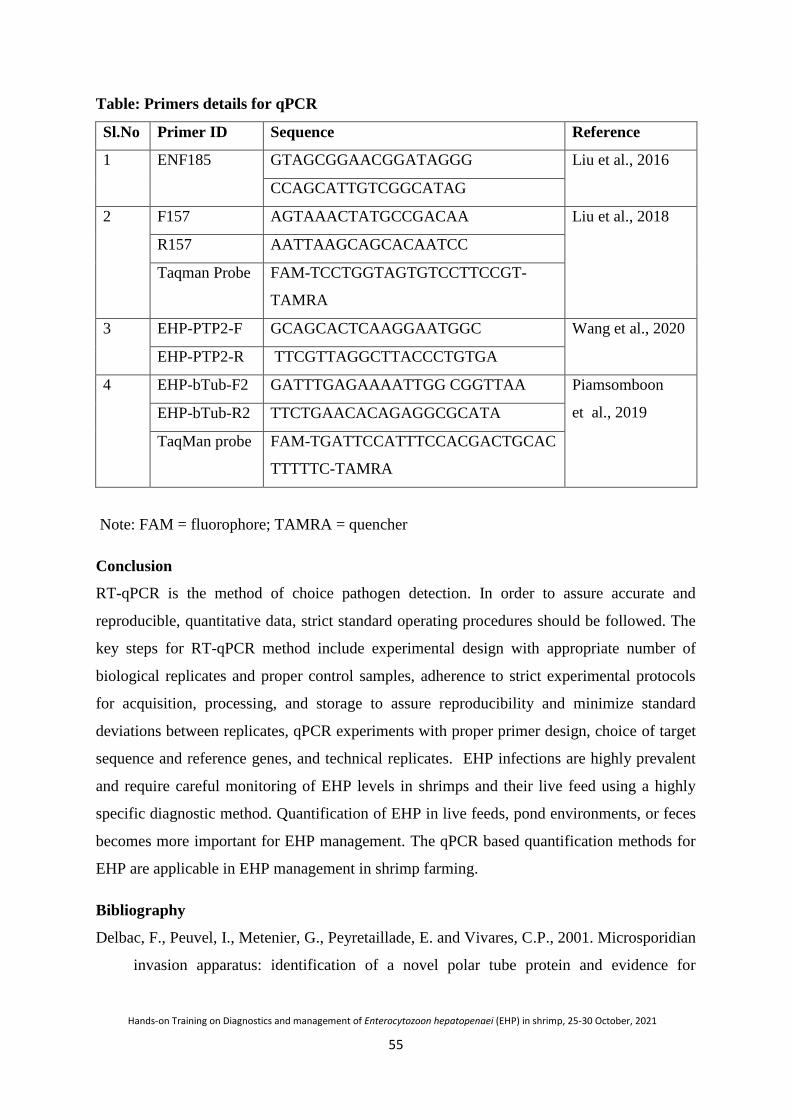
55
Hands-on Training on Diagnostics and management of Enterocytozoon hepatopenaei (EHP) in shrimp, 25-30 October, 2021
Table: Primers details for qPCR
Sl.No Primer ID Sequence Reference
1 ENF185 GTAGCGGAACGGATAGGG Liu et al., 2016
CCAGCATTGTCGGCATAG
2 F157 AGTAAACTATGCCGACAA Liu et al., 2018
R157 AATTAAGCAGCACAATCC
Taqman Probe FAM-TCCTGGTAGTGTCCTTCCGT-
TAMRA
3 EHP-PTP2-F GCAGCACTCAAGGAATGGC Wang et al., 2020
EHP-PTP2-R TTCGTTAGGCTTACCCTGTGA
4 EHP-bTub-F2 GATTTGAGAAAATTGG CGGTTAA Piamsomboon
et al., 2019 EHP-bTub-R2 TTCTGAACACAGAGGCGCATA
TaqMan probe FAM-TGATTCCATTTCCACGACTGCAC
TTTTTC-TAMRA
Note: FAM = fluorophore; TAMRA = quencher
Conclusion
RT-qPCR is the method of choice pathogen detection. In order to assure accurate and
reproducible, quantitative data, strict standard operating procedures should be followed. The
key steps for RT-qPCR method include experimental design with appropriate number of
biological replicates and proper control samples, adherence to strict experimental protocols
for acquisition, processing, and storage to assure reproducibility and minimize standard
deviations between replicates, qPCR experiments with proper primer design, choice of target
sequence and reference genes, and technical replicates. EHP infections are highly prevalent
and require careful monitoring of EHP levels in shrimps and their live feed using a highly
specific diagnostic method. Quantification of EHP in live feeds, pond environments, or feces
becomes more important for EHP management. The qPCR based quantification methods for
EHP are applicable in EHP management in shrimp farming.
Bibliography
Delbac, F., Peuvel, I., Metenier, G., Peyretaillade, E. and Vivares, C.P., 2001. Microsporidian
invasion apparatus: identification of a novel polar tube protein and evidence for
56
Hands-on Training on Diagnostics and management of Enterocytozoon hepatopenaei (EHP) in shrimp, 25-30 October, 2021
clustering of ptp1 and ptp2 genes in three Encephalitozoon species. Infection and
immunity, 69, 1016-1024.
Han, J.E., Tang, K.F. and Kim, J.H., 2018. The use of beta-tubulin gene for phylogenetic
analysis of the microsporidian parasite Enterocytozoonhepatopenaei (EHP) and in the
development of a nested PCR as its diagnostic tool. Aquaculture, 495, 899-902.
Liu, Y.M., Qiu, L., Sheng, A.Z., Wan, X.Y., Cheng, D.Y. and Huang, J., 2018. Quantitative
detection method of Enterocytozoonhepatopenaei using TaqMan probe real-time
PCR. Journal of Invertebrate Pathology, 151, 191-196.
Liu, Y.M., Qiu, L., Sheng, A.Z., Wan, X.Y., Cheng, D.Y. and Huang, J., 2018. Quantitative
detection method of Enterocytozoonhepatopenaei using TaqMan probe real-time
PCR. Journal of Invertebrate Pathology, 151, 191-196.
Piamsomboon, P., Choi, S.K., Hanggono, B., Nuraini, Y.L., Wati, F., Tang, K.F., Park, S.,
Kwak, D., Rhee, M.H., Han, J.E. and Kim, J.H., 2019. Quantification of
Enterocytozoonhepatopenaei (EHP) in penaeid shrimps from Southeast Asia and Latin
America using taqman probe-based quantitative PCR. Pathogens, 8, 233.
Wang, L., Lv, Q., He, Y., Gu, R., Zhou, B., Chen, J., Fan, X., Pan, G., Long, M. and Zhou,
Z., 2020. Integrated qPCR and staining methods for detection and quantification
of Enterocytozoon hepatopenaei in shrimp Litopenaeusvannamei. Microorganisms.
8,1366.

57
Hands-on Training on Diagnostics and management of Enterocytozoon hepatopenaei (EHP) in shrimp, 25-30 October, 2021
Loop-mediated isothermal amplification (LAMP) detection of
Enterocytozoon hepatopenaei (EHP)
T. Sathish Kumar
Introduction
Diagnostics plays a crucial part in aquatic biosecurity, and timely use of this tool
helps in successful aquatic animal health management and a successful crop. Most advanced
molecular diagnostics such as PCR and qPCR are commonly used in aquaculture for routine
screening compared to human/veterinary disease diagnostics. These advanced diagnostic
methods need expensive thermal cycling equipment for amplification, gel
electrophoresis/real-time monitoring (fluorimetry) for detection, and trained/ skilled
personnel for analysis.
LAMP is a simple, efficient, and highly selective diagnostic technique that can detect
the pathogen under isothermal settings in a short period of time. LAMP results can be quickly
inspected for color change with the naked eye by in-tube colorimetric indicators without any
toxic staining materials. The closed-tube LAMP assays were demonstrated to avoid cross-
contamination and opening of tubes after amplification. Visual detection of closed-tube
LAMP amplicons by naked eye was achieved by the accumulation of magnesium
pyrophosphate metal indicator dyes such as Hydroxynaphthol blue, malachite green, etc.,
intercalating dyes such as SYBR green I, Eva green, etc and pH-indicator dyes such as
phenol red, neutral red, cresol red, m-cresol purple, etc..
Principle
The LAMP assay is mainly based on the auto cycling and high strand displacement
activity mediated by the enzyme Bst polymerase at isothermal conditions. The LAMP assay
includes an initial step and a combination of amplification step with an elongation/recycling
step. The LAMP amplification is carried out at isothermal temperature (60–65 °C), which is
optimum for Bst polymerase activity. LAMP primers include two inner primers (forward
inner primer (FIP) and backward inner primer (BIP) and two outer primers (forward outer
primer (F3) and backward outer primer (B3). Inner primers consisted of two different
sequences that recognized a sense and an antisense sequence of the target DNA, while the
outer primers recognized only one external sequence of the target DNA. In order to accelerate
the reaction process two additional loop primers are also used in the LAMP amplification

58
Hands-on Training on Diagnostics and management of Enterocytozoon hepatopenaei (EHP) in shrimp, 25-30 October, 2021
LAMP assay for the field level detection of EHP
Many LAMP protocols have been demonstrated for the rapid diagnosis of EHP. Real-
time LAMP and visual LAMP assays have been established for the diagnosis of EHP.
Suebsing et al., 2013 established a loop-mediated isothermal amplification (LAMP) assay
combined with colorimetric nanogold (AuNP) for the rapid detection of EHP. Few Real-time
LAMP assays had been demonstrated for the sensitive detection of EHP. All these LAMP
protocols were developed based on SSU-rRNA gene sequence. In our earlier study also, we
demonstrated a closed tube visual LAMP detection of EHP based on SSU rRNA, where the
aerosol cross-contamination after amplification was avoided by adding the SYBRTM green I
dye by using needles and syringes into the reaction vials without opening the tubes. However,
SSU rRNA primers were reported to produce false-positive results. Recently an EHP LAMP
protocol based on spore wall protein (SWP) sequence colorimetric nanogold (AuNP) for the
specific and sensitive detection has been reported. Later, in 2021 ICAR-CIBA developed a
closed tube LAMP protocol based on spore wall protein (SWP) sequence using simple
indicator dyes such as phenol red and hydroxynaphthol blue.
Fig. Closed tube EHP LAMP using phenol red and HNB dyes (Sathish Kumar et al., 2021)
This LAMP assay showed 100% sensitivity, 100% specificity in clinical diagnostic
evaluation when compared with SWP-PCR. Also the LAMP assay showed the analytical
sensitivity as ten copies per reaction vial, equivalent to SWP-PCR. The LAMP assay is rapid,
can diagnose the EHP microsporidian within 40 minutes in a simple dry bath. Also, the end
59
Hands-on Training on Diagnostics and management of Enterocytozoon hepatopenaei (EHP) in shrimp, 25-30 October, 2021
results can be visualized with naked eye. LAMP assays do not need any post-amplification
harmful chemicals, expensive equipment, and skilled personnel. Thus, this LAMP protocol is
a comparatively simple, highly sensitive, and highly specific EHP diagnostic assay.
Conclusion
LAMP diagnostics, in particular, has wide applications in various fields and certainly
suited for testing at points of care. The EHP LAMP can be performed with minimum skills
by using a simple dry bath, and the results can be easily visualized with the naked eye. This
LAMP assay does not need any high resource setting/skilled personnel and has a great
potential to be a rapid, on-farm diagnostic tool. Additionally, this field-friendly, low-cost,
robust assay can be an effective tool for EHP transmission studies, epidemiology, and routine
surveillance.
Bibliography
Arunrut, N., Tondee, B., Khumwan, P., Kampeera, J. and Kiatpathomchai, W., 2021. Rapid
and sensitive colorimetric detection of microsporidian Enterocytozoonhepatopenaei
(EHP) based on spore wall protein (SWP) gene using loop-mediated isothermal
amplification combined with DNA functionalized gold nanoparticles as
probes. Aquaculture, 533, 736206.
Cai, S.X., Kong, F.D., Xu, S.F. and Yao, C.L., 2018. Real-time loop-mediated isothermal
amplification for rapid detection of Enterocytozoonhepatopenaei. PeerJ, 6, p.e5993.
Goto, M., Honda, E., Ogura, A., Nomoto, A. and Hanaki, K.I., 2009. Colorimetric detection
of loop-mediated isothermal amplification reaction by using hydroxynaphthol
blue. Biotechniques, 46, 167-172.
Karthik, K., Rathore, R., Thomas, P., Arun, T.R., Viswas, K.N., Dhama, K. and Agarwal, R.,
2014. New closed tube loop mediated isothermal amplification assay for prevention of
product cross-contamination. Methods X, 1, 137-143.
Karthikeyan, K., Sharma, A., Mekata, T., Itami, T. and Sudhakaran, R., 2017. Rapid and
sensitive real-time loop meditated isothermal amplification for the detection of
Enterocytozoonhepatopenaei of shrimp. Aquaculture, 481, 119-123.
Liang, C., Cheng, S., Chu, Y., Wu, H., Zou, B., Huang, H., Xi, T. and Zhou, G., 2013. A
closed-tube detection of loop-mediated isothermal amplification (LAMP) products

60
Hands-on Training on Diagnostics and management of Enterocytozoon hepatopenaei (EHP) in shrimp, 25-30 October, 2021
using a wax-sealed fluorescent intercalator. Journal of Nanoscience and
Nanotechnology, 13, 3999-4005.
Sathish Kumar, T., Navaneeth Krishnan, A., Joseph SahayaRajan, J., Makesh, M.,
Jithendran, K.P., Alavandi, S. V., Vijayan, K.K., 2018. Visual loop-mediated
isothermal amplification (LAMP) for the rapid diagnosis of
Enterocytozoonhepatopenaei (EHP) infection. Parasitol. Res. 117, 1485–1493.
https://doi.org/10.1007/s00436-018-5828-4
Suebsing, R., Prombun, P., Srisala, J. and Kiatpathomchai, W., 2013. Loop‐mediated
isothermal amplification combined with colorimetric nanogold for detection of the
microsporidian Enterocytozoonhepatopenaei in penaeid shrimp. Journal of Applied
Microbiology, 114, 1254-1263.

61
Hands-on Training on Diagnostics and management of Enterocytozoon hepatopenaei (EHP) in shrimp, 25-30 October, 2021
Management of Enterocytozoon hepatopenaei (EHP) in shrimp hatcheries
and farms
Vidya Rajendran, T. Sathish Kumar and K.P. Jithendran
Enterocytozoon hepatopenaei (EHP) is an obligate intracellular microsporidian
parasite which infects and proliferates in hepatopancreatic tubules and thereby impairing its
digestive and absorptive functions. EHP affected shrimps display slow and retarded growth
with high FCR (2.9 to 3.0), often found to be associated to White feces syndrome, however
its association is often refutable. A microsporidian infection was first detected from a growth
retarded Penaeus monodon in Thailand (2004) and later identified as EHP (2009). The first
case of EHP in India was reported from farmed vannamei shrimps from South east coast in
2016. Since then more cases were reported and in a short span of time EHP has emerged as a
serious threat to Indian shrimp farming industry with a prevalence rate of 27% in 2019 with a
production loss of 1.80 ± 0.24 ton/ ha/crop (2021).
Significance of EHP
EHP is a spore producing microsporidian parasite currently classified as a kind of
fungi and can be detected only through microscopy and specific PCR tests. It spreads
horizontally through oral route by consuming spores via cannibalism and cohabitation with
infected shrimps. EHP often associated with slow growth. However its conclusive association
with white feces syndrome is still debatable. There are no specific signs and symptoms
associated to EHP infections in shrimp. Only regular monitoring of intake of feed, slow
growth and monitoring hepatopancreas can assist EHP detection in shrimps.
Microscopic and PCR diagnosis are available to detect the infection as the earliest,
however many of times the low level infection remain undetected and find its entry to shrimp
hatcheries and grow out systems. The recommended methods to contain the infection are only
through enforcing tight biosecurity which additionally involves routine disinfection using
recommended chemicals before stocking the brooders and post larvae. Many of the
disinfection methods involve the inactivation of spores targeting the polar tube or polar
flagella extrusion which is essential for infecting the target animal. A high pH (9) usually
activate spore to extrude the flagella and release spore contents which make them inactive for
successive infection. Spores are hardy and thick walled hence they can survive from two
62
Hands-on Training on Diagnostics and management of Enterocytozoon hepatopenaei (EHP) in shrimp, 25-30 October, 2021
weeks to even 1 year depending on environmental condition. Use of chemicals and drugs are
regulated for aquaculture activities; however certain permitted chemicals can be effectively
used in controlling EHP in hatcheries and farms.
EHP prevention and management in hatcheries
Disease prevention and management in hatcheries are based on a biosecurity plan. It
involves preventing the entry and establishment of infectious agents in the hatchery facility
using prophylactic measures, e.g. sterilisation of input water, quarantine of brood stock, use
of vaccines, probiotics, and bacteriophages. Once infection establishes in the hatcheries
biosecurity plans enforce to shut down the facility, culling of infected stocks and initiate
disinfection process. Disinfection involves the killing of established infectious agent using
physical and chemical methods which are recommend by regulatory bodies, e.g. chlorination
in hatcheries and therapeutic treatment of infected animals.
Biosecurity
Biosecurity means a set of management and physical measures designed to reduce the
risk of introduction, establishment and spread of pathogenic agents (EHP) to, from and within
an aquatic animal population. Biosecurity plans apply important principles from HACCP
(Hazard Analysis and Critical Control Points) and quality assurance systems.
Identification of critical control points in EHP prevention in hatcheries
Critical control points are the physical locations or production steps at which shrimp
may be exposed to disease pathogens/EHP. Each of these represents a critical control point.
Through following critical control points pathogen/EHP can enter the hatchery.
• Infected brooder shrimp (wild or imported) and spores adhered to eggs
• Untreated water sources
• Contaminated diets: Algae, live feeds, polychaetes, frozen foods, commercial feeds
• Vectors: Personnel, contaminated equipment and vehicles
Disinfection process
Disinfection of tanks and pipelines
Treat tanks, pipelines, tools and accessories in hatcheries with 2.5% sodium
hydroxide for 3h and wash them with disinfected water followed by dry out them for 7days.
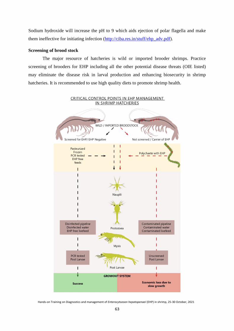
63
Hands-on Training on Diagnostics and management of Enterocytozoon hepatopenaei (EHP) in shrimp, 25-30 October, 2021
Sodium hydroxide will increase the pH to 9 which aids ejection of polar flagella and make
them ineffective for initiating infection (http://ciba.res.in/stuff/ehp_adv.pdf).
Screening of brood stock
The major resource of hatcheries is wild or imported brooder shrimps. Practice
screening of brooders for EHP including all the other potential disease threats (OIE listed)
may eliminate the disease risk in larval production and enhancing biosecurity in shrimp
hatcheries. It is recommended to use high quality diets to promote shrimp health.

64
Hands-on Training on Diagnostics and management of Enterocytozoon hepatopenaei (EHP) in shrimp, 25-30 October, 2021
Screening of brooder feed
Hatcheries use live polychaetes for feeding brood stock to enhance spawning
efficiency which often carries EHP. Try feeding frozen or pasteurised polychaetes or SPF
polychaetes which can eliminate the entry of this important pathogen.
Contaminated Artemia cysts
Artemia cysts used for hatchery operation may sometimes get contaminated with EHP
spores and other disease agents. So, screening or use of specific pathogen free Artemia cysts
may reduce the infection chances in larval production.
Screening PLs
Prior to release the seed stock from hatcheries check the health status of post larvae
by testing for EHP and other listed disease agents. However, the testing can only indicate the
status of the animal whereas the farms if already contaminated with disease agents become
potential threats to successful culture.
Regular monitoring
Examine the faecal threads of brood stock and PLs for EHP and closely monitor the
intake of feed and growth rate. These observations can guide the farmer to detect the
infection early and take adequate measures to reduce the economic loss.
EHP management in shrimp farms
Farm Management
EHP is contagious and spread through spores from infected shrimp to others. Spores
are method of reproduction in microsporidians and infected shrimp can produce large number
of spore and shed to the environment where they live. These spores may enter either laterally
through oral route or deposited in the pond bottom and available to re-infect the post larvae in
the successive cultures. Spores cannot be removed due to its microscopic nature and therefore
can only be deactivated using certain chemicals and disinfectants. Hence preparation of
shrimp ponds is critically important prior to stocking. At the farm level biosecurity plays an
important role in preventing this residual protozoan disease. Farm management has many
aspects and they are:
Larval management
PL stocking density plays an important role, whereas increased stocking may produce
stressful environment for shrimp, increasing their chances of disease expression. Farmers
65
Hands-on Training on Diagnostics and management of Enterocytozoon hepatopenaei (EHP) in shrimp, 25-30 October, 2021
should always buy seeds from registered shrimp hatcheries and should stock quality healthy
seeds free from EHP, in addition to other important pathogens.
Pond management
To maintain farm productivity, farmers should always follow better management
practices like providing adequate time (at least 3-4 weeks) for the ponds to dry after every
harvest. The ponds having history of disease should be disinfected using appropriate
protocols. High organic loads are typically related to spore loads. Once spores are in ponds, it
is very difficult to eradicate the disease. Spores can be inactivated by physical and chemical
methods. Heat treatment significantly inactivated spores. There is likely some intermediate
vector, and until we are sure what it is, use strategies to properly treat sediments before
stocking. As spores typically are resistant to a wide variety of environmental conditions, treat
pond bottom with a very caustic material to bring the pH to 12 and kill many of the spores.
Killing all of them may not be possible. It has been recommended that earthen ponds be
disinfected with very heavy use of calcium oxide, or quick lime, applied at a level of 6,000
kg/ha (https://enaca.org/?id=1064). Pond bottoms must be completely dry. Plough the quick
lime into the dried sediments to a depth of 10 to 12 cm; then moisten the sediments to
activate the lime. If the application is done properly, the pH of the soils will rise to 12 or
more within days and then gradually return to normal as the quick lime becomes calcium
carbonate.NACA also suggested >15 ppm KMnO4 or >40 ppm chlorine to inactivate spores
in the soil (https://enaca.org/?id=1064).
Feed management
Feed management is an important factor in successful shrimp culture. The feed should
be given in right amount to shrimp for proper growth. Insufficient feeding may lead to
cannibalism, while excess feed will deposit at the bottom of the pond, augmenting bacterial
growth and compromising water quality maintenance and thereby giving stress to the animal.
Water management
Water management is another crucial element of disease control in shrimp
aquaculture. Water is often pumped into the farm from water sources, such as the sea, estuary
or canal, which may introduce pathogens that can cause diseases. The use of water reservoirs
with proper duration of water ageing appears to be a regular practice on shrimp farms that can
reduce disease occurrence. Water ageing prior to stocking decreased the odds of EHP cases in
shrimp ponds. Treatment of water bodies with Calcium hypochlorite at the concentration of

66
Hands-on Training on Diagnostics and management of Enterocytozoon hepatopenaei (EHP) in shrimp, 25-30 October, 2021
18 mg/l before stocking PL can eradicate wild crustaceans and significantly reduced the
prevalence of microsporidian infection.
Continuous monitoring
Health and growth of shrimps must be monitored regularly and ensure healthy feed
and effective aeration and water movement in the pond. Once slow growth and low feed
intake is observed, check for EHP by PCR. Once EHP is diagnosed in an on-going culture
avoid over feeding and try to provide high protein diet. Post-culture disinfect the water and
dry the pond bottom for two weeks. Prior to next culture, remove mud or black soil from
pond bottom.
Conclusion
The disease emergence and its spread are due to the various linked events involving
the interactions between the host, the environment and the pathogens. It would be always
cautious to monitor and encourage producers to consider standard management strategies
such as the lowering of stocking densities, maintaining a good water quality, and the use of
EHP-free shrimp PLs for stocking, improved better management practices, routine farm
biosecurity measures and responsible trade practices to prevent epizootics in aquaculture.
Shrimp health is a shared responsibility, of every stakeholder‟s contribution to minimize
losses and sustain productivity.
Bibliography
Rajendran, K.V., Shivam, S., Praveena, P.E., Rajan, J.J.S., Kumar, T.S.,Avunje, S.,
Jagadeesan, V., Babu, S.P., Pande, A., Krishnan, A.N. and Alavandi, S.V., 2016.
Emergence of Enterocytozoonhepatopenaei (EHP) in farmed Penaeus (Litopenaeus)
vannamei in India. Aquaculture, 454, 272-280.

67
Hands-on Training on Diagnostics and management of Enterocytozoon hepatopenaei (EHP) in shrimp, 25-30 October, 2021
Disinfectants and therapeutics in the treatment and control of
Enterocytozoon hepatopenaei (EHP)
T. Sathish Kumar
Introduction
Microsporidia are obligate, unicellular organisms infecting wide range of eukaryotic
hosts belonging to the phylum Microspora. All microsporidia are spore-forming, intracellular
parasites that invade vertebrates and invertebrates. A characteristic feature of microsporidia is
the polar tube or polar filament found in the spore used to infiltrate host cells. Development
of the parasite generally occurs within the cytoplasm of the host-cell via nuclear proliferation,
and spore formation (sporogony), though certain genera are known to undergo similar
development within the host nucleoplasm.
Recently, Enterocytozoon hepatopenaei (EHP) (measure about 1.7 ×1 μm), has been
reported in shrimp farming nations of south-east Asia in cultured Penaeus vannamei. The
parasite was first recorded from growth retarded tiger shrimp, P. monodon from Thailand and
reported as an undesignated microsporidian. Subsequently, the parasite was identified and
characterized from P. monodon from Thailand. EHP infection in shrimp cannot be detected
by visual inspection of animals and there are no specifically distinctive gross signs, except
that it is suspected to be associated with growth retardation and white feces syndrome (WFS).
The target organ for EHP is shrimp hepatopancreas, the power house of the animal, and
infection in hepatopancreas may cause impairment in metabolism and ultimately result in
stunted growth. EHP infection can be transmitted horizontally through an oral route
(cannibalism, predation) and possibly by vertical transmission (trans-ovum). So far, no
intermediate hosts have been known to be involved. Thus, EHP is being emerged as a serious
threat to the shrimp farming industry. The studies on prophylactic and therapeutics strategies
for the treatment and control of EHP are very limited. This chapter discuss about the potential
disinfectants and drugs in the control and treatment of EHP.
EHP spore inactivation
Artificial germination of microsporidian spores can be triggered in vitro by using a
combination of nutrients, alterations in temperature, pH, hyperosmotic conditions, the
presence of anions or cations, or exposure to ultra-violet light or peroxides. Triggering the
artificial germination (extrusion of the polar tube) to render EHP mature spores non-
infectious is extremely important, as the polar tube extrusion is the key to successful

68
Hands-on Training on Diagnostics and management of Enterocytozoon hepatopenaei (EHP) in shrimp, 25-30 October, 2021
transmission of microsporidian to host cell. Recently, evaluation of EHP spores viability by
inducing extrusion of polar tubule was demonstrated with phloxin B. Microsporidians can be
killed by boiling for at least 5 min and by application of several disinfectants including
quaternary ammonium, 70% ethanol, formaldehyde, phenolic derivatives, hydrogen peroxide,
chloramines, sodium hydroxide, or amphoteric surfactants. The viability of the purified
spores has been studied by treating EHP spores with different chemical treatments such as
calcium hypochlorite, formalin, potassium permanganate (KMnO4) and ethanol. It was found
that 15ppm KMnO4 for 15min, 40ppm of 65% active chlorine for 15min or 10ppm of 65%
active chlorine for 24h and 20% ethanol for 15min rendering complete inhibition of EHP
spores activity. In our study in ICAR-CIBA the EHP spore artificial germination was
achieved with 1%KOH at pH 11 for 15 min incubation. It was found that the overnight
treatment of KMnO4 at 10 ppm, Formalin at 150 ppm, Calcium hypochlorite at 40 ppm ,
Sodium hydroxide at 3%, Hydrogen peroxide at 2% cause 100% inhibition of EHP spore
polar tubule extrusion and disintegration and denaturation of EHP spores
Fig: SEM image of a germinated EHP spore
Potential therapeutics for treating microsporidians
Microsporidia do not have true mitochondria and lack the tricarboxylic acid cycle for
energy production. It entirely depends on other metabolic pathways such as the glycolytic

69
Hands-on Training on Diagnostics and management of Enterocytozoon hepatopenaei (EHP) in shrimp, 25-30 October, 2021
pathway, pentose phosphate pathway, and trehalose metabolism for energy production. The
enzymes participating in the above pathways can be targeted as a potential target for evolving
therapeutic strategies. Triosephosphateisomerase (TIM) an important enzyme in glycolysis,
was inhibited by sulfhydryl reagents in Trypanosomacruzi, T. brucei, Entamoebahistolytica,
and Giardia duodenalis. Microsporidian microtubules are formed by polymerization of
tubulin protein; this polymerization can be inhibited by benzimidazole drugs such as
albendazole, cambendazole, benomyl, carbendazim, fenbendazole, mebendazole, and
triclabendazole. Methionine aminopeptidase 2 (MetAP2) is vital for tissue repair, protein
degradation, and angiogenesis in microsporidians. Anti-angiogenic drugs - ovalicin and
Fumagillin were reported to show anti-microsporidian activity. Chitin maintains the spore
wall structure and is vital for infection and extracellular survival. Chitin synthesis can be
inhibited by the activity of Nikkomycin and Polyoxin. Spore germination is calcium-
dependent and can be inhibited by calcium antagonists (Verapamil, Lanthanum), calmodulin
inhibitor (trifluoperazine, chlorpromazine), and calcium channel blocker (Nifedipine).
Studies on treatment or therapeutic drugs available for controlling EHP are limited. There is
no reported treatment, or therapeutic drug available for killing EHP spores. One study
reported Fumagillin-B to be ineffective in controlling EHP spores.
Disinfectants in the control of EHP
Disinfectants may be useful in eliminating extracellular spores in the soil, water, and
implements. It is recommended to use 2.5% sodium hydroxide solution (25gms NaOH/L
fresh water) for control the epizootics of EHP in hatcheries. After 3 hrs contact time, NaOH
should be washed off and dried for 7 days. Then it should be rinsed down with acidified
chlorine (200 ppm chlorine solution at pH <4. 5). This treatment was recommended for all
equipment, filters, reservoirs and pipes in hatcheries. For disinfecting shrimp earthen ponds
CaO (quicklime, burnt lime, unslaked lime or hot lime) was recommended at 6 Ton/ha. CaO
should be plowed into the dry pond sediment (10-12 cm) and then moisten the sediment to
activate the lime and left untouched for 1 week before drying or filling. After application of
CaO, the soil pH should rise to 12 or more for a couple of days and then fall back to the
normal range as it absorbs carbon dioxide and becomes CaCO3. Many disinfectants have
demonstrated the complete inhibition of spore germination. It is reported that 15 ppm KMnO4
for 15 min, 40 ppm of 65% active chlorine for 15 min or 10 ppm of 65% active chlorine for
24 h, and 20% ethanol for 15 min are effective in inhibiting spore germination. In the ICAR-
70
Hands-on Training on Diagnostics and management of Enterocytozoon hepatopenaei (EHP) in shrimp, 25-30 October, 2021
CIBA in-vivo challenge study, we found that potassium permanganate at 15 ppm, Calcium
hypochlorite 20 ppm, sodium hydroxide 3%, and hydrogen peroxide 3% are effective in
destroying EHP spores in the soil.
Conclusion
Microspordia are obligate intracellular pathogens that cause severe impact across all
trophic levels from mammals to planktonic copepods. Spores are the infective material of
microsporidians for transmitting disease. The epizootics of microsporidians in aquatic
animals are known in all freshwater, brackish water and marine water environments. Shrimp
hepatopancreas is the target organ for EHP infection. The studies on prophylactic and
therapeutics strategies for the treatment and control of EHP are very limited. Better
management practices and strict implementation of biosecurity protocols are the proper ways
to prevent the disease.
Bibliography
Aldama-Cano, D.J., Sanguanrut, P., Munkongwongsiri, N., Ibarra-Gámez, J.C.,
Itsathitphaisarn, O., Vanichviriyakit, R., Flegel, T.W., Sritunyalucksana, K. and
Thitamadee, S., 2018. Bioassay for spore polar tube extrusion of shrimp
Enterocytozoonhepatopenaei (EHP). Aquaculture, 490, 156-161.
Didier, E.S., Maddry, J.A., Brindley, P.J., Stovall, M.E. and Didier, P.J., 2005. Therapeutic
strategies for human microsporidia infections. Expert review of anti-infective
therapy, 3, 419-434.
Keeling, P.J. and Fast, N.M., 2002. Microsporidia: biology and evolution of highly reduced
intracellular parasites. Annual Reviews in Microbiology, 56, 93-116.
Leiro, J.M., Piazzon, C., Domínguez, B., Mallo, N. and Lamas, J., 2012. Evaluation of some
physical and chemical treatments for inactivating microsporidian spores isolated from
fish. International Journal of Food Microbiology, 156, 152-160.
Setlow, P., 2003. Spore germination. Current opinion in microbiology, 6(6), 550-556.
Weidner, E., 1982. The microsporidian spore invasion tube. III. Tube extrusion and
assembly. The Journal of Cell Biology, 93, 976-979.

71
Hands-on Training on Diagnostics and management of Enterocytozoon hepatopenaei (EHP) in shrimp, 25-30 October, 2021
Collection and preservation of shrimp samples for diagnosis of
Enterocytozoon hepatopenaei (EHP)
Sujeet Kumar and P. Ezhil Praveena
Sample collection
Acurate sampling and preservation of samples is one of the most crucial step for accurate
disease diagnosis.
• Before sampling, note any gross changes (behavioural abnormalities, size variation,
stunted growth, white fecal threads in the ponds, change in appearance, colour, lesions
etc.)
• Moribund (approaching death) or freshly dead shrimp is the most appropriate sample,
since autolysis takes place immediately.
Sending live shrimp samples for diseases diagnosis
A representative live sample of shrimp is the most ideal for diseases diagnostic lab as
this enhance the usage of various techniques for diagnostic purpose. For this purpose, 5-6
shrimps showing the sign of stunted growth, size variation should be collected and pack in
double polypack with oxygen. Pack these bags in a styroform or thermocol box to keep
temperature cool. Place a couple of ice pack or gel pack while sending samples long distance.
For smaller shrimp or post-larvae place adequate number ranging from 30 to 150 depending
on size in the same manner. LABEL and dispatch through courier with completed history
sheet (use separate photocopy for each batch of sample) within 24 hours.
Sending fixed shrimp samples for disease diagnosis
In case live sample delivery is not possible, the samples collected as above may be
fixed appropriately as detailed below for PCR analysis or for histopathology.
Shrimp samples for PCR analysis
Smaller shrimp samples or dissected hepatopancreas from larger shrimps may be
collected in 95% Ethyl alcohol in tightly capped container and submit to the disease
diagnostic lab at the earliest. Remember, that hepatopancreas is the target organ for EHP,
therefore, it must be included in the dissected sample.

72
Hands-on Training on Diagnostics and management of Enterocytozoon hepatopenaei (EHP) in shrimp, 25-30 October, 2021
Faecal sample for microscopy and PCR analysis
In addition to shrimp samples, faecal sample can be collected from the affected
animals in normal saline and send to laboratory for microscopical examination as well as for
PCR analysis for the presence of spore.
Shrimp samples for histopathology analysis
Fixation of sample: Shrimps are usually fixed in Davidson‟s OR Neutral Buffered
Formalin (NBF) fixative (composition shown below) in a wide mouth plastic bottle.
Smaller shrimp may be fixed directly by immersing in appropriate fixative for 24 hours and
for larger shrimps it should be in the fixative for 48 hours (sample size should not exceed
1/10th
of the volume of the fixative).
For larger shrimp (>1g), inject fixative into live animals, first in hepatopancreas, then the
region anterior to the hepatopancreas, and lastly two injections into the abdomen, one
anterior and the other posterior. Inject fixative @ 10% of shrimp body weight (e.g. 1 ml for a
10 g shrimp).
To ensure proper penetration of fixative, slit the cuticle open on both sides of the shrimp,
from the 6th
abdominal segment to the rostrum using scissors before placing in fixative. Seal
the container, LABEL and dispatch with history sheet as soon as possible.
Collection of water samples
Water samples may also be collected for examination of various physicochemical
parameters. Further, white, floating faecal strings from the affected disease ponds should be
collected from the affected ponds and transported on ice.
Collection of soil samples
About 500gm of soil sample from the four corners and centre of the pond was taken and then
packed in a zip lock pouch and label it properly and send it to the laboratory for diagnosis of
any EHP spore. The collected soil should be representative of the pond as a whole.
Precautions for transportation of samples
• Glass containers are not recommended due to breakage.
• Wrap samples in a non-fibrous material.

73
Hands-on Training on Diagnostics and management of Enterocytozoon hepatopenaei (EHP) in shrimp, 25-30 October, 2021
• Samples over ice may be packed in zip-lock bags.
• Labels should be written with pencil.
• Complete proforma with contact details.
Preparation of fixatives
1. Davidson’s Fixative 1000 ml
• Formalin (37% formaldehyde) 220 ml
• 95% ethanol 330 ml
• Distilled water (if not available, tap water) 335 ml
• Glacial acetic acid 115 ml
2. Neutral Buffered Formalin (NBF) 1000 ml
1. Formalin (37% formaldehyde) 100 ml
2. Distilled water (if not available, tap water) 900 ml
3. Sodium phosphate, monobasic monohydrate 4.0 gm
4. Sodium phosphate, dibasic anhydrous 6.5 gm
3. 10% Formalin 1000 ml
1. Formalin 100 ml
2. Distilled water 900 ml
4. 95% Ethyl alcohol 1000 ml
1. Absolute alcohol 950 ml
2. Distilled water 50 ml
Bibliography
Chaijarasphong, T., Munkongwongsiri, N., Stentiford, G.D., Aldama-Cano, D.J., Thansa, K.,
Flegel, T.W., Sritunyalucksana, K. and Itsathitphaisarn, O., 2020. The shrimp
microsporidian Enterocytozoon hepatopenaei (EHP): Biology, pathology, diagnostics
and control. Journal of Invertebrate Pathology, p.107458.
Otta, S.K., Patil, P.K., Jithendran, K.P., Rajendran, K.V., Alavandi, S.V. and Vijayan, K.K.,
2016. Managing Enterocytozoon hepatopenaei (EHP), microsporidial infections in
vannamei shrimp farming: An Advisory.

74
Hands-on Training on Diagnostics and management of Enterocytozoon hepatopenaei (EHP) in shrimp, 25-30 October, 2021
Detection of Enterocytozoon hepatopenaei (EHP) in shrimp pond soil
T. Sathish Kumar
Shrimp pond is naturally inhabited by many microorganisms including several
microsporidians hence monitoring of EHP spores and their elimination in natural
environment is highly tedious. However, in the context of alarming rate of detection of EHP
in farmed shrimps in-spite of stocking post larvae from specific pathogen free brood stock
raising alarms indicating the presence of infective spores in the rearing environment and
causing secondary infections in the grow-out period. ICAR-CIBA developed a simple
purifying method to recover EHP spores from soil and also developed a simple DNA
extraction procedure to extract DNA directly from EHP infected shrimp pond soil.
Purification of spores from soil
A method originally recommended by Medema et al., (1998) is modified for
concentrating EHP spores from pond soil and is found to be satisfactory in recovering EHP
spores from pond soil under laboratory trials
Method
Concentration of EHP spores from pond soil
Collect 20-100 g of soil (sieved through 40 μ mesh) from shrimp ponds and add 100 ml of
distilled water containing 1-2%Tween 80
Mix the suspension for 15 min in a magnetic stirrer for homogeneity/vortex thoroughly
Allow the suspension to settle for 15 min at room temperature
Collect the supernatant and pelletize the suspension containing spores by centrifugation at
5000 rpm for 15 min
This pellet containing spores can be viewed under a microscope (or) DNA can be extracted
from the pellet for EHP detection (Refer above article for DNA extraction)

75
Hands-on Training on Diagnostics and management of Enterocytozoon hepatopenaei (EHP) in shrimp, 25-30 October, 2021
DNA extraction from EHP infected shrimp pond soil
Soil sample collection
The soil sample (around 5 kg) should be collected from the four corners and centre of
the infected pond and for the detection of EHP.
Before DNA extraction, the soil collected from each pond was pooled.
Then 250 mg of soil sample with 0.5 mm glass beads (Omni International, USA) was taken in
a 2 ml tube, and 500 μl of DNA extraction buffer were added.
The mixture was vortexed at maximum speed for 15 min.
The sample was incubated at 65°C for 10 min and centrifuged at 12000 × g for 5 min at 4°C.
The supernatant was transferred to a fresh 2 ml centrifuge tube, and 100 μl of 3 M sodium
acetate and 400 μl of 30% PEG (MW-8000) was added.
The mixture was allowed to precipitate at -20°C for 20 minutes and centrifuged at 12000 × g
for 5 min at 4°C after thawing.
The resultant pellet was re-suspended in 500 μl of sterile TE buffer and 500 μl of chloroform:
isoamyl alcohol mixture (24:1) and centrifuged at 12,000 × g for 5 min at 4°C.
The aqueous phase was transferred into a new tube, 500 μl of isopropanol (ice-cold) was
added and centrifuged at 12,000 × g for 10 min at 4°C.
The precipitate was washed twice with 70% ethanol at 12,000× g for 2 min at 4°C. The DNA
pellet was dissolved in nuclease-free water and preserved at -20°C.
This pellet can be used for extraction of DNA as recommended for EHP detection

76
Hands-on Training on Diagnostics and management of Enterocytozoon hepatopenaei (EHP) in shrimp, 25-30 October, 2021
Bibliography
Medema, G.J., Schets, F.M., Teunis, P.F.M. and Havelaar, A.H., 1998. Sedimentation of free
and attached Cryptosporidium oocysts and Giardia cysts in water. Applied and
Environmental Microbiology, 64, 4460-4466.
Kumar, T.S., Radhika, K., Rajan, J.J.S., Makesh, M., Alavandi, S.V. and Vijayan, K.K.,
2021. Closed-tube field-deployable loop-mediated isothermal amplification (LAMP)
assay based on spore wall protein (SWP) for the visual detection of
Enterocytozoonhepatopenaei (EHP). Journal of Invertebrate Pathology, 107624.

77
Hands-on Training on Diagnostics and management of Enterocytozoon hepatopenaei (EHP) in shrimp, 25-30 October, 2021
PCR protocol for detection of Enterocytozoon hepatopenaei infection
In shrimp
T. Bhuvaneswari, R. Ananda Raja, J. J. S. Rajan and M. Poornima
Sample collection
For PCR detection, fresh or ethanol-preserved samples of post larvae (PL) from
hatchery or whole hepatopancreas in case of grow-out monitoring may be used. As the
microsporidian spores are expelled out through feces, fresh fecal sample from precious or
specific pathogen-free (SPF) broodstock may also be used as non-lethal sample for analysis.
DNA Extraction
• Take 20-30 mg of sample in 150 µl lysis buffer (50 mMTris pH 8.0- 606 mg, 1mM
EDTA- 37.2 mg, 500 mMNaCl- 2.922 gm, 1% SDS – 1 gm make up to 100 ml with
Proteinase K @ 10 µg/ml) and homogenize the sample with the micropestle.
• Add 350 µl of lysis buffer to the homogenate and mix well by vortexing.
• Incubate the sample at 95oC in dry bath for 1 min.
• Cool the lysate to room temperature and centrifuge the sample at 12,000 rpm for 10
min.
• Collect 200 µl of supernatant into fresh tube with a micropipette and 500 µl of 70%
ethanol and mix by invert mixing.
• Centrifuge the tube at 12,000 rpm for 10 min and decant the supernatant without
disturbing the DNA pellet.
• Invert the tube on a clean dry tissue wipe and air dry the pellet.
• Reconstitute the DNA pellet in 200 µl of water or TE buffer (1 ml 1M Tris (pH 8.3)
and 200 µl 0.5 M EDTA (pH 8.0) and adjust the volume to 100 ml of de-ionized
water).
• Use 1 µl of this sample as DNA template for the PCR.
Quantification of DNA
The concentration of DNA should be determined by measuring the absorbance at 260
nm in a spectrophotometer. For quantification of DNA, take 10 µl of DNA and dilute in 990

78
Hands-on Training on Diagnostics and management of Enterocytozoon hepatopenaei (EHP) in shrimp, 25-30 October, 2021
µl of TE buffer. Take the O.D at 260 nm and the concentration of DNA to be calculated as
follows.
1 OD of dsDNA at 260nm = 50 µg/ml
Therefore, DNA concentration (µg/ml) = O.D x Dilution factor x 50
1000
The concentration of DNA should be adjusted to 50 µg/µl with TE buffer for PCR analysis.
The extracted DNA can be kept for long period at 4oC.
Method : The PCR procedure followed as described by Jaroenlak et al. (2016)
PCR Primers details for the detection of E.hepatopenaei
Primers Sequence Amplicon Size
Spore wall protein
SWP 1F TTGCAGAGTGTTGTTAAGGGTTT First step
514 bp SWP 1R CACGATGTGTCTTTGCAATTTTC
SWP 2F TTGGCGGCACAATTCTCAAACA Nested
148 bp SWP 2R GCTGTTTGTCTCCAACTGTATTTGA
Setting up the PCR Reaction
Once we choose the appropriate substrate DNA and PCR primer sequences, the PCR
reaction can be set up as follows. Take care not to cross-contaminate the reagents, especially
the templates and primers. Pipetting order in general; we may add water first and the enzyme
last. For a large number of reactions, it is good practice to first set-up a master mix of the
common reagents and then aliquot them, rather than to pipette the reagents separately for
each individual tube. An example for a typical reaction of 25 μl set up is shown below:
Step 1 (First step PCR)
Reagents Vol
(µl) Vol (µl) (X) no. of
reactions
Water 9.5
2x Master Mix (contains Buffer with MgCl2,
dNTPs, Taq DNA polymerase) 12.5
Forward primer (10 µM) 1
Reverse primer (10 µM) 1
DNA template 1
* Original Rxn. Vol. 25 µl 25 µl

79
Hands-on Training on Diagnostics and management of Enterocytozoon hepatopenaei (EHP) in shrimp, 25-30 October, 2021
Step 2 (Nested PCR)
Reagents Vol
(µl) Vol (µl) (X) no. of
reactions
Water 9.5
2x Master Mix (contains Buffer with MgCl2,
dNTPs, Taq DNA polymerase) 12.5
Forward primer (10 µM) 1
Reverse primer (10 µM) 1
First PCR product (template) 1
* Original Rxn. Vol. 25 µl 25 µl
The reaction conditions for each step are provided above along with references
Observations and Documentation
After completion of the PCR, 1.6% agarose gel prepared either in 1x Tris-Acetate-
EDTA buffer (1 litre 50x TAE–242 g Tris Base,55 ml Glacial Acetic acid and 37.2 g EDTA,
pH 8) or 0.5 x Tris Boric acid EDTA buffer (1 litre 50x TBE – Tris base 540 g, Boric acid
275 g and EDTA 18.5 g, pH 8.0). Ethidium bromide is added to the molten agarose (0.5
μg/ml final concentration) when temperature reached 45-50oC and then poured into the
casting tray. Once the gels are solidified, it is submerged in the tank with the same buffer. A
total volume of 5-10 μl amplified product is directly loaded in sample wells and 4 µl of the
100 bp DNA ladder loaded to the marker well in gel to verify the size of the amplified
product. After loading, the tank is connected to a power pack and electrophoresis is carried
out at voltage of 80-120. Continue the electrophoresis until the dye migrates to the
appropriate distance in the gel and visualise under UV trans illuminator.
Representative PCR gel electrophoresis
Fig. Gel image of EHP spore wall protein PCR First step.

80
Hands-on Training on Diagnostics and management of Enterocytozoon hepatopenaei (EHP) in shrimp, 25-30 October, 2021
Fig.Gel image of EHP spore wall protein PCR second step
Bibliography
Jaroenlak, P., Sanguanrut, P., Williams, B.A., Stentiford, G.D., Flegel, T.W.,
Sritunyalucksana, K. and Itsathitphaisarn, O., 2016. A nested PCR assay to avoid false
positive detection of the microsporidian Enterocytozoonhepatopenaei (EHP) in
environmental samples in shrimp farms. PLoS One, 11, e0166320.

81
Hands-on Training on Diagnostics and management of Enterocytozoon hepatopenaei (EHP) in shrimp, 25-30 October, 2021
TaqMan probe based qPCR assay protocol for diagnosis of Enterocytozoon
hepatopenaei (EHP)
J.J.S.Rajan, T. Bhuvaneswari and M.Poornima
Generally no obvious clinical symptoms are observed in EHP infection. Hence the
quantitation of infection in terms of amount of EHP in infected shrimp is essential to adopt
the management practices in culture system. The qPCR method is a useful technique for the
detection of pathogens qualitatively with relatively higher sensitivity. Among the quantitative
detection methods, TaqMan probe based qPCR assay uses a specific probe which can
hybridize with the target sequence with a high specificity. As this qPCR tool could
effectively prevent nonspecific products in the PCR, it further improves the accuracy of
quantitative assay.
Method: The Taqman probe real-time PCR Assay described by Liu et a. (2018) for the
detection of EHP infection in shrimp is followed.
Procedure:
Required reagents:
• 2x Master mix
• EHP Primer probe mix
• Internal control Primer probe mix
• Nuclease free water
Primers details:
Primer ID 5′- 3 sequence′
Forward primer F157 AGT AAA CTA TGC CGA CAA
Reverse primer R157 AAT TAA GCA GCA CAA TCC
TaqMan probe FAM-TCC TGG TAG TGT CCT TCC GT-TAMRA
Master Mix:
Reagents
Vol. x1 rxn (20l)
Water 6.0
Master Mix (2X) 10.0
EHP Primer & Probe Mix* 1.6
ROX 50x 0.4
DNA Template 2.0
Total Volume 20.0

82
Hands-on Training on Diagnostics and management of Enterocytozoon hepatopenaei (EHP) in shrimp, 25-30 October, 2021
Protocol
A Master Mix is prepared in a tube by combining a 2X Master Mix Reagent and
Primer & Probe Mix with nuclease free water (protect from light).
After mixing thoroughly 18µl volumes of the Master Mix are quickly dispensed
into the plate.
Add 2µl of DNA test samples and controls
Pulse spin tubes at 3000rpm.
Keep the plate/strips/tubes in thermal cycler.
Run PCR program at 95°C for 30 sec, 40cycles of 95°C for 5 sec, 60°C for 30 sec.
After the completion of the reaction observe the Ct value of the samples
Detection channel
EHP -FAM
Decapod internal control- HEX / VIC
Control reactions
No template control (water)
Positive control (target plasmid or DNA dilutions)

83
Hands-on Training on Diagnostics and management of Enterocytozoon hepatopenaei (EHP) in shrimp, 25-30 October, 2021
Bibliography
Liu, Y.M., Qiu, L., Sheng, A.Z., Wan, X.Y., Cheng, D.Y. and Huang, J., 2018. Quantitative
detection method of Enterocytozoon hepatopenaei using TaqMan probe real-time
PCR. Journal of Invertebrate Pathology, 151, 91-196.

84
Hands-on Training on Diagnostics and management of Enterocytozoon hepatopenaei (EHP) in shrimp, 25-30 October, 2021
Microscopic diagnosis of Enterocytozoon hepatopenaei (EHP) -
Histopathology
P. Ezhil Praveena, T. Sathish Kumar and N. Jagan Mohan Raj
Microscopical examination of tissues for the presence of any pathological alterations
in it is called histopathology. This involves collection of morbid tissues from necropsy,
fixation, preparation of sections, staining and finally microscopic interpretation. The various
steps involved in histopathology are:
A. Collection of Materials
Collect hepatopancreas from three to five shrimp showing morbid changes along with
equal number of normal shrimps. Collect moribund and normal shrimp hepatopancreas
separately in containers and label it with all details.
B. Fixation
It is a process by which the cells and tissue constituents are fixed in a physical and
partly also in a chemical state so that they will withstand subsequent treatment with various
reagents with minimum loss of tissue architecture. This is attained by exposing the tissue to
various chemical compounds, called fixatives. Smaller shrimps (<1 g) are fixed directly by
immersing in samples to Davidson‟s fixative ratio of 1:10 for 24 h and larger shrimps (>1 g)
are fixed for 48 h in a wide mouth plastic bottle. For larger shrimp, the fixative is
injected shrimp @ 10% of shrimp body weight, first into HP, then into the ventral sinus
after removing haemolymph so that the fixative is equally distributed along the course of the
circulatory system. The cuticle is slit open on both sides of the shrimp from the 6th
abdominal
segment to the rostrum using scissors before placing it in the fixative. After 24-48 h fixation
in Davidson‟s fixative, the samples are transferred to 70% ethanol for long storage with
appropriate sealing and labelling. The shrimp samples should remain in fixative at room
temperature for 48-72, which mainly depends on the size of shrimps. The volume of the
fixative added should be 10 times more than the volume of the tissues. Thin pieces of various
organs of shrimps of 3-5 mm thickness are dissected out from it and are processed.
Common fixatives used for collection of shrimp are:
Davidson Fixative
Ethyl alcohol 95% 330 ml
Formalin 220 ml
Glacial acetic acid 115 ml

85
Hands-on Training on Diagnostics and management of Enterocytozoon hepatopenaei (EHP) in shrimp, 25-30 October, 2021
Tap/Distilled water 335 ml
Formal Saline
Formalin 100 ml
Sodium Chloride 8.5g
Tap/Distilled water 900 ml
C. Dehydration
This is the process by which the water is removed from the tissues. This is done to
prevent undue shrinkage to the tissues. The steps involved in this process are
Ethyl alcohol 70% - 1 hour
Ethyl alcohol 90% - 1 hour
Absolute alcohol I - 1 hour
Absolute alcohol II - 1 hour
D. Clearing
It is process of removal of alcohol from the tissues and prepares it for paraffin
penetration for embedding and the steps involved are
Xylene I - 1 hour
Xylene II - 1 hour
E. Embedding
This is the process by which impregnating the tissues completely with paraffin (54-
56°C). The steps involved are two changes of paraffin one hour each.
F. Blocking
Melted paraffin is poured into the moulds and the tissues are oriented in such a
position that the cutting surface of the tissue faces down. The blocks are removed from the
moulds and they are ready for sectioning.
G. Section cutting
The blocks are trimmed off the excess paraffin and the section is cut using a
microtome. Then the sections are transferred from the microtome to a tissue flotation bath
having warm water. Sections spread out uniformly are then taken on to a clean glass slides
coated with Meyer‟s albumin-glycerin mixture.
86
Hands-on Training on Diagnostics and management of Enterocytozoon hepatopenaei (EHP) in shrimp, 25-30 October, 2021
H. Staining of sections
Haematoxylin and eosin method of staining (H&E) is the routinely used stain for
tissue sections. The steps involved are
• Deparaffinise the section in Xylene for 5-10 minutes, two changes.
• Removal of xylene by treating with absolute alcohol for 5-10 minute, two changes.
• Treat the sections in 90%, 70% and 50% alcohol each about 5-10 minutes and then
wash it in tap water.
• Stain the tissues with Haematoxylin for 4-8 minutes and wash it in running tap water
for 5-10 minutes.
• Blue the sections by treating with ammonia water (0.5% Ammonium hydroxide)
• Wash in tap water.
• Counter stain with eosin 0.5% until the section appears light pink (15-30 seconds)
• Wash in tap water.
• Blot it dry
• Dehydrate in alcohol
• Clear in xylene.
• Mount in DPX mount, keep slides dry and remove air bubbles, if any.
The processed slides are ready for examination under microscope.

87
Hands-on Training on Diagnostics and management of Enterocytozoon hepatopenaei (EHP) in shrimp, 25-30 October, 2021
Loop-mediated isothermal amplification (LAMP) protocol for detection of
Enterocytozoon hepatopenaei (EHP)
T. Sathish Kumar
Loop-mediated isothermal amplification (LAMP) is an alternative nucleic acid
amplification technique based on the principal of strand displacement DNA synthesis and
production of stem-loop DNA structures under isothermal conditions. The LAMP assay is a
simple diagnostic protocol with numerous advantages such as high specificity and sensitivity,
working at isothermal conditions, field adaptability and no requirement for sophisticated
equipment.
Principle
The LAMP assay occasionally suffers from product cross contamination. The opening
of the reaction vial after amplification often results in aerosol contamination. Thus, to
overcome such aerosol contamination, we developed a simple and cheap closed tube method
using indicator dyes such as phenol red, hydroxyl naphthol blue.
Procedure
The procedure is followed as per method described by Sathish Kumar et al., 2021
Reagents
LAMP Primers stock preparation
1 FIP (Forward inner primer) and BIP (Backward inner primer) 1.6 µM each
2 F3 (Forward outward primer) and B3 (Forward outward primer) 0.2 µM each
3 LF (Loop forward primer), and LB (Loop backward primer) 0.4 µM each
Reaction mix
Sl no Reagents Test sample (µl) No template
control (µl)
Positive control
(µl)
1 2X Reaction Mix (RM) 12.5 12.5 12.5
2 Primer Mix (PM) 6 6 6
3 Sample (DNA) 1-2 (100 ng) 0 1(PC)
4 Nuclease free water
(H2O)
Make upto 25 Make upto 25 Make upto 25
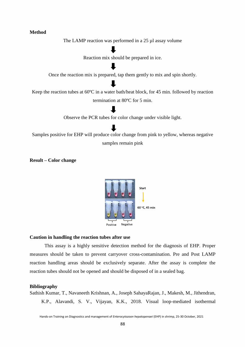
88
Hands-on Training on Diagnostics and management of Enterocytozoon hepatopenaei (EHP) in shrimp, 25-30 October, 2021
Method
The LAMP reaction was performed in a 25 µl assay volume
Reaction mix should be prepared in ice.
Once the reaction mix is prepared, tap them gently to mix and spin shortly.
Keep the reaction tubes at 60ºC in a water bath/heat block, for 45 min. followed by reaction
termination at 80ºC for 5 min.
Observe the PCR tubes for color change under visible light.
Samples positive for EHP will produce color change from pink to yellow, whereas negative
samples remain pink
Result – Color change
Caution in handling the reaction tubes after use
This assay is a highly sensitive detection method for the diagnosis of EHP. Proper
measures should be taken to prevent carryover cross-contamination. Pre and Post LAMP
reaction handling areas should be exclusively separate. After the assay is complete the
reaction tubes should not be opened and should be disposed of in a sealed bag.
Bibliography
Sathish Kumar, T., Navaneeth Krishnan, A., Joseph SahayaRajan, J., Makesh, M., Jithendran,
K.P., Alavandi, S. V., Vijayan, K.K., 2018. Visual loop-mediated isothermal

89
Hands-on Training on Diagnostics and management of Enterocytozoon hepatopenaei (EHP) in shrimp, 25-30 October, 2021
amplification (LAMP) for the rapid diagnosis of Enterocytozoonhepatopenaei (EHP)
infection. Parasitol. Res. 117, 1485–1493. https://doi.org/10.1007/s00436-018-5828-4
Kumar, T.S., Radhika, K., Rajan, J.J.S., Makesh, M., Alavandi, S.V. and Vijayan, K.K.,
2021. Closed-tube field-deployable loop-mediated isothermal amplification (LAMP)
assay based on spore wall protein (SWP) for the visual detection of
Enterocytozoonhepatopenaei (EHP). Journal of Invertebrate Pathology, 107624.

90
Hands-on Training on Diagnostics and management of Enterocytozoon hepatopenaei (EHP) in shrimp, 25-30 October, 2021
Trouble shooting in PCR and qPCR
M. Poornima, S. K. Otta, T. Bhuvaneswari, J. J. S. Rajan
General trouble shootings in PCR
Observation Possible Cause Solution
SEQUENCE
ERRORS
Low fidelity
polymerase • Choose a higher fidelity polymerase
Suboptimal reaction
conditions
• Reduce number of cycles
• Decrease extension time
• Decrease Mg++
concentration in the reaction
Unbalanced
nucleotide
concentrations • Prepare fresh deoxynucleotide mixes
Template DNA has
been damaged
• Start with a fresh template
• Try repairing good DNA template
• Repair Mix
• Limit UV exposure time when analyzing or
excising PCR product from the gel
Desired sequence
may be toxic to host
• Clone into a non-expression vector
• Use a low-copy number cloning vector
INCORRECT
PRODUCT
SIZE
Incorrect annealing
temperature
• Recalculate primer Tm values
Mispriming
• Verify that primers have no additional
complementary regions within the template
DNA
Improper Mg++
concentration
• Adjust Mg++
concentration in 0.2–1 mM
increments
Nuclease
contamination • Repeat reactions using fresh solutions
NO
PRODUCT
Incorrect annealing
temperature
• Recalculate primer Tm values
• Test an annealing temperature gradient, starting
at 5°C below the lower Tm of the primer pair
Poor primer design
• Check specific product literature for
recommended primer design
• Verify that primers are non-complementary,
both internally and to each other

91
Hands-on Training on Diagnostics and management of Enterocytozoon hepatopenaei (EHP) in shrimp, 25-30 October, 2021
• Increase length of primer
Poor primer
specificity • Verify that oligos are complementary to proper
target sequence
Insufficient primer
concentration
• Primer concentration can range from 0.05–1
µM in the reaction. Please see specific product
literature for ideal conditions
Missing reaction
component • Repeat reaction setup
Suboptimal reaction
conditions
• Optimize Mg++
concentration by testing 0.2–1
mM increments
• Thoroughly mix Mg++
solution and buffer prior
to adding to the reaction
• Optimize annealing temperature by testing an
annealing temperature gradient, starting at 5°C
below the lower Tm of the primer pair
Poor template quality
• Analyze DNA via gel electrophoresis before
and after incubation with Mg++
• Check 260/280 ratio of DNA template
Presence of inhibitor
in reaction
• Further purify starting template by alcohol
precipitation, drop dialysis or commercial clean
up kit
• Decrease sample volume
Insufficient number
of cycles • Rerun the reaction with more cycles
Incorrect
thermocycler
programming • Check program, verify times and temperatures
Inconsistent block
temperature • Test calibration of heating block
Contamination of
reaction tubes
or solutions
• Autoclave empty reaction tubes prior to use to
eliminate biological inhibitors
• Prepare fresh solutions or use new reagents and
new tubes
Complex template
• Use High-Fidelity DNA Polymerases
• Include the appropriate GC enhancer.
• For longer templates, use specific high fidelity
DNA polymerase
MULTIPLE
OR
NON-
Premature replication • Use a hot start polymerase
• Set up reactions on ice using chilled
components and add samples to thermocycler

92
Hands-on Training on Diagnostics and management of Enterocytozoon hepatopenaei (EHP) in shrimp, 25-30 October, 2021
SPECIFIC
PRODUCTS
preheated to the denaturation temperature
Primer annealing
temperature too low • Increase annealing temperature
Incorrect Mg++
concentration • Adjust Mg
++ in 0.2–1 mM increments
Poor primer design
• Check specific product literature for
recommended primer design
• Verify that primers are non-complementary,
both internally and to each other
• Increase length of primer
• Avoid GC-rich 3´ ends
Excess primer • Primer concentration can range from 0.05–1
µM in the reaction. Please see specific product
literature for ideal conditions.
Contamination with
exogenous DNA
• Use positive displacement pipettes or non-
aerosol tips
• Set-up dedicated work area and pipettor for
reaction setup
• Wear gloves during reaction setup
Incorrect template
concentration
• For low complexity templates (i.e. plasmid,
lambda, BAC DNA), use 1 pg–10 ng of DNA
per 50 µl reaction
• For higher complexity templates (i.e. genomic
DNA), use 1 ng–1 µg of DNA per 50 µl
reaction
General trouble shootings in Real Time –PCR
Problem Possible Causes Solution
The assay is insensitive and
the amplification plots look
abnormal
Inappropriate assay design
The probe inadequately
labeled.
The assay requires
optimization. Re-design the
assay if possible.
The Cq data for dilutions of
the standard curve are
irregularly spaced
Inappropriate assay design
leading to inefficient
priming.
Errors in the dilutions of the
template for the standard
Proper dilutions of template.
Purify sample template
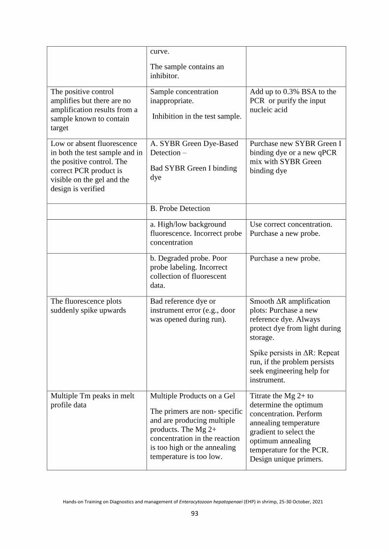
93
Hands-on Training on Diagnostics and management of Enterocytozoon hepatopenaei (EHP) in shrimp, 25-30 October, 2021
curve.
The sample contains an
inhibitor.
The positive control
amplifies but there are no
amplification results from a
sample known to contain
target
Sample concentration
inappropriate.
Inhibition in the test sample.
Add up to 0.3% BSA to the
PCR or purify the input
nucleic acid
Low or absent fluorescence
in both the test sample and in
the positive control. The
correct PCR product is
visible on the gel and the
design is verified
A. SYBR Green Dye-Based
Detection –
Bad SYBR Green I binding
dye
Purchase new SYBR Green I
binding dye or a new qPCR
mix with SYBR Green
binding dye
B. Probe Detection
a. High/low background
fluorescence. Incorrect probe
concentration
Use correct concentration.
Purchase a new probe.
b. Degraded probe. Poor
probe labeling. Incorrect
collection of fluorescent
data.
Purchase a new probe.
The fluorescence plots
suddenly spike upwards
Bad reference dye or
instrument error (e.g., door
was opened during run).
Smooth ΔR amplification
plots: Purchase a new
reference dye. Always
protect dye from light during
storage.
Spike persists in ΔR: Repeat
run, if the problem persists
seek engineering help for
instrument.
Multiple Tm peaks in melt
profile data Multiple Products on a Gel
The primers are non- specific
and are producing multiple
products. The Mg 2+
concentration in the reaction
is too high or the annealing
temperature is too low.
Titrate the Mg 2+ to
determine the optimum
concentration. Perform
annealing temperature
gradient to select the
optimum annealing
temperature for the PCR.
Design unique primers.

94
Hands-on Training on Diagnostics and management of Enterocytozoon hepatopenaei (EHP) in shrimp, 25-30 October, 2021
Artificial germination of EHP spore
T. Sathish Kumar
Spore germination is a rapid strategy carried out by spores to infect the host cell
without its knowledge. Spore germination starts with swelling of the polaroplast, and
expansion of the posterior vacuole resulting in swelling of the spore. Swelling of the spore
ultimately increases the osmotic pressure. Increasing pressure inside the spore ruptures the
thinner apex of the spore case and expels the polar tube/injection tube by eversion. Expansion
of the posterior vacuole forces the cytoplasm and nucleus through the tube. The ejected polar
tube length may range from 50–500 µm in length. The whole event of spore germination can
be completed within a few seconds. The ejected polar tube/injection tube can penetrate any
obstacle, including other microsporidian spores, and inject the sporoplasm like a needle into
the host cell cytoplasm. Spore germination is induced by different environmental triggers
specific to species and habitat. In vitro germination of spores is achieved by many physical
and chemical inducing factors such as changes in pH, dehydration followed by rehydration,
hyperosmotic settings, presence of anions or cations, and exposure to ultraviolet light or
peroxides.
Induced germination of EHP spore protocol:
10μl of purified EHP spores (1.0 × 10⁴/ml)
Add 20 μl of 1% KOH (pH > 10.5)
Incubate at room temperature for 15 min
Observeunder a lightmicroscope (100× objective lens)for spore germination
References
CIBA (Central Institute of Brackishwater Aquaculture), 2019, CIBA, Annual report 2019,
https://krishi.icar.gov.in/jspui/bitstream/123456789/43559/1/CIBA%20Annual%20Report%2
02019.pdf.

95
Hands-on Training on Diagnostics and management of Enterocytozoon hepatopenaei (EHP) in shrimp, 25-30 October, 2021
General laboratory biosafety
In view of rapid development, in both basic and applied research in the field of
aquaculture and related disciplines, there are concerns with respect risks to research staff
during laboratory operations and overall risks due to the waste generated during the
research activities. All the general laboratory safety regulations outlined for any R&D lab
also apply to the shared PCR laboratories as well. The purpose of this chapter is to provide
specific guidance and protocols for the protection of the users of the shared PCR
laboratories. The plan proposed here will help to optimize use of equipment, space and
safety of the workers at large.
Biosafety requirements in shared PCR laboratories
Biosafety for research staff
Personal protection
1. Laboratory coveralls, gowns or uniforms and appropriate gloves must be worn for
all procedures that may involve direct or accidental contact with hazardous
chemicals and other potentially infectious materials
2. After use, gloves should be removed aseptically and hands must then be washed.
3. Personnel must wash their hands after handling infectious materials and animals,
and before they leave the laboratory working areas.
4. Safety glasses, face shields (visors) or other protective devices must be worn when
it is necessary to protect the eyes and face from splashes, impacting objects and
sources of artificial ultraviolet radiation.
5. It is prohibited to wear protective laboratory clothing outside the laboratory, e.g. in
canteens, coffee rooms, offices, libraries, staff rooms and toilets.
6. Open-toed footwear must not be worn in laboratories.
7. Eating, drinking, smoking, applying cosmetics and handling contact lenses is
prohibited in the laboratory working areas.
8. Storing human foods or drinks anywhere in the laboratory working areas is
prohibited.
9. Protective laboratory clothing that has been used in the laboratory must not be
stored in the same lockers or cupboards as street clothing.

96
Hands-on Training on Diagnostics and management of Enterocytozoon hepatopenaei (EHP) in shrimp, 25-30 October, 2021
Procedures
1. Pipetting by mouth must be strictly forbidden.
2. Materials must not be placed in the mouth. Labels must not be licked.
3. All technical procedures should be performed in a way that minimizes the
formation of aerosols and droplets.
4. The use of hypodermic needles and syringes should be limited. They must not be
used as substitutes for pipetting devices or for any purpose other than injection or
aspiration of fluids from laboratory animals.
5. All spills, accidents and overt or potential exposures to infectious materials must
be reported to the laboratory supervisor. A written record of such accidents and
incidents should be maintained.
6. A written procedure for the clean-up of all spills must be developed and followed.
7. Contaminated liquids must be decontaminated (chemically or physically) before
discharge to the sanitary sewer. An effluent treatment system may be required,
depending on the risk assessment for the agent(s) being handled.
8. Written documents that are expected to be removed from the laboratory need to be
protected from contamination while in the laboratory.
Guidelines for biosafety - laboratory operations
1. „Learners‟ in the lab should be taught the „dos and don‟ts‟ of lab practices before
they start the work.
2. Laboratories should be clean and items arranged in an orderly manner, free from
materials that are not pertinent to the work.
3. Work surfaces must be decontaminated after any spill of potentially dangerous
material and at the end of the working day.
4. All contaminated materials, specimens and cultures must be decontaminated before
disposal or cleaning for reuse.
5. The cupboards/cabinets in the laboratory should be labeled /categorized properly for
easy availability/identification of materials (chemicals /glassware /plasticware/or
any other items) and items should be replaced properly.
6. Laboratory items/furniture (glassware/equipments/fumehood/ balances) have to be
used for the identified purpose only.
7. Beverages and other food items should never be served / consumed inside the lab.

97
Hands-on Training on Diagnostics and management of Enterocytozoon hepatopenaei (EHP) in shrimp, 25-30 October, 2021
8. Inflammable liquids should be kept in separate containers with red paint marking
(danger mark) and should be stored in fire- proof cabinets.
9. Acids and volatile solvents and digestion of biological samples should be handled
strictly under fume hood.
10. Every lab should have first aid box with emergency eye washer. Researchers/staff
should use aprons whenever/wherever they handle hazardous materials.
11. Corrosive liquids should be handled properly using corrosive-resistant hand
gloves.
12. Gas cylinders containing explosive gases should be kept outside the laboratories.
Guidelines for bio-safety- disposal of waste generated during research activities
Hazardous laboratory wastes need to be monitored and disposed properly. There is a
need for bio-safety guidelines for proper disposal of waste generated in research activities.
Materials for decontamination and disposal should be pre-sorted and placed in autoclavable
plastic bags that are colour-coded- according to whether the contents are bio-degradable,
non-biodegradable or sharps. Materials to be autoclaved or incinerated should be
separated. No pre-cleaning should be attempted of any contaminated materials before it is
decontaminated by autoclaving or disinfection. The procedure for handling/disposal for
contaminated materials/wastes along with separation system and categories to be followed
are listed as below:
1. Non-contaminated (non-infectious) waste can be disposed of as general waste.
2. Contaminated “sharps” – hypodermic needles, scalpels, knives and broken glass;
these should always be collected in puncture-proof containers fitted with covers
and treated as infectious. Disposable syringes, used alone or with needles, should
be placed in sharps disposal containers and incinerated, with prior autoclaving if
required. Sharps disposal containers must be disposed off before filled to the
capacity. Sharps disposal containers must not be discarded in landfills.
Contaminated material should be decontaminated by autoclaving and thereafter
washing and reuse or recycling should be done (such as glass petri-plates, test
tubes, glass wares used for microbiological work.
3. Biodegradable contaminated material (agar, culture broth, tissue culture slants,
cotton and swabs, protein and fats) should be segregated in colour coded bags and
decontaminated by autoclaving before disposal.

98
Hands-on Training on Diagnostics and management of Enterocytozoon hepatopenaei (EHP) in shrimp, 25-30 October, 2021
4. Non-biodegradable (plastic tubes and tips,organic recalcitrant chemicals, gloves, gel
wastes, glass, media tins and bottles, rubber, aluminium foils etc.) should be
segregated in colour coded bags before decontamination and disposal.
5. The research division/section/unit heads should train personnel working in various
labs to manage the waste generated as per these guidelines. Extra sensitization is
required for those working with disease pathogens and molecular biology work.
6. Concentrated stock solution of toxic chemicals such as heavy metals, nitrogenous,
sulfurous and phosphorous compounds, reagents used for routine analysis and
ethidium bromide should not be disposed in the wash basin. They should be
deactivated before disposal. To avoid the need for deactivation, minimum quantity
of stock solution as per requirement should be prepared, preserved properly and also
be used fully without any disposal. Some examples of hazardous chemicals and
their deactivation procedures are listed in Table 1.
7. All lab workers should wear appropriate safety gears while handling waste.
8. Research workers should handle gel containing ethidium bromide only with gloves.
Such gels should be biosorbed with activated charcoal before disposal. The gloves
used for the molecular work, should never be exposed outside the lab or allowed to
contaminate other areas.
9. Glassware / plasticware used for micro- organisms should properly be sterilized.
Hazards of microbial/other pathogen waste generated should properly be disposed
by adopting good lab practices, for which persons involved should be trained.
10. Dead and post-experimental animals should be disposed of by incinerating them in
pits covered with soil.
11. Based on the activity of hazardous waste, they can be identified as corrosive,
inflammable, ignitable, reactive, toxic, infectious and radioactive. If such wastes
are identified and their hazardous nature cannot be eliminated, special precautions
as mentioned in the Table-1 need to be taken and if required separate guidelines
would need to be developed.
12. Gloves (contaminated with ethidium bromide or other toxic substances) shouldnot
be worn to answer the telephone or use other equipment in the lab.
13. Ethidium bromide solutions should be disposed of in the red waste
containerlocated in the fume hood/staining bench of PCR lab. If hazardous waste
other than ethidium bromide is put into these containers itshould be clearly marked

99
Hands-on Training on Diagnostics and management of Enterocytozoon hepatopenaei (EHP) in shrimp, 25-30 October, 2021
on the label attached.
14. Ethidium bromide spills should be cleaned up immediately, using
properprecautions (wear gloves, put material used to clean up in bio-hazardous
wastebins, wipe contaminated area down well with ethanol or detergent).
15. Used gels and gel waste should be discarded in the bio- hazardous waste bin,
which is located beneath the gel electrophoresis benches.
16. Used gels and gel waste should not be put down any of the sinks.
17. Packing and transportation must follow applicable national and/or international
regulations.
Laboratory Rules for shared laboratory
Each laboratory should develop a system of regulations most suited for managing
smooth and ambient work flow.
1. Users should be provided with their own bench space when possible. It is the
responsibility of the user to keep their bench space clean. When bench space is
shared users should be respectful of the other user‟s space.
4. Users are responsible for their own dishes and spills. Clean up is NOT the
responsibility of the laboratory supervisors.
5. Dishes should NOT be left in the gel electrophoresis tank.
6. Broken or malfunctioning equipment should be reported to a laboratory supervisor
IMMEDIATELY.
7. Instructions on how to use the thermocyclers, spectrophotometer, Imaging System
and centrifuge are kept beside them.
8. All users must make entry and sign in the logbook prior to use.
9. Pipettes labeled for PCR use should be used for PCR only. NO EXCEPTIONS.
Table 1- Disposal of chemical wastes in a PCR laboratory
Chemical Method of deactivation
Ethidium bromide Filter the ethidium bromide solution through charcoal
filtration. Pour filtrate down the drain. Place charcoal
filter in sealed bag and place in bio hazardous waste box
for incineration

100
Hands-on Training on Diagnostics and management of Enterocytozoon hepatopenaei (EHP) in shrimp, 25-30 October, 2021
Recombinant DNA Deactivate recombinant DNA using DEPC or UV
radiation or by autoclaving
Heavy metals Minimum quantity of stock solution as per requirement
should be prepared, preserved properly and also be used
fully without any disposal because heavy metals cannot
be biodegraded or eliminated
Nitrogenous, sulfurous and
phosphorous compounds
Minimum quantity of stock solution as per requirement
should be prepared, preserved properly and also be used
fully without any disposal.
Reagents for routine analysis
of chemical and
biochemical parameters
Minimum quantity of reagents as per requirement
should be prepared and should be used fully.
Volatile solvents
and acids
Volatile solvents and acids should strictly be
handled under fume hood. In the case of any leakage or
spillage, all the doors and windows should be
opened and the entry of persons should be
restricted in the lab till complete escape of fumes.
In the case of person having any accident with the
spillage of acid, only water should be applied (please
avoid the use of alkali) and should immediately be
taken to nearby hospital. All the labs should have
medical aids.
Guidelines for ethidium bromide waste management and disposal
Ethidium bromide is a compound used in many PCR laboratories for visualization of
nucleic acid during gel electrophoresis applications. Ethidium bromide solution is
incorporated into the electrophoresis gel as a dye for the DNA, RNA, or other molecules
to be visualized. Ethidium bromide is mutagenic and moderately toxic and must be
handled with care.
The powder form is considered an irritant to the upper respiratory tract, eyes, and skin,
hence preparation of stock solutions and any operations capable of generating ethidium
bromide dust or aerosols should be done in a fume hood to prevent inhalation. Nitrile

101
Hands-on Training on Diagnostics and management of Enterocytozoon hepatopenaei (EHP) in shrimp, 25-30 October, 2021
gloves, a lab coat and eye protection should be worn at all times, as with working with
any hazardous material.
When working with ethidium bromide, minimize the potential for spills. Where practical,
purchase pre-mixed stock solutions from chemical manufacturers in lieu of preparing
solutions. If solutions of ethidium bromide must be prepared, consider performing this
process in a fume hood.
Spills must be addressed immediately; for small spills (<20 ml) of concentrations up
to 10 mg/ml; absorb with paper towel and then treat the area with 70-95%
ethanol/isopropanol and wipe up-all to be collected as hazardous waste. Repeat as
necessary (if still fluorescing). Final rinse with soap and water (paper towels for this
step were discarded into regular trash). DO NOT clean ethidium bromide spills with
bleach solutions.
When an ultraviolet light source is used in work with ethidium bromide, added
caution is required. As a general rule, avoid exposing unprotected skin and eyes to
intense UV sources.
Ethidium bromide waste should not be poured down the drain, or thrown in the
trash, unless the waste has been deactivated or filtered. The following are the
recommended disposal procedures for ethidium bromide:
Dry the gels under the hood in an open container before bringing to the waste room
for disposal. Dilute aqueous solutions containing <0.5 mg/ml ethidium bromide can
be filtered or deactivated. Concentrated solutions (>0.5 mg/ml) should be collected
for disposal as hazardous waste. Similarly, solutions that contain heavy metals,
organics, cyanides or sulfides should be collected and disposed of as hazardous
waste. Filtering the aqueous ethidium bromide waste solutions, free of other
contaminants, through a bed of activated charcoal is a relatively simple and effective
method for removal of ethidium bromide. The filtrate may then be poured down the
drain.
Autoclaving chemical waste creates exposure to the chemicals as they heat and
volatilizes; hence should not be attempted.
Consider switching to less-toxic alternatives to ethidium bromide (SYBR Safe DNA
gel stain) to reduce potential hazardous exposures in the lab.

102
Hands-on Training on Diagnostics and management of Enterocytozoon hepatopenaei (EHP) in shrimp, 25-30 October, 2021
Bibliography
Centers for Disease Control and Prevention. 2011. Guidelines for Safe Work Practices in
Human and Animal Medical Diagnostic Laboratories, MMWR 2011; U.S. Department
of Health and Human Services, Atlanta, GA 30333, 61 (Suppl), p 102.
CPCSEA, 2003. Guidelines For Laboratory Animal Facility, Committee for the Purpose of
Control and Supervision on Experiments on Animals, N. 13/1, 3rd Seaward Road,
Valmiki Nagar, Thiruvanmiyur, Chennai, Tamil Nadu (India), p 38.
DBT, 2011. Guidelines and Handbook for Institutional Biosafety Committees (IBSCs),
Second edition, Ministry of Science & Technology & Biotech Consortium India
Limited, New Delhi, p 120.
ICMR, 2008. Guidelines for Good Clinical Laboratory Practices (GCLP). Indian Council of
Medical Research, New Delhi, p 46.
WHO, 2004. Laboratory biosafety manual, Third edition, World Health Organization,
Geneva, p 178.

Notes

Notes

Notes

Notes

ICAR-Central Institute of Brackishwater AquacultureISO 9001:2015 Certified
75, Santhome High Road, M.R.C. Nagar, Chennai - 600 028.Phone: 044-24610565, 24618817, 24616948, Telefax: 044-24613818, 24610311
Email: [email protected], Website: www.ciba.res.in
/ icarciba
civer.indd 3 10/23/2021 9:32:19 AM