Embed Size (px)
Citation preview
Hand
Gross Anatomy
K. Bo Foreman, PT, PhD
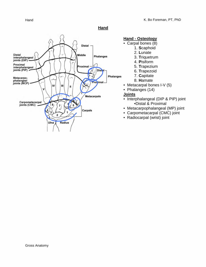
Hand - Osteology • Carpal bones (8)
1. Scaphoid 2. Lunate 3. Triquetrum 4. Pisiform 5. Trapezium 6. Trapezoid 7. Capitate 8. Hamate
• Metacarpal bones I-V (5) • Phalanges (14) Joints • Interphalangeal (DIP & PIP) joint
•Distal & Proximal • Metacarpophalangeal (MP) joint • Carpometacarpal (CMC) joint • Radiocarpal (wrist) joint
Hand
Hand
Gross Anatomy
K. Bo Foreman, PT, PhD
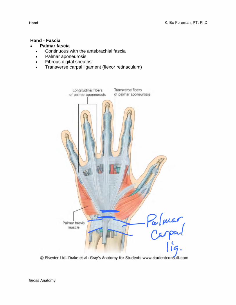
Hand - Fascia • Palmar fascia
• Continuous with the antebrachial fascia • Palmar aponeurosis • Fibrous digital sheaths • Transverse carpal ligament (flexor retinaculum)
Hand
Gross Anatomy
K. Bo Foreman, PT, PhD
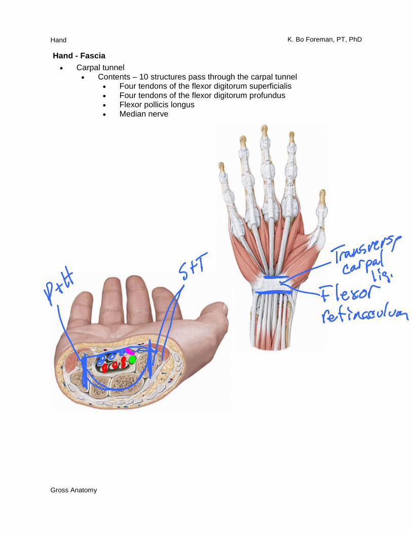
Hand - Fascia • Carpal tunnel
• Contents – 10 structures pass through the carpal tunnel • Four tendons of the flexor digitorum superficialis • Four tendons of the flexor digitorum profundus • Flexor pollicis longus • Median nerve
Hand
Gross Anatomy
K. Bo Foreman, PT, PhD
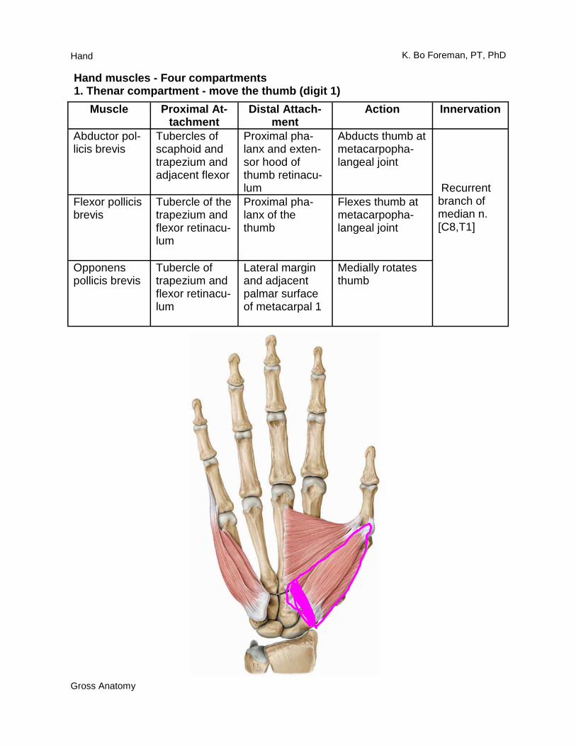
Hand muscles - Four compartments 1. Thenar compartment - move the thumb (digit 1)
Muscle Proximal At-tachment
Distal Attach-ment
Action Innervation
Abductor pol-licis brevis
Tubercles of scaphoid and trapezium and adjacent flexor
Proximal pha-lanx and exten-sor hood of thumb retinacu-lum
Abducts thumb at metacarpopha-langeal joint
Recurrent branch of median n. [C8,T1]
Flexor pollicis brevis
Tubercle of the trapezium and flexor retinacu-lum
Proximal pha-lanx of the thumb
Flexes thumb at metacarpopha-langeal joint
Opponens pollicis brevis
Tubercle of trapezium and flexor retinacu-lum
Lateral margin and adjacent palmar surface of metacarpal 1
Medially rotates thumb
Hand
Gross Anatomy
K. Bo Foreman, PT, PhD
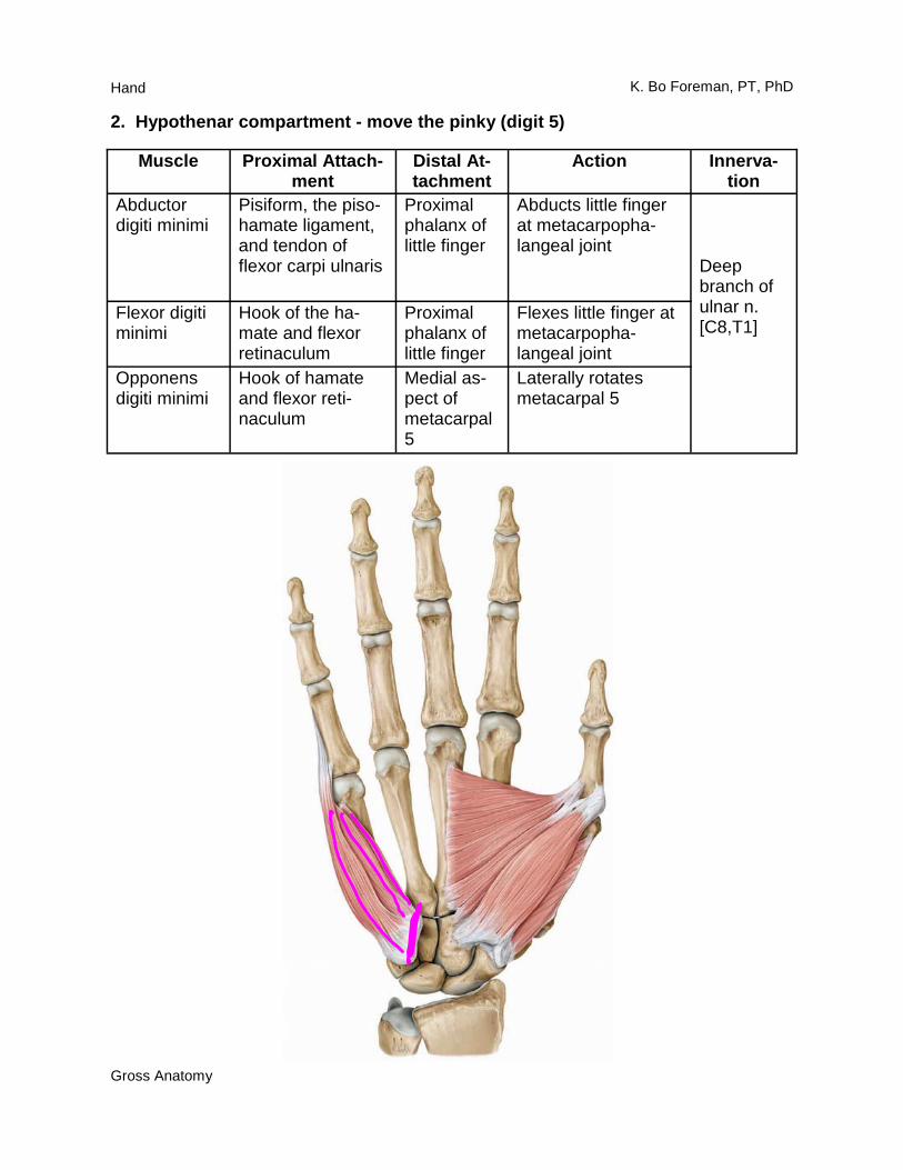
2. Hypothenar compartment - move the pinky (digit 5)
Muscle Proximal Attach-ment
Distal At-tachment
Action Innerva-tion
Abductor digiti minimi
Pisiform, the piso-hamate ligament, and tendon of flexor carpi ulnaris
Proximal phalanx of little finger
Abducts little finger at metacarpopha-langeal joint
Deep branch of ulnar n. [C8,T1]
Flexor digiti minimi
Hook of the ha-mate and flexor retinaculum
Proximal phalanx of little finger
Flexes little finger at metacarpopha-langeal joint
Opponens digiti minimi
Hook of hamate and flexor reti-naculum
Medial as-pect of metacarpal 5
Laterally rotates metacarpal 5
Hand
Gross Anatomy
K. Bo Foreman, PT, PhD
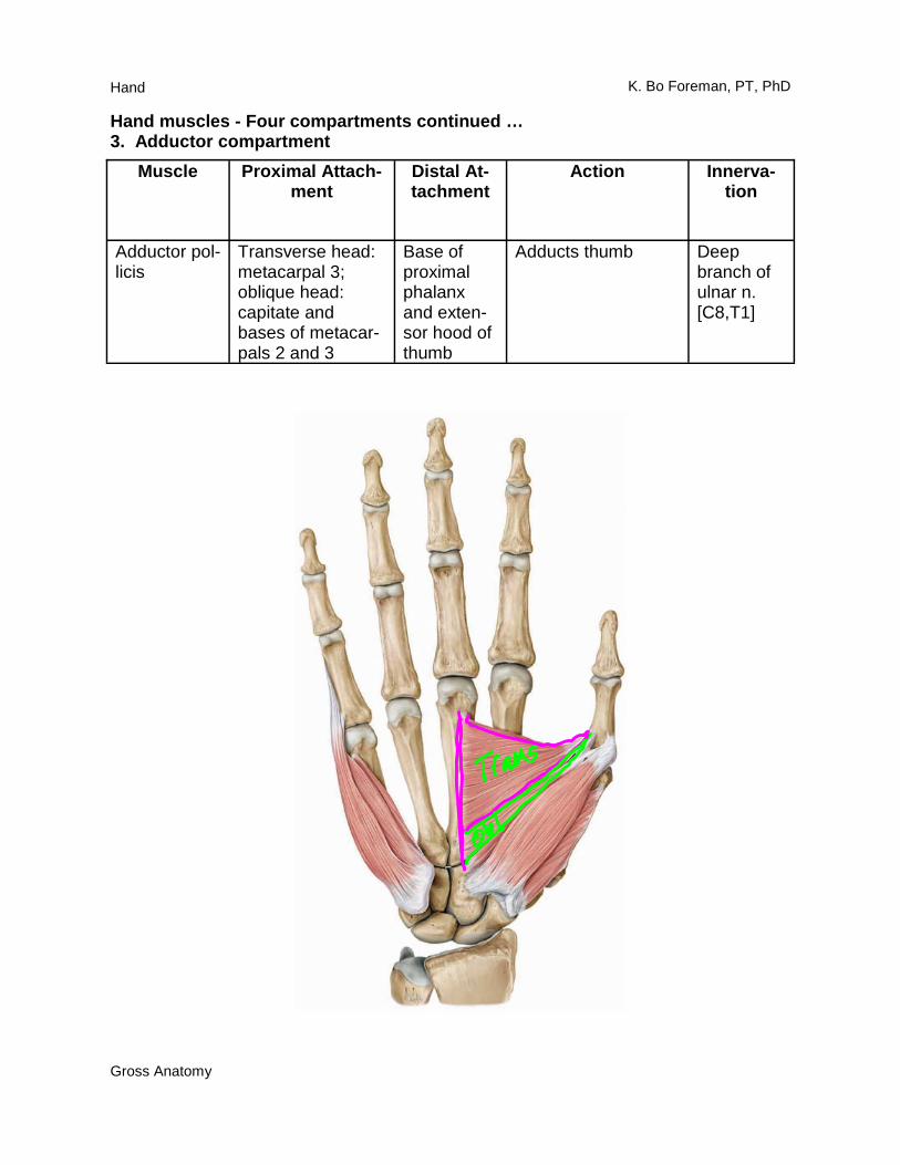
Muscle Proximal Attach-ment
Distal At-tachment
Action Innerva-tion
Adductor pol-licis
Transverse head: metacarpal 3; oblique head: capitate and bases of metacar-pals 2 and 3
Base of proximal phalanx and exten-sor hood of thumb
Adducts thumb Deep branch of ulnar n. [C8,T1]
Hand muscles - Four compartments continued … 3. Adductor compartment
Hand
Gross Anatomy
K. Bo Foreman, PT, PhD
Muscle Proximal Attachment
Distal At-tachment
Action Innervation
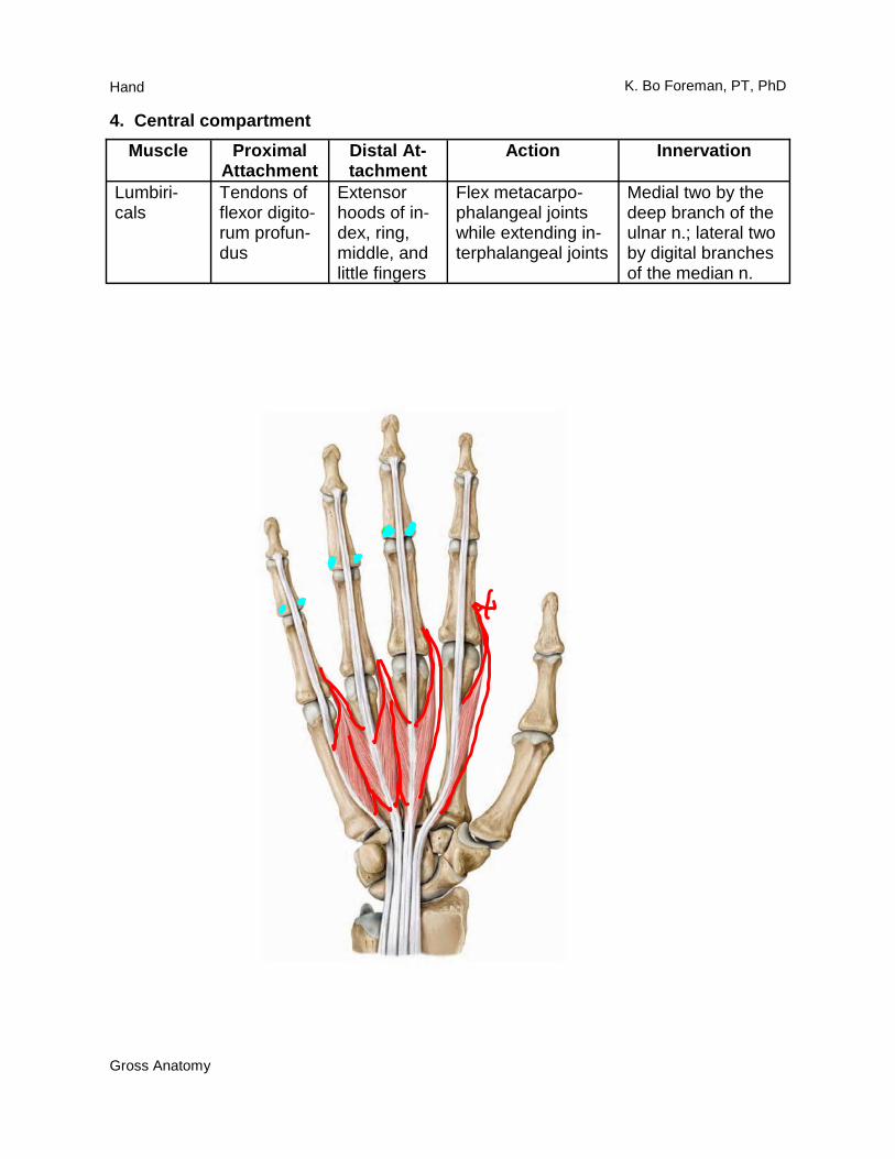
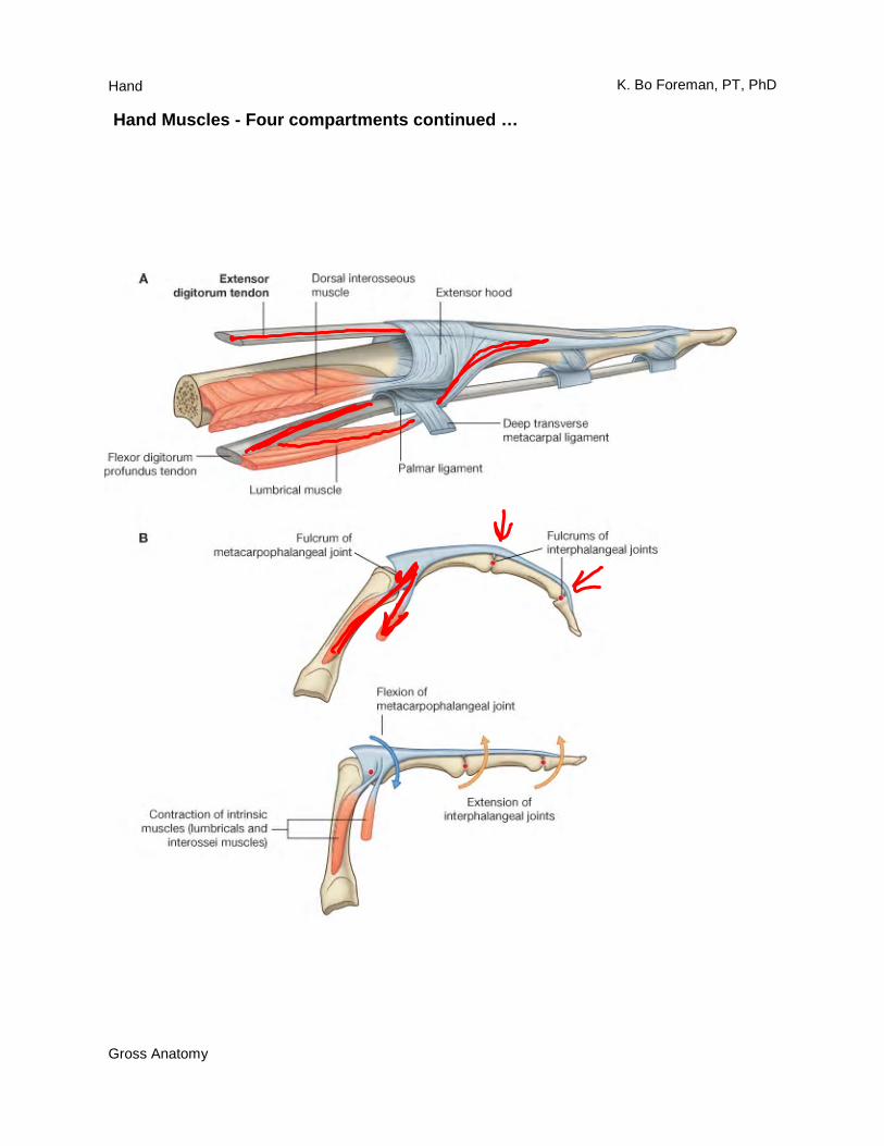
Lumbiri-cals
Tendons of flexor digito-rum profun-dus
Extensor hoods of in-dex, ring, middle, and little fingers
Flex metacarpo-phalangeal joints while extending in-terphalangeal joints
Medial two by the deep branch of the ulnar n.; lateral two by digital branches of the median n.
4. Central compartment
Hand
Gross Anatomy
K. Bo Foreman, PT, PhD
Muscle Proximal Attachment
Distal At-tachment
Action Innervation
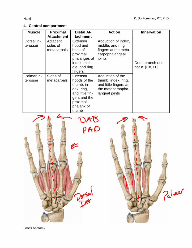
Dorsal in-terossei
Adjacent sides of metacarpals
Extensor hood and base of proximal phalanges of index, mid-dle, and ring fingers
Abduction of index, middle, and ring fingers at the meta-carpophalangeal joints
Deep branch of ul-nar n. [C8,T1]
Palmar in-terossei
Sides of metacarpals
Extensor hoods of the thumb, in-dex, ring, and little fin-gers and the proximal phalanx of thumb
Adduction of the thumb, index, ring, and little fingers at the metacarpopha-langeal joints
4. Central compartment
Hand
Gross Anatomy
K. Bo Foreman, PT, PhD
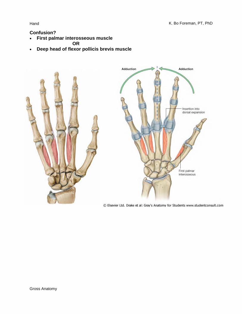
Confusion? • First palmar interosseous muscle OR • Deep head of flexor pollicis brevis muscle
Hand
Gross Anatomy
K. Bo Foreman, PT, PhD
Hand Muscles - Four compartments continued …
Hand
Gross Anatomy
K. Bo Foreman, PT, PhD
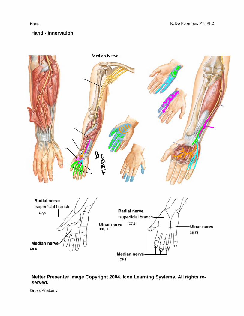
Hand - Innervation
Netter Presenter Image Copyright 2004. Icon Learning Systems. All rights re-served.
C7,8
C6-8
C8,T1 C7,8
C6-8
C8,T1
Hand
Gross Anatomy
K. Bo Foreman, PT, PhD
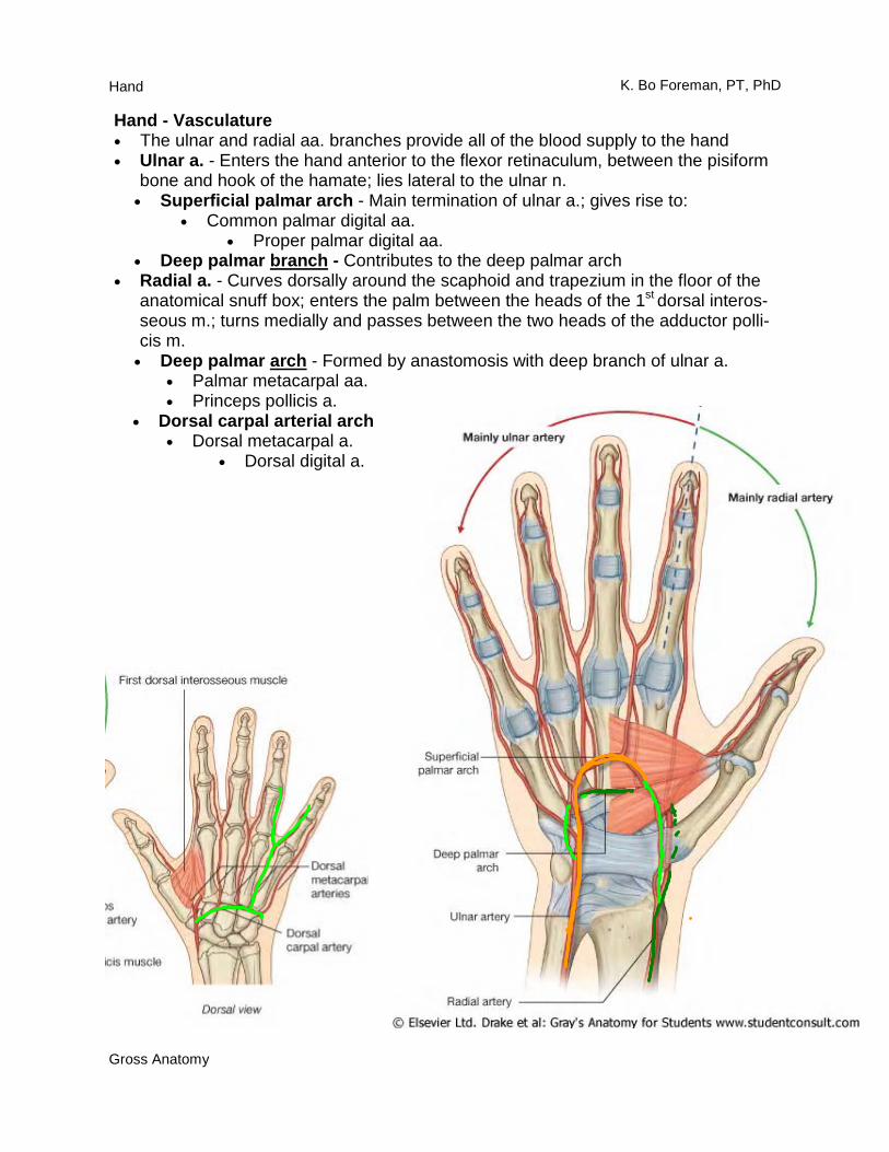
Hand - Vasculature • The ulnar and radial aa. branches provide all of the blood supply to the hand • Ulnar a. - Enters the hand anterior to the flexor retinaculum, between the pisiform
bone and hook of the hamate; lies lateral to the ulnar n. • Superficial palmar arch - Main termination of ulnar a.; gives rise to:
• Common palmar digital aa. • Proper palmar digital aa.
• Deep palmar branch - Contributes to the deep palmar arch • Radial a. - Curves dorsally around the scaphoid and trapezium in the floor of the
anatomical snuff box; enters the palm between the heads of the 1st dorsal interos-seous m.; turns medially and passes between the two heads of the adductor polli-cis m.
• Deep palmar arch - Formed by anastomosis with deep branch of ulnar a. • Palmar metacarpal aa. • Princeps pollicis a.
• Dorsal carpal arterial arch • Dorsal metacarpal a.
• Dorsal digital a.
Hand
Gross Anatomy
K. Bo Foreman, PT, PhD
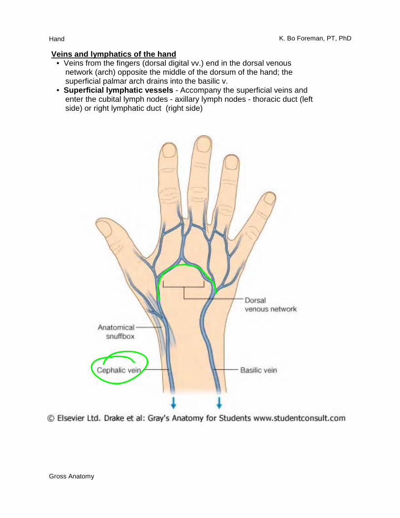
Veins and lymphatics of the hand • Veins from the fingers (dorsal digital vv.) end in the dorsal venous network (arch) opposite the middle of the dorsum of the hand; the superficial palmar arch drains into the basilic v. • Superficial lymphatic vessels - Accompany the superficial veins and enter the cubital lymph nodes - axillary lymph nodes - thoracic duct (left side) or right lymphatic duct (right side)
Hand
Gross Anatomy
K. Bo Foreman, PT, PhD
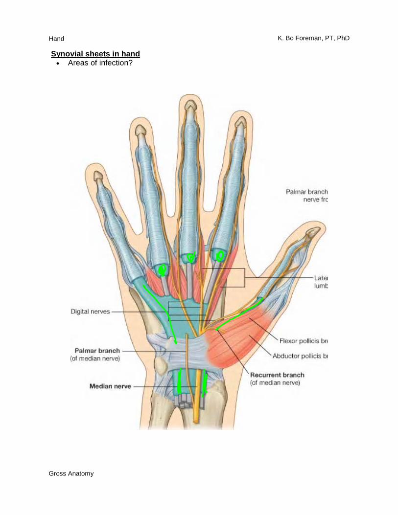
Synovial sheets in hand • Areas of infection?
Hand
Gross Anatomy
K. Bo Foreman, PT, PhD
Hand - Innervation • Median n.
• Palmar branch - Skin of the central region of the palm (sensory) • Enters the hand through carpal tunnel (the only nerve to do so) • Recurrent branch
• Thenar mm. (motor) • Opponens pollicis m. • Abductor pollicis brevis m. • Flexor pollicis brevis m.
• Common palmar digital nn. (mixed) • Lumbrical mm. 1-2 (motor) • Proper palmar digital nn. (sensory)
• Ulnar n.
• Dorsal branch (sensory) • Palmar branch (sensory) • Enters the hand by emerging deep from the flexor carpi ulnaris tendon
(Guyon’s canal) • Superficial branch of the ulnar n. (sensory)
• Common palmar digital nn. • Proper palmar digital nn.
• Deep branch (motor) • Adductor pollicis m. • Hypothenar mm.
• Opponens digiti minimi m. • Flexor digiti minimi m. • Abductor digiti minimi m.
• Lumbrical mm. 3-4 • Palmar interossei and dorsal interossei mm. • Deep head of the flexor pollicis brevis m.
• Radial n. • Superficial branch (sensory)
Hand
Gross Anatomy
K. Bo Foreman, PT, PhD
Upper limb nerves Terminal branches of the brachial plexus • Musculocutaneous n.
• Motor to anterior compartment of arm • Coracobrachialis m. • Biceps brachii m. • Brachialis m.
• Sensory to forearm • Lateral cutaneous nerve of the forearm
• Median n. • Motor to superficial anterior compartment of the forearm
• Pronator teres m. • Flexor carpi radialis m. • Palmaris longus m. • Flexor digitorum superficialis m.
• Motor to deep anterior compartment of the forearm via anterior interosseous n.
• Radial ½ of flexor digitorum profundus m. • Flexor pollicis longus m. • Pronator quadratus m.
• Motor to the hand via recurrent median n. • Abductor pollicis brevis m. • Opponens pollicis m. • Superficial head of the flexor pollicis brevis m.
• Motor to the hand via common palmar digital nn. • 1st and 2nd lumbrical mm.
• Sensory to hand • Palmar branch of median n. • Common palmar digital nn.
• Proper palmar digital nn. • Ulnar n.
• Motor to anterior compartment of the forearm • Ulnar ½ of flexor digitorum profundus m. • Flexor carpi ulnaris m.
• Motor to hand via superficial branch of the ulnar n. • Palmaris brevis m.
• Motor to hand via deep branch of the ulnar n. • Abductor digiti minimi m. • Flexor digiti minimi (brevis) m. • Opponens digiti minimi m. • Palmar interosseous mm. • Dorsal interosseous mm. • 3rd and 4th lumbrical mm. • Adductor pollicis m. • Deep head of the flexor pollicis brevis m.
Hand
Gross Anatomy
K. Bo Foreman, PT, PhD
• Ulnar n. continued • Sensory to hand
• Dorsal branch of the ulnar n. • Dorsal digital nn.
• Palmar branch of the ulnar n. • Superficial branch of the ulnar n.
• Common palmar digital nn. • Proper palmar digital nn.
• Axillary n. • Motor to posterior shoulder
• Deltoid m. • Teres minor m.
• Sensory to posterior shoulder • Superior lateral cutaneous nerve of the arm
• Radial n. • Motor to posterior compartment of the arm
• Triceps brachii m. • Motor to posterior compartment of the forearm
• Anconeus m. • Brachioradialis m. • Extensor carpi radials longus m.
• Motor to the lateral portion of the brachialis m. • Motor to posterior compartment of the forearm via the deep radial n.
• Extensor carpi radialis brevis m. • Motor to the posterior compartment of the forearm via the posterior interos-
seous n. • Supinator m. • Extensor carpi ulnaris m. • Extensor digitorum m. • Extensor digiti minimi m. • Extensor indicis m. • Extensor pollicis longus m. • Abductor pollicis longus m. • Extensor pollicis brevis m.
• Sensory to arm • Posterior cutaneous nerve of the arm • Inferior lateral cutaneous nerve of the arm
• Sensory to forearm • Posterior cutaneous nerve of the forearm
• Sensory to hand • Superficial branch of the radial n.
• Dorsal digital nn.