Embed Size (px)
Citation preview

Hand and Foot Color Change:Diagnosis and Management
Dustin E. Fleck, MD,* Mark F. Hoeltzel, MD*
*Division of Pediatric Rheumatology, Department of Pediatrics, University of Michigan, Ann Arbor, MI
Education Gap
Primary care clinicians are challenged to evaluate an array of different
hand and foot color changes in kids, as treatment may range from
reassurance of a benign, self-limited condition to further diagnostic
evaluation and treatment with vasodilator pharmacotherapy. The most
common and well-known causes of these color changes are Raynaud
phenomenon, acrocyanosis, and the more rare but serious
erythromelalgia. Clinicians should be aware of the clinical presentation,
symptoms, etiologic origins, diagnostic evaluation, treatment, and
potential complications of Raynaud phenomenon, acrocyanosis, and
erythromelalgia in children.
Objectives After completing this article, readers should be able to:
1. Identify the various causes of hand and foot color change in children
and adolescents.
2. Recognize the differences in signs and symptoms of acrocyanosis,
erythromelalgia, and Raynaud phenomenon.
3. Recognize complications associated with acrocyanosis,
erythromelalgia, and Raynaud phenomenon.
4. Describe current treatments for acrocyanosis, erythromelalgia, and
Raynaud phenomenon.
ACROCYANOSIS
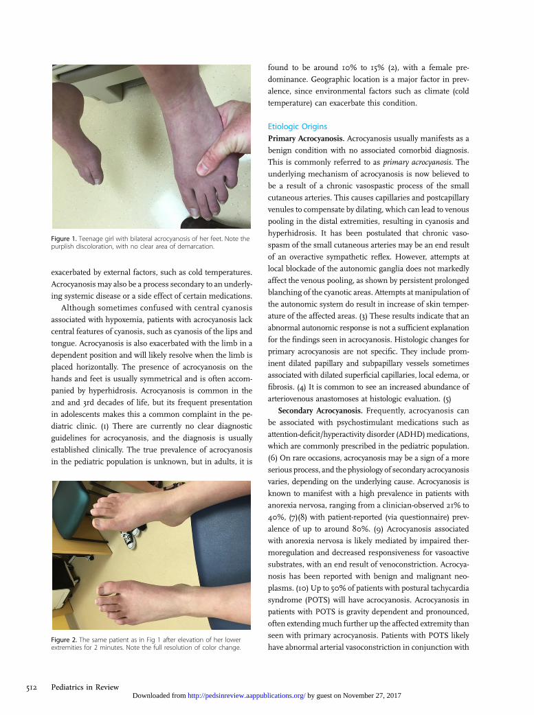
Clinical PresentationAcrocyanosis is a clinically benign process that is known to cause alarm in patients
and their parents. Acrocyanosis typically presents as symmetrical blue and/or
purple discoloration in the peripheral extremities, usually either the hands or the
feet (Figs 1, 2, 3, 4). In infants, acrocyanosis also includes the perioral area but not
the lips or tongue, where central cyanosis becomes a concern. It is often seen after
bathing, after feeding, or in a cool environment. It is evanescent and benign.
The bluish discoloration in acrocyanosis is often asymptomatic, without clear
demarcation of color change. Acrocyanosis episodes can be either precipitated or
AUTHORDISCLOSUREDrs Fleck and Hoeltzelhave disclosed no financial relationshipsrelevant to this article. This commentary doesnot contain a discussion of an unapproved/investigative use of a commercial product/device.
ABBREVIATIONS
ADHD attention-deficit/hyperactivity
disorder
ANA antinuclear antibody
POTS postural tachycardia syndrome
Vol. 38 No. 11 NOVEMBER 2017 511 by guest on November 27, 2017http://pedsinreview.aappublications.org/Downloaded from
exacerbated by external factors, such as cold temperatures.
Acrocyanosis may also be a process secondary to an underly-
ing systemic disease or a side effect of certain medications.
Although sometimes confused with central cyanosis
associated with hypoxemia, patients with acrocyanosis lack
central features of cyanosis, such as cyanosis of the lips and
tongue. Acrocyanosis is also exacerbated with the limb in a
dependent position and will likely resolve when the limb is
placed horizontally. The presence of acrocyanosis on the
hands and feet is usually symmetrical and is often accom-
panied by hyperhidrosis. Acrocyanosis is common in the
2nd and 3rd decades of life, but its frequent presentation
in adolescents makes this a common complaint in the pe-
diatric clinic. (1) There are currently no clear diagnostic
guidelines for acrocyanosis, and the diagnosis is usually
established clinically. The true prevalence of acrocyanosis
in the pediatric population is unknown, but in adults, it is
found to be around 10% to 15% (2), with a female pre-
dominance. Geographic location is a major factor in prev-
alence, since environmental factors such as climate (cold
temperature) can exacerbate this condition.
Etiologic OriginsPrimary Acrocyanosis. Acrocyanosis usually manifests as a
benign condition with no associated comorbid diagnosis.
This is commonly referred to as primary acrocyanosis. The
underlying mechanism of acrocyanosis is now believed to
be a result of a chronic vasospastic process of the small
cutaneous arteries. This causes capillaries and postcapillary
venules to compensate by dilating, which can lead to venous
pooling in the distal extremities, resulting in cyanosis and
hyperhidrosis. It has been postulated that chronic vaso-
spasm of the small cutaneous arteries may be an end result
of an overactive sympathetic reflex. However, attempts at
local blockade of the autonomic ganglia does not markedly
affect the venous pooling, as shown by persistent prolonged
blanching of the cyanotic areas. Attempts at manipulation of
the autonomic system do result in increase of skin temper-
ature of the affected areas. (3) These results indicate that an
abnormal autonomic response is not a sufficient explanation
for the findings seen in acrocyanosis. Histologic changes for
primary acrocyanosis are not specific. They include prom-
inent dilated papillary and subpapillary vessels sometimes
associated with dilated superficial capillaries, local edema, or
fibrosis. (4) It is common to see an increased abundance of
arteriovenous anastomoses at histologic evaluation. (5)
Secondary Acrocyanosis. Frequently, acrocyanosis can
be associated with psychostimulant medications such as
attention-deficit/hyperactivity disorder (ADHD)medications,
which are commonly prescribed in the pediatric population.
(6) On rare occasions, acrocyanosis may be a sign of a more
serious process, and the physiology of secondary acrocyanosis
varies, depending on the underlying cause. Acrocyanosis is
known to manifest with a high prevalence in patients with
anorexia nervosa, ranging from a clinician-observed 21% to
40%, (7)(8) with patient-reported (via questionnaire) prev-
alence of up to around 80%. (9) Acrocyanosis associated
with anorexia nervosa is likely mediated by impaired ther-
moregulation and decreased responsiveness for vasoactive
substrates, with an end result of venoconstriction. Acrocya-
nosis has been reported with benign and malignant neo-
plasms. (10) Up to 50% of patients with postural tachycardia
syndrome (POTS) will have acrocyanosis. Acrocyanosis in
patients with POTS is gravity dependent and pronounced,
often extendingmuch further up the affected extremity than
seen with primary acrocyanosis. Patients with POTS likely
have abnormal arterial vasoconstriction in conjunction with
Figure 1. Teenage girl with bilateral acrocyanosis of her feet. Note thepurplish discoloration, with no clear area of demarcation.
Figure 2. The same patient as in Fig 1 after elevation of her lowerextremities for 2 minutes. Note the full resolution of color change.
512 Pediatrics in Review by guest on November 27, 2017http://pedsinreview.aappublications.org/Downloaded from

decreased overall blood flow in the arms and legs, likely
secondary to decreased cutaneous neuronal nitric oxide pro-
duction. (11) Medications used in the outpatient pediatric
setting that may induce acrocyanosis include psychostimu-
lants and tricyclic antidepressants. Acrocyanosis can also
be seen in infections, disseminated intravascular coagula-
tion, and spinal cord injury (12), as well as genetic and/or
heritable conditions, such as mitochondrial diseases (13)
and Ehlers-Danlos syndrome. (14)
EvaluationDiagnosis of primary acrocyanosis is primarily based on a
through clinical history and physical examination. No lab-
oratory investigations have been shown to be helpful in the
evaluation of acrocyanosis. History and physical examination
will provide insight on possible secondary causes of acrocya-
nosis, such as medications, eating disorders, malignancy, or
infection.
Capillaroscopy has been widely studied in the evaluation
of acrocyanosis and is performed with a magnification
system that ranges from�20 to�600 by using a dedicated
capillary scope. In the primary care setting, a capillary scope
may not be readily available, so an otoscope or ophthalmo-
scope may be used. However, owing to inadequate magni-
fication produced with these devices (�2.5 magnification
with a standard otoscope,�15 magnification with a standard
ophthalmoscope), adequate visualization of the capillary
beds is not possible. Capillaroscopy can be used at the
conjunctiva, lip, or tongue, but is more commonly used at
the nail beds. Findings of acrocyanosis can include dilated
capillary loops and/or pericapillary edema. (15) These find-
ings are not always present in patients with acrocyanosis, nor
are they specific to this disease process, as they can be seen
in other connective tissue disorders.
TreatmentOne of the most important aspects of treating patients with
acrocyanosis is providing reassurance, since this is a benign
condition with no long-term complications. Beyond reassur-
ance, treatment for primary acrocyanosis is usually conserva-
tive, such as avoiding cold exposure and trauma and drinking
adequate fluids.
If an underlying cause is associated with acrocyanosis,
therapy should be directed at the primary diagnosis. If the
underlying cause is thought to be secondary to medications
such as ADHD medications, severity of the symptoms in
combination with the benefit received from the medication
will demonstrate whether the medication should be contin-
ued. Only in rare cases do symptoms impose a clinically
significant burden that warrants discontinuation.
ERYTHROMELALGIA
Clinical PresentationThe term erythromelalgia consists of the Greek words
“erythros” (red), “melos” (extremity), and “algos” (pain), which
adequately describes this condition. Erythromelalgia is char-
acterized by episodes of painful and red extremities, pri-
marily the hands and feet (Fig 5); however, on rare occasions
it can be isolated to other places, such as the ears. Duration
of an attack is highly variable and can last 2 to 3 hours, or the
condition may be present constantly, with no periods of
relief. Erythromelalgia episodes can be induced by heat ex-
posure and exercise and cause the affected areas to be warm
or hot, both subjectively and objectively. This is different
than other forms of hand color changes, such as those seen
in Raynaud phenomenon and acrocyanosis. Patients with
erythromelalgia will find some relief when the affected areas
Figure 4. Five seconds after applying pressure to the hand of the samepatient as in Fig 3, the blanching persists, which is consistent with thedelayed capillary refill seen in acrocyanosis.
Figure 3. Acrocyanosis in the hand of a 10-year-old boy.
Vol. 38 No. 11 NOVEMBER 2017 513 by guest on November 27, 2017http://pedsinreview.aappublications.org/Downloaded from

are exposed to cold temperatures. Inheritable forms of
erythromelalgia exist and make up a small portion of cases.
These are associated with autosomal dominant mutations
in sodium channel protein Na(V)1.7 subunit (SCN9A). (16)
These mutations are generally not found in nonheritable
cases of erythromelalgia. Erythromelalgia is the least prev-
alent of the hand color changes in children but has the
highest associated morbidity, given the severity of symptoms
in relation to its general resistance to multiple therapies. In
children, erythromelalgia is extremely rare and the preva-
lence is unknown, but it has been found to have a female
predominance andusuallymanifests during adolescence. (17)
Etiologic OriginsThere has been some debate regarding the classification of
erythromelalgia. Like acrocyanosis, it is generally catego-
rized into primary and secondary subtypes. Many have
proposed further classifications, including adult onset, child-
hood onset, aspirin responsive, and aspirin nonrespon-
sive. For the purpose of this review article, we will discuss
erythromelalgia in terms of primary and secondary sub-
types, with focus on the primary subtype. Secondary eryth-
romelalgia is extremely rare in children but constitutes 40%
of adult-onset cases. (17)
Primary Idiopathic Erythromelalgia. Owing to the rare
nature of erythromelalgia in children, data are scarce. In
pediatric patients with erythromelalgia, it has been shown
that most have intermittent episodes (81%), with approxi-
mately one-fifth of affected patients experiencing constant
symptoms. Nearly all patients report redness, pain, and
warmth. A retrospective study showed that 100% of patients
have involvement of the feet, with less than 50% having
symptoms proximal to the feet or in the upper extremities.
The severe nature of symptoms and pain associated with
erythromelalgia causes moderate to severe physical limita-
tions in most patients. (18) This has psychological implica-
tions, with multiple reported suicides in both pediatric and
adult populations. (18)(19) The pathophysiology of primary
erythromelalgia is not well understood. It has been shown
in children that 59% demonstrate evidence of small-fiber
neuropathy on neurophysiological studies. Electromyogra-
phy and nerve conduction tests have not demonstrated any
large-fiber involvement. (18) Histologic evaluation of adult
patients with primary erythromelalgia has shown that 81%
have epidermal fiber counts less than the 5th percentile.
Also, perivascular lymphocytic inflammation and smooth-
muscle hyperplasia was noted in most patients. None of the
patients who underwent biopsy demonstrated thrombi. (20)
The pathophysiology of erythromelalgia is not well under-
stood. Familial mutations linked to chromosomal arm 2q,
leading to abnormalities in voltage-gated sodium channels,
have been described. (21) Many theories have been postu-
lated to explain nonfamilial cases, including hypersensitivity
of C-fibers, (22) postganglionic sympathetic dysfunction,
and arteriovenous shunting that leads to imbalances in skin
perfusion. (23)
Secondary Erythromelalgia. As previously mentioned,
secondary erythromelalgia is rare in children. In adults,
the most common causes of secondary erythromelalgia are
myeloproliferative disorders with thrombocythemia. Eryth-
romelalgia becomes evident up to 2.5 years prior to diag-
nosis of the myeloproliferative disorder. Only a few cases of
erythromelalgia develop after the diagnosis is established.
(24) Like the primary form of disease, secondary erythro-
melalgia is also induced by exercise and warm exposure. In
contrast to the primary form, episodes are often asymmetrical.
(25) Secondary forms are more often associated with compli-
cations, such as distal digit necrosis. Histologic examination
of secondary erythromelalgia, particularly thrombocythemia
related, shows isolated arteriolar involvement with sparing
of capillaries, nerves, and venules. Arteriole changes include
endothelial swelling and vessel thickening, with or without
luminal occlusion by thrombi. (26)
EvaluationErythromelalgia is a clinical diagnosis based on a thorough
physical examination and patient history. Care should be
taken to assess any other systemic symptoms or physical
examinationfindings. Fabry disease, a genetic condition caused
by mutations in a-galactosidase, can occur with similar man-
ifestations of severeneuropathic limbpain. This disease is often
associated with other manifestations, such as telangiectasia,
angiokeratomas, and renal, ocular, and auditorymanifestations.
(27) Complete blood counts can be helpful in assessing
Figure 5. A 2-year-old with erythromelalgia of the feet. Note theerythema on the acral surface of the foot, with no marked involvementof the dorsum.
514 Pediatrics in Review by guest on November 27, 2017http://pedsinreview.aappublications.org/Downloaded from

polycythemia or thrombocythemia that can be seen in
secondary erythromelalgia, particularly in adult patients or
older adolescents. Genetic testing for mutations in SCN9A
can be considered to evaluate heritable forms. Neurophysi-
ological and vascular studies may demonstrate abnormalities
but are not diagnostic.
TreatmentMost patients with secondary erythromelalgia associated
with thrombocythemia will respond to aspirin therapy and
have substantially reduced morbidity. Primary idiopathic
erythromelalgia has no universally effective therapy. Topical
lidocaine can be one of the most useful therapies and has
been found to be effective in 55% of adult patients. (28)
Mexiletine, a nonselective voltage-gated sodium channel
blocker, has also been reported to be effective in children
with primary erythromelalgia. (29) In a retrospective review
of pediatric primary erythromelalgia, aspirin was found to
be helpful in only 7% of patients. Themedications that seem
most beneficial include antidepressants and gabapentin.
However, these were only found to have any benefit in
40% and 33% of patients, respectively. (18) Therapies that
have been found to be relatively ineffective in most patients
include anti-inflammatories (glucocorticoids, nonsteroidal
anti-inflammatory drugs), antihistamines, vasodilators, bio-
feedback, anticonvulsants, homeopathic agents, and sym-
pathectomy. (18)
Most patients have symptomatic relief with exposure to
cold, and the use of cooling gloves can be helpful. Complica-
tions such as frostbite and immersion injuries can arise when
patients seek relief from soaking and cold exposure. There
have been reported cases of self-induced near-fatal hypother-
mia, (30) as well the death of a patient secondary to infection
caused by prolonged soaking of the extremities. (18)
RAYNAUD PHENOMENON
Clinical PresentationRaynaud phenomenon is a well-known vasospastic disorder,
primarily of the distal extremities—particularly the fingers
and toes—but it has also been known to involve other areas
of the body, including the ears, nose, and nipples. Much like
the other causes of hand and foot color change, Raynaud
phenomenon is categorized as primary and secondary.
Primary Raynaud phenomenon is considered idiopathic.
Secondary Raynaud phenomenon is often associated with
connective tissue disorders and rheumatologic processes,
such as lupus, scleroderma, and mixed connective tissue dis-
orders. Raynaud phenomenon is characterized by episodic
color change, including pallor, cyanosis (Fig 6, 7), and/or
rubor. The color changes are classified as monophasic, bi-
phasic, or triphasic in relation to how many phases of color
changes take place during an episode. In children, mono-, bi-
and triphasic variants of Raynaud phenomenon occur with
nearly equal frequency, with a slight propensity for mono-
phasic color change. (31) In contrast to acrocyanosis, the color
changes seen with Raynaud phenomenon have a clear area of
demarcation. Most patients with both primary and second-
ary Raynaud phenomenon experience pain, discomfort, or
numbness in the affected areas during an episode. However,
some patients may be asymptomatic. Episodes can range
from minutes to hours. The frequency is highly variable and
ranges from seldom tomultiple times per day. Attacks can be
precipitated by environmental factors, such as exposure to
cold or physical trauma. Smoking, heavy alcohol consump-
tion, caffeine, or vasoactive medications can precipitate epi-
sodes. Certain medications, such as ADHDmedications, can
Figure 6. Teenage girl with secondary Raynaud phenomenon (cyanoticphase). Note that the color change is distal to the metacarpophalangealjoints, and there is evidence of skin breakdown in the tip of the third digit.
Figure 7. Raynaud phenomenon in the toes of a 16-year-old female.Note that the color change is distal to the metatarsophalangeal joints,with clear area of skin demarcation. Some area of skin breakdown isevident around the nail bed of the first digit.
Vol. 38 No. 11 NOVEMBER 2017 515 by guest on November 27, 2017http://pedsinreview.aappublications.org/Downloaded from

predispose individuals to Raynaud phenomenon. Attempts
have been made to estimate the prevalence of Raynaud phe-
nomenon in pediatric patients. A self-reported questionnaire
among children 12 to 15 years of age demonstrated a total
prevalence of 15%, with a female predominance; there was a
prevalence of 18% for girls versus 12% for boys. (32)
Etiologic OriginsPrimary Raynaud Phenomenon. Primary Raynaud phenom-
enon is an isolated process not in association with any other
disease process. Primary Raynaud phenomenon constitutes
most cases of Raynaud phenomenon, in both children and
adults. Primary Raynaud phenomenon generally follows a
more benign course than its secondary form. To date, the
pathophysiology behind the process is not clearly understood,
but it is believed to have many possible causes, such as
autonomic dysregulation, abnormal regulation of vasomotor
response, and intravascular abnormalities that may predis-
pose individuals to attacks. Regulation of blood flow and
vascular tone is the end result of a delicate equilibrium of
vasoconstriction and vasodilation. In individuals who are
predisposed to Raynaud phenomenon, disruption of this
balance by things such as trauma, temperature, or stress lead
to an exaggerated vascular response. The female predomi-
nance seen in Raynaud phenomenon is likelymediated by the
effects of estrogen on the vascular tone. (33) Exposure to cold is
the classical trigger for Raynaud phenomenon attacks.
The abnormal vascular response takes place in multiple
areas of the circulatory system, including pre- and postcapil-
lary venules, arterioles, and arteries. Vasoconstriction of all
elements leads to poor blood perfusion to the affected area,
which creates the color phase of pallor (white). The hypoxic
tissue responds by vasodilating the precapillary venules, lead-
ing to the accumulation of desaturated blood, which gives way
to the cyanotic phase of Raynaud phenomenon. In the third
phase of erythema and/or rubor, all elements—including
pre- and postcapillary venules—dilate, which can lead to
hyperperfusion and hyperemia of the affected area. (34)
Secondary Raynaud Phenomenon. The manifestation of
secondary Raynaud phenomenon is similar to that of pri-
mary Raynaud phenomenon, but the difference lies in its
severity and complications. Attacks of secondary Raynaud
phenomenon are generally more severe and frequent
and can be found in an asymmetrical pattern. Secondary
Raynaud phenomenon is more often associated with com-
plications such as skin breakdown, digital ulcerations, and,
sometimes, ischemic self-amputation. The most common
conditions associated with secondary Raynaud phenomenon
are systemic autoimmune disease, such as systemic lupus
erythematous, juvenile dermatomyositis, scleroderma,mixed
connective tissue disease, Sjogren syndrome, and undiffer-
entiated connective tissue diseases. It can also be present in
various forms of vasculitis.
The more aggressive course of secondary Raynaud phe-
nomenon is both explained and reflected in the patho-
physiology, as well as the histology. In secondary Raynaud
phenomenon associated with connective tissue disorders,
endothelial injury is the likely contributing factor, which
leads to structural vessel changes and disruption in vaso-
active regulation. Histologic evaluations have shown infil-
tration of lymphocytes and monocytes. In addition, vessel
fibrosis and intimal thickening are seen on the basis of
abnormal synthesis of the extracellularmatrix. These changes
lead to obliteration of the lumen of blood vessels, which
explains the higher incidence of ischemic-related injuries
when compared to primary Raynaud phenomenon. (35)
Vascular wall damage seen in connective tissue –related
secondary Raynaud phenomenon leads to subendothelial
collagen exposure with subsequent platelet activation. (36)
EvaluationDiagnosis of Raynaud phenomenon is primarily centered
on history and physical examination findings. There have
been multiple proposed classification criteria for Raynaud
phenomenon. However, none has been universally accepted
or validated for pediatric patients. Given the intermittent
nature of Raynaud phenomenon episodes, it is common for
them to go unwitnessed during a clinical visit. This neces-
sitates a descriptive history of the episode from the patient.
Readily available cameras on smartphones have made pho-
tographing episodes easy, and many patients may provide
pictures during their visit. Asking patients and their families
to take pictures of future episodes and then either sending
them to your office or bringing them to a future clinic visit
can help provide a more objective picture of the patient’s
episodes. Physical examination should emphasize exami-
nation of the affected areas and assessment of the evidence
of chronic or ischemic changes, such as skin ulceration or
disrupted skin integrity. Capillaroscopy is a very important
component in the evaluation of Raynaud disease. In primary
Raynaud disease, capillaroscopic findings are likely to be
normal. Abnormal findings can include capillary enlarge-
ment, decreased overall number of capillaries, and telangi-
ectasias. Patients with these abnormal findings are more
likely to develop a connective tissue disorder. (37)
Evaluation for autoantibodies can be helpful in differen-
tiating primary from secondary Raynaud phenomenon.
Positive antinuclear antibody (ANA) results does not nec-
essarily indicate secondary Raynaud phenomenon, since
there is a high rate of false-positive findings for ANA testing
516 Pediatrics in Review by guest on November 27, 2017http://pedsinreview.aappublications.org/Downloaded from

in healthy children. (38) Laboratory investigation is useful in
screening for and assessing the underlying causes of
Raynaud phenomenon, but results should be interpreted
along with the clinical status of the patient. Autoantibody
evaluation becomes more necessary in the context of se-
vere Raynaud phenomenon or associated chronic changes
at examination, such as skin breakdown or periungual
capillary change. One retrospective study in the pediatric
population of children with Raynaud phenomenon demon-
strated a positive ANA finding in 25% of primary Raynaud
phenomenon cases and in 85% of secondary Raynaud
phenomenon cases. Overall, ANA titers associated with
primary Raynaud phenomenon were lower than those asso-
ciated with the secondary form. (18) It remains unknown
whether autoantibodies have a direct pathophysiological
link to Raynaud disease, but it has been found that
anti-Scl-70 seen in patients with scleroderma can induce
endothelial apoptosis, possibly contributing to Raynaud
phenomenon. (39) Autoantibodies may also interact with
vasoactive intestinal peptide. (40) A higher incidence of
antiphospholipid antibodies has been found in patients with
Raynaud phenomenon, but the significance of this is not yet
clear. (18) Other advanced methods are used in assessing
Raynaud phenomenon, such as laser Doppler imaging,
angiography, and thermal imaging, but these are not pre-
dictive of severity or responsiveness to treatment. For that
reason, they have no real clinical utility at this time and are
reserved for research-related uses.
TreatmentThe aim of therapy is the reduction of ischemic episodes.
This is generally accomplished by therapies aimed at coun-
teracting and preventing the vasoconstrictive process. This
is more readily accomplished in treating primary Raynaud
phenomenon than secondary Raynaud phenomenon (par-
ticularly associated with scleroderma). Secondary forms
have a tendency to be refractory to therapy, which are likely
related to the extensive vascular remodeling that takes
place, which already compromises vascular flow to the
digits—even in the absence of external stimuli that lead
to vasoconstriction.
Typically, first-line therapy for primary Raynaud phenom-
enon is conservative. This consists of avoiding cold tem-
peratures and emotional and physical stress. In terms of
exposure to cold, emphasis is placed on adequately protect-
ing the affected areas, such as wearing gloves and multiple
layers of socks. Individuals should also focus on keeping
their core temperature warm to prevent peripheral vasocon-
striction. Avoidance of offending agents, such as cigarette smoke,
alcohol, and vasoconstrictive medications, is important.
If conservative measures fail, second-line therapy (first-
line pharmacotherapy) for primary and secondary Raynaud
phenomenon is dihydropyridine calcium channel blockers,
such as amlodipine and nifedipine. (41) These have been
effective in decreasing the frequency and severity of flares.
For patients who fail to respond to calcium channel block-
ers, multiple other agents exist. These include iloprost
(synthetic analogue of prostaglandin I2) and phosphodies-
terase inhibitors, such as sildenafil and tadalafil. (42)
Another option includes topical nitroglycerin. However, this
is associated with a high side-effect profile of hypotension
and headaches, which makes its use limited. These agents
all work by actively dilating the peripheral blood vessels,
increasing blood flow, and decreasing episodes of ischemia.
Iloprost is very effective for secondary Raynaud phenomenon
associated with scleroderma; it decreases both the severity and
incidence of digital ulcerations. (43) In general, treating the
underlying disease with immunosuppressive medications can
help decrease secondary Raynaud phenomenon associated
with autoimmune and/or connective tissue disease. This
appliesmore to lupus, vasculitis, and other systemic diseases.
Scleroderma and mixed connective tissue diseases that have
associated Raynaud phenomenon are generally not respon-
sive to treatment of the underlying condition.
References for this article are at http://pedsinreview.aappublications.
org/content/38/11/511.
SUMMARY1. On the basis of primarily consensus, owing to lack of relevant clinical
studies at this time, there are no universally accepted diagnosticcriteria for acrocyanosis, erythromelalgia, andRaynaudphenomenon.
2. On the basis of primarily consensus, owing to lack of relevantclinical studies at this time, providing reassurance is generallysufficient, along with conservative measures of hydration andavoidance of cold in the treatment of acrocyanosis.
3. On the basis of some evidence, topical lidocaine, (28) mexiletine,(29) gabapentin, and antidepressants (18) have been found to beeffective in some patients in the treatment of erythromelalgia.
4. On the basis of primarily consensus, owing to lack of relevantclinical studies at this time, mild Raynaud phenomenon caninitially be treated with conservative measures, such as avoidingexposure to cold and vasoconstrictive substances.
5. On the basis of strong research evidence and consensus, first-linepharmacotherapy for treatment of Raynaud phenomenon consistsof calcium channel blockers, such as amlodipine and nifedipine. (41)Phosphodiesterase inhibitors (42) and iloprost (43) have also beenfound to be effective in treating Raynaud phenomenon.
Vol. 38 No. 11 NOVEMBER 2017 517 by guest on November 27, 2017http://pedsinreview.aappublications.org/Downloaded from

PIR QuizThere are two ways to access the journal CME quizzes:
1. Individual CME quizzes are available via a handy blue CME link under the article title in the Table of Contents of any issue.
2. To access all CME articles, click “Journal CME” fromGateway’s orangemainmenu or go directly to: http://www.aappublications.org/content/journal-cme.
3. To learn how to claim MOC points, go to: http://www.aappublications.org/content/moc-credit.
REQUIREMENTS: Learnerscan take Pediatrics in Reviewquizzes and claim creditonline only at: http://pedsinreview.org.
To successfully complete2017 Pediatrics in Reviewarticles for AMA PRACategory 1 CreditTM, learnersmustdemonstrate aminimumperformance level of 60% orhigher on this assessment. Ifyou score less than60%on theassessment, you will be givenadditional opportunities toanswer questions until anoverall 60% or greater scoreis achieved.
This journal-based CMEactivity is available throughDec. 31, 2019, however, creditwill be recorded in the year inwhich the learner completesthe quiz.
2017 Pediatrics in Review nowis approved for a total of 30Maintenance of Certification(MOC) Part 2 credits by theAmerican Board of Pediatricsthrough the AAP MOCPortfolio Program. Completethe first 10 issues or a total of30 quizzes of journal CMEcredits, achieve a 60% passingscore on each, and startclaiming MOC credits as earlyas October 2017. To learn howto claim MOC points, go to:http://www.aappublications.org/content/moc-credit.
1. A previously healthy 14-year-old presents to the office with her mother in January dueto a concern with her feet turning blue when she goes outside. She and her motherhave not noted any color change of her face. She otherwise feels well. When she comesback inside and lays down the color of her feet returns to normal. She has not noted anyother rashes. She is not on any medications. Her menstrual periods have been normal.She has not had a significant weight gain or weight loss and her BMI is at the 75th
percentile. Her physical exam in the office is unremarkable. Which of the following isthe most likely diagnosis?
A. Primary acrocyanosis.B. Primary idiopathic erythromelalgia.C. Secondary erythromelalgia.D. Secondary Raynaud’s.E. Systemic lupus erythematosus.
2. For the same 14-year-old girl in the previous question which of the following is the mostappropriate laboratory testing?
A. Antinuclear antibody.B. Complete blood count and serum electrolyes.C. Erythrocyte sedimentation rate.D. Genetic testing for SCN9A mutation.E. Laboratory testing not indicated.
3. A previously healthy 17-year-old boy presents to the office with a 4-week history of his feetbecoming red and warm when he exercises. He has associated pain. He gets relief bytaking his shoes and socks off and applying an ice pack. He has photos which show rednessof the soles of his feet and plantar aspects of the toes but the dorsal aspects of hisfeet appear normal. His physical examination findings in the office are normal. Whichof the following is the most likely diagnosis?
A. Fabry disease.B. Myeloproliferative disorder with thrombocythemia.C. Primary idiopathic erythromelalgia.D. Secondary acrocyanosis.E. Secondary Raynaud phenomenon.
4. A 16-year-old girl presents to the office in February with a 5-week history of herfingers turning white when she goes outside in cold weather. She has photos that show aclear area of demarcation. She has associated pain and numbness of the area. The episodeslast 15 to 20 minutes. She has not noted any other rashes, fatigue, or weakness. Herphysical examination findings are normal. A complete blood count, serum electrolytelevels, alanine aminotransferase level, aspartate aminotransferase level, blood ureanitrogen level, and creatinine level are normal. Erythrocyte sedimentation rate is6 mm/hour. An antinuclear antibody test is positive at 1:40, and the double-strandedDNA antibody result is negative. Capillaroscopy findings are normal. Which of thefollowing is the most appropriate next step in diagnosis?
A. Angiography.B. Antiphospholipid antibodies.C. No further testing indicated.D. Rheumatoid factor.E. Thermal imaging.
518 Pediatrics in Review by guest on November 27, 2017http://pedsinreview.aappublications.org/Downloaded from

5. For the same 16-year-old girl as in the previous question, which of the following is themostappropriate initial treatment?
A. Nifedipine.B. No treatment indicated unless the episodes become more severe.C. Sildenafil.D. Topical nitroglycerin.E. Wearing gloves in cold weather.
Parent Resources from the AAP at HealthyChildren.org• Common Conditions in Newborns: https://www.healthychildren.org/English/ages-stages/baby/Pages/Common-Conditions-in-Newborns.aspx
• Apgar Scores: https://www.healthychildren.org/English/ages-stages/prenatal/delivery-beyond/pages/Apgar-Scores.aspx
For a comprehensive library of AAP parent handouts, please go to the Pediatric Patient Education site at http://patiented.aap.org.
Vol. 38 No. 11 NOVEMBER 2017 519 by guest on November 27, 2017http://pedsinreview.aappublications.org/Downloaded from

DOI: 10.1542/pir.2016-02342017;38;511Pediatrics in Review
Dustin E. Fleck and Mark F. HoeltzelHand and Foot Color Change: Diagnosis and Management
ServicesUpdated Information &
http://pedsinreview.aappublications.org/content/38/11/511including high resolution figures, can be found at:
Referenceshttp://pedsinreview.aappublications.org/content/38/11/511#BIBLThis article cites 43 articles, 8 of which you can access for free at:
Subspecialty Collections
n_vascular_-_other_multisystem_disorders_subhttp://classic.pedsinreview.aappublications.org/cgi/collection/collageCollagen Vascular & Other Multisystem Disorderstology:musculoskeletal_disorders_subhttp://classic.pedsinreview.aappublications.org/cgi/collection/rheumaRheumatology/Musculoskeletal Disorders_cmehttp://classic.pedsinreview.aappublications.org/cgi/collection/journalJournal CMEl_education_subhttp://classic.pedsinreview.aappublications.org/cgi/collection/medicaMedical Educationfollowing collection(s): This article, along with others on similar topics, appears in the
Permissions & Licensing
.xhtmlhttp://classic.pedsinreview.aappublications.org/site/misc/Permissionsin its entirety can be found online at: Information about reproducing this article in parts (figures, tables) or
Reprints
mlhttp://classic.pedsinreview.aappublications.org/site/misc/reprints.xhtInformation about ordering reprints can be found online:
by guest on November 27, 2017http://pedsinreview.aappublications.org/Downloaded from

DOI: 10.1542/pir.2016-02342017;38;511Pediatrics in Review
Dustin E. Fleck and Mark F. HoeltzelHand and Foot Color Change: Diagnosis and Management
http://pedsinreview.aappublications.org/content/38/11/511located on the World Wide Web at:
The online version of this article, along with updated information and services, is
Pediatrics. All rights reserved. Print ISSN: 0191-9601. Boulevard, Elk Grove Village, Illinois, 60007. Copyright © 2017 by the American Academy of published, and trademarked by the American Academy of Pediatrics, 141 Northwest Pointpublication, it has been published continuously since 1979. Pediatrics in Review is owned, Pediatrics in Review is the official journal of the American Academy of Pediatrics. A monthly
by guest on November 27, 2017http://pedsinreview.aappublications.org/Downloaded from

![Wrist Hand Foot Ankle 1.pptx [Read-Only] · Wrist, hand, foot, ankle ... • Joint space • Alignment • Bone density ... Microsoft PowerPoint - Wrist Hand Foot Ankle 1.pptx [Read-Only]](https://img.pdfslide.us/doc/110x75/5ac89e287f8b9aa3298c441d/wrist-hand-foot-ankle-1pptx-read-only-hand-foot-ankle-joint-space-.jpg)