Embed Size (px)
Citation preview

Haloferax sulfurifontis sp. nov., a Halophilic Archaeon Isolated from
a Low Salt Sulfide-rich Spring
K. N. Savage1, M. S. Elshahed1, A. Oren2, L. R. Krumholz1
1University of Oklahoma, Norman, OK, 2The Hebrew University of Jerusalem, Jerusalem,
ISRAEL
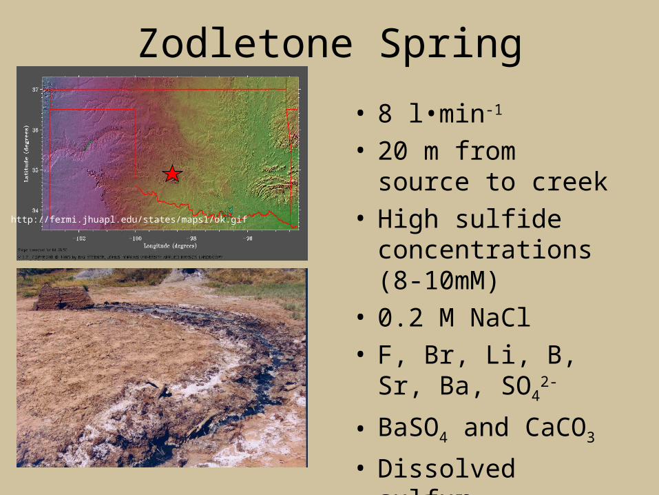
Zodletone Spring
http://fermi.jhuapl.edu/states/maps1/ok.gif
• 8 l•min-1
• 20 m from source to creek
• High sulfide concentrations (8-10mM)
• 0.2 M NaCl• F, Br, Li, B, Sr, Ba,
SO42-
• BaSO4 and CaCO3
• Dissolved sulfur
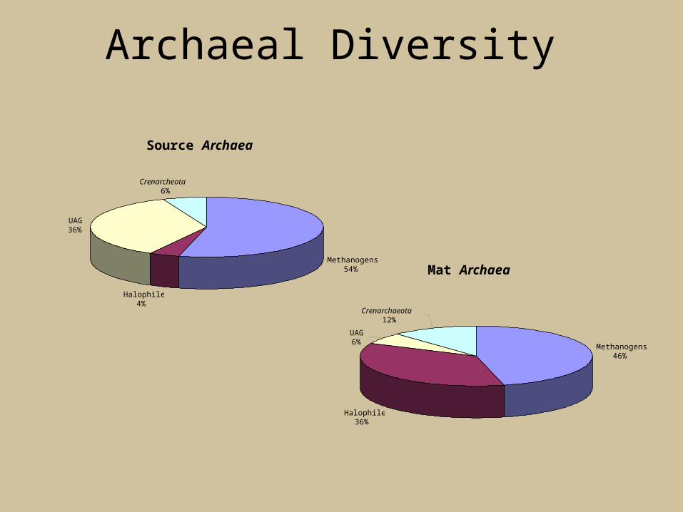
Archaeal Diversity
Source Archaea
Methanogens54%
Halophiles4%
UAG36%
Crenarcheota6%
Mat Archaea
Methanogens46%
Halophiles36%
UAG 6%
Crenarchaeota12%

Halophilic Archaea
• 14 validly described genera• Isolated from hypersaline environments (i.e. The
Dead Sea)• Rods, cocci and pleomorphic forms• Require high salt concentrations from 3.5 M NaCl
to saturation (5.2 M)• Contain pigment material (bacteriorhodopsin and
halorhodopsin) which gives them a distinct pink or red color

Isolation of Haloarchaea
• 36% of the mat and 4% of the source library• Isolated halophiles from the mat using a
high salt plus antibiotic medium with different salt concentrations (7, 12 and 18%)
• Serially diluted mat material and plated onto HM plates (immediately and after incubation)
• Cultures were incubated at 37ºC under light

A Novel Species of Haloferax
• 16S rRNA sequence analysis of strain M6 showed a 96.7-98.0% similarity to other validly described species of this genus
• M6 was 89% similar to Halogeometricum borinquense the most closely related species outside the genus
• DNA-DNA hybridization confirmed its novel speciation having a hybridization value of only 24% to Haloferax gibbonsii

G+C content % Relatedness with 3H-labelled DNA
(mol%) from strain M6
Source of unlabelled DNA
_________________________________________________________________________________
___
Strain M6 60.5 100
Haloferax volcanii NCIMB 2012T 63.4* 21
Haloferax gibbonsii ATCC 33959T 61.8* 24
Haloferax denitrificans DSM 4425T 64.2* 1
Haloferax mediterranei ATCC 33500T 60.0* 4
Haloferax lucentense JCM 9276T 64.5* 3
* Data taken from Mullakhanbhai & Larsen (1975), Rodriguez-Valera et al. (1983), Tomlinson et al.
(1986), Juez et al. (1986) and Gutierrez et al. (2002).
Phylogeny

Haloferax sulfurifontis• Isolated from Zodletone
spring• Extreme pleomorphism• Colonies were small (2-3
mm) and salmon pink in color at 37°C
• Growth occurred in a wide range of salt concentrations (6% to saturation)
• Required at least 1 mM Mg2+ for growth
• Anaerobically reduces Sº

Haloferax sulfurifontis
Characteristic Strain M6 H. volcanii H. gibbonsii H.denitrificans
H.mediterranei
H.alexandrinus
H.lucentense
Temperature optimum(0C)
32-37 40 35-40 50 40 37 37
Temperature range (0C) 18-50 N.D. 25-55 30-55 20-55 20-55 10-45NaCl range (M) 1-5.2 1-4.5 1.5-5.2 1.5-4.5 1.3-4.7 1.8-5.1 1.8-5.1
NaCl optimum (M) 2.1-2.6 1.7-2.5 2.5-4.3 2-3 2.9 4.3 4.3Cell stability (M NaCl) 0.5 0.5 0.5-0.7 1.5 0.5 1.7 ND
Motility + - - - + - +pH optimum 6.4-6.8 7 6.5-7 6-7 6.5 7.2 7.5
Gelatin hydrolysis + - + + + + -Starch hydrolysis - - - - + - -Anaerobic nitrate
reduction- - - + + - -
Tween 80 hydrolysis + - + - + + NDIndole production + + + - + + (check) +
H2S production fromthiosulfate
+ + + + - + +
G+C content (mol%) 60.5 63.4 61.8 64.2 59.1-62.2 59.5 64.5Casein hydrolysis - - + - + - -
Resistance to rifampicin + - - - - + ND

A Continued Search for Halophiles
• Clone libraries indicated the presence of a diverse halophilic community
• Originally18 strains were isolated from the mats present at the stream
• Studies indicated that these isolates were of the same species
• In order to encourage the growth of different isolates the media was prepared at 3 different salt concentrations (18, 25 and 30%) and 11 different carbon sources were used instead of yeast extract

Future Work
• Microscopic inspection of colonies to determine cell morphologies present and trends
• Further purification and isolation
• Colony PCR and sequencing
• Anaerobic reduction of S°
• Strain characterizations

Acknowledgements
• Dr. Krumholz• Dr. Elshahed• Aharon Oren and Antonio Ventosa• Roe Laboratory
Funding for this project is by the National Science Foundation