Embed Size (px)
Citation preview

Halitosis: A Clinical Review
The Academy of Dental Learning and OSHA Training, LLC, designates this
activity for 7 continuing education credits (7 CEs).
Mary Oeding, RDH, M.Ed.
Health Science Editor: Megan Wright, RDH, MS
Publication Date: October 2012
Updated Date: January 2017
Expiration Date: February 2020
The Academy of Dental Learning and OSHA Training, LLC is an ADA CERP Recognized
Provider. ADA CERP is a service of the American Dental Association to assist dental
professionals in identifying quality providers of continuing dental education. ADA CERP does not
approve or endorse individual courses or instructors, nor does it imply acceptance of credit hours
by boards of dentistry. Concerns or complaints about a CE provider may be directed to the
provider or to the Commission for Continuing Education Provider Recognition at ADA.org/CERP.
Conflict of Interest Disclosure: ADL does not accept promotional or commercial funding in
association with its courses. In order to promote quality and scientific integrity, ADL's evidence-
based course content is developed independent of commercial interests. Refund Policy: If you
are dissatisfied with the course for any reason, prior to taking the test and receiving your
certificate, return the printed materials within 15 days of purchase and we will refund your full
tuition. Shipping charges are nonrefundable.
California Registered Provider Number: RP5631
1
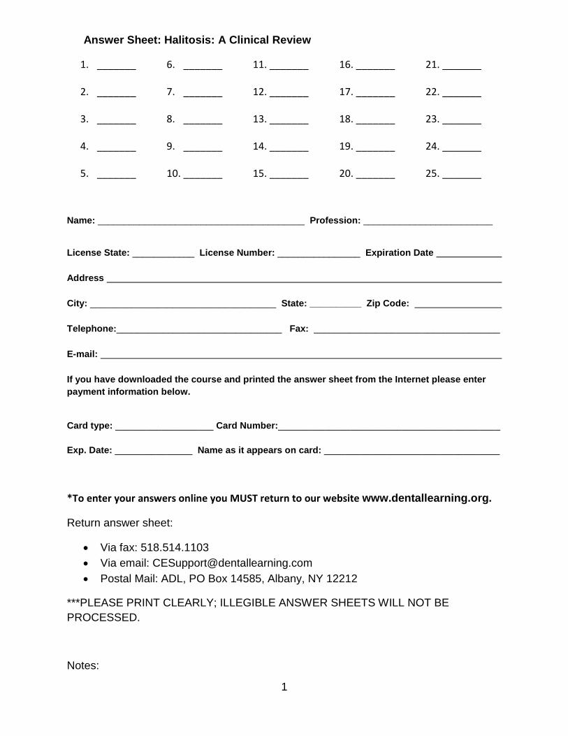
Answer Sheet: Halitosis: A Clinical Review
1. _______
2. _______
3. _______
4. _______
5. _______
6. _______
7. _______
8. _______
9. _______
10. _______
11. _______
12. _______
13. _______
14. _______
15. _______
16. _______
17. _______
18. _______
19. _______
20. _______
21. _______
22. _______
23. _______
24. _______
25. _______
Name: ________________________________________ Profession: _________________________
License State: ____________ License Number: ________________ Expiration Date
Address
City: ____________________________________ State: __________ Zip Code:
Telephone:________________________________ Fax: ____________________________________
E-mail:
If you have downloaded the course and printed the answer sheet from the Internet please enter
payment information below.
Card type: ___________________ Card Number:___________________________________________
Exp. Date: _______________ Name as it appears on card: __________________________________
*To enter your answers online you MUST return to our website www.dentallearning.org.
Return answer sheet:
Via fax: 518.514.1103
Via email: [email protected]
Postal Mail: ADL, PO Box 14585, Albany, NY 12212
***PLEASE PRINT CLEARLY; ILLEGIBLE ANSWER SHEETS WILL NOT BE
PROCESSED.
Notes:
2
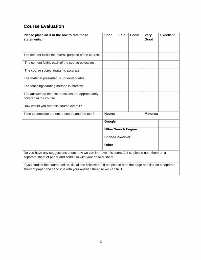
Course Evaluation
Please place an X in the box to rate these
statements:
Poor Fair Good Very
Good
Excellent
The content fulfills the overall purpose of the course.
The content fulfills each of the course objectives.
The course subject matter is accurate.
The material presented is understandable.
The teaching/learning method is effective.
The answers to the test questions are appropriately
covered in the course.
How would you rate this course overall?
Time to complete the entire course and the test? Hours: _________ Minutes: _______
Other Search Engine
Friend/Coworker
Other
Do you have any suggestions about how we can improve this course? If so please note them on a
separate sheet of paper and send it in with your answer sheet.
If you studied the course online, did all the links work? If not please note the page and link on a separate
sheet of paper and send it in with your answer sheet so we can fix it.
3
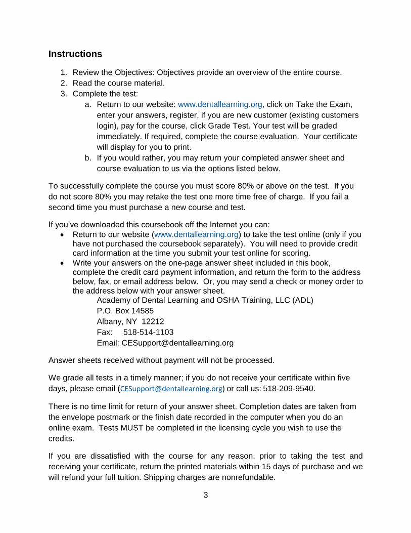
Instructions
1. Review the Objectives: Objectives provide an overview of the entire course.
2. Read the course material.
3. Complete the test:
a. Return to our website: www.dentallearning.org, click on Take the Exam,
enter your answers, register, if you are new customer (existing customers
login), pay for the course, click Grade Test. Your test will be graded
immediately. If required, complete the course evaluation. Your certificate
will display for you to print.
b. If you would rather, you may return your completed answer sheet and
course evaluation to us via the options listed below.
To successfully complete the course you must score 80% or above on the test. If you
do not score 80% you may retake the test one more time free of charge. If you fail a
second time you must purchase a new course and test.
If you’ve downloaded this coursebook off the Internet you can:
Return to our website (www.dentallearning.org) to take the test online (only if you have not purchased the coursebook separately). You will need to provide credit card information at the time you submit your test online for scoring.
Write your answers on the one-page answer sheet included in this book, complete the credit card payment information, and return the form to the address below, fax, or email address below. Or, you may send a check or money order to the address below with your answer sheet.
Academy of Dental Learning and OSHA Training, LLC (ADL)
P.O. Box 14585
Albany, NY 12212
Fax: 518-514-1103
Email: [email protected]
Answer sheets received without payment will not be processed.
We grade all tests in a timely manner; if you do not receive your certificate within five
days, please email ([email protected]) or call us: 518-209-9540.
There is no time limit for return of your answer sheet. Completion dates are taken from
the envelope postmark or the finish date recorded in the computer when you do an
online exam. Tests MUST be completed in the licensing cycle you wish to use the
credits.
If you are dissatisfied with the course for any reason, prior to taking the test and
receiving your certificate, return the printed materials within 15 days of purchase and we
will refund your full tuition. Shipping charges are nonrefundable.

4
If someone else would like to use this material after you are done, he or she may register with us and take advantage of a “sharing discount”. Courses downloaded from the Internet can be shared at the same tuition rate as currently available on our website. Please call us if you need an extra answer sheet or download one from our website. There is no “sharing discount” for online exams. The author and ADL have made every effort to include information in this course that is
factual and conforms to accepted standards of care. This course is not to be used as a
sole reference for treatment decisions. It is your responsibility to understand your legal
obligations and license requirements when treating patients. ADL is not responsible for
the misuse of information presented in this course. The material in this course cannot
be reproduced or transmitted in any way without the written consent of ADL.

5
Table of Contents
Answer Sheet 1
Evaluation 2
Instructions 3
Table of Contents 5
Objectives 6
Introduction 6
Oral Microflora and Volatile Compounds 7
Anaerobic Microflora and the Tongue 9
Diamines 10
Correlations Between Volatile Sulphur Compounds and Oral Measurements 11
Periodontal Disease Related to Bad Breath 12
Another Study About Oral Malodor and Periodontitis 13
Clinical Experiences in Israel 13
Diagnosis of Bad Breath 14
Measurement of Oral Malodor 14
Instruments Used to Analyze Mouth Odor 15
Classification of Halitosis and Treatment Needs 19
Oral Hygiene to Reduce Halitosis 20
History of Toothbrushes and Tooth Brushing 20
Today’s Toothbrushes 21
Toothbrush Selection 21
Basic Toothbrushing Procedure 22
Methods of Toothbrushing 23
Electric Toothbrushes 23
Tongue Care 23
Interdental Cleaning 24
Proper Flossing Procedure 24
Mouthrinses 24
Porobiotic Treatments 25
Other Sources of Bad Breath 25
Conclusion 27
The Future Direction of Bad Breath Research 28
References 29
Appendix: Patient Information Sheets 40
Appendix: Halimeter 45
Course Test 46
End Notes 51
6
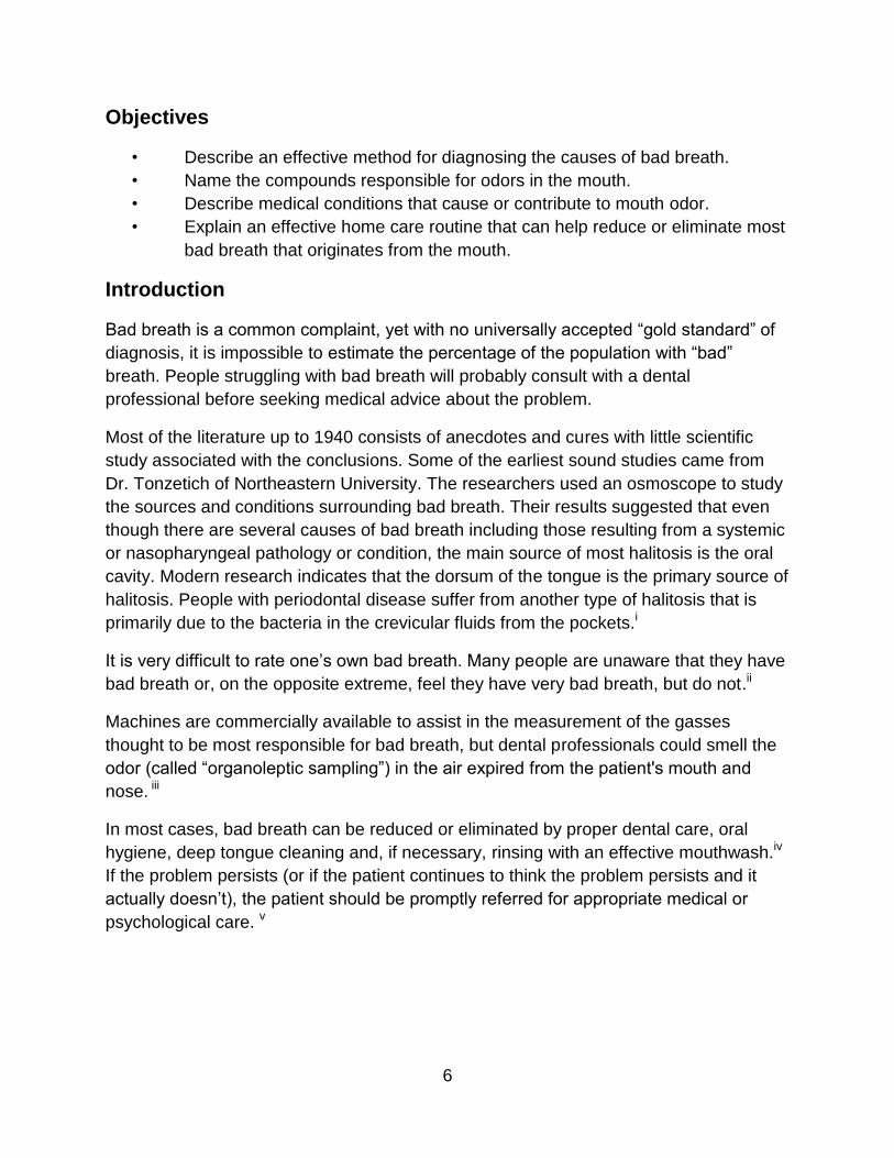
Objectives
• Describe an effective method for diagnosing the causes of bad breath.
• Name the compounds responsible for odors in the mouth.
• Describe medical conditions that cause or contribute to mouth odor.
• Explain an effective home care routine that can help reduce or eliminate most
bad breath that originates from the mouth.
Introduction
Bad breath is a common complaint, yet with no universally accepted “gold standard” of
diagnosis, it is impossible to estimate the percentage of the population with “bad”
breath. People struggling with bad breath will probably consult with a dental
professional before seeking medical advice about the problem.
Most of the literature up to 1940 consists of anecdotes and cures with little scientific
study associated with the conclusions. Some of the earliest sound studies came from
Dr. Tonzetich of Northeastern University. The researchers used an osmoscope to study
the sources and conditions surrounding bad breath. Their results suggested that even
though there are several causes of bad breath including those resulting from a systemic
or nasopharyngeal pathology or condition, the main source of most halitosis is the oral
cavity. Modern research indicates that the dorsum of the tongue is the primary source of
halitosis. People with periodontal disease suffer from another type of halitosis that is
primarily due to the bacteria in the crevicular fluids from the pockets.i
It is very difficult to rate one’s own bad breath. Many people are unaware that they have
bad breath or, on the opposite extreme, feel they have very bad breath, but do not.ii
Machines are commercially available to assist in the measurement of the gasses
thought to be most responsible for bad breath, but dental professionals could smell the
odor (called “organoleptic sampling”) in the air expired from the patient's mouth and
nose. iii
In most cases, bad breath can be reduced or eliminated by proper dental care, oral
hygiene, deep tongue cleaning and, if necessary, rinsing with an effective mouthwash.iv
If the problem persists (or if the patient continues to think the problem persists and it
actually doesn’t), the patient should be promptly referred for appropriate medical or
psychological care. v
7
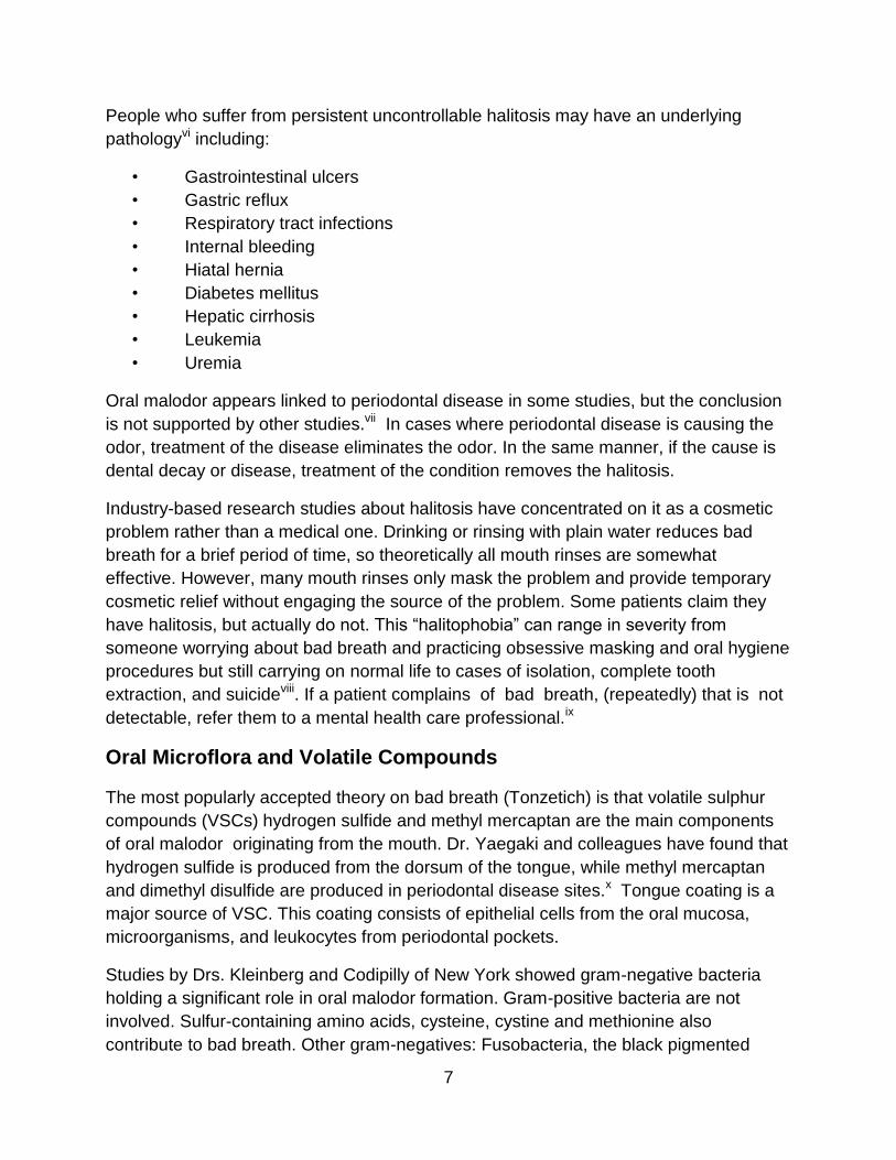
People who suffer from persistent uncontrollable halitosis may have an underlying
pathologyvi including:
• Gastrointestinal ulcers
• Gastric reflux
• Respiratory tract infections
• Internal bleeding
• Hiatal hernia
• Diabetes mellitus
• Hepatic cirrhosis
• Leukemia
• Uremia
Oral malodor appears linked to periodontal disease in some studies, but the conclusion
is not supported by other studies.vii In cases where periodontal disease is causing the
odor, treatment of the disease eliminates the odor. In the same manner, if the cause is
dental decay or disease, treatment of the condition removes the halitosis.
Industry-based research studies about halitosis have concentrated on it as a cosmetic
problem rather than a medical one. Drinking or rinsing with plain water reduces bad
breath for a brief period of time, so theoretically all mouth rinses are somewhat
effective. However, many mouth rinses only mask the problem and provide temporary
cosmetic relief without engaging the source of the problem. Some patients claim they
have halitosis, but actually do not. This “halitophobia” can range in severity from
someone worrying about bad breath and practicing obsessive masking and oral hygiene
procedures but still carrying on normal life to cases of isolation, complete tooth
extraction, and suicideviii. If a patient complains of bad breath, (repeatedly) that is not
detectable, refer them to a mental health care professional.ix
Oral Microflora and Volatile Compounds
The most popularly accepted theory on bad breath (Tonzetich) is that volatile sulphur
compounds (VSCs) hydrogen sulfide and methyl mercaptan are the main components
of oral malodor originating from the mouth. Dr. Yaegaki and colleagues have found that
hydrogen sulfide is produced from the dorsum of the tongue, while methyl mercaptan
and dimethyl disulfide are produced in periodontal disease sites.x Tongue coating is a
major source of VSC. This coating consists of epithelial cells from the oral mucosa,
microorganisms, and leukocytes from periodontal pockets.
Studies by Drs. Kleinberg and Codipilly of New York showed gram-negative bacteria
holding a significant role in oral malodor formation. Gram-positive bacteria are not
involved. Sulfur-containing amino acids, cysteine, cystine and methionine also
contribute to bad breath. Other gram-negatives: Fusobacteria, the black pigmented

8
anaerobes, Haemophilus, and Veillonella as well as amino acids other than VSC are
participants in malodor production.
When the main odoriferous volatiles are applied to an epithelial surface, hydrogen
sulfide and methyl mercaptan are lost rapidly and indole/skatole, putrescine,
cadaverine, and organic acids are lost more slowly, indicating the substances have an
ability to adhere to the surface and prolong the malodor.xi
Studies on whole saliva have yielded information about oral malodor and theories of
dental caries etiology. The whole saliva contains a sample of the microbial population
from the hard and soft tissues of the mouth and salivary components. Once centrifuged,
sediment of bacteria and epithelial cell elements can be sampled (called salivary
sediment). The bacteria are freely floating and attached to the epithelial components.
The fluid from the centrifugation is saliva from the oral glands and some gingival
crevicular fluid; found especially if the subject had significant inflammation. Studies have
shown salivary sediment is metabolically and microbiologically similar to pooled
dental plaque.(Denepitiya and Kleinberg, 1982; Singer and Kleinberg, 1983.)
One hundred years ago, Miller added teeth to a mixture of whole saliva and glucose
incubated 4 to 24 hours at 37 C. He found that the enamel on the teeth was
demineralized and the caries process began (The Miller Acid Decalcification Theory).
However, no malodor is produced. Whole saliva with glucose becomes acidic, whole
saliva by itself does not. Further studies show that the acidity inhibits bacterial
putrefication. The incubated saliva without the glucose is quite odoriferous. Early
studies (Sulser, 1939, Berg and Fosdick, 1946; Berg 1947) showed the odor was a
result of the bacteria acting on salivary proteins and peptides to yield compounds
producing the odor. Tonzetich (1977) confirmed the findings and also showed the air
around the incubated saliva is very similar to human breath air.xii Malodor is produced
in saliva that has a neutral or alkaline pH but is inhibited by an acidic pH. Fermentation
of sugars inhibit malodor generation but is the basis of caries formation.
Not only are H2S (hydrogen sulfide) and Ch3SH (methyl mercaptan) major components
of malodor, they are destructive to the oral tissues. They can penetrate and react with
the mucosa, making it more permeable to some ions and molecules. Researchers have
shown that mucosa treated with H2S can be restored to a normal state much more
readily than mucosa treated with CH3SH after the compound is removed.xiii
H2S and CH3SH damage the cell structure, the integrity of collagen, and the
metabolism of cells. Studies demonstrated a 70% lower collagen content in tissues
treated with the H2S and CH3SH as compared to the control tissues. Tests involving
periodontal disease show collagen components in crevicular fluid in the diseased sites,
and collagen loss of the same amount in surrounding tissues.xiv

9
Anaerobic Microflora and the Tonguevi
Investigators tested 16 people who complained of bad breath. They began with the
hypothesis: “If anaerobic flora on the tongue’s surface is the primary source of volatile
sulfur compounds in the mouth, then oral malodor should decrease with changes in the
bacterial content of the tongue coating, such as:
• a reduction in total bacterial counts;
• an increase in the percentage of facultative microorganisms; and a reduction in
the ratio of anaerobic over aerobic bacteria.”
Previous studies have shown proteolytic activity is important in the production of oral
malodor. The microorganisms on the tongue and in dental plaque putrefy proteins,
mucins, and peptides to release volatile sulfur compounds as gas in the breath. Studies
have shown gram-negative anaerobic bacteria combined with blood serum also produce
the VSC. Bacteria such as Treponema denticola, Porphyromonas gingivalis, Prevotella
intermedia, Bacteriodes forsythus, and Fusobacterium when combined with serum,
cysteine and methionine produce hydrogen sulfide and methylmercaptan.xv T.
denticola, P. gingivalis, and B. forsythus produce an enzyme similar to trypsin that can
be detected by a commercially available test called a BANA (benzoyl-DL-arginine-2-
napthylamide) Test.xvi
In another study, researchers set out to show that certain types of bacteria on the
dorsum of the tongue significantly contribute to the odor in most cases of halitosis. The
researchers gathered 8 men and 8 women who had complained of oral malodor.
Subjects limited eating and hygiene prior to the first appointment. Examiners reviewed
each medical history and documented the patient’s dental condition, including a
measurement of the oral odor with a portable sulfide monitor and organoleptic
assessment. The examiners collected scrapings from the dorsal surface of each
individual’s tongue. Samples were also taken of plaque on the mesial of the first molars
and two random interproximal sites. BANA tests were done on the incubated samples of
tongue scrapings and plaque to determine the type of bacteria present. Samples from
tongue scrapings were further incubated in anaerobic conditions for one week.
One week after the sample was taken, the subjects started a treatment regimen
consisting of tooth brushing, tongue brushing with a toothbrush dipped in chlorhexidine
gluconate, and a 60 second rinse of chlorhexidine gluconate twice a day for a week.
10
Findings of this study include:
• Full-mouth odor is related to tongue odor and the presence and amount of
tongue coating but not associated with periodontal factors (pockets,
bleeding).
• Surface characteristics of the tongue may effect the amount of odor
produced, deep fissures and an increased amount of tongue coating yielded
objectionable odors.
• The amount of coating is directly related to the bacterial load especially of
anaerobic proteolytic organisms.
• The scores relating to the anaerobic bacteria correlated with the amount of
breath odor, indicating these bacteria are primarily responsible for releasing
volatile fatty acids in the breath. These acids are perceived organoleptically,
but not by the portable sulfide monitor.
• Anaerobic bacteria were found on the tongue but not in interproximal plaque.
When the anaerobic bacteria count on the tongue was reduced, oral malodor
was reduced.
• Fusobacterium (including F. nucleatum, F. fusiform, and F. polymorphum)
and P. intermedia were found in a majority of the tongue samples. After
treatment, these anaerobic bacterium were significantly reduced.
This study gives additional evidence that bacterial activity on the tongue significantly
contributes to bad breath. VSC, anaerobic bacteria, tongue coating, and deep fissures
are all directly related to oral malodor. This study further supports the theory that gram-
negative anaerobic and assaccharolytic bacteria are key in the production of oral
malodor.
Diaminesxvii
Researchers Goldberg, Kozlovsky, and Rosenberg designed a study to evaluate the
contribution of diamines (especially cadaverine and putrescine) to bad breath. They
studied 52 people, most of whom complained of bad breath. The researchers
measured:
• the VSC with a portable sulfide monitor, mouth odor via organoleptic means,
plaque index,
• gingival index, probing depths,
• BANA test results, and
• levels of cadaverine and putrescine in the saliva.

11
Results of the study included:
• Cadaverine scores were associated with odor judge organoleptic scores,
plaque index scores, and gingival index scores. Cadaverine levels were also
relative to the BANA scores and mean probing depth. Cadaverine levels were
not associated with VSC levels.
• Putrescine levels were not significantly related to the malodor and periodontal
measurements, but were related to cadaverine levels.
In a second experiment, they compared saliva from a patient with periodontal disease to
saliva from a patient who had healthy gingiva. The sample from the periodontitis patient
had higher cadaverine levels. Putrescine levels in both samples were similar.
In a third experiment, they showed that higher levels of both cadaverine and putrescine
were found in deeper pockets than in shallow ones.
The results of this study showed cadaverine levels are associated with malodor and
periodontal disease, while putrescine’s role is still somewhat unknown.
Correlations Between Volatile Sulphur Compounds and Oral Measurementsx
Miyazaki et al. conducted a study in 1995 with 2,672 subjects ranging in age from 18 to
64. The research team assessed dental and periodontal conditions, plaque index, and
tongue coating. Subjects discussed their medical history, smoking habits, and oral
hygiene routines. Volatile sulphur compounds (VSC) were measured using a portable
sulphide monitor then analyzed by gender, age, and time of measurement.
Results of this study include:
• There are no significant differences between males and females in similar
age group.
• The highest measurement of malodor occurred in late morning and then late
afternoon, with the lowest value in the early afternoon.
• Measurements taken before lunch were 50% higher than those taken on
different subjects after lunch.
• There is a significant correlation between VSC value and both tongue coating
and periodontal conditions.
• Findings suggest that tongue coating may cause oral malodor in the younger
generation and periodontal diseases with tongue coating may cause oral
malodor in older subjects.
• Age was not commensurate with VSC increase.
• VSC increased quantitatively in subjects with gingivitis compared to control
subjects.
12
• There were no relationships between VSC and dental plaque, tooth brushing
habits, the number of decayed teeth, smoking habits, or the subject’s
perception of their own malodor.
Previous studies showed that age was a factor in increasing periodontal disease and/or
tongue coating that would lead to an increase in VSC and oral malodor.xviii These
results would suggest that halitosis may not occur in older people if their periodontal
condition is healthy and if there is little coating on the tongue.
Periodontal Disease Related to Bad Breathxix
Dr. Yaegaki of Yokohama, Japan conducted studies to investigate the relationship
between oral malodor and periodontal disease. He found zinc mouthwash reduced VSC
concentration more than 90% for 3 hours. (As an interesting side note: chlorhexidine
gluconate mouthwash was specifically not tested in this study because Japanese
Ministry of Health and Welfare does not allow it’s use in Japan because it has caused
anaphylactia in some Japanese people.)
Dr. Yaegaki combined human gingival fibroblast culture with methyl mercaptan in a
laboratory setting. He found that the methyl mercaptan interfered with collagen
synthesis and actually degraded the collagen cells. The presence of methyl mercaptan
may contribute to collagen degradation in periodontal disease.
He tested the effect of VSC on wound healing in a laboratory setting using rats. Results
indicated that the wounds treated with VSC did not heal well and the control group
without VSC healed very well. The presence of VSC after any kind of oral surgery can
delay healing of the area. He suggests zinc mouthwash to reduce the amount of VSC
after any oral surgery.
Dr. Yaegaki and Dr. Kato conducted a laboratory test to determine the effect of VSC on
oxygen production in human polymorphonuclear leukocytes. Their results indicated that
VSC stimulated active oxygen production in a periodontal pocket and that this
accelerates the destruction of periodontal tissues.
Findings presented in a combination of Dr. Yaegaki’s studies include:
• Volatile Sulfur Compounds increase the permeability of the cells of the
mucosa and collagen solubility.
• VSCs degrade protein and collagen.
• VSCs reduce the synthesis of protein, collagen and DNA. Periodontal patients
have a higher amount of VSC in their mouth air
• than those without periodontal disease.
13
• Periodontal patients had four times more tongue coating, which produced
60% of the VSC in their mouth air.
• The microorganisms responsible for periodontal disease, the tongue
coating, and the gingival crevicular fluid all contribute to VSC production in
patients who have periodontal disease.
• Zinc mouthwash would reduce VSC and be beneficial for
periodontal patients.
Another Study About Oral Malodor and Periodontitisxx
Drs. McCulloch and Bosy from Toronto, Canada set out to answer the question: “Is
there a quantifiable relationship between periodontitis and oral malodor”? All
professionals would agree that the earlier periodontitis is detected, the better the
chance of successful treatment. McCulloch and Bosy postulated that if oral malodor is
an indication of early periodontal disease, a diagnostic test could be devised to treat
periodontal disease in it’s earliest stage. They examined 127 patients, most from the
oral malodor clinic at the University of Toronto. Evaluation included: VSC peak, VSC
steady-state, organoleptic judging of whole mouth odor, tongue odor from scraping,
BANA test of tongue plaque, plaque index, periodontal probings, and organoleptic
judging of floss odor.
In a second phase of the test, the patients rinsed with 0.2% chlorhexidine gluconate
three times a day. The rinsing produced a reduction in the mouth odor, but not in
periodontal health.
Conclusions drawn from this study include:
• The halimeter was successfully used to make reproducible measurements of
VSC.
• VSC correlations with the halimeter were similar to organoleptic
measurements.
• The halimeter and organoleptic tests alone were not a good test for screening
of periodontal disease because improvements of halimeter and organoleptic
tests did not directly correlate with periodontal health improvements.
Clinical Experiences in Israelxxi
Drs. Rosenberg and Leib surveyed 308 people who came to the clinic between
February and December, 1992. Sixty percent of the participants were female. The
patients provided a medical history and information on their current oral hygiene
practices. The patients gave a description of the problem and how greatly it impacted
their daily lives. They rated their own oral malodor level on a scale from 1 to 5. Human
judges took organoleptic measurements for whole mouth odor, tongue dorsum odor,

14
and nasal odor. Drs. Rosenberg and Leib report three interesting cases from this study.
A 28 year old female presented with an oral odor, cigarette odor, and an atypical nasal
odor. The doctors referred the patient to an otolaryngologist. Surgery yielded a calcified
child’s bead that had been in her nose for 25 years.
The second subject was a 19 year old severely mentally retarded male. The odor was
typical of nasal odor, and was found especially around his nose, hair, hands, and
clothes. He was referred to an otolaryngologist who removed a putrefied paper tissue
lodged in the patient’s nasal cavity. The odor problem was cured.
The third case was a man who complained of oral malodor, but after examination,
malodor was not detected. The man continued to think he had bad breath, even after
his family and health professionals denied any problem.
Results of the clinical experiences in this study included:
• Odor judge scores were higher for men even though women gave higher self-
scores.
• The odor from the dorsum of the tongue was a major contributor to overall
mouth odor.
• No cases of malodor in this study were caused by the stomach.
• Most physiological causes of oral malodor are treatable, but some
“halitophobic” individuals may over exaggerate their perception of their own
breath. Even after treatment and improvement of the situation, these people
are not convinced their halitosis is gone.
Diagnosis of Bad Breath
Measurement of Oral Malodor
The measurement of bad breath is a difficult task because:
• the gasses are complex, it is difficult to sample,
• the samples change with exposure to air or with time, there is no standard to
measure against, and
• the subject population is limited. iii
Five main types of mouth odors are:
• periodontal-type odor from the dental floss or from crevicular fluid in the
periodontal pockets;
• odor from the posterior tongue dorsum from the spoon sample;
• denture odor detectable from removable appliances especially if placed in a
plastic bag for a few minutes;
15
• characteristic nasal odor as tested from the air expired from the nose only;
and
• smoker's breath.xxii
Instruments Used to Analyze Mouth Odor
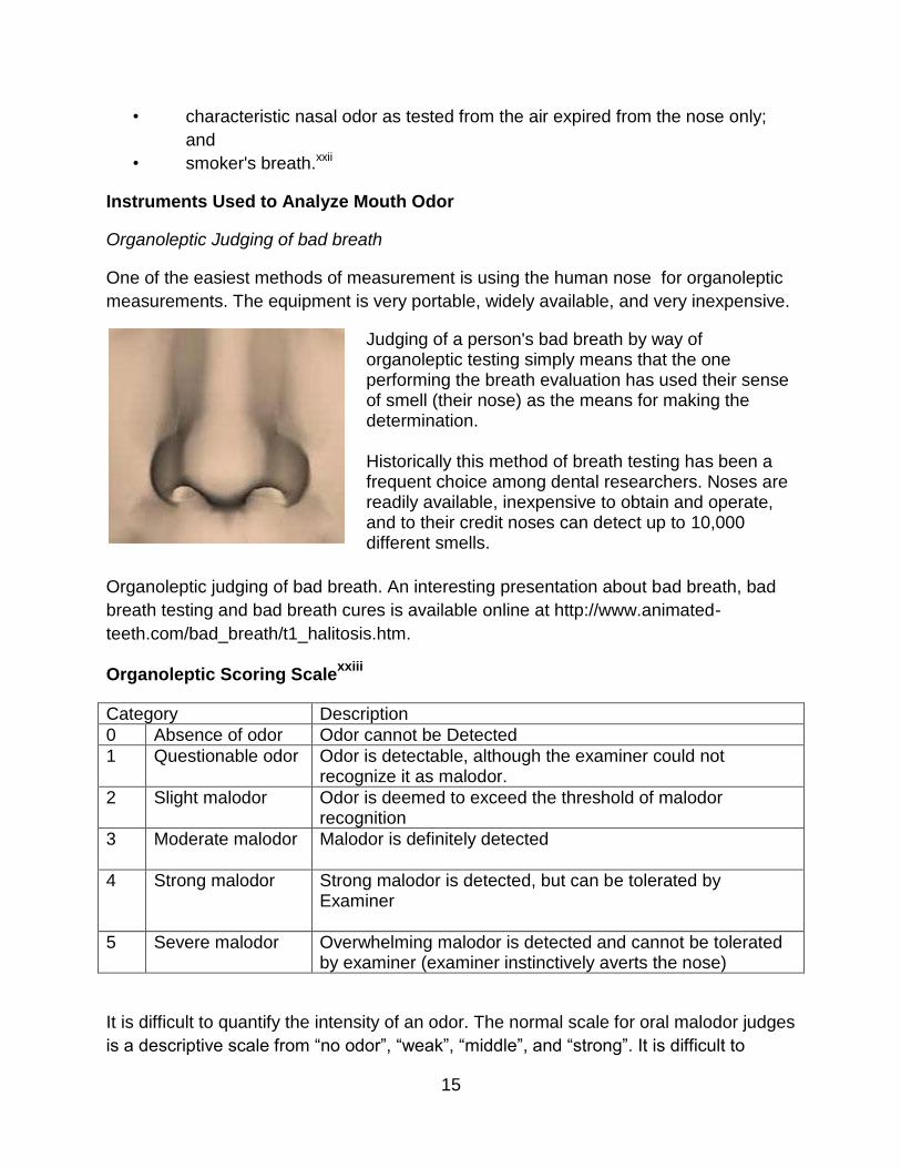
Organoleptic Judging of bad breath
One of the easiest methods of measurement is using the human nose for organoleptic
measurements. The equipment is very portable, widely available, and very inexpensive.
Judging of a person's bad breath by way of organoleptic testing simply means that the one performing the breath evaluation has used their sense of smell (their nose) as the means for making the determination. Historically this method of breath testing has been a frequent choice among dental researchers. Noses are readily available, inexpensive to obtain and operate, and to their credit noses can detect up to 10,000 different smells.
Organoleptic judging of bad breath. An interesting presentation about bad breath, bad
breath testing and bad breath cures is available online at http://www.animated-
teeth.com/bad_breath/t1_halitosis.htm.
Organoleptic Scoring Scalexxiii
Category Description
0 Absence of odor Odor cannot be Detected
1 Questionable odor Odor is detectable, although the examiner could not recognize it as malodor.
2 Slight malodor Odor is deemed to exceed the threshold of malodor recognition
3 Moderate malodor Malodor is definitely detected
4 Strong malodor Strong malodor is detected, but can be tolerated by Examiner
5 Severe malodor Overwhelming malodor is detected and cannot be tolerated by examiner (examiner instinctively averts the nose)
It is difficult to quantify the intensity of an odor. The normal scale for oral malodor judges
is a descriptive scale from “no odor”, “weak”, “middle”, and “strong”. It is difficult to
16
quantify a “middle” odor. The base measurement of “no odor” is concrete, but the
highest range of “extremely foul” is infinite.
Patients participating in an organoleptic examination should abstain from eating garlic,
onion and spicy food for 48 hours before the assessment. They should not use scented
cosmetics for 24 hours prior to the assessment. They should abstain from food, drink,
oral hygiene and breath fresheners for 12 hours before the assessment. The test should
take place 3 weeks after any antibiotic therapy. xxiv
Sample the patient’s breath by inserting a clear tube into the patient’s mouth. Instruct
them to exhale slowly and fill the tube with their breath (it will be undiluted by room air.)
Cap the tube and conduct the organoleptic test in another room, away from view of the
patient. xvi
Appraisal of bad breath can differ from one judge to another. Certain factors can
influence the individual’s organoleptic senses like: hunger, menstrual cycle, head
position, degree of attentiveness and expectation. This method may not be as reliable
as others because the olfactory sense acclimatizes to odors and therefore loses
sensitivity. If one person is unreliable, perhaps a panel of judges should conduct the
testing and the result based on a mean score. Panels were used in some studies
without success because of several complications. The most significant variable is that
the subject delivers the sample for one judge then the concentration and composition of
the gasses will be different in the following breaths for the other judges.xxv
People can be trained as oral malodor judges. A study showed trained judges gave a
more reproducible score, and were more closely related to sulfide analysis from a
machine.xviii Unfortunately, there is no reproducible standard for measuring bad breath,
so there is no standard training medium. There are some standard tests for selecting an
odor judgexix, but none specifically for the gasses commonly found in halitosis.
Probably the best judge is a dental professional, but people tend to become
accustomed to odors after prolonged exposure.
Measuring Bad Breath Scientifically
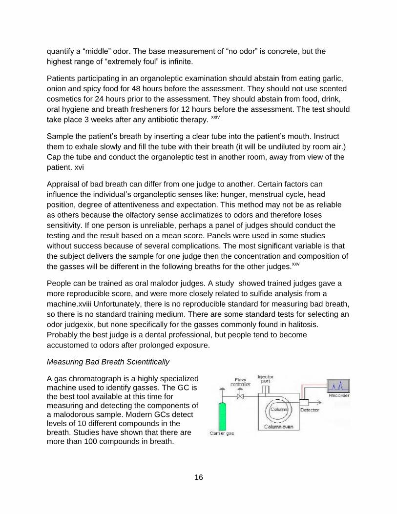
A gas chromatograph is a highly specialized machine used to identify gasses. The GC is the best tool available at this time for measuring and detecting the components of a malodorous sample. Modern GCs detect levels of 10 different compounds in the breath. Studies have shown that there are more than 100 compounds in breath.
17
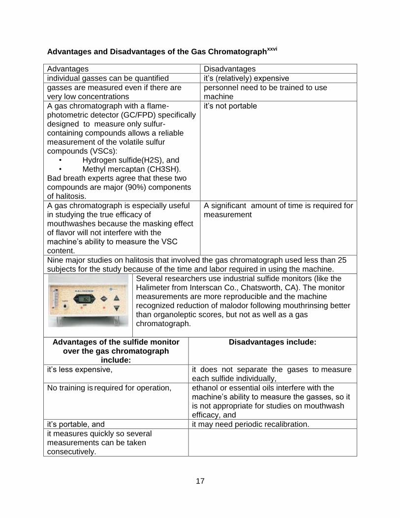
Advantages and Disadvantages of the Gas Chromatographxxvi
Advantages Disadvantages
individual gasses can be quantified it’s (relatively) expensive
gasses are measured even if there are very low concentrations
personnel need to be trained to use machine
A gas chromatograph with a flame- photometric detector (GC/FPD) specifically designed to measure only sulfur-containing compounds allows a reliable measurement of the volatile sulfur compounds (VSCs):
• Hydrogen sulfide(H2S), and • Methyl mercaptan (CH3SH).
Bad breath experts agree that these two compounds are major (90%) components of halitosis.
it’s not portable
A gas chromatograph is especially useful in studying the true efficacy of mouthwashes because the masking effect of flavor will not interfere with the machine’s ability to measure the VSC content.
A significant amount of time is required for measurement
Nine major studies on halitosis that involved the gas chromatograph used less than 25 subjects for the study because of the time and labor required in using the machine.
Several researchers use industrial sulfide monitors (like the Halimeter from Interscan Co., Chatsworth, CA). The monitor measurements are more reproducible and the machine recognized reduction of malodor following mouthrinsing better than organoleptic scores, but not as well as a gas chromatograph.
Advantages of the sulfide monitor over the gas chromatograph
include:
Disadvantages include:
it’s less expensive, it does not separate the gases to measure each sulfide individually,
No training is required for operation, ethanol or essential oils interfere with the machine’s ability to measure the gasses, so it is not appropriate for studies on mouthwash efficacy, and
it’s portable, and it may need periodic recalibration.
it measures quickly so several measurements can be taken consecutively.

18
A chemiluminescence detector (SCD) has become commercially available. It boasts better selectivity and sensitivity for measuring low levels of sulfur compounds than all commercially available sulfur- selective detectors. Comparative studies are needed to test this device. As technology increases, it may become possible to measure more of the compounds found in bad breath. Physicians might be able to diagnose certain diseases if they had the ability to detect trace amounts of certain compounds in the breath.
There are 2 other tests:
BANA test: measures for a specific enzyme produced by halitosis-causing bacteria
Beta-galactosidase test: levels of the enzyme beta-galactosidase have been found to
correlate with mouth odor.
Get an accurate picture of the problem
A complete medical history should be taken and evaluated for health problems or
medications that may exaggerate bad breath. An excellent way to get an accurate and
more objective view of the patient’s problem is to interview a member of their family.
They may help in establishing the severity of the problem and at what times of the day
the odor is at it’s worst, and if the odor at the time of the appointment is indicative of the
worst odor. If the odor is not present at the time of the dental appointment, interview to
find out the patient’s activities in preparing for the appointment.xxvii
A thorough dental and periodontal exam should be completed to rule out disease of soft
or hard tissues. Sample the coating of the tongue with a disposable plastic spoon.
Sample the plaque from the interdental spaces by drawing floss through a few molar
areas.
19
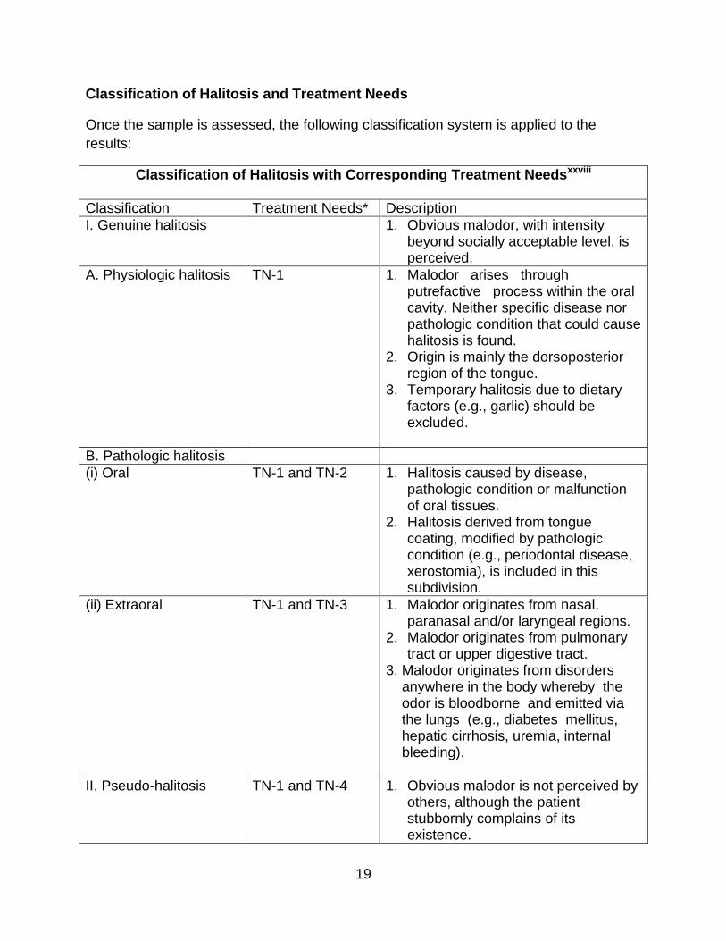
Classification of Halitosis and Treatment Needs
Once the sample is assessed, the following classification system is applied to the
results:
Classification of Halitosis with Corresponding Treatment Needsxxviii
Classification Treatment Needs* Description
I. Genuine halitosis 1. Obvious malodor, with intensity beyond socially acceptable level, is perceived.
A. Physiologic halitosis TN-1 1. Malodor arises through putrefactive process within the oral cavity. Neither specific disease nor pathologic condition that could cause halitosis is found.
2. Origin is mainly the dorsoposterior region of the tongue.
3. Temporary halitosis due to dietary factors (e.g., garlic) should be excluded.
B. Pathologic halitosis
(i) Oral TN-1 and TN-2 1. Halitosis caused by disease, pathologic condition or malfunction of oral tissues.
2. Halitosis derived from tongue coating, modified by pathologic condition (e.g., periodontal disease, xerostomia), is included in this subdivision.
(ii) Extraoral TN-1 and TN-3 1. Malodor originates from nasal, paranasal and/or laryngeal regions.
2. Malodor originates from pulmonary tract or upper digestive tract.
3. Malodor originates from disorders anywhere in the body whereby the odor is bloodborne and emitted via the lungs (e.g., diabetes mellitus, hepatic cirrhosis, uremia, internal bleeding).
II. Pseudo-halitosis TN-1 and TN-4 1. Obvious malodor is not perceived by others, although the patient stubbornly complains of its existence.

20
2. Condition is improved by counseling (using literature support, education and explanation of examination results) and simple oral hygiene measures.
III. Halitophobia TN-1 and TN-5 1. After treatment for genuine halitosis or pseudo- halitosis, the patient persists in believing that he/she has halitosis.
2. No physical or social evidence exists to suggest that halitosis is present.
*Treatment Needs (TN) for Breath Malodorxxix
Category Description
TN-1 Explanation of halitosis and instructions for oral hygiene (support and reinforcement of a patient’s own self-care for further improvement of their oral hygiene).
TN-2 Oral prophylaxis, professional cleaning and treatment for oral diseases, especially periodontal diseases.
TN-3 Referral to a physician or medical specialist.
TN-4 Explanation of examination data, further professional instruction, education and reassurance.
TN-5 Referral to a clinical psychologist, psychiatrist or other psychological specialist.
Note: TN-1 is applicable to all cases requiring TN-2 through TN-5
Oral Hygiene to Reduce Halitosisxxx
History of Toothbrushes and Tooth Brushing
Maryly Snow displays samples of her toothbrush collection. Online image available at
http://www.berkeley.edu/news/berkeleyan/2001/05/09_tooth.html
The earliest cleaning implement for teeth was excavated in Mesopotamia. It was a gold
toothpick thought to be used by Sumerians in 3000 B.C. Some primitive cultures
used “chewsticks” a flavorful twig of wood that is chewed or crushed at the end to form
a fibrous brush. References to chewsticks in Chinese literature appear as early as 1600
B.C. Chewsticks are still used by some Asian and African people today. Hippocrates’
writings around 300 B.C contain descriptions of gum disease, calculus, and treatment of
gums.
21
Toothbrushes resembling our modern versions appeared in England in the late 18th
Century. The first U.S. patent for a toothbrush was issued in the 19th Century.
The American Academy of Periodontology identified ideal specifications for
toothbrushes in 1919, attempting to develop a standard for the industry.
Today’s Toothbrushes
“1950. It all started with a dentist, back in 1950, who created the first Oral-B toothbrush
and its soft, end-rounded nylon bristles. Dr. Robert Hutson, a California periodontist,
designed and patented the first Oral-B toothbrush. He also created the Oral-B brand
name. "The first product was known as the Oral-B 60," said Dr. Hutson, "because it
had 60 tufts."
Dr. Hutson started small, with a family business. He would wrap the orders in the
basement, then load them in the back seat of the car and take them down to the
post office to be mailed.
The role of the dentist was critical from the very beginning. Dr. Hutson said, "I knew I
had a good thing but the acceptance by the dentist was something I never expected—
having a product that was accepted by the dentist and the dentist in turn told the people
about it." Available at http://www.oralb.com/aboutus/history.asp
According to the American Dental Association, an effective toothbrush should have the
following characteristics:xxxi
• A size, shape and texture that will conform to an individual’s needs,
• Easily and efficiently manipulated,
• Impervious to moisture and easily cleaned and dried,
• Durable, inexpensive,
• Flexible, soft bristles, strong, light weight, End-rounded filaments,
• Designed for utility, efficiency, and cleanliness.
Toothbrushes are available in a variety of handle types and bristle planes. Orthodontic
brushes have a bi-level plane. Others include dome-shaped, rippled and flat.
Toothbrush Selection
Choose brushes with filaments made of synthetic nylon. Brushes made of this material
will rinse and dry more completely between brushings. The standardization of the bristle
length and shape will facilitate more uniform cleaning of the tooth surface and gingival
margin. End-rounded filaments are more effective at the gingival margin and will reduce
gingival damage during brushing.
Consider the following factors when selecting a toothbrush for a particular patient:

22
• Patient ability: How well will the patient be able to use the brush to remove
plaque from all tooth surfaces without damaging the soft tissue or tooth
structure? What is the patient’s manual dexterity? Is the patient motivated and
willing to use the correct technique with this type of brush?
• Gingival Health: How resilient is the gingiva? Is the brush adaptable to the
anatomic configurations of the tissue?
• Tooth Position: Are teeth crowded? Displaced? Open contacts?
• Tooth Shape: Large? Small?
• Patient Preference: Does the patient prefer a type of brush. Can they be
instructed in proper use of this type of brush, or is it damaging to their
dentition?
• Brushing Method: The type of brush is dependent on the method of brushing
used.
When used correctly, a soft nylon brush with rounded filaments provide:xxxii
• Effective cleaning in cervical areas.
• Reduction of gingival trauma so patients can brush more thoroughly at the
gingival margin without laceration.
• Adaptation toward sulcus area and interproximal areas for better cleaning.
• Thorough cleaning around orthodontic and other fixed appliances. Reduction
of the chance of gingival recession or tooth abrasion.
• Effective and comfortable brushing for patients with gingivitis or traumatized
tissue.
Basic Toothbrushing Procedurexxxiii
Proper and thorough brushing is best accomplished through an orderly system.
Brushing is habitual. Patients may be neglecting areas that are harder to reach simply
because they are not thinking about the procedure. The toothbrush handle must be
grasped firmly with the whole hand for control of motion and tactile sense of direction.
An appropriate pressure must be applied to the tooth surface to remove the plaque
without flaring the bristles away from the target area.
Teeth should be brushed in a regular order so each surface is given equal attention.
Start with a facial molar region and work around to the opposite side. Then brush the
lingual from that side back to the beginning. Repeat procedure on the opposing arch.
The sequence should be varied each time: for example, start on the right side in the
morning, left side at night. Start in the upper arch one week, lower arch the next. Areas
that are difficult or frequently missed should be given double attention.
Most people brush their teeth too quickly, or spend more time with the front teeth than
with the back. Thorough brushing should take 3 to 4 minutes. The brusher should count

23
to seven or ten in each area to evenly distribute the brushing time.
People should brush their teeth at least 2 times a day and clean interdental areas at
least once a day. The last brushing of the day should be right before bed. Reduced
salivary flow and lack of mastication during the night facilitate bacterial growth.
Methods of Toothbrushing
The most widely used method of toothbrushing is the scrub brush method in which the
brusher vigorously brushes in a back and forth movement. Even though this may be the
easiest to teach, it can be detrimental to the tissues and tooth structure.
The Bass Technique or the Modified Bass Technique is currently considered the most
effective method for plaque removal at the gingival margin. The filaments should be
directed apically to the tooth at a 45° angle to the long axis of the tooth and straight into
the sulcus. Using light pressure, allow the bristles to go somewhat into the sulcus
without damaging the tissues. The brush is moved in a short, vibrating motion without
displacing the bristles from the sulcus for the count of ten. The brush is then moved to
the teeth directly next to those brushed, overlapping areas slightly. The lingual areas of
anterior teeth can be brushed by holding the brush vertically and using a rolling stroke
from the heel of the brush through the whole length of the brush. Occlusal surfaces
should be brushed with a scrub brush stroke.xxxiv
Electric Toothbrushes
Power brushes are very popular and widely available. Studies have shown several
electric toothbrushes to be effective in plaque removal when used correctly. Electric
toothbrushes may be indicated in patients who have limited manual dexterity. Electric
toothbrushes should have soft bristles and be used carefully to avoid gingival trauma or
cervical abrasion. Because electric toothbrushes have been proven to vibrate a
dislodging motion down into the teeth sulci on upward, it is widely accepted as the more
effective way to brush in 2016.
Tongue Care
The tongue’s filiform and fungiform papillae create a rough,uneven surface. Bacteria,
debris, and plaque collect in this surface. The posterior dorsum of the tongue is
particularly difficult to clean and has been demonstrated to be an area of odor. The
tongue can be gently brushed with a standard soft toothbrush. Use the brush soaked in
an antibacterial mouthwash to deliver the substance to the surface of the tongue. Try to
reach back to the dorsum of the tongue without choking and be gentle enough to avoid
damaging the tongue surface. Try different sized toothbrushes to find the most effective
size for you.
24
Tongue scrapers are made of a flexible material and have ridges to clean the surface of
the tongue. These should be used gently also. Plastic ones are most gentle. Tongue
scrapers are very effective in bacterial/plaque removal especial when compared to only
brushing the tongue with one’s toothbrush. If a patient has more of a gag reflex, it is
helpful to have him or her wet tongue scraper first, each time, before scraping, while
breathing out slowly or humming to keep soft palate relaxed. Then rinse and repeat for
both sides, as well as, midline of tongue. Tongue scraping at nighttime may decrease a
hyperactive gag reflex.
Interdental Cleaning
Plaque between the teeth cannot be removed with a toothbrush. Dental floss used
correctly removes plaque and debris from the interproximal area. Waterflossers can be
effective, easily accessing deep periodontal pockets, where floss cannot reach.
Waxed floss has been compared to unwaxed floss in several studiesxxxv . There has
been no significant difference in plaque removal between the two. Waxed floss resists
shredding while unwaxed floss spreads well once it’s passed through the contact for a
larger cleaning area. Floss before brushing so the fluoride from the toothpaste can be
worked into the proximal areas. Newer woven flosses absorb more plaque, while
flossing, but not recommended for patients with tight contacts as it can get stuck
interproximally, if not careful. Teaching patients to pull woven floss towards gingivae
and then guided out on tight areas can prevent fraying and woven floss getting stuck.
Also, newer floss brands that are still shred-resistant but still move plaque out contain
monofilaments with flexible MICRO-GROOVES® Technology.
Proper Flossing Procedure
Use approximately 12 inches of floss. Grasp floss between thumb and index finger of
each hand. Wrap remainder around middle fingers or tie the ends of the floss together
to form a large circle. Work floss gently through the contact of the teeth without
snapping it into the papillae. Curve the floss in a C shape and slide the floss up and
down the tooth surface a few times to dislodge the plaque. It should be moved far
enough under the gingiva so that it meets resistance. The distal of one tooth (unless it is
the back tooth) and the mesial of another are cleaned without removing the floss from
the contact. Floss from distal of the last tooth on one side across the arch to the distal of
the last tooth. Repeat with the other arch.
Mouthrinses
While many typical mouthwashes out on the market today merely temporarily cover up
bad breath, some do, in fact, help treat the cause. In order for a mouthrinse to do this, it
must do one of two things: kills the microorganisms responsible for producing the
25
volatile sulfur compounds or neutralize the sulfur compound themselves.
Recent studies have suggested that mouthrinses with certain active ingredients attack
bad breath at its cause and can be very effective treatments. The active components
include zinc, chlorine dioxide, cetylpyridium chloride and chlorhexidine gluconate.
These have been shown to kill, with varying degrees of success, anaerobic bacteria
and/or their chemical products. More research is necessary to know which ones are the
most effective and user friendly treatments.
Probiotic Treatments
Streptococcus salivarius K12, has been claimed to suppress malodorous bacteria
growth, however well designed randomized control clinical trials are needed to assess
this.
Other Sources of Bad Breath
If halitosis persists after debridement of the tongue and proper dental care (including
periodontal treatment if indicated and proper oral hygiene) the patient should be
referred to their physician for further investigation of a systemic cause. Nicotine usage
and alcohol consumption are huge factor for halitosis, among many other ailments.
Crash dieting can also cause halitosis due to the breakdown of fats producing
chemicals called ketones.
The following oral conditions present malodor that is not associated with the bacterial
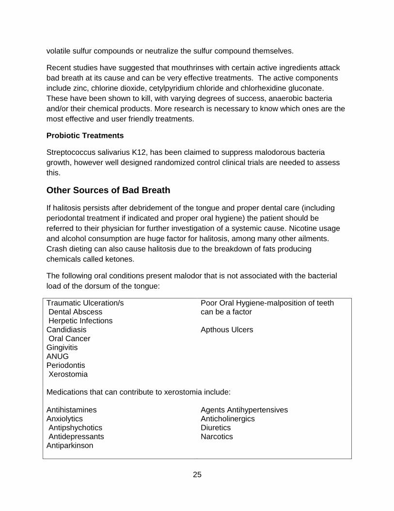
load of the dorsum of the tongue:
Traumatic Ulceration/s Dental Abscess Herpetic Infections Candidiasis Oral Cancer Gingivitis ANUG Periodontis Xerostomia
Poor Oral Hygiene-malposition of teeth can be a factor Apthous Ulcers
Medications that can contribute to xerostomia include: Antihistamines Anxiolytics Antipshychotics Antidepressants Antiparkinson
Agents Antihypertensives Anticholinergics Diuretics Narcotics
26
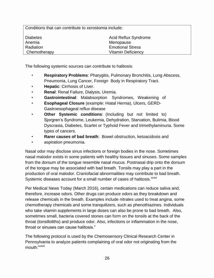
Conditions that can contribute to xerostomia include: Diabetes Anemia Radiation Chemotherapy
Acid Reflux Syndrome Menopause Emotional Stress Vitamin Deficiency
The following systemic sources can contribute to halitosis:
• Respiratory Problems: Pharygitis, Pulmonary Bronchitis, Lung Abscess,
Pneumonia, Lung Cancer, Foreign Body in Respiratory Tract.
• Hepatic: Cirrhosis of Liver.
• Renal: Renal Failure, Dialysis, Uremia.
• Gastrointestinal: Malabsorption Syndromes, Weakening of
• Esophageal Closure (example: Hiatal Hernia), Ulcers, GERD-
Gastroesophageal reflux disease
• Other Systemic conditions: (Including but not limited to)
Sjorgren’s Syndrome, Leukemia, Dehydration, Starvation, Bulimia, Blood
Dyscrasia, Diabetes, Scarlet or Typhoid Fever and trimethylaminuria. Some
types of cancers.
• Rarer causes of bad breath: Bowel obstruction, ketoacidosis and
• aspiration pneumonia.
Nasal odor may disclose sinus infections or foreign bodies in the nose. Sometimes
nasal malodor exists in some patients with healthy tissues and sinuses. Some samples
from the dorsum of the tongue resemble nasal mucus. Postnasal drip onto the dorsum
of the tongue may be associated with bad breath. Tonsils may play a part in the
production of oral malodor. Craniofacial abnormalities may contribute to bad breath.
Systemic diseases account for a small number of cases of halitosis.xxxvi
Per Medical News Today (March 2016), certain medications can reduce saliva and,
therefore, increase odors. Other drugs can produce odors as they breakdown and
release chemicals in the breath. Examples include nitrates used to treat angina, some
chemotherapy chemicals and some tranquilizers, such as phenothiazines. Individuals
who take vitamin supplements in large doses can also be prone to bad breath. Also,
sometimes small, bacteria covered stones can form on the tonsils at the back of the
throat (tonsilloliths) and produce odor. Also, infections or inflammation in the nose,
throat or sinuses can cause halitosis.”
The following protocol is used by the Chemosensory Clinical Research Center in
Pennsylvania to analyze patients complaining of oral odor not originating from the
mouth:xxxvii

27
1. Patient collects first morning urine.
2. Patient fasts and refrains from fragrant cosmetics.
3. Organoleptic evaluation by two judges on a scale of 1 to 10.
4. Analysis of breath by gas chromatograph.
5. Determination of resting salivary flow rate.
6. Analysis of nasal air for VSC.
7. Saliva sample taken for incubation and analysis.
8. Baseline saliva sample for Trimethylamine analysis.
9. Lung air collection.
10. Choline administered to patient.
11. Patient gathers samples of urine and saliva each 8 hours for the next 24 hours.
12. The next day, the patient returns all samples to be evaluated for choline (will help
in diagnosis of trimethylaminuria). Results from the previous day’s tests are
discussed. They are examined by a dentist and an Ear, Nose, and Throat
Specialist. A sensory examination is done.
Conclusion
Sometimes halitosis is linked with serious and sobering consequences. Dr. Yaegaki
relates a case involving a 60-year old professional man. Dr. Yaegaki examined the
patient to find tongue coating. The patient was instructed in tongue debridement and
given a zinc mouthrinse. He returned after 2 weeks and the odor remained. Dr. Yaegaki
referred the patient for counseling because the patient was depressed from his wife’s
death and other issues at his work. The counselor recommended a long vacation, but
the patient refused for fear of losing his job. The patient was referred to a psychiatrist,
but committed suicide before the treatment could begin.xxxviii
The following ideas are generally accepted among the Bad Breath Research
Community:
• Volatile sulfur compounds (hydrogen sulfide and methyl mercaptan)
• are found in bad breath.
• The coating on the dorsum of the tongue is the primary area of bacterial
putrefication responsible for most mouth odors.
• Proper home care including tongue cleansing will reduce oral malodor.
The following ideas seem to be conflicting in studies:
• The relationship between oral hygiene and bad breath.
• The relationship between periodontal disease and bad breath.
28
The Future Direction of Bad Breath Research
Science would benefit from the development of an easy to use machine to analyze
other compounds in the breath, especially amines. Studies have indicated that amines
are important components of oral malodor, but without being able to measure them, it is
not possible to prove.
Research in the area of identifying which compounds (though not malodorous
themselves) contribute to the intensity or quality of halitosis would be beneficial. For
example, halitosis due to age, an ulcer, and hepatic cirrhosis all contain similar
components of VSC but smell distinctly different. These differences in odor may be due
to ratios and mixtures of the sulfur-compounds, but there is a possibility that some non-
sulfur compound changes the quality of the odor.xxxix
Most cases of bad breath are due to microbial putrefication in the mouth. Oral bacteria
produce hydrogen sulfide when exposed to serum or saliva in vitro. Odor from the
dorsum of the tongue is different than the odor from subgingival plaque. Research to
identify the reasons for the difference, (for example, if it is from different chemical
constituents, the ratios of the components, microbial effects, or a combination of all)
would be beneficial.xl
Studies are necessary in the area of the effect of volatile H2S and CH3SH on cells and
tissues. The studies should also contain information about the effect of other salivary
products like putrescine and cadaverine. Researchers should investigate the role of
VSC in periodontal disease. xli
The study of oral malodor and its components is a growing science that may at the
surface appear to be a simplistic exercise in curing a merely social problem. Deeper
investigation into the chemical compounds found in halitosis and their effect on tissues
and cells may expand our understanding of cell function in normal and diseased tissue. xlii
Bad breath is difficult to describe or quantify. Odor judges in a clinical setting have a
more difficult task in trying to score the intensity or quality of the odor. A study
comparing the effectiveness of trained and untrained human judges when sampling bad
breath did not clearly demonstrated that training improves the reliability of the judge.xliii
Some non-sulfur compounds like indole, methylamine, and cadaverine are not volatized
from aqueous saliva but are released when they are allowed to dry on the skin. Clinical
evaluation by instrumental measurement alone should be discouraged.iii Ideally, bad
breath should be evaluated using a combination of human odor judges and machines.
The field of halitosis research would benefit from: more reliable, portable instruments for
29
measuring VSC, a standard scale for assessing oral malodor, further studies with larger
sections of the population, and development of site-specific measurements.
References
Abelson, D.; Barton, J. Maletti, G.; Cowherd, M. (1981). Evaluation of interproximal
cleaning by two types of dental floss. Clin. Prev. Dent., 3, 19.
Alexander, J. (1980). Toothbrushes and toothbrushing. The Biologic Basis of Dental
Caries Harper & Row; Hagerstown, MD. , 482-496.
American Dental Association, Council on Dental Therapeutics. (1984). Accepted Dental
Therapeutics, 40th ed. Chicago, IL. 386-387.
Amigoni, N.; Johnson, G.; Kalkwarf, K. (1987). The use of sodium bicarbonate and
hydrogen perioxide in periodontal therapy: a review. JADA , 114, 217-221.
Attia, E. and Marshall, K. (1982). Halitosis. Can. Med. Assoc. J, 126, 1281-1285.
Bass, C.C. (1954). An effective method of personal oral hygiene. J. Louisiana State
Med. Soc., 106, 100. Beaumont, R. (1990). Patient preference for waxed or unwaxed
dental floss. J. Periodonto, 61, 123.
Berg, M. and Fosdick, L. (1946). Studies in periodontal disease. II. Putrefactive
organisms in the mouth, J. Dent. Res, 25, 73-81.
Berg, M.; Burrill, D.; Fosdick, L. (1947). Chemical studies in periodontal disease. III.
Putrefaction of salivary protiens, J. Dent. Res., 25, 231-246.
Bosy, A.(1997). Oral malodor: philosopical and practical aspects. 63 (3), 196-201.
Bosy, A. (1997). Taste as a Predictor of oral malodour. The Third International
Conference on Breath Odour; Vancouver, BC, Canada, (S4), 1-6.
Bosy, A. and Celler, J. (1997). Ethics of bad breath [letter:comment], J Can Dent Assoc,
63 (4), 235.
Bosy, A.; Kulkarni, G.; Rosenberg, M.; McCulloch, C. (1994). Relationship of oral
malodor to periodontitis: evidence of independence in discrete subpopulations. J.
Periodontol, 65, 37-46.
Breitenmoser, J.; Mörmann, W., and Mühleman, H. (1979). Damaging effects of
toothbrush bristle end form on gingiva. J. Periodontol, 50, 212.
Brening, R.; Sulser, G.; Fosdick, L. (1939). The determination of halitosis by use of the
osmoscope and the cyroscopic method. J. Dent. Res , 18, 127.
30
Brown, E. Krabek, W. Skiffington, R. (1947). Glycerite of hydrogen peroxide A
comparison of its bacteriotoxic action with that of mercurial solutions. J. Bacteriol, 53,
793-799.
Cain, S.; Moskowitz, H. (1974). Psycophysical scaling of odor. Human Responses to
Environmental Odors. New York: Academic Press, 2-32.
Caldwell, R.; Pigman, W. (1966). Changes in protein and glycoprotein concentrations in
human submaxillary saliva under various stimulatory conditions. Arch Oral Biol, 11, 437-
449.
Cardash, H. and Rosenberg, M. (1990). An innovative method of monitoring denture
hygiene. Journal of Prosthetic Dentistry, 63, 661-664.
Cherniak, O.; Kozlovsky, A.; Gordon, D.; Gelernter, I.; Rosenberg, M. (1993). Self-
assessment of oral malodor. J. Dent. Res ., 72, 260.
Cimasoni, G. Crevicular fluid updated. Monographs in Oral Science. Basel: Karger, 2-
28.
Claesson, R.; Edlund, M.; Persson, S.; Carlsson, J. (1990). Production of volatile
sulfur compounds by various Fusobacterium species. Oral Micrbiol. Immunol, 5, 137-
142.
Dawes, C. (1987). Physiological factors affecting salivary flow rate, oral sugar
clearance, and the sensation of dry mouth in man. J. Dent. Res, 66, 648-653.
DeBoever, E.; De Uzeda, M.; Loesche, W. (1994). Relationship between volatile sulphur
compounds, BANA hydrolyzing bacteria and gingival health in patients with and without
complaints of oral malodor. J. Clin. Dent., 4, 114-119.
DeBoever, E.; Loesche, W. (1995). Assessing the contribution of anarobic microfl ora of
the tongue to oral malodor. JADA , 126, 1384-1393.
Denepitiya, L.; Kleinberg, I. (1982). A comparison of the microbial compositions of
pooled dental plaque and salivary sediment. Arch. Oral Biol., 27, 739-745.
DiSabato-Mordarski, T.; Kleinberg, I. (1989). Intra-oral variation in the residual saliva on
the oral tissues. J. Dent Res., 68, 316.
Doty, R.; Green, P.; Ram, C.; Yankell, S. (1982). Communication of gender from
human breath odors: relationship to perceived intensity and pleasantness. Horm.
Behav., 16, 13-22.
31
Eli, I.; Baht, R.; Kozlovsky, A. and Rosenberg, M. (1996).The complaint of oral
malodor - possible psychopathologic aspects. Psychosomatic Medicine, 58, 156-159.
Eli, I.; Baht, R.; Rosenberg, M. (1995). Psychological factors in self assessment of
oral malodor. Bad Breath: Research Perspectives , Ramot Publishing, Tel Aviv
University, 201-214. Engen, T. (1982). The Perception of Odors . New York, Academic
Press.
Feitosa, A.; Amalfitano, J.; Loesche, W. (1993). The effect of incubation temperature
on the specificity of the BANA -test. Oral Microbiol. Immunol, 8, 57-61.
Finkelstein, P. and Grossman, E. (1979). The effectiveness of dental floss in reducing
gingival inflammation. J. Dent. Res , 58, 1034.
Finkelstein, Y.(1995). The otolaryngolgist and the patient with halitosis. Bad Breath:
Research Perspectives Ramot Publishing, Tel Aviv University, 175-188.
Flotra, L. (1971). Side effects of chlorhexidine mouth washes. Scand J Dent Res , 79,
119-125.
Fox, S. and Bosworth, B. (1987). A morphological survey of proximal root concavities:
a consideration in periodontal therapy. JADA , 114: 811.
Frostell, G. (1960). Studies on the ammonia production and the ureolytic activity
of the dental plaque material. Acta Odont Scand, 18, 29-65.
Frostell, G.; Soder, P. (1970). The proteolytic activity of plaque and its relation to soft
tissue pathology. Int. Dent. J., 29, 436-50.
Geist H. (1957). Halitosis in ancient literature. Dent Abstr, 2, 417-418.
Gher, M. and Vernino, A. (1980). Root morphology — clinical significance in
pathogenesis and treatment of periodontal disease. JAMA, 101, 627.
Globerman, D.; Kleinberg, I. Intra-oral PO2 and its relation to bacterial accumulation
on the oral tissues. New York and Washington Information Retrieval, 275-291.
Gold, S. (1983). Early origins of hydrogen peroxide use in oral hygiene. J. Periodontol,
54, 247.
Goldberg, S.; and Rosenberg, M. (1991). Bacterial desorption by commercial
mouthwashes vs two-phase oil:water formulations. Biofouling, 3, 193-198.
Goldberg, S.; Kozlovsky, A.; Gordon, D.; Gelernter, I.; Sintov, A.; Rosenberg, M.
(1994). Cadaverine as a putative component of oral malodor. J Dent Res, 73, 1168-
1172.
32
Goldberg, S.; Kozlovsky, A.; Rosenberg, M. (1995). Association of diamines with oral
malodor. Bad Breath: Research Perspectives . Ramot Publishing, Tel Aviv University,
71-86.
Goldberg, S.;Konis, Y.; and Rosenberg, M. (1990). Effect of cetylpyridinium chloride on
microbial adhesion to hexadecane. Applied and Environmental Microbiology, 56, 1678-
1682.
Golub, L.; Borden, S.; Kleinberg, I. (1971). Urea content of gingival crevicular fluid
and its relation to periodontal disease in humans. J. Periodont. Res ., 6, 243-251.
Gordon, D.; Gibbons, R. (1966). Studies of the predominant cultivable
microorganisms from the human tongue. Arch. Oral Biol., 11, 627-632.
Grapp GL. (1933). Fetor oris (halitosis). A medical and dental responsibility. Northwest
Med, 32, 375-80. Graves, R.; Disney, J.; and Stamm, J. (1989). Comparative
effectiveness of flossing and brushing in reducing interproximal bleeding. J. Periodontol,
60, 243.
Greenwell, H.; Bissada, N.; Maybury, J. DeMarco, T. (1983). Clinical and
microbiologic effectiveness of Keyes’ method of oral hygiene on human periodontitis
trreated with and without surgery. J. Am. Dent. Assoc ., 106, 457-461.
Hanes, P.; O’Dell, N.; Baker, M.; Keagle, J.; and Davis, H. (1992). The effect of tensile
strength on the clinical effectiveness and patient acceptance of dental floss. J. Clin.
Periodontol, 19, 30.
Hawkins, C. (1987). Real and imaginary halitosis. Br. Med. J., 294, 200-201. Hawxhurst
DC. (1873). Offensive breath. Dent Register, 27, 104-110.
Hill, H.; Levi, P.; and Glickman, I. (1973). The effects of waxed and unwaxed dental
floss on interdental plaque accumulation and interdental gingival health. J. Periodontol,
44, 411.
Howe JW. (1898). The Breath and the Diseases Which Give it a Fetid Odor. 4th ed.,
New York: D. Appleton and Co.
Kanapka, J. and Kleinberg, I. (1983). Catabolism of arginine by the mixed
bacteria in human salivary sediment under conditions of low and high glucose
concentration. Arch. Oral Biol, 28, 10087-1015.
Katayama, T.; Suzuki, T.; Okada, S. (1975). Clinical observation of dental plaque
maturation. Application of oxidation-reduction indicator dyes. J Periodont. Res ., 46,
610-613.
33
Kiger, R.; Nylund, K; and Feller, R. (1991). A comparison of proximal plaque
removal using floss and interdental brushes. J. Clin. Periodontol, 18, 681.
Kimmery, M.; Stallard, R. (1968). The evolutionary development and contemporary
utilization of various oral hygiene procedures. Periodont, 16: 90.
Kleinberg, I. and Westbay, G. (1992). Salivary and metabolic factors involved in oral
malodor formation. J. Periodontol, 63, 768-775.
Kleinberg, I.; Codipilly, M. (1995). The biological basis of oral malodor formation,
Bad Breath:Research Perspectives. Ramot Publishing: Tel Aviv University, 13-39.
Kleinberg, I.; Westbay, G. (1990). Oral malodor. Oral Biol. Med., 1, 247-260.
Korayem, M. and Kleinberg, I. (1990). Constituents of salivary supernatant
responsible for stimulation of oxygen uptake activity by the bacteria in human salivary
sediment. Arch Oral Biol, 35, 145-152.
Korayem, M.; Traudt, M.; Kleinberg, I. (1990). Oxygen uptake and its relation to pH in a
human salivary system during fermentation of glucose. Arch Oral Biol., 35, 759-764.
Kornman, K.; Loesche, W. (1978). New medium for the isolation of
Acintomyces viscousus and Acintomyces naeslundii from dental plaque. J. clin.
Microbiol., 7, 514-8.
Kostelc, J.; Preti, G. Zelson, P.; Stoller, N.; Tonzetich, J. (1980). Salivary
volatiles as indicators of periodontitis. J. Periodont. Res., 15, 185-192.
Kostelc, J.; Preti, G.; Zelson, P.; Brauner, L.; Baehni, P. (1984). Oral odors in early
experimental gingivitis. J. Periodont. Res ., 19, 303-312.
Kostelc, J.; Preti, G.; Zelson, P.; Tonzetich, J.; Huggins, G. (1981). Volatiles of
exogenous origin from the human oral cavity. J. Chromatogr., 226, 315-323.
Kostelc, J.; Zelson, P.; Preti, G.; Tonzetich, J. (1981). Quantitative differences in
volatiles from healthy mouths and mouths with periodontitis. Clinic. Chemist., 27, 842-
845.
Kozlovsky, A.; Goldberg, S.; Natour, I.; Rogatky-Gat, A.; Gelernter, I.; and Rosenberg,
M. (1996). Efficacy of a 2-phase oil:water mouthrinse in controlling mouth odor,
gingivitis and plaque. Journal of Periodontology, 67, 577-582.
Kozlovsky, A.; Gordon, D.; Gelernter, I.; Loesche, W.; Rosenberg, M.(1994).
Correlation between the BANA test and oral malodor parameters. J Dent Res., 73,
1036-1042.

34
Kozlovsky, A.; Rogat ky, A.; Rosenberg, M. (1994). Malodor reduction by Assunta
vs. Listerine mouth wash. J. Dent. Res ., 73, 887.
Lamberts, D.; Wunderlich, R.; and Cafesse, R. (1982). The effect of waxed and
unwaxed dental floss on gingival health. Part II. Crevicular fluid flow and gingival
bleeding. J. Periodontol, 53, 397.
Lobene, R. ; Soparkar, P. and Newman, M. (1982). Use of dental floss, effect on plaque
and gingivitis. Clin. Prev. Dent., 4, 5.
Loesche, W. (1979). Clinical and microbiological aspects of chemotherapeutic agents
used according to the specific plaque hypothesis. J. Dent. Res ., 58, 404-412.
Loesche, W. (1986). The identification of bacteria associated with periodontal disease
and dental caries by enzymatic met hods. Oral Microbio. Immunol., 1, 65-70.
Loesche, W. and DeBoever, E. (1995). Strategies to identify the main microbial
contributors to oral malodor. Bad Breath: Research Perspectives . Ramot Publishing:
Tel Aviv University, 41-54.
Loesche, W.; Syed, S. (1973). The predominant cultivable flora of carious plaque
and carious dentine. Caries Res., 7, 201-16.
Loesche, W.J. (1999). The effects of antimicrobial mouthrinses on oral malodor and
their status relative to U.S. Food and Drug Administration regulations. Quintessence Int,
30, 311-8.
Lugassy, A.; Lautenschlager, E.; Katrana, D. (1971). Characterization of water spray
devices. J. Dent. Res., 50, 466.
Malcmacher, Louis. (1999). The hygienist’s role in halitosis treatment. RDH.
Mandel, I. Chemotherapeutic agents for controlling plaque and gingivitis. J. Clin
Periodontol., 15, 488-498.
Markovich, D.; Goldberg, S.; Eli, I.; Rosenberg, M. (1995). Appendix 2: In vitro oral
malodor assessment.Bad Breath: Research Perspectives . Ramot Publishing: Tel Aviv
University, 223-226.
Marshall, M.; Cancro, L.; and Fischman, S. (1995). Hydrogen Peroxide: a review of its
use in dentistry. J. Periodontol, 66, 786-796.
Massassati, A. and Frank, R. (1982). Scanning electron microscopy of unused
and used manual toothbrushes. J. Clin. Periodontol, 9, 148.
Massler, M.; Emslie, R.; Bolden, T. (1951). Fetor ex ore. Oral Surg, 4, 110-125.
35
McCauley, H. (1995). Toothbrushes, tooth brush materials and design. JADA , 33, 283.
McCulloch, C.; and Bosy, A. (1995). Relationship of oral malodor and periodontitis.Bad
Breath: Research Perspectives . Ramot Publishing: Tel Aviv University, 109-118.
McNamara, T.; Alexander, J.; Lee, M. (1972). The role of microorganisms in the
production of oral malodor.Oral Surg. Oral Med. Oral Pathol., 34, 41-48.
Meskin, Lawrence H. (1996). A Breath of Fresh Air. JADA .
Miyazaki, H.; Sakao, S.; Katoh, Y.; and Takehara, T. (1995). Correlations
between volatile sulphur compounds and certain oral health measurements in the
general population. J. Periodontol, 66, 679-684.
Miyazaki, H., Arao, M., Okamura, K, Kawaguchi, Y., Toyofuku, A., Hoshi, K, Yaegaki,
K. (2000). Tentative classification of halitosis and its treatment needs. J Can Dent
Assoc ., 66 (5), 257-61.
Moore, J.; Jessop, L.; Osborne, D. (1987). Gas-chromatographic and mass-
spectrometric analysis of the odor of human feces. Gastroenterology, 93, 1321-1329.
Morris, P. and Read, R. (1949). Hallitosis: variations in mouth and total breath odor
intensity resulting from prophylaxis and antisepsis. J. Dent. Res., 28, 324-333.
Morris A, Santos S, Sinatra K, et al. Plaque removal of a revolutionary monofilament
floss with flexible MICRO-GROOVES® Technology. J Dent Res. Abstract 1574.
2009;88 (Spec Issue A).
Miyazaki, H.; Sakao, S.; Katoh, Y.; Takehara, T. (1995). Oral malodor in the
general population of Japan.Bad Breath: Research Perspectives . Ramot Publishing:
Tel Aviv University, 119-136. Neiders, M. and Ramos, B. “Operation of bad breath
clinics.(1999). Quintessence Int , 30, 295-301.
Newman, Tim. Bad Breath (Halitosis): Causes, Diagnosis and Treatment Newman,
March 2016. http://www.medicalnewstoday.com/articles/166636.php. Accessed
Decemeber, 2016.
Niles, H. and Gaffar, A. (1995). Advances in mouth odor research.Bad Breath:
Research Perspectives .Ramot Publishing: Tel Aviv University, 55-70.
Niles, H. and Miller, S. (1993). Mouth odor reduction by a sodium bicarbonate dentifrice.
J. Dent Res., 72,1167.
Niles, H.P.; Vazquez, J.; Rustogi, K.N.; Williams, M.; Gaffar, A.; and Proskin, H.M.
(1999). The clinical effectiveness of a dentifrice containing triclosan and a copolymer for
36
providing long-term control of breath odor measured chromatographically. J. Clin Dent.,
10 (4), 135-8.
Persson, S.; Claesson, R.; Carlsson, J. (1989). The capacity of subgingival microbiotas
to produce volatile sulfur compounds in human serum. Oral Microbiol Immunol, 4, 169-
172.
Persson, S.; Edlund, M.; Claesson, R.; Carlsson, J. (1990) . The formation of hydrogen
sulfide and methyl- mercaptan by oral bacteria. Oral Microbiol. Immunol., 5 (4), 195-
201.
Philips, M. (1992). Breath tests in medicine. Sci. Am., 74-79.
Pianotti, R.; Lachette, S.; Dills, S. (1986). Desulfuration of cysteine and methionine by
Fusobacterium nucleatum. J. Dent. Res ., 65, 913-917.
Pitts, G. Pianotti, A. Feary, T.; McGuiness, J.; Masurrat, T. (1981). The in vivo effects of
an antiseptic mouthwash on odor-producing microorganisms. J Dent Res., 60, 1891-
1896.
Pitts, G.; Brogdon, C.; Hu, L.; Masurat, T.; Pianotti, R.; and Schumann, P. (1983).
Mechanism of action of an antiseptic, anti-odor mouthwash. J. Dent. Res ., 62, 738-
742.
Preti G.; Clark L.; Cowart BJ.; Feldman R.; Lowry L.; Weber E.; Young I-M. (1992). Non-
oral etiologies of oral malodor and altered chemosensation. J. Periodontol, 63, 790-796.
Preti, G.; Lawly, C.; Hormann, C.; Cowart, B.; Feldman, R.; Lowry, L; Young, I. (1995).
Non-oral and oral aspects of oral malodor. Bad Breath: Research Perspectives .
Ramot Publishing: Tel Aviv University, 149-174.
Prinz, H. “Offensive breath, its causes and its prevention.” Dent Cosmos 1930;72:700-
707. Quintessence Int . (1999). 30 (5), 328-33.
Ratcliff, P.A. and Johnson, P.W. (1999). The relationship between oral malodor,
gingivitis, and periodontitis. A review. J. Periodontol, 70 (5), 485-9.
Richter, V.; Tonzetich, J. (1964). The application of instrumental technique for the
evaluation of odoriferous volatiles from saliva and breath. Arch. Oral Biol., 9, 47-53.
Rizzo, A. (1967). The possible role of hydrogen sulfide in human periodontal disease. I
Hydrogen sulfide production in periodontal pockets. Periodontics, 5, 233-236.
Rosenberg, M. (1995). Appendix 1: conference report: first international workshop on
oral malodor.Bad Breath: Research Perspectives . Ramot Publishing: Tel Aviv
37
University, 215-222. Rosenberg, M. (1990). Bad breath: Diagnosis and treatment. U
Toronto Dent J., 3, 7-11.
Rosenberg, M. (1991). Basic and applied aspects of microbial adhesion at the
oil:water interface. CRC Critical Reviews in Microbiology, 18, 159-173.
Rosenberg, M. (1996). Clinical assessment of bad breath: current concepts. Journal of
the American Dental Association, 127, 475-482.
Rosenberg, M. (1996). Developments in diagnosis and treatment of bad breath.
Cosmetics & Toiletries, 111, 85-90.
Rosenberg, M. (1992). Foreword (Directions in Halitosis Research). Journal of
Periodontology, 9, 767. Rosenberg, M. (1992). Halitosis - The need for further research
and education. Journal of Dental Research, 71, 424.
Rosenberg, M. (1995). Introduction.Bad Breath: Research Perspectives . Ramot
Publishing: Tel Aviv University, 1-12.
Rosenberg, M. (1989). Microbial films in the mouth: some ecologically-relevant
observations. Microbial Mats: Ecological Physiology of Benthic Microbial Communities
Washington, D.C.: ASM Publications, 246-251.
Rosenberg, M.; and McCulloch, C. (1992). Measurement of oral malodor: current
methods and future prospects. J. Periodontol, 63, 776-782.
Rosenberg, M.; Barki, M.; and Portnoy S. (1989). A simple method for estimating oral
microbial levels. Journal of Microb iological Methods, 9, 253-256.
Rosenberg, M. (1995). Bad Breath: Research Perspectives . Ramot Publishing: Tel
Aviv University.
Rosenberg, M.; Gelernter I.; Barki M.; Bar-Ness R. (1992). Daylong reduction of oral
malodor by a two-phase oil:water mouthrinse, as compared to chlorhexidine and
placebo rinses. J. Periodontol, 63, 39-43.
Rosenberg, M.; Kozlovsky A.; Gelernter I.; Cherniak O.; Gabbay J.; Baht R.; Eli I.
(1995). Self estimation of oral malodor. J. Dent. Res., 1577-1582.
Rosenberg, M.; Kozlovsky, A.; Wind, Y.; Mindel, E. (1999). Self-assessment of
oral malodor 1 year following initial consultation. Quintessence Int ., 30 (5), 324-7.
Rosenberg, M.; Kulkarni, G.; Bosy, A.; and McCulloch, C. (1991). Reproducibility and
sensitivity of oral malodor measurements with a portable sulphide monitor. J. Dent.
Res., 70, 1436-1440.
38
Rosenberg, M.; Leib, E. “Experiences in an Israeli malodor clinic.(1995).
Bad Breath: Research Perspectives. Ramot Publishing: Tel Aviv University, 137-148.
Rosenberg, M.; McCulloch, C. (1992). Measurement of oral malodor: current methods
and future prospects. J. Periodontol, 63, 776-782.
Rosenberg, M.; Sept on, I.; Eli, I.; Brenner, S.; Gelernter, I. Gabbay, J. (1991). Halitosis
measurement with an industrial sulphide monitor. J. Periodontol, 62, 487-489.
Sato, H.; Ohkushi, T.; Kaizu, T.; Tsunoda, M.; Sato, T. (1980). A study of the
mechanisms of halitosis occurrence in periodontal patients. Bull Tokyo Dent Coll., 21,
171-178.
Schmidt, N. and Tarbet, W. (1978). The effect of oral rinses on organoleptic mouth odor
ratings and levels of volatile sulfur compounds. Oral Surg. Oral Med. Oral Pathol., 45,
876-883.
Schmidt, N., Missau, S.; Tarbet, W.; and Cooper, A. (1978). The correlation between
organoleptic mouth- odor ratings and levels of volatile sulfur compounds. Oral Surg.
Oral Med. Oral Pathol., 45, 560-567.
Scully, C.; Porter, S.; Greenman,.J. (1994). What to do about halitosis. Br Med J., 308,
217-218. Silverstone, L. and Featherstone, M. (1988). A scanning electron microscope
study of the end rounding of bristles in eight toot hbrush types. Quintessence, Int., 19,
3.
Smuckler, H; Nager, MC; and Tolmie, PC. (1989). Interproximal tooth morphology and
its effect on plaque removal. Quintessence Int., 20, 249.
Solis-Gaffar, M.; Niles, H.; Rainieri, W.; and Kestenbaum, R. (1975). Instrumental
evaluation of mouth odor in a human clinical study. J. Dent. Res., 54, 351-357.
Solis-Gaffar, M.; Rustogi, K.; and Gaffar, A. (1980). Hydrogen sulfide production from
gingival crevicular fluid. J. Periodontol, 51, 603-606.
Spouge, J. (1964). Halitosis: a review of its causes and treatment. Dent. Practit., 45,
560-567.
Sulser, G.; Brening, R.; and Fosdick, L. (1939). Some conditions that affect the
odor concentration of breath. J. Dent. Res ., 18, 355-359.
Syed, S.; Svandberg, M.; Svandberg, G. (1980). Predominant cultivable flora of
gingivitis associated plaque of beagle dogs. J. Periodont. Res., 15, 123.
39
Tal, H.; Rosenberg, M. (1990). Estimation of dental plaque levels and gingival
inflammation using a simple oral rinse tec hnique. J. Periodontol, 61, 339-342.
Tonzetich J. (1977). Oral malodour: An indicator of health status and oral cleanliness.
Int. Dent. J., 28, 309-319.
Tonzetich J. (1977). Production and origin of oral malodor: a review of mechanisms and
methods of analysis. J. Periodontol, 48, 13-20.
Tonzetich, J. (1971). Direct gas chromatographic analysis of sulphur compounds in
mouth air in man. Arch Oral Biol, 16, 587-597.
Tonzetich, J. and Ng, S. (1976). Reduction of malodor by oral cleansing procedures.
Oral Surg. Oral Med. Oral Pathol., 42, 172-181.
Tonzetich, J. and Spouge, J. (1979). Effect of periodontal therapy on volatile sulfur
content of mouth air. J. Dent. Res., 58, 175.
Tonzetich, J.; Eigen, E.; King, W.; Weiss, S. (1967). Volatility as a factor in the inability
of certain amines and indole to increase the odor of saliva. Arch Oral Biol, 12, 1167-
1175.
Tonzetich, J.; McBride, B. (1981). Characterization of volatile sulphur production by
pathogenic and non- pathogenic strains of oral Bacteriodes. Arch. Oral Biol., 26, 963-
969.
Tonzetich, J.; Preti, G.; Huggins, G. (1978). Changes in concentration of volatile
sulphur compounds of mouth air during the menstrual cycle. J. Int. Med. Res., 6, 245-
254.
Tonzetich, J.; Richter, V. (1964). Evaluation of volatile odoriferous components of
saliva. Arch. Oral Biol., 9, 39-45.
Van Winkelhoff, A.; Van der Velden, U.; Winkel, E.; De Graff, J. (1986). Black-
pigmented bacteriodes and motile organisms on oral mucosal surfaces in individuals
with and without periodontal breakdown. J. Periodont. Res ., 21, 434-9.
VanSteenberghe, D., Rosenberg, M. (1996). Bad Breath: A Multidisciplinary
Approach. Leuven University Press: Waler, S.M.
Wilkins, E. (1994). Clinical Practice of the Dental Hygienist. Lea & Febiger.
Wolff, L.; Pihlstrom, B.; Bakdash, M.; Schaffer, E. Aeppli, D.; and Bandt, C. (1989).
Four-year investigation of salt and peroxide regimen compared with conventional oral
hygiene. JADA , 111, 67-72.
40
Wolffe, G.; Van der Velden, U. (1987). Reproducibility of phase-contrast microscope
measurement of percentage motile microorganisms in samples removed from the
dorsum of the tongue. J. Periodont. Res., 22, 366-369.
Yaegaki, K. (1995). Oral malodor and periodontal disease. Bad Breath: Research
Perspectives . Ramot Publishing: Tel Aviv University, 87-108.
Yaegaki, K. and Coil, J.M. (1999). Clinical application of the questionnaire for diagnosis
and treatment for halitosis. Quintessence Int ., 30, 302-6.
Yaegaki, K. and Coil, J.M. Clinical dilemmas posed by patients with psychosomatic
halitosis.
Yaegaki, K. and Sanada, K. (1992). Biochemical an clinical factors influencing oral
malodor in periodontal patients. J. Periodontol, 63, 783-789.
Yaegaki, K. and Sanada, K. (1992). Effects of a two-phase oil-water mouthwash on
halitosis. Clin. Prev. Dent., 14, 5-9.
Yaegaki, K. and Sanada, K. (1992). Volatile sulfur compounds in mouth air from
clinically healthy subjects and patients with periodontal disease. J. Periodont. Res., 27,
233-238.
Yaegaki, K. Coil, J.M. (2000). Examination, classification, and treatment of halitosis;
clinical perspectives. J. Can. Dent. Assoc., 66 (5), 257-61.
Appendix: Patient Information Sheets
The following information sheets have been designed to be copied and distributed to
your patients. Anyone who has purchased this course may use the following sheets.
You may use them as they are or retype on office letterhead for personalization.
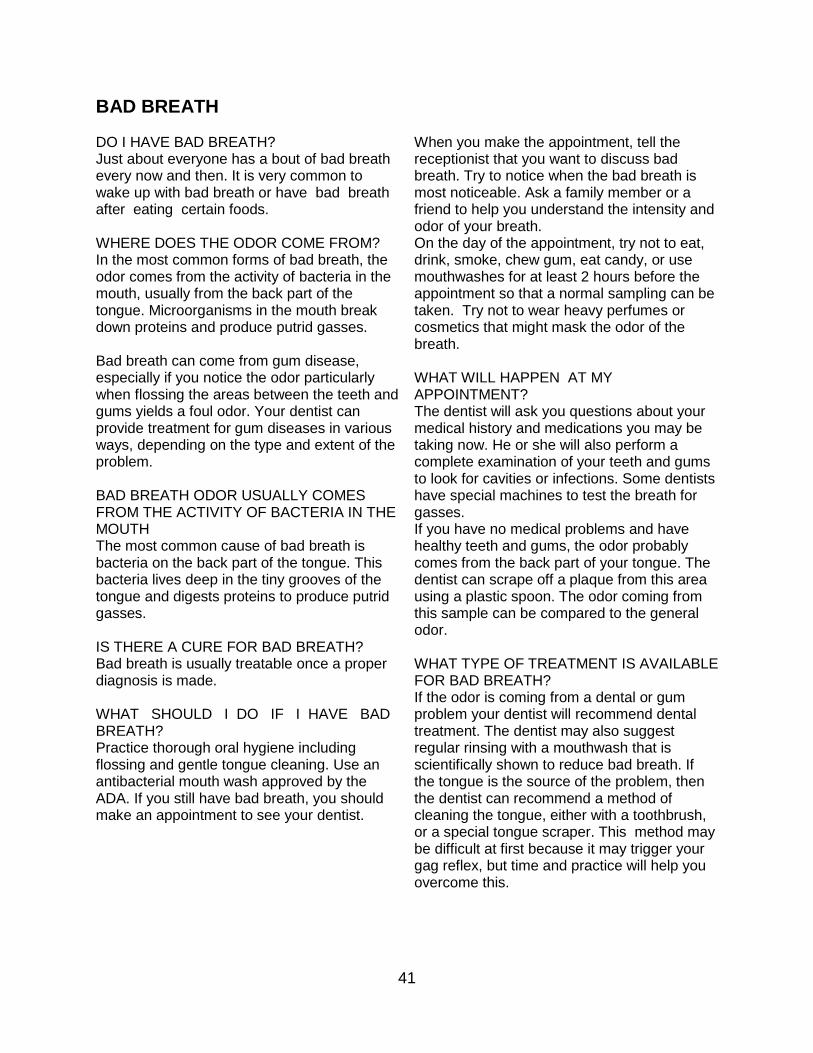
41
BAD BREATH
DO I HAVE BAD BREATH? Just about everyone has a bout of bad breath every now and then. It is very common to wake up with bad breath or have bad breath after eating certain foods. WHERE DOES THE ODOR COME FROM? In the most common forms of bad breath, the odor comes from the activity of bacteria in the mouth, usually from the back part of the tongue. Microorganisms in the mouth break down proteins and produce putrid gasses. Bad breath can come from gum disease, especially if you notice the odor particularly when flossing the areas between the teeth and gums yields a foul odor. Your dentist can provide treatment for gum diseases in various ways, depending on the type and extent of the problem. BAD BREATH ODOR USUALLY COMES FROM THE ACTIVITY OF BACTERIA IN THE MOUTH The most common cause of bad breath is bacteria on the back part of the tongue. This bacteria lives deep in the tiny grooves of the tongue and digests proteins to produce putrid gasses. IS THERE A CURE FOR BAD BREATH? Bad breath is usually treatable once a proper diagnosis is made. WHAT SHOULD I DO IF I HAVE BAD BREATH? Practice thorough oral hygiene including flossing and gentle tongue cleaning. Use an antibacterial mouth wash approved by the ADA. If you still have bad breath, you should make an appointment to see your dentist.
When you make the appointment, tell the receptionist that you want to discuss bad breath. Try to notice when the bad breath is most noticeable. Ask a family member or a friend to help you understand the intensity and odor of your breath. On the day of the appointment, try not to eat, drink, smoke, chew gum, eat candy, or use mouthwashes for at least 2 hours before the appointment so that a normal sampling can be taken. Try not to wear heavy perfumes or cosmetics that might mask the odor of the breath. WHAT WILL HAPPEN AT MY APPOINTMENT? The dentist will ask you questions about your medical history and medications you may be taking now. He or she will also perform a complete examination of your teeth and gums to look for cavities or infections. Some dentists have special machines to test the breath for gasses. If you have no medical problems and have healthy teeth and gums, the odor probably comes from the back part of your tongue. The dentist can scrape off a plaque from this area using a plastic spoon. The odor coming from this sample can be compared to the general odor. WHAT TYPE OF TREATMENT IS AVAILABLE FOR BAD BREATH? If the odor is coming from a dental or gum problem your dentist will recommend dental treatment. The dentist may also suggest regular rinsing with a mouthwash that is scientifically shown to reduce bad breath. If the tongue is the source of the problem, then the dentist can recommend a method of cleaning the tongue, either with a toothbrush, or a special tongue scraper. This method may be difficult at first because it may trigger your gag reflex, but time and practice will help you overcome this.
42
Your dentist may also refer you to a clinic that specializes in bad breath, or to another medical expert. WHAT ELSE CAN I DO? Normally, the treatment and regular home care your dentist recommends will take care of bad breath. It is important to practice home care daily to prevent the bacteria from colonizing on your tongue. VISIT THE BAD BREATH INTERNET SITE AT: http://www.smellwell.com/badBreathOverview.asp x Remember, bad breath is a problem that needs professional attention. Don't mask it - deal with it. Do's • Visit your dentist regularly. • Have your teeth cleaned periodically by a
dental professional. • Floss or otherwise clean between your
teeth, as recommended by your dentist. Choose unscented floss so that you can detect those areas between your teeth that give off odors, and clean them more carefully.
• Brush your teeth and gums properly. Ask your dentist to recommend a toothbrush or scraper for your tongue. Clean your tongue all the way back gently, but thoroughly.
• Drink plenty of liquids. • Chew sugar-free gum for a minute or two
at a time, especially if your mouth feels dry.
• Chewing parsley, mint, cloves or fennel seeds may also help.
• Clean your mouth after eating or drinking milk products, fish and meat.
• Unless your dentist advises otherwise, soak dentures overnight in antiseptic solution.
• Get control over the problem. Ask a family member to tell you whenever you have bad breath.
If someone in your family or a close friend has bad breath, find a kind way to let them know. If you can't tell them directly, leave this fact filelying around. They may get the message. Ask your dentist to recommend a mouthwash which has been shown to be clinically effective in fighting bad breath. Use it right before bed time for the best effect. Eat fresh, fibrous vegetables such as carrots. Don'ts • Let your concern about
having bad breath run your life.
• Don't be passive. • Don't be depressed. • Don't ignore your gums - you
can lose your teeth as well as smell bad.
• Don't drink too much coffee - it may make the situation worse.
• Don't forget to clean behind the back teeth in each row.
• Don't brush your tongue with regular toothpaste - it's better to dip your toothbrush in mouthwash for tongue cleaning.
• Don't run to the gastroenterologist for concerns of having bad breath - it usually comes from the mouth and almost never from the stomach.
• Don't clean your tongue so hard that it hurts.
• Don't rely on mouthwash alone - practice complete oral hygiene.
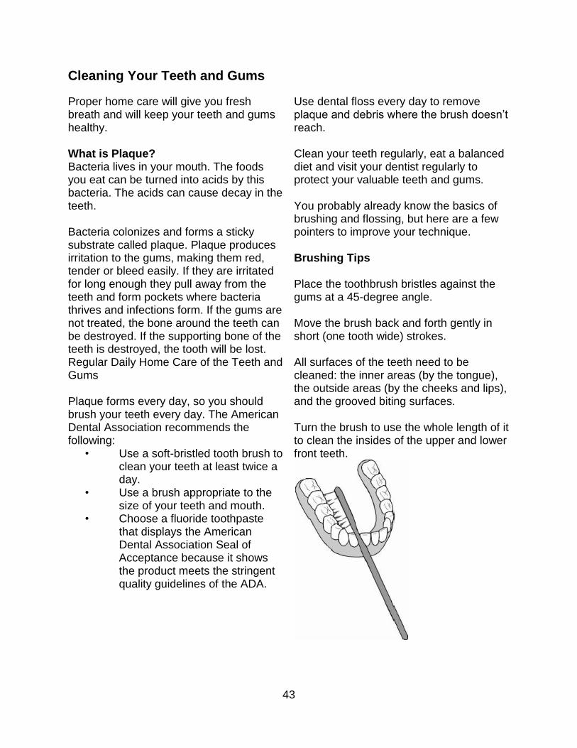
43
Cleaning Your Teeth and Gums
Proper home care will give you fresh breath and will keep your teeth and gums healthy. What is Plaque? Bacteria lives in your mouth. The foods you eat can be turned into acids by this bacteria. The acids can cause decay in the teeth. Bacteria colonizes and forms a sticky substrate called plaque. Plaque produces irritation to the gums, making them red, tender or bleed easily. If they are irritated for long enough they pull away from the teeth and form pockets where bacteria thrives and infections form. If the gums are not treated, the bone around the teeth can be destroyed. If the supporting bone of the teeth is destroyed, the tooth will be lost. Regular Daily Home Care of the Teeth and Gums Plaque forms every day, so you should brush your teeth every day. The American Dental Association recommends the following:
• Use a soft-bristled tooth brush to clean your teeth at least twice a day.
• Use a brush appropriate to the size of your teeth and mouth.
• Choose a fluoride toothpaste that displays the American Dental Association Seal of Acceptance because it shows the product meets the stringent quality guidelines of the ADA.
Use dental floss every day to remove plaque and debris where the brush doesn’t reach. Clean your teeth regularly, eat a balanced diet and visit your dentist regularly to protect your valuable teeth and gums. You probably already know the basics of brushing and flossing, but here are a few pointers to improve your technique. Brushing Tips Place the toothbrush bristles against the gums at a 45-degree angle. Move the brush back and forth gently in short (one tooth wide) strokes. All surfaces of the teeth need to be cleaned: the inner areas (by the tongue), the outside areas (by the cheeks and lips), and the grooved biting surfaces. Turn the brush to use the whole length of it to clean the insides of the upper and lower front teeth.
44
Flossing Break off about 12 inches of a strong floss and wind most of it around one of your middle fingers. Or you can tie the two ends of the floss together to form a circle. Wind some of the floss around a finger of the other hand. Hold the floss taunt between your thumbs and forefingers. Guide the floss between your teeth using a gentle back and forth motion. Don’t snap the floss between the teeth. After the floss is through the tight area between the two teeth, curve it into a C shape against one tooth. Gently slide it up and down the tooth and under the gum line. Do the same thing for the side of the other tooth. Advance the floss so you use a clean area between each tooth.
Tongue Cleaning Gently brush the tongue with a regular toothbrush dipped in an antibacterial mouth wash. Reach back as far as you can without hurting yourself. At first, you may gag, but practice will decrease this natural urge. Rinse the toothbrush well and repeat a few times until the white coating on the surface of the tongue is removed. Be gentle so the delicate tissues are not damaged. Special tongue cleaning devices may be used. Gently scrape the surface of the tongue according to the manufacturer’s directions. Mouth Washes Your dentist may recommend an antibacterial mouth wash. S/he will give you specific directions on when to use it.
45
Appendix: Halimeter
halimeter_brochure_2004.pdf
http://www.animated-teeth.com/bad_breath/t1_halitosis.htm
“Bad breath, also known as "halitosis", doesn't have to be an insurmountable
problem. Appropriate cures for bad breath are usually very simple once the
sufferer understands the fundamental causes of their breath odors. In most
cases a person's bad breath is due to anaerobic oral bacteria which have
accumulated on the person's tongue and also between and around their teeth.”
Our discussion will outline for you some simple tests you can use to determine if
you have bad breath, explain to you how and why oral bacteria create bad
breath, detail for you where these offending bacteria commonly accumulate and
how to effectively minimize them, provide you with explanations regarding the
use of tongue scrapers, mouthwashes, and other specialty products, and provide
you with some common sense tips which can help you to minimize bad breath.
Bad Breath. [Online] Available http://www.animated-teeth.com/ December 2016.

46
Course Test: Halitosis: A Clinical Review
1. Some of the earliest sound studies on bad breath indicated that the is the
source of most halitosis.
a. gastrointestinal tract
b. sinus
c. circulatory system
d. oral cavity
2. Organoleptic sampling means:
a. measurement of odor using a sophisticated commercially available machine.
b. measurement of bleeding and inflammation in the gingival sulcus using
a periodontal probe.
c. relying on the self-report of the patient for estimation of the degree of
malodor.
d. smelling the expired air or other sample of the subject for presence or lack of
odor.
3. The currently accepted conclusion is that volatile sulphur compounds (VSC) are the
main component of oral malodor.
a. True
b. False
4. The advantages of the gas chromatograph include:
a. individual gasses can be quantified.
b. gasses are measured even if there are very low concentrations.
c. it is portable.
d. a and b
5. Disadvantages of a gas chromatograph include:
a. (relatively) expensive,
b. personnel need to be trained to use machine,
c. significant amount of time required for measurement.
d. all of the above.

47
6. Industrial sulfide monitor measurements have been shown to be more reproducible
and recognized reduction of malodor following mouthrinsing better than
organoleptic scores.
a. True
b. False
7. Advantages of the sulfide monitor over the gas chromatograph include:
a. less expensive
b. no training required for operation
c. portable
d. measures quickly so several measurements can be taken consecutively
e. all of the above
8. Disadvantages of the sulfide monitor include:
a. does not separate the gases to measure each sulfide individually
b. ethanol or essential oils interfere with the machine’s ability to measure the
gasses, so it is not appropriate for studies on mouthwash efficacy
c. may need periodic recalibration
d. all of the above
9. In most cases, bad breath can be reduced or eliminated by:
a. proper dental care, oral hygiene, deep tongue cleaning and, if necessary, rinsing
with an effective mouthwash.
b. sinus surgery.
c. antacid use.
d. antibiotics.
10. Persistent uncontrollable halitosis may have:
a. Gastrointestinal ulcers
b. Internal bleeding
c. Hiatal hernia.
d. Diabetes mellitus
e. any of the above
11. The microorganisms on the tongue and in dental plaque putrefy proteins, mucins,
and peptides to release volatile sulfur compounds which are released as gas in the
breath.
a. True
b. False
48
12. Malodor is produced in saliva that has a neutral or alkaline pH but is inhibited by an
acidic pH. Fermentation of sugars inhibit malodor generation but is the basis of
caries formation.
a. True
b. False
13. In the study about diamines by Drs. Goldberg, Kozlovsky, and Rosenberg,
cadaverine levels were:
a. associated with odor judge organoleptic scores, plaque index scores, and
gingival index scores.
b. relative to the BANA scores and mean probing depth.
c. not associated with VSC levels.
d. All of the above.
14. The following conditions present malodor that is not associated with the bacterial
load of the dorsum of the tongue:
a. Traumatic ulceration
b. Dental Abscess
c. Herpetic Infections
d. Apthous Ulcers
e. all of the above
15. Medications that can contribute to xerostomia include:
a. Antihistamines
b. Anxiolytics
c. Antipshychotics
d. Anticholinergics
e. all of the above.
16. Conditions that can contribute to xerostomia include:
a. Diabetes
b. Anemia
c. Vitamin Deficiency
d. Radiation Treatment
e. all of the above.

49
17. The following systemic sources can contribute to halitosis:
a. Pharygitis,
b. cirrhosis of the liver
c. c. Renal Failure
d. Ulcers.
e. all of the above
18. If halitosis persists after debridement of the tongue and proper dental care (including
periodontal treatment if indicated and proper oral hygiene), the patient should be
referred to their physician for further investigation of a systemic cause.
a. true
b. false
19. A complete medical history does not need to be taken if the patient complains of
bad breath.
a. true
b. false
20. In a normal examination, five main types of odors can be found emanating from the
mouth. Which of these is not one of them?
a. periodontal-type odor
b. odor from the posterior tongue dorsum
c. denture odor
d. athlete’s mouth odor
e. characteristic nasal odor
21. Patients who have oral malodor related to dry mouth should be instructed to
a. drink extra water.
b. use a mouthwash containing alcohol.
c. breathe through their mouths.
d. All of the above
22. Toothbrush filaments should be made of
a. straw
b. wood
c. brick
d. synthetic nylon
e. natural materials
50
23. Toothbrush filaments should have filaments.
a. rounded
b. straight
c. square
d. uneven
24. The Bass Technique or the Modified Bass should be directed apically to the tooth at
an angle to the long axis of the tooth.
a. 60°
b. 80°
c. 45°
d. 35°
25. The tongue’s papillae create an uneven surface.
a. filiform and fungiform
b. villiform and williform
c. rugae and raphae
d. hydrogen dioxide and methyl mercaptan
51
Endnotes
i Tonzetich, J. “Preface” In Bad Breath: Research Perspectives. ii Rosenberg, M. “Clinical assessment of bad breath: current concepts.”
iii Rosenberg, M. “Clinical assessment of bad breath: current concepts.”
iv Rosenberg, M. “Clinical assessment of bad breath: current concepts.”
v Rosenberg, M. “Clinical assessment of bad breath: current concepts.”
vi Rosenberg, M. “Clinical assessment of bad breath: current concepts.”
vii Rosenberg, M. “Clinical assessment of bad breath: current concepts.”
viii Yaegaki, K.; Sanada, K. “Biochemical and clinical factors influencing oral malodor in periodontal patients.”
ix Rosenberg, M. “Introduction” In Bad Breath: Research Perspectives
x De Boever, E. and Loesche, W. “Assessing the contribution of anaerobic microflora of the tongue to oral
malodor.” xi Kleinberg, I. and Codipilly, M. “The biological basis of oral malodor formation.” In Bad Breath: Research
Perspectives . xii
Kleinberg, I. and Codipilly, M. “The biological basis of oral malodor formation.” In Bad Breath: Research Perspectives . xiii
Rosenberg, M. “Clinical assessment of bad breath: current concepts.” xiv
Rosenberg, M. “Clinical assessment of bad breath: current concepts.” xv
Persson, S.; Edlund, M.; Claesson, R. Carlsson, J. “The formation of hydrogen sulfide and methyl- mercaptan by oral bacteria.” and Claesson, R.; Edlund, M.; Persson, S.; Carlsson, J. “The production of volatile sulfur compounds by various Fusobacterium species.” xvi
McCulloch, C. and Bosy, A. “Relationship of oral malodor and periodontitis.” xvii
Goldberg, S.; Kozlovsky, A.; Rosenberg, M.; “Diamines and oral malodor.” In Bad Breath: Researc h Perspectives. xviii
Goldberg, S.; Kozlovsky, A.; Rosenberg, M.; “Diamines and oral malodor.” In Bad Breath: Researc h Perspectives. xix
Persson, S.; Edlund, M.; Claesson, R. Carlsson, J. “The formation of hydrogen sulfide and methyl- mercaptan by oral bacteria.” and Claesson, R.; Edlund, M.; Persson, S.; Carlsson, J. “The production of volatile sulfur compounds by various Fusobacterium species.” xx
Loesche, W.; De Boever, E. “Strategies to identify the main microbial contributors to oral malodor.” xxi
Yaegaki, K. Coil, J.M. “Examination, classification, and treatment of halitosis; clinical perspectives.”J Can Dent Assoc. 2000; May;66(5):257-61. xxii
Rosenberg, M. “Current Concepts in Clinical Asses sment of Bad Breath” xxiii
Miyazaki, H. et al, “Tentative classification of halitosis and its treatment needs.” In Yaegaki, K. Coil, J.M. “Examination, classification, and treatment of halitosis; clinical perspectives.”J Can Dent Assoc. 2000; May;66(5):257-61. xxiv
Yaegaki, K. Coil, J.M. “Examination, classification, and treatment of halitosis; clinical perspectives.”J Can Dent Assoc. 2000; May;66(5):257-61. xxv
Foster, D. “Limitations of subjective measurement of odor.”; Guadagni, D. “Requirements for coordination of instrumental and sensory techniques.”; and Langenau, E. “Correlation of objective -subjective methods as applied to the perfumery and cosmetics industries.” All In: Correlation of Subjective -Objective Methods in the Study of Odors and Taste. xxvi
Rosenberg, M. “Clinical assessment of bad breath: current concepts.” xxvii
Rosenberg, M. “Current Concepts in Clinical Asses sment of Bad Breath” xxviii
Miyazaki, H. et al, “Tentative classification of halitosis and its treatment needs.” In Yaegaki, K. Coil, J.M. “Examination, classification, and treatment of halitosis; clinical perspectives.”J Can Dent Assoc. 2000; May;66(5):257-61.

52
xxix
Miyazaki, H. et al, “Tentative classification of halitosis and its treatment needs.” In Yaegaki, K. Coil, J.M. “Examination, classification, and treatment of halitosis; clinical perspectives.”J Can Dent Assoc. 2000; May;66(5):257-61. xxx
Yaegaki, K. “Oral malodor and periodontal disease.” In Bad Breath: Research Perspectives.” xxxi
The American Dental Association web site: http://www.ada.org xxxii
Wilkins, E. Clinical Practice of the Dental Hygienist. 7th Edition. xxxiii
Wilkins, E. Clinical Practice of the Dental Hygienist. 7th Edition. xxxiv
Bass, C.C. “An effective method of personal oral hygiene.” J. Louisiana State Med. Soc. Mar 1954; 106: 100. xxxv
Wilkins, E. Clinical Practice of the Dental Hygienist. 7th Edition. xxxvi
Rosenberg, M. “Introduction” In Bad Breath: Research Perspectives xxxvii
Preti, Lawlwy, Hormann, Cowar, Feldman, Lowry and Young “Non-oral and oral aspect of oral malodor.” xxxviii
Yaegaki, K. “Oral malodor and periodontal disease.” In Bad Breath: Research Perspectives.” xxxix
Rosenberg, M. “Clinical assessment of bad breath: current concepts.” xl Rosenberg, M. “Clinical assessment of bad breath: current concepts.”
xli Rosenberg, M. “Clinical assessment of bad breath: current concepts.”
xlii Rosenberg, M. “Clinical assessment of bad breath: current concepts.”
xliii Rosenberg, M. “Clinical assessment of bad breath: current concepts.”