Embed Size (px)
Citation preview
1
Haemoglobinopathiesin pregnancy
Clinical Guideline
Directorate of Women’s HealthMinistry of Health
Trinidad and TobagoJanuary 2020
Women’s Health


Message from the Directorate The Directorate of Women’s Health commenced activities in 2017 at the Ministry of Health to improve maternal and perinatal outcomes and address international targets for Trinidad and Tobago in achieving the milestones towards the Sustainable Developmental Goals (SDGs), 2030. This document is one such response to create standardized clinical guidelines related to Obstetrics and Gynaecology.
In line with the SDGs 2030-Agenda and the Global Strategy for Women’s, Children’s and Adolescent’s Health (2016-2030), this document supports the objectives of “Survive, Thrive and Transform” by promoting the reduction of maternal and perinatal, infant and child morbidity and mortality.
Haemoglobinopathies are a heterogeneous group of single-gene disorders that include the structural haemoglobin variants and the thalassaemias. In Trinidad and Tobago, as maternal mortality from direct deaths has declined significantly in the past few years, deaths from indirect causes including complications related to sickle cell disease, have to be addressed with common initial care protocols that can be tailored and escalated based on each patient’s clinical status. This document is not intended to provide comprehensive details on the management of haemoglobinopathies.
We used an ‘adopt and adapt’ method in the production of this guideline based on existing resources and expertise. Consensus was obtained from recognized multidisciplinary stakeholders based on the evidence and publications at the time of producing this document.
This Guideline provides new information that was previously not contained in the Standard Operating Procedure (SOP) Manual for Obstetric and Midwifery Services produced by the Ministry of Health in June 2011. This guideline also recommends a change to the antenatal panel of blood tests as noted in Page 4 of the Maternal and Child Health Manual (MCHM) of April 2015. All other sections and updated clinical guidelines on other topics remain in force.
Acknowledgements
The Directorate wishes to recognize the contributions from all stakeholders including haematologists and staff at the RHAs. The Obstetrics & Gynaecology Department at the Eastern Regional Health Authority provided the initial draft working document.
The work of the team from the Corporate Communications department in finalizing this publication is also acknowledged.
Dr Adesh Sirjusingh Director
LIST OF ABBREVIATIONS ACE Angiotensin-converting enzyme
ACS Acute chest syndrome
ACOG The American College of Obstetricians and Gynecologists
ARB Angiotensin receptor blocker
CBC Complete blood count
CTG Cardio-tocography
CTPA Computed tomography pulmonary angiogram
CXR Chest X-Ray
FIGO International Federation of Gynecology and Obstetrics
Hb Haemoglobin
HPLC High Performance Liquid Chromatography
IEF Isoelectric focusing
IUGR Intrauterine growth restriction
IV Intravenous
LWMH Low molecular weight heparin
MCHM Maternal and Child Health Manual
MCOS Medical Chief of Staff
MCS Microscopy, culture and sensitivity
MOH Ministry of Health
MSU Midstream urine
PAHO Pan American Health Organization
PCA Patient Controlled Analgaesia
RCOG The Royal College of Obstetricians and Gynaecologists
RHA Regional Health Authority
SCD Sickle cell disease
SDGs Sustainable Developmental Goals
SIP Perinatal Information System
SMO Specialist Medical Officer
SOGC Society of Gynecologists and Obstetricians of Canada
SOP Standard Operating Procedure
V/Q Ventilation-perfusion
WHO World Health Organization
TABLE OF CONTENTS1.0 Key recommendations 1
2.0 Introduction 2
3.0 Thalassaemia and pregnancy 5
4.0 Sickle cell disease and pregnancy 5
5.0 Complications of SCD 7
6.0 Antenatal management of SCD 8
7.0 Admission for acute complications of SCD 10
8.0 Intrapartum care 13
9.0 Postpartum care 14
10.0 Audit and Research 14
11.0 Bibliography 14
1.0 Key recommendations Pre-pregnancy
All women (of childbearing age) enrolled in an existing clinic system for inherited blood disorders, should be counselled on family planning options and receive counselling regarding the risks of inheritance to their offspring. This will also involve haemoglobinopathy screening of their partners.
Antenatal Screening and Diagnosis
Where not currently practiced, women must have a complete blood count (CBC) instead of only a haemoglobin test (change from MCHM 2015).
The current sickle solubility screening test should be phased out at all centres and replaced by a diagnostic test. Currently Hb Electrophoresis is available but newer laboratory methods have been developed to diagnose and quantitate the abnormal haemoglobin(s) instead of gel electrophoresis and are beyond the scope of this document. These include Isoelectric focusing (IEF) and High performance liquid chromatography (HPLC). The RHAs will have to develop a plan to implement the relevant services for this new approach.
Multi-disciplinary clinic
All pregnant women with inherited blood disorders should be managed in collaboration with a haematologist.
Neonatal Screening
RHAs should collaborate and build on existing neonatal haemoglobinopathy screening programs.
Audit and Research
National data, cost-benefit analysis and research are important to guide the introduction of changes to current patient care management algorithms, including a national universal neonatal screening program. This is also required to inform and strengthen the national policy.
1
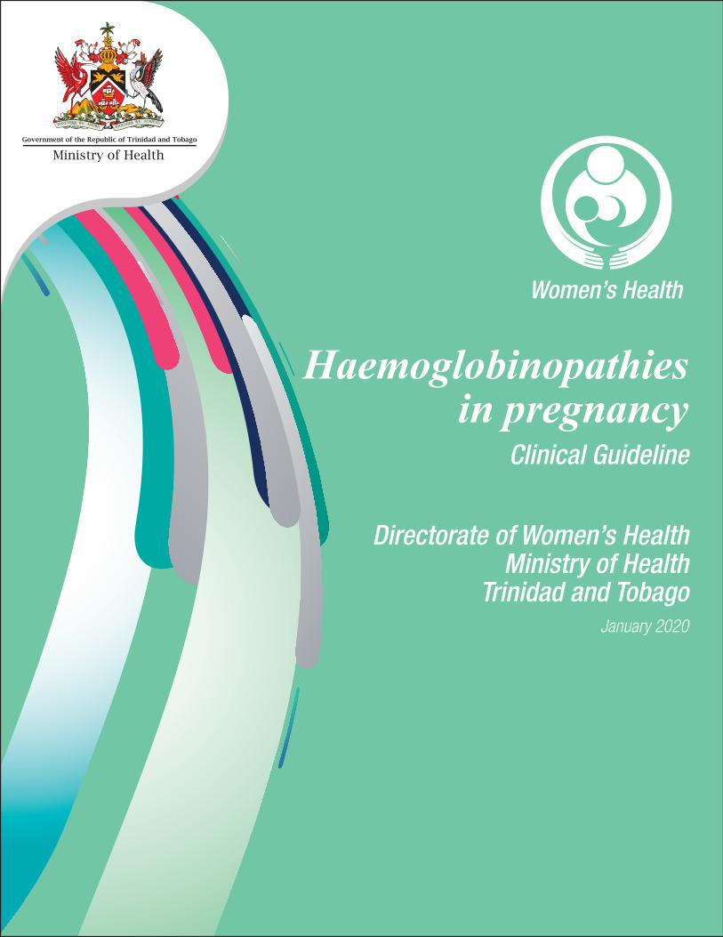
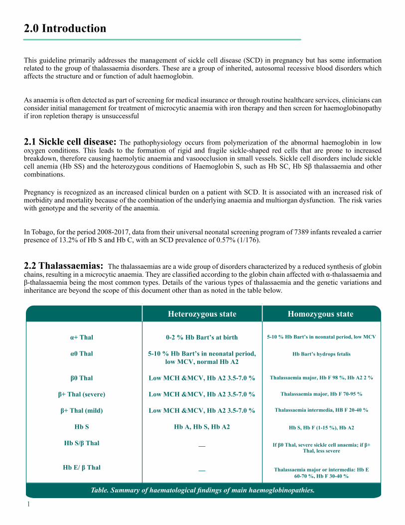
Heterozygous state Homozygous state
α+ Thal
α0 Thal
β0 Thal
β+ Thal (severe)
β+ Thal (mild)
Hb S
Hb S/β Thal
Hb E/ β Thal
0-2 % Hb Bart’s at birth
5-10 % Hb Bart’s in neonatal period, low MCV, normal Hb A2
Low MCH &MCV, Hb A2 3.5-7.0 %
Low MCH &MCV, Hb A2 3.5-7.0 %
Low MCH &MCV, Hb A2 3.5-7.0 %
Hb A, Hb S, Hb A2
__
__
5-10 % Hb Bart’s in neonatal period, low MCV
Hb Bart’s hydrops fetalis
Thalassaemia major, Hb F 98 %, Hb A2 2 %
Thalassaemia major, Hb F 70-95 %
Thalassaemia intermedia, HB F 20-40 %
Hb S, Hb F (1-15 %), Hb A2
If β0 Thal, severe sickle cell anaemia; if β+Thal, less severe
Thalassaemia major or intermedia: Hb E 60-70 %, Hb F 30-40 %
2.0 Introduction
This guideline primarily addresses the management of sickle cell disease (SCD) in pregnancy but has some information related to the group of thalassaemia disorders. These are a group of inherited, autosomal recessive blood disorders which affects the structure and or function of adult haemoglobin.
As anaemia is often detected as part of screening for medical insurance or through routine healthcare services, clinicians can consider initial management for treatment of microcytic anaemia with iron therapy and then screen for haemoglobinopathy if iron repletion therapy is unsuccessful
2.1 Sickle cell disease: The pathophysiology occurs from polymerization of the abnormal haemoglobin in low oxygen conditions. This leads to the formation of rigid and fragile sickle-shaped red cells that are prone to increased breakdown, therefore causing haemolytic anaemia and vasoocclusion in small vessels. Sickle cell disorders include sickle cell anemia (Hb SS) and the heterozygous conditions of Haemoglobin S, such as Hb SC, Hb Sβ thalassaemia and other combinations.
Pregnancy is recognized as an increased clinical burden on a patient with SCD. It is associated with an increased risk of morbidity and mortality because of the combination of the underlying anaemia and multiorgan dysfunction. The risk varies with genotype and the severity of the anaemia.
In Tobago, for the period 2008-2017, data from their universal neonatal screening program of 7389 infants revealed a carrier presence of 13.2% of Hb S and Hb C, with an SCD prevalence of 0.57% (1/176).
2.2 Thalassaemias: The thalassaemias are a wide group of disorders characterized by a reduced synthesis of globin chains, resulting in a microcytic anaemia. They are classified according to the globin chain affected with α-thalassaemia and β-thalassaemia being the most common types. Details of the various types of thalassaemia and the genetic variations and inheritance are beyond the scope of this document other than as noted in the table below.
Table. Summary of haematological findings of main haemoglobinopathies.
2
2.3 Pre-pregnancy general advice
From adolescence, counsel regarding planning of pregnancy and contraception. For SCD, they can be advised that pregnancy and delivery carries a greater risk of major complications including thrombosis.
Family planning and contraception options should be part of the regular haematology and general practice clinics. Stress the importance of safe and reliable contraception versus the risks of pregnancy. These are missed opportunities to prevent maternal and fetal morbidity and mortality.
Primary care health physicians and haematologists have a key role in preconception care in addition to screening for chronic disease complications.
RHAs should provide individualized information including how to access services. The MOH’s website has additional resources on the Women’s Health page, regarding pregnancy, contraception and other women’s health issues.
2.4 Antenatal Screening
Currently all pregnant patients are screened with at least a haemoglobin [complete blood count (CBC) is available at all RHAs] and a sickle cell test (solubility test), as part of the antenatal panel of blood tests. This information however will not exclude or quantify haemoglobinopathies.
A CBC is recommended to replace haemoglobin only testing, where Hb-only screening is being performed.
Where the information is not previously documented, a diagnostic test (patient and partner testing) is recommended instead of a solubility sickle test. This can identify and quantify important transmissible gene abnormalities including traits associated with thalassaemia, Hb C, E, B and D, that can affect fetal outcome. Currently Hb Electrophoresis is available at RHA hospital based laboratories.
RHAs are to ensure and strengthen the access to more reliable and automated laboratory diagnostic services including investing in the appropriate technology based on the RHA’s clinical requirements., such as Isoelectric focusing (IEF) and High performance liquid chromatography (HPLC)
Data and research on this topic are required from all RHAs to inform and strengthen these changes to the current national antenatal haemoglobinopathy screening program.
2.5 Documentation, data and reports
For all public sector patients, the Perinatal Information System (SIP) (hard and soft copy) is to be utilized to document all reports as mandated by the MoH as of August 2018. Once fully implemented by the RHAs, the SIP will provide valuable national data and reports on this clinical area.
The patient must be provided with a copy of the SIP form which is to be updated at each clinical encounter.
The electronic SIP version must be utilized at all maternity units and completed after delivery and before discharge from the institution.
3
The electronic SIP system will reduce the need and costs associated with repeating constant diagnostic tests with every pregnancy occasion for the same patient (e.g. haemoglobinopathy genotype, Blood Group, Rhesus) as this forms a permanent record.
2.6 Prenatal testing
Prenatal genetic testing, such as maternal serum, chorionic villus sampling or amniocentesis, is currently not routinely available at the maternity units. RHAs with high-risk obstetric referral services e.g. University based centres, are to open further dialogue on this topic.
2.7 Newborn screening
Currently universal RHA-newborn screening programs are not in place except in Tobago and the Port of Spain General Hospital. RHAs should collaborate and build on these programs.
3.0 Thalassaemia and pregnancyFor α-thalassaemia, the clinical course is not significantly affected.
For β-thalassaemia major, pregnancy is rare, but possible. These women must receive extensive risk assessment, review and counselling especially if cardiac function is abnormal.
For β-thalassaemia minor, a mild asymptomatic anaemia is usual. Care is required if administering iron therapy beyond prophylactic doses.
The safety of iron chelation therapy in pregnancy is unknown.
Fetal growth must be closely monitored throughout pregnancy.
4.0 Sickle cell disease and pregnancy
4.1 Counsel:
• How nausea and vomiting in pregnancy can result in dehydration and the precipitation of crises.
• That there are increased risks of worsening anaemia, crises, acute chest syndrome (ACS) and infection during pregnancy.
• That there is an increased risk of having a growth-restricted baby, induction of labour and caesarean section.
4
• There is a chance of their offspring inheriting SCD.
• There is need for up-to-date assessment for chronic disease complications.
4.2 Assessment for chronic disease complications should include:
• Screening for pulmonary hypertension with echocardiography. If there is a high risk of pulmonary hypertension, appropriate contraception should be advised due to the increased mortality risk.
• Blood pressure measurement and urinalysis to identify hypertension and/or proteinuria.
• Renal and liver function tests at least annually to identify sickle nephropathy and/or deranged hepatic function.
• Retinal screening. Proliferative retinopathy is common (especially with Hb SC) and can lead to loss of vision.
• Screening for iron overload.
• Screening for red cell antibodies (if available).
4.3 Assessment of partner’s status:
• Offer diagnostic haemoglobinopathy testing for their partner before they embark on pregnancy to allow appropriate counselling regarding the risk of having an affected offspring. Detailed genetic counseling is beyond the scope of this document.
4.4 Immunization and antibiotic prophylaxis:
• Review the latest recommendations for vaccinations including the annual flu vaccine.
• Consult haematologist for recommendations on the up to date requirements for penicillin prophylaxis.
4.5 Medication:
• Hydroxyurea should be stopped at least 3 months before conception
• ACE inhibitors and ARB’s should be stopped before conception
• Folic acid (5 mg) once daily preconception and throughout pregnancy.
5
5.0 Complications of SCD
5.1 Acute complications:
• Acute painful crisis is the most frequent complication of SCD encountered in pregnancy.
• Acute Chest Syndrome (ACS); characterized by respiratory symptoms (tachypnoea, chest pain or shortness of breath) in the presence of a new infiltrate on chest X-ray. ACS is the most frequent cause of death in adults with SCD and is rapidly progressive highlighting the need for prompt diagnosis and management.
• Acute Stroke; the diagnosis should be considered in any woman with SCD with acute neurological impairment.
• Acute Anaemia; this may be due to erythrovirus B19 which causes red cell maturation arrest and aplastic crisis. It is characterized by reticulopaenia.
5.2 Chronic complications include:
• Pulmonary Hypertension
• Renal Dysfunction
• Retinal Disease
• Leg Ulcers
• Cholelithiasis
• Avascular Necrosis (osteonecrosis)
5.3 Complications specific to pregnancy include:
• Miscarriage
• Stillbirth
• Fetal growth restriction
• Preterm labour and delivery
• Premature rupture of membranes
• Infections: urinary tract, respiratory, postpartum
• Thromboembolic events
• Increased risk of caesarean section
• There is a chance of their offspring inheriting SCD.
6
6.0 Antenatal management of SCD
6.1 General
• Provide antenatal care by a multidisciplinary team that includes an obstetrician with experience in high-risk obstetrics, in consultation with a haematologist.
• Educate and counsel throughout; keep communication channels open for easy patient access to the unit.
• Avoid precipitating factors such as cold, dehydration, stress, physical exertion and low oxygen concentration for example.
• Prophylactic exchange transfusion is not recommended as there is no agreed consensus on this area. The risks of repeated blood transfusions include alloimmunization, viral infections and iron overload.
• Transfuse only if clinically indicated as guided by the haematologist using the appropriate tested blood components rather than whole blood.
• Discuss and counsel on breastfeeding and contraception plans during the antenatal period.
The following are key milestones and recommendations. They do not include all current ‘routine’ pregnancy procedures, visits and practices.
6.2 First trimester
• Partner testing (if not done before)
• Review medications – stop hydroxyurea, ACE inhibitors and ARB’s
• Folic acid (4-5 mg) throughout
• Penicillin prophylaxis as directed by haematologist
• Ensure recommended vaccinations
• Retinal, cardiac and renal assessments
• Baseline oxygen saturation and blood pressure
• Baseline renal function test, liver function test, urine protein/creatinine ratio and ferritin level
• MSU for MCS and repeat every 4 weeks
• 7 to 9 weeks- ultrasound to confirm viability and accurate dating
• Introduce topic of family planning and discuss at each visit
7
6.3 11-14 weeks
• Ultrasound (see the MOH’s Ultrasound in Obstetrics Guideline)
• Low dose aspirin from 12 weeks
• Review partner’s electrophoresis’ results
6.4 18-22 weeks
• Detailed anomaly ultrasound scan
• Repeat MSU and CBC
6.5 24 weeks
• Ultrasound monitoring fetal growth and amniotic fluid volume
• Repeat MSU
6.6 28 weeks
• Ultrasound for growth, liquor volume, umbilical artery Doppler (Repeat every two weeks if IUGR detected; otherwise 4 weekly)
• Repeat MSU and CBC
6.7 32 weeks
• Ultrasound for growth, liquor volume, umbilical artery Doppler (Repeat every two weeks if IUGR detected; otherwise 4 weekly)
6.8 34 weeks
• Ultrasound for growth, liquor volume, umbilical artery Doppler
• MSU
8
6.9 36 Weeks
• Ultrasound for growth, liquor volume, umbilical artery Doppler
• Discuss mode of delivery (Caesarean Section is reserved for obstetric indications)
• Anaesthetic consultation
6.10 38 Weeks
• For those with a normally growing fetus, offer elective induction o f labour, or by elective caesarean section if indicated, after 38 weeks of gestation.
• If declined, for closer monitoring with ward reviews instead of weekly clinic visits
6.11 40 Weeks
• Offer Induction of labour if previously declined
7.0 Admission for acute complicationsThis must be on a suitable ward equipped for management of the medical requirements of the patent and not necessarily an obstetric or gynaecology ward
The SMO Obstetrician, haematologist and physician, along with the senior midwife (if applicable) are to be informed whenever a pregnant woman with SCD is admitted to hospital.
7.1 General measures
o Assess the steady state haemoglobin
o Previous transfusions
o Precipitating factors for pain crisis - exposure to extreme temperatures, dehydration and overexertion
o Any source of infection- fever, cough, chest pain, shortness of breath, runny nose, dysuria, vomiting, diarrhea etc.
o Description of pain
o Respiratory symptoms
o Review of medications
9
• Examination
o Rapid clinical assessment for complications requiring urgent intervention such as ACS, sepsis or dehydration
o Vital signs, particularly the presence of maternal tachycardia or pyrexia, and SpO2
o Cardiorespiratory
o Cause of pain
• Laboratory investigations include
o CBC (WBC counts are often raised in SCD and do not necessarily indicate infection)
o Reticulocyte count
o Arterial blood gas as indicated
o Renal function test
o Liver function test
o Depending on clinical scenario- Chest x ray, blood cultures, urine culture
• The role of an urgent CXR to detect consolidation (regardless of the stage of pregnancy) in known SCD patients with respiratory symptoms is underscored.
• CTG tracing (only if applicable) or fetal heart rate auscultation
• Fluid intake of at least 60ml/kg/24h (can be oral or intravenous)
• Prophylactic dose of low-weight molecular heparin (LWMH)
• Antibiotics: If febrile or high clinical suspicion of infection (administer after sepsis screening)
• Oxygen: If saturation falls below patient’s baseline or below 95%
• Senior specialist review- obstetrician, haematologist / physician
7.2 Management of acute pain
• Analgesia- start within 30 minutes of arriving at hospital with effective analgesia achieved within 1 hr. Consider the WHO’s analgaesia ladder for the escalation of treatment but there is individual variation in the pain response to opiates.
10
• Start with paracetamol for mild pain.
• NSAIDS for mild to moderate pain between 12 and 28 weeks’ gestation.
• Weak opioids for moderate pain. Opiates are not associated with teratogenicity or congenital malformations but may be associated with suppression of fetal movement and reduced baseline variability.
• Morphine for severe pain- oral/SC/IM/IV. IV can be given by intermittent bolus or by PCA system.
• Pethidine is not usually considered as a first line but care can be individualized based on previous patient experience with analgaesia.
• Facial oxygen therapy.
• Prescribe laxatives, anti-pruritic and antiemetic medication if required.
• Monitor pain, sedation, vital signs, respiratory rate and oxygen saturation every 20-30 minutes until pain is controlled and vital signs are stable, then every 2 hours (hourly if parenteral opiates given).
• Patients receiving parenteral opiates should be nursed in an area where they can undergo hourly observations.
• If respiratory rate is less than 10/min, omit maintenance analgesia and consider naloxone.
• Anaesthesiologist review as needed.
• Reduce analgesia after 2-3 days and replace injections with equivalent dose of oral analgesia. Patients should be discharged on tapering doses of oral opiates when pain free to prevent rebound pain from sudden discontinuation.
7.3 Management of acute chest syndrome
• Oxygen via face mask.
• Urgent Hematology review.
• The role of an urgent CXR to detect consolidation (irrespective of the stage of pregnancy) and prompt treatment (appropriate antibiotics, oxygen, transfusion, analgaesia) in known SCD patients with respiratory symptoms is underscored.
• CTPA or V/Q scanning are not helpful in diagnosing ACS (which is a combination of infarction) and should be considered only if CXR is normal.
• Urgent anaesthesiologist review especially if hypoxic and consider HDU/ICU admission.
• Intravenous antibiotics AFTER sepsis screen and include “Influenza swab” testing (refer to national guidance). Consider appropriate therapy with a third generation cephalosporin and a macrolide or approved quinolone.
• Blood transfusion (as advised by haematology team)
11
o Consider top up transfusion- if haemoglobin is falling or haemoglobin is less than 6.5 g/dl.
o Consider exchange transfusion- in severe hypoxia and if haemoglobin is maintained.
• There is a low threshold for considering pulmonary embolism among women with SCD.
• Inform neonatal team if the fetus is potentially viable.
7.4 Management of acute stroke
• Urgent brain imaging
• Hematology and neurology review
• Anaesthesiologist review
• Rapid-exchange blood transfusion- decreases long term neurological damage
• Thrombolysis is not indicated in acute stroke secondary to SCD
7.5 Management of acute anaemia
• Haematology review
• Acute anaemia may be attributable to erythrovirus leading to reticulocytopaenia
• Vertical transmission of erythrovirus to the fetus can lead to hydrops fetalis- review by specialist and arrange specialist ultrasound follow-up
• Transfusion, as guided by haematologist
• Place in isolation if infected with erythrovirus
8 Intrapartum care• Obstetric SMO, Paediatric staff, Anesthesiologist, Hematologist and Physician should be informed when patient is admitted
• Obtain IV access
• Cross match two (2) units of blood
12
• Adequate hydration during labour
• Keep warm
• Continuous CTG monitoring
• Vital signs hourly
• Low threshold for commencing broad spectrum antibiotics
• Continuous pulse oximetry. If SpO2< 94%, for ABG analysis and oxygen via face mask
• Adequate analgesia- avoid pethidine as first line but other opiates can be used (Consider PCA pump or epidural)
• Allow vaginal delivery if no obstetric indication for caesarean section
9 Postpartum care
• Maintain oxygen saturation above 94% and adequate hydration
• Administer prophylactic dose LMWH whilst hospitalized and seven (7) days post discharge for vaginal delivery and six (6) weeks for caesarean section
• Encourage mobilization
• Antithrombotic stockings
• Maintain vigilance for acute crisis and other complications of SCD
• Encourage breastfeeding as with all other mothers
• NSAIDs for analgesia
• Offer long term contraception prior to being discharged
• Neonatal screening
10 Audit and Research
• National data and research related to this clinical area are required in order to inform and develop a comprehensive package of services.
• Universal mandatory use of the SIP system is reinforced.
13
Bibliography
ACOG Practice Bulletin No. 78: Hemoglobinopathies in Pregnancy. January 2007, Reaffirmed 2018. (Replaces Practice Bulletin Number 64, July 2005). Available from https://www.acog.org/Clinical-Guidance-and-Publications/Practice-Bulletins/Committee-on-Practice-Bulletins-Obstetrics/Hemoglobinopathies-in-Pregnancy
Carrier Screening for Thalassaemia and Hemoglobinopathies in Canada. Joint SOGC-CCMG Clinical Practice Guideline. No 218, 2008. Available from +https://www.jogc.com/article/S1701-2163(16)32975-9/pdf
Charles KS, Osagie K, Battini RK. Hospital admissions for acute painful crisis in Trinidad and Tobago. Are the British Committee for Standards in Haematology (BCSH) guidelines applicable? Clinical and Laboratory Haematology. 2006; 28(5):299-302.
Genetic disorders and the fetus: diagnosis, prevention, and treatment. Edited by Milunsky A, and Milunsky JM. John Wiley & Sons; 7th Edition , 2016. Available from https://www.tuseb.gov.tr/enstitu/tacese/yuklemeler/ekitap/genetik/genetic_disorders_and_the_fetus_diagnosis_prevention_and_treatment_2016.pdf
Howard J, Hart N, Roberts‐Harewood M, Cummins M , Awogbade, M and Davis B. Guideline on the management of acute chest syndrome in sickle cell disease. British Journal of Haematology. 2015; 169: 492-505. doi:10.1111/bjh.13348
Knight-Madden, J; Lee, K; Elana, G; Elenga, N et al. Newborn Screening for Sickle Cell Disease in the Caribbean: An Update of the Present Situation and of the Disease Prevalence. Int. J. Neonatal Screen. 2019, 5(1), 5; https://doi.org/10.3390/ijns5010005
Management of Acute Chest Syndrome in Sickle Cell Disease. March 2015. British Society for Haematology. Available from https://b-s-h.org.uk/guidelines/guidelines/management-of-acute-chest-syndrome-in-sickle-cell-disease/
Management of Sickle Cell Disease in Pregnancy. RCOG Green-top Guideline No. 61. 1st Edition. July 2011(valid in 2019 until replaced by the British Society for Haematology). Available from https://www.rcog.org.uk/globalassets/documents/guidelines/gtg_61.pdf
Maternal and Child Health Manual 2015, Ministry of Health(TT) and the PAHO. Available from http://www.google.com/url?q=http%3A%2F%2Fwww.health.gov.tt%2Fdownloads%2FDownloadItem.aspx%3Fid%3D357&sa=D&sntz=1&usg=AFQjCNHZAug6ModRZqI8SYU7iNByeWu5hQ
14
Standard Operating Procedure Manual for Obstetric and Midwifery Services, June 2011. Ministry of Health Trinidad and Tobago. Available from http://www.google.com/url?q=http%3A%2F%2Fwww.health.gov.tt%2Fdownloads%2FDownloadItem.aspx%3Fid%3D244&sa=D&sntz=1&usg=AFQjCNHKP5dJvHFFEA0deoNHjUtOP1WTOw
The Directorate of Women’s Health Webpage of the Ministry of Health of Trinidad and Tobago. https://sites.google.com/health.gov.tt/womenshealth
Ultrasound Examination in Pregnancy- Clinical Guideline, 2018. Ministry of Health Trinidad and Tobago. Available from http://www.google.com/url?q=http%3A%2F%2Fwww.health.gov.tt%2Fdownloads%2FDownloadItem.aspx%3Fid%3D409&sa=D&sntz=1&usg=AFQjCNF44IDbh57Osc4qOw5ObWWhiyO8Gw
World Health Organization. Cancer Pain Relief With A Guide To Opioid Availability. 2nd ed. Geneva, Switzerland: World Health Organization; 1996.


![Raised Haemoglobin F (HbF) Level in Haemoglobinopathies ... · Haemoglobinopathies are the worldwide prevalent monogenic genetic disorders with variable geographic distribution [1]-[5].Although](https://img.pdfslide.us/doc/110x75/5f1b8427d7f40f077a680f2a/raised-haemoglobin-f-hbf-level-in-haemoglobinopathies-haemoglobinopathies.jpg)