Embed Size (px)
Citation preview

Morbidity and Mortality Weekly Report
Recommendations and Reports August 9, 2002 / Vol. 51 / No. RR-10
Centers for Disease Control and PreventionCenters for Disease Control and PreventionCenters for Disease Control and PreventionCenters for Disease Control and PreventionCenters for Disease Control and PreventionSAFER • HEALSAFER • HEALSAFER • HEALSAFER • HEALSAFER • HEALTHIER • PEOPLETHIER • PEOPLETHIER • PEOPLETHIER • PEOPLETHIER • PEOPLETM
Guidelines for the Prevention of IntravascularCatheter-Related Infections
MMWR
CONTENTS
Introduction ......................................................................... 1
Intravascular Catheter-Related Infections in Adult
and Pediatric Patients: An Overview ................................. 2
Epidemiology and Microbiology ........................................... 4
Pathogenesis ....................................................................... 5
Strategies for Prevention of Catheter-Related Infections
in Adult and Pediatric Patients .......................................... 5
Replacement of Catheters .................................................... 9
Special Considerations for Intravascular Catheter-Related
Infections In Pediatric Patients ........................................ 11
Recommendations for Placement of Intravascular
Catheters in Adults and Children.................................... 13
References ......................................................................... 19
Appendix A........................................................................ 27
Appendix B ........................................................................ 29
SUGGESTED CITATIONCenters for Disease Control and Prevention.Guidelines for the Prevention of IntravascularCatheter-Related Infections. MMWR 2002;51(No.RR-10):[inclusive page numbers].
The MMWR series of publications is published by theEpidemiology Program Office, Centers for DiseaseControl and Prevention (CDC), U.S. Department ofHealth and Human Services, Atlanta, GA 30333.
Centers for Disease Control and Prevention
Julie L. Gerberding, M.D., M.P.H.Director
David W. Fleming, M.D.Deputy Director for Science and Public Health
Dixie E. Snider, Jr., M.D., M.P.H.Associate Director for Science
Epidemiology Program Office
Stephen B. Thacker, M.D., M.Sc.Director
Office of Scientific and Health Communications
John W. Ward, M.D.Director
Editor, MMWR Series
Suzanne M. Hewitt, M.P.A.Managing Editor
Teresa F. RutledgeRachel J. Wilson
Project Editors
Malbea A. HeilmanBeverly J. Holland
Visual Information Specialists
Quang M. DoanErica R. Shaver
Information Technology Specialists
Vol. 51 / RR-10 Recommendations and Reports 1
The material in this report was prepared for publication by the National Centerfor Infectious Diseases, James M. Hughes, M.D., Director; Division of HealthcareQuality Promotion, Steven L. Solomon, M.D., Acting Director.
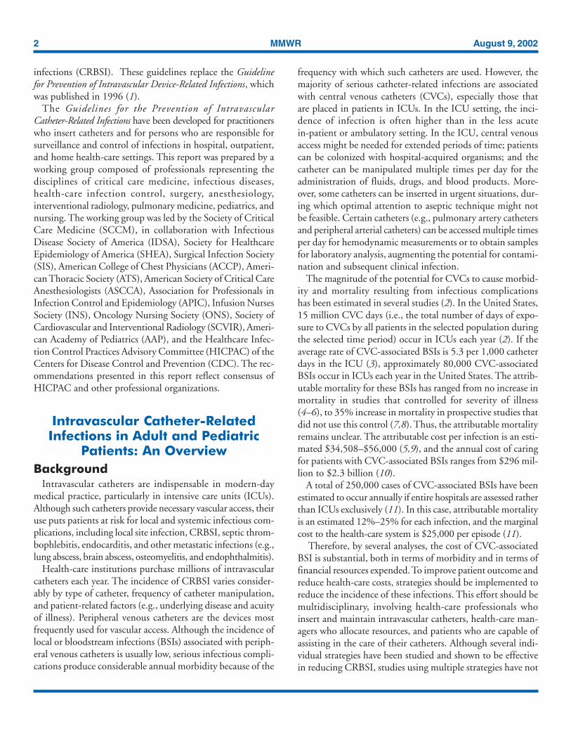
Guidelines for the Prevention of IntravascularCatheter-Related Infections
Prepared byNaomi P. O’Grady, M.D.1
Mary Alexander2
E. Patchen Dellinger, M.D.3
Julie L. Gerberding, M.D., M.P.H.4
Stephen O. Heard, M.D.5
Dennis G. Maki, M.D.6
Henry Masur, M.D.1
Rita D. McCormick, M.D.7
Leonard A. Mermel, D.O.8
Michele L. Pearson, M.D.9
Issam I. Raad, M.D.10
Adrienne Randolph, M.D., M.Sc.11
Robert A. Weinstein, M.D.12
1National Institutes of Health, Bethesda, Maryland2Infusion Nurses Society, Cambridge, Massachusetts
3University of Washington, Seattle, Washington4Office of the Director, CDC, Atlanta, Georgia
5University of Massachusetts Medical School, Worcester, Massachusetts6University of Wisconsin Medical School, Madison, Wisconsin
7University of Wisconsin Hospital and Clinics, Madison, Wisconsin8Rhode Island Hospital and Brown University School of Medicine, Providence, Rhode Island
9Division of Healthcare Quality Promotion, National Center for Infectious Diseases, CDC, Atlanta, Georgia10MD Anderson Cancer Center, Houston, Texas11The Children’s Hospital, Boston, Massachusetts
12Cook County Hospital and Rush Medical College, Chicago, Illinois
Summary
These guidelines have been developed for practitioners who insert catheters and for persons responsible for surveillance andcontrol of infections in hospital, outpatient, and home health-care settings. This report was prepared by a working group compris-ing members from professional organizations representing the disciplines of critical care medicine, infectious diseases, health-careinfection control, surgery, anesthesiology, interventional radiology, pulmonary medicine, pediatric medicine, and nursing. Theworking group was led by the Society of Critical Care Medicine (SCCM), in collaboration with the Infectious Disease Society ofAmerica (IDSA), Society for Healthcare Epidemiology of America (SHEA), Surgical Infection Society (SIS), American College ofChest Physicians (ACCP), American Thoracic Society (ATS), American Society of Critical Care Anesthesiologists (ASCCA),Association for Professionals in Infection Control and Epidemiology (APIC), Infusion Nurses Society (INS), Oncology NursingSociety (ONS), Society of Cardiovascular and Interventional Radiology (SCVIR), American Academy of Pediatrics (AAP), andthe Healthcare Infection Control Practices Advisory Committee (HICPAC) of the Centers for Disease Control and Prevention(CDC) and is intended to replace the Guideline for Prevention of Intravascular Device-Related Infections published in 1996.These guidelines are intended to provide evidence-based recommendations for preventing catheter-related infections. Major areasof emphasis include 1) educating and training health-care providers who insert and maintain catheters; 2) using maximal sterilebarrier precautions during central venous catheter insertion; 3) using a 2% chlorhexidine preparation for skin antisepsis;4) avoiding routine replacement of central venous catheters as a strategy to prevent infection; and 5) using antiseptic/antibioticimpregnated short-term central venous catheters if the rate of infection is high despite adherence to other strategies (i.e., educationand training, maximal sterile barrier precautions, and 2% chlorhexidine for skin antisepsis). These guidelines also identifyperformance indicators that can be used locally by health-care institutions or organizations to monitor their success in implement-ing these evidence-based recommendations.
IntroductionThis report provides health-care practitioners with back-
ground information and specific recommendations to reducethe incidence of intravascular catheter-related bloodstream
2 MMWR August 9, 2002
infections (CRBSI). These guidelines replace the Guidelinefor Prevention of Intravascular Device-Related Infections, whichwas published in 1996 (1).
The Guidelines for the Prevention of IntravascularCatheter-Related Infections have been developed for practitionerswho insert catheters and for persons who are responsible forsurveillance and control of infections in hospital, outpatient,and home health-care settings. This report was prepared by aworking group composed of professionals representing thedisciplines of critical care medicine, infectious diseases,health-care infection control, surgery, anesthesiology,interventional radiology, pulmonary medicine, pediatrics, andnursing. The working group was led by the Society of CriticalCare Medicine (SCCM), in collaboration with InfectiousDisease Society of America (IDSA), Society for HealthcareEpidemiology of America (SHEA), Surgical Infection Society(SIS), American College of Chest Physicians (ACCP), Ameri-can Thoracic Society (ATS), American Society of Critical CareAnesthesiologists (ASCCA), Association for Professionals inInfection Control and Epidemiology (APIC), Infusion NursesSociety (INS), Oncology Nursing Society (ONS), Society ofCardiovascular and Interventional Radiology (SCVIR), Ameri-can Academy of Pediatrics (AAP), and the Healthcare Infec-tion Control Practices Advisory Committee (HICPAC) of theCenters for Disease Control and Prevention (CDC). The rec-ommendations presented in this report reflect consensus ofHICPAC and other professional organizations.
Intravascular Catheter-RelatedInfections in Adult and Pediatric
Patients: An OverviewBackground
Intravascular catheters are indispensable in modern-daymedical practice, particularly in intensive care units (ICUs).Although such catheters provide necessary vascular access, theiruse puts patients at risk for local and systemic infectious com-plications, including local site infection, CRBSI, septic throm-bophlebitis, endocarditis, and other metastatic infections (e.g.,lung abscess, brain abscess, osteomyelitis, and endophthalmitis).
Health-care institutions purchase millions of intravascularcatheters each year. The incidence of CRBSI varies consider-ably by type of catheter, frequency of catheter manipulation,and patient-related factors (e.g., underlying disease and acuityof illness). Peripheral venous catheters are the devices mostfrequently used for vascular access. Although the incidence oflocal or bloodstream infections (BSIs) associated with periph-eral venous catheters is usually low, serious infectious compli-cations produce considerable annual morbidity because of the
frequency with which such catheters are used. However, themajority of serious catheter-related infections are associatedwith central venous catheters (CVCs), especially those thatare placed in patients in ICUs. In the ICU setting, the inci-dence of infection is often higher than in the less acutein-patient or ambulatory setting. In the ICU, central venousaccess might be needed for extended periods of time; patientscan be colonized with hospital-acquired organisms; and thecatheter can be manipulated multiple times per day for theadministration of fluids, drugs, and blood products. More-over, some catheters can be inserted in urgent situations, dur-ing which optimal attention to aseptic technique might notbe feasible. Certain catheters (e.g., pulmonary artery cathetersand peripheral arterial catheters) can be accessed multiple timesper day for hemodynamic measurements or to obtain samplesfor laboratory analysis, augmenting the potential for contami-nation and subsequent clinical infection.
The magnitude of the potential for CVCs to cause morbid-ity and mortality resulting from infectious complicationshas been estimated in several studies (2). In the United States,15 million CVC days (i.e., the total number of days of expo-sure to CVCs by all patients in the selected population duringthe selected time period) occur in ICUs each year (2). If theaverage rate of CVC-associated BSIs is 5.3 per 1,000 catheterdays in the ICU (3), approximately 80,000 CVC-associatedBSIs occur in ICUs each year in the United States. The attrib-utable mortality for these BSIs has ranged from no increase inmortality in studies that controlled for severity of illness(4–6), to 35% increase in mortality in prospective studies thatdid not use this control (7,8). Thus, the attributable mortalityremains unclear. The attributable cost per infection is an esti-mated $34,508–$56,000 (5,9), and the annual cost of caringfor patients with CVC-associated BSIs ranges from $296 mil-lion to $2.3 billion (10).
A total of 250,000 cases of CVC-associated BSIs have beenestimated to occur annually if entire hospitals are assessed ratherthan ICUs exclusively (11). In this case, attributable mortalityis an estimated 12%–25% for each infection, and the marginalcost to the health-care system is $25,000 per episode (11).
Therefore, by several analyses, the cost of CVC-associatedBSI is substantial, both in terms of morbidity and in terms offinancial resources expended. To improve patient outcome andreduce health-care costs, strategies should be implemented toreduce the incidence of these infections. This effort should bemultidisciplinary, involving health-care professionals whoinsert and maintain intravascular catheters, health-care man-agers who allocate resources, and patients who are capable ofassisting in the care of their catheters. Although several indi-vidual strategies have been studied and shown to be effectivein reducing CRBSI, studies using multiple strategies have not
Vol. 51 / RR-10 Recommendations and Reports 3
been conducted. Thus, it is not known whether implementingmultiple strategies will have an additive effect in reducing CRBSI,but it is logical to use multiple strategies concomitantly.
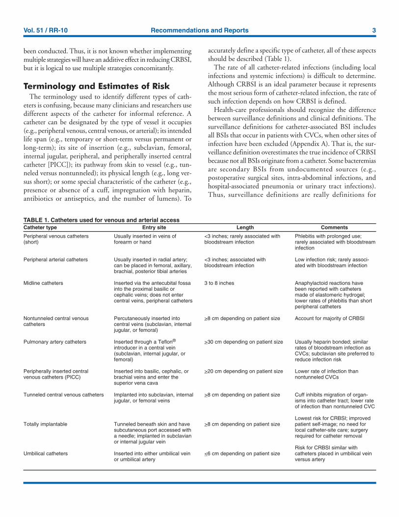
Terminology and Estimates of RiskThe terminology used to identify different types of cath-
eters is confusing, because many clinicians and researchers usedifferent aspects of the catheter for informal reference. Acatheter can be designated by the type of vessel it occupies(e.g., peripheral venous, central venous, or arterial); its intendedlife span (e.g., temporary or short-term versus permanent orlong-term); its site of insertion (e.g., subclavian, femoral,internal jugular, peripheral, and peripherally inserted centralcatheter [PICC]); its pathway from skin to vessel (e.g., tun-neled versus nontunneled); its physical length (e.g., long ver-sus short); or some special characteristic of the catheter (e.g.,presence or absence of a cuff, impregnation with heparin,antibiotics or antiseptics, and the number of lumens). To
accurately define a specific type of catheter, all of these aspectsshould be described (Table 1).
The rate of all catheter-related infections (including localinfections and systemic infections) is difficult to determine.Although CRBSI is an ideal parameter because it representsthe most serious form of catheter-related infection, the rate ofsuch infection depends on how CRBSI is defined.
Health-care professionals should recognize the differencebetween surveillance definitions and clinical definitions. Thesurveillance definitions for catheter-associated BSI includesall BSIs that occur in patients with CVCs, when other sites ofinfection have been excluded (Appendix A). That is, the sur-veillance definition overestimates the true incidence of CRBSIbecause not all BSIs originate from a catheter. Some bacteremiasare secondary BSIs from undocumented sources (e.g.,postoperative surgical sites, intra-abdominal infections, andhospital-associated pneumonia or urinary tract infections).Thus, surveillance definitions are really definitions for
Peripheral venous catheters(short)
Peripheral arterial catheters
Midline catheters
Nontunneled central venouscatheters
Pulmonary artery catheters
Peripherally inserted centralvenous catheters (PICC)
Tunneled central venous catheters
Totally implantable
Umbilical catheters
Usually inserted in veins offorearm or hand
Usually inserted in radial artery;can be placed in femoral, axillary,brachial, posterior tibial arteries
Inserted via the antecubital fossainto the proximal basilic orcephalic veins; does not entercentral veins, peripheral catheters
Percutaneously inserted intocentral veins (subclavian, internaljugular, or femoral)
Inserted through a Teflon®
introducer in a central vein(subclavian, internal jugular, orfemoral)
Inserted into basilic, cephalic, orbrachial veins and enter thesuperior vena cava
Implanted into subclavian, internaljugular, or femoral veins
Tunneled beneath skin and havesubcutaneous port accessed witha needle; implanted in subclavianor internal jugular vein
Inserted into either umbilical veinor umbilical artery
<3 inches; rarely associated withbloodstream infection
<3 inches; associated withbloodstream infection
3 to 8 inches
>8 cm depending on patient size
>30 cm depending on patient size
>20 cm depending on patient size
>8 cm depending on patient size
>8 cm depending on patient size
<6 cm depending on patient size
Phlebitis with prolonged use;rarely associated with bloodstreaminfection
Low infection risk; rarely associ-ated with bloodstream infection
Anaphylactoid reactions havebeen reported with cathetersmade of elastomeric hydrogel;lower rates of phlebitis than shortperipheral catheters
Account for majority of CRBSI
Usually heparin bonded; similarrates of bloodstream infection asCVCs; subclavian site preferred toreduce infection risk
Lower rate of infection thannontunneled CVCs
Cuff inhibits migration of organ-isms into catheter tract; lower rateof infection than nontunneled CVC
Lowest risk for CRBSI; improvedpatient self-image; no need forlocal catheter-site care; surgeryrequired for catheter removal
Risk for CRBSI similar withcatheters placed in umbilical veinversus artery
TABLE 1. Catheters used for venous and arterial accessCatheter type Entry site Length Comments
4 MMWR August 9, 2002
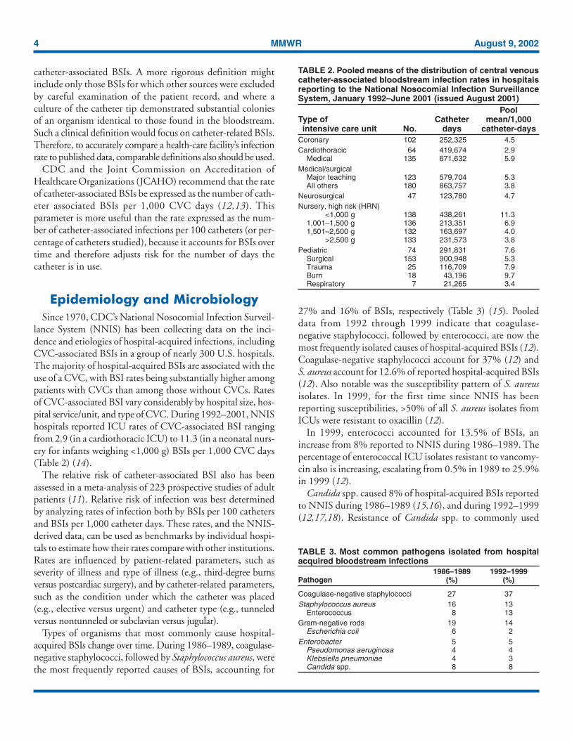
TABLE 2. Pooled means of the distribution of central venouscatheter-associated bloodstream infection rates in hospitalsreporting to the National Nosocomial Infection SurveillanceSystem, January 1992–June 2001 (issued August 2001)
PoolType of Catheter mean/1,000intensive care unit No. days catheter-days
Coronary 102 252,325 4.5Cardiothoracic 64 419,674 2.9
Medical 135 671,632 5.9Medical/surgical
Major teaching 123 579,704 5.3All others 180 863,757 3.8
Neurosurgical 47 123,780 4.7Nursery, high risk (HRN)
<1,000 g 138 438,261 11.31,001–1,500 g 136 213,351 6.91,501–2,500 g 132 163,697 4.0
>2,500 g 133 231,573 3.8Pediatric 74 291,831 7.6
Surgical 153 900,948 5.3Trauma 25 116,709 7.9Burn 18 43,196 9.7Respiratory 7 21,265 3.4
TABLE 3. Most common pathogens isolated from hospitalacquired bloodstream infections
1986–1989 1992–1999Pathogen (%) (%)
Coagulase-negative staphylococci 27 37Staphylococcus aureus 16 13
Enterococcus 8 13Gram-negative rods 19 14
Escherichia coli 6 2Enterobacter 5 5
Pseudomonas aeruginosa 4 4Klebsiella pneumoniae 4 3Candida spp. 8 8
catheter-associated BSIs. A more rigorous definition mightinclude only those BSIs for which other sources were excludedby careful examination of the patient record, and where aculture of the catheter tip demonstrated substantial coloniesof an organism identical to those found in the bloodstream.Such a clinical definition would focus on catheter-related BSIs.Therefore, to accurately compare a health-care facility’s infectionrate to published data, comparable definitions also should be used.
CDC and the Joint Commission on Accreditation ofHealthcare Organizations (JCAHO) recommend that the rateof catheter-associated BSIs be expressed as the number of cath-eter associated BSIs per 1,000 CVC days (12,13). Thisparameter is more useful than the rate expressed as the num-ber of catheter-associated infections per 100 catheters (or per-centage of catheters studied), because it accounts for BSIs overtime and therefore adjusts risk for the number of days thecatheter is in use.
Epidemiology and MicrobiologySince 1970, CDC’s National Nosocomial Infection Surveil-
lance System (NNIS) has been collecting data on the inci-dence and etiologies of hospital-acquired infections, includingCVC-associated BSIs in a group of nearly 300 U.S. hospitals.The majority of hospital-acquired BSIs are associated with theuse of a CVC, with BSI rates being substantially higher amongpatients with CVCs than among those without CVCs. Ratesof CVC-associated BSI vary considerably by hospital size, hos-pital service/unit, and type of CVC. During 1992–2001, NNIShospitals reported ICU rates of CVC-associated BSI rangingfrom 2.9 (in a cardiothoracic ICU) to 11.3 (in a neonatal nurs-ery for infants weighing <1,000 g) BSIs per 1,000 CVC days(Table 2) (14).
The relative risk of catheter-associated BSI also has beenassessed in a meta-analysis of 223 prospective studies of adultpatients (11). Relative risk of infection was best determinedby analyzing rates of infection both by BSIs per 100 cathetersand BSIs per 1,000 catheter days. These rates, and the NNIS-derived data, can be used as benchmarks by individual hospi-tals to estimate how their rates compare with other institutions.Rates are influenced by patient-related parameters, such asseverity of illness and type of illness (e.g., third-degree burnsversus postcardiac surgery), and by catheter-related parameters,such as the condition under which the catheter was placed(e.g., elective versus urgent) and catheter type (e.g., tunneledversus nontunneled or subclavian versus jugular).
Types of organisms that most commonly cause hospital-acquired BSIs change over time. During 1986–1989, coagulase-negative staphylococci, followed by Staphylococcus aureus, werethe most frequently reported causes of BSIs, accounting for
27% and 16% of BSIs, respectively (Table 3) (15). Pooleddata from 1992 through 1999 indicate that coagulase-negative staphylococci, followed by enterococci, are now themost frequently isolated causes of hospital-acquired BSIs (12).Coagulase-negative staphylococci account for 37% (12) andS. aureus account for 12.6% of reported hospital-acquired BSIs(12). Also notable was the susceptibility pattern of S. aureusisolates. In 1999, for the first time since NNIS has beenreporting susceptibilities, >50% of all S. aureus isolates fromICUs were resistant to oxacillin (12).
In 1999, enterococci accounted for 13.5% of BSIs, anincrease from 8% reported to NNIS during 1986–1989. Thepercentage of enterococcal ICU isolates resistant to vancomy-cin also is increasing, escalating from 0.5% in 1989 to 25.9%in 1999 (12).
Candida spp. caused 8% of hospital-acquired BSIs reportedto NNIS during 1986–1989 (15,16), and during 1992–1999(12,17,18). Resistance of Candida spp. to commonly used
Vol. 51 / RR-10 Recommendations and Reports 5
antifungal agents is increasing. Although NNIS has notreported the percentage of BSIs caused by nonalbicans speciesor fluconazole susceptibility data, other epidemiologic andclinical data document that fluconazole resistance is anincreasingly relevant consideration when designing empirictherapeutic regimens for CRBSIs caused by yeast. Data fromthe Surveillance and Control of Pathogens of EpidemiologicImportance (SCOPE) Program documented that 10% ofC. albicans bloodstream isolates from hospitalized patients wereresistant to fluconazole (17). Additionally, 48% of CandidaBSIs were caused by nonalbicans species, including C. glabrataand C. krusei, which are more likely than C. albicans to dem-onstrate resistance to fluconazole and itraconazole (18,19).
Gram-negative bacilli accounted for 19% of catheter-associated BSIs during 1986–1989 (15) compared with 14%of catheter-associated BSIs during 1992–1999 (12). Anincreasing percentage of ICU-related isolates are caused byEnterobacteriaceae that produce extended-spectrumß-lactamases (ESBLs), particularly Klebsiella pneumoniae (20).Such organisms not only are resistant to extended-spectrumcephalosporins, but also to frequently used, broad spectrumantimicrobial agents.
PathogenesisMigration of skin organisms at the insertion site into the
cutaneous catheter tract with colonization of the catheter tipis the most common route of infection for peripherallyinserted, short-term catheters (21,22). Contamination of thecatheter hub contributes substantially to intraluminal coloni-zation of long-term catheters (23–25). Occasionally, cathetersmight become hematogenously seeded from another focus ofinfection. Rarely, infusate contamination leads to CRBSI (26).
Important pathogenic determinants of catheter-relatedinfection are 1) the material of which the device is made and2) the intrinsic virulence factors of the infecting organism. Invitro studies demonstrate that catheters made of polyvinylchloride or polyethylene are likely less resistant to the adher-ence of microorganisms than are catheters made of Teflon®,silicone elastomer, or polyurethane (27,28). Therefore, themajority of catheters sold in the United States are no longermade of polyvinyl chloride or polyethylene. Some cathetermaterials also have surface irregularities that enhance the mi-crobial adherence of certain species (e.g., coagulase-negativestaphylococci, Acinetobacter calcoaceticus, and Pseudomonasaeruginosa) (29–31); catheters made of these materials areespecially vulnerable to microbial colonization and subsequentinfection. Additionally, certain catheter materials are morethrombogenic than others, a characteristic that also mightpredispose to catheter colonization and catheter-related
infection (31,32). This association has led to emphasis on pre-venting catheter-related thrombus as an additional mechanismfor reducing CRBSI.
The adherence properties of a given microorganism also areimportant in the pathogenesis of catheter-related infection.For example, S. aureus can adhere to host proteins (e.g.,fibronectin) commonly present on catheters (33,34). Also,coagulase-negative staphylococci adhere to polymer surfacesmore readily than do other pathogens (e.g., Escherichia coli orS. aureus). Additionally, certain strains of coagulase-negativestaphylococci produce an extracellular polysaccharide oftenreferred to as “slime” (35,36). In the presence of catheters,this slime potentiates the pathogenicity of coagulase-negativestaphylococci by allowing them to withstand host defensemechanisms (e.g., acting as a barrier to engulfment and kill-ing by polymorphonuclear leukocytes) or by making them lesssusceptible to antimicrobial agents (e.g., forming a matrix thatbinds antimicrobials before their contact with the organismcell wall) (37). Certain Candida spp., in the presence ofglucose-containing fluids, might produce slime similar to thatof their bacterial counterparts, potentially explaining theincreased proportion of BSIs caused by fungal pathogensamong patients receiving parenteral nutrition fluids (38).
Strategies for Preventionof Catheter-Related Infectionsin Adult and Pediatric Patients
Quality Assurance and ContinuingEducation
Measures to minimize the risk for infection associated withintravascular therapy should strike a balance between patientsafety and cost effectiveness. As knowledge, technology, andhealth-care settings change, infection control and preventionmeasures also should change. Well-organized programs thatenable health-care providers to provide, monitor, and evaluatecare and to become educated are critical to the success of thiseffort. Reports spanning the past two decades have consis-tently demonstrated that risk for infection declines followingstandardization of aseptic care (39–43), and that insertion andmaintenance of intravascular catheters by inexperienced staffmight increase the risk for catheter colonization and CRBSI(43,44). Specialized “IV teams” have shown unequivocal ef-fectiveness in reducing the incidence of catheter-related infec-tions and associated complications and costs (45–47).Additionally, infection risk increases with nursing staff reduc-tions below a critical level (48).
6 MMWR August 9, 2002
Site of Catheter InsertionThe site at which a catheter is placed influences the subse-
quent risk for catheter-related infection and phlebitis. Theinfluence of site on the risk for catheter infections is related inpart to the risk for thrombophlebitis and density of local skin flora.
Phlebitis has long been recognized as a risk for infection.For adults, lower extremity insertion sites are associated witha higher risk for infection than are upper extremity sites(49–51). In addition, hand veins have a lower risk for phlebi-tis than do veins on the wrist or upper arm (52).
The density of skin flora at the catheter insertion site is amajor risk factor for CRBSI. Authorities recommend thatCVCs be placed in a subclavian site instead of a jugular orfemoral site to reduce the risk for infection. No randomizedtrial satisfactorily has compared infection rates for cathetersplaced in jugular, subclavian, and femoral sites. Catheters in-serted into an internal jugular vein have beenassociated with higher risk for infection than those insertedinto a subclavian or femoral vein (22,53,54).
Femoral catheters have been demonstrated to have relativelyhigh colonization rates when used in adults (55). Femoral cath-eters should be avoided, when possible, because they are asso-ciated with a higher risk for deep venous thrombosis than areinternal jugular or subclavian catheters (56–60) and becauseof a presumption that such catheters are more likely tobecome infected. However, studies in pediatric patients havedemonstrated that femoral catheters have a low incidence ofmechanical complications and might have an equivalentinfection rate to that of nonfemoral catheters (61–63). Thus,in adult patients, a subclavian site is preferred for infectioncontrol purposes, although other factors (e.g., the potentialfor mechanical complications, risk for subclavian vein steno-sis, and catheter-operator skill) should be considered whendeciding where to place the catheter. In a meta-analysis of eightstudies, the use of bedside ultrasound for the placementof CVCs substantially reduced mechanical complicationscompared with the standard landmark placement technique(relative risk [RR] = 0.22; 95% confidence interval[CI] = 0.10–0.45) (64). Consideration of comfort, security,and maintenance of asepsis as well as patient-specific factors(e.g., preexisting catheters, anatomic deformity, and bleedingdiathesis), relative risk of mechanical complications (e.g., bleed-ing and pneumothorax), the availability of bedside ultrasound,and the risk for infection should guide site selection.
Type of Catheter MaterialTeflon® or polyurethane catheters have been associated with
fewer infectious complications than catheters made of polyvinylchloride or polyethylene (27,65,66). Steel needles used as an
alternative to catheters for peripheral venous access have thesame rate of infectious complications as do Teflon® catheters(67,68). However, the use of steel needles frequently iscomplicated by infiltration of intravenous (IV) fluids into thesubcutaneous tissues, a potentially serious complication if theinfused fluid is a vesicant (68).
Hand Hygiene and Aseptic TechniqueFor short peripheral catheters, good hand hygiene before
catheter insertion or maintenance, combined with proper asep-tic technique during catheter manipulation, provides protec-tion against infection. Good hand hygiene can be achievedthrough the use of either a waterless, alcohol-based product(69) or an antibacterial soap and water with adequate rinsing(70). Appropriate aseptic technique does not necessarilyrequire sterile gloves; a new pair of disposable nonsterile glovescan be used in conjunction with a “no-touch” technique forthe insertion of peripheral venous catheters. However, glovesare required by the Occupational Safety and Health Adminis-tration as standard precautions for the prevention ofbloodborne pathogen exposure.
Compared with peripheral venous catheters, CVCs carry asubstantially greater risk for infection; therefore, the level ofbarrier precautions needed to prevent infection during inser-tion of CVCs should be more stringent. Maximal sterile bar-rier precautions (e.g., cap, mask, sterile gown, sterile gloves,and large sterile drape) during the insertion of CVCs substan-tially reduces the incidence of CRBSI compared with stan-dard precautions (e.g., sterile gloves and small drapes) (22,71).Although the efficacy of such precautions for insertion ofPICCs and midline catheters has not been studied, the use ofmaximal barrier precautions also is probably applicable toPICCs.
Skin AntisepsisIn the United States, povidone iodine has been the most
widely used antiseptic for cleansing arterial catheter and CVC-insertion sites (72). However, in one study, preparation of cen-tral venous and arterial sites with a 2% aqueous chlorhexidinegluconate lowered BSI rates compared with site preparationwith 10% povidone-iodine or 70% alcohol (73). Commer-cially available products containing chlorhexidine have notbeen available until recently; in July 2000, the U.S. Food andDrug Administration (FDA) approved a 2% tincture ofchlorhexidine preparation for skin antisepsis. Other prepara-tions of chlorhexidine might not be as effective. Tincture ofchlorhexidine gluconate 0.5% is no more effective in prevent-ing CRBSI or CVC colonization than 10% povidone iodine,as demonstrated by a prospective, randomized study of adults
Vol. 51 / RR-10 Recommendations and Reports 7
(74). However, in a study involving neonates, 0.5%chlorhexidine reduced peripheral IV colonization comparedwith povidone iodine (20/418 versus 38/408 catheters;p = 0.01) (75). This study, which did not include CVCs, hadan insufficient number of participants to assess differences inBSI rates. A 1% tincture of chlorhexidine preparation is avail-able in Canada and Australia, but not yet in the United States.No published trials have compared a 1% chlorhexidine prepara-tion to povidone-iodine.
Catheter Site Dressing RegimensTransparent, semipermeable polyurethane dressings have
become a popular means of dressing catheter insertion sites.Transparent dressings reliably secure the device, permit con-tinuous visual inspection of the catheter site, permit patientsto bathe and shower without saturating the dressing, andrequire less frequent changes than do standard gauze and tapedressings; the use of these dressings saves personnel time.
In the largest controlled trial of dressing regimens onperipheral catheters, the infectious morbidity associated withthe use of transparent dressings on approximately 2,000peripheral catheters was examined (65). Data from this studysuggest that the rate of colonization among catheters dressedwith transparent dressings (5.7%) is comparable to that ofthose dressed with gauze (4.6%) and that no clinically sub-stantial differences exist in either the incidences of catheter-site colonization or phlebitis. Furthermore, these data suggestthat transparent dressings can be safely left on peripheral venouscatheters for the duration of catheter insertion withoutincreasing the risk for thrombophlebitis (65).
A meta-analysis has assessed studies that compared the riskfor catheter-related BSIs for groups using transparent dress-ings versus groups using gauze dressing (76). The risk forCRBSIs did not differ between the groups. The choice of dress-ing can be a matter of preference. If blood is oozing from thecatheter insertion site, gauze dressing might be preferred.
In a multi-center study, a chlorhexidine-impregnated sponge(Biopatch™) placed over the site of short-term arterial andCVCs reduced the risk for catheter colonization and CRBSI(77). No adverse systemic effects resulted from use of thisdevice.
Catheter Securement DevicesSutureless securement devices can be advantageous over
suture in preventing catheter-related BSIs. One study, whichinvolved only a limited number of patients and was under-powered, compared a sutureless device with suture for thesecurement of PICCS; in this study, CRBSI was reduced inthe group of patients that received the sutureless device (78).
In-Line FiltersIn-line filters reduce the incidence of infusion-related phle-
bitis (79,80). No data support their efficacy in preventinginfections associated with intravascular catheters and infusionsystems. Proponents of filters cite several potential benefits tousing these filters, including 1) reducing the risk for infectionfrom contaminated infusate or proximal contamination (i.e.,introduced proximal to the filter); 2) reducing the risk forphlebitis in patients who require high doses of medication orin those in whom infusion-related phlebitis already hasoccurred; 3) removing particulate matter that might contami-nate IV fluids (81); and 4) filtering endotoxin produced bygram-negative organisms in contaminated infusate (82). Thesetheoretical advantages should be tempered by the knowledgethat infusate-related BSI is rare and that filtration of medica-tions or infusates in the pharmacy is a more practical and lesscostly way to remove the majority of particulates. Further-more, in-line filters might become blocked, especially withcertain solutions (e.g., dextran, lipids, and mannitol), therebyincreasing the number of line manipulations and decreasingthe availability of administered drugs (83). Thus, for reduc-ing the risk for CRBSI, no strong recommendation can bemade in favor of using in-line filters.
Antimicrobial/Antiseptic ImpregnatedCatheters and Cuffs
Certain catheters and cuffs that are coated or impregnatedwith antimicrobial or antiseptic agents can decrease the riskfor CRBSI and potentially decrease hospital costs associatedwith treating CRBSIs, despite the additional acquisition costof an antimicrobial/antiseptic impregnated catheter (84). Allof the studies involving antimicrobial/antiseptic impregnatedcatheters have been conducted using triple-lumen, noncuffedcatheters in adult patients whose catheters remained in place<30 days. Although all of the studies have been conducted inadults, these catheters have been approved by FDA for use inpatients weighing >3 kg. No antiseptic or antimicrobialimpregnated catheters currently are available for use inweighing <3 kg.
Chlorhexidine/Silver sulfadiazine. Catheters coated withchlorhexidine/silver sulfadiazine only on the external luminalsurface have been studied as a means to reduce CRBSI. Twometa-analyses (2,85) demonstrated that such catheters reducedthe risk for CRBSI compared with standard noncoatedcatheters. The mean duration of catheter placement in onemeta-analysis ranged from 5.1 to 11.2 days (86). The half-lifeof antimicrobial activity against S. epidermidis is 3 days in vitrofor catheters coated with chlorhexidine/silver sulfadiazine; thisantimicrobial activity decreases over time (87). The benefit
8 MMWR August 9, 2002
for the patients who receive these catheters will be realizedwithin the first 14 days (86). A second-generation catheter isnow available with chlorhexidine coating both the internaland external luminal surfaces. The external surface has threetimes the amount of chlorhexidine and extended release ofthe surface bound antiseptics than that in the first generationcatheters. The external surface coating of chlorhexidine is com-bined with silver-sulfadiazine, and the internal surface is coatedwith chlorhexidine alone. Preliminary studies indicate thatprolonged anti-infective activity provides improved efficacyin preventing infections (88). Although rare, anaphylaxis hasbeen reported with the use of these chlorhexidine/silver sulfa-diazine catheters in Japan (89). Whether patients will becomecolonized or infected with organisms resistant to chlorhexidine/silver sulfadiazine has not been determined (86).
Chlorhexidine/silver sulfadiazine catheters are more expen-sive than standard catheters. However, one analysis has sug-gested that the use of chlorhexidine/silver sulfadiazine cathetersshould lead to a cost savings of $68 to $391 per catheter (90)in settings in which the risk for CRBSI is high despite adher-ence to other preventive strategies (e.g., maximal barrier pre-cautions and aseptic techniques). Use of these catheters mightbe cost effective in ICU patients, burn patients, neutropenicpatients, and other patient populations in which the rate ofinfection exceeds 3.3 per 1,000 catheter days (86).
Minocycline/Rifampin. In a multicenter randomized trial,CVCs impregnated on both the external and internal surfaceswith minocycline/rifampin were associated with lower ratesof CRBSI when compared with the first-generationchlorhexidine-silver sulfadiazine impregnated catheters (91).The beneficial effect began after day 6 of catheterization. Noneof the catheters were evaluated beyond 30 days. Nominocycline/rifampin-resistant organisms were reported. How-ever, in vitro data indicate that these impregnated catheterscould increase the incidence of minocycline and rifampinresistance among pathogens, especially staphylococci. The half-life of antimicrobial activity against S. epidermidis is 25 dayswith catheters coated with minocycline/rifampin, comparedwith 3 days for the first-generation catheters coated withchlorhexidine/silver sulfadiazine in vitro (87). In vivo, theduration of antimicrobial activity of the minocycline/rifampincatheter is longer than that of the first-generation chlorhexidine/silver sulfadiazine catheter (91). No comparative studies havebeen published using the second-generation chlorhexidine/silver sulfadiazine catheter. Studies are needed to evaluatedwhether the improved performance of the minocyline/rifampincatheters results from the antimicrobial agents used or fromthe coating of both the internal and external surfaces. As withchlorhexidine/silver sulfadiazine catheters, some clinicians haverecommended that the minocycline/rifampin catheters be
considered in patient populations when the rate of CRBSIexceeds 3.3 per 1,000 catheter days (86). Others suggest thatreducing all rates of CRBSI should be the goal (92). The deci-sion to use chlorhexidine/silver sulfadiazine or minocycline/rifampin impregnated catheters should be based on the needto enhance prevention of CRBSI after standard procedureshave been implemented (e.g., educating personnel, using maxi-mal sterile barrier precautions, and using 2% chlorhexidineskin antisepsis) and then balanced against the concern foremergence of resistant pathogens and the cost of implementingthis strategy.
Platinum/Silver. Ionic metals have broad antimicrobialactivity and are being used in catheters and cuffs to preventCRBSI. A combination platinum/silver impregnated catheteris available in Europe and has recently been approved by FDAfor use in the United States. Although these catheters arebeing marketed for their antimicrobial properties, no publishedstudies have been presented to support an antimicrobial effect.
Silver cuffs. Ionic silver has been used in subcutaneous col-lagen cuffs attached to CVCs (93). The ionic silver providesantimicrobial activity and the cuff provides a mechanical bar-rier to the migration of microorganisms along the externalsurface of the catheter. In studies of catheters left in place >20days, the cuff failed to reduce the incidence of CRBSI (94,95).Two other studies of short-term catheters could not demon-strate efficacy because of the minimal number of CRBSIsobserved (93,96).
Systemic Antibiotic ProphylaxisNo studies have demonstrated that oral or parenteral anti-
bacterial or antifungal drugs might reduce the incidence ofCRBSI among adults (97–99). However, among low birthweight infants, two studies have assessed vancomycin prophy-laxis; both demonstrated a reduction in CRBSI but no reduc-tion in mortality (100,101). Because the prophylactic use ofvancomycin is an independent risk factor for the acquisitionof vancomycin-resistant enterococcus (VRE) (102), the riskfor acquiring VRE likely outweighs the benefit of using pro-phylactic vancomycin.
Antibiotic/Antiseptic OintmentsPovidone-iodine ointment applied at the insertion site of
hemodialysis catheters has been studied as a prophylacticintervention to reduce the incidence of catheter-relatedinfections. One randomized study of 129 hemodialysis cath-eters demonstrated a reduction in the incidence of exit-siteinfections, catheter-tip colonization, and BSIs with the rou-tine use of povidone-iodine ointment at the catheter insertionsite compared with no ointment at the insertion site (103).
Vol. 51 / RR-10 Recommendations and Reports 9
Several studies have evaluated the effectiveness of mupirocinointment applied at the insertion sites of CVCs as a means toprevent CRBSI (104–106). Although mupirocin reduced therisk for CRBSI (106), mupirocin ointment also has been asso-ciated with mupirocin resistance (107,108), and might adverselyaffect the integrity of polyurethane catheters (109,110).
Nasal carriers of S. aureus have a higher risk for acquiringCRBSI than do noncarriers (103,111). Mupirocin ointmenthas been used intranasally to decrease nasal carriage of S. aureusand lessen the risk for CRBSI. However, resistance tomupirocin develops in both S. aureus and coagulase-negativestaphylococci soon after routine use of mupirocin is instituted(107,108).
Other antibiotic ointments applied to the catheter inser-tion site also have been studied and have yielded conflictingresults (112–114). In addition, rates of catheter colonizationwith Candida spp. might be increased with the use of antibi-otic ointments that have no fungicidal activity (112,114). Toavoid compromising the integrity of the catheter, any oint-ment that is applied to the catheter insertion site should bechecked against the catheter and ointment manufacturers’ rec-ommendations regarding compatibility.
Antibiotic Lock ProphylaxisTo prevent CRBSI, antibiotic lock prophylaxis has been
attempted by flushing and filling the lumen of the catheterwith an antibiotic solution and leaving the solution to dwellin the lumen of the catheter. Three studies have demonstratedthe usefulness of such prophylaxis in neutropenic patients withlong-term catheters (115–117). In two of the studies, patientsreceived either heparin alone (10 U/ml) or heparin plus 25micrograms/ml of vancomycin. The third study comparedvancomycin/ciprofloxacin/heparin (VCH) to vancomycin/heparin (VH)and then to heparin alone. The rate of CRBSIwith vancomycin-susceptible organisms was significantly lower(VCH p = 0.022; VH p = 0.028) and the time to the firstepisode of bacteremia with vancomycin-susceptible organismswas substantially longer (VCH p = 0.036; VH p = 0.011) inpatients receiving either vancomycin/ciprofloxacin/heparin orvancomycin/heparin compared with heparin alone (115–117).One study involving a limited number of children revealed nodifference in rates of CRBSI between children receiving aheparin flush compared with those receiving heparin and van-comycin (118). However, because the use of vancomycin is anindependent risk factor for the acquisition of VRE (102), thispractice is not recommended routinely.
An anticoagulant/antimicrobial combination comprisingminocycline and ethylenediaminetetraraacetic acid (EDTA)has been proposed as a lock solution because it has antibiofilm
and antimicrobial activity against gram-positive, gram-negative, and Candida organisms (119), as well as anticoagu-lant properties. However, no controlled or randomized trialshave demonstrated its efficacy.
AnticoagulantsAnticoagulant flush solutions are used widely to prevent cath-
eter thrombosis. Because thrombi and fibrin deposits on cath-eters might serve as a nidus for microbial colonization ofintravascular catheters (120,121), the use of anticoagulantsmight have a role in the prevention of CRBSI.
In a meta-analysis evaluating the benefit of heparin prophy-laxis (3 U/ml in TPN, 5,000 U every 6 or 12 hours flush, or2,500 U low molecular weight heparin subcutaneously) inpatients with short-term CVCs, the risk for catheter-relatedcentral venous thrombosis was reduced with the use of pro-phylactic heparin (122). However, no substantial differencein the rate for CRBSI was observed. Because the majority ofheparin solutions contain preservatives with antimicrobialactivity, whether any decrease in the rate of CRBSI is a resultof the reduced thrombus formation, the preservative, or bothis unclear.
The majority of pulmonary artery, umbilical, and centralvenous catheters are available with a heparin-bonded coating.The majority are heparin-bonded with benzalkonium chlo-ride, which provides the catheters with antimicrobial activity(123) and provides an anti-thrombotic effect (124).
Warfarin also has been evaluated as a means for reducingCRBSI by reducing thrombus formation on catheters(125,126). In patients with long-term CVCs, low-dose war-farin (i.e., 1 mg/day) reduced the incidence of catheter throm-bus. No data demonstrate that warfarin reduces the incidenceof CRBSI.
Replacement of CathetersPeripheral Venous Catheters
Scheduled replacement of intravascular catheters has beenproposed as a method to prevent phlebitis and catheter-related infections. Studies of short peripheral venous cathetersindicate that the incidence of thrombophlebitis and bacterialcolonization of catheters increases when catheters are left inplace >72 hours (66,67,127). However, rates of phlebitis arenot substantially different in peripheral catheters left in place72 hours compared with 96 hours (128). Because phlebitisand catheter colonization have been associated with anincreased risk for catheter-related infection, short peripheralcatheter sites commonly are rotated at 72–96-hour intervals
10 MMWR August 9, 2002
to reduce both the risk for infection and patient discomfortassociated with phlebitis.
Midline CathetersMidline catheters have been associated with lower rates of
phlebitis than short peripheral catheters and with lower ratesof infection than CVCs (129–131). In one prospective studyof 140 midline catheters, their use was associated with a BSIrate of 0.8 per 1,000 catheter-days (131). No specific risk fac-tors, including duration of catheterization, were associated withinfection. Midline catheters were in place a median of 7 days,but for as long as 49 days. Although the findings of this studysuggested that midline catheters can be changed only whenthere is a specific indication, no prospective, randomized stud-ies have assessed the benefit of routine replacement as a strat-egy to prevent CRBSI associated with midline catheters.
CVCs, Including PICCs andHemodialysis Catheters
Catheter replacement at scheduled time intervals as a methodto reduce CRBSI has not lowered rates. Two trials haveassessed a strategy of changing the catheter every 7 days com-pared with a strategy of changing catheters as needed (132,133).One of these studies involved 112 surgical ICU patients need-ing CVCs, pulmonary artery catheters, or peripheral arterialcatheters (132), whereas the other study involved only subcla-vian hemodialysis catheters (133). In both studies, no differ-ence in CRBSI was observed in patients undergoing scheduledcatheter replacement every 7 days compared with patientswhose catheters were replaced as needed.
Scheduled guidewire exchanges of CVCs is another proposedstrategy for preventing CRBSI. The results of a meta-analysisof 12 randomized controlled trials assessing CVC manage-ment failed to prove any reduction of CRBSI rates throughroutine replacement of CVCs by guidewire exchange com-pared with catheter replacement on an as-needed basis (134).Thus, routine replacement of CVCs is not necessary for cath-eters that are functioning and have no evidence of causinglocal or systemic complications.
Catheter replacement over a guidewire has become anaccepted technique for replacing a malfunctioning catheter orexchanging a pulmonary artery catheter for a CVC wheninvasive monitoring no longer is needed. Catheter insertionover a guidewire is associated with less discomfort and a sig-nificantly lower rate of mechanical complications than are thosepercutaneously inserted at a new site (135); in addition, thistechnique provides a means of preserving limited venousaccess in some patients. Replacement of temporary cathetersover a guidewire in the presence of bacteremia is not an
acceptable replacement strategy, because the source of infec-tion is usually colonization of the skin tract from the insertionsite to the vein (22,135). However, in selected patients withtunneled hemodialysis catheters and bacteremia, catheterexchange over a guidewire, in combination with antibiotictherapy, might be an alternative as a salvage strategy in pa-tients with limited venous access (136–139).
Hemodialysis CathetersThe use of catheters for hemodialysis is the most common
factor contributing to bacteremia in dialysis patients (140,141).The relative risk for bacteremia in patients with dialysis cath-eters is sevenfold the risk for patients with primary arterio-venous fistulas (142). Despite the National KidneyFoundation’s effort to reduce the number of hemodialysispatients maintained with catheter access, catheter use increasedfrom 12.7% in 1995 to 22.2% in 1999 (143). Rates for bac-teremia per 100 patient months were 0.2 for arteriovenousfistulas, 0.5 for grafts, 5.0 for cuffed catheters, and 8.5 fornoncuffed catheters (CDC, unpublished data, 1999).
To reduce the rate of infection, hemodialysis catheters shouldbe avoided in favor of arteriovenous fistulas and grafts. If tem-porary access is needed for dialysis, a cuffed catheter is prefer-able to a noncuffed catheter, even in the ICU setting, if thecatheter is expected to stay in place for >3 weeks (11,144).
Pulmonary Artery CathetersPulmonary artery catheters are inserted through a Teflon®
introducer and typically remain in place an average of 3 days.The majority of pulmonary artery catheters are heparin bonded,which reduces not only catheter thrombosis but also micro-bial adherence to the catheter (145). Meta-analysis indicatesthat standard nonheparin-bonded pulmonary artery catheterrates of CRBSI are 5.5 per 1,000 catheter days; for heparin-bonded pulmonary artery catheters, this rate is 2.6 per 1,000catheter days (11). Because the majority of pulmonary arterycatheters are heparin-bonded, the relative risk of infection withthese catheters is similar to that of CVC (2.6 versus 2.3 per1,000 catheter days) (11).
A prospective study of 442 pulmonary artery catheters dem-onstrated an increased risk for CRBSI after 5 days (0/442CRBSI before 5 days versus 5/442 CSBSI after 5 days;p < 0.001) (146). A prospective observational study of 71 pul-monary artery catheters demonstrated higher infection ratesin catheters left in place longer than 7 days (2% before 7 daysversus 16% after 7 days; p = 0.056) (147). However, no stud-ies indicate that catheter replacement at scheduled time inter-vals is an effective method to reduce CRBSI (132,135). Inpatients who continue to require hemodynamic monitoring,

Vol. 51 / RR-10 Recommendations and Reports 11
pulmonary artery catheters do not need to be changed morefrequently than every 7 days. No specific recommendationcan be made regarding routine replacement of catheters thatneed to be in place for >7 days.
Pulmonary artery catheters are usually packaged with a thinplastic sleeve that prevents touch contamination when placedover the catheter. In a study of 166 catheters, patients whowere randomly assigned to have their catheters self-containedwithin this sleeve had a reduced risk for CRBSI comparedwith those who had a pulmonary artery catheter placed with-out the sleeve (p = 0.002) (148).
Peripheral Arterial CathetersPeripheral arterial catheters are usually inserted into the
radial or femoral artery and permit continuous blood pressuremonitoring and blood gas measurements. The rate of CRBSIis comparable to that of temporary CVCs (2.9 versus 2.3 per1,000 catheter days) (11). One study of peripheral arterial cath-eters demonstrated no difference in infection rates betweenchanging catheters at scheduled times and changing arterialcatheters on an as-needed basis (132). One observational studyof 71 arterial catheters revealed that 10 local infections andfour CRBSIs occurred in patients who had peripheral arterialcatheters in place for >4 days compared with one local infec-tion and no CRBSIs in patients whose catheters were in place<4 days (p < 0.05) (147). Because the risk for CRBSI is likelysimilar to that of short-term CVCs, arterial catheters can beapproached in a similar way. No specific recommendation canbe made regarding replacement of catheters that need to be inplace for >5 days.
Replacement of Administration SetsThe optimal interval for routine replacement of IV admin-
istration sets has been examined in three well-controlledstudies. Data from each of these studies reveal that replacingadministration sets no more frequently than 72 hours afterinitiation of use is safe and cost-effective (149–151). Datafrom a more recent study demonstrated that rates of phlebitiswere not substantially different if administration sets were leftin place 96 hours compared with 72 hours (128). When afluid that enhances microbial growth is infused (e.g., lipidemulsions and blood products), more frequent changes ofadministration sets are indicated, because these products havebeen identified as independent risk factors for CRBSI (152–158).
Stopcocks (used for injection of medications, administra-tion of IV infusions, and collection of blood samples) repre-sent a potential portal of entry for microorganisms into vascularaccess catheters and IV fluids. Stopcock contamination is com-mon, occurring in 45% and 50% in the majority of series.
Whether such contamination is a substantial entry point ofCRBSI has been difficult to prove.
“Piggyback” systems are used as an alternative to stopcocks.However, they also pose a risk for contamination of the intra-vascular fluid if the device entering the rubber membrane ofan injection port is exposed to air or comes into direct contactwith nonsterile tape used to fix the needle to the port. Modi-fied piggyback systems have the potential to prevent contami-nation at these sites (159).
Needleless Intravascular CatheterSystems
Attempts to reduce the incidence of sharp injuries and theresultant risk for transmission of bloodborne infections tohealth-care workers have led to the design and introductionof needleless infusion systems. When the devices are usedaccording to manufacturers’ recommendations, they do notsubstantially affect the incidence of CRBSI (160–167).
Multidose Parenteral Medication VialsParenteral medications commonly are dispensed in
multidose, parenteral medication vials that might be used forprolonged periods for one or more patients. Although the over-all risk for extrinsic contamination of multidose vials is likelyminimal (168), the consequences of contamination mightresult in life-threatening infection (169,170). Single-use vialsare frequently preservative-free and might pose a risk forcontamination if they are punctured several times.
Special Considerationsfor Intravascular Catheter-Related
Infections in Pediatric PatientsPrevention of CRBSI in children requires additional con-
siderations, although only certain studies have been performedspecifically in children. Pediatric data have been derived largelyfrom studies in neonatal or pediatric ICUs and pediatric on-cology patients.
EpidemiologyAs in adults, the majority of BSIs in children are associated
with the use of an intravascular catheter. From 1995 through2000, the pooled mean catheter-associated BSI rate for allpediatric ICUs reporting data to NNIS was 7.7 per 1,000 cath-eter days (171,172). Umbilical catheter and CVC-associatedBSI rates for neonatal ICUs ranged from 11.3 per 1,000 cath-eter days in children with birth weight <1,000 g to 4.0 per1,000 catheter days in children whose birth weight was
12 MMWR August 9, 2002
>2,500 g (171). Catheter utilization rates were comparable inadult and pediatric ICUs (172,173).
MicrobiologyAs in adults, the majority of CRBSIs in children are caused
by coagulase-negative staphylococci. During 1992–1999, thesebacteria accounted for 37.7% of BSIs in pediatricICUs reporting to NNIS (12). Exposure to lipids has beenidentified as an independent risk factor for development ofcoagulase-negative staphylococcal bacteremia in very low birthweight infants (i.e., those weighing <1,000 g) (odds ratio [OR]= 9.4; 95% CI = 1.2–74.2) (155), as well as candidemia in theneonatal ICU (OR = 5.33; 95% CI = 1.23–48.4) (154). Gram-negative bacteria accounted for 25% of BSIs reported in pedi-atric ICUs (172), whereas enterococci and Candida spp.accounted for 10% and 9%, respectively (172).
Peripheral Venous CathetersAs in adults, the use of peripheral venous catheters in pedi-
atric patients might be complicated by phlebitis, infusionextravasation, and catheter infection (174). Catheter location,infusion of parenteral nutritional fluids with continuous IVlipid emulsions, and length of ICU stay before catheter inser-tion have all increased pediatric patients’ risk for phlebitis.However, contrary to the risk in adults, the risk for phlebitisin children has not increased with the duration of catheteriza-tion (174,175).
Peripheral Arterial CathetersIn a prospective study of 340 peripheral arterial catheters in
children, the following two risk factors for catheter-relatedinfection were identified: 1) use of an arterial system thatpermitted backflow of blood into the pressure tubing and2) duration of catheterization (176). Although a correlationwas found between duration of arterial catheterization andrisk for catheter colonization, the risk remained constant for2–20 days at 6.2% (176).
Umbilical CathetersAlthough the umbilical stump becomes heavily colonized
soon after birth, umbilical-vessel catheterization often is usedfor vascular access in newborn infants. Umbilical vessels canbe cannulated easily and permit both collection of bloodsamples and measurement of hemodynamic status. The inci-dences of catheter colonization and BSI are similar forumbilical vein catheters and umbilical artery catheters. In sev-eral studies, an estimated 40%–55% of umbilical artery cath-eters were colonized and 5% resulted in CRBSI; umbilical
vein catheters were associated with colonization in 22%–59%of cases (177–179) and with CRBSI in 3%–8% of cases (178).Although CRBSI rates are similar for umbilical catheters inthe high position (i.e., above the diaphragm) compared withthe low position (i.e., below the diaphragm and above the aor-tic bifurcation), catheters placed in the high position result ina lower incidence of vascular complications without anincrease in adverse sequelae (178).
Risk factors for infection differ for umbilical artery andumbilical vein catheters. In one study, neonates with very lowbirth weight who also received antibiotics for >10 days wereat increased risk for umbilical artery CRBSIs (178). In com-parison, those with higher birth weight and receipt of parenteralnutrition fluids were at increased risk for umbilical vein CRBSI.Duration of catheterization was not an independent risk fac-tor for infection of either type of umbilical catheter.
CVCsBecause of the limited vascular sites in children, attention
should be given to the frequency with which catheters arereplaced in these patients. In a study in which survival analy-sis techniques were used to examine the relation between theduration of central venous catheterization and complicationsin pediatric ICU patients, all of the patients studied (n = 397)remained uninfected for a median of 23.7 days (180). Inaddition, no relation was found between duration of catheter-ization and the daily probability of infection (r = 0.21;p > 0.1), suggesting that routine replacement of CVCs likelydoes not reduce the incidence of catheter-related infection(180).
Catheter Site CareAlthough data regarding the use of the chlorhexidine-
impregnated sponge (Biopatch™) in children are limited, onerandomized, controlled study involving 705 neonatesreported a substantial decrease in colonized catheter tips ininfants in the Biopatch™ group compared with the group thathad standard dressings (15% versus 24%; RR = 0.6; 95%CI = 0.5–0.9), but no difference in the rates of CRBSI or BSIwithout a source. Biopatch™ was associated with localizedcontact dermatitis in infants of very low birth weight. Of 98neonates with very low birth weight, 15 (15%) developedlocalized contact dermatitis; four (1.5%) of 237 neonatesweighing >1,000 g developed this reaction (p < 0.0001).Infants with gestational age <26 weeks who had CVCs placedat age <8 days were at increased risk for having localized con-tact dermatitis, whereas no infants in the control group devel-oped this local reaction (181).

Vol. 51 / RR-10 Recommendations and Reports 13
Performance IndicatorsPerformance indicators for reducing CRBSI are 1) imple-
mentation of educational programs that include didactic andinteractive components for those who insert and maintaincatheters; 2) use of maximal sterile barrier precautions duringcatheter placement; 3) use of chlorhexidine for skin antisep-sis; and 4) rates of catheter discontinuation when the catheteris no longer essential for medical management. The impactthese recommendations will have on individual institutionsshould be evaluated using specific performance indicators.
Recommendations for Placementof Intravascular Catheters
in Adults and ChildrenThese recommendations are designed to reduce the infec-
tious complications associated with intravascular catheter use.Recommendations should be considered in the context of theinstitution’s experience with catheter-related infections, expe-rience with other adverse catheter-related complications (e.g.,thrombosis, hemorrhage, and pneumothorax), and availabil-ity of personnel skilled in the placement of intravasculardevices. Recommendations are provided for 1) intravascular-catheter use in general; 2) specific devices; and 3) special cir-cumstances (i.e., intravascular-device use in pediatric patientsand CVC use for parenteral nutrition and hemodialysisaccess). Recommendations regarding the frequency of replac-ing catheters, dressings, administration sets, and fluids alsoare provided (Appendix B).
As in previous guidelines issued by CDC and HICPAC,each recommendation is categorized on the basis of existingscientific data, theoretical rationale, applicability, and economicimpact. The CDC/HICPAC system for categorizing recom-mendations is as follows:
Category IA. Strongly recommended for implementationand strongly supported by well-designed experimental, clini-cal, or epidemiologic studies.
Category IB. Strongly recommended for implementationand supported by some experimental, clinical, or epidemio-logic studies, and a strong theoretical rationale.
Category IC. Required by state or federal regulations, rules,or standards.
Category II. Suggested for implementation and supportedby suggestive clinical or epidemiologic studies or a theoreticalrationale.
Unresolved issue. Represents an unresolved issue for whichevidence is insufficient or no consensus regarding efficacyexists.
I. Health-care worker education and trainingA. Educate health-care workers regarding the indica-
tions for intravascular catheter use, proper proce-dures for the insertion and maintenance ofintravascular catheters, and appropriate infection-control measures to prevent intravascular catheter-related infections (39,43,45–47,182–187).Category IA
B. Assess knowledge of and adherence to guidelinesperiodically for all persons who insert and manageintravascular catheters (39,43,46,182,188).Category IA
C. Ensure appropriate nursing staff levels in ICUs tominimize the incidence of CRBSIs (48,189,190).Category IB
II. SurveillanceA. Monitor the catheter sites visually or by palpation
through the intact dressing on a regular basis,depending on the clinical situation of individualpatients. If patients have tenderness at the insertionsite, fever without obvious source, or other mani-festations suggesting local or BSI, the dressing shouldbe removed to allow thorough examination of thesite (1,191–193). Category IB
B. Encourage patients to report to their health-care pro-vider any changes in their catheter site or any newdiscomfort. Category II
C. Record the operator, date, and time of catheterinsertion and removal, and dressing changes on astandardized form. Category II
D. Do not routinely culture catheter tips (8,194,195).Category IA
III. Hand hygieneA. Observe proper hand-hygiene procedures either by
washing hands with conventional antiseptic-containing soap and water or with waterlessalcohol-based gels or foams. Observe hand hygienebefore and after palpating catheter insertion sites, aswell as before and after inserting, replacing, access-ing, repairing, or dressing an intravascular catheter.Palpation of the insertion site should not be per-formed after the application of antiseptic, unlessaseptic technique is maintained (43,70,196–200).Category IA
B. Use of gloves does not obviate the need for handhygiene (43,198,199). Category IA
IV. Aseptic technique during catheter insertion and careA. Maintain aseptic technique for the insertion and care of
intravascular catheters (22,71,201,202). Category IA
14 MMWR August 9, 2002
B. Wear clean or sterile gloves when inserting an intra-vascular catheter as required by the OccupationalSafety and Health Administration BloodbornePathogens Standard. Category IC. Wearing cleangloves rather than sterile gloves is acceptable for theinsertion of peripheral intravascular catheters if theaccess site is not touched after the application ofskin antiseptics. Sterile gloves should be worn forthe insertion of arterial and central catheters(201,203). Category IA
C. Wear clean or sterile gloves when changing the dress-ing on intravascular catheters. Category IC
V. Catheter insertionDo not routinely use arterial or venous cutdown
procedures as a method to insert catheters (204–206).Category IA
VI. Catheter site careA. Cutaneous antisepsis
1. Disinfect clean skin with an appropriate anti-septic before catheter insertion and during dress-ing changes. Although a 2% chlorhexidine-based preparation is preferred, tincture ofiodine, an iodophor, or 70% alcohol can be used(73,75,207,208). Category IA
2. No recommendation can be made for the useof chlorhexidine in infants aged <2 months.Unresolved issue
3. Allow the antiseptic to remain on the insertionsite and to air dry before catheter insertion.Allow povidone iodine to remain on the skinfor at least 2 minutes, or longer if it is not yet drybefore insertion (73,75,207,208). Category IB
4. Do not apply organic solvents (e.g., acetone andether) to the skin before insertion of cathetersor during dressing changes (209). Category IA
VII. Catheter-site dressing regimensA. Use either sterile gauze or sterile, transparent, semi-
permeable dressing to cover the catheter site(146,210–212). Category IA
B. Tunneled CVC sites that are well healed might notrequire dressings. Category II
C. If the patient is diaphoretic, or if the site is bleedingor oozing, a gauze dressing is preferable to a trans-parent, semi-permeable dressing (146,210–212).Category II
D. Replace catheter-site dressing if the dressing becomesdamp, loosened, or visibly soiled (146,210).Category IB
E. Change dressings at least weekly for adult and ado-lescent patients depending on the circumstances ofthe individual patient (211). Category II
F. Do not use topical antibiotic ointment or creamson insertion sites (except when using dialysis cath-eters) because of their potential to promote fungalinfections and antimicrobial resistance (107,213).Category IA (See Central Venous Catheters, Includ-ing PICCs, Hemodialysis, and Pulmonary ArteryCatheters, in Adult and Pediatric Patients, SectionII.I.)
G. Do not submerge the catheter under water. Show-ering should be permitted if precautions can be takento reduce the likelihood of introducing organismsinto the catheter (e.g., if the catheter and connect-ing device are protected with an impermeable coverduring the shower (214,215). Category II
VIII. Selection and replacement of intravascular cathetersA. Select the catheter, insertion technique, and inser-
tion site with the lowest risk for complications(infectious and noninfectious) for the anticipatedtype and duration of IV therapy (22,55,59,216–218). Category IA
B. Promptly remove any intravascular catheter that isno longer essential (219,220). Category IA
C. Do not routinely replace central venous or arterialcatheters solely for the purposes of reducing theincidence of infection (134,135,221). Category IB
D. Replace peripheral venous catheters at least every72–96 hours in adults to prevent phlebitis (128).Leave peripheral venous catheters in place in chil-dren until IV therapy is completed, unless compli-cations (e.g., phlebitis and infiltration) occur(174,175,222,223). Category IB
E. When adherence to aseptic technique cannot beensured (i.e., when catheters are inserted during amedical emergency), replace all catheters as soon aspossible and after no longer than 48 hours(22,71,201,202). Category II
F. Use clinical judgment to determine when to replacea catheter that could be a source of infection (e.g.,do not routinely replace catheters in patients whoseonly indication of infection is fever). Do not rou-tinely replace venous catheters in patients who arebacteremic or fungemic if the source of infection isunlikely to be the catheter (224). Category II
G. Replace any short-term CVC if purulence isobserved at the insertion site, which indicatesinfection (224,225). Category IB
Vol. 51 / RR-10 Recommendations and Reports 15
H. Replace all CVCs if the patient is hemodynamicallyunstable and CRBSI is suspected (224,225).Category II
I. Do not use guidewire techniques to replace cath-eters in patients suspected of having catheter-relatedinfection (134,135). Category IB
IX. Replacement of administration sets*, needleless systems,and parenteral fluidsA. Administration sets
1. Replace administration sets, including second-ary sets and add-on devices, no more frequentlythan at 72-hour intervals, unless catheter-relatedinfection is suspected or documented (23,149–151). Category IA
2. Replace tubing used to administer blood, bloodproducts, or lipid emulsions (those combinedwith amino acids and glucose in a 3-in-1admixture or infused separately) within 24 hoursof initiating the infusion (158,226–229).Category IB. If the solution contains only dex-trose and amino acids, the administration setdoes not need to be replaced more frequentlythan every 72 hours (226). Category II
3. Replace tubing used to administer propofolinfusions every 6 or 12 hours, depending on itsuse, per the manufacturer’s recommendation(230). Category IA
B. Needleless intravascular devices1. Change the needleless components at least as
frequently as the administration set (160–162,164–167). Category II
2. Change caps no more frequently than every 72hours or according to manufacturers’ recom-mendations (160,162,165,166). Category II
3. Ensure that all components of the system arecompatible to minimize leaks and breaks in thesystem (163). Category II
4. Minimize contamination risk by wiping theaccess port with an appropriate antiseptic andaccessing the port only with sterile devices(162,163,165). Category IB
C. Parenteral fluids1. Complete the infusion of lipid-containing
solutions (e.g., 3-in-1 solutions) within 24 hours
of hanging the solution (156–158,226,229).Category IB
2. Complete the infusion of lipid emulsions alonewithin 12 hours of hanging the emulsion. Ifvolume considerations require more time, theinfusion should be completed within 24 hours(156–158). Category IB
3. Complete infusions of blood or other bloodproducts within 4 hours of hanging the blood(231–234). Category II
4. No recommendation can be made for the hangtime of other parenteral fluids. Unresolved issue
X. IV-injection portsA. Clean injection ports with 70% alcohol or an
iodophor before accessing the system (164,235,236).Category IA
B. Cap all stopcocks when not in use (235). Category IBXI. Preparation and quality control of IV admixtures
A. Admix all routine parenteral fluids in the pharmacyin a laminar-flow hood using aseptic technique(237,238). Category IB
B. Do not use any container of parenteral fluid thathas visible turbidity, leaks, cracks, or particulatematter or if the manufacturer’s expiration date haspassed (237). Category IB
C. Use single-dose vials for parenteral additives or medi-cations when possible (237,239). Category II
D. Do not combine the leftover content of single-usevials for later use (237,239). Category IA
E. If multidose vials are used1. Refrigerate multidose vials after they are opened
if recommended by the manufacturer.Category II
2. Cleanse the access diaphragm of multidosevials with 70% alcohol before inserting adevice into the vial (236). Category IA
3. Use a sterile device to access a multidose vialand avoid touch contamination of the devicebefore penetrating the access diaphragm(235,240). Category IA
4. Discard multidose vial if sterility is compro-mised (235,240). Category IA
XII. In-line filtersDo not use filters routinely for infection-control pur-
poses (80,241). Category IAXIII. IV-therapy personnel
Designate trained personnel for the insertion andmaintenance of intravascular catheters (46,47,210,242).Category IA
* Administration sets include the area from the spike of tubing entering the fluidcontainer to the hub of the vascular access device. However, a short extensiontube might be connected to the catheter and might be considered a portion ofthe catheter to facilitate aseptic technique when changing administration sets.
16 MMWR August 9, 2002
XIV. Prophylactic antimicrobialsDo not administer intranasal or systemic antimicro-
bial prophylaxis routinely before insertion or during useof an intravascular catheter to prevent catheter coloni-zation or BSI (97,98,108,243). Category IA
Peripheral Venous Catheters, IncludingMidline Catheters, in Adultand Pediatric Patients I. Selection of peripheral catheter
A. Select catheters on the basis of the intended purposeand duration of use, known complications (e.g., phle-bitis and infiltration), and experience of individualcatheter operators (67,68,244). Category IB
B. Avoid the use of steel needles for the administrationof fluids and medication that might cause tissuenecrosis if extravasation occurs (67,68). Category IA
C. Use a midline catheter or PICC when the duration ofIV therapy will likely exceed 6 days (244). Category IB
II. Selection of peripheral-catheter insertion siteA. In adults, use an upper- instead of a lower-extremity
site for catheter insertion. Replace a catheter insertedin a lower-extremity site to an upper-extremity site assoon as possible (67,245). Category IA
B. In pediatric patients, the hand, the dorsum of thefoot, or the scalp can be used as the catheter insertionsite. Category II
C. Replacement of catheter1. Evaluate the catheter insertion site daily, by pal-
pation through the dressing to discern tendernessand by inspection if a transparent dressing is inuse. Gauze and opaque dressings should not beremoved if the patient has no clinical signs infec-tion. If the patient has local tenderness or othersigns of possible CRBSI, an opaque dressingshould be removed and the site inspected visu-ally. Category II
2. Remove peripheral venous catheters if the patientdevelops signs of phlebitis (e.g., warmth, tender-ness, erythema, and palpable venous cord),infection, or a malfunctioning catheter (66).Category IB
3. In adults, replace short, peripheral venous cath-eters at least 72–96 hours to reduce the risk forphlebitis. If sites for venous access are limited andno evidence of phlebitis or infection is present,peripheral venous catheters can be left in placefor longer periods, although the patient and the
insertion sites should be closely monitored(66,128,246). Category IB
4. Do not routinely replace midline catheters toreduce the risk for infection (131). Category IB
5. In pediatric patients, leave peripheral venous cath-eters in place until IV therapy is completed,unless a complication (e.g., phlebitis and infil-tration) occurs (174,175,222,223). Category IB
III. Catheter and catheter-site careDo not routinely apply prophylactic topical antimi-
crobial or antiseptic ointment or cream to the insertionsite of peripheral venous catheters (107,213). CategoryIA
Central Venous Catheters, IncludingPICCs, Hemodialysis, and PulmonaryArtery Catheters, in Adultand Pediatric Patients
I. SurveillanceA. Conduct surveillance in ICUs and other patient popu-
lations to determine CRBSI rates, monitor trends inthose rates, and assist in identifying lapses in infection-control practices (3,12,16,247–250). Category IA
B. Express ICU data as the number of catheter-associatedBSIs per 1,000 catheter-days for both adults and chil-dren and stratify by birth weight categories for neo-natal ICUs to facilitate comparisons with national datain comparable patient populations and health-caresettings (3,12,16,247–250). Category IB
C. Investigate events leading to unexpected life-threateningor fatal outcomes. This includes any process varia-tion for which a recurrence would likely present anadverse outcome (13). Category IC
II. General principlesA. Use a CVC with the minimum number of ports or
lumens essential for the management of the patient(251–254). Category IB
B. Use an antimicrobial or antiseptic-impregnated CVCin adults whose catheter is expected to remain in place>5 days if, after implementing a comprehensive strat-egy to reduce rates of CRBSI, the CRBSI rate remainsabove the goal set by the individual institution basedon benchmark rates (Table 2) and local factors. Thecomprehensive strategy should include the followingthree components: educating persons who insert andmaintain catheters, use of maximal sterile barrierprecautions, and a 2% chlorhexidine preparationfor skin antisepsis during CVC insertion(84–86,90,91,255). Category IB
Vol. 51 / RR-10 Recommendations and Reports 17
C. No recommendation can be made for the use ofimpregnated catheters in children. Unresolved issue
D. Designate personnel who have been trained andexhibit competency in the insertion of catheters tosupervise trainees who perform catheter insertion(39,43,46,182,187,188). Category IA
E. Use totally implantable access devices for patients whorequire long-term, intermittent vascular access. Forpatients requiring frequent or continuous access, aPICC or tunneled CVC is preferable (256,257).Category II
F. Use a cuffed CVC for dialysis if the period of tempo-rary access is anticipated to be prolonged (e.g., >3weeks) (144,258). Category IB
G. Use a fistula or graft instead of a CVC for permanentaccess for dialysis (142). Category IB
H. Do not use hemodialysis catheters for blood drawingor applications other than hemodialysis except dur-ing dialysis or under emergency circumstances.Category II
I. Use povidone-iodine antiseptic ointment at thehemodialysis catheter exit site after catheter insertionand at the end of each dialysis session only if thisointment does not interact with the material of thehemodialysis catheter per manufacturer’s recommen-dation (103,114,144). Category II
III. Selection of catheter insertion siteA. Weigh the risk and benefits of placing a device at a
recommended site to reduce infectious complicationsagainst the risk for mechanical complications (e.g.,pneumothorax, subclavian artery puncture, subclavianvein laceration, subclavian vein stenosis, hemotho-rax, thrombosis, air embolism, and catheter misplace-ment) (22,55,59,218). Category IA
B. Use a subclavian site (rather than a jugular or a femo-ral site) in adult patients to minimize infection riskfor nontunneled CVC placement (22,55,59,60).Category IA
C. No recommendation can be made for a preferredsite of insertion to minimize infection risk for anontunneled CVC (61–63). Unresolved issue
D. Place catheters used for hemodialysis and pheresis ina jugular or femoral vein rather than a subclavian veinto avoid venous stenosis if catheter access is needed(259–263). Category IA
IV. Maximal sterile barrier precautions during catheterinsertionA. Use aseptic technique including the use of a cap, mask,
sterile gown, sterile gloves, and a large sterile sheet,
for the insertion of CVCs (including PICCS) orguidewire exchange (22,71). Category IA
B. Use a sterile sleeve to protect pulmonary artery cath-eters during insertion (148). Category IB
V. Replacement of catheterA. Do not routinely replace CVCs, PICCs, hemodialy-
sis catheters, or pulmonary artery catheters to pre-vent catheter-related infections (132,134,135).Category IB
B. Do not remove CVCs or PICCs on the basis of feveralone. Use clinical judgment regarding the appropri-ateness of removing the catheter if infection is evi-denced elsewhere or if a noninfectious cause of feveris suspected (224,264). Category II
C. Guidewire exchange1. Do not use guidewire exchanges routinely for
nontunneled catheters to prevent infection(135,265). Category IB
2. Use a guidewire exchange to replace a malfunc-tioning nontunneled catheter if no evidence ofinfection is present (135,265). Category IB
3. Use a new set of sterile gloves before handlingthe new catheter when guidewire exchanges areperformed (22,71). Category II
VI. Catheter and catheter-site careA. General measures
Designate one port exclusively for hyperalimen-tation if a multilumen catheter is used to administerparenteral nutrition (266). Category II
B. Antibiotic lock solutionsDo not routinely use antibiotic lock solutions to
prevent CRBSI. Use prophylactic antibiotic locksolution only in special circumstances (e.g., in treat-ing a patient with a long-term cuffed or tunneled cath-eter or port who has a history of multiple CRBSIsdespite optimal maximal adherence to aseptic tech-nique) (115,116,267,268). Category II
C. Catheter-site dressing regimens1. Replace the catheter-site dressing when it becomes
damp, loosened, or soiled or when inspection ofthe site is necessary (65,146,211). Category IA
2. Replace dressings used on short-term CVC sitesevery 2 days for gauze dressings and at least every7 days for transparent dressings, except in thosepediatric patients in which the risk for dis-lodging the catheter outweighs the benefit ofchanging the dressing (211). Category IB
3. Replace dressings used on tunneled or implantedCVC sites no more than once per week, until theinsertion site has healed (211). Category IB
18 MMWR August 9, 2002
4. No recommendation can be made regarding thenecessity for any dressing on well-healed exit sitesof long-term cuffed and tunneled CVCs.Unresolved issue
D. No recommendation can be made for the use ofchlorhexidine sponge dressings to reduce the incidenceof infection. Unresolved issue
E. Do not use chlorhexidine sponge dressings in neo-nates aged <7 days or of gestational age <26 weeks(181). Category II
F. No recommendation can be made for the use ofsutureless securement devices. Unresolved issue
G. Ensure that catheter-site care is compatible with thecatheter material (109,110). Category IB
H. Use a sterile sleeve for all pulmonary artery catheters(148). Category IB
Additional Recommendationsfor Peripheral Arterial Cathetersand Pressure Monitoring Devicesfor Adult and Pediatric Patients I. Selection of pressure monitoring system
Use disposable, rather than reusable, transducer assem-blies when possible (269–273). Category IB
II. Replacement of catheter and pressure monitoring systemA. Do not routinely replace peripheral arterial catheters
to prevent catheter-related infections (132,147,221,274). Category II
B. Replace disposable or reusable transducers at 96-hourintervals. Replace other components of the system(including the tubing, continuous-flush device, andflush solution) at the time the transducer is replaced(22,270). Category IB
III. Care of pressure monitoring systemsA. General measures
1. Keep all components of the pressure monitoringsystem (including calibration devices and flushsolution) sterile (269,275–277). Category IA
2. Minimize the number of manipulations of andentries into the pressure monitoring system. Usea closed-flush system (i.e., continuous flush),rather than an open system (i.e., one that requiresa syringe and stopcock), to maintain the patencyof the pressure monitoring catheters (272,278).Category II
3. When the pressure monitoring system is accessedthrough a diaphragm rather than a stopcock, wipethe diaphragm with an appropriate antisepticbefore accessing the system (272). Category IA
4. Do not administer dextrose-containing solutionsor parenteral nutrition fluids through the pressuremonitoring circuit (272,279,280). Category IA
B. Sterilization or disinfection of pressure monitoringsystems1. Use disposable transducers (272,279–282).
Category IB2. Sterilize reusable transducers according to the
manufacturers’ instructions if the use of dispos-able transducers is not feasible (272,279–282).Category IA
Recommendations for UmbilicalCatheters
I. Replacement of cathetersA. Remove and do not replace umbilical artery catheters
if any signs of CRBSI, vascular insufficiency, or throm-bosis are present (283). Category II
B. Remove and do not replace umbilical venous cath-eters if any signs of CRBSI or thrombosis are present(283). Category II
C. No recommendation can be made for treating throughan umbilical venous catheter suspected of beinginfected. Unresolved issue
D. Replace umbilical venous catheters only if the cath-eter malfunctions. Category II
II. Catheter-site careA. Cleanse the umbilical insertion site with an antisep-
tic before catheter insertion. Avoid tincture of iodinebecause of the potential effect on the neonatalthyroid. Other iodine-containing products (e.g.,povidone-iodine) can be used (75,177,178,284,285).Category IB
B. Do not use topical antibiotic ointment or creams onumbilical catheter insertion sites because of thepotential to promote fungal infections and antimi-crobial resistance (107,213). Category IA
C. Add low doses of heparin (0.25–1.0 F/ml) to the fluidinfused through umbilical arterial catheters(286–288). Category IB
D. Remove umbilical catheters as soon as possible whenno longer needed or when any sign of vascular insuf-ficiency to the lower extremities is observed. Opti-mally, umbilical artery catheters should not be left inplace >5 days (283,289). Category II
E. Umbilical venous catheters should be removed as soonas possible when no longer needed but can be usedup to 14 days if managed aseptically (290,291).Category II
Vol. 51 / RR-10 Recommendations and Reports 19
References1. Pearson ML. Guideline for prevention of intravascular device-related
infections. Part I. Intravascular device-related infections: an overview.The Hospital Infection Control Practices Advisory Committee. Am JInfect Control 1996;24:262–77.
2. Mermel LA. Prevention of intravascular catheter-related infections. AnnIntern Med 2000;132:391–402.
3. CDC. National Nosocomial Infections Surveillance (NNIS) Systemreport, data summary from October 1986–April 1998, issued June1998. Am J Infect Control 1998;26:522–33.
4. Digiovine B, Chenoweth C, Watts C, Higgins M. The attributablemortality and costs of primary nosocomial bloodstream infections inthe intensive care unit. Am J Respir Crit Care Med 1999;160:976–81.
5. Rello J, Ochagavia A, Sabanes E, et al. Evaluation of outcome of intra-venous catheter-related infections in critically ill patients. Am J RespirCrit Care Med 2000;162:1027–30.
6. Soufir L, Timsit JF, Mahe C, Carlet J, Regnier B, Chevret S. Attribut-able morbidity and mortality of catheter-related septicemia in criti-cally ill patients: a matched, risk-adjusted, cohort study. Infect ControlHosp Epidemiol 1999;20:396–401.
7. Collignon PJ. Intravascular catheter associated sepsis: a common prob-lem. The Australian Study on Intravascular Catheter Associated Sep-sis. Med J Aust 1994;161:374–8.
8. Pittet D, Tarara D, Wenzel RP. Nosocomial bloodstream infection incritically ill patients. Excess length of stay, extra costs, and attributablemortality. JAMA 1994;271:1598–601.
9. Dimick JB, Pelz RK, Consunji R, Swoboda SM, Hendrix CW, LipsettPA. Increased resource use associated with catheter-related bloodstreaminfection in the surgical intensive care unit. Arch Surg 2001;136:229–34.
10. Mermel LA. Correction: catheter related bloodstream-infections. AnnIntern Med 2000;133:395.
11. Kluger DM, Maki DG. The relative risk of intravascular device relatedbloodstream infections in adults [Abstract]. In: Abstracts of the 39thInterscience Conference on Antimicrobial Agents and Chemotherapy.San Francisco, CA: American Society for Microbiology, 1999:514.
12. CDC. National Nosocomial Infections Surveillance (NNIS) Systemreport, data summary from January 1990–May 1999, issued June 1999.Am J Infect Control 1999;27:520–32.
13. Joint Commission on the Accreditation of Healthcare Organizations.Accreditation manual for hospitals. In: Joint Commission on theAccreditation of Healthcare Organizations, ed. Chicago, IL: JointCommission on the Accreditation of Healthcare Organizations,1994:121–40.
14. CDC. National Nosocomial Infections Surveillance (NNIS) Systemreport, data summary from January 1992–June 2001, issued August2001. Am J Infect Control 2001;6:404–21.
15. Schaberg DR, Culver DH, Gaynes RP. Major trends in the microbialetiology of nosocomial infection. Am J Med 1991;91(suppl):S72–S75.
16. Banerjee SN, Emori TG, Culver DH, et al. Secular trends in nosoco-mial primary bloodstream infections in the United States, 1980–1989.National Nosocomial Infections Surveillance System. Am J Med1991;91(suppl):S86–S89.
17. Pfaller MA, Jones RN, Messer SA, Edmond MB, Wenzel RP. Nationalsurveillance of nosocomial blood stream infection due to Candidaalbicans: frequency of occurrence and antifungal susceptibility in theSCOPE Program. Diagn Microbiol Infect Dis 1998;31:327–32.
18. Pfaller MA, Jones RN, Messer SA, Edmond MB, Wenzel RP. Nationalsurveillance of nosocomial blood stream infection due to species ofCandida other than Candida albicans: frequency of occurrence andantifungal susceptibility in the SCOPE Program. Diagn MicrobiolInfect Dis 1998;30:121–9.
19. Nguyen MH, Peacock JE Jr., Morris AJ, et al. The changing face ofcandidemia: emergence of non-Candida albicans species and antifun-gal resistance. Am J Med 1996;100:617–23.
20. Fridkin SK, Gaynes RP. Antimicrobial resistance in intensive care units.Clin Chest Med 1999;20:303–16.
21. Maki DG, Weise CE, Sarafin HW. A semiquantitative culture methodfor identifying intravenous-catheter-related infection. N Engl J Med1977;296:1305–9.
22. Mermel LA, McCormick RD, Springman SR, Maki DG. The patho-genesis and epidemiology of catheter-related infection with pulmo-nary artery Swan-Ganz catheters: a prospective study utilizing molecularsubtyping. Am J Med 1991;91(suppl):S197–S205.
23. Sitges-Serra A, Linares J, Perez JL, Jaurrieta E, Lorente L. A random-ized trial on the effect of tubing changes on hub contamination andcatheter sepsis during parenteral nutrition. Parenter Enteral Nutr1985;9:322–5.
24. Linares J, Sitges-Serra A, Garau J, Perez JL, Martin R. Pathogenesis ofcatheter sepsis: a prospective study with quantitative andsemiquantitative cultures of catheter hub and segments. J Clin Microbiol1985;21:357–60.
25. Raad II, Costerton W, Sabharwal U, Sacilowski M, Anaissie E, BodeyGP. Ultrastructural analysis of indwelling vascular catheters: a quanti-tative relationship between luminal colonization and duration of place-ment. J Infect Dis 1993;168:400–7.
26. Maki DG. Infections associated with intravascular lines. In: RemingtonJS, ed. Current Clinical Topics in Infectious Diseases. New York:McGraw-Hill, 1982:309–63.
27. Sheth NK, Franson TR, Rose HD, Buckmire FL, Cooper JA, SohnlePG. Colonization of bacteria on polyvinyl chloride and Teflon intra-vascular catheters in hospitalized patients. J Clin Microbiol1983;18:1061–3.
28. Ashkenazi S, Weiss E, Drucker MM, Bodey GP. Bacterial adherenceto intravenous catheters and needles and its influence by cannula typeand bacterial surface hydrophobicity. J Lab Clin Med 1986;107:136–40.
29. Locci R, Peters G, Pulverer G. Microbial colonization of prostheticdevices. IV. Scanning electron microscopy of intravenous catheters invadedby yeasts. Zentralbl Bakteriol Mikrobiol Hyg [B] 1981;173:419–24.
30. Locci R, Peters G, Pulverer G. Microbial colonization of prostheticdevices. I. Microtopographical characteristics of intravenous cathetersas detected by scanning electron microscopy. Zentralbl BakteriolMikrobiol Hyg [B] 1981;173:285–92.
31. Nachnani GH, Lessin LS, Motomiya T, Jensen WN, Bodey GP. Scan-ning electron microscopy of thrombogenesis on vascular catheter sur-faces. N Engl J Med 1972;286:139–40.
32. Stillman RM, Soliman F, Garcia L, Sawyer PN. Etiology of catheter-associated sepsis. Correlation with thrombogenicity. Arch Surg1977;112:1497–9.
33. Herrmann M, Lai QJ, Albrecht RM, Mosher DF, Proctor RA. Adhe-sion of Staphylococcus aureus to surface-bound platelets: role offibrinogen/fibrin and platelet integrins. J Infect Dis 1993;167:312–22.
34. Herrmann M, Suchard SJ, Boxer LA, Waldvogel FA, Lew PD.Thrombospondin binds to Staphylococcus aureus and promotes staphy-lococcal adherence to surfaces. Infect Immun 1991;59:279–88.
20 MMWR August 9, 2002
35. Ludwicka A, Uhlenbruck G, Peters G, et al. Investigation on extracel-lular slime substance produced by Staphylococcus epidermidis. ZentralblBakteriol Mikrobiol Hyg 1984;258:256–67.
36. Gray ED, Peters G, Verstegen M, Regelmann WE. Effect of extracel-lular slime substance from Staphylococcus epidermidis on the humancellular immune response. Lancet 1984;1:365–7.
37. Farber BF, Kaplan MH, Clogston AG. Staphylococcus epidermidis ex-tracted slime inhibits the antimicrobial action of glycopeptide antibi-otics. J Infect Dis 1990;161:37–40.
38. Branchini ML, Pfaller MA, Rhine-Chalberg J, Frempong T, IsenbergHD. Genotypic variation and slime production among blood and cath-eter isolates of Candida parapsilosis. J Clin Microbiol 1994;32:452–6.
39. Sherertz RJ, Ely EW, Westbrook DM, et al. Education of physicians-in-training can decrease the risk for vascular catheter infection. AnnIntern Med 2000;132:641–8.
40. Ryan JA Jr., Abel RM, Abbott WM, et al. Catheter complications intotal parenteral nutrition: a prospective study of 200 consecutivepatients. N Engl J Med 1974;290:757–61.
41. Sanders RA, Sheldon GF. Septic complications of total parenteralnutrition: a five year experience. Am J Surg 1976;132:214–20.
42. Murphy LM, Lipman TO. Central venous catheter care in parenteralnutrition: a review. Parenter Enteral Nutr 1987;11:190–201.
43. Eggimann P, Harbarth S, Constantin MN, Touveneau S, ChevroletJC, Pittet D. Impact of a prevention strategy targeted at vascular-access care on incidence of infections acquired in intensive care.Lancet 2000;355:1864–8.
44. Armstrong CW, Mayhall CG, Miller KB, et al. Prospective study ofcatheter replacement and other risk factors for infection ofhyperalimentation catheters. J Infect Dis 1986;154:808–16.
45. Nehme AE. Nutritional support of the hospitalized patient: the teamconcept. JAMA 1980;243:1906–8.
46. Soifer NE, Borzak S, Edlin BR, Weinstein RA. Prevention of periph-eral venous catheter complications with an intravenous therapy team:a randomized controlled trial. Arch Intern Med 1998;158:473–7.
47. Tomford JW, Hershey CO. The IV therapy team: impact on patientcare and costs of hospitalization. NITA 1985;8:387–9.
48. Fridkin SK, Pear SM, Williamson TH, Galgiani JN, Jarvis WR. Therole of understaffing in central venous catheter-associated bloodstreaminfections. Infect Control Hosp Epidemiol 1996;17:150–8.
49. Bansmer G, Keith D, Tesluk H. Complications following use of ind-welling catheters of inferior vena cava. JAMA 1958;167:1606–11.
50. Crane C. Venous interruption of septic thrombophlebitis. N Engl JMed 1960;262:947–51.
51. Indar R. The dangers of indwelling polyethelene cannulae in deep veins.Lancet 1959;1:284–6.
52. Maki DG, Mermel LA. Infections due to infusion therapy. In: BennettJV, Brachman PS, eds. Hospital Infections. 4th ed. Philadelphia:Lippencott-Raven, 1998:689–724.
53. Heard SO, Wagle M, Vijayakumar E, et al. Influence of triple-lumencentral venous catheters coated with chlorhexidine and silver sulfadi-azine on the incidence of catheter-related bacteremia. Arch Intern Med1998;158:81–7.
54. Richet H, Hubert B, Nitemberg G, et al. Prospective multicenter studyof vascular-catheter-related complications and risk factors for positivecentral-catheter cultures in intensive care unit patients. J Clin Microbiol1990;28:2520–5.
55. Goetz AM, Wagener MM, Miller JM, Muder RR. Risk of infectiondue to central venous catheters: effect of site of placement and cathetertype. Infect Control Hosp Epidemiol 1998;19:842–5.
56. Joynt GM, Kew J, Gomersall CD, Leung VY, Liu EK. Deep venousthrombosis caused by femoral venous catheters in critically ill adultpatients. Chest 2000;117:178–83.
57. Mian NZ, Bayly R, Schreck DM, Besserman EB, Richmand D. Inci-dence of deep venous thrombosis associated with femoral venous cath-eterization. Acad Emerg Med 1997;4:1118–21.
58. Durbec O, Viviand X, Potie F, Vialet R, Albanese J, Martin C. A pro-spective evaluation of the use of femoral venous catheters in criticallyill adults. Crit Care Med 1997;25:1986–9.
59. Trottier SJ, Veremakis C, O’Brien J, Auer AI. Femoral deep vein throm-bosis associated with central venous catheterization: results from a pro-spective, randomized trial. Crit Care Med 1995;23:52–9.
60. Merrer J, De Jonghe B, Golliot F, et al. Complications of femoral andsubclavian venous catheterization in critically ill patients: a random-ized controlled trial. JAMA 2001;286:700–7.
61. Venkataraman ST, Thompson AE, Orr RA. Femoral vascular cath-eterization in critically ill infants and children. Clin Pediatr 1997;36:311–9.
62. Stenzel JP, Green TP, Fuhrman BP, Carlson PE, Marchessault RP. Per-cutaneous femoral venous catheterizations: a prospective study of com-plications. J Pediatr 1989;114:411–5.
63. Goldstein AM, Weber JM, Sheridan RL. Femoral venous access is safein burned children: an analysis of 224 catheters. J Pediatr1997;130:442–6.
64. Randolph AG, Cook DJ, Gonzales CA, Pribble CG. Ultrasound guid-ance for placement of central venous catheters: a meta-analysis of theliterature. Crit Care Med 1996;24:2053–8.
65. Maki DG, Ringer M. Evaluation of dressing regimens for preventionof infection with peripheral intravenous catheters: gauze, a transparentpolyurethane dressing, and an iodophor-transparent dressing. JAMA1987;258:2396–403.
66. Maki DG, Ringer M. Risk factors for infusion-related phlebitis withsmall peripheral venous catheters: a randomized controlled trial. AnnIntern Med 1991;114:845–54.
67. Band JD, Maki DG. Steel needles used for intravenous therapy: mor-bidity in patients with hematologic malignancy. Arch Intern Med1980;140:31–4.
68. Tully JL, Friedland GH, Baldini LM, Goldmann DA. Complicationsof intravenous therapy with steel needles and Teflon® catheters: a com-parative study. Am J Med 1981;70:702–6.
69. Pittet D, Hugonnet S, Harbath S, et al. Effectiveness of a hospital-wide programme to improve compliance with hand hygiene. Lancet2000;356:1307–9.
70. Larson EL, Rackoff WR, Weiman M, et al. APIC guideline forhandwashing and hand antisepsis in health care settings. Am J InfectControl 1995;23:251–69.
71. Raad II, Hohn DC, Gilbreath BJ, et al. Prevention of central venouscatheter-related infections by using maximal sterile barrier precautionsduring insertion. Infect Control Hosp Epidemiol 1994;15:231–8.
72. Clemence MA, Walker D, Farr BM. Central venous catheter prac-tices: results of a survey. Am J Infect Control 1995;23:5–12.
73. Maki DG, Ringer M, Alvarado CJ. Prospective randomised trial ofpovidone-iodine, alcohol, and chlorhexidine for prevention of infec-tion associated with central venous and arterial catheters. Lancet1991;338:339–43.
74. Humar A, Ostromecki A, Direnfeld J, et al. Prospective randomizedtrial of 10% povidone-iodine versus 0.5% tincture of chlorhexidine ascutaneous antisepsis for prevention of central venous catheter infec-tion. Clin Infect Dis 2000;31:1001–7.
Vol. 51 / RR-10 Recommendations and Reports 21
75. Garland JS, Buck RK, Maloney P, et al. Comparison of 10%povidone-iodine and 0.5% chlorhexidine gluconate for the preventionof peripheral intravenous catheter colonization in neonates: a prospec-tive trial. Pediatr Infect Dis J 1995;14:510–6.
76. Hoffmann KK, Weber DJ, Samsa GP, Rutala WA. Transparent poly-urethane film as an intravenous catheter dressing: a meta-analysis ofthe infection risks. JAMA 1992;267:2072–6.
77. Maki DG, Mermel LA, Klugar D, et al. The efficacy of a chlorhexidineimpregnated sponge (Biopatch) for the prevention of intravascularcatheter-related infection- a prospective randomized controlledmulticenter study [Abstract]. Presented at the Interscience Conferenceon Antimicrobial Agents and Chemotherapy. Toronto, Ontario,Canada: American Society for Microbiology, 2000.
78. Yamamoto AJ, Solomon JA, Soulen MC, et al. Sutureless securementdevice reduces complications of peripherally inserted central venouscatheters. J Vasc Interv Radiol 2001 (in press).
79. Rusho WJ, Bair JN. Effect of filtration on complications of postopera-tive intravenous therapy. Am J Hosp Pharm 1979;36:1355–6.
80. Maddox RR, John JF Jr., Brown LL, Smith CE. Effect of inline filtra-tion on postinfusion phlebitis. Clin Pharm 1983;2:58–61.
81. Turco SJ, Davis NM. Particulate matter in intravenous infusion flu-ids—phase 3. Am J Hosp Pharm 1973;30:611–3.
82. Baumgartner TG, Schmidt GL, Thakker KM, et al. Bacterial endot-oxin retention by inline intravenous filters. Am J Hosp Pharm1986;43:681–4.
83. Butler DL, Munson JM, DeLuca PP. Effect of inline filtration on thepotency of low-dose drugs. Am J Hosp Pharm 1980;37:935–41.
84. Raad II, Darouiche R, Dupuis J, et al. Central venous catheters coatedwith minocycline and rifampin for the prevention of catheter-relatedcolonization and bloodstream infections: a randomized, double-blindtrial. The Texas Medical Center Catheter Study Group. Ann InternMed 1997;127:267–74.
85. Veenstra DL, Saint S, Saha S, Lumley T, Sullivan SD. Efficacy ofantiseptic-impregnated central venous catheters in preventingcatheter-related bloodstream infection: a meta-analysis. JAMA1999;281:261–7.
86. Maki DG, Stolz SM, Wheeler S, Mermel LA. Prevention of centralvenous catheter-related bloodstream infection by use of an antiseptic-impregnated catheter: a randomized, controlled trial. Ann Intern Med1997;127:257–66.
87. Raad II, Darouiche R, Hachem R, Mansouri M, Bodey GP. The broad-spectrum activity and efficacy of catheters coated with minocyclineand rifampin. J Infect Dis 1996;173:418–24.
88. Bassetti S, Hu J, D’Agostino RB Jr., Sherertz RJ. Prolonged antimicro-bial activity of a catheter containing chlorhexidine-silver sulfadiazineextends protection against catheter infections in vivo. Antimicrob AgentsChemother 2001;45:1535–8.
89. Oda T, Hamasaki J, Kanda N, Mikami K. Anaphylactic shockinduced by an antiseptic-coated central venous catheter. Anesthesiol-ogy 1997;87:1242–4.
90. Veenstra DL, Saint S, Sullivan SD. Cost-effectiveness of antiseptic-impregnated central venous catheters for the prevention of catheter-related bloodstream infection. JAMA 1999;282:554–60.
91. Darouiche RO, Raad II, Heard SO, et al. A comparison of two antimi-crobial-impregnated central venous catheters. Catheter Study Group.N Engl J Med 1999;340:1–8.
92. Institute of Medicine. To err is human: building a safer health system.Washington, DC: National Academy Press, 2000.
93. Maki DG, Cobb L, Garman JK, Shapiro JM, Ringer M, HelgersonRB. An attachable silver-impregnated cuff for prevention of infectionwith central venous catheters: a prospective randomized multicentertrial. Am J Med 1988;85:307–14.
94. Dahlberg PJ, Agger WA, Singer JR, et al. Subclavian hemodialysis cath-eter infections: a prospective, randomized trial of an attachable silver-impregnated cuff for prevention of catheter-related infections. InfectControl Hosp Epidemiol 1995;16:506–11.
95. Groeger JS, Lucas AB, Coit D, et al. A prospective, randomized evalu-ation of the effect of silver impregnated subcutaneous cuffs for pre-venting tunneled chronic venous access catheter infections in cancerpatients. Ann Surg 1993;218:206–10.
96. Bonawitz SC, Hammell EJ, Kirkpatrick JR. Prevention of central venouscatheter sepsis: a prospective randomized trial. Am Surg 1991;57:618–23.
97. McKee R, Dunsmuir R, Whitby M, Garden OJ. Does antibioticprophylaxis at the time of catheter insertion reduce the incidence ofcatheter-related sepsis in intravenous nutrition? J Hosp Infect1985;6:419–25.
98. Ranson MR, Oppenheim BA, Jackson A, Kamthan AG, Scarffe JH.Double-blind placebo controlled study of vancomycin prophylaxis forcentral venous catheter insertion in cancer patients. J Hosp Infect1990;15:95–102.
99. Ljungman P, Hagglund H, Bjorkstrand B, Lonnqvist B, Ringden O.Peroperative teicoplanin for prevention of gram-positive infections inneutropenic patients with indwelling central venous catheters: a ran-domized, controlled study. Support Care Cancer 1997;5:485–8.
100. Kacica MA, Horgan MJ, Ochoa L, Sandler R, Lepow ML, VeneziaRA. Prevention of gram-positive sepsis in neonates weighing less than1500 g. J Pediatr 1994;125:253–8.
101. Spafford PS, Sinkin RA, Cox C, Reubens L, Powell KR. Prevention ofcentral venous catheter-related coagulase-negative staphylococcal sep-sis in neonates. J Pediatr 1994;125:259–63.
102. CDC. Recommendations for preventing the spread of vancomycinresistance. Recommendations of the Hospital Infection Control Prac-tices Advisory Committee (HICPAC). MMWR 1995;44(No. RR-12).
103. Levin A, Mason AJ, Jindal KK, Fong IW, Goldstein MB. Preventionof hemodialysis subclavian vein catheter infections by topicalpovidone-iodine. Kidney Int 1991;40:934–8.
104. Casewell MW. The nose: an underestimated source of Staphylococcusaureus causing wound infection. J Hosp Infect 1998;40(suppl):S3–S11.
105. Hill RL, Fisher AP, Ware RJ, Wilson S, Casewell MW. Mupirocin forthe reduction of colonization of internal jugular cannulae—a random-ized controlled trial. J Hosp Infect 1990;15:311–21.
106. Sesso R, Barbosa D, Leme IL, et al. Staphylococcus aureus prophylaxisin hemodialysis patients using central venous catheter: effect ofmupirocin ointment. J Am Soc Nephrol 1998;9:1085–92.
107. Zakrzewska-Bode A, Muytjens HL, Liem KD, Hoogkamp-KorstanjeJA. Mupirocin resistance in coagulase-negative staphylococci, after topi-cal prophylaxis for the reduction of colonization of central venous cath-eters. J Hosp Infect 1995;31:189–93.
108. Miller MA, Dascal A, Portnoy J, Mendelson J. Development ofmupirocin resistance among methicillin-resistant Staphylococcus aureusafter widespread use of nasal mupirocin ointment. Infect Control HospEpidemiol 1996;17:811–3.
109. Rao SP, Oreopoulos DG. Unusual complications of a polyurethanePD catheter. Perit Dial Int 1997;17:410–2.
110. Riu S, Ruiz CG, Martinez-Vea A, Peralta C, Oliver JA. Spontaneousrupture of polyurethane peritoneal catheter: a possible deleteriouseffect of mupirocin ointment. Nephrol Dial Transplant 1998;13:1870–1.
22 MMWR August 9, 2002
111. von Eiff C, Becker K, Machka K, Stammer H, Peters G. Nasal carriageas a source of Staphylococcus aureus bacteremia. N Engl J Med2001;344:11–6.
112. Zinner SH, Denny-Brown BC, Braun P, Burke JP, Toala P, Kass EH.Risk of infection with intravenous indwelling catheters: effect ofapplication of antibiotic ointment. J Infect Dis 1969;120:616–9.
113. Norden CW. Application of antibiotic ointment to the site of venouscatheterization—a controlled trial. J Infect Dis 1969;120:611–5.
114. Maki DG, Band JD. A comparative study of polyantibiotic andiodophor ointments in prevention of vascular catheter-related infec-tion. Am J Med 1981;70:739–44.
115. Henrickson KJ, Axtell RA, Hoover SM, et al. Prevention of centralvenous catheter-related infections and thrombotic events inimmunocompromised children by the use of vancomycin/ciprofloxacin/heparin flush solution: a randomized, multicenter, double-blind trial.J Clin Oncol 2000;18:1269–78.
116. Carratala J, Niubo J, Fernandez-Sevilla A, et al. Randomized, double-blind trial of an antibiotic-lock technique for prevention of gram-positive central venous catheter-related infection in neutropenicpatients with cancer. Antimicrob Agents Chemother 1999;43:2200–4.
117. Schwartz C, Henrickson KJ, Roghmann K, Powell K. Prevention ofbacteremia attributed to luminal colonization of tunneled central venouscatheters with vancomycin-susceptible organisms. J Clin Oncol1990;8:1591–7.
118. Rackoff WR, Weiman M, Jakobowski D, et al. A randomized, con-trolled trial of the efficacy of a heparin and vancomycin solution inpreventing central venous catheter infections in children. J Pediatr1995;127:147–51.
119. Raad II, Buzaid A, Rhyne J, et al. Minocycline and ethylene-diaminetetraacetate for the prevention of recurrent vascular catheterinfections. Clin Infect Dis 1997;25:149–51.
120. Raad II, Luna M, Khalil SA, Costerton JW, Lam C, Bodey GP. Therelationship between the thrombotic and infectious complications ofcentral venous catheters. JAMA 1994;271:1014–6.
121. Timsit JF, Farkas JC, Boyer JM, et al. Central vein catheter-relatedthrombosis in intensive care patients: incidence, risk factors, and rela-tionship with catheter-related sepsis. Chest 1998;114:207–13.
122. Randolph AG, Cook DJ, Gonzales CA, Andrew M. Benefit of heparinin central venous and pulmonary artery catheters: a meta-analysis ofrandomized controlled trials. Chest 1998;113:165–71.
123. Mermel LA, Stolz SM, Maki DG. Surface antimicrobial activity ofheparin-bonded and antiseptic-impregnated vascular catheters.J Infect Dis 1993;167:920–4.
124. Pierce CM, Wade A, Mok Q. Heparin-bonded central venous linesreduce thrombotic and infective complications in critically ill chil-dren. Intensive Care Med 2000;26:967–72.
125. Bern MM, Lokich JJ, Wallach SR, et al. Very low doses of warfarin canprevent thrombosis in central venous catheters: a randomized prospec-tive trial. Ann Intern Med 1990;112:423–8.
126. Boraks P, Seale J, Price J, et al. Prevention of central venous catheterassociated thrombosis using minidose warfarin in patients withhaematological malignancies. Br J Haematol 1998;101:483–6.
127. Collin J, Collin C. Infusion thrombophlebitis. Lancet 1975;2:458.128. Lai KK. Safety of prolonging peripheral cannula and i.v. tubing use
from 72 hours to 96 hours. Am J Infect Control 1998;26:66–70.129. Fontaine PJ. Performance of a new softening expanding midline cath-
eter in home intravenous therapy patients. J Intraven Nurs 1991;14:91–9.
130. Harwood IR, Greene LM, Kozakowski-Koch JA, Rasor JS. Newperipherally inserted midline catheter: a better alternative for intrave-nous antibiotic therapy in patients with cystic fibrosis. Pediatr Pulmonol1992;12:233–9.
131. Mermel LA, Parenteau S, Tow SM. The risk of midline catheterizationin hospitalized patients. A prospective study. Ann Intern Med1995;123:841–4.
132. Eyer S, Brummitt C, Crossley K, Siegel R, Cerra F. Catheter-relatedsepsis: prospective, randomized study of three methods of long-termcatheter maintenance. Crit Care Med 1990;18:1073–9.
133. Uldall PR, Merchant N, Woods F, Yarworski U, Vas S. Changing sub-clavian haemodialysis cannulas to reduce infection. Lancet 1981;1:1373.
134. Cook D, Randolph A, Kernerman P, et al. Central venous catheterreplacement strategies: a systematic review of the literature. Crit CareMed 1997;25:1417–24.
135. Cobb DK, High KP, Sawyer RG, et al. A controlled trial of scheduledreplacement of central venous and pulmonary-artery catheters. N EnglJ Med 1992;327:1062–8.
136. Robinson D, Suhocki P, Schwab SJ. Treatment of infected tunneledvenous access hemodialysis catheters with guidewire exchange. KidneyInt 1998;53:1792–4.
137. Beathard GA. Management of bacteremia associated with tunneled-cuffed hemodialysis catheters. J Am Soc Nephrol 1999;10:1045–9.
138. Saad TF. Bacteremia associated with tunneled, cuffed hemodialysiscatheters. Am J Kidney Dis 1999;34:1114–24.
139. Duszak R Jr., Haskal ZJ, Thomas-Hawkins C, et al. Replacement offailing tunneled hemodialysis catheters through pre-existing subcuta-neous tunnels: a comparison of catheter function and infection ratesfor de novo placements and over-the-wire exchanges. J Vasc IntervRadiol 1998;9:321–7.
140. Jaar BG, Hermann JA, Furth SL, Briggs W, Powe NR. Septicemia indiabetic hemodialysis patients: comparison of incidence, risk factors,and mortality with nondiabetic hemodialysis patients. Am J KidneyDis 2000;35:282–92.
141. Powe NR, Jaar B, Furth SL, Hermann J, Briggs W. Septicemia indialysis patients: incidence, risk factors, and prognosis. Kidney Int1999;55:1081–90.
142. Hoen B, Paul-Dauphin A, Hestin D, Kessler M. EPIBACDIAL: amulticenter prospective study of risk factors for bacteremia in chronichemodialysis patients. J Am Soc Nephrol 1998;9:869–76.
143. Tokars JI, Miller ER, Alter MJ, et al. National surveillance of dialysis-associated diseases in the United States, 1997. Semin Dial 2000;13:75–85.
144. Foundation NK. III. NKF-K/DOQI Clinical practice guidelines forvascular access: update 2000. Am J Kidney Dis 2001;37(suppl):S137–S81.
145. Mermel LA. Intravascular catheters impregnated with benzalkoniumchloride. J Antimicrob Chemother 1993;32:905–6.
146. Maki DG, Stolz SS, Wheeler S, Mermel LA. A prospective, random-ized trial of gauze and two polyurethane dressings for site care of pul-monary artery catheters: implications for catheter management. CritCare Med 1994;22:1729–37.
147. Raad II, Umphrey J, Khan A, Truett LJ, Bodey GP. The duration ofplacement as a predictor of peripheral and pulmonary arterial catheterinfections. J Hosp Infect 1993;23:17–26.
148. Cohen Y, Fosse JP, Karoubi P, et al. The “hands-off ” catheter and theprevention of systemic infections associated with pulmonary arterycatheter: a prospective study. Am J Respir Crit Care Med1998;157:284–7.
Vol. 51 / RR-10 Recommendations and Reports 23
149. Josephson A, Gombert ME, Sierra MF, Karanfil LV, Tansino GF. Therelationship between intravenous fluid contamination and the frequencyof tubing replacement. Infect Control 1985;6:367–70.
150. Maki DG, Botticelli JT, LeRoy ML, Thielke TS. Prospective study ofreplacing administration sets for intravenous therapy at 48- vs 72-hourintervals: 72 hours is safe and cost-effective. JAMA 1987;258:1777–81.
151. Snydman DR, Donnelly-Reidy M, Perry LK, Martin WJ. Intravenoustubing containing burettes can be safely changed at 72 hour intervals.Infect Control 1987;8:113–6.
152. Hanna HA, Raad II. Blood products: a significant risk factor for long-term catheter-related bloodstream infections in cancer patients. InfectControl Hosp Epidemiol 2001;22:165–6.
153. Raad II, Hanna HA, Awad A, et al. Optimal frequency of changingintravenous administration sets: is it safe to prolong use beyond 72hours? Infect Control Hosp Epidemiol 2001;22:136–9.
154. Saiman L, Ludington E, Dawson JD, et al. Risk factors for Candidaspecies colonization of neonatal intensive care unit patients. PediatrInfect Dis J 2001;20:1119–24.
155. Avila-Figueroa C, Goldmann DA, Richardson DK, Gray JE, FerrariA, Freeman J. Intravenous lipid emulsions are the major determinantof coagulase-negative staphylococcal bacteremia in very low birth weightnewborns. Pediatr Infect Dis J 1998;17:10–7.
156. Crocker KS, Noga R, Filibeck DJ, Krey SH, Markovic M, Steffee WP.Microbial growth comparisons of five commercial parenteral lipidemulsions. J Parenter Enteral Nutr 1984;8:391–5.
157. Jarvis WR, Highsmith AK. Bacterial growth and endotoxin produc-tion in lipid emulsion. J Clin Microbiol 1984;19:17–20.
158. Melly MA, Meng HC, Schaffner W. Microbiol growth in lipid emul-sions used in parenteral nutrition. Arch Surg 1975;110:1479–81.
159. Inoue Y, Nezu R, Matsuda H, et al. Prevention of catheter-relatedsepsis during parenteral nutrition: effect of a new connection device.J Parenter Enteral Nutr 1992;16:581–5.
160. Arduino MJ, Bland LA, Danzig LE, McAllister SK, Aguero SM.Microbiologic evaluation of needleless and needle-access devices. AmJ Infect Control 1997;25:377–80.
161. Brown JD, Moss HA, Elliott TS. The potential for catheter microbialcontamination from a needleless connector. J Hosp Infect 1997;36:181–9.
162. Cookson ST, Ihrig M, O’Mara EM, et al. Increased bloodstream in-fection rates in surgical patients associated with variation from recom-mended use and care following implementation of a needleless device.Infect Control Hosp Epidemiol 1998;19:23–7.
163. Do AN, Ray BJ, Banerjee SN, et al. Bloodstream infection associatedwith needleless device use and the importance of infection-controlpractices in the home health care setting. J Infect Dis 1999;179:442–8.
164. Luebke MA, Arduino MJ, Duda DL, et al. Comparison of the micro-bial barrier properties of a needleless and a conventional needle-basedintravenous access system. Am J Infect Control 1998;26:437–41.
165. McDonald LC, Banerjee SN, Jarvis WR. Line-associated bloodstreaminfections in pediatric intensive-care-unit patients associated with aneedleless device and intermittent intravenous therapy. Infect ControlHosp Epidemiol 1998;19:772–7.
166. Mendelson MH, Short LJ, Schechter CB, et al. Study of a needlelessintermittent intravenous-access system for peripheral infusions: analy-sis of staff, patient, and institutional outcomes. Infect Control HospEpidemiol 1998;19:401–6.
167. Seymour VM, Dhallu TS, Moss HA, Tebbs SE, Elliot TS. A prospec-tive clinical study to investigate the microbial contamination of aneedleless connector. J Hosp Infect 2000;45:165–8.
168. Longfield RN, Smith LP, Longfield JN, Coberly J, Cruess D.Multiple-dose vials: persistence of bacterial contaminants and infec-tion control implications. Infect Control 1985;6:194–9.
169. Henry B, Plante-Jenkins C, Ostrowska K. An outbreak of Serratiamarcescens associated with the anesthetic agent propofol. Am J InfectControl 2001;29:312–5.
170. Grohskopf LA, Roth VR, Feikin DR, et al. Serratia liquefaciens blood-stream infections from contamination of epoetin alfa at a hemodialysiscenter. N Engl J Med 2001;344:1491–7.
171. CDC. National Nosocomial Infections Surveillance (NNIS) Systemreport, data summary from April 1995–April 2000, issued June 2000.Am J Infect Control 2000;28:429–35.
172. Richards MJ, Edwards JR, Culver DH, Gaynes RP. Nosocomial infec-tions in pediatric intensive care units in the United States: NationalNosocomial Infections Surveillance System. Pediatrics 1999;103:103–9.
173. Richards MJ, Edwards JR, Culver DH, Gaynes RP. Nosocomial infec-tions in medical intensive care units in the United States: NationalNosocomial Infections Surveillance System. Crit Care Med1999;27:887–92.
174. Garland JS, Dunne WM Jr., Havens P, et al. Peripheral intravenouscatheter complications in critically ill children: a prospective study.Pediatrics 1992;89:1145–50.
175. Garland JS, Nelson DB, Cheah TE, Hennes HH, Johnson TM. Infec-tious complications during peripheral intravenous therapy with Tefloncatheters: a prospective study. Pediatr Infect Dis J 1987;6:918–21.
176. Furfaro S, Gauthier M, Lacroix J, Nadeau D, Lafleur L, Mathews S.Arterial catheter-related infections in children: a 1-year cohort analy-sis. Am J Dis Child 1991;145:1037–43.
177. Krauss AN, Albert RF, Kannan MM. Contamination of umbilical cath-eters in the newborn infant. J Pediatr 1970;77:965–9.
178. Landers S, Moise AA, Fraley JK, Smith EO, Baker CJ. Factors associ-ated with umbilical catheter-related sepsis in neonates. Am J Dis Child1991;145:675–80.
179. Balagtas RC, Bell CE, Edwards LD, Levin S. Risk of local and sys-temic infections associated with umbilical vein catheterization: a pro-spective study in 86 newborn patients. Pediatrics 1971;48:359–67.
180. Stenzel JP, Green TP, Fuhrman BP, Carlson PE, Marchessault RP. Per-cutaneous central venous catheterization in a pediatric intensive careunit: a survival analysis of complications. Crit Care Med 1989;17:984–8.
181. Garland JS, Alex CP, Mueller CD, et al. A randomized trial comparingpovidone-iodine to a chlorhexidine gluconate-impregnated dressingfor prevention of central venous catheter infections in neonates. Pedi-atrics 2001;107:1431–6.
182. Davis D, O’Brien MA, Freemantle N, Wolf FM, Mazmanian P,Taylor-Vaisey A. Impact of formal continuing medical education: doconferences, workshops, rounds, and other traditional continuing edu-cation activities change physician behavior or health care outcomes?JAMA 1999;282:867–74.
183. Conly JM, Hill S, Ross J, Lertzman J, Louie TJ. Handwashing prac-tices in an intensive care unit: the effects of an educational programand its relationship to infection rates. Am J Infect Control 1989;17:330–9.
184. East SA. Planning, implementation, and evaluation of a successfulhospital-based peripherally inserted central catheter program. J IntravenNurs 1994;17:189–92.
185. Kyle KS, Myers JS. Peripherally inserted central catheters. Develop-ment of a hospital-based program. J Intraven Nurs 1990;13:287–90.
186. BeVier PA, Rice CE. Initiating a pediatric peripherally inserted centralcatheter and midline catheter program. J Intraven Nurs 1994;17:201–5.

24 MMWR August 9, 2002
187. Tomford JW, Hershey CO, McLaren CE, Porter DK, Cohen DI.Intravenous therapy team and peripheral venous catheter-associatedcomplications: a prospective controlled study. Arch Intern Med1984;144:1191–4.
188. Wenzel RP, Wentzel RP. The development of academic programs forquality assessment. Arch Intern Med 1991;151:653–4.
189. Robert J, Fridkin SK, Blumberg HM, et al. The influence of the com-position of the nursing staff on primary bloodstream infection rates ina surgical intensive care unit. Infect Control Hosp Epidemiol2000;21:12–7.
190. Vicca AF. Nursing staff workload as a determinant of methicillin-resistant Staphylococcus aureus spread in an adult intensive therapy unit.J Hosp Infect 1999;43:109–13.
191. White MC, Ragland KE. Surveillance of intravenous catheter-relatedinfections among home care clients. Am J Infect Control 1994;22:231–5.
192. Lorenzen AN, Itkin DJ. Surveillance of infection in home care.Am J Infect Control 1992;20:326–9.
193. White MC. Infections and infection risks in home care settings. InfectControl Hosp Epidemiol 1992;13:535–9.
194. Raad II, Baba M, Bodey GP. Diagnosis of catheter-related infections:the role of surveillance and targeted quantitative skin cultures. ClinInfect Dis 1995;20:593–7.
195. Widmer AF, Nettleman M, Flint K, Wenzel RP. The clinical impact ofculturing central venous catheters: a prospective study. Arch InternMed 1992;152:1299–302.
196. Boyce JM, Farr BM, Jarvis WR, et al. Guideline for hand hygiene inthe healthcare setting. Am J Infect Control 2002 (in press).
197. Bischoff WE, Reynolds TM, Sessler CN, Edmond MB, Wenzel RP.Handwashing compliance by health care workers: the impact of intro-ducing an accessible, alcohol-based hand antiseptic. Arch Intern Med2000;160:1017–21.
198. Pittet D, Dharan S, Touveneau S, Sauvan V, Perneger TV. Bacterialcontamination of the hands of hospital staff during routine patientcare. Arch Intern Med 1999;159:821–6.
199. Simmons B, Bryant J, Neiman K, Spencer L, Arheart K. The role ofhandwashing in prevention of endemic intensive care unit infections.Infect Control Hosp Epidemiol 1990;11:589–94.
200. Boyce JM, Kelliher S, Vallande N. Skin irritation and dryness associ-ated with two hand-hygiene regimens: soap-and-water hand washingversus hand antisepsis with an alcoholic hand gel. Infect Control HospEpidemiol 2000;21:442–8.
201. Capdevila JA. Catheter-related infection: an update on diagnosis, treat-ment, and prevention. Int J Infect Dis 1998;2:230–6.
202. Abi-Said D, Raad II, Umphrey J, Gonzalez V, Richardson D, Marts K,Hohn D. Infusion therapy team and dressing changes of central venouscatheters. Infect Control Hosp Epidemiol 1999;20:101–5.
203. CDC. Update: universal precautions for prevention of transmission ofhuman immunodeficiency virus, hepatitis B virus, and other bloodbornepathogens in health-care settings. MMWR 1988;37:377–82, 388.
204. Povoski SP. A prospective analysis of the cephalic vein cutdownapproach for chronic indwelling central venous access in 100 consecu-tive cancer patients. Ann Surg Oncol 2000;7:496–502.
205. Arrighi DA, Farnell MB, Mucha P Jr., Iistrup DM, Anderson DL.Prospective, randomized trial of rapid venous access for patients inhypovolemic shock. Ann Emerg Med 1989;18:927–30.
206. Ahmed Z, Mohyuddin Z. Complications associated with differentinsertion techniques for Hickman catheters. Postgrad Med J1998;74:104–7.
207. Little JR, Murray PR, Traynor PS, Spitznagel E. A randomized trial ofpovidone-iodine compared with iodine tincture for venipuncture sitedisinfection: effects on rates of blood culture contamination. Am JMed 1999;107:119–25.
208. Mimoz O, Pieroni L, Lawrence C, et al. Prospective, randomized trialof two antiseptic solutions for prevention of central venous or arterialcatheter colonization and infection in intensive care unit patients. CritCare Med 1996;24:1818–23.
209. Maki DG, McCormack KN. Defatting catheter insertion sites in totalparenteral nutrition is of no value as an infection control measure.Controlled clinical trial. Am J Med 1987;83:833–40.
210. Bijma R, Girbes AR, Kleijer DJ, Zwaveling JH. Preventing centralvenous catheter-related infection in a surgical intensive-care unit.Infect Control Hosp Epidemiol 1999;20:618–20.
211. Rasero L, Degl’Innocenti M, Mocali M, et al. Comparison of twodifferent time interval protocols for central venous catheter dressing inbone marrow transplant patients: results of a randomized, multicenterstudy. Haematologica 2000;85:275–9.
212. Madeo M, Martin CR, Turner C, Kirkby V, Thompson DR. A ran-domized trial comparing Arglaes (a transparent dressing containingsilver ions) to Tegaderm (a transparent polyurethane dressing) for dress-ing peripheral arterial catheters and central vascular catheters. Inten-sive Crit Care Nurs 1998;14:187–91.
213. Flowers RH, Schwenzer KJ, Kopel RF, Fisch MJ, Tucker SI, Farr BM.Efficacy of an attachable subcutaneous cuff for the prevention of intra-vascular catheter-related infection: a randomized, controlled trial. JAMA1989;261:878–83.
214. Robbins J, Cromwell P, Korones DN. Swimming and central venouscatheter-related infections in the child with cancer. J Pediatr OncolNurs 1999;16:51–6.
215. Howell PB, Walters PE, Donowitz GR, Farr BM. Risk factors forinfection of adult patients with cancer who have tunneled central venouscatheters. Cancer 1995;75:1367–75.
216. Goetz AM, Miller J, Wagener MM, Muder RR. Complications relatedto intravenous midline catheter usage: a 2-year study. J Intraven Nurs1998;21:76–80.
217. Martin C, Viviand X, Saux P, Gouin F. Upper-extremity deep veinthrombosis after central venous catheterization via the axillary vein.Crit Care Med 1999;27:2626–9.
218. Robinson JF, Robinson WA, Cohn A, Garg K, Armstrong JD. Perfora-tion of the great vessels during central venous line placement. ArchIntern Med 1995;155:1225–8.
219. Lederle FA, Parenti CM, Berskow LC, Ellingson KJ. The idle intrave-nous catheter. Ann Intern Med 1992;116:737–8.
220. Parenti CM, Lederle FA, Impola CL, Peterson LR. Reduction ofunnecessary intravenous catheter use: internal medicine house staffparticipate in a successful quality improvement project. Arch InternMed 1994;154:1829–32.
221. Thomas F, Burke JP, Parker J, et al. The risk of infection related toradial vs femoral sites for arterial catheterization. Crit Care Med1983;11:807–12.
222. Nelson DB, Garland JS. The natural history of Teflon catheter-associated phlebitis in children. Am J Dis Child 1987;141:1090–2.
223. Shimandle RB, Johnson D, Baker M, Stotland N, Karrison T, ArnowPM. Safety of peripheral intravenous catheters in children. Infect Con-trol Hosp Epidemiol 1999;20:736–40.
Vol. 51 / RR-10 Recommendations and Reports 25
224. O’Grady NP, Barie PS, Bartlett J, et al. Practice parameters for evalu-ating new fever in critically ill adult patients. Task Force of the Ameri-can College of Critical Care Medicine of the Society of Critical CareMedicine in collaboration with the Infectious Disease Society ofAmerica. Crit Care Med 1998;26:392–408.
225. Mermel LA, Farr BM, Sherertz RJ, et al. Guidelines for the manage-ment of intravascular catheter-related infections. Clin Infect Dis2001;32:1249–72.
226. Mershon J, Nogami W, Williams JM, Yoder C, Eitzen HE, LemonsJA. Bacterial/fungal growth in a combined parenteral nutrition solu-tion. J Parenter Enteral Nutr 1986;10:498–502.
227. Gilbert M, Gallagher SC, Eads M, Elmore MF. Microbial growth pat-terns in a total parenteral nutrition formulation containing lipid emul-sion. J Parenter Enteral Nutr 1986;10:494–7.
228. Maki DG, Martin WT. Nationwide epidemic of septicemia caused bycontaminated infusion products. IV. Growth of microbial pathogensin fluids for intravenous infusions. J Infect Dis 1975;131:267–72.
229. Didier ME, Fischer S, Maki DG. Total nutrient admixtures appearsafer than lipid emulsion alone as regards microbial contamination:growth properties of microbial pathogens at room temperature.J Parenter Enteral Nutr 1998;22:291–6.
230. Bennett SN, McNeil MM, Bland LA, et al. Postoperative infectionstraced to contamination of an intravenous anesthetic, propofol. N EnglJ Med 1995;333:147–54.
231. Roth VR, Arduino MJ, Nobiletti J, et al. Transfusion-related sepsisdue to Serratia liquefaciens in the United States. Transfusion2000;40:931–5.
232. Blajchman MA. Reducing the risk of bacterial contamination of cellu-lar blood components. Dev Biol Stand 2000;102:183–93.
233. Barrett BB, Andersen JW, Anderson KC. Strategies for the avoidanceof bacterial contamination of blood components. Transfusion1993;33:228–33.
234. Wagner SJ, Friedman LI, Dodd RY. Transfusion-associated bacterialsepsis. Clin Microbiol Rev 1994;7:290–302.
235. Plott RT, Wagner RF Jr., Tyring SK. Iatrogenic contamination ofmultidose vials in simulated use. A reassessment of current patientinjection technique. Arch Dermatol 1990;126:1441–4.
236. Salzman MB, Isenberg HD, Rubin LG. Use of disinfectants to reducemicrobial contamination of hubs of vascular catheters. J Clin Microbiol1993;31:475–9.
237. ASPH Council on Professional Affairs. ASHP guidelines on qualityassurance for pharmacy-prepared sterile products. Am J Health SystPharm 2000;57:1150–69.
238. Herruzo-Cabrera R, Garcia-Caballero J, Vera-Cortes ML, Vazquez-Encinar A, Garcia-Caballero F, Rey-Calero J, Garcia de Lorenzo A.Growth of microorganisms in parenteral nutrient solutions. Am J HospPharm 1984;41:1178–80.
239. Green KA, Shouldachi B, Schoer K, Moro D, Blend R, McGeer A.Gadolinium-based MR contrast media: potential for growth of micro-bial contaminants when single vials are used for multiple patients. AmJ Roentgenol 1995;165:669–71.
240. Arrington ME, Gabbert KC, Mazgaj PW, Wolf MT. Multidose vialcontamination in anesthesia. Aana J 1990;58:462–6.
241. Falchuk KH, Peterson L, McNeil BJ. Microparticulate-induced phlebi-tis: its prevention by in-line filtration. N Engl J Med 1985;312:78–82.
242. Cohran J, Larson E, Roach H, Blane C, Pierce P. Effect of intravascu-lar surveillance and education program on rates of nosocomial blood-stream infections. Heart Lung 1996;25:161–4.
243. Netto dos Santos KR, de Souza Fonseca L, Gontijo Filho PP. Emer-gence of high-level mupirocin resistance in methicillin-resistantStaphylococcus aureus isolated from Brazilian university hospitals.Infect Control Hosp Epidemiol 1996;17:813–6.
244. Ryder MA. Peripheral access options. Surg Oncol Clin N Am1995;4:395–427.
245. Maki DG, Goldman DA, Rhame FS. Infection control in intravenoustherapy. Ann Intern Med 1973;79:867–87.
246. Tager IB, Ginsberg MB, Ellis SE, et al. An epidemiologic study of therisks associated with peripheral intravenous catheters. Am J Epidemiol1983;118:839–51.
247. Horan TC, Emori TG. Definitions of key terms used in the NNISSystem. Am J Infect Control 1997;25:112–6.
248. Khuri-Bulos NA, Shennak M, Agabi S, et al. Nosocomial infections inthe intensive care units at a university hospital in a developing coun-try: comparison with National Nosocomial Infections Surveillance in-tensive care unit rates. Am J Infect Control 1999;27:547–52.
249. Pittet D, Wenzel RP. Nosocomial bloodstream infections. Secular trendsin rates, mortality, and contribution to total hospital deaths. ArchIntern Med 1995;155:1177–84.
250. CDC. Monitoring hospital-acquired infections to promote patientsafety—United States, 1990–1999. MMWR 2000;49:149–53.
251. Clark-Christoff N, Watters VA, Sparks W, Snyder P, Grant JP. Use oftriple-lumen subclavian catheters for administration of total parenteralnutrition. J Parenter Enteral Nutr 1992;16:403–7.
252. Early TF, Gregory RT, Wheeler JR, Snyder SO Jr., Gayle RG.Increased infection rate in double-lumen versus single-lumen Hickmancatheters in cancer patients. South Med J 1990;83:34–6.
253. Hilton E, Haslett TM, Borenstein MT, Tucci V, Isenberg HD, SingerC. Central catheter infections: single- versus triple-lumen catheters:influence of guide wires on infection rates when used for replacementof catheters. Am J Med 1988;84:667–72.
254. Yeung C, May J, Hughes R. Infection rate for single lumen v triplelumen subclavian catheters. Infect Control Hosp Epidemiol1988;9:154–8.
255. Collin GR. Decreasing catheter colonization through the use of anantiseptic-impregnated catheter: a continuous quality improvementproject. Chest 1999;115:1632–40.
256. Groeger JS, Lucas AB, Thaler HT, et al. Infectious morbidity associ-ated with long-term use of venous access devices in patients with can-cer. Ann Intern Med 1993;119:1168–74.
257. Pegues D, Axelrod P, McClarren C, et al. Comparison of infections inHickman and implanted port catheters in adult solid tumor patients. JSurg Oncol 1992;49:156–62.
258. Moss AH, Vasilakis C, Holley JL, Foulks CJ, Pillai K, McDowell DE.Use of a silicone dual-lumen catheter with a Dacron cuff as a long-term vascular access for hemodialysis patients. Am J Kidney Dis1990;16:211–5.
259. Schillinger F, Schillinger D, Montagnac R, Milcent T. Post catheter-ization vein stenosis in haemodialysis: comparative angiographic studyof 50 subclavian and 50 internal jugular accesses. Nephrol Dial Trans-plant 1991;6:722–4.
260. Cimochowski GE, Worley E, Rutherford WE, Sartain J, Blondin J,Harter H. Superiority of the internal jugular over the subclavian accessfor temporary dialysis. Nephron 1990;54:154–61.
261. Barrett N, Spencer S, McIvor J, Brown EA. Subclavian stenosis: a majorcomplication of subclavian dialysis catheters. Nephrol Dial Transplant1988;3:423–5.
26 MMWR August 9, 2002
262. Trerotola SO, Kuhn-Fulton J, Johnson MS, Shah H, Ambrosius WT,Kneebone PH. Tunneled infusion catheters: increased incidence ofsymptomatic venous thrombosis after subclavian versus internal jugu-lar venous access. Radiology 2000;217:89–93.
263. Macdonald S, Watt AJ, McNally D, Edwards RD, Moss JG. Compari-son of technical success and outcome of tunneled catheters insertedvia the jugular and subclavian approaches. J Vasc Interv Radiol2000;11:225–31.
264. Widmer AF. Management of catheter-related bacteremia and fungemiain patients on total parenteral nutrition. Nutrition 1997;13(suppl):S18–S25.
265. Powell C, Kudsk KA, Kulich PA, Mandelbaum JA, Fabri PJ. Effect offrequent guidewire changes on triple-lumen catheter sepsis. J ParenterEnteral Nutr 1988;12:462–4.
266. Snydman DR, Murray SA, Kornfeld SJ, Majka JA, Ellis CA. Totalparenteral nutrition-related infections: prospective epidemiologic studyusing semiquantitative methods. Am J Med 1982;73:695–9.
267. Easom A. Prophylactic antibiotic lock therapy for hemodialysis cath-eters. Nephrol Nurs J 2000;27:75.
268. Vercaigne LM, Sitar DS, Penner SB, Bernstein K, Wang GQ, BurczynskiFJ. Antibiotic-heparin lock: in vitro antibiotic stability combined withheparin in a central venous catheter. Pharmacotherapy 2000;20:394–9.
269. Donowitz LG, Marsik FJ, Hoyt JW, Wenzel RP. Serratia marcescensbacteremia from contaminated pressure transducers. JAMA1979;242:1749–51.
270. Luskin RL, Weinstein RA, Nathan C, Chamberlin WH, Kabins SA.Extended use of disposable pressure transducers: a bacteriologic evalu-ation. JAMA 1986;255:916–20.
271. Maki DG, Hassemer CA. Endemic rate of fluid contamination andrelated septicemia in arterial pressure monitoring. Am J Med1981;70:733–8.
272. Mermel LA, Maki DG. Epidemic bloodstream infections from hemo-dynamic pressure monitoring: signs of the times. Infect Control HospEpidemiol 1989;10:47–53.
273. Tenold R, Priano L, Kim K, Rourke B, Marrone T. Infection potentialof nondisposable pressure transducers prepared prior to use. Crit CareMed 1987;15:582–3.
274. Leroy O, Billiau V, Beuscart C, et al. Nosocomial infections associatedwith long-term radial artery cannulation. Intensive Care Med1989;15:241–6.
275. Fisher MC, Long SS, Roberts EM, Dunn JM, Balsara RK. Pseudomo-nas maltophilia bacteremia in children undergoing open heart surgery.JAMA 1981;246:1571–4.
276. Stamm WE, Colella JJ, Anderson RL, Dixon RE. Indwelling arterialcatheters as a source of nosocomial bacteremia: an outbreak caused byFlavobacterium Species. N Engl J Med 1975;292:1099–102.
277. Weinstein RA, Emori TG, Anderson RL, Stamm WE. Pressure trans-ducers as a source of bacteremia after open heart surgery: report of anoutbreak and guidelines for prevention. Chest 1976;69:338–44.
278. Shinozaki T, Deane RS, Mazuzan JE Jr., Hamel AJ, Hazelton D. Bac-terial contamination of arterial lines: a prospective study. JAMA1983;249:223–5.
279. Solomon SL, Alexander H, Eley JW, et al. Nosocomial fungemia inneonates associated with intravascular pressure-monitoring devices.Pediatr Infect Dis 1986;5:680–5.
280. Weems JJ Jr., Chamberland ME, Ward J, Willy M, Padhye AA,Solomon SL. Candida parapsilosis fungemia associated with parenteralnutrition and contaminated blood pressure transducers. J ClinMicrobiol 1987;25:1029–32.
281. Beck-Sague CM, Jarvis WR, Brook JH, et al. Epidemic bacteremiadue to Acinetobacter baumannii in five intensive care units. Am JEpidemiol 1990;132:723–33.
282. Villarino ME, Jarvis WR, O’Hara C, Bresnahan J, Clark N. Epidemicof Serratia marcescens bacteremia in a cardiac intensive care unit. J ClinMicrobiol 1989;27:2433–6.
283. Boo NY, Wong NC, Zulkifli SS, Lye MS. Risk factors associated withumbilical vascular catheter-associated thrombosis in newborn infants.J Paediatr Child Health 1999;35:460–5.
284. Cronin WA, Germanson TP, Donowitz LG. Intravascular catheter colo-nization and related bloodstream infection in critically ill neonates.Infect Control Hosp Epidemiol 1990;11:301–8.
285. Miller KL, Coen PE, White WJ, Hurst WJ, Achey BE, Lang CM.Effectiveness of skin absorption of tincture of I in blocking radioiod-ine from the human thyroid gland. Health Phys 1989;56:911–4.
286. Ankola PA, Atakent YS. Effect of adding heparin in very low concen-tration to the infusate to prolong the patency of umbilical artery cath-eters. Am J Perinatol 1993;10:229–32.
287. Horgan MJ, Bartoletti A, Polansky S, Peters JC, Manning TJ, LamontBM. Effect of heparin infusates in umbilical arterial catheters on fre-quency of thrombotic complications. J Pediatr 1987;111:774–8.
288. David RJ, Merten DF, Anderson JC, Gross S. Prevention of umbilicalartery catheter clots with heparinized infusates. Dev Pharmacol Ther1981;2:117–26.
289. Fletcher MA, Brown DR, Landers S, Seguin J. Umbilical arterial cath-eter use: report of an audit conducted by the Study Group for Compli-cations of Perinatal Care. Am J Perinatol 1994;11:94–9.
290. Seguin J, Fletcher MA, Landers S, Brown D, Macpherson T. Umbili-cal venous catheterizations: audit by the Study Group for Complica-tions of Perinatal Care. Am J Perinatol 1994;11:67–70.
291. Loisel DB, Smith MM, MacDonald MG, Martin GR. Intravenousaccess in newborn infants: impact of extended umbilical venous cath-eter use on requirement for peripheral venous lines. J Perinatol1996;16:461–6.
292. Garner JS, Jarvis WR, Emori TG, Horan TC, Hughes JM. CDC defi-nitions for nosocomial infections, 1988. Am J Infect Control1988;16:128–40.
293. Garner JS, Jarvis WR, Emori TG, Horan TC, Hughes JM. CDC defi-nitions for nosocomial infections, 1988. Erratum. Am J Infect Con-trol 1988;16:177.
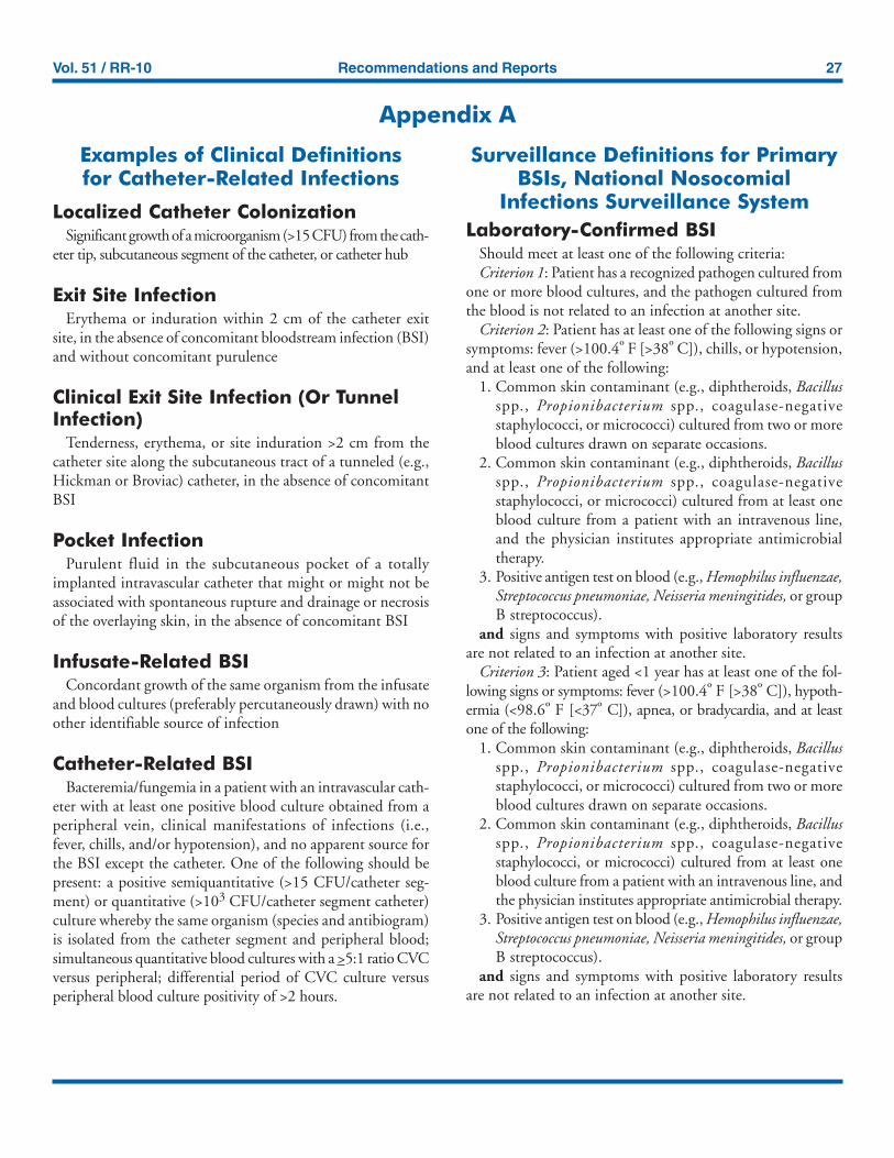
Vol. 51 / RR-10 Recommendations and Reports 27
Appendix A
Examples of Clinical Definitionsfor Catheter-Related Infections
Localized Catheter ColonizationSignificant growth of a microorganism (>15 CFU) from the cath-
eter tip, subcutaneous segment of the catheter, or catheter hub
Exit Site InfectionErythema or induration within 2 cm of the catheter exit
site, in the absence of concomitant bloodstream infection (BSI)and without concomitant purulence
Clinical Exit Site Infection (Or TunnelInfection)
Tenderness, erythema, or site induration >2 cm from thecatheter site along the subcutaneous tract of a tunneled (e.g.,Hickman or Broviac) catheter, in the absence of concomitantBSI
Pocket InfectionPurulent fluid in the subcutaneous pocket of a totally
implanted intravascular catheter that might or might not beassociated with spontaneous rupture and drainage or necrosisof the overlaying skin, in the absence of concomitant BSI
Infusate-Related BSIConcordant growth of the same organism from the infusate
and blood cultures (preferably percutaneously drawn) with noother identifiable source of infection
Catheter-Related BSIBacteremia/fungemia in a patient with an intravascular cath-
eter with at least one positive blood culture obtained from aperipheral vein, clinical manifestations of infections (i.e.,fever, chills, and/or hypotension), and no apparent source forthe BSI except the catheter. One of the following should bepresent: a positive semiquantitative (>15 CFU/catheter seg-ment) or quantitative (>103 CFU/catheter segment catheter)culture whereby the same organism (species and antibiogram)is isolated from the catheter segment and peripheral blood;simultaneous quantitative blood cultures with a >5:1 ratio CVCversus peripheral; differential period of CVC culture versusperipheral blood culture positivity of >2 hours.
Surveillance Definitions for PrimaryBSIs, National Nosocomial
Infections Surveillance SystemLaboratory-Confirmed BSI
Should meet at least one of the following criteria:Criterion 1: Patient has a recognized pathogen cultured from
one or more blood cultures, and the pathogen cultured fromthe blood is not related to an infection at another site.
Criterion 2: Patient has at least one of the following signs orsymptoms: fever (>100.4º F [>38º C]), chills, or hypotension,and at least one of the following:
1. Common skin contaminant (e.g., diphtheroids, Bacillusspp., Propionibacterium spp., coagulase-negativestaphylococci, or micrococci) cultured from two or moreblood cultures drawn on separate occasions.
2. Common skin contaminant (e.g., diphtheroids, Bacillusspp., Propionibacterium spp., coagulase-negativestaphylococci, or micrococci) cultured from at least oneblood culture from a patient with an intravenous line,and the physician institutes appropriate antimicrobialtherapy.
3. Positive antigen test on blood (e.g., Hemophilus influenzae,Streptococcus pneumoniae, Neisseria meningitides, or groupB streptococcus).
and signs and symptoms with positive laboratory resultsare not related to an infection at another site.
Criterion 3: Patient aged <1 year has at least one of the fol-lowing signs or symptoms: fever (>100.4º F [>38º C]), hypoth-ermia (<98.6º F [<37º C]), apnea, or bradycardia, and at leastone of the following:
1. Common skin contaminant (e.g., diphtheroids, Bacillusspp., Propionibacterium spp., coagulase-negativestaphylococci, or micrococci) cultured from two or moreblood cultures drawn on separate occasions.
2. Common skin contaminant (e.g., diphtheroids, Bacillusspp., Propionibacterium spp., coagulase-negativestaphylococci, or micrococci) cultured from at least oneblood culture from a patient with an intravenous line, andthe physician institutes appropriate antimicrobial therapy.
3. Positive antigen test on blood (e.g., Hemophilus influenzae,Streptococcus pneumoniae, Neisseria meningitides, or groupB streptococcus).
and signs and symptoms with positive laboratory resultsare not related to an infection at another site.
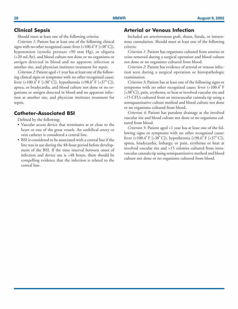
28 MMWR August 9, 2002
Clinical SepsisShould meet at least one of the following criteria:Criterion 1: Patient has at least one of the following clinical
signs with no other recognized cause: fever (>100.4º F [>38º C]),hypotension (systolic pressure <90 mm Hg), or oliguria(<20 mL/hr), and blood culture not done or no organisms orantigen detected in blood and no apparent infection atanother site, and physician institutes treatment for sepsis.
Criterion 2: Patient aged <1 year has at least one of the follow-ing clinical signs or symptoms with no other recognized cause:fever (>100.4º F [>38º C]), hypothermia (<98.6º F [<37º C]),apnea, or bradycardia, and blood culture not done or no or-ganisms or antigen detected in blood and no apparent infec-tion at another site, and physician institutes treatment forsepsis.
Catheter-Associated BSIDefined by the following:• Vascular access device that terminates at or close to the
heart or one of the great vessels. An umbilical artery orvein catheter is considered a central line.
• BSI is considered to be associated with a central line if theline was in use during the 48-hour period before develop-ment of the BSI. If the time interval between onset ofinfection and device use is >48 hours, there should becompelling evidence that the infection is related to thecentral line.
Arterial or Venous InfectionIncluded are arteriovenous graft, shunt, fistula, or intrave-
nous cannulation. Should meet at least one of the followingcriteria:
Criterion 1: Patient has organisms cultured from arteries orveins removed during a surgical operation and blood culturenot done or no organisms cultured from blood.
Criterion 2: Patient has evidence of arterial or venous infec-tion seen during a surgical operation or histopathologicexamination.
Criterion 3: Patient has at least one of the following signs orsymptoms with no other recognized cause: fever (>100.4º F[>38º C]), pain, erythema, or heat at involved vascular site and>15 CFUs cultured from an intravascular cannula tip using asemiquantitative culture method and blood culture not doneor no organisms cultured from blood.
Criterion 4: Patient has purulent drainage at the involvedvascular site and blood culture not done or no organisms cul-tured from blood.
Criterion 5: Patient aged <1 year has at least one of the fol-lowing signs or symptoms with no other recognized cause:fever (>100.4º F [>38º C]), hypothermia (<98.6º F [<37º C]),apnea, bradycardia, lethargy, or pain, erythema or heat atinvolved vascular site and >15 colonies cultured from intra-vascular cannula tip using semiquantitative method and bloodculture not done or no organisms cultured from blood.
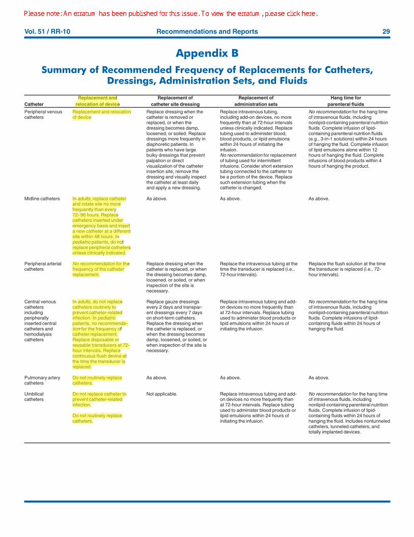
Vol. 51 / RR-10 Recommendations and Reports 29
Appendix BSummary of Recommended Frequency of Replacements for Catheters,
Dressings, Administration Sets, and Fluids
Peripheral venouscatheters
Midline catheters
Peripheral arterialcatheters
Central venouscathetersincludingperipherallyinserted centralcatheters andhemodialysiscatheters
Pulmonary arterycatheters
Umbilicalcatheters
Replacement and relocationof device
In adults, replace catheterand rotate site no morefrequently than every72–96 hours. Replacecatheters inserted underemergency basis and inserta new catheter at a differentsite within 48 hours. Inpediatric patients, do notreplace peripheral cathetersunless clinically indicated.
No recommendation for thefrequency of the catheterreplacement.
In adults, do not replacecatheters routinely toprevent catheter-relatedinfection. In pediatricpatients, no recommenda-tion for the frequency ofcatheter replacement.Replace disposable orreusable transducers at 72-hour intervals. Replacecontinuous flush device atthe time the transducer isreplaced.
Do not routinely replacecatheters.
Do not replace catheter toprevent catheter-relatedinfection.
Do not routinely replacecatheters.
Replace dressing when thecatheter is removed orreplaced, or when thedressing becomes damp,loosened, or soiled. Replacedressings more frequently indiaphoretic patients. Inpatients who have largebulky dressings that preventpalpation or directvisualization of the catheterinsertion site, remove thedressing and visually inspectthe catheter at least dailyand apply a new dressing.
As above.
Replace dressing when thecatheter is replaced, or whenthe dressing becomes damp,loosened, or soiled, or wheninspection of the site isnecessary.
Replace gauze dressingsevery 2 days and transpar-ent dressings every 7 dayson short-term catheters.Replace the dressing whenthe catheter is replaced, orwhen the dressing becomesdamp, loosened, or soiled, orwhen inspection of the site isnecessary.
As above.
Not applicable.
Replace intravenous tubing,including add-on devices, no morefrequently than at 72-hour intervalsunless clinically indicated. Replacetubing used to administer blood,blood products, or lipid emulsionswithin 24 hours of initiating theinfusion.No recommendation for replacementof tubing used for intermittentinfusions. Consider short extensiontubing connected to the catheter tobe a portion of the device. Replacesuch extension tubing when thecatheter is changed.
As above.
Replace the intravenous tubing at thetime the transducer is replaced (i.e.,72-hour intervals).
Replace intravenous tubing and add-on devices no more frequently thanat 72-hour intervals. Replace tubingused to administer blood products orlipid emulsions within 24 hours ofinitiating the infusion.
As above.
Replace intravenous tubing and add-on devices no more frequently thanat 72-hour intervals. Replace tubingused to administer blood products orlipid emulsions within 24 hours ofinitiating the infusion.
No recommendation for the hang timeof intravenous fluids, includingnonlipid-containing parenteral nutritionfluids. Complete infusion of lipid-containing parenteral nutrition fluids(e.g., 3-in-1 solutions) within 24 hoursof hanging the fluid. Complete infusionof lipid emulsions alone within 12hours of hanging the fluid. Completeinfusions of blood products within 4hours of hanging the product.
As above.
Replace the flush solution at the timethe transducer is replaced (i.e., 72-hour intervals).
No recommendation for the hang timeof intravenous fluids, includingnonlipid-containing parenteral nutritionfluids. Complete infusions of lipid-containing fluids within 24 hours ofhanging the fluid.
As above.
No recommendation for the hang timeof intravenous fluids, includingnonlipid-containing parenteral nutritionfluids. Complete infusion of lipid-containing fluids within 24 hours ofhanging the fluid. Includes nontunneledcatheters, tunneled catheters, andtotally implanted devices.
Replacement and Replacement of Replacement of Hang time forCatheter relocation of device catheter site dressing administration sets parenteral fluids

30 MMWR August 9, 2002

Vol. 51 / RR-10 Recommendations and Reports 31

32 MMWR August 9, 2002
All MMWR references are available on the Internet at http://www.cdc.gov/mmwr. Use the search function to find specific articles.——————
Use of trade names and commercial sources is for identification only and does not imply endorsement by the U.S. Department of Healthand Human Services.
——————References to non-CDC sites on the Internet are provided as a service to MMWR readers and do not constitute or imply endorsement ofthese organizations or their programs by CDC or the U.S. Department of Health and Human Services. CDC is not responsible for thecontent of these sites. URL addresses listed in MMWR were current as of the date of publication.
Healthcare Infection Control Practices Advisory CommitteeMembership List, May 2001
Chairman: Robert A. Weinstein, M.D., Cook County Hospital, Chicago, Illinois.Co-Chairman: Jane D. Siegel, M.D., University of Texas Southwestern Medical Center, Dallas, Texas.Executive Secretary: Michele L. Pearson, M.D., Medical Epidemiologist, National Center for Infectious Diseases, Centers for Disease Control and Prevention,Atlanta, Georgia.Members: Raymond Y.W. Chinn, M.D., Sharp Memorial Hospital, San Diego, California; Alfred DeMaria, Jr., M.D., Massachusetts Department of PublicHealth, Jamaica Plain, Massachusetts; James T. Lee, M.D., PhD., University of Minnesota and VA Medical Center, St. Paul, Minnesota; Ramon E. Moncada,M.D., Coronado Physician’s Medical Center, Coronado, California; William A. Rutala, PhD., University of North Carolina School of Medicine, Chapel Hill,North Carolina; William E. Scheckler, M.D., University of Wisconsin Medical School, Madison, Wisconsin; Beth H. Stover, Kosair Children’s Hospital,Louisville, Kentucky; Majorie A. Underwood, Mt. Diablo Medical Center, Concord, California.
Disclosure of Financial Interests or RelationshipsDennis Maki: Research grant sponsorship from Arrow, 3M, Becton-Dickinson, Johnson and Johnson; consultant for Micrologix.Issam Raad: Holds two patents associated with minocylcine-rifampin catheters. Patents are the property of University of Texas M.D. Anderson CancerCenter, Baylor College of Medicine and Wake Forest University. Both patents are liscensed to Cook Critical Care with royalty rights to the institutionsinvolved, and a percentage of royalties to the inventors. Co-inventor of patents associated with minocycline-EDTA flush solution. These patents are not yetlicensed. Becton-Dickenson holds rights to this technology. Grant and research support from Kimberly Clark, Becton-Dickinson, Abbott Labs, Immunomedics,and Cook Critical Care, Inc.Stephen Heard: Research grant sponsorship from Cook Critical Care, Inc., and Edwards Lifesciences.E. Patchen Dellinger: Research support, lectured for honoraria, or served on advisory boards for SmithKline Beecham, Wyeth-Ayerst, Pharmacia, Bristol-Myers Squibb, Zeneca, Pfizer, Aventis, Hoffman-LaRoche, Arrow, NABI, Ortho-McNeil, Park-Davis, Abbott, ICOS, Immunex, Chiron, Searle, Ican, Inc.,and Centercor.Andrienne Randolph: Research support from Cook Critical Care.Leonard Mermel: Research support from Arrow, Johnson and Johnson; consultant to 3M.
MMWR
The Morbidity and Mortality Weekly Report (MMWR) series is prepared by the Centers for Disease Control and Prevention (CDC) andis available free of charge in electronic format and on a paid subscription basis for paper copy. To receive an electronic copy each week, sendan e-mail message to [email protected]. The body content should read SUBscribe mmwr-toc. Electronic copy also is available fromCDC’s Internet server at http://www.cdc.gov/mmwr or from CDC’s file transfer protocol server at ftp://ftp.cdc.gov/pub/publications/mmwr. Tosubscribe for paper copy, contact Superintendent of Documents, U.S. Government Printing Office, Washington, DC 20402; telephone202-512-1800.
Data in the weekly MMWR are provisional, based on weekly reports to CDC by state health departments. The reporting weekconcludes at close of business on Friday; compiled data on a national basis are officially released to the public on the following Friday.Address inquiries about the MMWR series, including material to be considered for publication, to Editor, MMWR Series, Mailstop C-08,CDC, 1600 Clifton Rd., N.E., Atlanta, GA 30333; telephone 888-232-3228.
All material in the MMWR series is in the public domain and may be used and reprinted without permission; however, citation of thesource is appreciated.
✩U.S. Government Printing Office: 2002-733-100/69045 Region IV


![Guidelines for the Prevention of Intravascular Catheter-related ......complications (e.g., phlebitis and infiltration), and experience of individual catheter operators [33–35]](https://img.pdfslide.us/doc/110x75/607205cffb3e78705d41a900/guidelines-for-the-prevention-of-intravascular-catheter-related-complications.jpg)