Embed Size (px)
Citation preview
Draft Guideline For The Prevention Of Intravascular Catheter-Related Infections
Naomi P. O’Grady,1 Mary Alexander,2 E. Patchen Dellinger,3 Julie L. Gerberding,4 Stephen O. Heard,5 Dennis G. Maki,6 Henry Masur,1 Rita D. McCormick,7 Leonard A.
Mermel,8 Michele L. Pearson,4 Issam I. Raad,9 Adrienne Randolph,10 Robert A. Weinstein11 and the Healthcare Infection Control Practices Advisory Committee
(HICPAC)
Developed and sponsored by the American College of Critical Care Medicine of the Society of Critical Care Medicine (SCCM) in cooperation with the Infectious Disease Society of America (IDSA), Society for Healthcare Epidemiology of America (SHEA), Surgical Infection Society
(SIS), American College of Chest Physicians (ACCP), American Thoracic Society (ATS), American Society of Critical Care Anesthesiologists (ASCCA), Association for Professionals in Infection Control and Epidemiology (APIC), Intravenous Nurses Society (INS), Oncology
Nursing Society (ONS), Society of Cardiovascular and Interventional Radiology (SCVIR), American Academy of Pediatrics (AAP), and the Healthcare Infection Control Practices
Advisory Committee (HICPAC)* of the Centers for Disease Control and Prevention (CDC)
1. Clinical Center, National Institute of Health, Bethesda, MD Representing SCCM and IDSA
2. Intravenous Nurses Society, Cambridge, MA Representing INS
3. Department of Surgery, University of Washington, Seattle, WA
Representing SIS and SCCM 4. Division of Healthcare Quality Promotion, Centers for Disease Control and Prevention, Atlanta, GA
Representing HICPAC 5. Department of Anesthesiology, University of Massachusetts Medical School, Worcester, MA
Representing ACCP and ASCCA 6. Infectious Diseases Section, University of Wisconsin Medical School, Madison, WI 7. University of Wisconsin Hospital and Clinics Representing APIC 8. Division of Infectious Diseases, Rhode Island Hospital, and Brown University School of Medicine,
Providence, RI 9. Department of Medical Specialties, MD Anderson Cancer Center, Houston, TX 10. Departments of Anesthesia and Pediatrics, The Children’s Hospital, Boston, MA
11. Division of Infectious Disease, Cook County Hospital and Rush Medical College, Chicago, IL
Representing HICPAC and SHEA *HICPAC members listed in Appendix 2
2
Preface The “Guideline for Prevention of Intravascular Catheter Related Infections” provides
healthcare practitioners with background information and specific recommendations to reduce the incidence of intravascular catheter-related bloodstream infections (CR-BSI). This guideline replaces the “Guideline for Prevention of Intravascular Device-Related Infections” published in 1996 [1].
Part I of this current document (Intravascular Catheter-Related Infections: An Overview) reviews important issues in intravascular catheter use and maintenance. These issues include definitions and diagnosis of catheter-related infection, barrier precautions during catheter insertion, skin antisepsis, intervals for replacement of catheters and intravenous (IV) fluids and administration sets, catheter site care, the role of specialized intravascular catheter personnel, and the use of antimicrobial/antiseptic-impregnated catheters, prophylactic systemic antibiotics, flush solutions, and anticoagulants. Part II (Recommendations for Prevention of Intravascular Catheter- Related Infections) provides consensus recommendations of the Healthcare Infection Control Practices Advisory Committee (HICPAC) and other professional societies.
This document was prepared by a working group composed of members from professional organizations (listed as sponsors or endorsing organizations) representing the disciplines of critical care medicine, infectious diseases, healthcare infection control, surgery, anesthesiology, interventional radiology, pulmonary medicine, pediatrics, and nursing. The working group was led by the Society of Critical Care Medicine (SCCM). The active participation and endorsement of multiple professional societies has reduced the confusion and redundancy that can occur when different disciplines provide conflicting or inconsistent recommendations or guidelines to healthcare providers.
The “Guideline for Prevention of Intravascular Catheter-Related Infections” has been developed for practitioners who insert catheters and for personnel who are responsible for surveillance and control of infections in the hospital setting as well as in the outpatient and home healthcare setting. Most recommendations are appropriate for the inpatient, outpatient, and home care setting, unless otherwise noted.
3PART I: INTRAVASCULAR CATHETER-RELATED INFECTIONS IN ADULT AND
PEDIATRIC PATIENTS: AN OVERVIEW INTRODUCTION
Intravascular catheters are indispensable in modern-day medical practice, particularly in intensive care units (ICUs). While such catheters provide necessary vascular access, their use puts patients at risk for local and systemic infectious complications, including local site infection, CR-BSI, septic thrombophlebitis, endocarditis, and other metastatic infections (e.g., lung abscess, brain abscess, osteomyelitis, endophthalmitis).
Healthcare institutions purchase millions of intravascular catheters each year. The incidence of CR-BSI varies considerably with the type of catheter, frequency of catheter manipulation, and patient-related factors such as underlying disease and acuity of illness. Peripheral venous catheters are the most frequently used venous access devices. While the incidence of local or bloodstream infections assoxiated with peripheral venous catheters is usually low, serious infectious complications are recognized by clinicians because of the large numbers of such catheters that are placed. Most serious catheter-related infections, however, are associated with central venous catheters (CVCs), especially those that are placed in patients in the ICU. In the ICU setting the incidence of infection is often higher than in the less acute in-patient or ambulatory setting. In the ICU, central venous access may be needed for extended periods of time, patients may be colonized with hospital acquired organisms, and the catheter may be manipulated multiple times per day for the administration of fluids, drugs, and blood products. Moreover, some catheters may be inserted in urgent situations during which optimal attention to aseptic technique may not be feasible. Certain catheters such as pulmonary artery catheters and peripheral arterial catheters may be accessed multiple times per day for hemodynamic measurements or to obtain samples for laboratory analysis, augmenting the potential for contamination and subsequent clinical infection.
The magnitude of the potential for CVCs to cause morbidity and mortality due to infectious complications has been estimated in several studies [2-5]. One study estimated that there are 15 million CVC days per year in ICUs alone in the United States [2]. If, on average, the rate of CVC associated blood stream infections is 5.3 per 1000 catheter days in the ICU [3], approximately 80,000 CVC-associated BSIs occur in ICUs each year in the United States. Thus, it would appear that 9600-20,000 patients per year die in ICUs in the United States due to catheter-associated BSI. The attributable mortality for these BSIs has ranged from 12-25% in prospective studies [4, 5], and the attributable cost per infection has been estimated to be $3,700-$29,000 [5, 6]. The annual cost of caring for patients with CVC-associated BSIs is between $296 million and $2.3 billion [7].
An analysis by Kluger and Maki has suggested that a total of 250,000 cases of CVC- associated BSIs occur annually if one assesses the entire hospital rather than focusing exclusively on the ICU. They estimated 12-25% attributable mortality for each infection and a marginal cost to the healthcare system of $25,000 per episode [8].
Thus, by a variety of analyses, the cost of CVC-associated BSI is substantial both in terms of morbidity and in terms of financial resources expended. The data are compelling that a major effort is warranted to implement strategies to reduce the incidence of these infections if we are to improve patient outcome and reduce healthcare costs. This effort must be multidisciplinary, involving healthcare professionals who insert and maintain intravascular catheters, healthcare managers who allocate resources, and patients who are capable of assisting in the care of their catheters.
4Terminology and Estimates of Risk
The terminology to identify catheters is confusing because many clinicians and authors use one aspect of the catheter for informal reference. A catheter may be designated by the type of vessel it occupies (peripheral venous, central venous, or arterial); its intended life span (temporary or short-term, such as less than 30 days vs. permanent or long-term, such as more than 30 days); its site of insertion (subclavian, femoral, internal jugular, peripheral, peripherally inserted central catheter (PICC)); its pathway from skin to vessel (tunneled vs. non-tunneled); its physical length (long vs. short); or some special characteristic of the catheter (presence or absence of a cuff, impregnation with heparin, antibiotics or antiseptics, or the number of lumens). To accurately identify a catheter, all of these aspects must be described. Table 1 provides a summary of terminology, along with some of the unique characteristics of catheters.
The rate of all catheter-related infections (i.e., local infections and systemic infections in which no blood culture was positive, as well as CR-BSI) is difficult to determine since most surveillance systems capture only catheter-associated BSIs and do not enumerate local infections. CR-BSI is an ideal parameter to follow since it represents the most serious form of catheter-related infection. However, the rate of such infection depends on how CR-BSI is defined. Healthcare professionals must recognize the difference between surveillance definitions and more precise and specific clinical definitions. As indicated in Appendix 1, the surveillance definitions for catheter associated BSI includes all BSIs that occur in patients with CVCs, when other sites of infection have been excluded. That is, the surveillance definition overestimates the true incidence of CR-BSI because not all BSIs originate from a catheter. Some bacteremias are secondary BSIs from undocumented sources such as post-operative surgical sites, intra-abdominal infections, and hospital-associated pneumonia or urinary tract infections. Thus, surveillance definitions are really definitions for catheter associated BSIs. A more rigorous definition might include only those BSIs for which other sources were excluded by careful examination of the patient record, and where a culture of the catheter tip demonstrated a significant number of colonies of an organism identical to the bloodstream isolate. That is, such a clinical definition would focus on catheter-related BSIs. Therefore, when comparing a healthcare facility’s infection rate to published data, it is crucial to ascertain that one is using a comparable definition if one is to accurately compare one’s data to a benchmark.
The Centers for Disease Control and Prevention and the Joint Commission on Accreditation of Healthcare Organizations both recommend that the risk of catheter associated BSIs be expressed as number of catheter associated BSIs per 1000 CVC days (catheter days are the total number of days of exposure to CVCs, i.e. one catheter in place for one day=one catheter-day; two catheters in place for one day=one catheter day) [9, 10].This parameter is more useful than the rate expressed as the number of catheter-associated infections per 100 catheters (or percent of catheters studied), because it accounts for BSIs over time and therefore adjusts risk for the number of days the catheter is in use.
EPIDEMIOLOGY AND MICROBIOLOGY
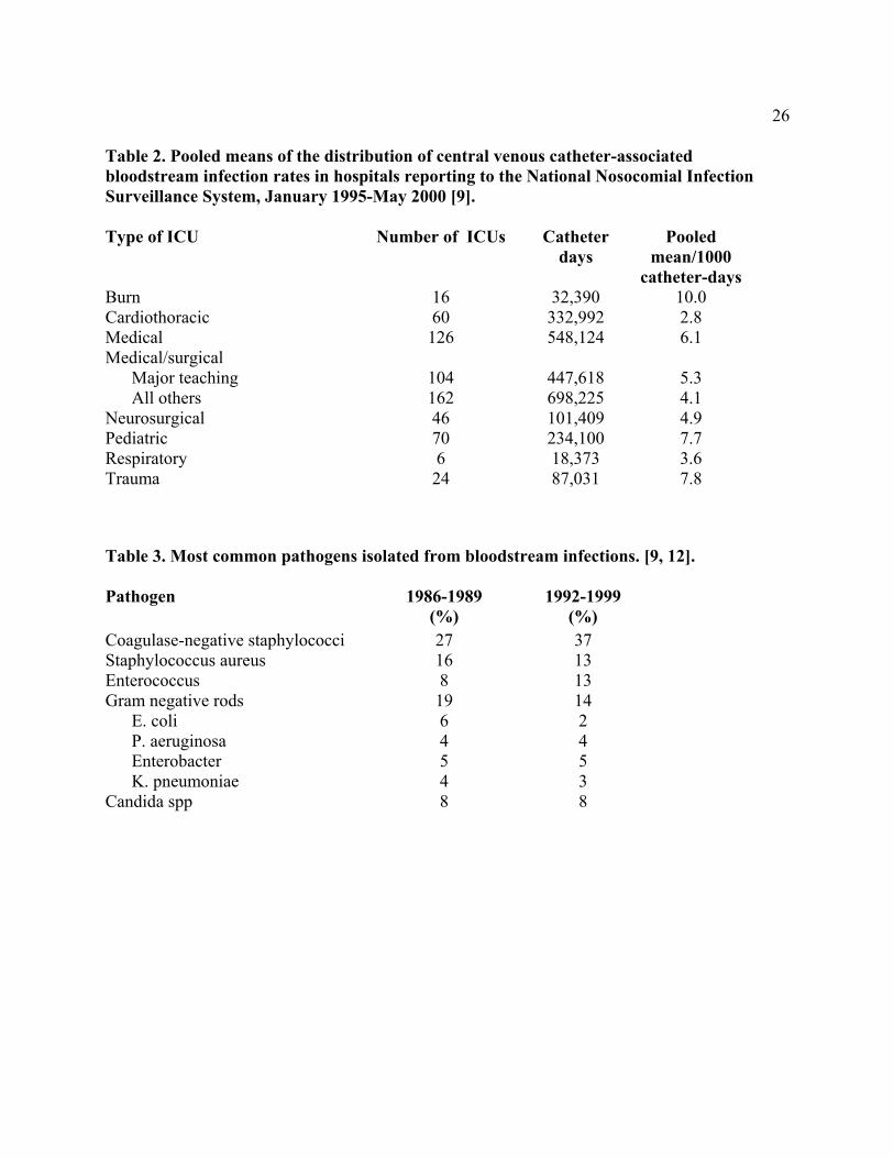
Since 1970, the CDC’s National Nosocomial Infection Surveillance System (NNIS) has been collecting data on incidence and etiologies of hospital-acquired infections, including CVC-associated BSIs in a group of almost 300 hospitals nationwide. Most hospital-acquired BSIs are associated with the use of a CVC, with BSI rates being substantially higher among patients with CVCs than among those without CVCs. Rates of CVC-associated BSI vary considerably by hospital size, hospital service/unit, and type of CVC. For1995-2000, the NNIS hospitals reported ICU rates of CVC-associated BSI ranging from 2.8 (cardiothoracic ICU) to 10.0 (burn ICU) BSIs per 1000 central catheter days (Table 2) [11].
5The relative risk of intravascular catheter-associated BSI has also been assessed in a
meta-analysis of 223 prospective studies in adult patients [8]. Relative risk of infection was best appreciated by analyzing rates of infection both by BSIs per 100 catheters (%) and BSIs per 1000 catheter days. These rates, and the NNIS-derived data, are important benchmarks for individual hospitals to use to estimate how their rates compare to other institutions. Rates are influenced by patient-related parameters, such as severity of illness and type of illness (e.g., third-degree burns vs. post-cardiac surgery), and by catheter related parameters, such as the condition under which the catheter was placed (e.g., elective vs. urgent) and catheter type (e.g., tunneled vs. nontunneled, or subclavian vs. jugular).
Types of organisms that most commonly cause hospital-acquired BSIs have been changing. Between 1986 and 1989, coagulase-negative staphylococci followed by Staphylococcus aureus were the most frequently reported causes of BSIs, accounting for 27% and 16% of BSIs respectively (Table 3) [12]. Pooled data from 1992-1999 indicate that coagulase-negative staphylococci followed by enterococci are now the most frequently isolated causes of hospital-acquired BSIs [9]. Coagulase-negative staphylococci account for 37 % [9] and S. aureus account for 12.6% of reported hospital-acquired BSIs [9]. Also notable was the susceptibility pattern of the S. aureus isolates. In 1999, for the first time since NNIS has been reporting susceptibilities, more than 50% of all S. aureus ICU isolates were oxacillin resistant [9].
Enterococci accounted for 13.5% of BSIs, an increase from 8% reported to NNIS from 1986 through 1989. There has also been a dramatic rise in enterococcal ICU isolates resistant to vancomycin, escalating from 0.5% in 1989 to 25.9% in 1999 [9].
Candida spp. caused 8% of hospital-acquired BSIs reported to NNIS during 1986-1989 [12, 13] and 1992-1999 [9, 14, 15]. Resistance of Candida spp. to commonly used antifungal agents has become a clinically important issue. While NNIS has not reported the fraction of BSIs due to non-albicans species or fluconazole susceptibility data, other epidemiological and clinical data document that fluconazole resistance is an increasingly relevant consideration when designing empiric therapeutic regimens for CR-BSIs due to yeast. Data from the Surveillance and Control of Pathogens of Epidemiologic Importance (SCOPE) Program documented that 10% of C. albicans bloodstream isolates from hospitalized patients were resistant to fluconazole [14]. Additionally, 48% of Candida BSIs were caused by non-albicans species, including C. glabrata and C. krusei, which are more likely than C. albicans to demonstrate resistance to fluconazole and itraconazole [15, 16].
Gram-negative bacilli accounted for 19% of catheter-associated BSIs between 1986 and 1989 [12] compared to 14% of catheter-associated BSIs between 1992-1999 [9]. An increasing fraction of ICU-related isolates are caused by Enterobacteriaceae -producing extended-spectrum β-lactamases (ESBLs), particularly Klebsiella pneumoniae [17]. Such organisms are resistant to many commonly used cephalosporins. PATHOGENESIS
Migration of skin organisms at the insertion site into the cutaneous catheter tract with colonization of the catheter tip is the most common route of infection for peripherally inserted, short-term catheters [18, 19]. Contamination of the catheter hub is an important contributor to intraluminal colonization of long-term catheters [20-22]. Occasionally, catheters may become hematogenously seeded from another focus of infection. Rarely, infusate contamination leads to CR-BSI [23].
Important pathogenic determinants of catheter-related infection are (1) the material of which the device is made and (2) the virulence of the infecting organism. In vitro studies show that catheters made of polyvinyl chloride or polyethylene appear to be less resistant to the
6adherence of microorganisms than are catheters made of Teflon, silicone elastomer, or polyurethane [24, 25]. Thus, most catheters sold in the United States are no longer made of polyvinyl chloride or polyethylene. Some catheter materials also have surface irregularities that enhance the microbial adherence of certain species (e.g., coagulase-negative staphylococci, Acinetobacter calcoaceticus, and Pseudomonas aeruginosa) [26-28]. Thus catheters made of certain materials may be especially vulnerable to microbial colonization and subsequent infection. Additionally, certain catheter materials are more thrombogenic than others, a characteristic that also may predispose to catheter colonization and catheter-related infection [28, 29]. This association has led to emphasis on preventing catheter-related thrombus as an additional mechanism for reducing CR-BSI.
The adherence properties of a given microorganism are also important in the pathogenesis of catheter-related infection. For example, S. aureus can adhere to host proteins (e.g., fibronectin) commonly present on catheters [30, 31]. Also, coagulase-negative staphylococci adhere to polymer surfaces more readily than do other pathogens such as Escherichia coli or S. aureus. Additionally, certain strains of coagulase-negative staphylococci produce an extracellular polysaccharide often referred to as "slime" [32, 33]. In the presence of catheters, this slime potentiates the pathogenicity of coagulase-negative staphylococci by allowing them to withstand host defense mechanisms (e.g., acting as a barrier to engulfment and killing by polymorphonuclear leukocytes) or by making them less susceptible to antimicrobial agents (e.g., forming a matrix that binds antimicrobials before their contact with the organism cell wall) [34]. Certain Candida spp., in the presence of glucose-containing fluids, may produce slime similar to that of their bacterial counterparts, potentially explaining the increased proportion of BSIs due to fungal pathogens among patients receiving parenteral nutrition fluids [35].
STRATEGIES FOR PREVENTION OF CATHETER-RELATED INFECTIONS IN ADULT AND PEDIATRIC PATIENTS Quality Assurance and Continuing Education
Measures to minimize the risk of infection associated with intravascular therapy must strike a balance between patient safety and cost effectiveness. As knowledge, technology, and healthcare settings change, infection control and prevention measures must change. This implies the need for well-organized programs that provide, monitor, and evaluate care and provide education to all caregivers. Reports spanning the past two decades have consistently found that risk of infection declines following standardization of aseptic care [36-39] and that insertion and maintenance of intravascular catheters by inexperienced staff may increase the risk of catheter colonization and CR-BSI [40]. Specialized "IV teams" have shown unequivocal effectiveness in reducing the incidence of catheter-related infections and associated complications and costs [41-44]. Additionally, infection risk increases with nursing staff reductions below a critical level [45].
Site of Catheter Insertion
The site at which a catheter is placed influences the subsequent risk of catheter-related infection and phlebitis. The influence of site on the risk for catheter infections is related in part to the risk of thrombophlebitis and density of local skin flora.
Phlebitis has long been recognized as a risk for infection. For adults, lower extremity insertion sites are associated with a higher risk of infection than are upper extremity sites [46-
748]. In addition, hand veins have a lower risk of phlebitis than do veins on the wrist or upper arm [49].
The density of skin flora at the catheter insertion site is a major risk factor for CR-BSI. Many authorities recommend that central catheters be placed in a subclavian site in preference to a jugular or femoral site to reduce the risk of infection. No randomized trial has satisfactorily compared infection rates for catheters placed in jugular, subclavian, and femoral sites. Catheters inserted into an internal jugular vein were associated with higher risk of infection than were those inserted into a subclavian or femoral vein in observational studies and in one prospective study [19, 50, 51].
Regarding femoral catheters, several investigations have suggested that such catheters have a relatively high colonization rate [52]. Femoral catheters are usually avoided if possible because they are associated with a higher risk of deep venous thrombosis than are internal jugular or subclavian catheters [53-56], and because of a presumption that such catheters are more likely to become infected, particularly in children with diapers and incontinent adults.
Thus, a subclavian site is preferred for infection control purposes, although other considerations such as the potential for mechanical complications, risk of subclavian vein stenosis, and operator skill must enter into the ultimate decision about where to place the catheter. Consideration of comfort, security, and maintenance of asepsis as well as patient-specific factors (e.g., preexisting catheters, anatomic deformity, bleeding diathesis), relative risk of mechanical complications (e.g., bleeding, pneumothorax), and the risk of infection should guide site selection.
Type of Catheter Material
As indicated above in the section on pathogenesis, Teflon or polyurethane catheters appear to be associated with fewer infectious complications than catheters made of polyvinyl chloride or polyethylene [24, 57, 58]. Steel needles used as an alternative to catheters for peripheral venous access have the same rate of infectious complications as do Teflon catheters [59, 60]. However, the use of steel needles frequently is complicated by infiltration of IV fluids into the subcutaneous tissues, a potentially serious complication if the infused fluid is a vesicant [60].
8Hand Hygiene and Aseptic Technique
For short peripheral catheters, good hand hygiene before catheter insertion or maintenance, combined with proper aseptic technique during catheter manipulation, provides protection against infection. Hand hygiene may utilize either an alcohol-based product [61] or an antibacterial soap with adequate rinsing [62]. Appropriate aseptic technique does not necessarily require sterile gloves: a new pair of disposable non-sterile gloves may be used in conjunction with a “no-touch” technique for the insertion of peripheral venous catheters. However, gloves are recommended as standard precautions for the prevention of bloodborne pathogen exposure.
Compared to peripheral venous catheters, CVCs carry a significantly greater risk of infection, and the level of barrier precautions needed to prevent infection during insertion of CVCs should therefore be more stringent. Full barrier precautions (cap, mask, sterile gown, sterile gloves, large sterile drape) during the insertion of CVCs reduce the incidence of CR-BSI compared to standard precautions (sterile gloves and small drapes) [19, 63]. The efficacy of such precautions for insertion of PICCs and midline catheters has not been studied, but it is reasonable to extrapolate the use of maximal barrier precautions to PICCs. Skin Antisepsis In the United States, povidone iodine is the most widely used antiseptic for cleansing arterial catheter and central catheter insertion sites [64]. However, preparation of central venous and arterial sites with 2% chlorhexidine gluconate has been shown to lower BSI rates compared to site preparation with 10% povidone-iodine or 70% alcohol [65]. However, tincture of chlorhexidine gluconate 0.5% has not been shown to be more effective than 10% povidone iodine in adults. A prospective randomized study comparing 0.5% chlorhexidine gluconate to povidone iodine showed no difference in preventing CR-BSI or CVC colonization in adults [66]. In neonates, however, 0.5% chlorhexidine reduced peripheral IV colonization compared to povidone iodine (20/418 vs. 38/408 catheters; p = 0.01) [67]. The study was not powered to assess differences in CR-BSI rates. Catheter site dressing regimens.
Transparent semipermeable polyurethane dressings have become a popular means of dressing catheter insertion sites. Transparent dressings reliably secure the device, permit continuous visual inspection of the catheter site, permit patients to bathe and shower without saturating the dressing, and require less frequent changes than do standard gauze and tape dressings, thus saving personnel time.
In the largest controlled trial of dressing regimens on peripheral catheters to date, Maki and Ringer examined the infectious morbidity associated with the use of transparent dressings on more than 2,000 peripheral catheters [57]. Their findings suggest that the rate of colonization among catheters dressed with transparent dressings (5.7%) is comparable to that of those dressed with gauze (4.6%) and that there are no clinically important differences in either the incidences of catheter site colonization or phlebitis. Furthermore, these data suggest that transparent dressings can be safely left on peripheral venous catheters for the duration of catheter insertion without increasing the risk of thrombophlebitis [57].
Regarding CVC dressings, a meta-analysis has assessed studies that compared the risk of catheter-related BSIs for groups using transparent dressings versus groups using gauze dressing [68]. The risk for CR-BSIs did not differ between the groups. The choice of dressing may be a matter of preference. If blood is oozing from the catheter insertion site, gauze dressing may be preferred.
9A chlorhexidine-impregnated sponge (Biopatch) placed over the site of short-term
arterial and CVCs reduced the risk of catheter colonization and CR-BSI in a multi-center study [69]. There were no adverse systemic effects from using this device. Routine use of chlorhexidine–impregnated sponges may reduce the risk of CR-BSI in adult and adolescent patients with short-term catheters, particularly with uncuffed CVCs and arterial catheters. In-line filters
In-line filters reduce the incidence of infusion-related phlebitis [70, 71]. There are no data to support their efficacy in preventing infections associated with intravascular catheters and infusion systems. Proponents of filters cite a number of potential benefits: (1) reducing the risk of infection from contaminated infusate or proximal contamination (i.e., introduced proximal to the filter); (2) reducing the risk of phlebitis in patients who require high doses of medication or in those in whom infusion-related phlebitis already has occurred; (3) removing particulate matter that may contaminate IV fluids [72] and (4) filtering endotoxin produced by gram-negative organisms in contaminated infusate [73]. These theoretical advantages must be tempered by the knowledge that infusate-related BSI is rare and that filtration of medications or infusates in the pharmacy is a more practical and less costly way to remove most particulates. Furthermore, in-line filters may become blocked, especially with certain solutions (i.e., dextran, lipids, mannitol), thereby increasing the number of line manipulations and decreasing the availability of administered drugs [74]. Thus, for the purposes of reducing the risk for CR-BSI, no strong recommendation can be made in favor of using in-line filters. Antimicrobial/antiseptic impregnated catheters and cuffs
Certain antimicrobial or antiseptic impregnated or coated catheters and cuffs can decrease the risk of CR-BSI in selected patient populations and potentially decrease hospital costs associated with treating CR-BSIs, despite the additional cost of an antimicrobial/antiseptic impregnated catheter [75]. All of the studies involving antimicrobial/antiseptic impregnated catheters have been conducted using triple-lumen, noncuffed catheters in adult patients whose catheters remained in place less than 30 days. And although all of the studies have been conducted in adults, these catheters have been approved by the FDA for marketing in patients as small as 3 kg. There are no antiseptic or antimicrobial impregnated catheters currently available for neonates.
Chlorhexidine/silver sulfadiazine. Catheters coated with chlorhexidine/silver sulfadiazine only on the external luminal surface have been studied as a means to reduce CR-BSI. Two meta-analyses [2, 76] demonstrated that such catheters reduced the risk for CR-BSI compared to standard noncoated catheters. The mean duration of catheter placement in one meta-analysis ranged between 5.1 and 11.2 days [77]. The half-life of antimicrobial activity against S. epidermidis is 3 days in vitro for catheters coated with chlorhexidine/silver sulfadiazine, and the antimicrobial activity decreases over time [78]. The benefit for the patients who receive these catheters will be realized within the first 14 days [77]. Although rare, anaphylaxis has been reported with the use of these chlorhexidine/silver sulfadiazine catheters in Japan [79]. Whether patients will become colonized or infected with organisms resistant to chlorhexidine/silver sulfadiazine remains to be determined [77].
Chlorhexidine/silver sulfadiazine catheters are more expensive than standard catheters. However, one analysis has suggested that the use of chlorhexidine/silver sulfadiazine catheters should lead to a cost savings of $68 to $391 per catheter [80] in settings in which the risk of CR-BSI is high despite the adherence to other preventive strategies such as maximal barrier precautions and aseptic technique. Use of these catheters may be cost effective in ICU patients, burn patients, neutropenic patients and patients who have had catheters placed under emergency conditions.
10Minocycline/rifampin. In a multicenter randomized trial, CVCs impregnated on both
the external and internal surfaces with minocycline/rifampin were associated with lower rates of CR-BSI when compared with chlorhexidine-silver sulfadiazine impregnated catheters [81]. The beneficial effect began after day 5 of catheterization. None of the catheters were evaluated beyond 30 days. No minocycline/rifampin-resistant organisms were reported. However, based on in vitro data, there is concern that these impregnated catheters could increase the incidence of minocycline and rifampin resistance among important pathogens, especially staphylococci. The half-life of antimicrobial activity against S. epidermidis is 25 days with catheters coated with minocycline/rifampin, compared to 3 days for catheters coated with chlorhexidine/silver sulfadiazine in vitro [78]. In vivo, the duration of antimicrobial activity of the minocycline/rifampin catheter is longer than that of the chlorhexidine/silver sulfadiazine catheter [81]. As with chlorhexidine/silver sulfadiazine catheters, some clinicians have recommended that the minocycline/rifampin catheters be considered in patient populations when the rate of CR-BSI exceeds 3.3 per 1000 catheter days [77]. Others suggest that reducing all rates of CR-BSI should be the goal [82]. The decision to use chlorhexidine/silver sulfadiazine or minocycline/rifampin impregnated catheters should be based on the need to enhance prevention of CR-BSI balanced against the concern for emergence of resistant pathogens and the cost of implementing this strategy.
Platinum/Silver. Ionic metals have broad antimicrobial activity and are being used in catheters and cuffs to prevent CR-BSI. A combination platinum/silver impregnated catheter is available in Europe and has recently been approved by the FDA for use in the United States. Although these catheters are being marketed for their antimicrobial properties, no published studies support a recommendation for their use to prevent CR-BSI.
Silver cuffs. Ionic silver has been used in subcutaneous collagen cuffs attached to CVCs [83].The ionic silver provides antimicrobial activity and the cuff provides a mechanical barrier to the migration of microorganisms along the external surface of the catheter. In studies of catheters left in place 20 days or longer, the cuff failed to reduce the incidence of CR-BSI [84, 85]. Two other studies of short-term catheters could not demonstrate efficacy because of the small number of CR-BSI observed [83, 86]. Systemic Antibiotic Prophylaxis
There is no evidence that oral or parenteral antibacterial or antifungal drugs can reduce the incidence of CR-BSI among adults [87-89]. However, among low birth-weight infants, two studies have assessed vancomycin prophylaxis: both demonstrated a reduction in CR-BSI but no reduction in mortality [90, 91]. Since the prophylactic use of vancomycin is an independent risk factor for the acquisition of vancomycin-resistant enterococcus (VRE) [92], most authorities agree that the risk of acquiring VRE outweighs the benefit of using prophylactic vancomycin. Mupirocin Ointment Several studies have evaluated the effectiveness of mupirocin ointment applied at the insertion sites of CVCs as a means to prevent CR-BSI [93-95]. Although mupirocin reduced the risk for CR-BSI [95], mupirocin ointment has also been associated with mupirocin resistance [96, 97] and may adversely affect the integrity of polyurethane catheters [98, 99].
Nasal carriers of S. aureus have a higher risk for acquiring CR-BSI than do noncarriers [100, 101]. Mupirocin ointment has been used intranasally to decrease nasal carriage of S. aureus and lessen the risk for CR-BSI. However, resistance to mupirocin develops in both S. aureus and coagulase-negative staphylococci soon after routine use of mupirocin is instituted [96, 97]. Antibiotic Lock Prophylaxis
Antibiotic lock prophylaxis has been attempted by flushing and filling the lumen of the catheter with an antibiotic solution and leaving the solution to dwell in the lumen of the catheter
11in order to prevent CR-BSI. Three studies have shown this to be useful in neutropenic patients with long-term catheters [102-104]. In two of the studies, patients received heparin alone (10 U/ ml) or heparin plus 25 micrograms/ml of vancomycin. The third study compared vancomycin/ciprofloxacin/heparin to vancomycin/heparin to heparin alone. The rate of CR-BSI with vancomycin-susceptible organisms was significantly lower and the time to the first episode of bacteremia with vancomycin-susceptible organisms was significantly longer in patients receiving either vancomycin/ciprofloxacin/heparin or vancomycin/heparin compared to heparin alone [102-104]. One smaller study in children showed no difference between children receiving a heparin flush compared to heparin and vancomycin [105]. However, since the use of vancomycin is an independent risk factor for the acquisition of VRE [92], this practice is not routinely recommended.
An anticoagulant/antimicrobial combination consisting of minocycline and ethylenediaminetraacetic acid (EDTA) has been proposed as a lock solution because it has antibiofilm and antimicrobial activity against gram-positive, gram-negative, and Candida organisms [106], as well as anticoagulant properties. However, no controlled or randomized trials have demonstrated its efficacy. Anticoagulants
Anticoagulant flush solutions are widely used to prevent catheter thrombosis. Since thrombi and fibrin deposits on catheters may serve as a nidus for microbial colonization of intravascular catheters [107, 108], the use of anticoagulants may have a role in the prevention of CR-BSI. In a meta-analysis evaluating the benefit of heparin prophylaxis (3 U/ml in TPN, 5000 U q 6 or 12 hours flush, or 2500 U low molecular weight heparin subcutaneously) in patients with short-term CVCs, the risk for catheter-related central venous thrombosis was reduced with the use of prophylactic heparin [109]. However, there was no significant difference in the rate for CR-BSI. Since most heparin solutions contain preservatives with antimicrobial activity, it is unclear if any decrease in the rate of CR-BSI would be due to the reduced thrombus formation, the preservative, or both.
Most pulmonary artery, umbilical, and central venous catheters are available with a heparin-bonded coating. Most are heparin-bonded with benzalkonium chloride, which provides the catheters with some antimicrobial activity [110], in addition to providing an anti-thrombotic effect [111]. Warfarin has also been evaluated as a means for reducing CR-BSI by reducing thrombus formation on catheters [112, 113]. In patients with long-term CVCs, very low dose warfarin (1 mg/day) reduced the incidence of catheter thrombus. There are no data that demonstrate that warfarin reduces the incidence of CR-BSI. REPLACEMENT OF CATHETERS Peripheral Venous Catheters
Scheduled replacement of intravascular catheters has been proposed as a method to prevent phlebitis and catheter-related infections. Studies of short peripheral venous catheters show that the incidence of thrombophlebitis and bacterial colonization of catheters increases when catheters are left in place more than 72 hours [59, 114]. However, rates of phlebitis are not significantly different in peripheral catheters left in place 72 hours compared to 96 hours [115]. Because phlebitis and catheter colonization have been associated with an increased risk of catheter-related infection, short peripheral catheter sites commonly are rotated at 72-96 hour intervals to reduce the risk of infection, as well as the patient discomfort associated with phlebitis. Midline Catheters
12Midline catheters appear to be associated with lower rates of phlebitis than are short
peripheral catheters and lower rates of infection than CVCs [116-118]. In one prospective study of 140 midline catheters, their use was associated with a BSI rate of 0.8 per 1000 catheter-days [118]. No specific risk factors, including duration of catheterization, were associated with infection. Midline catheters were in place a median of 7 days, but as long as 49 days. Although the findings of this study suggested that midline catheters can be changed only when there is a specific indication, there are no prospective, randomized studies assessing the need for routine replacement as a strategy to prevent CR-BSI associated with midline catheters. CVCs, Including PICCs and Hemodialysis Catheters
No studies show an advantage for catheter replacement at scheduled time intervals as a method to reduce CR-BSI. Two trials assessed changing the catheter every 7 days in comparison to changing catheters as needed [119, 120]. One study involved 112 surgical ICU patients needing central venous catheters, pulmonary artery catheters, or peripheral arterial catheters [119], while the other study involved only subclavian hemodialysis catheters [120]. In both studies, there was no difference in CR-BSI in patients undergoing scheduled catheter replacement every 7 days compared to catheter replacement as needed.
Scheduled guidewire exchanges of CVCs is another strategy that has been proposed to prevent CR-BSI. The results of a meta-analysis of 12 randomized controlled trials assessing CVC management failed to prove any benefit for the reduction of CR-BSI by routine replacement of CVCs by guidewire exchange compared to catheter replacement on an as-needed basis [121]. Routine replacement of CVCs is not necessary for catheters that are functioning and have no evidence of local or systemic complications.
Catheter replacement over a guidewire has become an accepted technique for replacing a malfunctioning catheter or exchanging a pulmonary artery catheter for a CVC when invasive monitoring no longer is needed. Catheter insertion over a guidewire is associated with less discomfort and a significantly lower rate of mechanical complications than are those percutaneously inserted at a new site [122] and provide an important means of preserving limited venous access in some patients. Replacement of temporary catheters over a guidewire in the setting of bacteremia is not an acceptable replacement strategy, since the source of infection is usually colonization of the skin tract from the insertion site to the vein [19, 122]. However, in selected patients with tunneled hemodialysis catheters and bacteremia, catheter exchange over a guidewire in combination with antibiotic therapy may be an alternative as a salvage strategy in patients with limited venous access [123-126]. Hemodialysis Catheters Hemodialysis catheters are the most important factor contributing to bacteremia in dialysis patients [127, 128]. The relative risk for bacteremia in patients with dialysis catheters is more than 7 times the risk for patients with primary AV fistulas [129]. Despite the National Kidney Foundation’s effort to reduce the number of hemodialysis patients maintained with catheter access, the CDC annual survey indicates that catheter use increased from 12.7% in 1995 to 22.2% in 1999 [130]. Rates for bacteremia per 100 patient months were 0.2 for AV fistulas, 0.46 for grafts, 5.02 for cuffed catheters, and 8.48 for noncuffed catheters (CDC, unpublished data). Hemodialysis catheters should be avoided in favor of AV fistulas and grafts. If temporary access is needed for dialysis, a cuffed catheter is preferable to an noncuffed catheter, even in the ICU setting if the catheter is expected to stay in place for more than 2 weeks [8]. Pulmonary Artery Catheters Pulmonary artery catheters are inserted through a Teflon introducer and typically remain in place an average of 3 days. Most pulmonary artery catheters are heparin bonded, which
13reduces not only catheter thrombosis but also microbial adherence to the catheter [131]. Meta-analysis shows standard nonheparin-bonded pulmonary artery catheter rates of CR-BSI to be 5.5 per 1000 catheter days, compared to 2.6 per 1000 catheter days for heparin-bonded pulmonary artery catheters [8]. Since most pulmonary artery catheters in use today are heparin-bonded, the relative risk of infection with these catheters is similar to that of CVC (2.6 vs. 2.3 per 1000 catheter days) [8].
A prospective study of 442 pulmonary artery catheters showed an increased risk for CR-BSI after 5 days (0/442 CR-BSI before 5 days vs. 5/442 CS-BSI after 5 days; p<0.001) [132]. A small prospective observational study of 71 pulmonary artery catheters demonstrated higher infection rates in catheters left in place longer than 7 days (2% before 7 days vs. 16% after 7 days; p=0.056) [133]. In patients who continue to require hemodynamic monitoring, pulmonary artery catheters do not need to be changed more frequently than 5 days. Since there are no prospective randomized trials assessing routine replacement of pulmonary artery catheters to reduce CR-BSI, no specific recommendation can be made regarding routine replacement of catheters that need to be in place for more than 5 days.
Pulmonary artery catheters are usually packaged with a thin plastic sleeve that prevents touch contamination when placed over the catheter. In a study of 166 catheters, patients who were randomly assigned to have their catheters self-contained within this sleeve had a reduced risk for CR-BSI compared to those who had a pulmonary artery catheter placed without the sleeve (p=0.002) [134]. Peripheral Arterial Catheters
Peripheral arterial catheters are usually inserted into the radial or femoral artery and permit continuous blood pressure monitoring and blood gas measurements. The rate of CR-BSI is comparable to that of temporary central venous catheters (2.9 vs. 2.3 per 1000 catheter days) [8]. There have been no studies of scheduled catheter changes compared to changing arterial catheters on an as needed basis. One prospective observational study of 71 arterial catheters showed that 10 local infections and 4 CR-BSIs developed in patients who had peripheral arterial catheters in place for more than 4 days, compared to 1 local infection and zero CR-BSIs in patients whose catheters were in place 4 days or less (p<0.05) [133]. Since the risk of CR-BSI appears to be similar to that of short-term CVCs, it is logical to approach arterial catheters in a similar way. Since there are no prospective randomized trials assessing routine replacement of arterial catheters to reduce CR-BSI, no specific recommendation can be made regarding replacement of catheters that need to be in place for more than 5 days. Replacement of Administration Sets
The optimal interval for routine replacement of IV administration sets has been examined in three well-controlled studies. Data from each of these studies show that replacing administration sets no more frequently than 72 hours after initiation of use is safe and cost-beneficial [135-137]. Data from a more recent study showed that rates of phlebitis were not significantly different if administration sets were left in place 96 hours compared to 72 hours [115]. It may be logical to change administration sets more frequently if a fluid that enhances microbial growth is used (i.e., lipid emulsions or blood products) [138-140].
Stopcocks, used for injection of medications, administration of IV infusions, or collection of blood samples, represent an important potential portal of entry for microorganisms into vascular access catheters and IV fluids. Stopcock contamination is common, occurring in 45% and 50% in most series. Whether such contamination is an important entry point of CR-BSI has been difficult to prove.
"Piggyback" systems are used as an alternative to stopcocks. However, they also pose a risk for contamination of the intravascular fluid if the device entering the rubber membrane of an
14injection port is exposed to air or comes into direct contact with nonsterile tape used to fix the needle to the port. Modified piggyback systems have the potential to prevent contamination at these sites [141]. Needleless Intravascular Catheter Systems
Attempts to reduce the incidence of sharp injuries and the resultant risk of transmission of bloodborne infections to healthcare workers have led to the design and introduction of needleless infusion systems. When the devices are used according to manufacturers’ recommendations, they do not appear to have a major influence on the incidence of CR-BSI [142-149]. Multidose Parenteral Medication Vials
Parenteral medications commonly are dispensed in multidose parenteral medication vials that may be used for prolonged periods for one or more patients. Although the overall risk of extrinsic contamination of multidose vials appears to be small [150], the consequences of contamination may be serious. SPECIAL CONSIDERATIONS FOR INTRAVASCULAR CATHETER-RELATED INFECTIONS IN PEDIATRIC PATIENTS
Prevention of CR-BSI in children requires a few additional considerations, although only a limited number of studies have been performed specifically in children. Pediatric data have been derived largely from studies in neonatal or pediatric intensive care units and pediatric oncology patients. Epidemiology
As in adults, most BSIs in pediatric patients are related to the use of an intravascular catheter. From 1995 through 2000, the pooled mean catheter-associated BSI rate for all pediatric ICUs reporting data to NNIS was 7.7 per 1000 catheter days [11, 151]. Umbilical and central catheter-associated BSI rates for neonatal ICUs ranged from 11.6 per 1000 catheter days in children with birthweight <1000 grams to 4.0 per 1000 catheter days in children whose birthweight was >2500 grams[11]. Compared with the rate of CR-BSI in most adult ICUs [9], catheter utilization rates were comparable in adult and pediatric ICUs [151, 152]. Microbiology
As in adults, most CR-BSIs in children are caused by coagulase-negative staphylococci, which accounted for 37.7% of BSIs in pediatric ICUs reporting to NNIS during 1992-1999 [9]. Gram-negative bacteria accounted for 25% of BSIs reported in pediatric ICUs [151], whereas enterococci and Candida spp. accounted for 10% and 9%, respectively [151]. Peripheral Venous Catheters
As in adults, the use of peripheral venous catheters in pediatric patients may be complicated by phlebitis, infusion extravasation, and catheter infection [153]. Catheter location, infusion of parenteral nutritional fluids with continuous IV lipid emulsions, and length of ICU stay before catheter insertion have all increased a pediatric patient's risk for phlebitis. However, contrary to the risk in adults, the risk of phlebitis in children has not been shown to increase with the duration of catheterization [153, 154]. Peripheral Arterial Catheters
In a prospective study of 340 peripheral arterial catheters in children, two risk factors for catheter-related infection were identified: (1) use of an arterial system that permitted backflow of blood into the pressure tubing, and (2) duration of catheterization [155]. Although there was a correlation between duration of arterial catheterization and risk of catheter colonization, the risk remained constant for 2 to 20 days at 6.2% [155].
15 Umbilical Catheters
Although the umbilical stump becomes heavily colonized soon after birth, umbilical vessel catheterization often is used for vascular access in newborn infants. Umbilical vessels can be cannulated easily and permit both collection of blood samples and measurement of hemodynamic status. The incidences of catheter colonization and BSI appear to be similar for umbilical vein catheters and umbilical artery catheters. An estimated 40-55% of umbilical artery catheters were colonized and 5% resulted in CR-BSI [156-158]. Umbilical vein catheters were reported to be associated with colonization in 22% to 59% of cases [156-158], and CR-BSI in 3% to 8% of cases [157]. Although CR-BSI rates do not appear to be different with umbilical catheters in the high position (above the diaphragm) compared to the low position (below the diaphragm and above the aortic bifurcation), there is a lower incidence of vascular complications without an increase in adverse sequelae with catheters in the high position [157].
Risk factors for infection appear to differ between umbilical artery and vein catheters. Neonates with very low birth weight and prolonged receipt of antibiotic were at increased risk for umbilical artery CR-BSIs [157]. In contrast, those with higher birth weight and receipt of parenteral nutrition fluids were at increased risk for umbilical vein CR-BSI. Duration of catheterization was not an independent risk factor for infection of either type of umbilical catheter. CVCs
Because of the limited vascular sites, the required frequency of catheter replacement in children is particularly important. In a study that examined the frequency of central venous catheter replacement in PICU patients by using survival analysis techniques, the group of catheters studied (n=397) remained free of infection for a median of 23.7 days [159]. More importantly, there was no relationship between duration of catheterization and the daily probability of infection (r=0.21, p>0.1), suggesting that routine replacement of central venous catheters would not be expected to reduce the incidence of catheter-related infection [159].
16 PART II: RECOMMENDATIONS
These recommendations are designed to reduce the infectious complications associated with intravascular catheter use. Recommendations should be considered in the context of the institution's experience with catheter-related infections, experience with other adverse catheter-related complications (e.g., thrombosis, hemorrhage, pneumothorax), and availability of personnel skilled in the placement of intravascular devices. Recommendations are provided for (1) intravascular-catheter use in general; (2) specific devices; and (3) special circumstances (i.e. intravascular-device use in pediatric patients and CVC use for parenteral nutrition and hemodialysis access).
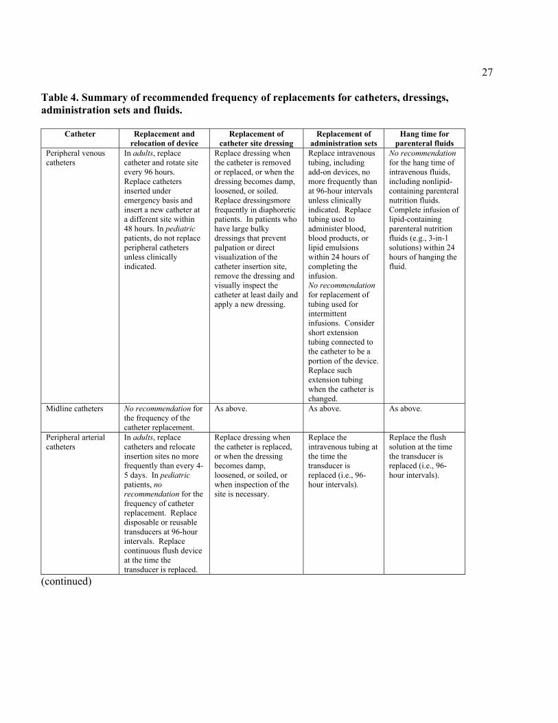
As in previous Centers for Disease Control and Prevention (CDC)/Healthcare Infection Control Practices Advisory Committee (HICPAC) guidelines, each recommendation is categorized on the basis of existing scientific data, theoretical rationale, applicability, and economic impact. The CDC/HICPAC system for categorizing recommendations is as follows: Category IA. Strongly recommended for implementation and strongly supported by well-designed experimental, clinical, or epidemiological studies. Category IB. Strongly recommended for implementation and supported by some experimental, clinical, or epidemiological studies and a strong theoretical rationale. Category IC. Required by state or federal regulations, rules, or standards. Category II. Suggested for implementation and supported by suggestive clinical or epidemiological studies or a theoretical rationale. No recommendation; unresolved issue. Practices for which evidence is insufficient or no consensus regarding efficacy exist. Recommendations regarding the frequency of replacing catheters, dressings, administration sets, and fluids are summarized in Table 4.
17
GENERAL RECOMMENDATIONS FOR ALL INTRAVASCULAR CATHETERS IN ADULT AND PEDIATRIC PATIENTS I. Healthcare worker education and training
A. Educate healthcare workers regarding indications for intravascular catheter use, proper procedures for the insertion and maintenance of intravascular catheters, and appropriate infection control measures to prevent intravascular catheter-related infections. Category IA [36, 41-44, 160-165].
B. Assess competence and compliance with guidelines periodically for all individuals who insert and manage intravascular catheters. Category IA [36, 42, 43, 160, 166].
C. Ensure appropriate nursing staff-to-patient ratios in ICUs. Category IB [45, 167, 168].
II. Surveillance for catheter-related infection A. Monitor the catheter sites visually or by palpation through the intact dressing on a
regular basis. The frequency of examination will depend on the clinical situation for the individual patient. If the patient has tenderness at the insertion site, fever without obvious source, or other manifestations suggesting local or bloodstream infection, the dressing should be removed to allow thorough examination of the site. Category IB [1, 169-171].
B. Encourage patients to report any changes in their catheter site or any new discomfort to their healthcare provider. Category II.
C. Record the operator, date and time of catheter insertion and removal, and dressing changes in a standardized location. Category II.
D. Do not routinely culture catheter tips. Category IA [172-174]. III. Hand hygiene
A. Observe proper hand hygiene either by washing hands with conventional antiseptic-containing soap and water or with waterless alcohol-based gels or foams. Observe hand hygiene before and after palpating catheter insertion sites, as well as before and after inserting, replacing, or dressing an intravascular catheter. Category IA [42, 62, 175-179].
B. Use of gloves does not obviate the need for hand hygiene. Category IA [42, 177, 178].
IV. Aseptic technique during catheter insertion and care A. Maintain aseptic technique for the insertion and care of intravascular catheters.
Category IA [19, 63, 180, 181]. B. Wear clean or sterile gloves when inserting an intravascular catheter as required
by the Occupational Safety and Health Administration (OSHA) Bloodborne Pathogens Standard. Category IC. Wearing clean gloves is acceptable for the insertion of peripheral intravascular catheters if aseptic technique can be maintained. Sterile gloves must be worn for the insertion of arterial and central catheters. Category IA [180, 182].
C. Wear clean or sterile gloves when changing the dressing on intravascular catheters. Category IC.
18 V. Catheter insertion
Do not routinely use cutdown procedures as a method to insert catheters. Category IA [183-185].
VI. Catheter site care A. Cutaneous antisepsis
1. Disinfect clean skin with an appropriate antiseptic before catheter insertion and at the time of dressing changes. A 2% chlorhexidine-based preparation is preferred. Alternatively, tincture of iodine, an iodophor, or 70% alcohol could be used. Category IA [65, 67, 186, 187]. No recommendation for the use of chlorhexidine in infants less than 2 months of age. Unresolved issue.
2. Allow the antiseptic to remain on the insertion site and to dry before inserting the catheter. Allow povidone iodine to remain on the skin for at least 2 minutes, or longer if it is not yet dry before inserting the catheter. Category IB [65, 67, 186, 187 ].
3. Do not apply organic solvents (e.g., acetone or ether) to the skin before insertion of catheters or during dressing changes. Category IB [188].
VII. Catheter site dressing regimens A. Use either a sterile gauze or sterile transparent semipermeable dressing to cover
the catheter site. Category IA [132, 189-191]. B. Well-healed tunneled CVC sites may not require dressings. Category II. C. If the patient is diaphoretic, or if the site is bleeding or oozing, a gauze dressing is
preferred. Category II [132, 189-191]. D. Replace catheter site dressing if the dressing becomes damp, loosened, or visibly
soiled. Category IB [132, 189]. E. Change dressings regularly for adult and adolescent patients; the frequency of
such changes must be determined individually depending on the circumstances of the patient, but should be done at least weekly. Category II [190].
F. Do not use topical antibiotic ointment or creams on insertion sites because of their potential to promote fungal infections and antimicrobial resistance. Category IA [96, 192].
G. Do not submerge the catheter under water. Showering may be permitted if precautions can be taken to reduce the likelihood of introducing organisms into the catheter, e.g., use an impermeable covering for the catheter and connecting device during the shower. Category II [193, 194].
VIII. Selection and replacement of intravascular catheters A. Select the catheter, the insertion technique, and the insertion site with the lowest
risk of complications (infectious and noninfectious) for the anticipated type and duration of intravenous (IV) therapy. Category IA [19, 52, 56, 195-197].
B. Remove any intravascular catheter that is no longer essential. Category IA [198, 199].
C. Do not routinely replace central venous or arterial catheters solely for the purposes of reducing the incidence of infection. Category IB [121, 122, 200].
D. Replace peripheral venous catheters every 96 hours in adults to prevent phlebitis. Category IB [115]. In pediatric patients, leave peripheral venous catheters in place until intravenous therapy is completed, unless a complication occurs. Category IB [153, 154, 201, 202].

19E. Replace all catheters inserted under emergency conditions within 48 hours
when adherence to aseptic technique cannot be enssured. Category II [19, 63, 180, 181].
F. Use clinical judgement to determine when to replace a catheter that could be a source of infection. For example, do not routinely replace catheters in patients whose only indication of infection is fever. Venous catheters do not necessarily need to be replaced routinely in patients who are bacteremic or fungemic if the source of infection is unlikely to be the catheter. Category II [203].
G. Replace all CVCs if the patient is hemodynamically unstable and CR-BSI is suspected. Category II [203].
H. Do not use guidewire techniques to replace catheters for which there is a clinical suspicion for CR-BSI. Category IB [121, 122].
IX. Replacement of administration sets*, needleless systems, and parenteral fluids A. Replace administration sets, including secondary sets and add-on devices, no
more frequently than at 96-hour intervals, unless CR-BSI is suspected or documented. Category IA [115, 135-137].
B. Replace tubing used to administer blood, blood products, or lipid emulsions within 24 hours of initiating the infusion. Category II [140, 204, 205].
C. Needleless intravascular devices 1. Change the needleless components at least as often as the administration
set. Category II [142-144, 146-149]. 2. Change caps no more frequently than every 96 hours or according to
manufacturers’ recommendations. Category II [142, 144, 147, 148]. 3. Minimize leaks and breaks in the system by making sure that all
components of the system are compatible. Category II [145]. 4. Minimize contamination risk by using aseptic technique when accessing
needleless systems. Category IB [144, 145, 147]. D. Parenteral fluids
1. Complete the infusion of lipid emulsions within 24 hours of hanging the emulsion. Category IB [138-140, 206].
2. Complete infusions of blood within 4 hours of hanging the blood. Category IC [207-210].
3. No recommendation for the hang time of other parenteral fluids. Unresolved issue.
X. Intravenous injection ports A. Clean injection ports with 70% alcohol or an iodophor before accessing the
system. Category II [211, 212]. B. Cap all stopcocks when not in use. Category IB [211]. XI. Preparation and quality control of intravenous admixtures
A. Admix all routine parenteral fluids in the pharmacy in a laminar-flow hood using aseptic technique. Category IB [213, 214].
B. Do not use any container of parenteral fluid with visible turbidity, leaks, cracks, particulate matter, or if the manufacturer’s expiration date has passed. Category IB [213].
C. Use single-dose vials for parenteral additives or medications whenever possible. Category II [213, 215].
D. If multidose vials are used
201. Refrigerate multidose vials after they are opened, if recommended by
the manufacturer. Category II. 2. Cleanse the access diaphragm of multidose vials with 70% alcohol before
inserting a device into the vial. Category II [212]. 3. Use a sterile device to access a multidose vial and avoid touch
contamination of the device before penetrating the access diaphragm. Category IA [211, 216].
4. Discard multidose vial if sterility is compromised. Category IA [211, 216]. XII. In-line filters
Do not use filters routinely for infection control purposes. Category IA [71, 217]. XIII. Intravenous therapy personnel
Designate trained personnel for the insertion and maintenance of intravascular catheters. Category IB [43, 44, 189].
XIV. Prophylactic antimicrobials Do not administer intranasal or systemic antimicrobial prophylaxis routinely before insertion or during use of an intravascular catheter as a method to prevent catheter colonization or bloodstream infection. Category IA [87, 88, 97, 218].
PERIPHERAL VENOUS CATHETERS, INCLUDING MIDLINE CATHETERS, IN ADULT AND PEDIATRIC PATIENTS I. Selection of peripheral catheter
A. Select catheters based on the intended purpose and duration of use, known complications (e.g., phlebitis and infiltration), and experience at the institution. Category IB [59, 60, 219].
B. Avoid the use of steel needles for the administration of fluids and medication that may cause tissue necrosis if extravasation occurs. Category IA [59, 60].
C. Use a midline catheter or PICC when the duration of IV therapy is expected to exceed 6 days. Category IB [219].
II. Selection of peripheral catheter insertion site A. In adults, use an upper extremity site in preference to one on a lower extremity for
catheter insertion. Replace a catheter inserted in a lower extremity site to an upper extremity site as soon as it is feasible. Category IB [59, 220].
B. In pediatric patients, the hand, the dorsum of the foot, or the scalp may be used as the catheter insertion site. Category II.
C. Replacement of catheter 1. Inspect the catheter insertion site daily. Category II. 2. Remove peripheral venous catheters if the patient develops signs of
phlebitis (i.e., warmth, tenderness, erythema, palpable venous cord), infection, or a malfunctioning catheter. Category IB [58].
3. In adults, replace short, peripheral venous catheters every 96 hours to reduce the risk of phlebitis. Category IA [115, 221].
4. Do not routinely replace midline catheters as a means to reduce the risk for infection. Category IB [118].
5. In pediatric patients, peripheral venous catheters may be left in place until intravenous therapy is completed, unless a complication occurs. Category IB [153, 154, 201, 202].
III. Catheter and catheter site care
21Do not routinely apply prophylactic topical antimicrobial or antiseptic ointment or cream to the insertion site of peripheral venous catheters. Category IA [96, 192].
CENTRAL VENOUS CATHETERS, INCLUDING PICC, HEMODIALYSIS ,AND PULMONARY ARTERY CATHETERS, IN ADULT AND PEDIATRIC PATIENTS I. Surveillance for catheter-related infection
A. Conduct surveillance for CR-BSI in ICUs and other patient populations suspected of having a high rate of CR-BSI to determine catheter-specific bloodstream infection rates, to monitor trends in those rates, and to assist in identifying lapses in infection control practices. Category IA [3, 9, 13, 222-225].
B. Express ICU data as the number of catheter-associated BSIs per 1000 catheter-days to facilitate comparisons with national data in comparable patient populations and healthcare settings. Category IB [3, 9, 13, 222-225].
C. Investigate events leading to serious, unexpected, life-threatening or fatal outcomes. This includes any process variation for which a recurrence would carry a significant chance of a serious adverse outcome. Category IC [10].
II. General Principles A. Use a CVC with the minimum number of ports or lumens essential for the
management of the patient. Category IB [226-229]. B. Use an antimicrobial or antiseptic-impregnated CVC in adults if
1) despite full adherence to maximal barrier precautions, there is still a high rate of CR-BSI (e.g., more than 3.3/1000 catheter days). Category IB [75-77, 81, 230].
2) patients are expected to be at high risk for CR-BSI (e.g. patients are receiving total parentral nutrition (TPN), are neutropenic, or are in an ICU and the catheter is expected to remain in place more than 4 days. Category II [75-77, 81, 230].
3) No Recommendation in other patients whose catheter is expected to stay in place more than 4 days. Unresolved issue.
4) No Recommendation for the use of impregnated catheters in children. Unresolved issue.
C. Designate trained personnel with documented competence and provide adequate supervision for trainees who perform catheter insertion. Category IA [36, 42, 43, 160, 165, 166].
D. Use totally implantable access devices for patients who require long-term, intermittent vascular access. For patients requiring frequent or continuous access, a PICC or tunneled central venous catheter is preferable. Category II [231, 232].
E. Use a cuffed central venous catheter for dialysis if the period of temporary access is anticipated to be prolonged (e.g., more than 2 weeks) Category IB [233].
F. Use a fistula or graft instead of a central venous catheter for permanent access for dialysis. Category IB [129].
G. Do not use hemodialysis catheters for blood drawing or applications other than hemodialysis except during dialysis or under emergency circumstances. Category II.
III. Selection of catheter insertion site A. Weigh the risk and benefits of placing a device at a recommended site to reduce
infectious complications against the risk of mechanical complications (e.g.,
22pneumothorax, subclavian artery puncture, subclavian vein laceration, subclavian vein stenosis, hemothorax, thrombosis, air embolism, catheter misplacement). Category IA [19, 52, 56, 197].
B. To minimize infection risk, use a subclavian site for nontunneled CVC placement rather than a jugular or a femoral site. Category IB [19, 52, 56].
C. Place catheters used for hemodialysis and pheresis in a jugular or femoral vein rather than a subclavian vein to avoid venous stenosis if catheter access is needed. Category IA [234-238].
IV. Barrier precautions during catheter insertion A. Use sterile technique including the use of a cap, mask, sterile gown, sterile gloves,
and a large sterile sheet, for the insertion of central venous catheters or guidewire exchange. Category IA [19, 63].
B. Use a sterile sleeve to protect pulmonary artery catheters during insertion. Category IB [134].
V. Replacement of catheter A. Do not routinely replace central venous catheters or PICCs as a method to prevent
catheter-related infections. Category IB [119, 121, 122]. B. Do not routinely replace hemodialysis catheters as a method to prevent catheter-
related infection. Category IB [120]. C. Do not routinely replace pulmonary artery catheters more frequently than 7 days
as a method to prevent catheter-related infections. Category IB [132, 133]. D. Do not remove central venous catheter or PICCs solely because of fever. Use
clinical judgement regarding the appropriateness of removing the catheter if there is evidence of infection elsewhere. Category II [239].
E. Guidewire exchange 1. Do not use guidewire exchanges routinely for non-tunneled catheters.
Category IB [122, 240]. 2. Use a guidewire exchange to replace a malfunctioning non-tunneled
catheter if there is no evidence of infection and the risk of inserting a catheter into a new site is unacceptably high (e.g., due to obesity, coagulopathy). Category IB [122, 240].
3. Use a new set of sterile gloves prior to handling the new catheter when guidewire exchanges are performed. Category II [19, 63].
VI. Catheter and catheter site care A. General measures
Designate one port exclusively for hyperalimentation if a multilumen catheter is used to administer parenteral nutrition. Category II [241].
B. Antibiotic lock solutions Do not routinely use antibiotic lock solutions as a means of preventing CR-BSI.
Use prophylactic antibiotic lock solution only in special circumstances (e.g., in treating a patient with a long-term catheter or port who has a history of multiple CR-BSIs despite optimal aseptic technique). Category II [102, 103, 242, 243].
C. Catheter site dressing regimens 1. Replace catheter site dressing when the dressing becomes damp, loosened,
or soiled, or when inspection of the site is necessary. Category IA [57, 132, 190].
2. Replace dressings used on short-term central catheter sites every 2 days for gauze dressings and every 7 days for transparent dressings, except in
23pediatric patients because in these patients the risk of dislodging the catheter outweighs the benefit of changing the dressing. Category IB [190].
3. Replace dressings used on tunneled or implanted central catheter sites no more than once per week, until the insertion site has healed. Category IB [190]. The frequency of catheter dressing changes over well-healed catheter exit sites is an unresolved issue.
D. No recommendation for the use of chlorhexidine sponge dressings to reduce the incidence of infection. Unresolved issue.
E. Do not use chlorhexidine sponge dressings in neonatesless than 7 days old or of gestational age less than 26 weeks. Category II [244].
F. Ensure that catheter site care is compatible with the catheter material. Category IB [98, 99].
G. Use a sterile sleeve for all pulmonary artery catheters. Category IB[134].
ADDITIONAL RECOMMENDATIONS FOR PERIPHERAL ARTERIAL CATHETERS AND PRESSURE MONITORING DEVICES FOR ADULT AND PEDIATRIC PATIENTS I. Selection of pressure monitoring system
Use disposable, rather than reusable, transducer assemblies when possible. Category IB [247-251].
II. Replacement of catheter and pressure monitoring system A. Replace peripheral arterial catheters and relocate catheter insertion sites no more
frequently than every 5 days for infection control purposes. Category IB [133, 200, 252].
B. Replace disposable or reusable transducers at 96-hour intervals. Replace other components of the system, including the tubing, continuous-flush device, and flush solution, at the time the transducer is replaced. Category IB [19, 248].
III. Care of pressure monitoring systems A. General measures
1. Keep all components of the pressure monitoring system (including calibration devices and flush solution) sterile. Category IA [247, 253-255].
2. Minimize the number of manipulations of and entries into the pressure monitoring system. Use a closed-flush system (i.e., continuous flush), rather than an open system (i.e., one that requires a syringe and stopcock), to maintain the patency of the pressure monitoring catheters. Category II [250, 256].
3. When the pressure monitoring system is accessed through a diaphragm rather than a stopcock, wipe the diaphragm with an appropriate antiseptic before accessing the system. Category IA [250].
4. Do not administer dextrose-containing solutions or parenteral nutrition fluids through the pressure monitoring circuit. Category IA [250, 257, 258].
B. Sterilization or disinfection of pressure monitoring systems 1. Use disposable transducers. Category IB [250, 257-260].

242. Sterilize reusable transducers according to the manufacturers' instructions if
the use of disposable transducers is not feasible. Category IA [250, 257-260].
RECOMMENDATIONS FOR UMBILICAL CATHETERS I. Replacement of catheters
A. Umbilical artery catheters should be removed and not replaced if any signs of CR-BSI, vascular insufficiency, or thrombosis occur. Category II [261].
B. Umbilical venous catheters should be removed and not replaced if any signs of CR-BSI or thrombosis occur. Category II [261].
C. No recommendation for treating through an umbilical venous catheter suspected of being infected. Unresolved issue.
D. Umbilical venous catheters should be replaced only if mechanical malfunction of the catheter occurs. Category II.
II. Catheter site care A. Cleanse the umbilical insertion site with an appropriate antiseptic before catheter
insertion. Avoid iodine-containing agents because of the potential effect on the neonatal thyroid. Category IB [67, 156, 157, 262, 263].
B. Do not use topical antibiotic ointment or creams on umbilical catheter insertion sites because of the potential to promote fungal infections and antimicrobial resistance. Category IA [96, 192].
C. Add low doses of heparin (0.5–1.0 u/ml TPN, 5000 u q 6 hr or 12 hr) to the fluid infused through umbilical arterial catheters. Category IB [264-266].
D. Remove umbilical catheters as soon as possible when not needed or when there is any sign of vascular insufficiency to the lower extremities. Optimally, umbilical artery catheters should not be left in place over 5 days. Category II [261, 267].
E. Umbilical venous catheters should be removed as soon as possible when not needed but can be used up to 14 days if managed aseptically. Category II [268, 269].
* Administration sets include the area from the spike of tubing entering the fluid container to the hub of the vascular access device. However, a short extension tube may be connected to the catheter and may be considered a portion of the catheter to facilitate aseptic technique when changing administration sets.

25
Table 1. Catheters used for venous and arterial access. Catheter Type Entry Site Length Comments Peripheral venous catheters usually inserted less than 3 inches phlebitis with prolonged use; (short) in veins of forearm or hand rarely associated with bloodstream infection Peripheral arterial catheters usually inserted in radial less than 3 inches low infection risk; rarely artery; can be placed in associated with bloodstream femoral, brachial, or posterior infection tibial arteries Midline catheters inserted via the antecubital 3 to 8 inches anaphylactoid reactions have fossa into the proximal been reported with catheters basilic or cephalic veins; made of elastomeric hydrogel;
does not enter central veins lower rates of phlebitis than short peripheral catheters
Nontunneled central percutaneously inserted into 8 cm or longer account for majority of CR-BSI venous catheters central veins (subclavian, depending on patient internal jugular, or femoral) size Pulmonary artery catheters inserted through a teflon 30 cm or longer usually heparin bonded; similar introducer in a central vein depending on patient rates of bloodtream infection as (subclavian, internal size CVC; subclavian site jugular, or femoral) preferred to reduce infection risk Peripherally inserted inserted into basilic, cephalic 20 cm or longer lower rate of infection than central venous catheters or brqaachial veins and enter depending on patient nontunneled CVCs (PICC) the superior vena cava size Tunneled central venous implanted into subclavian, 8 cm or longer cuff inhibits migration catheters internal jugular or femoral depending on patient of organisms into catheter tract; veins size lower rate of infection than nontunnelled CVC Totally implantable tunneled beneath skin and have 8 cm or longer lowest risk for CR-BSI; devices subcutaneous port accessed depending on patient improved patient self-image; with a needle; implanted size no need for local catheter in subclavian or internal site care; surgery required for jugular vein catheter removal Umbilical catheters inserted into either umbilical 6 cm or less, depending risk for CR-BSI similar with vein or umbilical artery on patient size catheters placed in umbilical vein vs. artery
26
Table 2. Pooled means of the distribution of central venous catheter-associated bloodstream infection rates in hospitals reporting to the National Nosocomial Infection Surveillance System, January 1995-May 2000 [9]. Type of ICU Number of ICUs Catheter
days Pooled
mean/1000 catheter-days
Burn 16 32,390 10.0 Cardiothoracic 60 332,992 2.8 Medical 126 548,124 6.1 Medical/surgical
Major teaching 104 447,618 5.3 All others 162 698,225 4.1
Neurosurgical 46 101,409 4.9 Pediatric 70 234,100 7.7 Respiratory 6 18,373 3.6 Trauma 24 87,031 7.8 Table 3. Most common pathogens isolated from bloodstream infections. [9, 12]. Pathogen 1986-1989
(%) 1992-1999
(%) Coagulase-negative staphylococci 27 37 Staphylococcus aureus 16 13 Enterococcus 8 13 Gram negative rods 19 14
E. coli 6 2 P. aeruginosa 4 4 Enterobacter 5 5 K. pneumoniae 4 3
Candida spp 8 8
27
Table 4. Summary of recommended frequency of replacements for catheters, dressings, administration sets and fluids.
Catheter Replacement and
relocation of device Replacement of
catheter site dressing Replacement of
administration sets Hang time for
parenteral fluids Peripheral venous catheters
In adults, replace catheter and rotate site every 96 hours. Replace catheters inserted under emergency basis and insert a new catheter at a different site within 48 hours. In pediatric patients, do not replace peripheral catheters unless clinically indicated.
Replace dressing when the catheter is removed or replaced, or when the dressing becomes damp, loosened, or soiled. Replace dressingsmore frequently in diaphoretic patients. In patients who have large bulky dressings that prevent palpation or direct visualization of the catheter insertion site, remove the dressing and visually inspect the catheter at least daily and apply a new dressing.
Replace intravenous tubing, including add-on devices, no more frequently than at 96-hour intervals unless clinically indicated. Replace tubing used to administer blood, blood products, or lipid emulsions within 24 hours of completing the infusion. No recommendation for replacement of tubing used for intermittent infusions. Consider short extension tubing connected to the catheter to be a portion of the device. Replace such extension tubing when the catheter is changed.
No recommendation for the hang time of intravenous fluids, including nonlipid-containing parenteral nutrition fluids. Complete infusion of lipid-containing parenteral nutrition fluids (e.g., 3-in-1 solutions) within 24 hours of hanging the fluid.
Midline catheters No recommendation for the frequency of the catheter replacement.
As above. As above. As above.
Peripheral arterial catheters
In adults, replace catheters and relocate insertion sites no more frequently than every 4-5 days. In pediatric patients, no recommendation for the frequency of catheter replacement. Replace disposable or reusable transducers at 96-hour intervals. Replace continuous flush device at the time the transducer is replaced.
Replace dressing when the catheter is replaced, or when the dressing becomes damp, loosened, or soiled, or when inspection of the site is necessary.
Replace the intravenous tubing at the time the transducer is replaced (i.e., 96-hour intervals).
Replace the flush solution at the time the transducer is replaced (i.e., 96-hour intervals).
(continued)
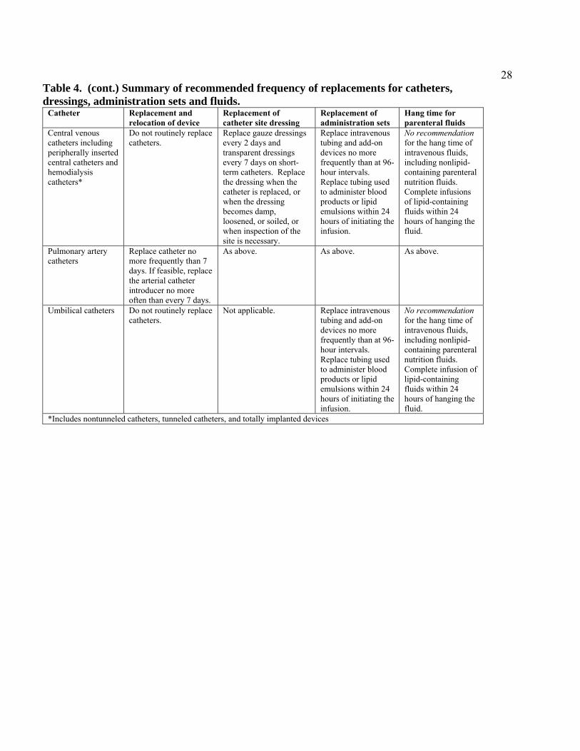
28Table 4. (cont.) Summary of recommended frequency of replacements for catheters, dressings, administration sets and fluids.
Catheter Replacement and relocation of device
Replacement of catheter site dressing
Replacement of administration sets
Hang time for parenteral fluids
Central venous catheters including peripherally inserted central catheters and hemodialysis catheters*
Do not routinely replace catheters.
Replace gauze dressings every 2 days and transparent dressings every 7 days on short-term catheters. Replace the dressing when the catheter is replaced, or when the dressing becomes damp, loosened, or soiled, or when inspection of the site is necessary.
Replace intravenous tubing and add-on devices no more frequently than at 96-hour intervals. Replace tubing used to administer blood products or lipid emulsions within 24 hours of initiating the infusion.
No recommendation for the hang time of intravenous fluids, including nonlipid-containing parenteral nutrition fluids. Complete infusions of lipid-containing fluids within 24 hours of hanging the fluid.
Pulmonary artery catheters
Replace catheter no more frequently than 7 days. If feasible, replace the arterial catheter introducer no more often than every 7 days.
As above. As above. As above.
Umbilical catheters Do not routinely replace catheters.
Not applicable. Replace intravenous tubing and add-on devices no more frequently than at 96-hour intervals. Replace tubing used to administer blood products or lipid emulsions within 24 hours of initiating the infusion.
No recommendation for the hang time of intravenous fluids, including nonlipid-containing parenteral nutrition fluids. Complete infusion of lipid-containing fluids within 24 hours of hanging the fluid.
*Includes nontunneled catheters, tunneled catheters, and totally implanted devices
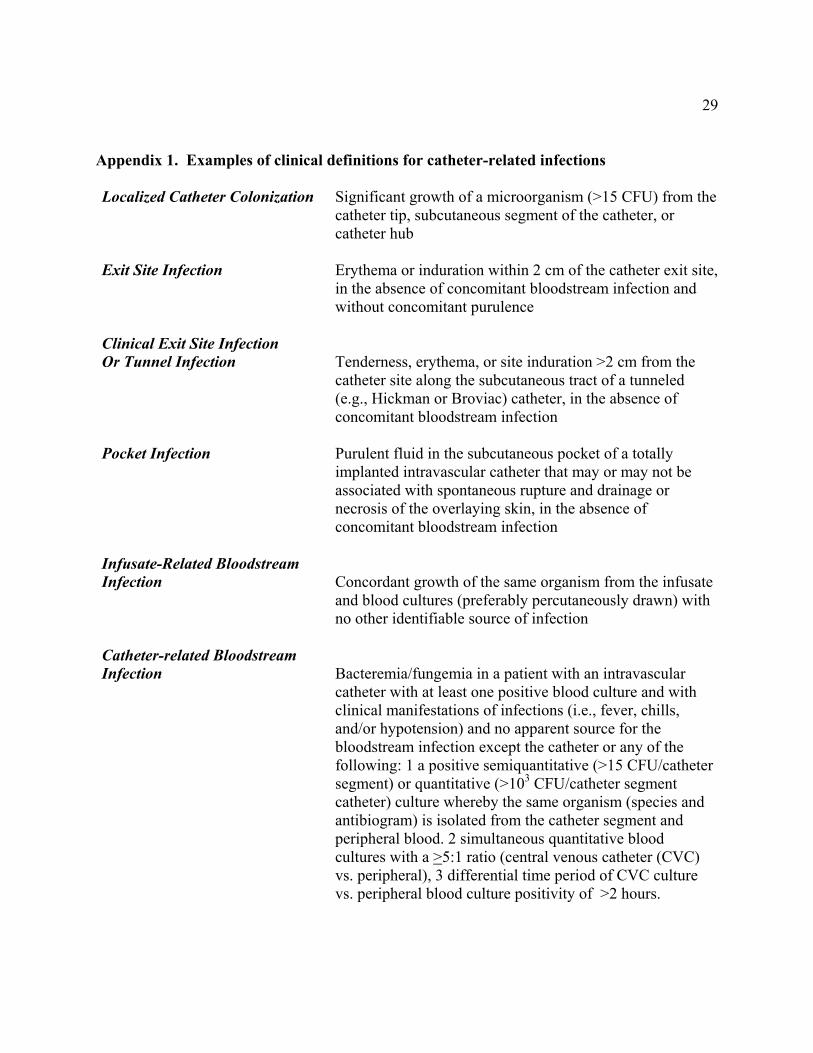
29
Appendix 1. Examples of clinical definitions for catheter-related infections Localized Catheter Colonization Significant growth of a microorganism (>15 CFU) from the
catheter tip, subcutaneous segment of the catheter, or catheter hub
Exit Site Infection Erythema or induration within 2 cm of the catheter exit site,
in the absence of concomitant bloodstream infection and without concomitant purulence
Clinical Exit Site Infection Or Tunnel Infection Tenderness, erythema, or site induration >2 cm from the
catheter site along the subcutaneous tract of a tunneled (e.g., Hickman or Broviac) catheter, in the absence of concomitant bloodstream infection
Pocket Infection Purulent fluid in the subcutaneous pocket of a totally
implanted intravascular catheter that may or may not be associated with spontaneous rupture and drainage or necrosis of the overlaying skin, in the absence of concomitant bloodstream infection
Infusate-Related Bloodstream Infection Concordant growth of the same organism from the infusate
and blood cultures (preferably percutaneously drawn) with no other identifiable source of infection
Catheter-related Bloodstream Infection Bacteremia/fungemia in a patient with an intravascular
catheter with at least one positive blood culture and with clinical manifestations of infections (i.e., fever, chills, and/or hypotension) and no apparent source for the bloodstream infection except the catheter or any of the following: 1 a positive semiquantitative (>15 CFU/catheter segment) or quantitative (>103 CFU/catheter segment catheter) culture whereby the same organism (species and antibiogram) is isolated from the catheter segment and peripheral blood. 2 simultaneous quantitative blood cultures with a >5:1 ratio (central venous catheter (CVC) vs. peripheral), 3 differential time period of CVC culture vs. peripheral blood culture positivity of >2 hours.
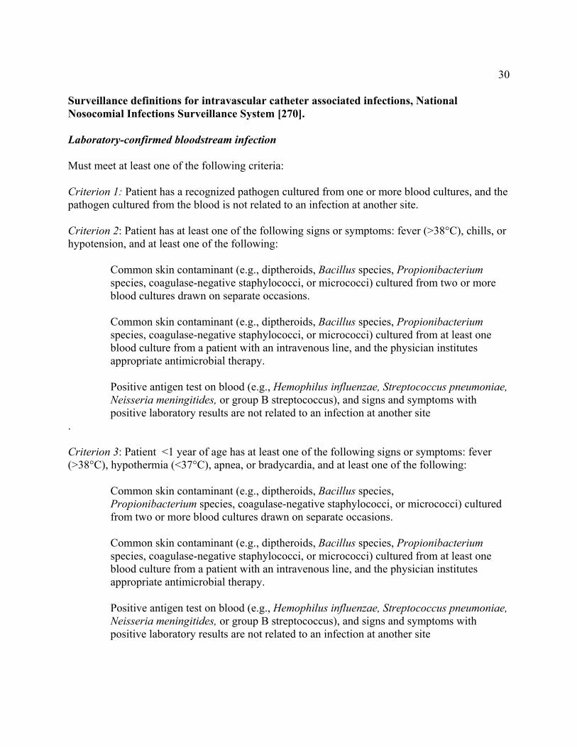
30 Surveillance definitions for intravascular catheter associated infections, National Nosocomial Infections Surveillance System [270]. Laboratory-confirmed bloodstream infection Must meet at least one of the following criteria: Criterion 1: Patient has a recognized pathogen cultured from one or more blood cultures, and the pathogen cultured from the blood is not related to an infection at another site. Criterion 2: Patient has at least one of the following signs or symptoms: fever (>38°C), chills, or hypotension, and at least one of the following:
Common skin contaminant (e.g., diptheroids, Bacillus species, Propionibacterium species, coagulase-negative staphylococci, or micrococci) cultured from two or more blood cultures drawn on separate occasions.
Common skin contaminant (e.g., diptheroids, Bacillus species, Propionibacterium
species, coagulase-negative staphylococci, or micrococci) cultured from at least one blood culture from a patient with an intravenous line, and the physician institutes appropriate antimicrobial therapy.
Positive antigen test on blood (e.g., Hemophilus influenzae, Streptococcus pneumoniae, Neisseria meningitides, or group B streptococcus), and signs and symptoms with positive laboratory results are not related to an infection at another site
. Criterion 3: Patient <1 year of age has at least one of the following signs or symptoms: fever (>38°C), hypothermia (<37°C), apnea, or bradycardia, and at least one of the following:
Common skin contaminant (e.g., diptheroids, Bacillus species, Propionibacterium species, coagulase-negative staphylococci, or micrococci) cultured from two or more blood cultures drawn on separate occasions.
Common skin contaminant (e.g., diptheroids, Bacillus species, Propionibacterium species, coagulase-negative staphylococci, or micrococci) cultured from at least one blood culture from a patient with an intravenous line, and the physician institutes appropriate antimicrobial therapy.
Positive antigen test on blood (e.g., Hemophilus influenzae, Streptococcus pneumoniae, Neisseria meningitides, or group B streptococcus), and signs and symptoms with positive laboratory results are not related to an infection at another site
31Clinical sepsis Must meet at least one of the following criteria: Criterion 1: Patient has at least one of the following clinical signs with no other recognized cause: fever (>38°C), hypotension (systolic pressure <90 mm Hg), or oliguria (<20 mL/hr), and blood culture not done or no organisms or antigen detected in blood and no apparent infection at another site, and physician institutes treatment for sepsis. Criterion 2: Patient <1 year of age has at least one of the following clinical signs or symptoms with no other recognized cause: fever (>38°C), hypothermia (<37°C), apnea, or bradycardia, and blood culture not done or no organisms or antigen detected in blood and no apparent infection at another site, and physician institutes treatment for sepsis. Catheter associated bloodstream infection Defined by the following:
Vascular access device that terminates at or close to the heart or one of the great vessels. An umbilical artery or vein catheter is considered a central line.
Bloodstream infection is considered to be associated with a central line if the line was in use during the 48-hour period before development of the bloodstream infection. If the time interval between onset of infection and device use is longer than 48 hours, there must be compelling evidence that the infection is related to the central line.
Arterial or venous infection Included are arteriovenous graft, shunt, fistula, or intravenous cannulation. Must meet at least one of the following criteria:
Criterion 1: Patient has organisms cultured from arteries or veins removed during a surgical operation and blood culture not done or no organisms cultured from blood.
Criterion 2: Patient has evidence of arterial or venous infection seen during a surgical operation or histopathologic examination.
Criterion 3: Patient has at least one of the following signs or symptoms with no other recognized cause: fever (>38°C), pain, erythema, or heat at involved vascular site and >15 CFUs cultured from an intravascular cannula tip using a semiquantitative culture method and blood culture not done or no organisms cultured from blood. Criterion 4: Patient has purulent drainage at the involved vascular site and blood culture not done or no organisms cultured from blood.
32Appendix 2. Healthcare Infection Control Practices Advisory Committee Membership, August 2001 CHAIR Weinstein, Robert A., MD Cook County Hospital Chicago, Illinois CO-CHAIR Siegel, Jane D., MD University of Texas Southwestern Medical Center Dallas, Texas EXECUTIVE SECRETARY Pearson, Michele L., MD Centers for Disease Control and Prevention Atlanta, Georgia
MEMBERSHIP Chinn, Raymond Y. W., MD Sharp Memorial Hospital San Diego, California DeMaria, Alfred, Jr., MD Massachusetts Department of Public Health Jamaica Plain, Massachusetts Larson, Elaine L., RN, PhD, FAN, CIC Cloumbia University School of Nursing New York, New York Lee, James T., MD, PhD University of Minnesota, VA Medical Center St. Paul, Minnesota Moncada, Ramon E., MD Coronado Physician’s Medical Center Coronado, California Rutala, William A., PhD University of North Carolina School of Medicine Chapel Hill, North Carolina Scheckler, William E., MD University of Wisconsin Medical School Madison, Wisconsin Stover, Beth H., RN, CIC Kosair Children’s Hospital Louisville, Kentucky
Underwood, Marjorie A., RN, BSN, CIC Mt. Diablo Medical Center Concord, California

33
References
1. Pearson ML. Guideline for prevention of intravascular device-related infections. Part I. Intravascular device-related infections: an overview. The Hospital Infection Control Practices Advisory Committee. Am J Infect Control 1996;24:262-77.
2. Mermel LA. Prevention of intravascular catheter-related infections. Ann Intern Med 2000;132:391-402. 3. Centers for Disease Control and Prevention. National Nosocomial Infections Surveillance (NNIS) System report, data
summary from October 1986-April 1998, issued June 1998. Am J Infect Control 1998;26:522-33. 4. Collignon PJ. Intravascular catheter associated sepsis: a common problem. The Australian Study on Intravascular
Catheter Associated Sepsis. Med J Aust 1994;161:374-8. 5. Heiselman D. Nosocomial bloodstream infections in the critically ill. JAMA 1994;272:1819-20. 6. Arnow PM, Quimosing EM, Beach M. Consequences of intravascular catheter sepsis. Clin Infect Dis 1993;16:778-
84. 7. Mermel LA. Correction: Catheter-related bloodstream infections. Ann Intern Med 2000;133:395. 8. Kluger DM, Maki DG. The relative risk of intravascular device related bloodstream infections in adults. Abstracts of
the 39th Interscience Conference on Antimicrobial Agents and Chemotherapy 1999:514. 9. Centers for Disease Control and Prevention. National Nosocomial Infections Surveillance (NNIS) System report, data
summary from January 1990-May 1999, issued June 1999. Am J Infect Control 1999;27:520-32. 10. Joint Commission on the Accreditation of Healthcare Organizations. Accreditation manual for hospitals. In: Joint
Commission on the Accreditation of Healthcare Organizations, ed. Chicago, IL, 1994:121-140. 11. Centers for Disease Control and Prevention. National Nosocomial Infections Surveillance (NNIS) System report, data
summary from April 1995-April 2000, issued June 2000. Am J Infect Control 2000;28:429-435. 12. Schaberg DR, Culver DH, Gaynes RP. Major trends in the microbial etiology of nosocomial infection. Am J Med
1991;91:72S-75S. 13. Banerjee SN, Emori TG, Culver DH, et al. Secular trends in nosocomial primary bloodstream infections in the United
States, 1980-1989. National Nosocomial Infections Surveillance System. Am J Med 1991;91:86S-89S. 14. Pfaller MA, Jones RN, Messer SA, et al. National surveillance of nosocomial blood stream infection due to Candida
albicans: frequency of occurrence and antifungal susceptibility in the SCOPE Program. Diagn Microbiol Infect Dis 1998;31:327-32.
15. Pfaller MA, Jones RN, Messer SA, et al. National surveillance of nosocomial blood stream infection due to species of Candida other than Candida albicans: frequency of occurrence and antifungal susceptibility in the SCOPE Program. SCOPE Participant Group. Surveillance and Control of Pathogens of Epidemiologic. Diagn Microbiol Infect Dis 1998;30:121-9.
16. Nguyen MH, Peacock JE, Jr., Morris AJ, et al. The changing face of candidemia: emergence of non-Candida albicans species and antifungal resistance. Am J Med 1996;100:617-23.
17. Fridkin SK, Gaynes RP. Antimicrobial resistance in intensive care units. Clin Chest Med 1999;20:303-16, viii. 18. Maki DG, Weise CE, Sarafin HW. A semiquantitative culture method for identifying intravenous-catheter-related
infection. N Engl J Med 1977;296:1305-9. 19. Mermel LA, McCormick RD, Springman SR, et al. The pathogenesis and epidemiology of catheter-related infection
with pulmonary artery Swan-Ganz catheters: a prospective study utilizing molecular subtyping. Am J Med 1991;91:197S-205S.
20. Sitges-Serra A, Linares J, Perez JL, et al. A randomized trial on the effect of tubing changes on hub contamination and catheter sepsis during parenteral nutrition. JPEN J Parenter Enteral Nutr 1985;9:322-5.
21. Linares J, Sitges-Serra A, Garau J, et al. Pathogenesis of catheter sepsis: a prospective study with quantitative and semiquantitative cultures of catheter hub and segments. J Clin Microbiol 1985;21:357-60.
22. Raad I, Costerton W, Sabharwal U, et al. Ultrastructural analysis of indwelling vascular catheters: a quantitative relationship between luminal colonization and duration of placement. J Infect Dis 1993;168:400-7.
23. Maki DG. Infections associated with intravascular lines. In: Remington JS, Schwartz, M., ed. Current Clinical Topics in Infectious Diseases. New York: McGraw-Hill, 1982:309-363.
24. Sheth NK, Franson TR, Rose HD, et al. Colonization of bacteria on polyvinyl chloride and Teflon intravascular catheters in hospitalized patients. J Clin Microbiol 1983;18:1061-3.
25. Ashkenazi S, Weiss E, Drucker MM, et al. Bacterial adherence to intravenous catheters and needles and its influence by cannula type and bacterial surface hydrophobicity. J Lab Clin Med 1986;107:136-40.
26. Locci R, Peters G, Pulverer G. Microbial colonization of prosthetic devices. IV. Scanning electron microscopy of intravenous catheters invaded by yeasts. Zentralbl Bakteriol Mikrobiol Hyg [B] 1981;173:419-24.
27. Locci R, Peters G, Pulverer G. Microbial colonization of prosthetic devices. I. Microtopographical characteristics of intravenous catheters as detected by scanning electron microscopy. Zentralbl Bakteriol Mikrobiol Hyg [B] 1981;173:285-92.
34
28. Nachnani GH, Lessin LS, Motomiya T, et al. Scanning electron microscopy of thrombogenesis on vascular catheter surfaces. Ultrastructural analysis of indwelling vascular catheters: a quantitative relationship between luminal colonization and duration of placement. N Engl J Med 1972;286:139-40.
29. Stillman RM, Soliman F, Garcia L, et al. Etiology of catheter-associated sepsis. Correlation with thrombogenicity. Arch Surg 1977;112:1497-9.
30. Herrmann M, Lai QJ, Albrecht RM, et al. Adhesion of Staphylococcus aureus to surface-bound platelets: role of fibrinogen/fibrin and platelet integrins. J Infect Dis 1993;167:312-22.
31. Herrmann M, Suchard SJ, Boxer LA, et al. Thrombospondin binds to Staphylococcus aureus and promotes staphylococcal adherence to surfaces. Infect Immun 1991;59:279-88.
32. Ludwicka A, Uhlenbruck G, Peters G, et al. Investigation on extracellular slime substance produced by Staphylococcus epidermidis. Zentralbl Bakteriol Mikrobiol Hyg [A] 1984;258:256-67.
33. Gray ED, Peters G, Verstegen M, et al. Effect of extracellular slime substance from Staphylococcus epidermidis on the human cellular immune response. Lancet 1984;1:365-7.
34. Farber BF, Kaplan MH, Clogston AG. Staphylococcus epidermidis extracted slime inhibits the antimicrobial action of glycopeptide antibiotics. J Infect Dis 1990;161:37-40.
35. Branchini ML, Pfaller MA, Rhine-Chalberg J, et al. Genotypic variation and slime production among blood and catheter isolates of Candida parapsilosis. J Clin Microbiol 1994;32:452-6.
36. Sherertz RJ, Ely EW, Westbrook DM, et al. Education of physicians-in-training can decrease the risk for vascular catheter infection. Ann Intern Med 2000;132:641-8.
37. Ryan JA, Jr., Abel RM, Abbott WM, et al. Catheter complications in total parenteral nutrition. A prospective study of 200 consecutive patients. N Engl J Med 1974;290:757-61.
38. Sanders RA, Sheldon GF. Septic complications of total parenteral nutrition. A five year experience. Am J Surg 1976;132:214-20.
39. Murphy LM, Lipman TO. Central venous catheter care in parenteral nutrition: a review. JPEN J Parenter Enteral Nutr 1987;11:190-201.
40. Armstrong CW, Mayhall CG, Miller KB, et al. Prospective study of catheter replacement and other risk factors for infection of hyperalimentation catheters. J Infect Dis 1986;154:808-16.
41. Nehme AE. Nutritional support of the hospitalized patient. The team concept. JAMA 1980;243:1906-8. 42. Eggimann P, Harbarth S, Constantin MN, et al. Impact of a prevention strategy targeted at vascular-access care on
incidence of infections acquired in intensive care. Lancet 2000;355:1864-8. 43. Soifer NE, Borzak S, Edlin BR, et al. Prevention of peripheral venous catheter complications with an intravenous
therapy team: a randomized controlled trial. Arch Intern Med 1998;158:473-7. 44. Tomford JW, Hershey CO. The i.v. therapy team: impact on patient care and costs of hospitalization. NITA
1985;8:387-9. 45. Fridkin SK, Pear SM, Williamson TH, et al. The role of understaffing in central venous catheter-associated
bloodstream infections. Infect Control Hosp Epidemiol 1996;17:150-8. 46. Bansmer G, Keith D, Tesluk H. Complications following use of indwelling catheters of inferior vena cava. JAMA
1958;167:1606-11. 47. Crane C. Venous interruption of septic thrombophlebitis. N Engl J Med 1960;262:947-51. 48. Indar R. The dangers of indwelling polyethelene cannulae in deep veins. Lancet 1959;1:284-6. 49. Maki DG, Mermel LA. Infections due to infusion therapy. In: Bennett JV and Brachman PS, eds. Hospital Infections.
4th ed. Philadelphia: Lippencott-Raven, 1998:689-724. 50. Heard SO, Wagle M, Vijayakumar E, et al. Influence of triple-lumen central venous catheters coated with
chlorhexidine and silver sulfadiazine on the incidence of catheter-related bacteremia. Arch Intern Med 1998;158:81-7.
51. Richet H, Hubert B, Nitemberg G, et al. Prospective multicenter study of vascular-catheter-related complications and risk factors for positive central-catheter cultures in intensive care unit patients. J Clin Microbiol 1990;28:2520-5.
52. Goetz AM, Wagener MM, Miller JM, et al. Risk of infection due to central venous catheters: effect of site of placement and catheter type. Infect Control Hosp Epidemiol 1998;19:842-5.
53. Joynt GM, Kew J, Gomersall CD, et al. Deep venous thrombosis caused by femoral venous catheters in critically ill adult patients. Chest 2000;117:178-83.
54. Mian NZ, Bayly R, Schreck DM, et al. Incidence of deep venous thrombosis associated with femoral venous catheterization. Acad Emerg Med 1997;4:1118-21.
55. Durbec O, Viviand X, Potie F, et al. A prospective evaluation of the use of femoral venous catheters in critically ill adults. Crit Care Med 1997;25:1986-9.
56. Trottier SJ, Veremakis C, O'Brien J, et al. Femoral deep vein thrombosis associated with central venous catheterization: results from a prospective, randomized trial. Crit Care Med 1995;23:52-9.
57. Maki DG, Ringer M. Evaluation of dressing regimens for prevention of infection with peripheral intravenous catheters. Gauze, a transparent polyurethane dressing, and an iodophor-transparent dressing. JAMA 1987;258:2396-403.
35
58. Maki DG, Ringer M. Risk factors for infusion-related phlebitis with small peripheral venous catheters. A randomized controlled trial. Ann Intern Med 1991;114:845-54.
59. Band JD, Maki DG. Steel needles used for intravenous therapy. Morbidity in patients with hematologic malignancy. Arch Intern Med 1980;140:31-4.
60. Tully JL, Friedland GH, Baldini LM, et al. Complications of intravenous therapy with steel needles and Teflon catheters. A comparative study. Am J Med 1981;70:702-6.
61. Pittet D, Hugonnet S, Harbath S, et al. Effectiveness of a hospital-wide programme to improve compliance with hand hygiene. Lancet 2000;356:1307-9.
62. Larson EL, Rackoff WR, Weiman M, et al. APIC guideline for handwashing and hand antisepsis in health care settings. Am J Infect Control 1995;23:251-69.
63. Raad, II, Hohn DC, Gilbreath BJ, et al. Prevention of central venous catheter-related infections by using maximal sterile barrier precautions during insertion. Infect Control Hosp Epidemiol 1994;15:231-8.
64. Clemence MA, Walker D, Farr BM. Central venous catheter practices: results of a survey. Am J Infect Control 1995;23:5-12.
65. Maki DG, Ringer M, Alvarado CJ. Prospective randomised trial of povidone-iodine, alcohol, and chlorhexidine for prevention of infection associated with central venous and arterial catheters. Lancet 1991;338:339-43.
66. Humar A, Ostromecki A, Direnfeld J, et al. Prospective randomized trial of 10% povidone-iodine versus 0.5% tincture of chlorhexidine as cutaneous antisepsis for prevention of central venous catheter infection. Clin Infect Dis 2000;31:1001-7.
67. Garland JS, Buck RK, Maloney P, et al. Comparison of 10% povidone-iodine and 0.5% chlorhexidine gluconate for the prevention of peripheral intravenous catheter colonization in neonates: a prospective trial. Pediatr Infect Dis J 1995;14:510-6.
68. Hoffmann KK, Weber DJ, Samsa GP, et al. Transparent polyurethane film as an intravenous catheter dressing. A meta-analysis of the infection risks. Jama 1992;267:2072-6.
69. Maki DG, Mermel LA, Klugar D, et al. The efficacy of a chlorhixidine impregnated sponge (Biopatch) for the prevention of intravascular catheter-related infection - a prospective randomized controlled multicenter study. In: Interscience Conference on Antimicrobial Agents and Chemotherapy, Toronto, Ontario, Canada:,2000.
70. Rusho WJ, Bair JN. Effect of filtration on complications of postoperative intravenous therapy. Am J Hosp Pharm 1979;36:1355-6.
71. Maddox RR, John JF, Jr., Brown LL, et al. Effect of inline filtration on postinfusion phlebitis. Clin Pharm 1983;2:58-61.
72. Turco SJ, Davis NM. Particulate matter in intravenous infusion fluids--phase 3. Am J Hosp Pharm 1973;30:611-3. 73. Baumgartner TG, Schmidt GL, Thakker KM, et al. Bacterial endotoxin retention by inline intravenous filters. Am J
Hosp Pharm 1986;43:681-4. 74. Butler DL, Munson JM, DeLuca PP. Effect of inline filtration on the potency of low-dose drugs. Am J Hosp Pharm
1980;37:935-41. 75. Raad I, Darouiche R, Dupuis J, et al. Central venous catheters coated with minocycline and rifampin for the
prevention of catheter-related colonization and bloodstream infections. A randomized, double-blind trial. The Texas Medical Center Catheter Study Group. Ann Intern Med 1997;127:267-74.
76. Veenstra DL, Saint S, Saha S, et al. Efficacy of antiseptic-impregnated central venous catheters in preventing catheter-related bloodstream infection: a meta-analysis. JAMA 1999;281:261-7.
77. Maki DG, Stolz SM, Wheeler S, et al. Prevention of central venous catheter-related bloodstream infection by use of an antiseptic-impregnated catheter. A randomized, controlled trial. Ann Intern Med 1997;127:257-66.
78. Raad I, Darouiche R, Hachem R, et al. The broad-spectrum activity and efficacy of catheters coated with minocycline and rifampin. J Infect Dis 1996;173:418-24.
79. Oda T, Hamasaki J, Kanda N, et al. Anaphylactic shock induced by an antiseptic-coated central venous catheter. Anesthesiology 1997;87:1242-4.
80. Veenstra DL, Saint S, Sullivan SD. Cost-effectiveness of antiseptic-impregnated central venous catheters for the prevention of catheter-related bloodstream infection. JAMA 1999;282:554-60.
81. Darouiche RO, Raad, II, Heard SO, et al. A comparison of two antimicrobial-impregnated central venous catheters. Catheter Study Group. N Engl J Med 1999;340:1-8.
82. Institute of Medicine. To err is human: building a safer health system. Washington, DC: Naional Academy Press, 2000.
83. Maki DG, Cobb L, Garman JK, et al. An attachable silver-impregnated cuff for prevention of infection with central venous catheters: a prospective randomized multicenter trial. Am J Med 1988;85:307-14.
84. Dahlberg PJ, Agger WA, Singer JR, et al. Subclavian hemodialysis catheter infections: a prospective, randomized trial of an attachable silver-impregnated cuff for prevention of catheter-related infections. Infect Control Hosp Epidemiol 1995;16:506-11.
36
85. Groeger JS, Lucas AB, Coit D, et al. A prospective, randomized evaluation of the effect of silver impregnated subcutaneous cuffs for preventing tunneled chronic venous access catheter infections in cancer patients. Ann Surg 1993;218:206-10.
86. Bonawitz SC, Hammell EJ, Kirkpatrick JR. Prevention of central venous catheter sepsis: a prospective randomized trial. Am Surg 1991;57:618-23.
87. McKee R, Dunsmuir R, Whitby M, et al. Does antibiotic prophylaxis at the time of catheter insertion reduce the incidence of catheter-related sepsis in intravenous nutrition? J Hosp Infect 1985;6:419-25.
88. Ranson MR, Oppenheim BA, Jackson A, et al. Double-blind placebo controlled study of vancomycin prophylaxis for central venous catheter insertion in cancer patients. J Hosp Infect 1990;15:95-102.
89. Ljungman P, Hagglund H, Bjorkstrand B, et al. Peroperative teicoplanin for prevention of gram-positive infections in neutropenic patients with indwelling central venous catheters: a randomized, controlled study. Support Care Cancer 1997;5:485-8.
90. Kacica MA, Horgan MJ, Ochoa L, et al. Prevention of gram-positive sepsis in neonates weighing less than 1500 grams. J Pediatr 1994;125:253-8.
91. Spafford PS, Sinkin RA, Cox C, et al. Prevention of central venous catheter-related coagulase-negative staphylococcal sepsis in neonates. J Pediatr 1994;125:259-63.
92. Centers for Disease Control and Prevention. Recommendations for preventing the spread of vancomycin resistance. Recommendations of the Hospital Infection Control Practices Advisory Committee (HICPAC). MMWR Morb Mortal Wkly Rep 1995;44:1-13.
93. Casewell MW. The nose: an underestimated source of Staphylococcus aureus causing wound infection. J Hosp Infect 1998;40:S3-11.
94. Hill RL, Fisher AP, Ware RJ, et al. Mupirocin for the reduction of colonization of internal jugular cannulae--a randomized controlled trial. J Hosp Infect 1990;15:311-21.
95. Sesso R, Barbosa D, Leme IL, et al. Staphylococcus aureus prophylaxis in hemodialysis patients using central venous catheter: effect of mupirocin ointment. J Am Soc Nephrol 1998;9:1085-92.
96. Zakrzewska-Bode A, Muytjens HL, Liem KD, et al. Mupirocin resistance in coagulase-negative staphylococci, after topical prophylaxis for the reduction of colonization of central venous catheters. J Hosp Infect 1995;31:189-93.
97. Miller MA, Dascal A, Portnoy J, et al. Development of mupirocin resistance among methicillin-resistant Staphylococcus aureus after widespread use of nasal mupirocin ointment. Infect Control Hosp Epidemiol 1996;17:811-3.
98. Rao SP, Oreopoulos DG. Unusual complications of a polyurethane PD catheter. Perit Dial Int 1997;17:410-2. 99. Riu S, Ruiz CG, Martinez-Vea A, et al. Spontaneous rupture of polyurethane peritoneal catheter. A possible
deleterious effect of mupirocin ointment. Nephrol Dial Transplant 1998;13:1870-1. 100. von Eiff C, Becker K, Machka K, et al. Nasal carriage as a source of Staphylococcus aureus bacteremia. N Engl J
Med 2001;344:11-16. 101. Levin A, Mason AJ, Jindal KK, et al. Prevention of hemodialysis subclavian vein catheter infections by topical
povidone-iodine. Kidney Int 1991;40:934-8. 102. Henrickson KJ, Axtell RA, Hoover SM, et al. Prevention of central venous catheter-related infections and thrombotic
events in immunocompromised children by the use of vancomycin/ciprofloxacin/heparin flush solution: A randomized, multicenter, double-blind trial. J Clin Oncol 2000;18:1269-78.
103. Carratala J, Niubo J, Fernandez-Sevilla A, et al. Randomized, double-blind trial of an antibiotic-lock technique for prevention of gram-positive central venous catheter-related infection in neutropenic patients with cancer. Antimicrob Agents Chemother 1999;43:2200-4.
104. Schwartz C, Henrickson KJ, Roghmann K, et al. Prevention of bacteremia attributed to luminal colonization of tunneled central venous catheters with vancomycin-susceptible organisms. J Clin Oncol 1990;8:1591-7.
105. Rackoff WR, Weiman M, Jakobowski D, et al. A randomized, controlled trial of the efficacy of a heparin and vancomycin solution in preventing central venous catheter infections in children. J Pediatr 1995;127:147-51.
106. Raad I, Buzaid A, Rhyne J, et al. Minocycline and ethylenediaminetetraacetate for the prevention of recurrent vascular catheter infections. Clin Infect Dis 1997;25:149-51.
107. Raad, II, Luna M, Khalil SA, et al. The relationship between the thrombotic and infectious complications of central venous catheters. JAMA 1994;271:1014-6.
108. Timsit JF, Farkas JC, Boyer JM, et al. Central vein catheter-related thrombosis in intensive care patients: incidence, risks factors, and relationship with catheter-related sepsis. Chest 1998;114:207-13.
109. Randolph AG, Cook DJ, Gonzales CA, et al. Benefit of heparin in central venous and pulmonary artery catheters: a meta-analysis of randomized controlled trials. Chest 1998;113:165-71.
110. Mermel LA, Stolz SM, Maki DG. Surface antimicrobial activity of heparin-bonded and antiseptic-impregnated vascular catheters. J Infect Dis 1993;167:920-4.
111. Pierce CM, Wade A, Mok Q. Heparin-bonded central venous lines reduce thrombotic and infective complications in critically ill children. Intensive Care Med 2000;26:967-72.

37
112. Bern MM, Lokich JJ, Wallach SR, et al. Very low doses of warfarin can prevent thrombosis in central venous catheters. A randomized prospective trial. Ann Intern Med 1990;112:423-8.
113. Boraks P, Seale J, Price J, et al. Prevention of central venous catheter associated thrombosis using minidose warfarin in patients with haematological malignancies. Br J Haematol 1998;101:483-6.
114. Collin J, Collin C. Infusion thrombophlebitis. Lancet 1975;2:458. 115. Lai KK. Safety of prolonging peripheral cannula and i.v. tubing use from 72 hours to 96 hours. Am J Infect Control
1998;26:66-70. 116. Fontaine PJ. Performance of a new softening expanding midline catheter in home intravenous therapy patients. J
Intraven Nurs 1991;14:91-9. 117. Harwood IR, Greene LM, Kozakowski-Koch JA, et al. New peripherally inserted midline catheter: a better alternative
for intravenous antibiotic therapy in patients with cystic fibrosis. Pediatr Pulmonol 1992;12:233-9. 118. Mermel LA, Parenteau S, Tow SM. The risk of midline catheterization in hospitalized patients. A prospective study.
Ann Intern Med 1995;123:841-4. 119. Eyer S, Brummitt C, Crossley K, et al. Catheter-related sepsis: prospective, randomized study of three methods of
long-term catheter maintenance. Crit Care Med 1990;18:1073-9. 120. Uldall PR, Merchant N, Woods F, et al. Changing subclavian haemodialysis cannulas to reduce infection. Lancet
1981;1:1373. 121. Cook D, Randolph A, Kernerman P, et al. Central venous catheter replacement strategies: a systematic review of the
literature. Crit Care Med 1997;25:1417-24. 122. Cobb DK, High KP, Sawyer RG, et al. A controlled trial of scheduled replacement of central venous and pulmonary-
artery catheters. N Engl J Med 1992;327:1062-8. 123. Robinson D, Suhocki P, Schwab SJ. Treatment of infected tunneled venous access hemodialysis catheters with
guidewire exchange. Kidney Int 1998;53:1792-4. 124. Beathard GA. Management of bacteremia associated with tunneled-cuffed hemodialysis catheters. J Am Soc Nephrol
1999;10:1045-9. 125. Saad TF. Bacteremia associated with tunneled, cuffed hemodialysis catheters. Am J Kidney Dis 1999;34:1114-24. 126. Duszak R, Jr., Haskal ZJ, Thomas-Hawkins C, et al. Replacement of failing tunneled hemodialysis catheters through
pre-existing subcutaneous tunnels: a comparison of catheter function and infection rates for de novo placements and over-the-wire exchanges. J Vasc Interv Radiol 1998;9:321-7.
127. Jaar BG, Hermann JA, Furth SL, et al. Septicemia in diabetic hemodialysis patients: comparison of incidence, risk factors, and mortality with nondiabetic hemodialysis patients. Am J Kidney Dis 2000;35:282-92.
128. Powe NR, Jaar B, Furth SL, et al. Septicemia in dialysis patients: incidence, risk factors, and prognosis. Kidney Int 1999;55:1081-90.
129. Hoen B, Paul-Dauphin A, Hestin D, et al. EPIBACDIAL: a multicenter prospective study of risk factors for bacteremia in chronic hemodialysis patients. J Am Soc Nephrol 1998;9:869-76.
130. Tokars JI, Miller ER, Alter MJ, et al. National surveillance of dialysis-associated diseases in the United States, 1997. Semin Dial 2000;13:75-85.
131. Mermel LA. Intravascular catheters impregnated with benzalkonium chloride. J Antimicrob Chemother 1993;32:905-6.
132. Maki DG, Stolz SS, Wheeler S, et al. A prospective, randomized trial of gauze and two polyurethane dressings for site care of pulmonary artery catheters: implications for catheter management. Crit Care Med 1994;22:1729-37.
133. Raad I, Umphrey J, Khan A, et al. The duration of placement as a predictor of peripheral and pulmonary arterial catheter infections. J Hosp Infect 1993;23:17-26.
134. Cohen Y, Fosse JP, Karoubi P, et al. The "hands-off" catheter and the prevention of systemic infections associated with pulmonary artery catheter: a prospective study. Am J Respir Crit Care Med 1998;157:284-7.
135. Josephson A, Gombert ME, Sierra MF, et al. The relationship between intravenous fluid contamination and the frequency of tubing replacement. Infect Control 1985;6:367-70.
136. Maki DG, Botticelli JT, LeRoy ML, et al. Prospective study of replacing administration sets for intravenous therapy at 48- vs 72-hour intervals. 72 hours is safe and cost-effective. JAMA 1987;258:1777-81.
137. Snydman DR, Donnelly-Reidy M, Perry LK, et al. Intravenous tubing containing burettes can be safely changed at 72 hour intervals. Infect Control 1987;8:113-6.
138. Crocker KS, Noga R, Filibeck DJ, et al. Microbial growth comparisons of five commercial parenteral lipid emulsions. J Parenter Enteral Nutr 1984;8:391-5.
139. Jarvis WR, Highsmith AK. Bacterial growth and endotoxin production in lipid emulsion. J Clin Microbiol 1984;19:17-20.
140. Melly MA, Meng HC, Schaffner W. Microbiol growth in lipid emulsions used in parenteral nutrition. Arch Surg 1975;110:1479-81.
141. Inoue Y, Nezu R, Matsuda H, et al. Prevention of catheter-related sepsis during parenteral nutrition: effect of a new connection device. J Parenter Enteral Nutr 1992;16:581-5.
38
142. Arduino MJ, Bland LA, Danzig LE, et al. Microbiologic evaluation of needleless and needle-access devices. Am J Infect Control 1997;25:377-80.
143. Brown JD, Moss HA, Elliott TS. The potential for catheter microbial contamination from a needleless connector. J Hosp Infect 1997;36:181-9.
144. Cookson ST, Ihrig M, O'Mara EM, et al. Increased bloodstream infection rates in surgical patients associated with variation from recommended use and care following implementation of a needleless device. Infect Control Hosp Epidemiol 1998;19:23-7.
145. Do AN, Ray BJ, Banerjee SN, et al. Bloodstream infection associated with needleless device use and the importance of infection-control practices in the home health care setting. J Infect Dis 1999;179:442-8.
146. Luebke MA, Arduino MJ, Duda DL, et al. Comparison of the microbial barrier properties of a needleless and a conventional needle-based intravenous access system. Am J Infect Control 1998;26:437-41.
147. McDonald LC, Banerjee SN, Jarvis WR. Line-associated bloodstream infections in pediatric intensive-care-unit patients associated with a needleless device and intermittent intravenous therapy. Infect Control Hosp Epidemiol 1998;19:772-7.
148. Mendelson MH, Short LJ, Schechter CB, et al. Study of a needleless intermittent intravenous-access system for peripheral infusions: analysis of staff, patient, and institutional outcomes. Infect Control Hosp Epidemiol 1998;19:401-6.
149. Seymour VM, Dhallu TS, Moss HA, et al. A prospective clinical study to investigate the microbial contamination of a needleless connector. J Hosp Infect 2000;45:165-8.
150. Longfield RN, Smith LP, Longfield JN, et al. Multiple-dose vials: persistence of bacterial contaminants and infection control implications. Infect Control 1985;6:194-9.
151. Richards MJ, Edwards JR, Culver DH, et al. Nosocomial infections in pediatric intensive care units in the United States. National Nosocomial Infections Surveillance System. Pediatrics 1999;103:103-9.
152. Richards MJ, Edwards JR, Culver DH, et al. Nosocomial infections in medical intensive care units in the United States. National Nosocomial Infections Surveillance System. Crit Care Med 1999;27:887-92.
153. Garland JS, Dunne WM, Jr., Havens P, et al. Peripheral intravenous catheter complications in critically ill children: a prospective study. Pediatrics 1992;89:1145-50.
154. Garland JS, Nelson DB, Cheah TE, et al. Infectious complications during peripheral intravenous therapy with Teflon catheters: a prospective study. Pediatr Infect Dis J 1987;6:918-21.
155. Furfaro S, Gauthier M, Lacroix J, et al. Arterial catheter-related infections in children. A 1-year cohort analysis. Am J Dis Child 1991;145:1037-43.
156. Krauss AN, Albert RF, Kannan MM. Contamination of umbilical catheters in the newborn infant. J Pediatr 1970;77:965-9.
157. Landers S, Moise AA, Fraley JK, et al. Factors associated with umbilical catheter-related sepsis in neonates. Am J Dis Child 1991;145:675-80.
158. Balagtas RC, Bell CE, Edwards LD, et al. Risk of local and systemic infections associated with umbilical vein catheterization: a prospective study in 86 newborn patients. Pediatrics 1971;48:359-67.
159. Stenzel JP, Green TP, Fuhrman BP, et al. Percutaneous central venous catheterization in a pediatric intensive care unit: a survival analysis of complications. Crit Care Med 1989;17:984-8.
160. Davis D, O'Brien MA, Freemantle N, et al. Impact of formal continuing medical education: do conferences, workshops, rounds, and other traditional continuing education activities change physician behavior or health care outcomes? JAMA 1999;282:867-74.
161. Conly JM, Hill S, Ross J, et al. Handwashing practices in an intensive care unit: the effects of an educational program and its relationship to infection rates. Am J Infect Control 1989;17:330-9.
162. East SA. Planning, implementation, and evaluation of a successful hospital-based peripherally inserted central catheter program. J Intraven Nurs 1994;17:189-92.
163. Kyle KS, Myers JS. Peripherally inserted central catheters. Development of a hospital-based program. J Intraven Nurs 1990;13:287-90.
164. BeVier PA, Rice CE. Initiating a pediatric peripherally inserted central catheter and midline catheter program. J Intraven Nurs 1994;17:201-5.
165. Tomford JW, Hershey CO, McLaren CE, et al. Intravenous therapy team and peripheral venous catheter-associated complications. A prospective controlled study. Arch Intern Med 1984;144:1191-4.
166. Wenzel RP, Wentzel RP. The development of academic programs for quality assessment. Arch Intern Med 1991;151:653-4.
167. Robert J, Fridkin SK, Blumberg HM, et al. The influence of the composition of the nursing staff on primary bloodstream infection rates in a surgical intensive care unit. Infect Control Hosp Epidemiol 2000;21:12-7.
168. Vicca AF. Nursing staff workload as a determinant of methicillin-resistant Staphylococcus aureus spread in an adult intensive therapy unit. J Hosp Infect 1999;43:109-13.
169. White MC, Ragland KE. Surveillance of intravenous catheter-related infections among home care clients. Am J Infect Control 1994;22:231-5.
39
170. Lorenzen AN, Itkin DJ. Surveillance of infection in home care. Am J Infect Control 1992;20:326-9. 171. White MC. Infections and infection risks in home care settings. Infect Control Hosp Epidemiol 1992;13:535-9. 172. Raad, II, Baba M, Bodey GP. Diagnosis of catheter-related infections: the role of surveillance and targeted
quantitative skin cultures. Clin Infect Dis 1995;20:593-7. 173. Widmer AF, Nettleman M, Flint K, et al. The clinical impact of culturing central venous catheters. A prospective
study. Arch Intern Med 1992;152:1299-302. 174. Pittet D, Tarara D, Wenzel RP. Nosocomial bloodstream infection in critically ill patients. Excess length of stay,
extra costs, and attributable mortality. JAMA 1994;271:1598-601. 175. Healthcare Infections Control Practices Advisory Committee/Society for Healthcare Epidemiology of America/
Infectious Disease Society of America. Guideline for Hand Hygiene, 2001 in press. 176. Bischoff WE, Reynolds TM, Sessler CN, et al. Handwashing compliance by health care workers: The impact of
introducing an accessible, alcohol-based hand antiseptic. Arch Intern Med 2000;160:1017-21. 177. Pittet D, Dharan S, Touveneau S, et al. Bacterial contamination of the hands of hospital staff during routine patient
care. Arch Intern Med 1999;159:821-6. 178. Simmons B, Bryant J, Neiman K, et al. The role of handwashing in prevention of endemic intensive care unit
infections. Infect Control Hosp Epidemiol 1990;11:589-94. 179. Boyce JM, Kelliher S, Vallande N. Skin irritation and dryness associated with two hand-hygiene regimens: soap-and-
water hand washing versus hand antisepsis with an alcoholic hand gel. Infect Control Hosp Epidemiol 2000;21:442-8. 180. Capdevila JA. Catheter-related infection: an update on diagnosis, treatment, and prevention. Int J Infect Dis
1998;2:230-6. 181. Abi-Said D, Raad I, Umphrey J, et al. Infusion therapy team and dressing changes of central venous catheters. Infect
Control Hosp Epidemiol 1999;20:101-5. 182. Centers for Disease Control and Prevention. Update: universal precautions for prevention of transmission of human
immunodeficiency virus, hepatitis B virus, and other bloodborne pathogens in health-care settings. MMWR 1988;24:377-82.
183. Povoski SP. A prospective analysis of the cephalic vein cutdown approach for chronic indwelling central venous access in 100 consecutive cancer patients [In Process Citation]. Ann Surg Oncol 2000;7:496-502.
184. Arrighi DA, Farnell MB, Mucha P, Jr., et al. Prospective, randomized trial of rapid venous access for patients in hypovolemic shock. Ann Emerg Med 1989;18:927-30.
185. Ahmed Z, Mohyuddin Z. Complications associated with different insertion techniques for Hickman catheters. Postgrad Med J 1998;74:104-7.
186. Little JR, Murray PR, Traynor PS, et al. A randomized trial of povidone-iodine compared with iodine tincture for venipuncture site disinfection: effects on rates of blood culture contamination. Am J Med 1999;107:119-25.
187. Mimoz O, Pieroni L, Lawrence C, et al. Prospective, randomized trial of two antiseptic solutions for prevention of central venous or arterial catheter colonization and infection in intensive care unit patients. Crit Care Med 1996;24:1818-23.
188. Maki DG, McCormack KN. Defatting catheter insertion sites in total parenteral nutrition is of no value as an infection control measure. Controlled clinical trial. Am J Med 1987;83:833-40.
189. Bijma R, Girbes AR, Kleijer DJ, et al. Preventing central venous catheter-related infection in a surgical intensive-care unit. Infect Control Hosp Epidemiol 1999;20:618-20.
190. Laura R, Degl'Innocenti M, Mocali M, et al. Comparison of two different time interval protocols for central venous catheter dressing in bone marrow transplant patients: results of a randomized, multicenter study. Haematologica 2000;85:275-9.
191. Madeo M, Martin CR, Turner C, et al. A randomized trial comparing Arglaes (a transparent dressing containing silver ions) to Tegaderm (a transparent polyurethane dressing) for dressing peripheral arterial catheters and central vascular catheters. Intensive Crit Care Nurs 1998;14:187-91.
192. Flowers RH, Schwenzer KJ, Kopel RF, et al. Efficacy of an attachable subcutaneous cuff for the prevention of intravascular catheter-related infection. A randomized, controlled trial. JAMA 1989;261:878-83.
193. Robbins J, Cromwell P, Korones DN. Swimming and central venous catheter-related infections in the child with cancer. J Pediatr Oncol Nurs 1999;16:51-6.
194. Howell PB, Walters PE, Donowitz GR, et al. Risk factors for infection of adult patients with cancer who have tunnelled central venous catheters. Cancer 1995;75:1367-75.
195. Goetz AM, Miller J, Wagener MM, et al. Complications related to intravenous midline catheter usage. A 2-year study. J Intraven Nurs 1998;21:76-80.
196. Martin C, Viviand X, Saux P, et al. Upper-extremity deep vein thrombosis after central venous catheterization via the axillary vein. Crit Care Med 1999;27:2626-9.
197. Robinson JF, Robinson WA, Cohn A, et al. Perforation of the great vessels during central venous line placement. Arch Intern Med 1995;155:1225-8.
198. Lederle FA, Parenti CM, Berskow LC, et al. The idle intravenous catheter. Ann Intern Med 1992;116:737-8.

40
199. Parenti CM, Lederle FA, Impola CL, et al. Reduction of unnecessary intravenous catheter use. Internal medicine house staff participate in a successful quality improvement project. Arch Intern Med 1994;154:1829-32.
200. Thomas F, Burke JP, Parker J, et al. The risk of infection related to radial vs femoral sites for arterial catheterization. Crit Care Med 1983;11:807-12.
201. Nelson DB, Garland JS. The natural history of Teflon catheter-associated phlebitis in children. Am J Dis Child 1987;141:1090-2.
202. Shimandle RB, Johnson D, Baker M, et al. Safety of peripheral intravenous catheters in children. Infect Control Hosp Epidemiol 1999;20:736-40.
203. O'Grady NP, Barie PS, Bartlett J, et al. Practice parameters for evaluating new fever in critically ill adult patients. Task Force of the American College of Critical Care Medicine of the Society of Critical Care Medicine in collaboration with the Infectious Disease Society of America. Crit Care Med 1998;26:392-408.
204. Gilbert M, Gallagher SC, Eads M, et al. Microbial growth patterns in a total parenteral nutrition formulation containing lipid emulsion. JPEN J Parenter Enteral Nutr 1986;10:494-7.
205. Maki DG, Martin WT. Nationwide epidemic of septicemia caused by contaminated infusion products. IV. Growth of microbial pathogens in fluids for intravenous infusions. J Infect Dis 1975;131:267-72.
206. Didier ME, Fischer S, Maki DG. Total nutrient admixtures appear safer than lipid emulsion alone as regards microbial contamination: growth properties of microbial pathogens at room temperature. J Parenter Enteral Nutr 1998;22:291-6.
207. Roth VR, Arduino MJ, Nobiletti J, et al. Transfusion-related sepsis due to Serratia liquefaciens in the United States. Transfusion 2000;40:931-5.
208. Blajchman MA. Reducing the risk of bacterial contamination of cellular blood components. Dev Biol Stand 2000;102:183-93.
209. Barrett BB, Andersen JW, Anderson KC. Strategies for the avoidance of bacterial contamination of blood components. Transfusion 1993;33:228-33.
210. Wagner SJ, Friedman LI, Dodd RY. Transfusion-associated bacterial sepsis. Clin Microbiol Rev 1994;7:290-302. 211. Plott RT, Wagner RF, Jr., Tyring SK. Iatrogenic contamination of multidose vials in simulated use. A reassessment of
current patient injection technique. Arch Dermatol 1990;126:1441-4. 212. Salzman MB, Isenberg HD, Rubin LG. Use of disinfectants to reduce microbial contamination of hubs of vascular
catheters. J Clin Microbiol 1993;31:475-9. 213. American Society of Health System Pharmacists. ASHP guidelines on quality assurance for pharmacy-prepared
sterile products. Am J Health Syst Pharm 2000;57:1150-69. 214. Herruzo-Cabrera R, Garcia-Caballero J, Vera-Cortes ML, et al. Growth of microorganisms in parenteral nutrient
solutions. Am J Hosp Pharm 1984;41:1178-80. 215. Green KA, Mustachi B, Schoer K, et al. Gadolinium-based MR contrast media: potential for growth of microbial
contaminants when single vials are used for multiple patients. AJR Am J Roentgenol 1995;165:669-71. 216. Arrington ME, Gabbert KC, Mazgaj PW, et al. Multidose vial contamination in anesthesia. AANA J 1990;58:462-6. 217. Falchuk KH, Peterson L, McNeil BJ. Microparticulate-induced phlebitis. Its prevention by in-line filtration. N Engl J
Med 1985;312:78-82. 218. Netto dos Santos KR, de Souza Fonseca L, Gontijo Filho PP. Emergence of high-level mupirocin resistance in
methicillin-resistant Staphylococcus aureus isolated from Brazilian university hospitals. Infect Control Hosp Epidemiol 1996;17:813-6.
219. Ryder MA. Peripheral access options. Surg Oncol Clin N Am 1995;4:395-427. 220. Maki DG, Goldman DA, Rhame FS. Infection control in intravenous therapy. Ann Intern Med 1973;79:867-87. 221. Tager IB, Ginsberg MB, Ellis SE, et al. An epidemiologic study of the risks associated with peripheral intravenous
catheters. Am J Epidemiol 1983;118:839-51. 222. Horan TC, Emori TG. Definitions of key terms used in the NNIS System. Am J Infect Control 1997;25:112-6. 223. Khuri-Bulos NA, Shennak M, Agabi S, et al. Nosocomial infections in the intensive care units at a university hospital
in a developing country: comparison with National Nosocomial Infections Surveillance intensive care unit rates. Am J Infect Control 1999;27:547-52.
224. Pittet D, Wenzel RP. Nosocomial bloodstream infections. Secular trends in rates, mortality, and contribution to total hospital deaths. Arch Intern Med 1995;155:1177-84.
225. Centers for Disease Control and Prevention. Monitoring hospital-acquired infections to promote patient safety--United States, 1990-1999. MMWR Morb Mortal Wkly Rep 2000;49:149-53.
226. Clark-Christoff N, Watters VA, Sparks W, et al. Use of triple-lumen subclavian catheters for administration of total parenteral nutrition. J Parenter Enteral Nutr 1992;16:403-7.
227. Early TF, Gregory RT, Wheeler JR, et al. Increased infection rate in double-lumen versus single-lumen Hickman catheters in cancer patients. South Med J 1990;83:34-6.
228. Hilton E, Haslett TM, Borenstein MT, et al. Central catheter infections: single- versus triple-lumen catheters. Influence of guide wires on infection rates when used for replacement of catheters. Am J Med 1988;84:667-72.

41
229. Yeung C, May J, Hughes R. Infection rate for single lumen v triple lumen subclavian catheters. Infect Control Hosp Epidemiol 1988;9:154-8.
230. Collin GR. Decreasing catheter colonization through the use of an antiseptic-impregnated catheter: a continuous quality improvement project. Chest 1999;115:1632-40.
231. Groeger JS, Lucas AB, Thaler HT, et al. Infectious morbidity associated with long-term use of venous access devices in patients with cancer [see comments]. Ann Intern Med 1993;119:1168-74.
232. Pegues D, Axelrod P, McClarren C, et al. Comparison of infections in Hickman and implanted port catheters in adult solid tumor patients. J Surg Oncol 1992;49:156-62.
233. Moss AH, McLaughlin MM, Lempert KD, Holley JL, et al. Use of a silicone catheter with a Dacron cuff for dialysis short-term vascular access. Am J Kidney Dis 1988;6:496-8.
234. Schillinger F, Schillinger D, Montagnac R, et al. Post catheterisation vein stenosis in haemodialysis: comparative angiographic study of 50 subclavian and 50 internal jugular accesses. Nephrol Dial Transplant 1991;6:722-4.
235. Cimochowski GE, Worley E, Rutherford WE, et al. Superiority of the internal jugular over the subclavian access for temporary dialysis. Nephron 1990;54:154-61.
236. Barrett N, Spencer S, McIvor J, et al. Subclavian stenosis: a major complication of subclavian dialysis catheters. Nephrol Dial Transplant 1988;3:423-5.
237. Trerotola SO, Kuhn-Fulton J, Johnson MS, et al. Tunneled infusion catheters: increased incidence of symptomatic venous thrombosis after subclavian versus internal jugular venous access. Radiology 2000;217:89-93.
238. Macdonald S, Watt AJ, McNally D, et al. Comparison of technical success and outcome of tunneled catheters inserted via the jugular and subclavian approaches. J Vasc Interv Radiol 2000;11:225-31.
239. Widmer AF. Management of catheter-related bacteremia and fungemia in patients on total parenteral nutrition. Nutrition 1997;13:18S-25S.
240. Powell C, Kudsk KA, Kulich PA, et al. Effect of frequent guidewire changes on triple-lumen catheter sepsis. JPEN J Parenter Enteral Nutr 1988;12:462-4.
241. Snydman DR, Murray SA, Kornfeld SJ, et al. Total parenteral nutrition-related infections. Prospective epidemiologic study using semiquantitative methods. Am J Med 1982;73:695-9.
242. Easom A. Prophylactic antibiotic lock therapy for hemodialysis catheters. Nephrol Nurs J 2000;27:75. 243. Vercaigne LM, Sitar DS, Penner SB, et al. Antibiotic-heparin lock: in vitro antibiotic stability combined with heparin
in a central venous catheter. Pharmacotherapy 2000;20:394-9. 244. Garland JS, Alex CP, Mueller CD, et al. Local reactions to a chlorhexidine gluconate-impregnated antimicrobial
dressing in very low birth weight infants. Pediatr Infect Dis J 1996;15:912-4. 245. Nilsson M, Frykberg L, Flock JI, et al. A fibrinogen-binding protein of Staphylococcus epidermidis. Infect Immun
1998;66:2666-73. 246. Elliott TS, Curran A. Effects of heparin and chlorbutol on bacterial colonisation of intravascular cannulae in an in
vitro model. J Hosp Infect 1989;14:193-200. 247. Donowitz LG, Marsik FJ, Hoyt JW, et al. Serratia marcescens bacteremia from contaminated pressure transducers.
JAMA 1979;242:1749-51. 248. Luskin RL, Weinstein RA, Nathan C, et al. Extended use of disposable pressure transducers. A bacteriologic
evaluation. JAMA 1986;255:916-20. 249. Maki DG, Hassemer CA. Endemic rate of fluid contamination and related septicemia in arterial pressure monitoring.
Am J Med 1981;70:733-8. 250. Mermel LA, Maki DG. Epidemic bloodstream infections from hemodynamic pressure monitoring: signs of the times.
Infect Control Hosp Epidemiol 1989;10:47-53. 251. Tenold R, Priano L, Kim K, et al. Infection potential of nondisposable pressure transducers prepared prior to use. Crit
Care Med 1987;15:582-3. 252. Leroy O, Billiau V, Beuscart C, et al. Nosocomial infections associated with long-term radial artery cannulation.
Intensive Care Med 1989;15:241-6. 253. Fisher MC, Long SS, Roberts EM, et al. Pseudomonas maltophilia bacteremia in children undergoing open heart
surgery. JAMA 1981;246:1571-4. 254. Stamm WE, Colella JJ, Anderson RL, et al. Indwelling arterial catheters as a source of nosocomial bacteremia. An
outbreak caused by Flavobacterium species. N Engl J Med 1975;292:1099-102. 255. Weinstein RA, Emori TG, Anderson RL, et al. Pressure transducers as a source of bacteremia after open heart
surgery. Report of an outbreak and guidelines for prevention. Chest 1976;69:338-44. 256. Shinozaki T, Deane RS, Mazuzan JE, Jr., et al. Bacterial contamination of arterial lines. A prospective study. JAMA
1983;249:223-5. 257. Solomon SL, Alexander H, Eley JW, et al. Nosocomial fungemia in neonates associated with intravascular pressure-
monitoring devices. Pediatr Infect Dis 1986;5:680-5. 258. Weems JJ, Jr., Chamberland ME, Ward J, et al. Candida parapsilosis fungemia associated with parenteral nutrition
and contaminated blood pressure transducers. J Clin Microbiol 1987;25:1029-32.
42
259. Beck-Sague CM, Jarvis WR, Brook JH, et al. Epidemic bacteremia due to Acinetobacter baumannii in five intensive care units. Am J Epidemiol 1990;132:723-33.
260. Villarino ME, Jarvis WR, O'Hara C, et al. Epidemic of Serratia marcescens bacteremia in a cardiac intensive care unit. J Clin Microbiol 1989;27:2433-6.
261. Boo NY, Wong NC, Zulkifli SS, et al. Risk factors associated with umbilical vascular catheter-associated thrombosis in newborn infants. J Paediatr Child Health 1999;35:460-5.
262. Cronin WA, Germanson TP, Donowitz LG. Intravascular catheter colonization and related bloodstream infection in critically ill neonates. Infect Control Hosp Epidemiol 1990;11:301-8.
263. Miller KL, Coen PE, White WJ, et al. Effectiveness of skin absorption of tincture of I in blocking radioiodine from the human thyroid gland. Health Phys 1989;56:911-4.
264. Ankola PA, Atakent YS. Effect of adding heparin in very low concentration to the infusate to prolong the patency of umbilical artery catheters. Am J Perinatol 1993;10:229-32.
265. Horgan MJ, Bartoletti A, Polansky S, et al. Effect of heparin infusates in umbilical arterial catheters on frequency of thrombotic complications. J Pediatr 1987;111:774-8.
266. David RJ, Merten DF, Anderson JC, et al. Prevention of umbilical artery catheter clots with heparinized infusates. Dev Pharmacol Ther 1981;2:117-26.
267. Fletcher MA, Brown DR, Landers S, et al. Umbilical arterial catheter use: report of an audit conducted by the Study Group for Complications of Perinatal Care. Am J Perinatol 1994;11:94-9.
268. Seguin J, Fletcher MA, Landers S, et al. Umbilical venous catheterizations: audit by the Study Group for Complications of Perinatal Care. Am J Perinatol 1994;11:67-70.
269. Loisel DB, Smith MM, MacDonald MG, et al. Intravenous access in newborn infants: impact of extended umbilical venous catheter use on requirement for peripheral venous lines. J Perinatol 1996;16:461-6.
270. Garner JS, Jarvis WR, Emori TG, et al. CDC definitions for nosocomial infections, 1988. Am J Infect Control 1988;16:128-40.