Embed Size (px)
Citation preview

Doc/Beetham/Guidelines Cerebrospinal Fluid for bilirubin
8th November 2001
ANALYSIS OF CEREBROSPINAL FLUID FOR BILIRUBIN IN SUSPECTED SUBARACHNOID HAEMORRHAGE
Covering Remarks to Proposed Guidelines
The proposed guidelines are designed to assist laboratories in what can be a difficult area, the analysis of CSF and the interpretation of this analysis in suspected subarachnoid haemorrhage which is not supported by a CT scan. In these guidelines we have tried to distil the evidence from the literature, gather evidence where it did not previously exist, and add experience gained from practice where we thought that this would be helpful. The reasoning behind the recommendations is presented either in the traditional form of references or in the form of notes which carry the brief results of work that the group or acknowledged associates has undertaken. While the guidelines have been produced by a group with some considerable experience in this field, and have been commented upon unofficially by neurologists, they have not been subject to peer review. We anticipate that this will follow in time. Nevertheless, because we are aware of a need for guidance now we are releasing them in the belief that they will help to raise standards in this difficult area. As in all such documents the contents are designed to guide and laboratories who adopt them are strongly recommended to discuss any change in practice based on the guidelines with their users. We would very much welcome comments and constructive criticism and be keen to learn of any difficulties encountered in putting the guidelines into practice. These can be addressed in the first instance to Peter White. Robert Beetham Chairman Advisory Group; Pilot EQA for CSF Proteins and Biochemistry
Doc/Beetham/Guidelines Cerebrospinal Fluid for bilirubin
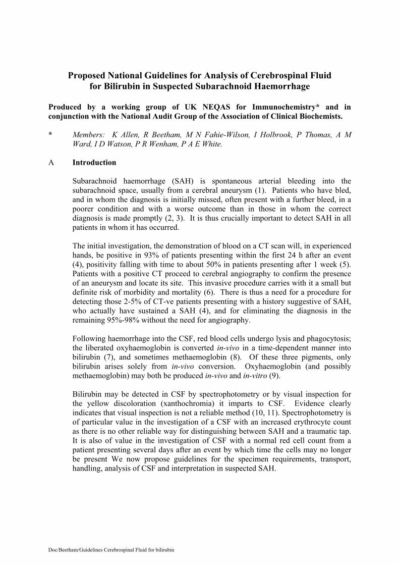
Proposed National Guidelines for Analysis of Cerebrospinal Fluid for Bilirubin in Suspected Subarachnoid Haemorrhage
Produced by a working group of UK NEQAS for Immunochemistry* and in conjunction with the National Audit Group of the Association of Clinical Biochemists. * Members: K Allen, R Beetham, M N Fahie-Wilson, I Holbrook, P Thomas, A M
Ward, I D Watson, P R Wenham, P A E White. A Introduction Subarachnoid haemorrhage (SAH) is spontaneous arterial bleeding into the
subarachnoid space, usually from a cerebral aneurysm (1). Patients who have bled, and in whom the diagnosis is initially missed, often present with a further bleed, in a poorer condition and with a worse outcome than in those in whom the correct diagnosis is made promptly (2, 3). It is thus crucially important to detect SAH in all patients in whom it has occurred.
The initial investigation, the demonstration of blood on a CT scan will, in experienced hands, be positive in 93% of patients presenting within the first 24 h after an event (4), positivity falling with time to about 50% in patients presenting after 1 week (5). Patients with a positive CT proceed to cerebral angiography to confirm the presence of an aneurysm and locate its site. This invasive procedure carries with it a small but definite risk of morbidity and mortality (6). There is thus a need for a procedure for detecting those 2-5% of CT-ve patients presenting with a history suggestive of SAH, who actually have sustained a SAH (4), and for eliminating the diagnosis in the remaining 95%-98% without the need for angiography.
Following haemorrhage into the CSF, red blood cells undergo lysis and phagocytosis; the liberated oxyhaemoglobin is converted in-vivo in a time-dependent manner into bilirubin (7), and sometimes methaemoglobin (8). Of these three pigments, only bilirubin arises solely from in-vivo conversion. Oxyhaemoglobin (and possibly methaemoglobin) may both be produced in-vivo and in-vitro (9).
Bilirubin may be detected in CSF by spectrophotometry or by visual inspection for the yellow discoloration (xanthochromia) it imparts to CSF. Evidence clearly indicates that visual inspection is not a reliable method (10, 11). Spectrophotometry is of particular value in the investigation of a CSF with an increased erythrocyte count as there is no other reliable way for distinguishing between SAH and a traumatic tap. It is also of value in the investigation of CSF with a normal red cell count from a patient presenting several days after an event by which time the cells may no longer be present We now propose guidelines for the specimen requirements, transport, handling, analysis of CSF and interpretation in suspected SAH.

Doc/Beetham/Guidelines Cerebrospinal Fluid for bilirubin
B Specimen requirements and transport A protocol for specimen requirements and transport is provided in Appendix 1
although modification may be required to meet local needs. Essentially, the requirements are:
Specimens must be collected sequentially even if no other investigations are
required. The specimen for spectrophotometry should always be the last fraction to be
taken, and ideally at least the fourth (Note a). The volume requested must be that which enables the analysis to be
undertaken without dilution (Note b). The specimen must be protected from light (Note c).
Pneumatic tube systems should not be used to transport the specimen to the
laboratory (12). A simultaneous blood specimen should be taken for serum bilirubin and total
protein measurement. Record the timing of sampling relative to that of possible haemorrhage. This
should be no less than 12h. It is advised that prospective protocols are discussed with users of the service.
C Specimen handling
The specimen designated for spectrophotometry should be centrifuged at >2000 rpm for 5 min as soon after receipt as possible and in any case within 1h of collection. The supernatant should be stored in the dark at 4 C until analysis (Note c).

Doc/Beetham/Guidelines Cerebrospinal Fluid for bilirubin
D Analysis
Perform a zero-order spectrophotometric scan on the supernatant between 360 and 600 nm using a recording spectrophotometer and a cuvette with a 1 cm path length. Use an initial full-scale deflection (FSD) of 0.1 absorbance units (AU). If any peaks exceed 0.1 AU, scale as appropriate but never use a FSD < 0.1 AU (note d).
The specimen should not be diluted.
Inspect the scan and identify and record the presence of the following haem pigments: Oxyhaemoglobin: absorbance maximum between 412 and 418 nm.
Bilirubin: either a broad peak in the range 450 to 460 nm or a shoulder
adjacent to an oxyhaemoglobin peak if present. Methaemoglobin: the rarest pigment and if present usually manifest as a
broader peak than oxyhaemoglobin occurring between 403 and 410 nm. If methaemoglobin is suspected, check that the wavelength maximum really is less than 410 nm by comparison with a scan of supernatant from lysed fresh red cells after centrifugation when the absorbance maximum should be between 413 and 416 nm. The presence of methaemoglobin is best confirmed by diluting the CSF with an equal volume of 100 mmol/L potassium cyanide. Any methaemoglobin should show a shift to 419 nm as methaemoglobin is converted to cyanmethaemoglobin.
Calculate the net bilirubin absorbance (NBA) according to Chalmers’
modification (13) to the original method of Chalmers and Kiley (14). (Appendix 2) (Note e).
Illustrative zero-order spectra are shown in Figures 1a-e.
E Reporting and Interpretation (i) NBA 0.007 AU and no oxyhaemoglobin present.
Report the calculated NBA obtained alongside reference range e.g. NBA 0.004 AU (reference range 0.000 to 0.007 AU). Where the NBA is 0.007 add the interpretative comment : - ‘Not consistent with SAH’ (Notes f, g).
(ii) The NBA is initially > 0.007 AU but correction for serum bilirubin (Equation
1, Appendix 2) gives a value 0.007.
Report the value given by equation 1 as follows: e.g. NBA (corrected for increased serum bilirubin) 0.004 AU (Reference Range 0.000 – 0. 007). Add the interpretive comment:- ‘Not consistent with SAH’.
Doc/Beetham/Guidelines Cerebrospinal Fluid for bilirubin
(iii) The NBA is >0.007 (even after correction for increased serum bilirubin) (Equation 1, Appendix 2) with a normal CSF protein concentration.
Report the NBA and the reference range and add the interpretative comment: - ‘Consistent with SAH or other source of CSF blood’ (Note h).
(iv) The NBA is > 0.007 AU and the CSF protein concentration is raised.
Report the NBA and the reference range together with the interpretive comment: - ‘Consistent with SAH, other source of CSF blood, or increased bilirubin accompanying increased CSF protein – interpret results with caution’(see Appendix 2).
(v) NBA 0.007 AU but methaemoglobin is present.
This will occur rarely. Report the NBA and the reference range. Add the interpretative comment: - ‘Methaemoglobin detected. Consistent with SAH or other source of CSF blood.’
(vi) Oxyhaemoglobin present but NBA 0.007 AU.
The detection of bilirubin in CSF is the cardinal finding which, subject to the conditions outlined above, supports a diagnosis of SAH. Oxyhaemoglobin may be present as a result of SAH or of in-vitro haemolysis of red cells introduced during the lumbar puncture. In the absence of bilirubin in an appropriately timed specimen its presence is most likely to be due to the latter. The finding of oxyhaemoglobin alone should therefore not be reported, but only the NBA as in (i) above (note g).
NB. When reporting on spectrophotometry, bear in mind (i)that a normal erythrocyte count in a CSF taken definitely between 12 and 72h after an event is evidence against a SAH; and (ii) that spectrophotometric findings on a CSF taken at a second or subsequent lumbar puncture some hours or more after the previous puncture only reflect the probability that blood has been introduced traumatically into the subarachnoid space at an earlier puncture.
F Decision tree A decision tree (Figure 2) outlines the steps involved in producing the key laboratory
information for the detection of an intra-cranial bleed.

Doc/Beetham/Guidelines Cerebrospinal Fluid for bilirubin
G Standards based on these guidelines
1. The laboratory should provide instructions for users which provide details of requesting, specimen requirements, transport and interpretation (see example in Appendix 1).
2. There should be in place SOPs for specimen handling, analysis, reporting and
interpretation. 3. The laboratory must participate in an appropriate external quality assurance
scheme. 4. It is unlikely that a laboratory will build up sufficient expertise unless a
minimum of 25 specimens are analysed annually. 5. The nature of the analytical service which a laboratory provides , e.g. whether
it is available only within certain hours or at all times, will be dependent upon local needs. In particular these will be determined by the Tertiary centre’s referral policy, access to its beds and availability of angiography. Both the analytical and interpretative aspects of the service should be provided together.
6. To meet the requirements of Clinical Governance all spectrophotometric scans
should be kept in an appropriate form for recall for a minimum 2 years. 7. Spectrophotometers should be serviced regularly and undergo regular
absorption and wavelength checks.
Acknowledgements The working group acknowledge with thanks the assistance of D O’Connell, H Gunawardena and P L M Lynch in the provision of data which has been used by the group. It also acknowledges the formulation of guidelines by other groups particularly the All Wales Audit Group and the Northern Ireland Audit Group.

Doc/Beetham/Guidelines Cerebrospinal Fluid for bilirubin
References 1. Vermeulen M, van Gijn J. The diagnosis of subarachnoid haemorrhage. J Neurol Neurosurg Psychiatry. 1990; 53:365-72. 2. Mayer P L., Awad I A, Todor R. Misdiagnosis of symptomatic cerebral
aneurysm; prevalence and correlation with outcome at four institutions. Stroke 1996; 27:1558-63.
3. Jacobsson K E, Saveland H, Hillman J, Edner G, Zygmunt S, Pellettieri L.
Warning leak and management outcome in aneurysmal subarachnoid haemorrhage. J Neurosurg 1996: 85:995-9.
4. van der Wee N, Rinkel G J, Hasan D, van Gijn J. Detection of subarachnoid
haemorrhage on early CT: is lumbar puncture still needed after a negative scan? J Neurol Neurosurg Psychiatry 1995; 58:357-9.
5. van Gijn J, van Dongen K J. The time course of aneurysmal haemorrhage on
computed tomograms. Neuroradiology 1982; 23:153-6. 6. Dion J E, Gates P C, Fox A J, Barnett H J M, Blom R J. Clinical events
following neuroangiography: a prospective study. Stroke 1987; 18:997-1004. 7. Fishman R A. Cerebral Fluid in Diseases of the Nervous System. London:
WB Saunders Co., 1980. 8. Wahlgren N G, Lindquist C. Haem derivates in the cerebrospinal fluid after
intracranial haemorrhage. Eur Neurol 1987; 26:216-21. 9. Fahie-Wilson M, Park D. Spectrophotometric examination of CSF in
suspected subarachnoid haemorrhage – what should we look for? In Martin S M, Halloran S P, eds. Proceedings of the XV1 International Congress of Clinical Chemistry, 1996 Jul 8-12, London. London: Association of Clinical Biochemists, 1996:85.
10. Kjellin K G, Söderström C E. Diagnostic significance of CSF
spectrophotometry in cerebrovascular diseases. J Neurol Sci 1974; 23:359-69. 11. Marden N A, Thomas P H, Stansbie D. Is the naked eye as sensitive as the
spectrophotometer for detecting xanthochromia in cerebrospinal fluid? In Martin S M, ed. Proceedings of the National Meeting, 2001 April 30-May 4, London. London: Association of Clinical Biochemists, 2001:53.
12. Wenham P R, Hanson T, Ashby J P. Interference in spectrophotometric
analysis of cerebrospinal fluid by haemolysis induced by transport through a pneumatic tube system. Ann Clin Biochem 2001; 38:371-5.
13. Chalmers A H. Cerebrospinal fluid xanthochromia testing simplified. Clin
Chem 2001; 47:147-8.
Doc/Beetham/Guidelines Cerebrospinal Fluid for bilirubin
14. Chalmers A H, Kiley M. Detection of xanthochromia in cerebrospinal fluid. Clin Chem 1998; 44:1740-2.
15. Kjellin K G. The binding of xanthochromic compounds in the cerebrospinal
fluid. J Neurol Sci 1969; 9:597-601. 16. Kronholm V, Lintrup J. Spectrophotometric investigations of the
cerebrospinal fluid in the near-ultraviolet region. Acta Psychiat Scand 1960; 35:314-29.
17. Wahlgren N G, Bergstrom K. Determination of haem derivatives in the
cerebrospinal fluid - a semi-quantitative method. J Neurol Neurosurg Psychiatry 1983; 653-8.
Doc/Beetham/Guidelines Cerebrospinal Fluid for bilirubin
Notes to the Guidelines (a) In addition to the oxyhaemoglobin which appears after a SAH, it also commonly
arises either from the in-vitro lysis of red cells in the CSF obtained following puncture, or from the trauma of the puncture itself. Such oxyhaemoglobin may interfere with the detection of bilirubin and, as explained in note (g) below, is a confounding element in interpretation. Therefore every effort should be made to eliminate it. It is for this reason that CSF taken for spectrophotometry should be collected into a separate container to those in which the first few mL of fluid are placed, and why transport by pneumatic tube is not recommended.
(b) As explained in note (h) even small increases above the reference range are sufficient
to be consistent with a SAH and therefore indicate the need for angiography. Dilution of the specimen will decrease the certainty with which such increases may be detected.
(c) Stability studies have shown that CSF stored in a plastic tube and exposed to spring
daylight through a north-facing window showed a bilirubin decay rate of at least 0.005 AU/h. CSF specimens must therefore be protected from light to avoid this phenomenon which may lead to false negative results
(d) Derivative spectroscopy has been found to be of value by some analysts, but requires considerable experience in interpretation. It is therefore not recommended.
(e) We have confirmed that, on 58 CSF specimens with NBA 0.003 – 0.251 (24 of which
contained oxyhaemoglobin in addition to bilirubin) there was no significant difference between the NBA obtained by the original Chalmers and Kiley method (14) and that by the modification of Chalmers (13).
(f) Out of 740 spectrophotometric scans reviewed from CT-ve patients in four
participating centres, 672 had a NBA 0.007. Angiograms were performed in 62 of these 672 patients and aneurysms were confirmed in 6 of them. Two of these presented 4 weeks or more after the event, two with unruptured aneurysms, and two with ruptured aneurysms. The odds of finding a ruptured aneurysm with a NBA 0.007 are thus 1 in 31 within 4 weeks of the event.
(g) We discourage the reporting of oxyhaemoglobin. From the same series, 204 CSFs
were reported as containing oxyhaemoglobin with NBA 0.007. 30 of these patients had angiography, and in only two instances was an aneurysm found. Oxyhaemoglobin thus has a low predictive value for SAH. However, we recognise that rarely, early on after a bleed, oxyhaemoglobin may be present without bilirubin.
(h) Originally Chalmers and Kiley (14) indicated a reference range for NBA of 0 to
0.007; values 0.010 – 0.014 were classed as equivocal and values > 0.014 as positive. In the series quoted above, CSFs from three patients with proven ruptured aneurysms have yielded NBA of 0.008, 0.015, 0.016. In addition we are aware of 3 CT+ve patients with proven aneurysms where the CSFs have yielded NBA of 0.008, 0.012, 0.019. We therefore recommend that values of a NBA > 0.007 are a clear indication for angiography. In the series quoted above, 29 patients with NBA > 0.007 proceeded to angiography of which 13 were found to have aneurysms.

Doc/Beetham/Guidelines Cerebrospinal Fluid for bilirubin
Appendix 1
Exemplar protocol for the collection, handling and transport to the laboratory of CSF requiring spectrophotometric scanning for the detection
of bilirubin
Principle This test is performed to try to identify those patients who have had a subarachnoid haemorrhage (SAH) but in whom the CT scan is negative. The spectrophotometric scan detects bilirubin in CSF and this finding is consistent with a bleed into the CSF. The formation of bilirubin after haemorrhage is a time-dependent process and bilirubin may not be detectable soon after the event (e.g. onset of severe headache). On current evidence it is recommended that CSF is not sampled until at least 12h after a suspected event. The opening pressure should always be recorded when performing a lumbar puncture. Please indicate on the request form
Clinical indication for request Result of CT scan Time of onset of symptoms/event Time of lumbar puncture If the differential diagnosis includes meningitis
Specimens CSF may also be required for microbiological examination and for protein and
glucose estimation. Sufficient CSF will therefore be needed for all of these required investigations.
Label three 28mL sterile universal containers and one yellow-top fluoride EDTA tube
each with the patient’s name, hospital number, ward, date of birth, time that the CSF was obtained and the sequence order of sampling.
The first specimen should be a minimum of 0.5mL of CSF placed in a yellow-top
fluoride EDTA tube for glucose and protein estimations. This specimen should be sent to Clinical Biochemistry.
Microbiology requires at least 5mL of CSF divided into 2 sequentially numbered
sterile 28mL universal containers labeled “second” and “third”. These 2 specimens must be delivered to the Microbiology Department as soon as possible. Do NOT use the pneumatic tube delivery system.
A further minimum of 1mL CSF should be placed in the final (labeled “fourth”)
sterile 28mL universal container for the spectrophotometric scan. (NB 1mL is about 20 drops from the Luer connector on a needle). Protect this sample from the light by placing it in a thick brown envelope outside the usual plastic specimen bag.
A blood specimen be should taken at the same time for serum bilirubin, total protein and glucose estimation that are needed to aid interpretation.
Doc/Beetham/Guidelines Cerebrospinal Fluid for bilirubin
These samples must also be delivered to the Clinical Biochemistry Department as soon as possible. Do NOT use the pneumatic tube delivery system. If this procedure is not followed analysis is likely to be compromised. Text in italics indicates those details subject to local requirements.

Doc/Beetham/Guidelines Cerebrospinal Fluid for bilirubin
Appendix 2
Spectrophotometric detection of bilirubin in CSF by spectrophotometry
From the spectrophotometric scan the calculate Net Bilirubin Absorbance (NBA) (13) as follows: 1. Draw a predicted baseline from 360 nm to a point where this will form a tangent to
the scan at about 510 – 530 nm. Measure the absorbance of the scan above this baseline at 476 nm which is the net bilirubin absorbance (NBA).
2. A NBA of 0.007 AU indicates no excess bilirubin in CSF. 3. A NBA of > 0.007 AU indicates excess bilirubin in the CSF which is consistent with
SAH or other source of CSF blood provided the serum bilirubin is less than 20 mmol/L and the CSF protein concentration is within the reference range. If either of these conditions does not pertain then proceed as follows:
(i) Serum bilirubin 20 mol/L. Calculate the corrected NBA in the CSF in the
following manner: Corrected NBA = NBA – predicted absorbance (PA)
………… (Equation 1) This takes into account the increase in CSF bilirubin absorbance which can be
attributed to the increased serum bilirubin. The PA is calculated (15,16,17)according to the following equation:
PA = CSF total protein (g/L) x serum bilirubin ( mol/L) x 0.042 AU. Serum total protein (g/L) ……………(Equation 2)
If the corrected NBA yields a value 0.007 then this indicates that the increased serum bilirubin accounts for the excess bilirubin in the CSF.
If the corrected NBA yields a value > 0.007, this indicates that some bilirubin
has originated from a SAH or other bleed into CSF. (ii) The CSF protein is increased. If the CSF protein is increased this may itself give rise to an increase in CSF
bilirubin. It may also indicate meningeal inflammation, which if severe may cause microhaemorrhages. Thus in the presence of an increased CSF protein caution should be exercised in the interpretation of a NBA >0.007 AU. In this instance, conditions other than SAH are more likely as a cause of increased NBA, particularly in the presence of an abnormal CSF white cell count.

Doc/Beetham/Guidelines Cerebrospinal Fluid for bilirubin
Figures 1a-e. Representative spectrophotometric scans showing NBA at 476 nm above a tangential baseline as described in the text. 1a. A normal CSF with essentially no bilirubin;scan and baseline (not drawn) are superimposable. 1b.A NBA within the reference range. 1c.Oxyhaemoglobin with zero NBA. 1d. Oxyhaemoglobin with NBA within the reference range. 1e. Oxyhaemoglobin with an increased NBA.
In practice such scans are best visualised filling the whole of an A4 page in landscape mode.

Doc/Beetham/Guidelines Cerebrospinal Fluid for bilirubin
Figure 2. Bilirubin absorbance in CSF for detection of intra-cranial bleed
CT
Angiography Lumbar puncture
Specimens 2, 3
Fluoride EDTA 1
Specimen 4 Serum sample
Microbiology
Clinical Biochemistry
Total Protein & Glucose
Spectro -photometry
Serum bilirubin
Oxy Hb
Chalmers calculation
Calculate serum bilirubin contribution to CSF (predicted absorbance)
Present Absent
No Met Hb
NEGATIVE
Inconclusive
POSITIVE
Angiography
+ve -ve
NBA – predictedabsorbance
NBA > 0.007
NBA <0.007
> 20 umol/L
NBA = Net Bilirubin Absorbance

Doc/Beetham/Guidelines Cerebrospinal Fluid for bilirubin

Doc/Beetham/Guidelines Cerebrospinal Fluid for bilirubin

Doc/Beetham/Guidelines Cerebrospinal Fluid for bilirubin