Embed Size (px)
Citation preview
Biochemical and genetic markers after subarachnoid
haemorrhage
Ludvig Csajbok
Department of Anaesthesiology and Intensive Care, Institute of Clinical Sciences, Sahlgrenska Academy
at University of Gothenburg, Göteborg, Sweden
Gothenburg 2015
Ludvig Zoltán Csajbók

Cover illustration: Subarachnoid haemorrhage on CT scan, with a giant aneurysm by courtesy of Dr. Hironao Yuzawa, Tohoku University Hospital, Sendai, Japan.
Biochemical and genetic markers after subarachnoid haemorrhage © Ludvig Csajbok 2015 [email protected]
ISBN 978-91-628-9554-9 http://hdl.handle.net/2077/39549
Printed in Bohus, Sweden 2015 Ale Tryckteam AB, Bohus Papers I, II, and III are reprinted with permission from Group BMJ Publishing and Wiley&sons Publishing Ltd

“A ship rests safely in harbour, but it is not what ships are built for.” William G.T. Shedd
To my father, who inspired me, to my mother, who made it all possible
and to my family, who made it all worthwhile

I
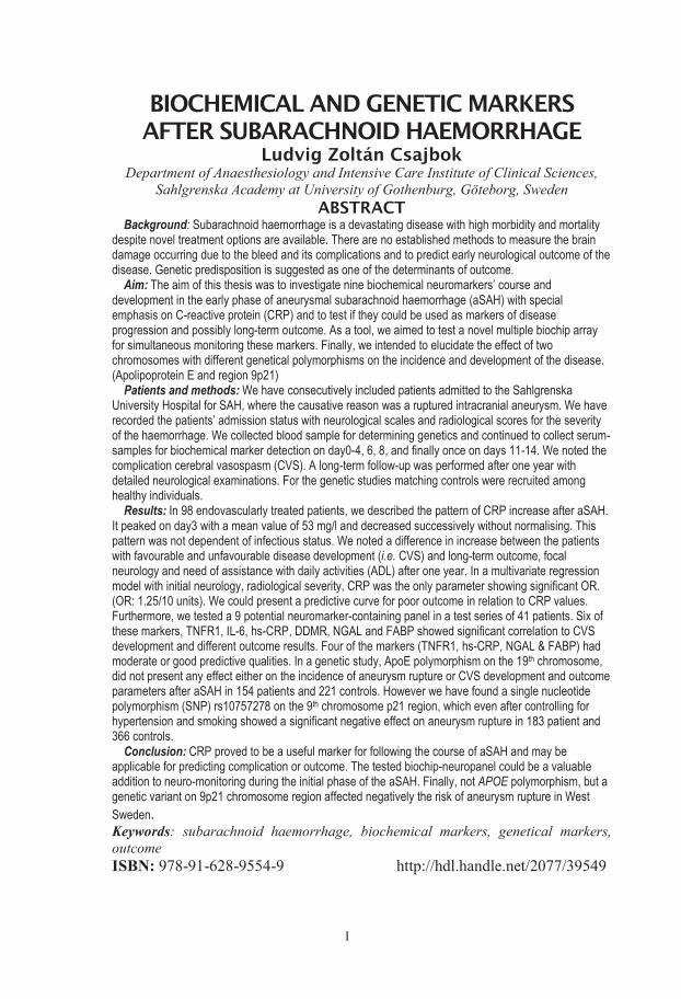
BIOCHEMICAL AND GENETIC MARKERS AFTER SUBARACHNOID HAEMORRHAGE
Ludvig Zoltán Csajbok Department of Anaesthesiology and Intensive Care Institute of Clinical Sciences,
Sahlgrenska Academy at University of Gothenburg, Göteborg, Sweden ABSTRACT
Background: Subarachnoid haemorrhage is a devastating disease with high morbidity and mortality despite novel treatment options are available. There are no established methods to measure the brain damage occurring due to the bleed and its complications and to predict early neurological outcome of the disease. Genetic predisposition is suggested as one of the determinants of outcome. Aim: The aim of this thesis was to investigate nine biochemical neuromarkers’ course and development in the early phase of aneurysmal subarachnoid haemorrhage (aSAH) with special emphasis on C-reactive protein (CRP) and to test if they could be used as markers of disease progression and possibly long-term outcome. As a tool, we aimed to test a novel multiple biochip array for simultaneous monitoring these markers. Finally, we intended to elucidate the effect of two chromosomes with different genetical polymorphisms on the incidence and development of the disease. (Apolipoprotein E and region 9p21) Patients and methods: We have consecutively included patients admitted to the Sahlgrenska University Hospital for SAH, where the causative reason was a ruptured intracranial aneurysm. We have recorded the patients’ admission status with neurological scales and radiological scores for the severity of the haemorrhage. We collected blood sample for determining genetics and continued to collect serum-samples for biochemical marker detection on day0-4, 6, 8, and finally once on days 11-14. We noted the complication cerebral vasospasm (CVS). A long-term follow-up was performed after one year with detailed neurological examinations. For the genetic studies matching controls were recruited among healthy individuals. Results: In 98 endovascularly treated patients, we described the pattern of CRP increase after aSAH. It peaked on day3 with a mean value of 53 mg/l and decreased successively without normalising. This pattern was not dependent of infectious status. We noted a difference in increase between the patients with favourable and unfavourable disease development (i.e. CVS) and long-term outcome, focal neurology and need of assistance with daily activities (ADL) after one year. In a multivariate regression model with initial neurology, radiological severity, CRP was the only parameter showing significant OR. (OR: 1.25/10 units). We could present a predictive curve for poor outcome in relation to CRP values. Furthermore, we tested a 9 potential neuromarker-containing panel in a test series of 41 patients. Six of these markers, TNFR1, IL-6, hs-CRP, DDMR, NGAL and FABP showed significant correlation to CVS development and different outcome results. Four of the markers (TNFR1, hs-CRP, NGAL & FABP) had moderate or good predictive qualities. In a genetic study, ApoE polymorphism on the 19th chromosome, did not present any effect either on the incidence of aneurysm rupture or CVS development and outcome parameters after aSAH in 154 patients and 221 controls. However we have found a single nucleotide polymorphism (SNP) rs10757278 on the 9th chromosome p21 region, which even after controlling for hypertension and smoking showed a significant negative effect on aneurysm rupture in 183 patient and 366 controls. Conclusion: CRP proved to be a useful marker for following the course of aSAH and may be applicable for predicting complication or outcome. The tested biochip-neuropanel could be a valuable addition to neuro-monitoring during the initial phase of the aSAH. Finally, not APOE polymorphism, but a genetic variant on 9p21 chromosome region affected negatively the risk of aneurysm rupture in West Sweden. Keywords: subarachnoid haemorrhage, biochemical markers, genetical markers, outcome ISBN: 978-91-628-9554-9 http://hdl.handle.net/2077/39549
II
LIST OF PAPERS
This thesis is based on the following papers, which will be referred to in the text by their Roman numerals
I. Csajbok, L. Z., Nylen, K., Ost, M., Sonander, H., & Nellgard, B. (2015).
In-hospital C-reactive protein predicts outcome after aneurysmal subarachnoid haemorrhage treated by endovascular coiling. Acta Anaesthesiol Scand, 59(2), 255-264. doi: 10.1111/aas.12441
II. Csajbok, L. Z., Nylen, K., Ost, M., Blennow, K., Zetterberg, H., Nellgard, P., & Nellgard, B. (2015). Apolipoprotein E polymorphism in aneurysmal subarachnoid haemorrhage in West-Sweden. Acta Neurol Scand, Epub. Ahaed of publication. doi: 10.1111/ane.12487
III.Olsson, S., Csajbok, L. Z., Jood, K., Nylen, K., Nellgard, B., & Jern, C. (2011). Association between genetic variation on chromosome 9p21 and aneurysmal subarachnoid haemorrhage. J Neurol Neurosurg Psychiatry, 82(4), 384-388. doi: 10.1136/jnnp.2009.187427
IV. Csajbok, L. Z., Nylen, K., Ost, M., Blennow, K., Zetterberg, H., Nellgard, P., & Nellgard, B. (2015). Biochip Neuromarker Array, a possible monitoring and prognostic tool after subarachnoid hemorrhage. Manuscript.
III
POPULÄRVETENSKAPLIG SAMMANFATTNING
Biokemiska och genetiska markörer efter subarachnoidalblödning
Subarachnoidalblödning är en typ av hjärnblödning, som förekommer mellan hjärnan och spindelnäthinnan, utgående ifrån ett brustet pulsåder-bråck på skallbotten och är ett förödande sjukdomstillstånd. Cirka en tredjedel av patienter, som drabbas dör, en tredjedel ådrar dig svåra neurologiska men och en tredjedel återhämtar sig så, att de kan ta hand om sig själva. Det existerar inga metoder att mäta den hjärnskada som blödningen och dess svåra komplikationer åstadkommer samt vi kan inte prediktera sjukdomsförloppet i ett tidigt skede. Man har dock visat, att genetiska faktorer påverkar hur hjärnan återhämtar sig efter sjukdomen.
Ett kroppseget äggvite-ämne, C-reaktiv protein, som hittills använts för att följa infektion i kroppen, kunde vi påvisa att koncentrationerna i blodet av denna följer kroppens hjärnblödningsförlopp tidigt under sjukdomen. Hos hundra blödningsdrabbade patienter mätte vi variationer i nivån av detta protein och observerade olika förlopp mellan de komplikationsdrabbade patienterna med dålig prognos samt de som återhämtade sig bra efter sjukdomen. Utifrån denna skillnad, kunde vi beräkna en prognostisk modell, som så tidigt som två dagar efter blödningen, kunde förutse hur stor risk patienten hade för en dålig sjukdomsprognos ett år efter insjuknandet. Denna prognostiska egenskap var oberoende om patienten blev infekterad eller inte under vårdförloppet.
Vi har dessutom testat en ny biochip styrd mätmetod för 9 olika små proteiner, som man samtidigt kunde analysera från en droppe blod i denna hjärnblödningsdrabbade patientgrupp. Vi ville testa om dessa markörer kunde komplettera eller ersätta de andra mycket farligare mätmetoderna (kateter-tryckmätning i hjärnan, upprepade röntgenkontroller) som används för att förutspå komplikationer och sjukdomsprognos efter genomgången sjukdom. Vi kunde konstatera att sex av dessa ämnen visade en nära korrelation till den långsiktiga sjukdomsprognosen efter sjukdomen och 4 av dessa kunde med

IV
en stor säkerhet förutspå denna. Dessa fynd bereder plats för ett införande av denna undersökningsmetod på sjukhuset.
Härutöver har vi testat två genetiska variationer på två olika kromosomer och tittat på deras effekter på bristning av pulsåderbråck i hjärnans kärl och komplikation samt sjukdomsprognos efter spindelvävshinneblödning. Den ena variation (Apolipoprotein E), som är känd för sin negativa påverkan på sjukdomsförloppet vid traumatiska hjärnskador och hjärnpropp (stroke) samt vid Alzheimers sjukdom, visade dock ingen effekt på denna typ av blödning. Den andra genetiska variationen, på den 9:e kromosomens p21 region visade däremot en statistiskt säkerställd negativ effekt på kärlens bristningstendens. Dessa studier hjälper oss att bättre förstå de ärftliga faktorer, som påverkar hur våra hjärnor hanterar dessa blödningar.
ABBREVIATIONS
ADL Activity of daily living H&H Hunt and Hess score ANRIL Antisense non-coding RNA in INK4
locus HDL High density lipoprotein
ApoE Apolipoprotein E protein HS Haemorrhagic stroke APOE Human gene coding apolipoprotein E ICP Intracranial pressure aSAH Aneurysmal subarachnoidal
haemorrhage ICU Intensive care unit
AUC Area under the curve IL-6 Interleukin 6 Aβ Amyloid beta ISAT International subarachnoid aneurysm
trial BBB Blood brain barrier LP Lumbar puncture BDNF Brain derived neutrophic factor LR Likelihood reaction BMN Biochemical neuromarker LTα Lymphotoxin alfa CAD Coronary artery disease MABP Mean arterial blood pressure CDKN2B Cyclin dependent kinase inhibitor 2B MAF Minor allele frequency CI Cerebral infarction MRA Magnetic resonance imaging
angiography CNS Cerebrospinal fluid MRI Magnetic resonance imaging CNS Central nervous system NCS Non-convulsive seizures COPD Chronic obstructive pulmonary disease NGAL Neutrophil gelatinase associated
lipocalin CPP Cerebral perfusion pressure NGF Nerve growth factor CRP C-reactive protein NIHSS National institute of health stroke scale CSF Cerebrospinal fluid NIVA Neurointensivvårds avdelning CT Computed tomography NSE Neuron specific enolase CTA Computed tomography angiography OR Odds ratio CVD Cardiovasular disease PE Pulmonary embolism CVS Cerebral vasospasm PNS Peripheral nervous system CVS Cerebral vasospasm POX Pulse-oximetry DCI Delayed cerebral ischemia PPV Positive predictive value DDMR D-dimer RLS85 Reaction level scale DIC Disseminated intravasal coagulation RNA Ribonucleic acid DIND Delayed ischemic neurological deficit ROC Receiver operator characteristic curve DNA Deoxyribonucleic acid SAH Subarachnoidal haemorrhage DSA Digital subtraktion angiography SEM Standard error of the mean DVT Deep venous thrombosis sIL-6R Soluble interleukin 6 receptor ELISA Enzyme linked immunoassay SIRS Systemic inflammatory reaction
syndrome EOS Early onset seizures SNP Single nucleotide polymorphism FABP FAtty acid binding protein TCD Transcranial doppler FDP Fibrin degradation protein TGFβ Tumour growth factor beta GCS Glasgow Coma Scale TNFR1 Tumour necrosis factor receptor 1 GFAP Glial fibrillary acidic protein TNFα Tumour necrosis factor alfa GFAP Glial fibrillary acidic protein WFNS World Federation of Neurological
Surgeons scale GOS Glasgow outcome scale GOSE Glasgow outcome scale extended GWAS Genome wide association study

V
ABBREVIATIONS
ADL Activity of daily living H&H Hunt and Hess score ANRIL Antisense non-coding RNA in INK4
locus HDL High density lipoprotein
ApoE Apolipoprotein E protein HS Haemorrhagic stroke APOE Human gene coding apolipoprotein E ICP Intracranial pressure aSAH Aneurysmal subarachnoidal
haemorrhage ICU Intensive care unit
AUC Area under the curve IL-6 Interleukin 6 Aβ Amyloid beta ISAT International subarachnoid aneurysm
trial BBB Blood brain barrier LP Lumbar puncture BDNF Brain derived neutrophic factor LR Likelihood reaction BMN Biochemical neuromarker LTα Lymphotoxin alfa CAD Coronary artery disease MABP Mean arterial blood pressure CDKN2B Cyclin dependent kinase inhibitor 2B MAF Minor allele frequency CI Cerebral infarction MRA Magnetic resonance imaging
angiography CNS Cerebrospinal fluid MRI Magnetic resonance imaging CNS Central nervous system NCS Non-convulsive seizures COPD Chronic obstructive pulmonary disease NGAL Neutrophil gelatinase associated
lipocalin CPP Cerebral perfusion pressure NGF Nerve growth factor CRP C-reactive protein NIHSS National institute of health stroke scale CSF Cerebrospinal fluid NIVA Neurointensivvårds avdelning CT Computed tomography NSE Neuron specific enolase CTA Computed tomography angiography OR Odds ratio CVD Cardiovasular disease PE Pulmonary embolism CVS Cerebral vasospasm PNS Peripheral nervous system CVS Cerebral vasospasm POX Pulse-oximetry DCI Delayed cerebral ischemia PPV Positive predictive value DDMR D-dimer RLS85 Reaction level scale DIC Disseminated intravasal coagulation RNA Ribonucleic acid DIND Delayed ischemic neurological deficit ROC Receiver operator characteristic curve DNA Deoxyribonucleic acid SAH Subarachnoidal haemorrhage DSA Digital subtraktion angiography SEM Standard error of the mean DVT Deep venous thrombosis sIL-6R Soluble interleukin 6 receptor ELISA Enzyme linked immunoassay SIRS Systemic inflammatory reaction
syndrome EOS Early onset seizures SNP Single nucleotide polymorphism FABP FAtty acid binding protein TCD Transcranial doppler FDP Fibrin degradation protein TGFβ Tumour growth factor beta GCS Glasgow Coma Scale TNFR1 Tumour necrosis factor receptor 1 GFAP Glial fibrillary acidic protein TNFα Tumour necrosis factor alfa GFAP Glial fibrillary acidic protein WFNS World Federation of Neurological
Surgeons scale GOS Glasgow outcome scale GOSE Glasgow outcome scale extended GWAS Genome wide association study
VI1
INTRODUCTION
I. Background
Aneurysmal subarachnoid haemorrhage
(aSAH) is a devastating neurological
emergency leaving “one third of the affected
patients dead, one third with severe handicap
and merely one-third with a good recovery”
according to the 1950’s well-known Swedish
pioneers of neurosurgery Gösta Norlén and
Herbert Olivecrona (Norlen and Olivecrona,
1953). This particular entity of haemorrhagic
stroke has been studied in thousands of
scientific studies and experiments from
Walton’s prognostic description (Walton,
1952) – 15% mortality in the first 24 h, 12%
after 1 week, 14% after 2 weeks and 11%
after 4 weeks, giving a cumulative
percentage of 32% mortality in the first
month – until Magni’s study, fairly recently
describing a 6 month mortality of 34%
(Magni et al., 2015). The overall outcome
figures remain surprisingly unchanged
throughout the years. From a sceptic’s point
of view, no progress has been achieved
during the work of two generations. From the
optimist’s point of view however, we have
come a long way and in the fields of
diagnostics, treatment, care and rehabilitation
the subarachnoid haemorrhage patients
receive an entirely different attention
compared to 60 years ago. The truth lies
nevertheless somewhere in between.
Although computed tomographic- (CTA),
magnetic resonance imaging- (MRA) and
digital subtractions-angiography (DSA) are
available for diagnostics, micro-neurosurgery
with titanium aneurysm-clips and
interventional neuroradiology with titanium
coils, and titanium-alloy stents are used in
therapy approaches, patients are cared for
and monitored in specialised neuro-intensive
care units and after acute ward, recovery is
undertaken in neuro-rehabilitation centres,
the outcome after aSAH is still bleak. One
could argue, that with better and more
efficient ambulance service more patients
survive the initial ictus and reach hospital in
a worse condition (worse initial neurology),
the patients are older and more affected by
co-morbidity, and reach therapy centres with
intra-ventricular haematomas, which was
impossible earlier (Naval et al., 2013). If the
outcome-study is controlled for all these
parameters, then the outcome after aSAH,
has indeed improved (Macdonald, 2013;
Grunwald et al., 2014; Naval et al., 2013).
The incidence of aSAH in the world is
around 9 of 100,000 individuals, but it has a
considerable geographical (Steiner et al.,
2013) and socio-economical (Jakovljevic et
al., 2001) variation. In Finland and Japan,
incidence over 20/100,000 were reported.
CONTENT ABSTRACT ........................................................................................................ I LIST OF PAPERS ................................................................................................ II SUMMARY IN SWEDISH .................................................................................. III ABBREVIATIONS .............................................................................................. V
CONTENT ......................................................................................................... VI
INTRODUCTION ................................................................................................ 1
I. Background ........................................................................................ 1 A. Mechanism .................................................................................... 3 B. Diagnosis ....................................................................................... 4 C. Treatment ....................................................................................... 7 D. Complications .............................................................................. 10
II. Neurological and radiological assessment . .................................... 11 A. Admission assessment ................................................................. 11 B. Outcome assessment .................................................................... 14 C. Physiological parameters ............................................................. 17
III. Genetic neuromarkers ................................................................... 17 IV. Biochemical neuromarkers ........................................................... 19
AIMS .............................................................................................................. 26
PATIENTS AND METHODS .............................................................................. 27
I. Inclusion .......................................................................................... 27 II. Regime............................................................................................ 27 III. Data collection and analysis .......................................................... 29 IV. Clinical and radiological assessment ............................................ 32 V. Statistics ......................................................................................... 33
RESULTS ........................................................................................................ 35
I. Biochemical neuromarkers .............................................................. 35 II. Genetic neuromarkers ..................................................................... 44
DISCUSSION ................................................................................................... 47
I. General considerations ..................................................................... 47 II. Patient considerations ..................................................................... 48 III. Methodological considerations ..................................................... 50 IV. Classification considerations ........................................................ 52 V. Remarks on genetic markers .......................................................... 55 VI. Remarks on biochemical markers ................................................. 57
CONCLUSION ................................................................................................. 63
FUTURE PERSPECTIVES .................................................................................. 64
ACKNOWLEDGEMENT .................................................................................... 65
REFERENCES .................................................................................................. 67
PAPER I. PAPER II. PAPER III. PAPER IV.
11
INTRODUCTION
I. Background
Aneurysmal subarachnoid haemorrhage
(aSAH) is a devastating neurological
emergency leaving “one third of the affected
patients dead, one third with severe handicap
and merely one-third with a good recovery”
according to the 1950’s well-known Swedish
pioneers of neurosurgery Gösta Norlén and
Herbert Olivecrona (Norlen and Olivecrona,
1953). This particular entity of haemorrhagic
stroke has been studied in thousands of
scientific studies and experiments from
Walton’s prognostic description (Walton,
1952) – 15% mortality in the first 24 h, 12%
after 1 week, 14% after 2 weeks and 11%
after 4 weeks, giving a cumulative
percentage of 32% mortality in the first
month – until Magni’s study, fairly recently
describing a 6 month mortality of 34%
(Magni et al., 2015). The overall outcome
figures remain surprisingly unchanged
throughout the years. From a sceptic’s point
of view, no progress has been achieved
during the work of two generations. From the
optimist’s point of view however, we have
come a long way and in the fields of
diagnostics, treatment, care and rehabilitation
the subarachnoid haemorrhage patients
receive an entirely different attention
compared to 60 years ago. The truth lies
nevertheless somewhere in between.
Although computed tomographic- (CTA),
magnetic resonance imaging- (MRA) and
digital subtractions-angiography (DSA) are
available for diagnostics, micro-neurosurgery
with titanium aneurysm-clips and
interventional neuroradiology with titanium
coils, and titanium-alloy stents are used in
therapy approaches, patients are cared for
and monitored in specialised neuro-intensive
care units and after acute ward, recovery is
undertaken in neuro-rehabilitation centres,
the outcome after aSAH is still bleak. One
could argue, that with better and more
efficient ambulance service more patients
survive the initial ictus and reach hospital in
a worse condition (worse initial neurology),
the patients are older and more affected by
co-morbidity, and reach therapy centres with
intra-ventricular haematomas, which was
impossible earlier (Naval et al., 2013). If the
outcome-study is controlled for all these
parameters, then the outcome after aSAH,
has indeed improved (Macdonald, 2013;
Grunwald et al., 2014; Naval et al., 2013).
The incidence of aSAH in the world is
around 9 of 100,000 individuals, but it has a
considerable geographical (Steiner et al.,
2013) and socio-economical (Jakovljevic et
al., 2001) variation. In Finland and Japan,
incidence over 20/100,000 were reported.
CONTENT ABSTRACT ........................................................................................................ I LIST OF PAPERS ................................................................................................ II SUMMARY IN SWEDISH .................................................................................. III ABBREVIATIONS .............................................................................................. V
CONTENT ......................................................................................................... VI
INTRODUCTION ................................................................................................ 1
I. Background ........................................................................................ 1 A. Mechanism .................................................................................... 3 B. Diagnosis ....................................................................................... 4 C. Treatment ....................................................................................... 7 D. Complications .............................................................................. 10
II. Neurological and radiological assessment . .................................... 11 A. Admission assessment ................................................................. 11 B. Outcome assessment .................................................................... 14 C. Physiological parameters ............................................................. 17
III. Genetic neuromarkers ................................................................... 17 IV. Biochemical neuromarkers ........................................................... 19
AIMS .............................................................................................................. 26
PATIENTS AND METHODS .............................................................................. 27
I. Inclusion .......................................................................................... 27 II. Regime............................................................................................ 27 III. Data collection and analysis .......................................................... 29 IV. Clinical and radiological assessment ............................................ 32 V. Statistics ......................................................................................... 33
RESULTS ........................................................................................................ 35
I. Biochemical neuromarkers .............................................................. 35 II. Genetic neuromarkers ..................................................................... 44
DISCUSSION ................................................................................................... 47
I. General considerations ..................................................................... 47 II. Patient considerations ..................................................................... 48 III. Methodological considerations ..................................................... 50 IV. Classification considerations ........................................................ 52 V. Remarks on genetic markers .......................................................... 55 VI. Remarks on biochemical markers ................................................. 57
CONCLUSION ................................................................................................. 63
FUTURE PERSPECTIVES .................................................................................. 64
ACKNOWLEDGEMENT .................................................................................... 65
REFERENCES .................................................................................................. 67
PAPER I. PAPER II. PAPER III. PAPER IV.
23
productivity loss and informal care (Joo et
al., 2014).
A. Mechanism
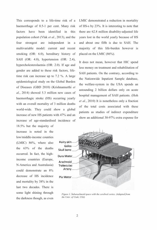
Subarachnoid haemorrhage is a result of a
bleeding from a blood-vessel within the
subarachnoid space (Fig 1). The source of the
bleeding can be traumatic, from around the
injured brain parenchyma (contusion-leak
from parenchymal capillaries), venous e.g.
from the subarachnoid venous network
often described as, bridging-veins or
perimesencephalic/prepontin bleed,
localised to those basal cisterns with
possible extension to the suprasellar
cistern (Schievink et al., 1994) or
arterial from small subarachnoid arteries
(malign hypertension bleed) frequently
with a parenchymal component. Most
often, however (ca. 85%) the
subarachnoid haemorrhage originates
from the large arteries on the base of the
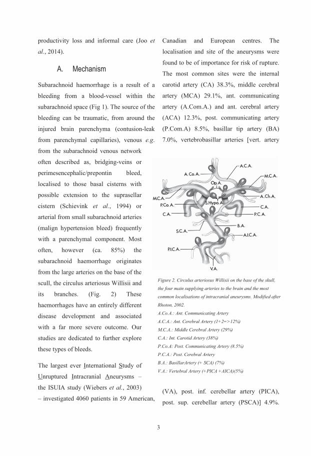
scull, the circulus arteriosus Willisii and
its branches. (Fig. 2) These
haemorrhages have an entirely different
disease development and associated
with a far more severe outcome. Our
studies are dedicated to further explore
these types of bleeds.
The largest ever International Study of
Unruptured Intracranial Aneurysms –
the ISUIA study (Wiebers et al., 2003)
– investigated 4060 patients in 59 American,
Canadian and European centres. The
localisation and site of the aneurysms were
found to be of importance for risk of rupture.
The most common sites were the internal
carotid artery (CA) 38.3%, middle cerebral
artery (MCA) 29.1%, ant. communicating
artery (A.Com.A.) and ant. cerebral artery
(ACA) 12.3%, post. communicating artery
(P.Com.A) 8.5%, basillar tip artery (BA)
7.0%, vertebrobasillar arteries [vert. artery
(VA), post. inf. cerebellar artery (PICA),
post. sup. cerebellar artery (PSCA)] 4.9%.
Figure 2. Circulus arteriosus Willisii on the base of the skull,
the four main supplying arteries to the brain and the most
common localisations of intracranial aneurysms. Modified after
Rhoton, 2002.
A.Co.A.: Ant. Communicating Artery
A.C.A.: Ant. Cerebral Artery (1+2=>12%)
M.C.A.: Middle Cerebral Artery (29%)
C.A.: Int. Carotid Artery (38%)
P.Co.A: Post. Communicating Artery (8.5%)
P.C.A.: Post. Cerebral Artery
B.A.: BasillarArtery (+ SCA) (7%)
V.A.: Vertebral Artery (+PICA +AICA)(5%)
2
This corresponds to a life-time risk of a
haemorrhage of 0.5-1 per cent. Many risk
factors have been identified in this
population cohort (Vlak et al., 2013), and the
four strongest are independent in a
multivariable model: current and recent
smoking (OR: 6.0), hereditary history of
SAH (OR: 4.0), hypertension (OR: 2.4),
hypercholesterolaemia (OR: 2.0). If age and
gender are added to these risk factors, life-
time risk can increase up to 7.2 %. A large
epidemiological study on the Global Burden
of Diseases (GBD 2010) (Krishnamurthi et
al., 2014) showed 5.3 million new cases of
haemorrhagic stroke (HS) occurring yearly
with an overall mortality of 3 million deaths
world-wide. They could show a global
increase of new HS patients with 47% and an
increase of age-standardised incidence of
18.5% but the majority of
increase is noted in the
low/middle-income counties
(LMIC) 86%, where also
the 63% of the deaths
occurred. In fact, the high-
income countries (Europe,
N-America and Australasia)
could demonstrate an 8%
decrease of HS incidence
and mortality by 38% in the
last two decades. There is
some light shining through
the darkness though, as even
LMIC demonstrated a reduction in mortality
of HS-s by 23%. It is interesting to note that
there are 62.8 million disability-adjusted life
years lost in the world yearly because of HS
and about one fifth is due to SAH. The
majority of this life-burden however is
placed on the LMIC (86%).
It does not mean, however that HIC spend
less money on treatment and rehabilitation of
SAH patients. On the contrary, according to
the Nationwide Inpatient Sample database,
the welfare-system in the USA spends an
astounding 2 billion dollars only on acute
hospital management of SAH patients. (Hoh
et al., 2010) It is nonetheless only a fraction
of the total costs associated with these
patients as studies of indirect expenditure
show an additional 30-97% extra expense for
Figure 1. Subarachnoid space with the cerebral cortex. (Adapted from the Univ. of Utah, USA)
33
productivity loss and informal care (Joo et
al., 2014).
A. Mechanism
Subarachnoid haemorrhage is a result of a
bleeding from a blood-vessel within the
subarachnoid space (Fig 1). The source of the
bleeding can be traumatic, from around the
injured brain parenchyma (contusion-leak
from parenchymal capillaries), venous e.g.
from the subarachnoid venous network
often described as, bridging-veins or
perimesencephalic/prepontin bleed,
localised to those basal cisterns with
possible extension to the suprasellar
cistern (Schievink et al., 1994) or
arterial from small subarachnoid arteries
(malign hypertension bleed) frequently
with a parenchymal component. Most
often, however (ca. 85%) the
subarachnoid haemorrhage originates
from the large arteries on the base of the
scull, the circulus arteriosus Willisii and
its branches. (Fig. 2) These
haemorrhages have an entirely different
disease development and associated
with a far more severe outcome. Our
studies are dedicated to further explore
these types of bleeds.
The largest ever International Study of
Unruptured Intracranial Aneurysms –
the ISUIA study (Wiebers et al., 2003)
– investigated 4060 patients in 59 American,
Canadian and European centres. The
localisation and site of the aneurysms were
found to be of importance for risk of rupture.
The most common sites were the internal
carotid artery (CA) 38.3%, middle cerebral
artery (MCA) 29.1%, ant. communicating
artery (A.Com.A.) and ant. cerebral artery
(ACA) 12.3%, post. communicating artery
(P.Com.A) 8.5%, basillar tip artery (BA)
7.0%, vertebrobasillar arteries [vert. artery
(VA), post. inf. cerebellar artery (PICA),
post. sup. cerebellar artery (PSCA)] 4.9%.
Figure 2. Circulus arteriosus Willisii on the base of the skull,
the four main supplying arteries to the brain and the most
common localisations of intracranial aneurysms. Modified after
Rhoton, 2002.
A.Co.A.: Ant. Communicating Artery
A.C.A.: Ant. Cerebral Artery (1+2=>12%)
M.C.A.: Middle Cerebral Artery (29%)
C.A.: Int. Carotid Artery (38%)
P.Co.A: Post. Communicating Artery (8.5%)
P.C.A.: Post. Cerebral Artery
B.A.: BasillarArtery (+ SCA) (7%)
V.A.: Vertebral Artery (+PICA +AICA)(5%)
2
This corresponds to a life-time risk of a
haemorrhage of 0.5-1 per cent. Many risk
factors have been identified in this
population cohort (Vlak et al., 2013), and the
four strongest are independent in a
multivariable model: current and recent
smoking (OR: 6.0), hereditary history of
SAH (OR: 4.0), hypertension (OR: 2.4),
hypercholesterolaemia (OR: 2.0). If age and
gender are added to these risk factors, life-
time risk can increase up to 7.2 %. A large
epidemiological study on the Global Burden
of Diseases (GBD 2010) (Krishnamurthi et
al., 2014) showed 5.3 million new cases of
haemorrhagic stroke (HS) occurring yearly
with an overall mortality of 3 million deaths
world-wide. They could show a global
increase of new HS patients with 47% and an
increase of age-standardised incidence of
18.5% but the majority of
increase is noted in the
low/middle-income counties
(LMIC) 86%, where also
the 63% of the deaths
occurred. In fact, the high-
income countries (Europe,
N-America and Australasia)
could demonstrate an 8%
decrease of HS incidence
and mortality by 38% in the
last two decades. There is
some light shining through
the darkness though, as even
LMIC demonstrated a reduction in mortality
of HS-s by 23%. It is interesting to note that
there are 62.8 million disability-adjusted life
years lost in the world yearly because of HS
and about one fifth is due to SAH. The
majority of this life-burden however is
placed on the LMIC (86%).
It does not mean, however that HIC spend
less money on treatment and rehabilitation of
SAH patients. On the contrary, according to
the Nationwide Inpatient Sample database,
the welfare-system in the USA spends an
astounding 2 billion dollars only on acute
hospital management of SAH patients. (Hoh
et al., 2010) It is nonetheless only a fraction
of the total costs associated with these
patients as studies of indirect expenditure
show an additional 30-97% extra expense for
Figure 1. Subarachnoid space with the cerebral cortex. (Adapted from the Univ. of Utah, USA)
45
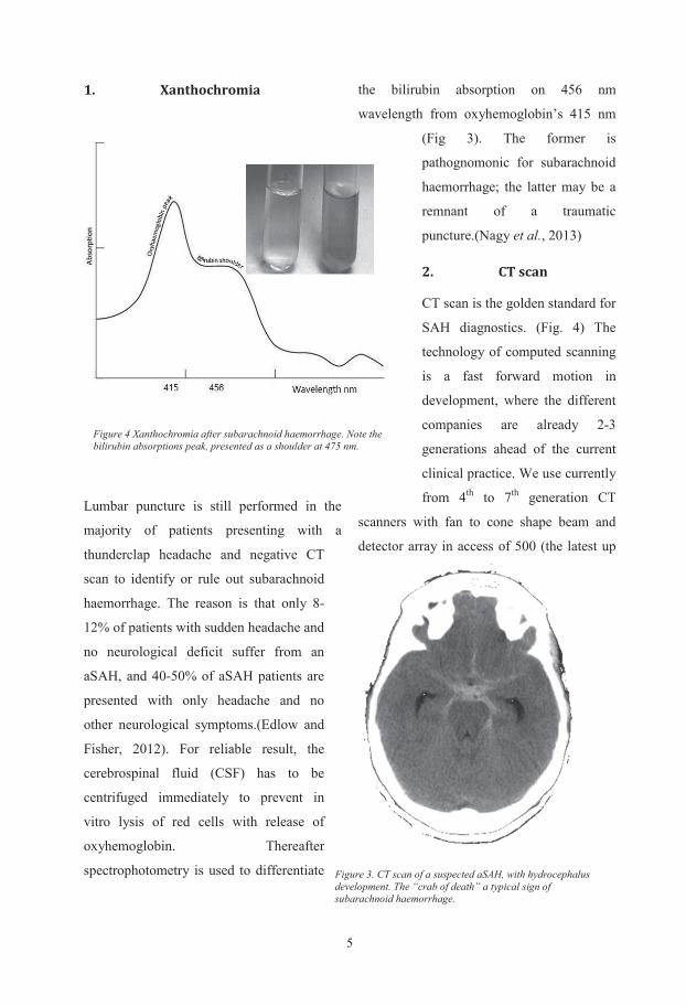
1. Xanthochromia
Lumbar puncture is still performed in the
majority of patients presenting with a
thunderclap headache and negative CT
scan to identify or rule out subarachnoid
haemorrhage. The reason is that only 8-
12% of patients with sudden headache and
no neurological deficit suffer from an
aSAH, and 40-50% of aSAH patients are
presented with only headache and no
other neurological symptoms.(Edlow and
Fisher, 2012). For reliable result, the
cerebrospinal fluid (CSF) has to be
centrifuged immediately to prevent in
vitro lysis of red cells with release of
oxyhemoglobin. Thereafter
spectrophotometry is used to differentiate
the bilirubin absorption on 456 nm
wavelength from oxyhemoglobin’s 415 nm
(Fig 3). The former is
pathognomonic for subarachnoid
haemorrhage; the latter may be a
remnant of a traumatic
puncture.(Nagy et al., 2013)
2. CT scan
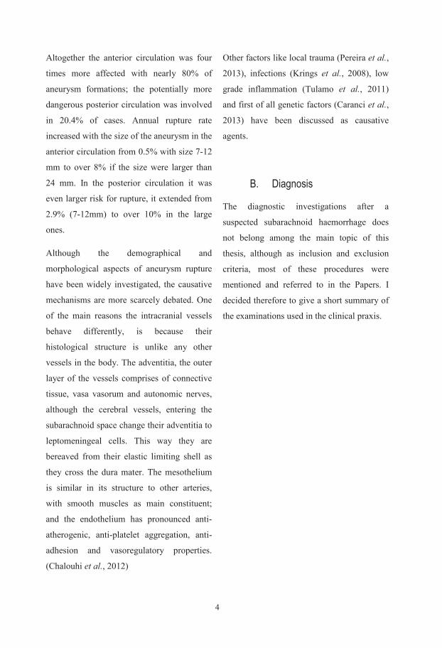
CT scan is the golden standard for
SAH diagnostics. (Fig. 4) The
technology of computed scanning
is a fast forward motion in
development, where the different
companies are already 2-3
generations ahead of the current
clinical practice. We use currently
from 4th to 7th generation CT
scanners with fan to cone shape beam and
detector array in access of 500 (the latest up
Figure 3. CT scan of a suspected aSAH, with hydrocephalus development. The “crab of death” a typical sign of subarachnoid haemorrhage.
Figure 4 Xanthochromia after subarachnoid haemorrhage. Note the bilirubin absorptions peak, presented as a shoulder at 475 nm.
4
Altogether the anterior circulation was four
times more affected with nearly 80% of
aneurysm formations; the potentially more
dangerous posterior circulation was involved
in 20.4% of cases. Annual rupture rate
increased with the size of the aneurysm in the
anterior circulation from 0.5% with size 7-12
mm to over 8% if the size were larger than
24 mm. In the posterior circulation it was
even larger risk for rupture, it extended from
2.9% (7-12mm) to over 10% in the large
ones.
Although the demographical and
morphological aspects of aneurysm rupture
have been widely investigated, the causative
mechanisms are more scarcely debated. One
of the main reasons the intracranial vessels
behave differently, is because their
histological structure is unlike any other
vessels in the body. The adventitia, the outer
layer of the vessels comprises of connective
tissue, vasa vasorum and autonomic nerves,
although the cerebral vessels, entering the
subarachnoid space change their adventitia to
leptomeningeal cells. This way they are
bereaved from their elastic limiting shell as
they cross the dura mater. The mesothelium
is similar in its structure to other arteries,
with smooth muscles as main constituent;
and the endothelium has pronounced anti-
atherogenic, anti-platelet aggregation, anti-
adhesion and vasoregulatory properties.
(Chalouhi et al., 2012)
Other factors like local trauma (Pereira et al.,
2013), infections (Krings et al., 2008), low
grade inflammation (Tulamo et al., 2011)
and first of all genetic factors (Caranci et al.,
2013) have been discussed as causative
agents.
B. Diagnosis
The diagnostic investigations after a
suspected subarachnoid haemorrhage does
not belong among the main topic of this
thesis, although as inclusion and exclusion
criteria, most of these procedures were
mentioned and referred to in the Papers. I
decided therefore to give a short summary of
the examinations used in the clinical praxis.
55
1. Xanthochromia
Lumbar puncture is still performed in the
majority of patients presenting with a
thunderclap headache and negative CT
scan to identify or rule out subarachnoid
haemorrhage. The reason is that only 8-
12% of patients with sudden headache and
no neurological deficit suffer from an
aSAH, and 40-50% of aSAH patients are
presented with only headache and no
other neurological symptoms.(Edlow and
Fisher, 2012). For reliable result, the
cerebrospinal fluid (CSF) has to be
centrifuged immediately to prevent in
vitro lysis of red cells with release of
oxyhemoglobin. Thereafter
spectrophotometry is used to differentiate
the bilirubin absorption on 456 nm
wavelength from oxyhemoglobin’s 415 nm
(Fig 3). The former is
pathognomonic for subarachnoid
haemorrhage; the latter may be a
remnant of a traumatic
puncture.(Nagy et al., 2013)
2. CT scan
CT scan is the golden standard for
SAH diagnostics. (Fig. 4) The
technology of computed scanning
is a fast forward motion in
development, where the different
companies are already 2-3
generations ahead of the current
clinical practice. We use currently
from 4th to 7th generation CT
scanners with fan to cone shape beam and
detector array in access of 500 (the latest up
Figure 3. CT scan of a suspected aSAH, with hydrocephalus development. The “crab of death” a typical sign of subarachnoid haemorrhage.
Figure 4 Xanthochromia after subarachnoid haemorrhage. Note the bilirubin absorptions peak, presented as a shoulder at 475 nm.
4
Altogether the anterior circulation was four
times more affected with nearly 80% of
aneurysm formations; the potentially more
dangerous posterior circulation was involved
in 20.4% of cases. Annual rupture rate
increased with the size of the aneurysm in the
anterior circulation from 0.5% with size 7-12
mm to over 8% if the size were larger than
24 mm. In the posterior circulation it was
even larger risk for rupture, it extended from
2.9% (7-12mm) to over 10% in the large
ones.
Although the demographical and
morphological aspects of aneurysm rupture
have been widely investigated, the causative
mechanisms are more scarcely debated. One
of the main reasons the intracranial vessels
behave differently, is because their
histological structure is unlike any other
vessels in the body. The adventitia, the outer
layer of the vessels comprises of connective
tissue, vasa vasorum and autonomic nerves,
although the cerebral vessels, entering the
subarachnoid space change their adventitia to
leptomeningeal cells. This way they are
bereaved from their elastic limiting shell as
they cross the dura mater. The mesothelium
is similar in its structure to other arteries,
with smooth muscles as main constituent;
and the endothelium has pronounced anti-
atherogenic, anti-platelet aggregation, anti-
adhesion and vasoregulatory properties.
(Chalouhi et al., 2012)
Other factors like local trauma (Pereira et al.,
2013), infections (Krings et al., 2008), low
grade inflammation (Tulamo et al., 2011)
and first of all genetic factors (Caranci et al.,
2013) have been discussed as causative
agents.
B. Diagnosis
The diagnostic investigations after a
suspected subarachnoid haemorrhage does
not belong among the main topic of this
thesis, although as inclusion and exclusion
criteria, most of these procedures were
mentioned and referred to in the Papers. I
decided therefore to give a short summary of
the examinations used in the clinical praxis.
67
pictures is well in the parity of a medium
quality DSA.
5. Digital subtraction
angiography (DSA)
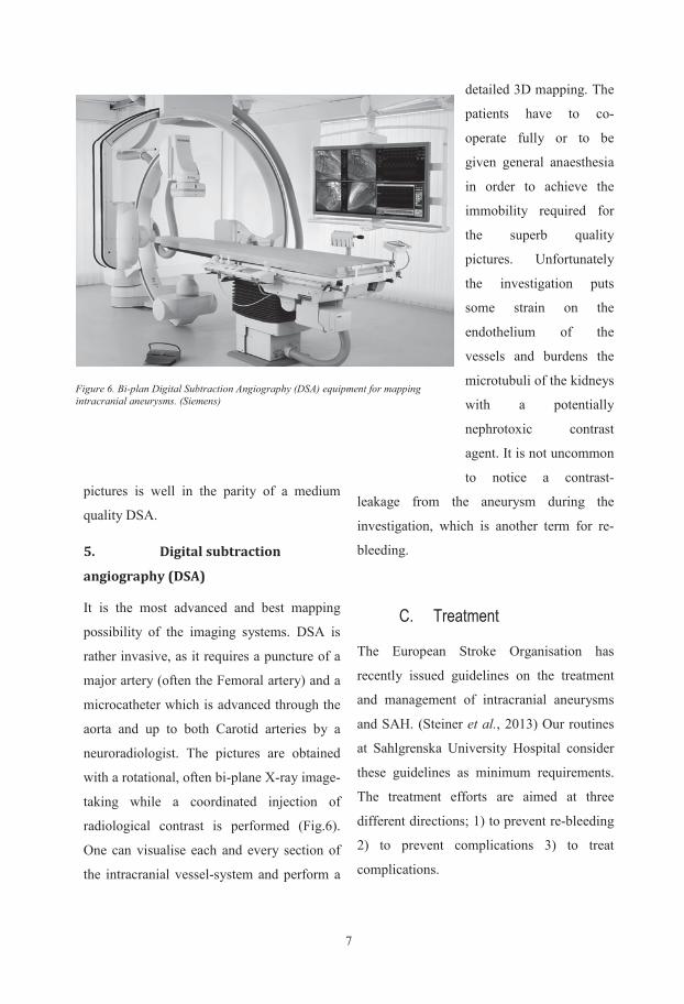
It is the most advanced and best mapping
possibility of the imaging systems. DSA is
rather invasive, as it requires a puncture of a
major artery (often the Femoral artery) and a
microcatheter which is advanced through the
aorta and up to both Carotid arteries by a
neuroradiologist. The pictures are obtained
with a rotational, often bi-plane X-ray image-
taking while a coordinated injection of
radiological contrast is performed (Fig.6).
One can visualise each and every section of
the intracranial vessel-system and perform a
detailed 3D mapping. The
patients have to co-
operate fully or to be
given general anaesthesia
in order to achieve the
immobility required for
the superb quality
pictures. Unfortunately
the investigation puts
some strain on the
endothelium of the
vessels and burdens the
microtubuli of the kidneys
with a potentially
nephrotoxic contrast
agent. It is not uncommon
to notice a contrast-
leakage from the aneurysm during the
investigation, which is another term for re-
bleeding.
C. Treatment
The European Stroke Organisation has
recently issued guidelines on the treatment
and management of intracranial aneurysms
and SAH. (Steiner et al., 2013) Our routines
at Sahlgrenska University Hospital consider
these guidelines as minimum requirements.
The treatment efforts are aimed at three
different directions; 1) to prevent re-bleeding
2) to prevent complications 3) to treat
complications.
Figure 6. Bi-plan Digital Subtraction Angiography (DSA) equipment for mapping intracranial aneurysms. (Siemens)
6
to 2400) in rotational array or in static
detectors all around. The 6th generation, so
called helical CT with source/detector
pairwise rotation and the 7th generation
multi-slice CT scanners can give 17 slices
per second and is fast enough to examine the
heart between beats. Studies started to
emerge, which showed that sensitivity and
specificity of 3rd generation and newer CT
scans are sufficient to diagnose or exclude
SAH not later than 6 hours after the onset of
symptoms, entirely on the basis of the scan if
neuroradiological expertise was at hand.
(Backes et al., 2012).
3. CT angiography (CTA)
The latest 5th and 6th generation
CT scanners with their speed of
scanning and their software
interface enabled to perform not
only a synchronised contrast X-
ray scan, but a high quality 3D
reconstruction of the intracranial
vessel system, and a first 3D
picture of a potential vessel
malformation. Many times the
quality is good enough to set the
diagnosis and initiate the
treatment. There are some voices
however; speaking out that CT
angiography should be used with
caution to rule out aneurysm
initially, because of the risk of diagnosing
asymptomatic aneurysms instead of a
haemorrhage. (Edlow and Fisher, 2012)
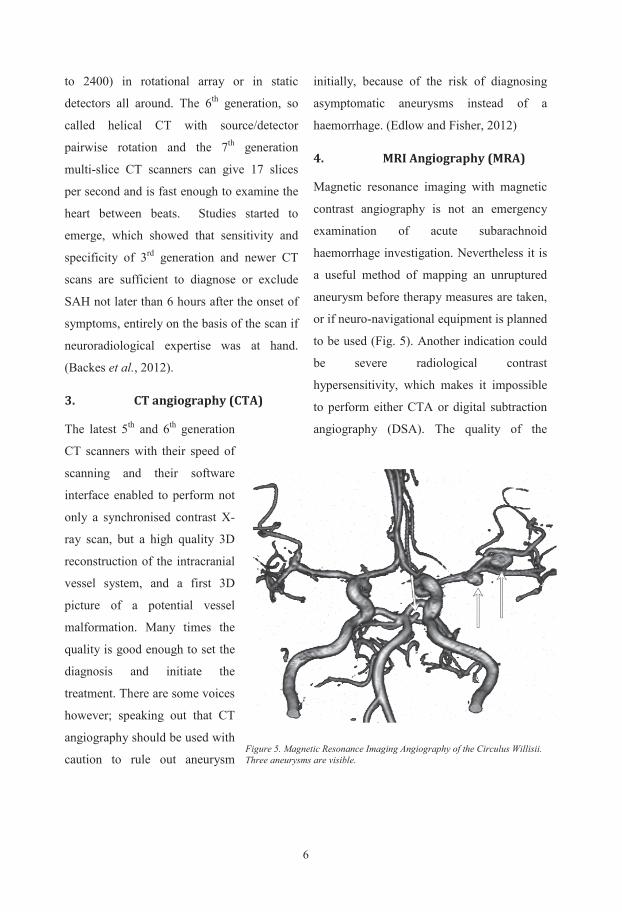
4. MRI Angiography (MRA)
Magnetic resonance imaging with magnetic
contrast angiography is not an emergency
examination of acute subarachnoid
haemorrhage investigation. Nevertheless it is
a useful method of mapping an unruptured
aneurysm before therapy measures are taken,
or if neuro-navigational equipment is planned
to be used (Fig. 5). Another indication could
be severe radiological contrast
hypersensitivity, which makes it impossible
to perform either CTA or digital subtraction
angiography (DSA). The quality of the
Figure 5. Magnetic Resonance Imaging Angiography of the Circulus Willisii. Three aneurysms are visible.
77
pictures is well in the parity of a medium
quality DSA.
5. Digital subtraction
angiography (DSA)
It is the most advanced and best mapping
possibility of the imaging systems. DSA is
rather invasive, as it requires a puncture of a
major artery (often the Femoral artery) and a
microcatheter which is advanced through the
aorta and up to both Carotid arteries by a
neuroradiologist. The pictures are obtained
with a rotational, often bi-plane X-ray image-
taking while a coordinated injection of
radiological contrast is performed (Fig.6).
One can visualise each and every section of
the intracranial vessel-system and perform a
detailed 3D mapping. The
patients have to co-
operate fully or to be
given general anaesthesia
in order to achieve the
immobility required for
the superb quality
pictures. Unfortunately
the investigation puts
some strain on the
endothelium of the
vessels and burdens the
microtubuli of the kidneys
with a potentially
nephrotoxic contrast
agent. It is not uncommon
to notice a contrast-
leakage from the aneurysm during the
investigation, which is another term for re-
bleeding.
C. Treatment
The European Stroke Organisation has
recently issued guidelines on the treatment
and management of intracranial aneurysms
and SAH. (Steiner et al., 2013) Our routines
at Sahlgrenska University Hospital consider
these guidelines as minimum requirements.
The treatment efforts are aimed at three
different directions; 1) to prevent re-bleeding
2) to prevent complications 3) to treat
complications.
Figure 6. Bi-plan Digital Subtraction Angiography (DSA) equipment for mapping intracranial aneurysms. (Siemens)
6
to 2400) in rotational array or in static
detectors all around. The 6th generation, so
called helical CT with source/detector
pairwise rotation and the 7th generation
multi-slice CT scanners can give 17 slices
per second and is fast enough to examine the
heart between beats. Studies started to
emerge, which showed that sensitivity and
specificity of 3rd generation and newer CT
scans are sufficient to diagnose or exclude
SAH not later than 6 hours after the onset of
symptoms, entirely on the basis of the scan if
neuroradiological expertise was at hand.
(Backes et al., 2012).
3. CT angiography (CTA)
The latest 5th and 6th generation
CT scanners with their speed of
scanning and their software
interface enabled to perform not
only a synchronised contrast X-
ray scan, but a high quality 3D
reconstruction of the intracranial
vessel system, and a first 3D
picture of a potential vessel
malformation. Many times the
quality is good enough to set the
diagnosis and initiate the
treatment. There are some voices
however; speaking out that CT
angiography should be used with
caution to rule out aneurysm
initially, because of the risk of diagnosing
asymptomatic aneurysms instead of a
haemorrhage. (Edlow and Fisher, 2012)
4. MRI Angiography (MRA)
Magnetic resonance imaging with magnetic
contrast angiography is not an emergency
examination of acute subarachnoid
haemorrhage investigation. Nevertheless it is
a useful method of mapping an unruptured
aneurysm before therapy measures are taken,
or if neuro-navigational equipment is planned
to be used (Fig. 5). Another indication could
be severe radiological contrast
hypersensitivity, which makes it impossible
to perform either CTA or digital subtraction
angiography (DSA). The quality of the
Figure 5. Magnetic Resonance Imaging Angiography of the Circulus Willisii. Three aneurysms are visible.
89
refined by Kenichiro Sugita at the Nagoya
University in the middle of the 1970-s
(Sugita et al., 1984), is applied to the neck of
the aneurysm, thereby obstructing the flow to
it (Fig. 7).
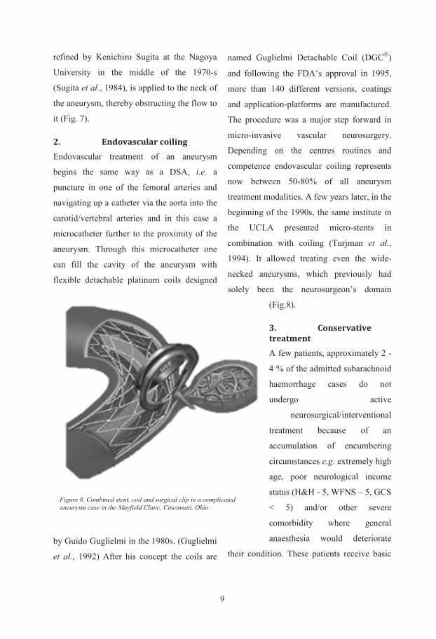
2. Endovascular coiling Endovascular treatment of an aneurysm
begins the same way as a DSA, i.e. a
puncture in one of the femoral arteries and
navigating up a catheter via the aorta into the
carotid/vertebral arteries and in this case a
microcatheter further to the proximity of the
aneurysm. Through this microcatheter one
can fill the cavity of the aneurysm with
flexible detachable platinum coils designed
by Guido Guglielmi in the 1980s. (Guglielmi
et al., 1992) After his concept the coils are
named Guglielmi Detachable Coil (DGC)
and following the FDA’s approval in 1995,
more than 140 different versions, coatings
and application-platforms are manufactured.
The procedure was a major step forward in
micro-invasive vascular neurosurgery.
Depending on the centres routines and
competence endovascular coiling represents
now between 50-80% of all aneurysm
treatment modalities. A few years later, in the
beginning of the 1990s, the same institute in
the UCLA presented micro-stents in
combination with coiling (Turjman et al.,
1994). It allowed treating even the wide-
necked aneurysms, which previously had
solely been the neurosurgeon’s domain
(Fig.8).
3. Conservative treatment A few patients, approximately 2 -
4 % of the admitted subarachnoid
haemorrhage cases do not
undergo active
neurosurgical/interventional
treatment because of an
accumulation of encumbering
circumstances e.g. extremely high
age, poor neurological income
status (H&H - 5, WFNS – 5, GCS
< 5) and/or other severe
comorbidity where general
anaesthesia would deteriorate
their condition. These patients receive basic
Figure 8. Combined stent, coil and surgical clip in a complicated aneurysm case in the Mayfield Clinic, Cincinnati, Ohio
8
To prevent re-bleeding
It starts immediately after first physician
contact of the patient through stabilising
his/her condition, ensuring adequate
oxygenation, and circulation. If needed,
airways have to be secured and artificial
ventilation started. Nearly one third of the
patients have initial loss of consciousness
and one fourth have convulsive seizures.
(Fung et al., 2015) It is imperative to cease
these seizures to continue the patient
management. To establish monitoring is
fundamental as oxygenation, circulatory
stability and neurological assessment are
determinant factors in therapeutical
decision-making. Invasive blood-
pressure monitoring,
plethysmographic pulse-oximetry
(POX), ECG monitoring, urinary
output and neurological valuation are
minimum monitoring standards even
during transport to tertiary
(neurosurgical) therapy centres.
Systolic arterial pressure should be
kept under 180 mmHg, but should
not be lowered to more than to a
mean arterial pressure (MAP) of 90
mmHg. (Steiner et al., 2013).
Tranexamic acid (Cyklokapron i.v.
1g three times daily), a fibrinolysis inhibitor
is recommended in our centre as a
pharmacologic re-bleeding prophylaxis,
given directly after the diagnosis is
established. (Hillman et al., 2002) This
treatment is continued until the aneurysm is
secured.
The efforts of re-bleeding prevention
continue in the neurosurgical department,
where the aneurysm is mapped with DSA
and a 3D image reconstruction is created.
After a discussion between the neurosurgeon
and the interventional neuroradiologist, a
joint decision is made how to secure the
aneurysm.
1. Surgical clipping One of the options is to use an intraoperative
method, involving an open craniotomy and
surgical exploration of the aneurysm. It is a
major neurosurgical operation when a special
clip, first used by Walter Dandy at the Johns
Hopkins Hospital in Baltimore, 1937 and
Figure 7. Titanium surgical Sugita clips for different aneurysm applications
99
refined by Kenichiro Sugita at the Nagoya
University in the middle of the 1970-s
(Sugita et al., 1984), is applied to the neck of
the aneurysm, thereby obstructing the flow to
it (Fig. 7).
2. Endovascular coiling Endovascular treatment of an aneurysm
begins the same way as a DSA, i.e. a
puncture in one of the femoral arteries and
navigating up a catheter via the aorta into the
carotid/vertebral arteries and in this case a
microcatheter further to the proximity of the
aneurysm. Through this microcatheter one
can fill the cavity of the aneurysm with
flexible detachable platinum coils designed
by Guido Guglielmi in the 1980s. (Guglielmi
et al., 1992) After his concept the coils are
named Guglielmi Detachable Coil (DGC)
and following the FDA’s approval in 1995,
more than 140 different versions, coatings
and application-platforms are manufactured.
The procedure was a major step forward in
micro-invasive vascular neurosurgery.
Depending on the centres routines and
competence endovascular coiling represents
now between 50-80% of all aneurysm
treatment modalities. A few years later, in the
beginning of the 1990s, the same institute in
the UCLA presented micro-stents in
combination with coiling (Turjman et al.,
1994). It allowed treating even the wide-
necked aneurysms, which previously had
solely been the neurosurgeon’s domain
(Fig.8).
3. Conservative treatment A few patients, approximately 2 -
4 % of the admitted subarachnoid
haemorrhage cases do not
undergo active
neurosurgical/interventional
treatment because of an
accumulation of encumbering
circumstances e.g. extremely high
age, poor neurological income
status (H&H - 5, WFNS – 5, GCS
< 5) and/or other severe
comorbidity where general
anaesthesia would deteriorate
their condition. These patients receive basic
Figure 8. Combined stent, coil and surgical clip in a complicated aneurysm case in the Mayfield Clinic, Cincinnati, Ohio
8
To prevent re-bleeding
It starts immediately after first physician
contact of the patient through stabilising
his/her condition, ensuring adequate
oxygenation, and circulation. If needed,
airways have to be secured and artificial
ventilation started. Nearly one third of the
patients have initial loss of consciousness
and one fourth have convulsive seizures.
(Fung et al., 2015) It is imperative to cease
these seizures to continue the patient
management. To establish monitoring is
fundamental as oxygenation, circulatory
stability and neurological assessment are
determinant factors in therapeutical
decision-making. Invasive blood-
pressure monitoring,
plethysmographic pulse-oximetry
(POX), ECG monitoring, urinary
output and neurological valuation are
minimum monitoring standards even
during transport to tertiary
(neurosurgical) therapy centres.
Systolic arterial pressure should be
kept under 180 mmHg, but should
not be lowered to more than to a
mean arterial pressure (MAP) of 90
mmHg. (Steiner et al., 2013).
Tranexamic acid (Cyklokapron i.v.
1g three times daily), a fibrinolysis inhibitor
is recommended in our centre as a
pharmacologic re-bleeding prophylaxis,
given directly after the diagnosis is
established. (Hillman et al., 2002) This
treatment is continued until the aneurysm is
secured.
The efforts of re-bleeding prevention
continue in the neurosurgical department,
where the aneurysm is mapped with DSA
and a 3D image reconstruction is created.
After a discussion between the neurosurgeon
and the interventional neuroradiologist, a
joint decision is made how to secure the
aneurysm.
1. Surgical clipping One of the options is to use an intraoperative
method, involving an open craniotomy and
surgical exploration of the aneurysm. It is a
major neurosurgical operation when a special
clip, first used by Walter Dandy at the Johns
Hopkins Hospital in Baltimore, 1937 and
Figure 7. Titanium surgical Sugita clips for different aneurysm applications
1011
3. Cerebral infarction (CI)
CI is defined as radiological (CT, MRI) signs
of infarction within 6 weeks after an aSAH,
the latest CT prior to death (in 6 weeks) or an
autopsy verified infarction. These signs
should not be directly connected to operation
or embolisation. These radiological signs
though must not be present within 48 hours
of the bleeding (Vergouwen et al., 2010). To
be more confusing, in American literature
this could be named as DCI, in contrast to
older infarction or infarction directly related
to treatment (post-operative or post-
embolisation complication). It is
understandable, that review articles and
meta-analyses have problems defining the
end-points of the studies.
Apart from an active neurosurgical
management and optimised neuro-intensive
care, the only drug which is documented to
improve outcome is nimodipin. This is the
reason why all aSAH patients receive iv. or
oral nimodipin, during 10-14 days after the
bleeding.
II. Neurological, radiological assessment
A. Admission assessment
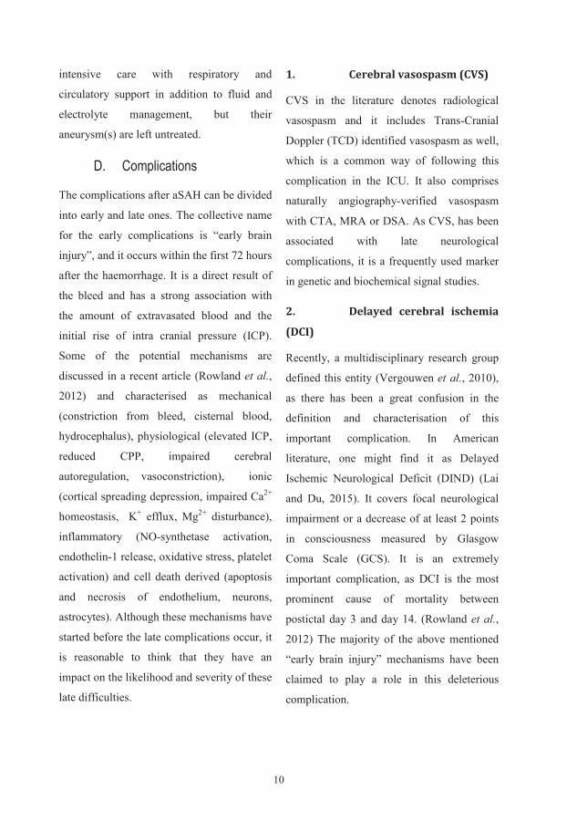
1. Hunt and Hess scale
SAH patients’ early evaluation has been
advocated from the early 1950’s (Norlen and
Olivecrona,
1953) and a
classification has
been
systematically
used since
Botterell
published his
article on
assessment of the
perioperative risk
of SAH patients.
(Botterell et al., 1956) From his five-grade
scale evolved the most used SAH grading
scale developed by William Hunt and Robert
Hess from Ohio and was the standard
assessment instrument for half a century.
(Table 1) (Hunt and Hess, 1968). As it has
been used world-wide and extensively
validated, we have chosen this instrument in
Table 1. Hunt and Hess grading scale and expected rate of survival at the time of publishing(Hunt and Hess, 1968)
10
intensive care with respiratory and
circulatory support in addition to fluid and
electrolyte management, but their
aneurysm(s) are left untreated.
D. Complications
The complications after aSAH can be divided
into early and late ones. The collective name
for the early complications is “early brain
injury”, and it occurs within the first 72 hours
after the haemorrhage. It is a direct result of
the bleed and has a strong association with
the amount of extravasated blood and the
initial rise of intra cranial pressure (ICP).
Some of the potential mechanisms are
discussed in a recent article (Rowland et al.,
2012) and characterised as mechanical
(constriction from bleed, cisternal blood,
hydrocephalus), physiological (elevated ICP,
reduced CPP, impaired cerebral
autoregulation, vasoconstriction), ionic
(cortical spreading depression, impaired Ca2+
homeostasis, K+ efflux, Mg2+ disturbance),
inflammatory (NO-synthetase activation,
endothelin-1 release, oxidative stress, platelet
activation) and cell death derived (apoptosis
and necrosis of endothelium, neurons,
astrocytes). Although these mechanisms have
started before the late complications occur, it
is reasonable to think that they have an
impact on the likelihood and severity of these
late difficulties.
1. Cerebral vasospasm (CVS)
CVS in the literature denotes radiological
vasospasm and it includes Trans-Cranial
Doppler (TCD) identified vasospasm as well,
which is a common way of following this
complication in the ICU. It also comprises
naturally angiography-verified vasospasm
with CTA, MRA or DSA. As CVS, has been
associated with late neurological
complications, it is a frequently used marker
in genetic and biochemical signal studies.
2. Delayed cerebral ischemia
(DCI)
Recently, a multidisciplinary research group
defined this entity (Vergouwen et al., 2010),
as there has been a great confusion in the
definition and characterisation of this
important complication. In American
literature, one might find it as Delayed
Ischemic Neurological Deficit (DIND) (Lai
and Du, 2015). It covers focal neurological
impairment or a decrease of at least 2 points
in consciousness measured by Glasgow
Coma Scale (GCS). It is an extremely
important complication, as DCI is the most
prominent cause of mortality between
postictal day 3 and day 14. (Rowland et al.,
2012) The majority of the above mentioned
“early brain injury” mechanisms have been
claimed to play a role in this deleterious
complication.
1111
3. Cerebral infarction (CI)
CI is defined as radiological (CT, MRI) signs
of infarction within 6 weeks after an aSAH,
the latest CT prior to death (in 6 weeks) or an
autopsy verified infarction. These signs
should not be directly connected to operation
or embolisation. These radiological signs
though must not be present within 48 hours
of the bleeding (Vergouwen et al., 2010). To
be more confusing, in American literature
this could be named as DCI, in contrast to
older infarction or infarction directly related
to treatment (post-operative or post-
embolisation complication). It is
understandable, that review articles and
meta-analyses have problems defining the
end-points of the studies.
Apart from an active neurosurgical
management and optimised neuro-intensive
care, the only drug which is documented to
improve outcome is nimodipin. This is the
reason why all aSAH patients receive iv. or
oral nimodipin, during 10-14 days after the
bleeding.
II. Neurological, radiological assessment
A. Admission assessment
1. Hunt and Hess scale
SAH patients’ early evaluation has been
advocated from the early 1950’s (Norlen and
Olivecrona,
1953) and a
classification has
been
systematically
used since
Botterell
published his
article on
assessment of the
perioperative risk
of SAH patients.
(Botterell et al., 1956) From his five-grade
scale evolved the most used SAH grading
scale developed by William Hunt and Robert
Hess from Ohio and was the standard
assessment instrument for half a century.
(Table 1) (Hunt and Hess, 1968). As it has
been used world-wide and extensively
validated, we have chosen this instrument in
Table 1. Hunt and Hess grading scale and expected rate of survival at the time of publishing(Hunt and Hess, 1968)
10
intensive care with respiratory and
circulatory support in addition to fluid and
electrolyte management, but their
aneurysm(s) are left untreated.
D. Complications
The complications after aSAH can be divided
into early and late ones. The collective name
for the early complications is “early brain
injury”, and it occurs within the first 72 hours
after the haemorrhage. It is a direct result of
the bleed and has a strong association with
the amount of extravasated blood and the
initial rise of intra cranial pressure (ICP).
Some of the potential mechanisms are
discussed in a recent article (Rowland et al.,
2012) and characterised as mechanical
(constriction from bleed, cisternal blood,
hydrocephalus), physiological (elevated ICP,
reduced CPP, impaired cerebral
autoregulation, vasoconstriction), ionic
(cortical spreading depression, impaired Ca2+
homeostasis, K+ efflux, Mg2+ disturbance),
inflammatory (NO-synthetase activation,
endothelin-1 release, oxidative stress, platelet
activation) and cell death derived (apoptosis
and necrosis of endothelium, neurons,
astrocytes). Although these mechanisms have
started before the late complications occur, it
is reasonable to think that they have an
impact on the likelihood and severity of these
late difficulties.
1. Cerebral vasospasm (CVS)
CVS in the literature denotes radiological
vasospasm and it includes Trans-Cranial
Doppler (TCD) identified vasospasm as well,
which is a common way of following this
complication in the ICU. It also comprises
naturally angiography-verified vasospasm
with CTA, MRA or DSA. As CVS, has been
associated with late neurological
complications, it is a frequently used marker
in genetic and biochemical signal studies.
2. Delayed cerebral ischemia
(DCI)
Recently, a multidisciplinary research group
defined this entity (Vergouwen et al., 2010),
as there has been a great confusion in the
definition and characterisation of this
important complication. In American
literature, one might find it as Delayed
Ischemic Neurological Deficit (DIND) (Lai
and Du, 2015). It covers focal neurological
impairment or a decrease of at least 2 points
in consciousness measured by Glasgow
Coma Scale (GCS). It is an extremely
important complication, as DCI is the most
prominent cause of mortality between
postictal day 3 and day 14. (Rowland et al.,
2012) The majority of the above mentioned
“early brain injury” mechanisms have been
claimed to play a role in this deleterious
complication.
1213
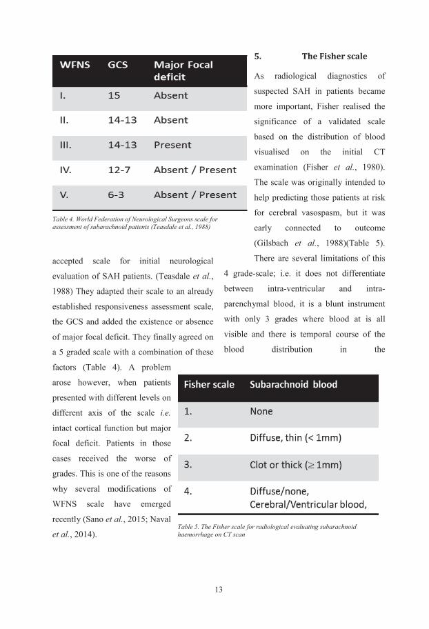
accepted scale for initial neurological
evaluation of SAH patients. (Teasdale et al.,
1988) They adapted their scale to an already
established responsiveness assessment scale,
the GCS and added the existence or absence
of major focal deficit. They finally agreed on
a 5 graded scale with a combination of these
factors (Table 4). A problem
arose however, when patients
presented with different levels on
different axis of the scale i.e.
intact cortical function but major
focal deficit. Patients in those
cases received the worse of
grades. This is one of the reasons
why several modifications of
WFNS scale have emerged
recently (Sano et al., 2015; Naval
et al., 2014).
5. The Fisher scale
As radiological diagnostics of
suspected SAH in patients became
more important, Fisher realised the
significance of a validated scale
based on the distribution of blood
visualised on the initial CT
examination (Fisher et al., 1980).
The scale was originally intended to
help predicting those patients at risk
for cerebral vasospasm, but it was
early connected to outcome
(Gilsbach et al., 1988)(Table 5).
There are several limitations of this
4 grade-scale; i.e. it does not differentiate
between intra-ventricular and intra-
parenchymal blood, it is a blunt instrument
with only 3 grades where blood at is all
visible and there is temporal course of the
blood distribution in the
Table 4. World Federation of Neurological Surgeons scale for assessment of subarachnoid patients (Teasdale et al., 1988)
Table 5. The Fisher scale for radiological evaluating subarachnoid haemorrhage on CT scan
12
most of our papers for assessing SAH
severity at admission.
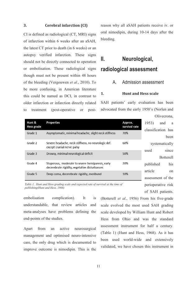
2. Glasgow Coma Scale (GCS)
As different kinds of neurological
emergencies started to be admitted to
dedicated Emergency Units, Teasdale and
Jennett have realised the importance of an
aetiology-independent grading scale
(Teasdale and Jennett, 1974) and introduced
a behavioural assessment grading based on
the best motor, verbal response and eye
opening, awarding points for each activity.
The total sum of the points, (max.: 15, min.:
3) provide the GCS. (Table 2) This scale has
since then been used for assessment of
altered consciousness of all possible causes
in the emergency departments.
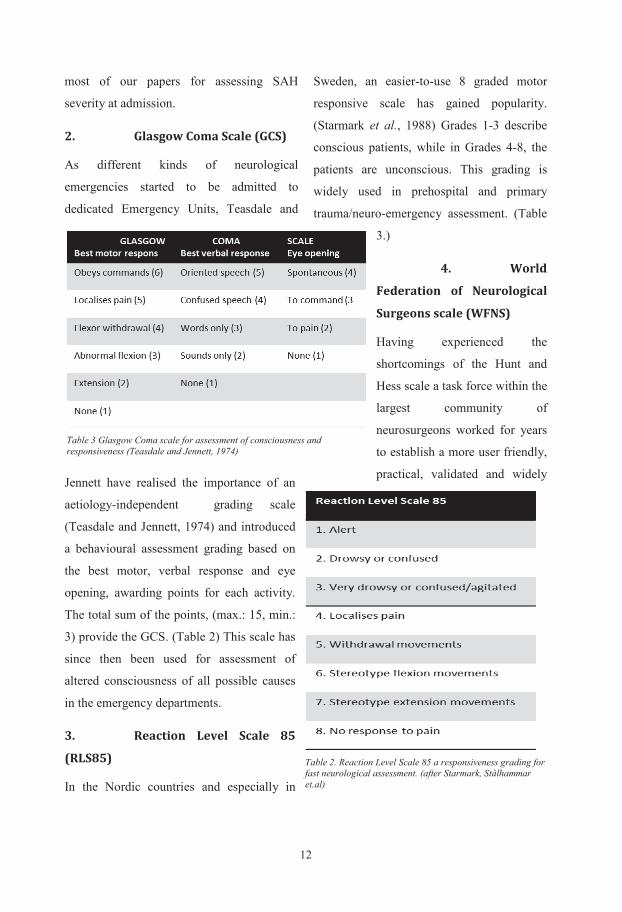
3. Reaction Level Scale 85
(RLS85)
In the Nordic countries and especially in
Sweden, an easier-to-use 8 graded motor
responsive scale has gained popularity.
(Starmark et al., 1988) Grades 1-3 describe
conscious patients, while in Grades 4-8, the
patients are unconscious. This grading is
widely used in prehospital and primary
trauma/neuro-emergency assessment. (Table
3.)
4. World
Federation of Neurological
Surgeons scale (WFNS)
Having experienced the
shortcomings of the Hunt and
Hess scale a task force within the
largest community of
neurosurgeons worked for years
to establish a more user friendly,
practical, validated and widely
Table 3 Glasgow Coma scale for assessment of consciousness and responsiveness (Teasdale and Jennett, 1974)
Table 2. Reaction Level Scale 85 a responsiveness grading for fast neurological assessment. (after Starmark, Stålhammar et.al)
1313
accepted scale for initial neurological
evaluation of SAH patients. (Teasdale et al.,
1988) They adapted their scale to an already
established responsiveness assessment scale,
the GCS and added the existence or absence
of major focal deficit. They finally agreed on
a 5 graded scale with a combination of these
factors (Table 4). A problem
arose however, when patients
presented with different levels on
different axis of the scale i.e.
intact cortical function but major
focal deficit. Patients in those
cases received the worse of
grades. This is one of the reasons
why several modifications of
WFNS scale have emerged
recently (Sano et al., 2015; Naval
et al., 2014).
5. The Fisher scale
As radiological diagnostics of
suspected SAH in patients became
more important, Fisher realised the
significance of a validated scale
based on the distribution of blood
visualised on the initial CT
examination (Fisher et al., 1980).
The scale was originally intended to
help predicting those patients at risk
for cerebral vasospasm, but it was
early connected to outcome
(Gilsbach et al., 1988)(Table 5).
There are several limitations of this
4 grade-scale; i.e. it does not differentiate
between intra-ventricular and intra-
parenchymal blood, it is a blunt instrument
with only 3 grades where blood at is all
visible and there is temporal course of the
blood distribution in the
Table 4. World Federation of Neurological Surgeons scale for assessment of subarachnoid patients (Teasdale et al., 1988)
Table 5. The Fisher scale for radiological evaluating subarachnoid haemorrhage on CT scan
12
most of our papers for assessing SAH
severity at admission.
2. Glasgow Coma Scale (GCS)
As different kinds of neurological
emergencies started to be admitted to
dedicated Emergency Units, Teasdale and
Jennett have realised the importance of an
aetiology-independent grading scale
(Teasdale and Jennett, 1974) and introduced
a behavioural assessment grading based on
the best motor, verbal response and eye
opening, awarding points for each activity.
The total sum of the points, (max.: 15, min.:
3) provide the GCS. (Table 2) This scale has
since then been used for assessment of
altered consciousness of all possible causes
in the emergency departments.
3. Reaction Level Scale 85
(RLS85)
In the Nordic countries and especially in
Sweden, an easier-to-use 8 graded motor
responsive scale has gained popularity.
(Starmark et al., 1988) Grades 1-3 describe
conscious patients, while in Grades 4-8, the
patients are unconscious. This grading is
widely used in prehospital and primary
trauma/neuro-emergency assessment. (Table
3.)
4. World
Federation of Neurological
Surgeons scale (WFNS)
Having experienced the
shortcomings of the Hunt and
Hess scale a task force within the
largest community of
neurosurgeons worked for years
to establish a more user friendly,
practical, validated and widely
Table 3 Glasgow Coma scale for assessment of consciousness and responsiveness (Teasdale and Jennett, 1974)
Table 2. Reaction Level Scale 85 a responsiveness grading for fast neurological assessment. (after Starmark, Stålhammar et.al)
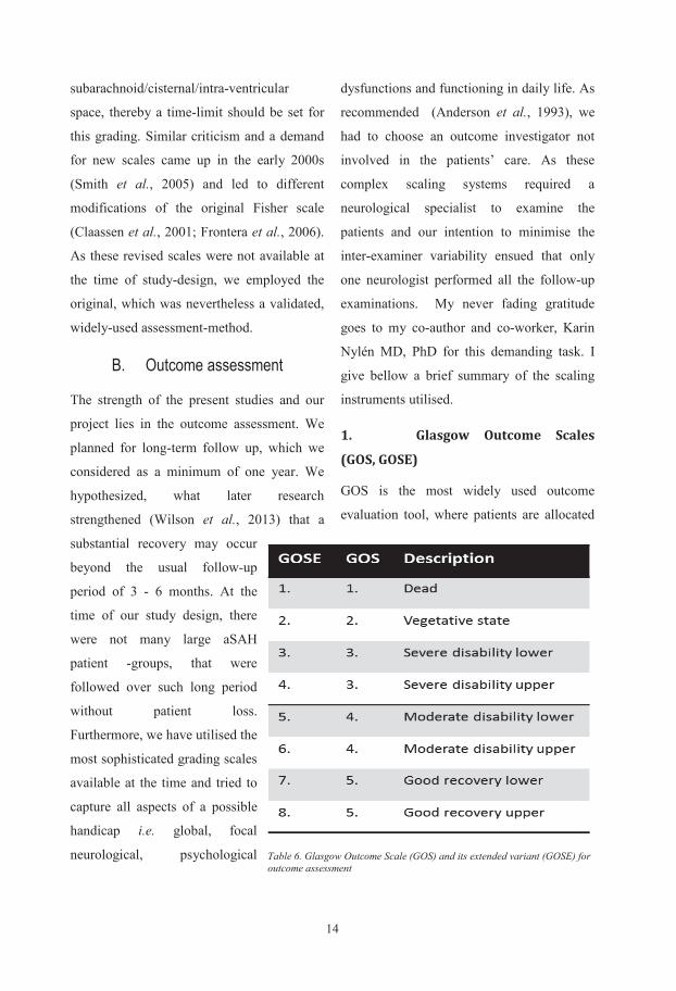
1415
to a category on a 5 or in the extended
version (GOSE) 8 hierarchical categories
during an interview (Teasdale and Jennett,
1974). This relatively easy-to-use scale gives
a general index of overall outcome and it
reflects disability, rather than impairment
compared to pre-morbid status; i.e. how the
handicap affects functioning in major areas
of life (Table 6). It allows comparison
between different patient-groups (Marschall,
1987) and been suggested as a measure of
outcome in clinical trials (Clifton et al.,
1992). The questions are based on the areas
of everyday life e.g. independence at home,
independence outside home, employability,
engaging in pre-morbid social and leisure
activity and interpersonal relationship
(Jennett et al., 1981). If inter-observer
variability is to be kept at minimum a
structured questionnaire-based interview with
few, educated raters have to be employed
(Wilson et al., 1998). We used the
questionnaire for the extended GOS (GOSE)
to allow a wider differentiation of the data.
Table 7. National Institute of Health Stroke Scale (NIHSS), instrument to measure focal neurological deficit
14
subarachnoid/cisternal/intra-ventricular
space, thereby a time-limit should be set for
this grading. Similar criticism and a demand
for new scales came up in the early 2000s
(Smith et al., 2005) and led to different
modifications of the original Fisher scale
(Claassen et al., 2001; Frontera et al., 2006).
As these revised scales were not available at
the time of study-design, we employed the
original, which was nevertheless a validated,
widely-used assessment-method.
B. Outcome assessment
The strength of the present studies and our
project lies in the outcome assessment. We
planned for long-term follow up, which we
considered as a minimum of one year. We
hypothesized, what later research
strengthened (Wilson et al., 2013) that a
substantial recovery may occur
beyond the usual follow-up
period of 3 - 6 months. At the
time of our study design, there
were not many large aSAH
patient -groups, that were
followed over such long period
without patient loss.
Furthermore, we have utilised the
most sophisticated grading scales
available at the time and tried to
capture all aspects of a possible
handicap i.e. global, focal
neurological, psychological
dysfunctions and functioning in daily life. As
recommended (Anderson et al., 1993), we
had to choose an outcome investigator not
involved in the patients’ care. As these
complex scaling systems required a
neurological specialist to examine the
patients and our intention to minimise the
inter-examiner variability ensued that only
one neurologist performed all the follow-up
examinations. My never fading gratitude
goes to my co-author and co-worker, Karin
Nylén MD, PhD for this demanding task. I
give bellow a brief summary of the scaling
instruments utilised.
1. Glasgow Outcome Scales
(GOS, GOSE)
GOS is the most widely used outcome
evaluation tool, where patients are allocated
Table 6. Glasgow Outcome Scale (GOS) and its extended variant (GOSE) for outcome assessment
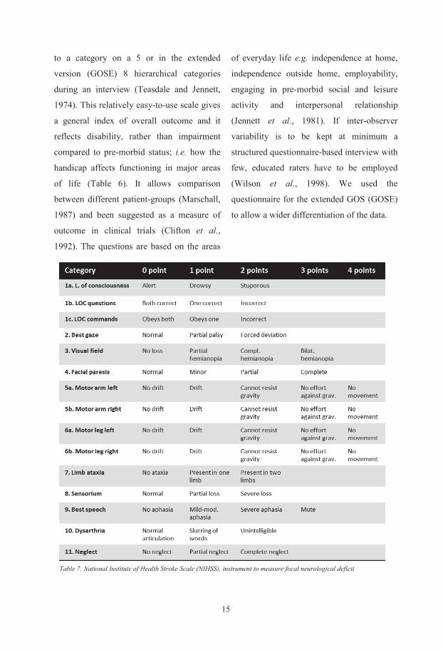
1515
to a category on a 5 or in the extended
version (GOSE) 8 hierarchical categories
during an interview (Teasdale and Jennett,
1974). This relatively easy-to-use scale gives
a general index of overall outcome and it
reflects disability, rather than impairment
compared to pre-morbid status; i.e. how the
handicap affects functioning in major areas
of life (Table 6). It allows comparison
between different patient-groups (Marschall,
1987) and been suggested as a measure of
outcome in clinical trials (Clifton et al.,
1992). The questions are based on the areas
of everyday life e.g. independence at home,
independence outside home, employability,
engaging in pre-morbid social and leisure
activity and interpersonal relationship
(Jennett et al., 1981). If inter-observer
variability is to be kept at minimum a
structured questionnaire-based interview with
few, educated raters have to be employed
(Wilson et al., 1998). We used the
questionnaire for the extended GOS (GOSE)
to allow a wider differentiation of the data.
Table 7. National Institute of Health Stroke Scale (NIHSS), instrument to measure focal neurological deficit
14
subarachnoid/cisternal/intra-ventricular
space, thereby a time-limit should be set for
this grading. Similar criticism and a demand
for new scales came up in the early 2000s
(Smith et al., 2005) and led to different
modifications of the original Fisher scale
(Claassen et al., 2001; Frontera et al., 2006).
As these revised scales were not available at
the time of study-design, we employed the
original, which was nevertheless a validated,
widely-used assessment-method.
B. Outcome assessment
The strength of the present studies and our
project lies in the outcome assessment. We
planned for long-term follow up, which we
considered as a minimum of one year. We
hypothesized, what later research
strengthened (Wilson et al., 2013) that a
substantial recovery may occur
beyond the usual follow-up
period of 3 - 6 months. At the
time of our study design, there
were not many large aSAH
patient -groups, that were
followed over such long period
without patient loss.
Furthermore, we have utilised the
most sophisticated grading scales
available at the time and tried to
capture all aspects of a possible
handicap i.e. global, focal
neurological, psychological
dysfunctions and functioning in daily life. As
recommended (Anderson et al., 1993), we
had to choose an outcome investigator not
involved in the patients’ care. As these
complex scaling systems required a
neurological specialist to examine the
patients and our intention to minimise the
inter-examiner variability ensued that only
one neurologist performed all the follow-up
examinations. My never fading gratitude
goes to my co-author and co-worker, Karin
Nylén MD, PhD for this demanding task. I
give bellow a brief summary of the scaling
instruments utilised.
1. Glasgow Outcome Scales
(GOS, GOSE)
GOS is the most widely used outcome
evaluation tool, where patients are allocated
Table 6. Glasgow Outcome Scale (GOS) and its extended variant (GOSE) for outcome assessment
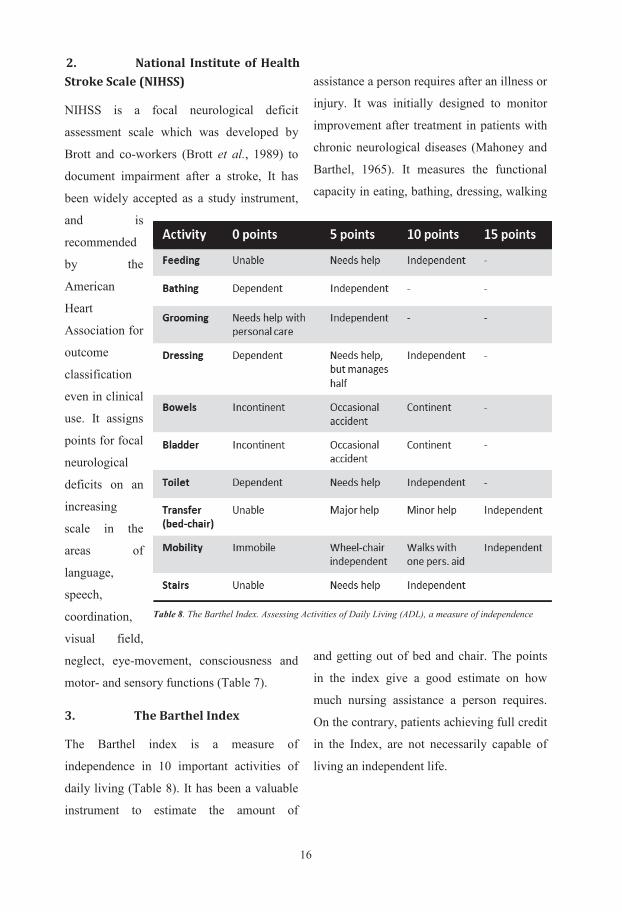
1617
C. Physiological parameters
To keep physiological parameters within
normal limits is important in any disease’s
therapeutical regime, notwithstanding it is
even more important in aSAH, when the
central coordination of the different
mechanisms is failing. Comprehensive
therapeutical guidelines for the management
of SAH patients in the Neurointensive Care
setting were published a few years ago
(Wartenberg, 2011; Smith, 2007)
summarising the main
aspects of the treatment
protocols. I describe our
intensions regarding the
physiological limits, at
the time our study-
patients were treated in
the NICU, Sahlgrenska
University Hospital.
Haemoglobin >120 g/l,
S-sodium >135<150
mmol/l, S-potassium 4.0-5.0 mmol/l, S-
albumin 35-50 g/l, core temperature 37 ± 0.5
° C, MABP between 70-100 mmHg,
intracranial pressure (ICP) <20 mmHg,
Cerebral perfusion pressure (CPP = MABP-
ICP) >60 mmHg, pO2 12-18 kPa, pCO2 ca
4.5 kPa and normalized pH. Blood glucose
was kept stringently between 4-6 mmol/l.
III. Genetic neuromarkers
1. Apolipoprotein E (ApoE)
Apolipoprotein E (ApoE) is the protein-part
of very low density lipoprotein (VLDL)
group, which are often remnants of
chylomicrons. The protein is only 299
amino-acid long and circulates in blood,
cerebrospinal fluid (CSF) and central nervous
system (CNS) tissue interstitial fluid (Fig.9).
In the CNS, it is produced by the astrocytes
and one of its functions is to transport
cholesterol and other lipids through
membranes, thereby ApoE is directly
responsible for the clearance of plasma
lipoproteins from the cell. The mechanism is
that ApoE serves as a critical ligand to low
density lipoprotein receptors. This function,
to redistribute lipoproteins into the cells is
Figure 9. Apolipoprotein E3 and E4’s 3D structure. Note the difference in mobility of the arm, and thence difference in function! With permission. (Mahley and Huang, 2012)
16
Stroke Scale (NIHSS)
NIHSS is a focal neurological deficit
assessment scale which was developed by
Brott and co-workers (Brott et al., 1989) to
document impairment after a stroke, It has
been widely accepted as a study instrument,
and is
recommended
by the
American
Heart
Association for
outcome
classification
even in clinical
use. It assigns
points for focal
neurological
deficits on an
increasing
scale in the
areas of
language,
speech,
coordination,
visual field,
neglect, eye-movement, consciousness and
motor- and sensory functions (Table 7).
3. The Barthel Index
The Barthel index is a measure of
independence in 10 important activities of
daily living (Table 8). It has been a valuable
instrument to estimate the amount of
assistance a person requires after an illness or
injury. It was initially designed to monitor
improvement after treatment in patients with
chronic neurological diseases (Mahoney and
Barthel, 1965). It measures the functional
capacity in eating, bathing, dressing, walking
and getting out of bed and chair. The points
in the index give a good estimate on how
much nursing assistance a person requires.
On the contrary, patients achieving full credit
in the Index, are not necessarily capable of
living an independent life.
Table 8. The Barthel Index. Assessing Activities of Daily Living (ADL), a measure of independence
2. National Institute of Health
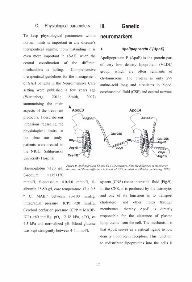
1717
C. Physiological parameters
To keep physiological parameters within
normal limits is important in any disease’s
therapeutical regime, notwithstanding it is
even more important in aSAH, when the
central coordination of the different
mechanisms is failing. Comprehensive
therapeutical guidelines for the management
of SAH patients in the Neurointensive Care
setting were published a few years ago
(Wartenberg, 2011; Smith, 2007)
summarising the main
aspects of the treatment
protocols. I describe our
intensions regarding the
physiological limits, at
the time our study-
patients were treated in
the NICU, Sahlgrenska
University Hospital.
Haemoglobin >120 g/l,
S-sodium >135<150
mmol/l, S-potassium 4.0-5.0 mmol/l, S-
albumin 35-50 g/l, core temperature 37 ± 0.5
° C, MABP between 70-100 mmHg,
intracranial pressure (ICP) <20 mmHg,
Cerebral perfusion pressure (CPP = MABP-
ICP) >60 mmHg, pO2 12-18 kPa, pCO2 ca
4.5 kPa and normalized pH. Blood glucose
was kept stringently between 4-6 mmol/l.
III. Genetic neuromarkers
1. Apolipoprotein E (ApoE)
Apolipoprotein E (ApoE) is the protein-part
of very low density lipoprotein (VLDL)
group, which are often remnants of
chylomicrons. The protein is only 299
amino-acid long and circulates in blood,
cerebrospinal fluid (CSF) and central nervous
system (CNS) tissue interstitial fluid (Fig.9).
In the CNS, it is produced by the astrocytes
and one of its functions is to transport
cholesterol and other lipids through
membranes, thereby ApoE is directly
responsible for the clearance of plasma
lipoproteins from the cell. The mechanism is
that ApoE serves as a critical ligand to low
density lipoprotein receptors. This function,
to redistribute lipoproteins into the cells is
Figure 9. Apolipoprotein E3 and E4’s 3D structure. Note the difference in mobility of the arm, and thence difference in function! With permission. (Mahley and Huang, 2012)
16
Stroke Scale (NIHSS)
NIHSS is a focal neurological deficit
assessment scale which was developed by
Brott and co-workers (Brott et al., 1989) to
document impairment after a stroke, It has
been widely accepted as a study instrument,
and is
recommended
by the
American
Heart
Association for
outcome
classification
even in clinical
use. It assigns
points for focal
neurological
deficits on an
increasing
scale in the
areas of
language,
speech,
coordination,
visual field,
neglect, eye-movement, consciousness and
motor- and sensory functions (Table 7).
3. The Barthel Index
The Barthel index is a measure of
independence in 10 important activities of
daily living (Table 8). It has been a valuable
instrument to estimate the amount of
assistance a person requires after an illness or
injury. It was initially designed to monitor
improvement after treatment in patients with
chronic neurological diseases (Mahoney and
Barthel, 1965). It measures the functional
capacity in eating, bathing, dressing, walking
and getting out of bed and chair. The points
in the index give a good estimate on how
much nursing assistance a person requires.
On the contrary, patients achieving full credit
in the Index, are not necessarily capable of
living an independent life.
Table 8. The Barthel Index. Assessing Activities of Daily Living (ADL), a measure of independence
2. National Institute of Health
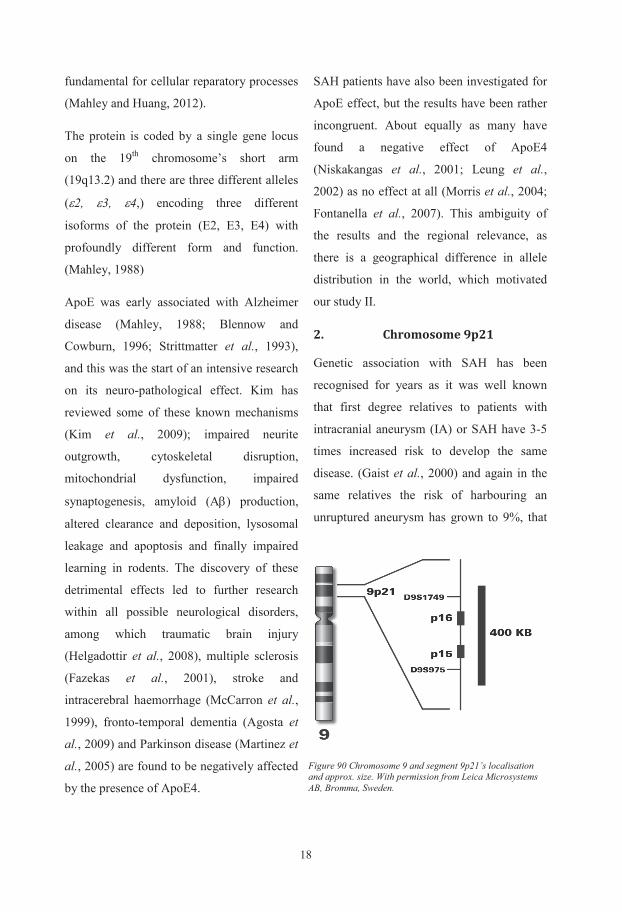
1819
is 4.5-time-increase of risk
compared to the general population.
(Ronkainen et al., 1997)
These family-linkage studies and
large genome-wide association
studies (GWAS) identifying a locus
on the 9th chromosome’s short arm
(9p21) as risk area for IA (Bilguvar
et al., 2008) and even other blood-
vessel related anomalies, like
coronary artery disease (CAD),
aortic aneurysm, and arterial
stiffness encouraged further investigation in
the region to search for the responsible
genetic variant (Fig.10). As no causative
genotype had been identified on this location
at that time, we had to further narrow down
the pursuit to single nucleotide
polymorphisms (SNPs) – one single base-
pair interchanges, which are the most
frequent occurring genetic alterations. We
have investigated in Paper III, if any of the
identified SNPs correlate to ruptured IAs
independently from other risk factors.
IV. Biochemical neuromarkers
1. C-Reactive Protein (CRP)
CRP was discovered by Tillett and acquired
its name from a reaction with the C-
polysaccharide of Pneumococcus (Tillett and
Francis, 1930). It is one of the body’s most
important acute-phase proteins mainly
synthesised in the liver. It responds to
cytokines, mainly IL-6, secreted by
macrophages, T-cells and also adipocytes. It
has an annular form and belongs among the
pentameric proteins, which are common as
ionic-channel receptors or viral capsids. They
can form a channel, expressing receptors and
with their ligand attached, they often undergo
a conformational change, thereby interacting
with the molecule (often neutralising, or
facilitating transport). It conforms to its
putative roll to detoxify and neutralise
harmful substances escaping into the
circulation (Fig.11).
As an acute-phase protein, CRP’s
concentration rises quickly and can increase
well over 1000-fold and peaks around 48 h
after the insult and returns to base-line after
Figure 10. C-Reactive Protein with its 5 sub-units organised in an annular, pentameric form. (Public domain)
18
fundamental for cellular reparatory processes
(Mahley and Huang, 2012).
The protein is coded by a single gene locus
on the 19th chromosome’s short arm
(19q13.2) and there are three different alleles
(ε2, ε3, ε4,) encoding three different
isoforms of the protein (E2, E3, E4) with
profoundly different form and function.
(Mahley, 1988)
ApoE was early associated with Alzheimer
disease (Mahley, 1988; Blennow and
Cowburn, 1996; Strittmatter et al., 1993),
and this was the start of an intensive research
on its neuro-pathological effect. Kim has
reviewed some of these known mechanisms
(Kim et al., 2009); impaired neurite
outgrowth, cytoskeletal disruption,
mitochondrial dysfunction, impaired
synaptogenesis, amyloid (Aβ) production,
altered clearance and deposition, lysosomal
leakage and apoptosis and finally impaired
learning in rodents. The discovery of these
detrimental effects led to further research
within all possible neurological disorders,
among which traumatic brain injury
(Helgadottir et al., 2008), multiple sclerosis
(Fazekas et al., 2001), stroke and
intracerebral haemorrhage (McCarron et al.,
1999), fronto-temporal dementia (Agosta et
al., 2009) and Parkinson disease (Martinez et
al., 2005) are found to be negatively affected
by the presence of ApoE4.
SAH patients have also been investigated for
ApoE effect, but the results have been rather
incongruent. About equally as many have
found a negative effect of ApoE4
(Niskakangas et al., 2001; Leung et al.,
2002) as no effect at all (Morris et al., 2004;
Fontanella et al., 2007). This ambiguity of
the results and the regional relevance, as
there is a geographical difference in allele
distribution in the world, which motivated
our study II.
2. Chromosome 9p21
Genetic association with SAH has been
recognised for years as it was well known
that first degree relatives to patients with
intracranial aneurysm (IA) or SAH have 3-5
times increased risk to develop the same
disease. (Gaist et al., 2000) and again in the
same relatives the risk of harbouring an
unruptured aneurysm has grown to 9%, that
Figure 90 Chromosome 9 and segment 9p21’s localisation and approx. size. With permission from Leica Microsystems AB, Bromma, Sweden.
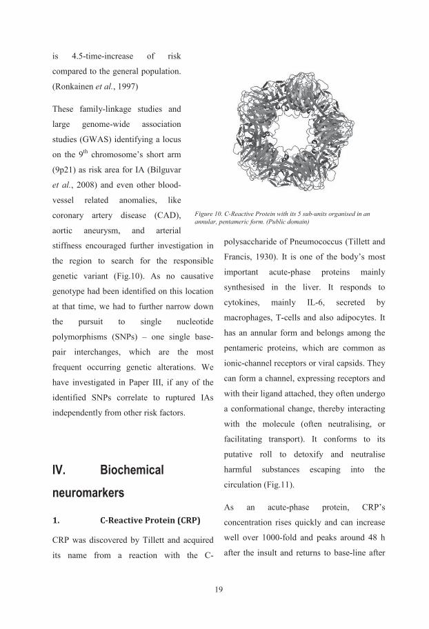
1919
is 4.5-time-increase of risk
compared to the general population.
(Ronkainen et al., 1997)
These family-linkage studies and
large genome-wide association
studies (GWAS) identifying a locus
on the 9th chromosome’s short arm
(9p21) as risk area for IA (Bilguvar
et al., 2008) and even other blood-
vessel related anomalies, like
coronary artery disease (CAD),
aortic aneurysm, and arterial
stiffness encouraged further investigation in
the region to search for the responsible
genetic variant (Fig.10). As no causative
genotype had been identified on this location
at that time, we had to further narrow down
the pursuit to single nucleotide
polymorphisms (SNPs) – one single base-
pair interchanges, which are the most
frequent occurring genetic alterations. We
have investigated in Paper III, if any of the
identified SNPs correlate to ruptured IAs
independently from other risk factors.
IV. Biochemical neuromarkers
1. C-Reactive Protein (CRP)
CRP was discovered by Tillett and acquired
its name from a reaction with the C-
polysaccharide of Pneumococcus (Tillett and
Francis, 1930). It is one of the body’s most
important acute-phase proteins mainly
synthesised in the liver. It responds to
cytokines, mainly IL-6, secreted by
macrophages, T-cells and also adipocytes. It
has an annular form and belongs among the
pentameric proteins, which are common as
ionic-channel receptors or viral capsids. They
can form a channel, expressing receptors and
with their ligand attached, they often undergo
a conformational change, thereby interacting
with the molecule (often neutralising, or
facilitating transport). It conforms to its
putative roll to detoxify and neutralise
harmful substances escaping into the
circulation (Fig.11).
As an acute-phase protein, CRP’s
concentration rises quickly and can increase
well over 1000-fold and peaks around 48 h
after the insult and returns to base-line after
Figure 10. C-Reactive Protein with its 5 sub-units organised in an annular, pentameric form. (Public domain)
18
fundamental for cellular reparatory processes
(Mahley and Huang, 2012).
The protein is coded by a single gene locus
on the 19th chromosome’s short arm
(19q13.2) and there are three different alleles
(ε2, ε3, ε4,) encoding three different
isoforms of the protein (E2, E3, E4) with
profoundly different form and function.
(Mahley, 1988)
ApoE was early associated with Alzheimer
disease (Mahley, 1988; Blennow and
Cowburn, 1996; Strittmatter et al., 1993),
and this was the start of an intensive research
on its neuro-pathological effect. Kim has
reviewed some of these known mechanisms
(Kim et al., 2009); impaired neurite
outgrowth, cytoskeletal disruption,
mitochondrial dysfunction, impaired
synaptogenesis, amyloid (Aβ) production,
altered clearance and deposition, lysosomal
leakage and apoptosis and finally impaired
learning in rodents. The discovery of these
detrimental effects led to further research
within all possible neurological disorders,
among which traumatic brain injury
(Helgadottir et al., 2008), multiple sclerosis
(Fazekas et al., 2001), stroke and
intracerebral haemorrhage (McCarron et al.,
1999), fronto-temporal dementia (Agosta et
al., 2009) and Parkinson disease (Martinez et
al., 2005) are found to be negatively affected
by the presence of ApoE4.
SAH patients have also been investigated for
ApoE effect, but the results have been rather
incongruent. About equally as many have
found a negative effect of ApoE4
(Niskakangas et al., 2001; Leung et al.,
2002) as no effect at all (Morris et al., 2004;
Fontanella et al., 2007). This ambiguity of
the results and the regional relevance, as
there is a geographical difference in allele
distribution in the world, which motivated
our study II.
2. Chromosome 9p21
Genetic association with SAH has been
recognised for years as it was well known
that first degree relatives to patients with
intracranial aneurysm (IA) or SAH have 3-5
times increased risk to develop the same
disease. (Gaist et al., 2000) and again in the
same relatives the risk of harbouring an
unruptured aneurysm has grown to 9%, that
Figure 90 Chromosome 9 and segment 9p21’s localisation and approx. size. With permission from Leica Microsystems AB, Bromma, Sweden.
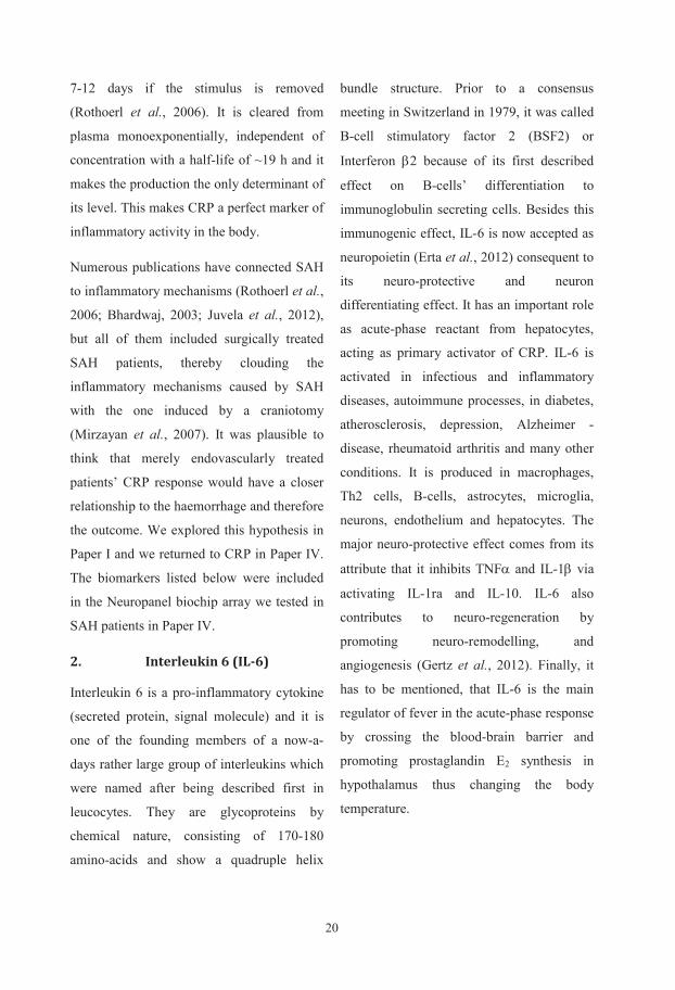
2021
3. Tumor Necrosis Factor
Receptor 1 (TNRF1)
TNFR1, together with TNFR2 are receptors
for tumour necrosis factor α (TNFα) and
lymphotoxin α (LTα), both pro-
inflammatory cytokines and they are
expressed on most cell types in the body.
TNFα and LTα play a major role in immune
regulation and in host-defence reactions.
They are expressed mainly by macrophages
after cell injury or inflammation but other
haematopoietic or non-hematopoietic cell-
types can be involved. (Probert et al., 2000)
TNFα is responsible for recruitment of
leucocytes to inflammation area and
regulates cell-death by proliferation,
cytotoxicity and apoptosis. (Wallach et al.,
1997) The majority of biological response to
TNFα manifests through TNFR1 receptors,
while TNFR2 has some special functions. It
mediates tymocyte proliferation and certain
suppression of inflammatory processes
(Peschon et al., 1998). TNFR1 receptor
mediates endotoxin shock and overexpressed
TNFα/ LTα or TNRF1 is detrimental,
resulting in rheumatoid arthritis or multiple
sclerosis. TNFα/ LTα’s pro-inflammatory
properties demonstrate local inflammation in
transgenic mice and their cytotoxic effects
can trigger apoptosis and cell-death in
oligodendrocytes. (Selmaj and Raine, 1988).
TNFα studies are however contradictory and
it may depend on the short half-life of this
cytokine, and that it can bind to undetectable
complexes (Beutler et al., 1985). It has been
suggested that the soluble forms of TNFR1
and 2 with their longer half-life are more
reliable indicator of the TNF system
activation than the short-lived cytokine itself
(Kreuzer et al., 1996). Moreover the soluble
TNFR1 can be viewed as an independent
inflammation marker as its transition to
soluble isomer is a complex activity,
dependent on other inflammatory
mechanisms (Diez-Ruiz et al., 1995).
4. Neutrophil Gelatinase
Associated Lipocalin (NGAL)
NGAL or Lipocalin-2 is a 25 kDa
glycoprotein and has its principal function in
innate immunity where it concentrates and
sequesters iron in the form of siderophores.
This defence mechanism is pivotal in
bacterial protection (Yang et al., 2002). It
was first described in neutrophils, but now
nearly all other cell-types have been shown
to express NGAL under different
circumstances e.g. in the brain under
oxidative stress (Naude et al., 2012).
It is widely used as marker for acute kidney
injury and a general inflammation signalling
substance. During inflammatory processes
the expression of NGAL is altered i.e. in
meningitis, myocarditis, psoriasis,
20
7-12 days if the stimulus is removed
(Rothoerl et al., 2006). It is cleared from
plasma monoexponentially, independent of
concentration with a half-life of ~19 h and it
makes the production the only determinant of
its level. This makes CRP a perfect marker of
inflammatory activity in the body.
Numerous publications have connected SAH
to inflammatory mechanisms (Rothoerl et al.,
2006; Bhardwaj, 2003; Juvela et al., 2012),
but all of them included surgically treated
SAH patients, thereby clouding the
inflammatory mechanisms caused by SAH
with the one induced by a craniotomy
(Mirzayan et al., 2007). It was plausible to
think that merely endovascularly treated
patients’ CRP response would have a closer
relationship to the haemorrhage and therefore
the outcome. We explored this hypothesis in
Paper I and we returned to CRP in Paper IV.
The biomarkers listed below were included
in the Neuropanel biochip array we tested in
SAH patients in Paper IV.
2. Interleukin 6 (IL-6)
Interleukin 6 is a pro-inflammatory cytokine
(secreted protein, signal molecule) and it is
one of the founding members of a now-a-
days rather large group of interleukins which
were named after being described first in
leucocytes. They are glycoproteins by
chemical nature, consisting of 170-180
amino-acids and show a quadruple helix
bundle structure. Prior to a consensus
meeting in Switzerland in 1979, it was called
B-cell stimulatory factor 2 (BSF2) or
Interferon β2 because of its first described
effect on B-cells’ differentiation to
immunoglobulin secreting cells. Besides this
immunogenic effect, IL-6 is now accepted as
neuropoietin (Erta et al., 2012) consequent to
its neuro-protective and neuron
differentiating effect. It has an important role
as acute-phase reactant from hepatocytes,
acting as primary activator of CRP. IL-6 is
activated in infectious and inflammatory
diseases, autoimmune processes, in diabetes,
atherosclerosis, depression, Alzheimer -
disease, rheumatoid arthritis and many other
conditions. It is produced in macrophages,
Th2 cells, B-cells, astrocytes, microglia,
neurons, endothelium and hepatocytes. The
major neuro-protective effect comes from its
attribute that it inhibits TNFα and IL-1β via
activating IL-1ra and IL-10. IL-6 also
contributes to neuro-regeneration by
promoting neuro-remodelling, and
angiogenesis (Gertz et al., 2012). Finally, it
has to be mentioned, that IL-6 is the main
regulator of fever in the acute-phase response
by crossing the blood-brain barrier and
promoting prostaglandin E2 synthesis in
hypothalamus thus changing the body
temperature.
2121
3. Tumor Necrosis Factor
Receptor 1 (TNRF1)
TNFR1, together with TNFR2 are receptors
for tumour necrosis factor α (TNFα) and
lymphotoxin α (LTα), both pro-
inflammatory cytokines and they are
expressed on most cell types in the body.
TNFα and LTα play a major role in immune
regulation and in host-defence reactions.
They are expressed mainly by macrophages
after cell injury or inflammation but other
haematopoietic or non-hematopoietic cell-
types can be involved. (Probert et al., 2000)
TNFα is responsible for recruitment of
leucocytes to inflammation area and
regulates cell-death by proliferation,
cytotoxicity and apoptosis. (Wallach et al.,
1997) The majority of biological response to
TNFα manifests through TNFR1 receptors,
while TNFR2 has some special functions. It
mediates tymocyte proliferation and certain
suppression of inflammatory processes
(Peschon et al., 1998). TNFR1 receptor
mediates endotoxin shock and overexpressed
TNFα/ LTα or TNRF1 is detrimental,
resulting in rheumatoid arthritis or multiple
sclerosis. TNFα/ LTα’s pro-inflammatory
properties demonstrate local inflammation in
transgenic mice and their cytotoxic effects
can trigger apoptosis and cell-death in
oligodendrocytes. (Selmaj and Raine, 1988).
TNFα studies are however contradictory and
it may depend on the short half-life of this
cytokine, and that it can bind to undetectable
complexes (Beutler et al., 1985). It has been
suggested that the soluble forms of TNFR1
and 2 with their longer half-life are more
reliable indicator of the TNF system
activation than the short-lived cytokine itself
(Kreuzer et al., 1996). Moreover the soluble
TNFR1 can be viewed as an independent
inflammation marker as its transition to
soluble isomer is a complex activity,
dependent on other inflammatory
mechanisms (Diez-Ruiz et al., 1995).
4. Neutrophil Gelatinase
Associated Lipocalin (NGAL)
NGAL or Lipocalin-2 is a 25 kDa
glycoprotein and has its principal function in
innate immunity where it concentrates and
sequesters iron in the form of siderophores.
This defence mechanism is pivotal in
bacterial protection (Yang et al., 2002). It
was first described in neutrophils, but now
nearly all other cell-types have been shown
to express NGAL under different
circumstances e.g. in the brain under
oxidative stress (Naude et al., 2012).
It is widely used as marker for acute kidney
injury and a general inflammation signalling
substance. During inflammatory processes
the expression of NGAL is altered i.e. in
meningitis, myocarditis, psoriasis,
20
7-12 days if the stimulus is removed
(Rothoerl et al., 2006). It is cleared from
plasma monoexponentially, independent of
concentration with a half-life of ~19 h and it
makes the production the only determinant of
its level. This makes CRP a perfect marker of
inflammatory activity in the body.
Numerous publications have connected SAH
to inflammatory mechanisms (Rothoerl et al.,
2006; Bhardwaj, 2003; Juvela et al., 2012),
but all of them included surgically treated
SAH patients, thereby clouding the
inflammatory mechanisms caused by SAH
with the one induced by a craniotomy
(Mirzayan et al., 2007). It was plausible to
think that merely endovascularly treated
patients’ CRP response would have a closer
relationship to the haemorrhage and therefore
the outcome. We explored this hypothesis in
Paper I and we returned to CRP in Paper IV.
The biomarkers listed below were included
in the Neuropanel biochip array we tested in
SAH patients in Paper IV.
2. Interleukin 6 (IL-6)
Interleukin 6 is a pro-inflammatory cytokine
(secreted protein, signal molecule) and it is
one of the founding members of a now-a-
days rather large group of interleukins which
were named after being described first in
leucocytes. They are glycoproteins by
chemical nature, consisting of 170-180
amino-acids and show a quadruple helix
bundle structure. Prior to a consensus
meeting in Switzerland in 1979, it was called
B-cell stimulatory factor 2 (BSF2) or
Interferon β2 because of its first described
effect on B-cells’ differentiation to
immunoglobulin secreting cells. Besides this
immunogenic effect, IL-6 is now accepted as
neuropoietin (Erta et al., 2012) consequent to
its neuro-protective and neuron
differentiating effect. It has an important role
as acute-phase reactant from hepatocytes,
acting as primary activator of CRP. IL-6 is
activated in infectious and inflammatory
diseases, autoimmune processes, in diabetes,
atherosclerosis, depression, Alzheimer -
disease, rheumatoid arthritis and many other
conditions. It is produced in macrophages,
Th2 cells, B-cells, astrocytes, microglia,
neurons, endothelium and hepatocytes. The
major neuro-protective effect comes from its
attribute that it inhibits TNFα and IL-1β via
activating IL-1ra and IL-10. IL-6 also
contributes to neuro-regeneration by
promoting neuro-remodelling, and
angiogenesis (Gertz et al., 2012). Finally, it
has to be mentioned, that IL-6 is the main
regulator of fever in the acute-phase response
by crossing the blood-brain barrier and
promoting prostaglandin E2 synthesis in
hypothalamus thus changing the body
temperature.
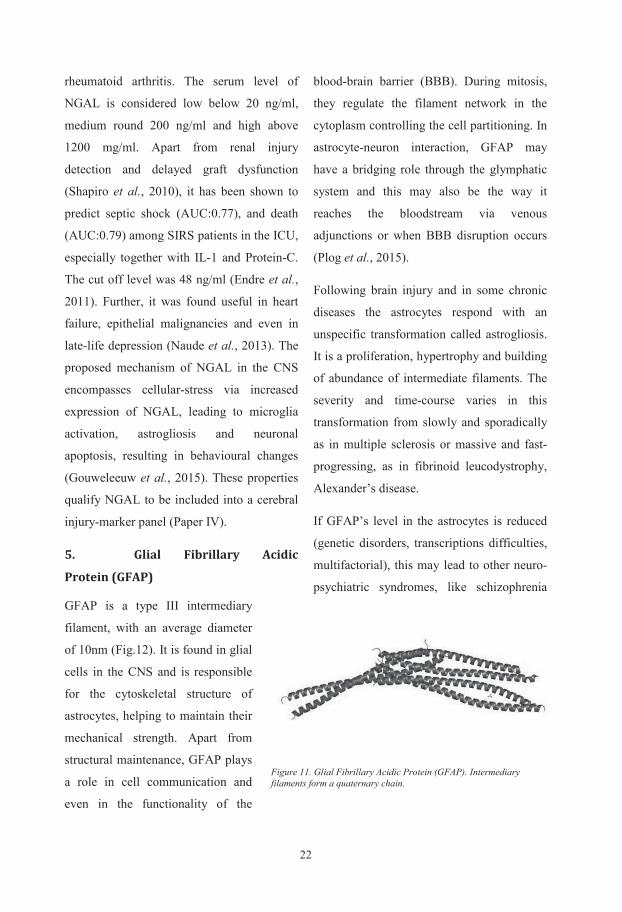
2223
and depression. The serum levels are
normally undetectable if not an immense
disruption of BBB occurs e.g. SAH, cerebral
trauma, intra or cerebral haemorrhage
(Mayer et al., 2013). In that case the
concentration is closely associated with the
injury size (Nylen et al., 2007; Nylen et al.,
2006; Nylen, 2007).
6. Brain-Derived Neutrophic
Factor (BDNF)
BDNF is a signal protein and one of the most
prominent members of the neurotrophins or
nerve growth factors. Its closest relatives in
this family are the neurotrophins 3 and 4 and
nerve growth factor (NGF). It is synthesised
in the endoplasmic reticulum and passed on
into vesicles in the brain and the periphery,
like in retina, kidneys saliva and the prostate.
It acts on the neurons, promoting neuronal
survival, neurogenesis and synaptogenesis.
(Lu, 2003) In animal studies it reduces
ischemic injury and improves recovery and
post-injury regeneration (Almeida et al.,
2005).
BDNF is most active in the cortex,
hippocampus, basal ganglia and areas, where
learning, memory and higher thinking take
place. It may work even in adult brain
through neural stem-cells, by promoting
neuroneogenesis (Pencea et al., 2001). In
knock-out mice, it produces severe brain
developmental defects and even perinatal
death. This emphasises the importance of this
neurotrophin in the embryonal brain
formation and neuronal growth. Normal
serum level was reported round 30 ng/ml and
low values were considered around half that
level. It is easy to understand why this
neuromarker is included in a neuropanel-
array.
7. Fatty-Acid Binding Protein
(FABP)
Lipids, where fatty-acids, eicosanoids and
retinoids are included function not only as
fuel source, building-blocks to membranes
and cellular structures, but also as intra- and
extra-cellular signalling substances. Recent
studies, summarised in a review
(Hotamisligil and Bernlohr, 2015), enlighten
us that lipids take part (i) in modifying the
actions and locations of proteins, such as
kinases or ion-channels; (ii) signal through
proteins to cell surface or between cells as G-
protein; (iii) ligand for transcription factors,
modifying expressions of other regulators;
(iv) regulate hormone actions, like PI3K, and
NFκβK pathways and finally (v) pattern
recognition receptors.
The insolubility and toxic effects of the free
forms of these molecules, require, however a
type of non-catalytic binding-protein e.g.
FABPs. The original FABP was described in
small intestine as a 12 kDa intracellular
protein (Ockner et al., 1972) but since then
22
rheumatoid arthritis. The serum level of
NGAL is considered low below 20 ng/ml,
medium round 200 ng/ml and high above
1200 mg/ml. Apart from renal injury
detection and delayed graft dysfunction
(Shapiro et al., 2010), it has been shown to
predict septic shock (AUC:0.77), and death
(AUC:0.79) among SIRS patients in the ICU,
especially together with IL-1 and Protein-C.
The cut off level was 48 ng/ml (Endre et al.,
2011). Further, it was found useful in heart
failure, epithelial malignancies and even in
late-life depression (Naude et al., 2013). The
proposed mechanism of NGAL in the CNS
encompasses cellular-stress via increased
expression of NGAL, leading to microglia
activation, astrogliosis and neuronal
apoptosis, resulting in behavioural changes
(Gouweleeuw et al., 2015). These properties
qualify NGAL to be included into a cerebral
injury-marker panel (Paper IV).
5. Glial Fibrillary Acidic
Protein (GFAP)
GFAP is a type III intermediary
filament, with an average diameter
of 10nm (Fig.12). It is found in glial
cells in the CNS and is responsible
for the cytoskeletal structure of
astrocytes, helping to maintain their
mechanical strength. Apart from
structural maintenance, GFAP plays
a role in cell communication and
even in the functionality of the
blood-brain barrier (BBB). During mitosis,
they regulate the filament network in the
cytoplasm controlling the cell partitioning. In
astrocyte-neuron interaction, GFAP may
have a bridging role through the glymphatic
system and this may also be the way it
reaches the bloodstream via venous
adjunctions or when BBB disruption occurs
(Plog et al., 2015).
Following brain injury and in some chronic
diseases the astrocytes respond with an
unspecific transformation called astrogliosis.
It is a proliferation, hypertrophy and building
of abundance of intermediate filaments. The
severity and time-course varies in this
transformation from slowly and sporadically
as in multiple sclerosis or massive and fast-
progressing, as in fibrinoid leucodystrophy,
Alexander’s disease.
If GFAP’s level in the astrocytes is reduced
(genetic disorders, transcriptions difficulties,
multifactorial), this may lead to other neuro-
psychiatric syndromes, like schizophrenia
Figure 11. Glial Fibrillary Acidic Protein (GFAP). Intermediary filaments form a quaternary chain.
2323
and depression. The serum levels are
normally undetectable if not an immense
disruption of BBB occurs e.g. SAH, cerebral
trauma, intra or cerebral haemorrhage
(Mayer et al., 2013). In that case the
concentration is closely associated with the
injury size (Nylen et al., 2007; Nylen et al.,
2006; Nylen, 2007).
6. Brain-Derived Neutrophic
Factor (BDNF)
BDNF is a signal protein and one of the most
prominent members of the neurotrophins or
nerve growth factors. Its closest relatives in
this family are the neurotrophins 3 and 4 and
nerve growth factor (NGF). It is synthesised
in the endoplasmic reticulum and passed on
into vesicles in the brain and the periphery,
like in retina, kidneys saliva and the prostate.
It acts on the neurons, promoting neuronal
survival, neurogenesis and synaptogenesis.
(Lu, 2003) In animal studies it reduces
ischemic injury and improves recovery and
post-injury regeneration (Almeida et al.,
2005).
BDNF is most active in the cortex,
hippocampus, basal ganglia and areas, where
learning, memory and higher thinking take
place. It may work even in adult brain
through neural stem-cells, by promoting
neuroneogenesis (Pencea et al., 2001). In
knock-out mice, it produces severe brain
developmental defects and even perinatal
death. This emphasises the importance of this
neurotrophin in the embryonal brain
formation and neuronal growth. Normal
serum level was reported round 30 ng/ml and
low values were considered around half that
level. It is easy to understand why this
neuromarker is included in a neuropanel-
array.
7. Fatty-Acid Binding Protein
(FABP)
Lipids, where fatty-acids, eicosanoids and
retinoids are included function not only as
fuel source, building-blocks to membranes
and cellular structures, but also as intra- and
extra-cellular signalling substances. Recent
studies, summarised in a review
(Hotamisligil and Bernlohr, 2015), enlighten
us that lipids take part (i) in modifying the
actions and locations of proteins, such as
kinases or ion-channels; (ii) signal through
proteins to cell surface or between cells as G-
protein; (iii) ligand for transcription factors,
modifying expressions of other regulators;
(iv) regulate hormone actions, like PI3K, and
NFκβK pathways and finally (v) pattern
recognition receptors.
The insolubility and toxic effects of the free
forms of these molecules, require, however a
type of non-catalytic binding-protein e.g.
FABPs. The original FABP was described in
small intestine as a 12 kDa intracellular
protein (Ockner et al., 1972) but since then
22
rheumatoid arthritis. The serum level of
NGAL is considered low below 20 ng/ml,
medium round 200 ng/ml and high above
1200 mg/ml. Apart from renal injury
detection and delayed graft dysfunction
(Shapiro et al., 2010), it has been shown to
predict septic shock (AUC:0.77), and death
(AUC:0.79) among SIRS patients in the ICU,
especially together with IL-1 and Protein-C.
The cut off level was 48 ng/ml (Endre et al.,
2011). Further, it was found useful in heart
failure, epithelial malignancies and even in
late-life depression (Naude et al., 2013). The
proposed mechanism of NGAL in the CNS
encompasses cellular-stress via increased
expression of NGAL, leading to microglia
activation, astrogliosis and neuronal
apoptosis, resulting in behavioural changes
(Gouweleeuw et al., 2015). These properties
qualify NGAL to be included into a cerebral
injury-marker panel (Paper IV).
5. Glial Fibrillary Acidic
Protein (GFAP)
GFAP is a type III intermediary
filament, with an average diameter
of 10nm (Fig.12). It is found in glial
cells in the CNS and is responsible
for the cytoskeletal structure of
astrocytes, helping to maintain their
mechanical strength. Apart from
structural maintenance, GFAP plays
a role in cell communication and
even in the functionality of the
blood-brain barrier (BBB). During mitosis,
they regulate the filament network in the
cytoplasm controlling the cell partitioning. In
astrocyte-neuron interaction, GFAP may
have a bridging role through the glymphatic
system and this may also be the way it
reaches the bloodstream via venous
adjunctions or when BBB disruption occurs
(Plog et al., 2015).
Following brain injury and in some chronic
diseases the astrocytes respond with an
unspecific transformation called astrogliosis.
It is a proliferation, hypertrophy and building
of abundance of intermediate filaments. The
severity and time-course varies in this
transformation from slowly and sporadically
as in multiple sclerosis or massive and fast-
progressing, as in fibrinoid leucodystrophy,
Alexander’s disease.
If GFAP’s level in the astrocytes is reduced
(genetic disorders, transcriptions difficulties,
multifactorial), this may lead to other neuro-
psychiatric syndromes, like schizophrenia
Figure 11. Glial Fibrillary Acidic Protein (GFAP). Intermediary filaments form a quaternary chain.
2425
9. D-dimer (DDMR)
DDMR is fibrin degradation protein (FDP),
which is a marker of the coagulation system
activation. The coagulation system can be
activated by intrinsic (endothelial activation)
or extrinsic (tissue activating factors via
Factor VII) pathways and both of them
interconnect to fibrinogen, which in the
presence of Ca2+ ions as cofactors and an
activated thrombin converts to fibrinogen -
polymers and further with the help of a
thrombin-activated Factor XIII to fibrin clot.
The activated Factor XIIIa binds a glutamyl -
lysyl amid crosslink which stabilises the
blood clot. When the wound heals the
coagulation stimulus ceases and fibrinolytic
mechanisms start to take over. The enzyme
plasmin starts to break down the fibin-clot to
high molecular-weight polymers and then
further to small polymers, FDPs. One of
these FDPs is DDMR.
It received its name that fibrinogen’s two D
domains and one E domain is cross-linked
together (Fig.13). It is normally undetectable
in serum and increases rapidly when the
coagulo-fibrinolytic system is activated, like
in DIC, thrombosis, DVT, PE,
and sinus thrombosis. DDMR
was discovered in 1973 and it
came to routine use at the end
of 1990s. It is tested with
monoclonal antibodies with a
reference value below 0.5
mg/L.
As SAH produces blood-clot
and microvascular coagulation
is suggested as causative
mechanism for DCI, the
coagulation system is involved in the
pathological mechanism of the disease. This
makes DDMR a neuro-marker to
consecutively analyse.
Figure 13. Coagulation mechanism and the early fibrinolysis, producing D-dimer (Public domain)
24
similar molecules were shown in kidneys,
adipose-tissue, myocardium, liver and brain.
Their function were further elucidated and
revealed that they not only buffer and
physically transport lipids, but they serve as
mediators and as communicating agents
within and between cells and between organs
thus supporting immunometabolism. In the
CNS, 4 isoforms are detected B-(brain), H-
(heart), E-(epidermal) and M-(myelin)FABP.
B-FABP is present mainly in astrocytes, H-
FABP, the most prominent of all FABSs, in
neurons and finally M-FABP is found in
peripheral nerves (Pelsers and Glatz, 2005).
Their role could be demonstrated in all
metabolic diseases, like diabetes, obesity and
also in coronary artery disease, inflammatory
conditions and in the CNS: in stroke, cerebral
injury and neurodegenerative diseases, like
Alzheimers and Creutzfeldt-Jakob disease. It
is a fast-reacting injury marker, and after 2-3
hours B- & H-FABP rises in serum and stay
elevated up to 120 hours. The cut-off serum
concentration for cell injury is 5 - 6 µg/L.
(Pelsers et al., 2004)
8. Neuron-Specific Enolase
(NSE)
NSE was first discovered in 1965 (Moore
and McGregor, 1965) as an intracellular
protein from the brain. Later it was described
as a 78 kDa large, dimeric isoenzyme to a
glycolytic enzyme, enolase. NSE can be
found in the neuron’s cytoplasm; where it
circulates with axoplasmic transport. (Cheng
et al., 2014) but normally it is not secreted
out of the cell. It can appear in CSF and
thereafter in the serum with neuronal
damage. It is hardly detectable in healthy
individuals but increasing rapidly with neural
disruption. This quality makes it theoretically
an ideal neuronal injury marker, explaining
why it has been investigated intensively.
Gradisek recommended it as prognostic
biomarker and therapeutic indicator after
traumatic brain injury. (Gradisek et al., 2012;
Olivecrona and Koskinen, 2012)
Recent meta-analysis in 16 studies found a
strong correlation between NSE serum levels
and outcome or death. (Cheng et al., 2014)
They found 100 % sensitivity for poor
outcome and mortality at the levels of 11.6 –
20 µg/L and > 20 µg/L respectively. NSE
rises within 12 hours of the neural injury and
decreases within hours if the neural cell-
disintegration ceases as the protein has a
half-life of ~ 24 hours.

2525
9. D-dimer (DDMR)
DDMR is fibrin degradation protein (FDP),
which is a marker of the coagulation system
activation. The coagulation system can be
activated by intrinsic (endothelial activation)
or extrinsic (tissue activating factors via
Factor VII) pathways and both of them
interconnect to fibrinogen, which in the
presence of Ca2+ ions as cofactors and an
activated thrombin converts to fibrinogen -
polymers and further with the help of a
thrombin-activated Factor XIII to fibrin clot.
The activated Factor XIIIa binds a glutamyl -
lysyl amid crosslink which stabilises the
blood clot. When the wound heals the
coagulation stimulus ceases and fibrinolytic
mechanisms start to take over. The enzyme
plasmin starts to break down the fibin-clot to
high molecular-weight polymers and then
further to small polymers, FDPs. One of
these FDPs is DDMR.
It received its name that fibrinogen’s two D
domains and one E domain is cross-linked
together (Fig.13). It is normally undetectable
in serum and increases rapidly when the
coagulo-fibrinolytic system is activated, like
in DIC, thrombosis, DVT, PE,
and sinus thrombosis. DDMR
was discovered in 1973 and it
came to routine use at the end
of 1990s. It is tested with
monoclonal antibodies with a
reference value below 0.5
mg/L.
As SAH produces blood-clot
and microvascular coagulation
is suggested as causative
mechanism for DCI, the
coagulation system is involved in the
pathological mechanism of the disease. This
makes DDMR a neuro-marker to
consecutively analyse.
Figure 13. Coagulation mechanism and the early fibrinolysis, producing D-dimer (Public domain)
24
similar molecules were shown in kidneys,
adipose-tissue, myocardium, liver and brain.
Their function were further elucidated and
revealed that they not only buffer and
physically transport lipids, but they serve as
mediators and as communicating agents
within and between cells and between organs
thus supporting immunometabolism. In the
CNS, 4 isoforms are detected B-(brain), H-
(heart), E-(epidermal) and M-(myelin)FABP.
B-FABP is present mainly in astrocytes, H-
FABP, the most prominent of all FABSs, in
neurons and finally M-FABP is found in
peripheral nerves (Pelsers and Glatz, 2005).
Their role could be demonstrated in all
metabolic diseases, like diabetes, obesity and
also in coronary artery disease, inflammatory
conditions and in the CNS: in stroke, cerebral
injury and neurodegenerative diseases, like
Alzheimers and Creutzfeldt-Jakob disease. It
is a fast-reacting injury marker, and after 2-3
hours B- & H-FABP rises in serum and stay
elevated up to 120 hours. The cut-off serum
concentration for cell injury is 5 - 6 µg/L.
(Pelsers et al., 2004)
8. Neuron-Specific Enolase
(NSE)
NSE was first discovered in 1965 (Moore
and McGregor, 1965) as an intracellular
protein from the brain. Later it was described
as a 78 kDa large, dimeric isoenzyme to a
glycolytic enzyme, enolase. NSE can be
found in the neuron’s cytoplasm; where it
circulates with axoplasmic transport. (Cheng
et al., 2014) but normally it is not secreted
out of the cell. It can appear in CSF and
thereafter in the serum with neuronal
damage. It is hardly detectable in healthy
individuals but increasing rapidly with neural
disruption. This quality makes it theoretically
an ideal neuronal injury marker, explaining
why it has been investigated intensively.
Gradisek recommended it as prognostic
biomarker and therapeutic indicator after
traumatic brain injury. (Gradisek et al., 2012;
Olivecrona and Koskinen, 2012)
Recent meta-analysis in 16 studies found a
strong correlation between NSE serum levels
and outcome or death. (Cheng et al., 2014)
They found 100 % sensitivity for poor
outcome and mortality at the levels of 11.6 –
20 µg/L and > 20 µg/L respectively. NSE
rises within 12 hours of the neural injury and
decreases within hours if the neural cell-
disintegration ceases as the protein has a
half-life of ~ 24 hours.
26
AIMS
In this clinical study on patients with aneurysmal subarachnoid haemorrhage (aSAH) we aimed to investigate the following questions:
To study the development of a well-known inflammatory marker C-reactive protein
during the early course of the disease and investigate, if these changes can be
associated to an established complication, cerebral vasospasm and the long-term
outcome.
To evaluate if a disadvantageous genetic variation in coding Apolipoprotein E has an
effect on disease incidence, complication frequency and outcome after aSAH
To elucidate, with the tools of genetics if proposed chromosome region 9p21 contains
information to predispose for aSAH.
To evaluate a novel investigation method, biochip-array neuropanel’s applicability in
aSAH patent-monitoring and its potential for outcome prediction.
1
PATIENTS AND METHODS
I. Inclusion
The study protocol was approved by the
University Ethics Committee Gothenburg,
Sweden with the allocation number S 161-00
and all the studies conformed to the Helsinki
Declaration on human research.
All patients were admitted to the
Neurointensive Care Unit at Sahlgrenska
University Hospital, Gothenburg, Sweden
between October 2000 and December 2002.
They were consecutively considered for
inclusion in the study if satisfying the
following criteria:
i. SAH was seen on CT-scan or
detected with cerebrospinal fluid
(CSF) analysis with increased red-
blood-cell count or the detection of
xanthochromia on CSF
spectrophotometry.
ii. The debut of symptoms, typically
thunderclap headache with altered
consciousness must have occurred
within 48 hours of admission,
although some patients might have
experienced headaches previously
(warning headache).
iii. After admission, a cerebral
angiography was performed with CT
angiography or conventional digital
subtractions angiography (DSA) with
3D image reconstruction and the
intracranial aneurysm/s had to be
identified in relation to the bleeding.
iv. The patient had to be permanently
residing in Sweden for long-term
outcome follow-up.
v. Finally an informed consent from the
patient or next of kin had to be
obtained within the first week of
treatment.
202 patients fulfilled the inclusion criteria
and were eligible for the studies but for other
criteria listed beneath different numbers of
patients were enlisted in the different studies.
II. Regime
Already after the diagnosis was established a
first-physician neurological assessment was
performed, (RLS, GCS, H&H, WFNS),
previous and present medical conditions were
noted, iv. tranexamic acid (Cyclocapron®)
administered, and intensified monitoring was
instituted. This involved invasive arterial
pressure recording and systolic blood
pressure management, keeping it below 160
mmHg even during transport from primary or
secondary health care centres. Securing the
airways and ventilation were mandatory if
the patient became unconscious. After
admittance to the university hospital, the
patients were treated and observed in the
intensive care unit, most of them in a
specialised neuro-intensive care unit (NICU).
27
AIMS
In this clinical study on patients with aneurysmal subarachnoid haemorrhage (aSAH) we aimed to investigate the following questions:
To study the development of a well-known inflammatory marker C-reactive protein
during the early course of the disease and investigate, if these changes can be
associated to an established complication, cerebral vasospasm and the long-term
outcome.
To evaluate if a disadvantageous genetic variation in coding Apolipoprotein E has an
effect on disease incidence, complication frequency and outcome after aSAH
To elucidate, with the tools of genetics if proposed chromosome region 9p21 contains
information to predispose for aSAH.
To evaluate a novel investigation method, biochip-array neuropanel’s applicability in
aSAH patent-monitoring and its potential for outcome prediction.
1
PATIENTS AND METHODS
I. Inclusion
The study protocol was approved by the
University Ethics Committee Gothenburg,
Sweden with the allocation number S 161-00
and all the studies conformed to the Helsinki
Declaration on human research.
All patients were admitted to the
Neurointensive Care Unit at Sahlgrenska
University Hospital, Gothenburg, Sweden
between October 2000 and December 2002.
They were consecutively considered for
inclusion in the study if satisfying the
following criteria:
i. SAH was seen on CT-scan or
detected with cerebrospinal fluid
(CSF) analysis with increased red-
blood-cell count or the detection of
xanthochromia on CSF
spectrophotometry.
ii. The debut of symptoms, typically
thunderclap headache with altered
consciousness must have occurred
within 48 hours of admission,
although some patients might have
experienced headaches previously
(warning headache).
iii. After admission, a cerebral
angiography was performed with CT
angiography or conventional digital
subtractions angiography (DSA) with
3D image reconstruction and the
intracranial aneurysm/s had to be
identified in relation to the bleeding.
iv. The patient had to be permanently
residing in Sweden for long-term
outcome follow-up.
v. Finally an informed consent from the
patient or next of kin had to be
obtained within the first week of
treatment.
202 patients fulfilled the inclusion criteria
and were eligible for the studies but for other
criteria listed beneath different numbers of
patients were enlisted in the different studies.
II. Regime
Already after the diagnosis was established a
first-physician neurological assessment was
performed, (RLS, GCS, H&H, WFNS),
previous and present medical conditions were
noted, iv. tranexamic acid (Cyclocapron®)
administered, and intensified monitoring was
instituted. This involved invasive arterial
pressure recording and systolic blood
pressure management, keeping it below 160
mmHg even during transport from primary or
secondary health care centres. Securing the
airways and ventilation were mandatory if
the patient became unconscious. After
admittance to the university hospital, the
patients were treated and observed in the
intensive care unit, most of them in a
specialised neuro-intensive care unit (NICU).
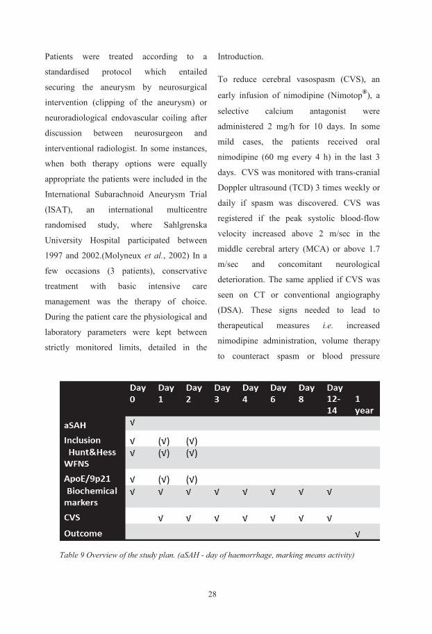
283
management.
During the patient treatment venous blood
samples were collected on days 0, 1, 2, 3, 4,
6, 8, and between days 10-14. Physiological
and laboratory parameters were registered
and any surgical interventions recorded.
After one year (11-13 month), a complete
neurological evaluation was performed and
patient status recorded with GOSE, NIHSS
and Barthel score by the same neurologist,
blinded to the investigated
(biochemical/genetical) markers. The study
plan is summarised on Table 9.
III. Data collection and analysis
A. Paper I. (CRP)
202 patients were considered for inclusion
with SAH, who were admitted to NICU. The
following patients were excluded: 3 patients
were admitted later than day 2 after the
haemorrhage, in 33 patients, no aneurysm
was detectable, no angiographic examination
could be performed in 4 patients and no
informed consent could be attained or the
consent withdrawn in 9 cases. As described
in the introduction craniectomy, as most
major surgical interventions increase CRP,
we excluded patients with surgical clipping
as therapy options (39 patients) and have
chosen to follow percutaneously treated
patients. As 3 patients were lost for follow-
up and in 13 patients consecutive data were
missing for AUC calculation, 98 patients
remained in this patient selection. The
patients’ demographics and characteristics at
admission and during observation can be
seen in Table 1 in Paper I.
Data collection routines were described
above (Table 1). Data analysis was
performed instantaneously in the hospitals
accredited laboratory with an
immunochemical kinetic turbidimetry
technique on a BH/Hitachi 917 analyser.
(Roche-Diagnostic, 2001)
B. Paper II. (APOE)
196 consecutive SAH patients with APOE
genetic information available, were
considered to be included to this genetic
study and 148 patients remained finally
enrolled. Of those non-eligible (mostly the
same as in Paper I), 3 patients were admitted
later than day 2, no aneurysm was detected in
33 patients, no informed consent/consent
withdrawal in 9 cases and finally 4 patients
lost for long-term outcome. 105 patients
treated with endovascular technique, in 39
patients the aneurysm was secured with
neurosurgical clipping. 4 patients were
conservatively treated since their clinical
condition did not permit active intervention.
221 population-based, healthy individuals
acted as control cohort, since we had the
2
Patients were treated according to a
standardised protocol which entailed
securing the aneurysm by neurosurgical
intervention (clipping of the aneurysm) or
neuroradiological endovascular coiling after
discussion between neurosurgeon and
interventional radiologist. In some instances,
when both therapy options were equally
appropriate the patients were included in the
International Subarachnoid Aneurysm Trial
(ISAT), an international multicentre
randomised study, where Sahlgrenska
University Hospital participated between
1997 and 2002.(Molyneux et al., 2002) In a
few occasions (3 patients), conservative
treatment with basic intensive care
management was the therapy of choice.
During the patient care the physiological and
laboratory parameters were kept between
strictly monitored limits, detailed in the
Introduction.
To reduce cerebral vasospasm (CVS), an
early infusion of nimodipine (Nimotop®), a
selective calcium antagonist were
administered 2 mg/h for 10 days. In some
mild cases, the patients received oral
nimodipine (60 mg every 4 h) in the last 3
days. CVS was monitored with trans-cranial
Doppler ultrasound (TCD) 3 times weekly or
daily if spasm was discovered. CVS was
registered if the peak systolic blood-flow
velocity increased above 2 m/sec in the
middle cerebral artery (MCA) or above 1.7
m/sec and concomitant neurological
deterioration. The same applied if CVS was
seen on CT or conventional angiography
(DSA). These signs needed to lead to
therapeutical measures i.e. increased
nimodipine administration, volume therapy
to counteract spasm or blood pressure
Table 9 Overview of the study plan. (aSAH - day of haemorrhage, marking means activity)
293
management.
During the patient treatment venous blood
samples were collected on days 0, 1, 2, 3, 4,
6, 8, and between days 10-14. Physiological
and laboratory parameters were registered
and any surgical interventions recorded.
After one year (11-13 month), a complete
neurological evaluation was performed and
patient status recorded with GOSE, NIHSS
and Barthel score by the same neurologist,
blinded to the investigated
(biochemical/genetical) markers. The study
plan is summarised on Table 9.
III. Data collection and analysis
A. Paper I. (CRP)
202 patients were considered for inclusion
with SAH, who were admitted to NICU. The
following patients were excluded: 3 patients
were admitted later than day 2 after the
haemorrhage, in 33 patients, no aneurysm
was detectable, no angiographic examination
could be performed in 4 patients and no
informed consent could be attained or the
consent withdrawn in 9 cases. As described
in the introduction craniectomy, as most
major surgical interventions increase CRP,
we excluded patients with surgical clipping
as therapy options (39 patients) and have
chosen to follow percutaneously treated
patients. As 3 patients were lost for follow-
up and in 13 patients consecutive data were
missing for AUC calculation, 98 patients
remained in this patient selection. The
patients’ demographics and characteristics at
admission and during observation can be
seen in Table 1 in Paper I.
Data collection routines were described
above (Table 1). Data analysis was
performed instantaneously in the hospitals
accredited laboratory with an
immunochemical kinetic turbidimetry
technique on a BH/Hitachi 917 analyser.
(Roche-Diagnostic, 2001)
B. Paper II. (APOE)
196 consecutive SAH patients with APOE
genetic information available, were
considered to be included to this genetic
study and 148 patients remained finally
enrolled. Of those non-eligible (mostly the
same as in Paper I), 3 patients were admitted
later than day 2, no aneurysm was detected in
33 patients, no informed consent/consent
withdrawal in 9 cases and finally 4 patients
lost for long-term outcome. 105 patients
treated with endovascular technique, in 39
patients the aneurysm was secured with
neurosurgical clipping. 4 patients were
conservatively treated since their clinical
condition did not permit active intervention.
221 population-based, healthy individuals
acted as control cohort, since we had the
2
Patients were treated according to a
standardised protocol which entailed
securing the aneurysm by neurosurgical
intervention (clipping of the aneurysm) or
neuroradiological endovascular coiling after
discussion between neurosurgeon and
interventional radiologist. In some instances,
when both therapy options were equally
appropriate the patients were included in the
International Subarachnoid Aneurysm Trial
(ISAT), an international multicentre
randomised study, where Sahlgrenska
University Hospital participated between
1997 and 2002.(Molyneux et al., 2002) In a
few occasions (3 patients), conservative
treatment with basic intensive care
management was the therapy of choice.
During the patient care the physiological and
laboratory parameters were kept between
strictly monitored limits, detailed in the
Introduction.
To reduce cerebral vasospasm (CVS), an
early infusion of nimodipine (Nimotop®), a
selective calcium antagonist were
administered 2 mg/h for 10 days. In some
mild cases, the patients received oral
nimodipine (60 mg every 4 h) in the last 3
days. CVS was monitored with trans-cranial
Doppler ultrasound (TCD) 3 times weekly or
daily if spasm was discovered. CVS was
registered if the peak systolic blood-flow
velocity increased above 2 m/sec in the
middle cerebral artery (MCA) or above 1.7
m/sec and concomitant neurological
deterioration. The same applied if CVS was
seen on CT or conventional angiography
(DSA). These signs needed to lead to
therapeutical measures i.e. increased
nimodipine administration, volume therapy
to counteract spasm or blood pressure
Table 9 Overview of the study plan. (aSAH - day of haemorrhage, marking means activity)
305
rs1537378 recently found in association with
ischemic stroke and large vessel disease
although it was not in the same (linkage
disequilibrium) LD structure with the others.
We used TaqMan Custom Assays to
genotype patients and controls with primers
and probes specially made at Applied
Biosystems (Carlsbad, CA, USA). 384 Well
GeneAmp PCR system 9700 was used for
amplification and fluorescence imaging was
carried out on an ABI PRISM 7900HT
Sequence Detector (both from Applied
Biosystems). The analyses were blinded to
patient-control status. One assay, rs1333040
had technical problems and could not be
repeated. The 5 successfully genotyped SNPs
tagged 90% of the SNPs in HapMap in the
9p21’s region of interest.
D. Paper IV (Neuropanel)
The aim of this study was to evaluate a novel
neuromarker biochip array and to test if the
course of the 9 markers in the array can be
associated to initial neurological condition
measured with Hunt and Hess scale (H&H),
cerebral vasospasm (CVS) during the first
two weeks or long-term general outcome
measured with the Extended Glasgow
Outcome Scale (GOSE), the focal
neurological deficit with the National
Institute of Health Stroke Scale (NIHSS) and
their activity of daily living with the Barthel
Index score. We wanted to assess if the
markers individually or together could
predict any of these parameters during the
course of aSAH.
We aimed to select randomly approximately
40 patients equally distributed among the
H&H categories (H&H 1-5) with full set of
blood samples (see above). 41 patients were
recruited and their demographics, disease,
therapy, complication and outcome attributes
are presented in Table 10.
In the next subchapter I describe the clinical
scales utilised and how we dichotomise them
for easier clinical interpretation.
We tested the selected series of serum
samples for the putative brain damage
markers’ concentration (GFAP, IL6, CRP,
NGAL, NSE, BDNF and DDMR) in a
biochip array cerebral panel (Randox,
Crumlin, UK). It is a semi-automated system,
where the array is a solid substrate,
containing the immobilised specific
antibodies in separate areas for separate
protein markers. The technique is designed to
simultaneously quantify the different
substrates in a drop (35-100 µl) of serum,
plasma or cerebrospinal fluid. The analyses
were performed blinded for the clinical data.
4
intention to study the variant allele’s effect
on the incidence of aSAH. These healthy
individuals were recruited from controls to
other studies from the same geographical
area (Blennow et al., 2000; Prince et al.,
2004).
We collected blood at admission from the
patients for genetic examination and at
samplings’ occasion from the controls (Table
1, Paper II). Genomic DNA was extracted
from the samples with Geno-Prep kit
(Genolution Pharmaceuticals Inc. Seoul,
Korea) and GenoM-48 DNA purification
system (GenoVision VmbH, Vienna,
Austria), which is a magnetic particle-based
technology. The solid phase for capturing
and purifying nucleic acid (NA) uses
magnetic stand, magnetic bead and
chaotropic agents (GTC GuHCl) as lysis and
binding agents, alcohols as washing and
water as elution of isolated NAs. (Genolution
Pharmaceutical, 2015; Blennow et al., 2000).
Genotypes were obtained using a solid-phase
minisequencing method as previously
described (Blennow et al., 2000).
C. Paper III (9p21)
183 patients with verified aSAH who were
admitted to the University hospital were
enrolled in this genetical study where the
analysis material was blood-sample drawn
from the patients on the admission day. Their
allelic frequencies were compared to 366
healthy controls, who were recruited from
population based health survey (Wilhelmsen
et al., 1997) and the Swedish Population
Register. These controls were matched for
age, sex and geographical area. As
recognised risk factors for SAH,
hypertension and current or previous
smoking habits were noted. As described in
the Introduction the region 21 on
chromosome 9p is recognised to show an
association with intracranial aneurysms (IA)
in a large GWAS on different populations
(Bilguvar et al., 2008) and also in a candidate
gene study (Helgadottir et al., 2008).
Additionally more cardiovascular diseases
were associated to this region like aortic
aneurysm, coronary artery disease, arterial
stiffness and also ischemic stroke. As
different vascular properties are connected to
this area (Wellcome Trust Case Control,
2007) it became interesting for susceptibility
for intracranial vascular weakness, thereby
aneurysm formation.
A 44 kbp (kilo base-pair) region was tagged
by using HapMap Central European (CEU)
genetical data and Haploview 4.1 program
(Broad Institute; Cambridge, MA, USA).
This tagging with r2=0.8 and minor allele
frequency (MAF) of 0.1, resulted in six
single-nucleotide polymorphisms (SNPs):
rs10965227, rs1547705, rs7857345,
rs1333045, rs1333040, and rs10757278. To
further analyses, we added one extra SNP,
315
rs1537378 recently found in association with
ischemic stroke and large vessel disease
although it was not in the same (linkage
disequilibrium) LD structure with the others.
We used TaqMan Custom Assays to
genotype patients and controls with primers
and probes specially made at Applied
Biosystems (Carlsbad, CA, USA). 384 Well
GeneAmp PCR system 9700 was used for
amplification and fluorescence imaging was
carried out on an ABI PRISM 7900HT
Sequence Detector (both from Applied
Biosystems). The analyses were blinded to
patient-control status. One assay, rs1333040
had technical problems and could not be
repeated. The 5 successfully genotyped SNPs
tagged 90% of the SNPs in HapMap in the
9p21’s region of interest.
D. Paper IV (Neuropanel)
The aim of this study was to evaluate a novel
neuromarker biochip array and to test if the
course of the 9 markers in the array can be
associated to initial neurological condition
measured with Hunt and Hess scale (H&H),
cerebral vasospasm (CVS) during the first
two weeks or long-term general outcome
measured with the Extended Glasgow
Outcome Scale (GOSE), the focal
neurological deficit with the National
Institute of Health Stroke Scale (NIHSS) and
their activity of daily living with the Barthel
Index score. We wanted to assess if the
markers individually or together could
predict any of these parameters during the
course of aSAH.
We aimed to select randomly approximately
40 patients equally distributed among the
H&H categories (H&H 1-5) with full set of
blood samples (see above). 41 patients were
recruited and their demographics, disease,
therapy, complication and outcome attributes
are presented in Table 10.
In the next subchapter I describe the clinical
scales utilised and how we dichotomise them
for easier clinical interpretation.
We tested the selected series of serum
samples for the putative brain damage
markers’ concentration (GFAP, IL6, CRP,
NGAL, NSE, BDNF and DDMR) in a
biochip array cerebral panel (Randox,
Crumlin, UK). It is a semi-automated system,
where the array is a solid substrate,
containing the immobilised specific
antibodies in separate areas for separate
protein markers. The technique is designed to
simultaneously quantify the different
substrates in a drop (35-100 µl) of serum,
plasma or cerebrospinal fluid. The analyses
were performed blinded for the clinical data.
4
intention to study the variant allele’s effect
on the incidence of aSAH. These healthy
individuals were recruited from controls to
other studies from the same geographical
area (Blennow et al., 2000; Prince et al.,
2004).
We collected blood at admission from the
patients for genetic examination and at
samplings’ occasion from the controls (Table
1, Paper II). Genomic DNA was extracted
from the samples with Geno-Prep kit
(Genolution Pharmaceuticals Inc. Seoul,
Korea) and GenoM-48 DNA purification
system (GenoVision VmbH, Vienna,
Austria), which is a magnetic particle-based
technology. The solid phase for capturing
and purifying nucleic acid (NA) uses
magnetic stand, magnetic bead and
chaotropic agents (GTC GuHCl) as lysis and
binding agents, alcohols as washing and
water as elution of isolated NAs. (Genolution
Pharmaceutical, 2015; Blennow et al., 2000).
Genotypes were obtained using a solid-phase
minisequencing method as previously
described (Blennow et al., 2000).
C. Paper III (9p21)
183 patients with verified aSAH who were
admitted to the University hospital were
enrolled in this genetical study where the
analysis material was blood-sample drawn
from the patients on the admission day. Their
allelic frequencies were compared to 366
healthy controls, who were recruited from
population based health survey (Wilhelmsen
et al., 1997) and the Swedish Population
Register. These controls were matched for
age, sex and geographical area. As
recognised risk factors for SAH,
hypertension and current or previous
smoking habits were noted. As described in
the Introduction the region 21 on
chromosome 9p is recognised to show an
association with intracranial aneurysms (IA)
in a large GWAS on different populations
(Bilguvar et al., 2008) and also in a candidate
gene study (Helgadottir et al., 2008).
Additionally more cardiovascular diseases
were associated to this region like aortic
aneurysm, coronary artery disease, arterial
stiffness and also ischemic stroke. As
different vascular properties are connected to
this area (Wellcome Trust Case Control,
2007) it became interesting for susceptibility
for intracranial vascular weakness, thereby
aneurysm formation.
A 44 kbp (kilo base-pair) region was tagged
by using HapMap Central European (CEU)
genetical data and Haploview 4.1 program
(Broad Institute; Cambridge, MA, USA).
This tagging with r2=0.8 and minor allele
frequency (MAF) of 0.1, resulted in six
single-nucleotide polymorphisms (SNPs):
rs10965227, rs1547705, rs7857345,
rs1333045, rs1333040, and rs10757278. To
further analyses, we added one extra SNP,
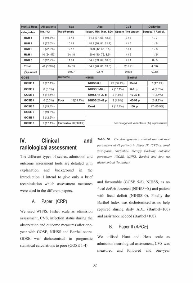
327
outcome evaluation was performed with
GOSE, NIHSS, and Barthel score.
Dichotomisation was performed as in Paper
I. Area and gender matched control group
with known APOE constitution was
recruited.
C. Paper III (9p21)
Only incidence of aSAH was confirmed and
genetical variation investigated in a case-
control study.
D. Paper IV (Neuropanel)
Hunt and Hess scale was utilised as selection
criteria and the levels of 9 biochemical
markers were followed and the course was
compared to H&H status, the presence or
absence of CVS and outcome parameters
with GOSE, NIHSS, and Barthel score.
V. Statistics
Statistical analyses were performed with
SPSS 21.1 (Statistical Packages Software
System – IBM Corp. Armonk, NY, USA)
and SAS 9.4 (Statistical Analysis Software –
SAS Inst. Inc. Cary, NC, USA). In planning
(i.e. sample size calculation) and result
interpretation (correct statistics) professional
statistical expertise was entrusted (Statistiska
Konsultgruppen, Gothenburg, Sweden). I
listed the statistical methods, employed in the
separate papers in the table below. (Tabl. 11)
Apart from the above mentioned statistical
methods, one additional genetic statistical
software was utilised in Paper III in
conjunction with haplotype frequency
calculation. The THESIAS Java based
program is constructed to performed
Table 11 The applied statistical methods for the individual papers. Abbreviations: ROC, -receiver operator characteristic curve, LR, -likelihood ratio, PPV, -positive predictive value
6
IV. Clinical and radiological assessment The different types of scales, admission and
outcome assessment tools are detailed with
explanation and background in the
Introduction. I intend to give only a brief
recapitulation which assessment measures
were used in the different papers.
A. Paper I (CRP)
We used WFNS, Fisher scale as admission
assessment, CVS, infection status during the
observation and outcome measures after one-
year with GOSE, NIHSS and Barthel score.
GOSE was dichotomised in prognostic
statistical calculations to poor (GOSE 1-4)
Table 10. The demographics, clinical and outcome
parameters of 41 patients in Paper IV. (CVS-cerebral
vasospasm, Op/Embol- therapy modality, outcome
parameters (GOSE, NIHSS, Barthel and how we
dichotomised the scales)
and favourable (GOSE 5-8), NIHSS, as no
focal deficit detected (NIHSS=0,) and patient
with focal deficit (NIHSS>0). Finally the
Barthel Index was dichotomised as no help
required during daily ADL (Barthel=100)
and assistance nedded (Barthel<100).
B. Paper II (APOE)
We utilised Hunt and Hess scale as
admission neurological assessment, CVS was
measured and followed and one-year
Hunt & Hess All patients Sex Age CVS Op/Embol
categories No. (%) Male/Female (Mean, Min, Max, SD) Spasm / No spasm Surgical / Radiol.
H&H 1 8 (19.5%) 5 / 3 51.3 (37, 66, 12.5) 3 / 5 1 / 7
H&H 2 9 (22.0%) 0 / 9 48.2 (20, 81, 21.7) 4 / 5 1 / 8
H&H 3 9 (22.0%) 2 / 7 56.0 (42, 65, 8.5) 5 / 4 1 / 8
H&H 4 10 (24.4%) 0 / 10 60.0 (40, 75, 8.9) 4 / 6 1 / 9
H&H 5 5 (12.2%) 1 / 4 54.2 (39, 69, 10.8) 4 / 1 0 / 5
Total 41 (100%) 8 / 33 54.2 (20, 81, 13.5) 20 / 21 4 / 37
χ2(p-value) - 0.007 0.575 0.575 0.958
GOSE
Outcome NIHSS
Barthel
GOSE 1 7 (17.1%) NIHSS 0 p 23 (56.1%) Dead 7 (17.1%) GOSE 2 0 (0.0%) NIHSS 1-10 p 7 (17.1%) 0-9 p 4 (9.8%) GOSE 3 6 (14.6%) NIHSS 11-20 p 2 (4.9%) 10-39 p 1 (2.4%) GOSE 4 0 (0.0%) Poor 13(31.7%) NIHSS 21-42 p 2 (4.9%) 40-99 p 2 (4.9%) GOSE 5 8 (19.5%) Dead 7 (17.1%) 100 p 27 (65.9%) GOSE 6 8 (19.5%)
GOSE 7 5 (12.2%)
GOSE 8 7 (17.1%) Favorable 28(68.3%) For categorical variables n (%) is presented.
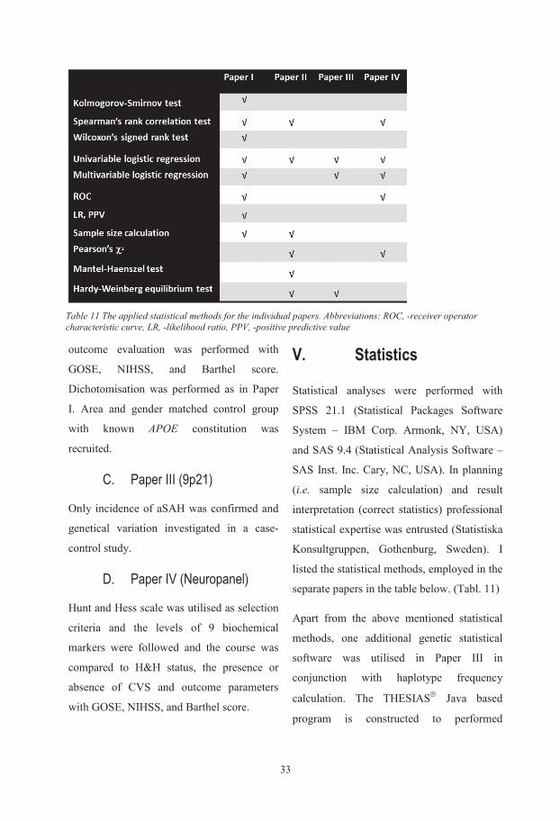
337
outcome evaluation was performed with
GOSE, NIHSS, and Barthel score.
Dichotomisation was performed as in Paper
I. Area and gender matched control group
with known APOE constitution was
recruited.
C. Paper III (9p21)
Only incidence of aSAH was confirmed and
genetical variation investigated in a case-
control study.
D. Paper IV (Neuropanel)
Hunt and Hess scale was utilised as selection
criteria and the levels of 9 biochemical
markers were followed and the course was
compared to H&H status, the presence or
absence of CVS and outcome parameters
with GOSE, NIHSS, and Barthel score.
V. Statistics
Statistical analyses were performed with
SPSS 21.1 (Statistical Packages Software
System – IBM Corp. Armonk, NY, USA)
and SAS 9.4 (Statistical Analysis Software –
SAS Inst. Inc. Cary, NC, USA). In planning
(i.e. sample size calculation) and result
interpretation (correct statistics) professional
statistical expertise was entrusted (Statistiska
Konsultgruppen, Gothenburg, Sweden). I
listed the statistical methods, employed in the
separate papers in the table below. (Tabl. 11)
Apart from the above mentioned statistical
methods, one additional genetic statistical
software was utilised in Paper III in
conjunction with haplotype frequency
calculation. The THESIAS Java based
program is constructed to performed
Table 11 The applied statistical methods for the individual papers. Abbreviations: ROC, -receiver operator characteristic curve, LR, -likelihood ratio, PPV, -positive predictive value
6
IV. Clinical and radiological assessment The different types of scales, admission and
outcome assessment tools are detailed with
explanation and background in the
Introduction. I intend to give only a brief
recapitulation which assessment measures
were used in the different papers.
A. Paper I (CRP)
We used WFNS, Fisher scale as admission
assessment, CVS, infection status during the
observation and outcome measures after one-
year with GOSE, NIHSS and Barthel score.
GOSE was dichotomised in prognostic
statistical calculations to poor (GOSE 1-4)
Table 10. The demographics, clinical and outcome
parameters of 41 patients in Paper IV. (CVS-cerebral
vasospasm, Op/Embol- therapy modality, outcome
parameters (GOSE, NIHSS, Barthel and how we
dichotomised the scales)
and favourable (GOSE 5-8), NIHSS, as no
focal deficit detected (NIHSS=0,) and patient
with focal deficit (NIHSS>0). Finally the
Barthel Index was dichotomised as no help
required during daily ADL (Barthel=100)
and assistance nedded (Barthel<100).
B. Paper II (APOE)
We utilised Hunt and Hess scale as
admission neurological assessment, CVS was
measured and followed and one-year
Hunt & Hess All patients Sex Age CVS Op/Embol
categories No. (%) Male/Female (Mean, Min, Max, SD) Spasm / No spasm Surgical / Radiol.
H&H 1 8 (19.5%) 5 / 3 51.3 (37, 66, 12.5) 3 / 5 1 / 7
H&H 2 9 (22.0%) 0 / 9 48.2 (20, 81, 21.7) 4 / 5 1 / 8
H&H 3 9 (22.0%) 2 / 7 56.0 (42, 65, 8.5) 5 / 4 1 / 8
H&H 4 10 (24.4%) 0 / 10 60.0 (40, 75, 8.9) 4 / 6 1 / 9
H&H 5 5 (12.2%) 1 / 4 54.2 (39, 69, 10.8) 4 / 1 0 / 5
Total 41 (100%) 8 / 33 54.2 (20, 81, 13.5) 20 / 21 4 / 37
χ2(p-value) - 0.007 0.575 0.575 0.958
GOSE
Outcome NIHSS
Barthel
GOSE 1 7 (17.1%) NIHSS 0 p 23 (56.1%) Dead 7 (17.1%) GOSE 2 0 (0.0%) NIHSS 1-10 p 7 (17.1%) 0-9 p 4 (9.8%) GOSE 3 6 (14.6%) NIHSS 11-20 p 2 (4.9%) 10-39 p 1 (2.4%) GOSE 4 0 (0.0%) Poor 13(31.7%) NIHSS 21-42 p 2 (4.9%) 40-99 p 2 (4.9%) GOSE 5 8 (19.5%) Dead 7 (17.1%) 100 p 27 (65.9%) GOSE 6 8 (19.5%)
GOSE 7 5 (12.2%)
GOSE 8 7 (17.1%) Favorable 28(68.3%) For categorical variables n (%) is presented.

34
RESULTS
I. Biochemical neuromarkers (BNMs)
A. Neuron cell-injury markers: The BNMs below comprise an admixture of
proteins recognized to be found in the
central nervous system. They have been
shown to have variable functions like
structural-, transport-, receptor- and special
purpose proteins within inflammation and
coagulation. The common attribute of these
proteins is that all of their concentrations in
body fluids have been found to increase
after neuronal injury. We have investigated
nine BNMs in a pilot study in 41 patients
after aSAH in Paper IV. The correlation
calculations between the investigated BNMs
and admission neurology, CVS and outcome
measurements (GOSE, NIHSS, Barthel) are
described in Table 12.
Neuron Specific Enolase
(NSE)
NSE followed an irregular pattern, but a
continuous rise from Day 0 (mean 2.82
ng/ml, SD 2.78) to Day 12-14 (8.57 ng/ml,
SD 7.38) could be observed. The maximum
or mean values showed, however no
correlation with H&H, vasospasm or the
Tabell 12 Correlation table for neuropanel markers; max and mean values vs. Hunt &Hess scale, CVS, GOSE, NIHSS and Barthel index. Spearman’s rho coefficients and p-values are presented. Statistically significant values are highlighted and marked in grey. 8
haplotype-based association analysis in
unrelated individuals. This program is based
on the maximum likelihood model and
estimates ORs for each haplotype in relation
to a reference haplotype (Tregouet and
Garelle, 2007).
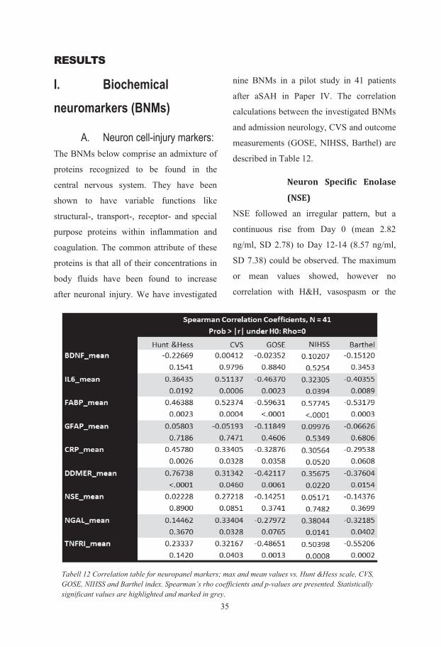
35
RESULTS
I. Biochemical neuromarkers (BNMs)
A. Neuron cell-injury markers: The BNMs below comprise an admixture of
proteins recognized to be found in the
central nervous system. They have been
shown to have variable functions like
structural-, transport-, receptor- and special
purpose proteins within inflammation and
coagulation. The common attribute of these
proteins is that all of their concentrations in
body fluids have been found to increase
after neuronal injury. We have investigated
nine BNMs in a pilot study in 41 patients
after aSAH in Paper IV. The correlation
calculations between the investigated BNMs
and admission neurology, CVS and outcome
measurements (GOSE, NIHSS, Barthel) are
described in Table 12.
Neuron Specific Enolase
(NSE)
NSE followed an irregular pattern, but a
continuous rise from Day 0 (mean 2.82
ng/ml, SD 2.78) to Day 12-14 (8.57 ng/ml,
SD 7.38) could be observed. The maximum
or mean values showed, however no
correlation with H&H, vasospasm or the
Tabell 12 Correlation table for neuropanel markers; max and mean values vs. Hunt &Hess scale, CVS, GOSE, NIHSS and Barthel index. Spearman’s rho coefficients and p-values are presented. Statistically significant values are highlighted and marked in grey. 8
haplotype-based association analysis in
unrelated individuals. This program is based
on the maximum likelihood model and
estimates ORs for each haplotype in relation
to a reference haplotype (Tregouet and
Garelle, 2007).
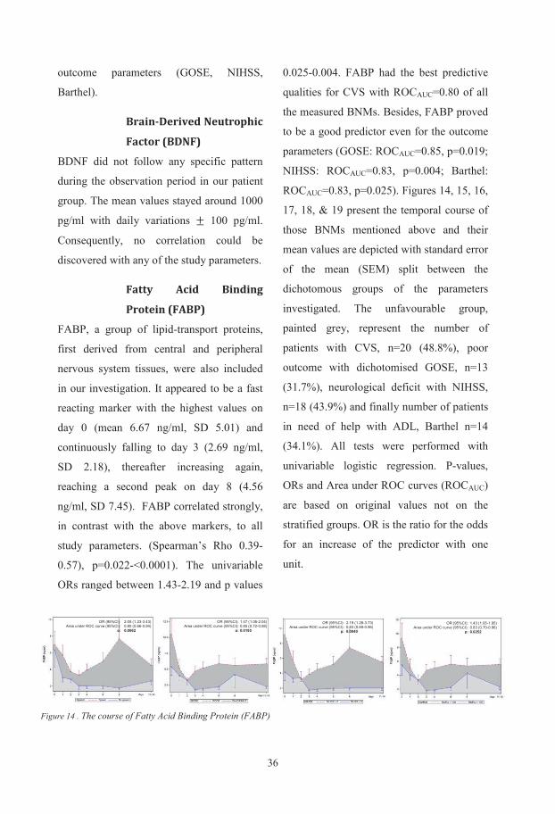
363
B. Glia cell-marker
Glial Fibrillary Acidic
Protein (GFAP)
This glia cell-injury marker has been
investigated in a previous study in partially
overlapping aSAB patient material (Nylen et
al. 2007). In our 41-patient material with the
new biochip-array technique, we could not
find an association between either the
maximum or mean values of GFAP and the
initial neurology (H&H), cerebral
vasospasm (CVS) or any of the outcome
parameters tested. (Spearman’s Rho was at
best Rho= − 0.19, p=0.22). However, when
plotting the results, there was a tendency of
the poor outcome patients having higher
initial values (Fig.15).
C. Coagulation protein D-dimer (DDMR)
DDMR is a fibrin degradation protein which
is a product of fibrinolysis, a natural process
of the organism dissolving a thrombus or a
clot. As SAH entails a clot-formation, it has
been proposed that the level of DDMR
corresponds to the amount of blood
exsanguinated from the aneurysm and
consequently to the risk of vasospasm and
outcome (Fujii et al. 1997). As DDMR is
included in the neuropanel biochip of
Randox UK, we
aimed to test this
coagulation
protein, relatively
new to SAB
diagnostics.
We found
initially medium
high values
(mean: 376
ng/ml, SD: 503)
which decreased
gradually,
reaching its nadir
on day 2 (180 ng/ml, SD: 134), thereafter
increasing again to even higher levels and
staying high during the whole observation
period (day 12-14: mean 477 ng/ml, SD:
372). Although, there is no difference in the
initial course and levels of DDMR between
the favourable and unfavourable group, the
Figure 15 The course of GFAP in patients with poor and favourable outcome (dichotomous GOSE). Mean values and standard error of the mean (SEM) are presented
2
outcome parameters (GOSE, NIHSS,
Barthel).
Brain-Derived Neutrophic
Factor (BDNF)
BDNF did not follow any specific pattern
during the observation period in our patient
group. The mean values stayed around 1000
pg/ml with daily variations ± 100 pg/ml.
Consequently, no correlation could be
discovered with any of the study parameters.
Fatty Acid Binding
Protein (FABP)
FABP, a group of lipid-transport proteins,
first derived from central and peripheral
nervous system tissues, were also included
in our investigation. It appeared to be a fast
reacting marker with the highest values on
day 0 (mean 6.67 ng/ml, SD 5.01) and
continuously falling to day 3 (2.69 ng/ml,
SD 2.18), thereafter increasing again,
reaching a second peak on day 8 (4.56
ng/ml, SD 7.45). FABP correlated strongly,
in contrast with the above markers, to all
study parameters. (Spearman’s Rho 0.39-
0.57), p=0.022-<0.0001). The univariable
ORs ranged between 1.43-2.19 and p values
0.025-0.004. FABP had the best predictive
qualities for CVS with ROCAUC=0.80 of all
the measured BNMs. Besides, FABP proved
to be a good predictor even for the outcome
parameters (GOSE: ROCAUC=0.85, p=0.019;
NIHSS: ROCAUC=0.83, p=0.004; Barthel:
ROCAUC=0.83, p=0.025). Figures 14, 15, 16,
17, 18, & 19 present the temporal course of
those BNMs mentioned above and their
mean values are depicted with standard error
of the mean (SEM) split between the
dichotomous groups of the parameters
investigated. The unfavourable group,
painted grey, represent the number of
patients with CVS, n=20 (48.8%), poor
outcome with dichotomised GOSE, n=13
(31.7%), neurological deficit with NIHSS,
n=18 (43.9%) and finally number of patients
in need of help with ADL, Barthel n=14
(34.1%). All tests were performed with
univariable logistic regression. P-values,
ORs and Area under ROC curves (ROCAUC)
are based on original values not on the
stratified groups. OR is the ratio for the odds
for an increase of the predictor with one
unit.
OR (95%CI): Area under ROC curve (95%CI):
p: 1.43 (1.05-1.95) 0.83 (0.70-0.96) 0.0252
OR (95%CI): Area under ROC curve (95%CI):
p: 2.19 (1.28-3.73) 0.83 (0.69-0.96) 0.0040
OR (95%CI): Area under ROC curve (95%CI):
p: OR (95%CI):
Area under ROC curve (95%CI): p:
1.47 (1.06-2.04) 0.85 (0.72-0.98) 0.0195
2.05 (1.23-3.43) 0.80 (0.66-0.94) 0.0062
Figure 14 . The course of Fatty Acid Binding Protein (FABP)
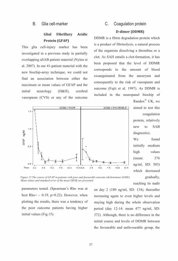
373
B. Glia cell-marker
Glial Fibrillary Acidic
Protein (GFAP)
This glia cell-injury marker has been
investigated in a previous study in partially
overlapping aSAB patient material (Nylen et
al. 2007). In our 41-patient material with the
new biochip-array technique, we could not
find an association between either the
maximum or mean values of GFAP and the
initial neurology (H&H), cerebral
vasospasm (CVS) or any of the outcome
parameters tested. (Spearman’s Rho was at
best Rho= − 0.19, p=0.22). However, when
plotting the results, there was a tendency of
the poor outcome patients having higher
initial values (Fig.15).
C. Coagulation protein D-dimer (DDMR)
DDMR is a fibrin degradation protein which
is a product of fibrinolysis, a natural process
of the organism dissolving a thrombus or a
clot. As SAH entails a clot-formation, it has
been proposed that the level of DDMR
corresponds to the amount of blood
exsanguinated from the aneurysm and
consequently to the risk of vasospasm and
outcome (Fujii et al. 1997). As DDMR is
included in the neuropanel biochip of
Randox UK, we
aimed to test this
coagulation
protein, relatively
new to SAB
diagnostics.
We found
initially medium
high values
(mean: 376
ng/ml, SD: 503)
which decreased
gradually,
reaching its nadir
on day 2 (180 ng/ml, SD: 134), thereafter
increasing again to even higher levels and
staying high during the whole observation
period (day 12-14: mean 477 ng/ml, SD:
372). Although, there is no difference in the
initial course and levels of DDMR between
the favourable and unfavourable group, the
Figure 15 The course of GFAP in patients with poor and favourable outcome (dichotomous GOSE). Mean values and standard error of the mean (SEM) are presented
2
outcome parameters (GOSE, NIHSS,
Barthel).
Brain-Derived Neutrophic
Factor (BDNF)
BDNF did not follow any specific pattern
during the observation period in our patient
group. The mean values stayed around 1000
pg/ml with daily variations ± 100 pg/ml.
Consequently, no correlation could be
discovered with any of the study parameters.
Fatty Acid Binding
Protein (FABP)
FABP, a group of lipid-transport proteins,
first derived from central and peripheral
nervous system tissues, were also included
in our investigation. It appeared to be a fast
reacting marker with the highest values on
day 0 (mean 6.67 ng/ml, SD 5.01) and
continuously falling to day 3 (2.69 ng/ml,
SD 2.18), thereafter increasing again,
reaching a second peak on day 8 (4.56
ng/ml, SD 7.45). FABP correlated strongly,
in contrast with the above markers, to all
study parameters. (Spearman’s Rho 0.39-
0.57), p=0.022-<0.0001). The univariable
ORs ranged between 1.43-2.19 and p values
0.025-0.004. FABP had the best predictive
qualities for CVS with ROCAUC=0.80 of all
the measured BNMs. Besides, FABP proved
to be a good predictor even for the outcome
parameters (GOSE: ROCAUC=0.85, p=0.019;
NIHSS: ROCAUC=0.83, p=0.004; Barthel:
ROCAUC=0.83, p=0.025). Figures 14, 15, 16,
17, 18, & 19 present the temporal course of
those BNMs mentioned above and their
mean values are depicted with standard error
of the mean (SEM) split between the
dichotomous groups of the parameters
investigated. The unfavourable group,
painted grey, represent the number of
patients with CVS, n=20 (48.8%), poor
outcome with dichotomised GOSE, n=13
(31.7%), neurological deficit with NIHSS,
n=18 (43.9%) and finally number of patients
in need of help with ADL, Barthel n=14
(34.1%). All tests were performed with
univariable logistic regression. P-values,
ORs and Area under ROC curves (ROCAUC)
are based on original values not on the
stratified groups. OR is the ratio for the odds
for an increase of the predictor with one
unit.
OR (95%CI): Area under ROC curve (95%CI):
p: 1.43 (1.05-1.95) 0.83 (0.70-0.96) 0.0252
OR (95%CI): Area under ROC curve (95%CI):
p: 2.19 (1.28-3.73) 0.83 (0.69-0.96) 0.0040
OR (95%CI): Area under ROC curve (95%CI):
p: OR (95%CI):
Area under ROC curve (95%CI): p:
1.47 (1.06-2.04) 0.85 (0.72-0.98) 0.0195
2.05 (1.23-3.43) 0.80 (0.66-0.94) 0.0062
Figure 14 . The course of Fatty Acid Binding Protein (FABP)
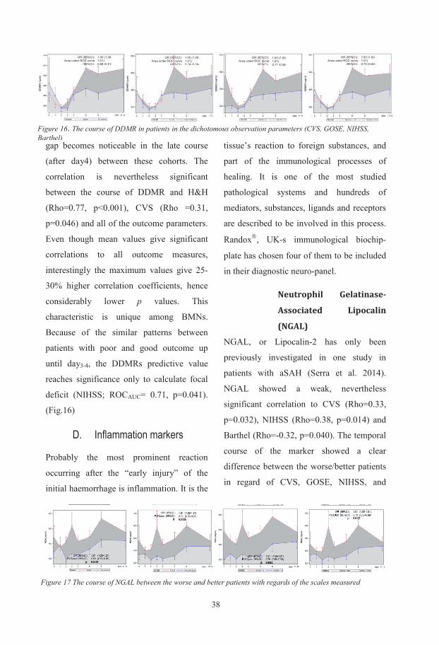
385
Barthel. The predictive qualities were all
significant, ranging between ROCAUC=0.69-
0.75. (Fig. 17)
Tumour Necrosis Factor
Receptor 1. (TNFR1)
TNFR1 is one of the receptors of TNF-α,
which is recognised to act as mediator in
inflammatory processes. The soluble
TNFR1 and 2 are activated by TNF-α and
having a longer half-life than the actual
cytokine, thereby acting as a more reliable
measure of inflammation activity than its
ligand. TNFR1 demonstrated a continuously
rising pattern. The correlation between the
early diagnostic scales are weak (H&H –
non significant, CVS – Rho=0.32, p=0.040),
but becomes strong (Rho>0.5, p<0.01) with
the outcome measures. It has the best
predictive strength of all the BNMs
investigated for poor – good outcome with
ROCAUC=0.87, p=0.0028, bordering the
conclusion: excellent predictor according to
Swets (Swets 1988). (Fig.18)
Interleukin-6 (IL-6)
IL6 is one of the most important pro-
inflammatory cytokines and mediator of
fever through prostaglandin (especially
PGE2) production. As found in smooth
muscle cells in blood vessel walls it is
involved in vessel regulation during
inflammation. It has recently been assigned
new functions as neuropoietin for its
positive neural properties (Vezzani and
Viviani 2015). Not surprisingly, IL6 is
interesting as a marker of inflammation and
regeneration after SAB. As nearly all
cytokines are fast reacting mediators, IL-6
demonstrated rapid rise after the insult,
reaching its peak on day2-4. The correlation
was good on all the observation parameters
(Rho=0.36-0.51, p=0.019-0.0006), but due
to its fast reaction and with regard of its
OR (95%CI):Area under ROC curve (95%CI):
765.8 (8.50-69038)0 84 (0 71 0 96)
OR (95%CI):Area under ROC curve (95%CI):
287.8 (4.18-19812)0 76 (0 60 0 91)
OR (95%CI):Area under ROC curve (95%CI):
1514.8 (12.46-184188)0 87 (0 75 0 98)
OR (95%CI):Area under ROC curve (95%CI):
21.08(0.95-469.0)
( )
Figure 18. TNFR1’s course with CVS and outcome measures
OR (95%CI):Area under ROC curve
(95%CI):
1.03 (0.99-1.08)0 76 (0 60
OR (95%CI):Area under ROC curve
(95%CI):
1.02 (0.98-1.05)0 65 (0 48
OR (95%CI):Area under ROC curve
(95%CI):
1.03 (0.99-1.07)0 75 (0 59
OR (95%CI):Area under ROC curve
(95%CI):
1.09 (1.02-1.16)0 80 (0 65
Figure 19. The development of Interleukin-6 (IL6) during the first two weeks in patients with or without vasospasmand favourable/unfavourable dichotomous groups of outcome scales
4
gap becomes noticeable in the late course
(after day4) between these cohorts. The
correlation is nevertheless significant
between the course of DDMR and H&H
(Rho=0.77, p<0.001), CVS (Rho =0.31,
p=0.046) and all of the outcome parameters.
Even though mean values give significant
correlations to all outcome measures,
interestingly the maximum values give 25-
30% higher correlation coefficients, hence
considerably lower p values. This
characteristic is unique among BMNs.
Because of the similar patterns between
patients with poor and good outcome up
until day3-4, the DDMRs predictive value
reaches significance only to calculate focal
deficit (NIHSS; ROCAUC= 0.71, p=0.041).
(Fig.16)
D. Inflammation markers
Probably the most prominent reaction
occurring after the “early injury” of the
initial haemorrhage is inflammation. It is the
tissue’s reaction to foreign substances, and
part of the immunological processes of
healing. It is one of the most studied
pathological systems and hundreds of
mediators, substances, ligands and receptors
are described to be involved in this process.
Randox, UK-s immunological biochip-
plate has chosen four of them to be included
in their diagnostic neuro-panel.
Neutrophil Gelatinase-
Associated Lipocalin
(NGAL)
NGAL, or Lipocalin-2 has only been
previously investigated in one study in
patients with aSAH (Serra et al. 2014).
NGAL showed a weak, nevertheless
significant correlation to CVS (Rho=0.33,
p=0.032), NIHSS (Rho=0.38, p=0.014) and
Barthel (Rho=-0.32, p=0.040). The temporal
course of the marker showed a clear
difference between the worse/better patients
in regard of CVS, GOSE, NIHSS, and
OR (95%CI):Area under ROC curve
(95%CI):
1.00 (1.00-1.01) 0.75 (0.60-
OR (95%CI):Area under ROC curve
(95%CI):
1.00 (1.00-1.01) 0.68 (0.51-
OR (95%CI):Area under ROC curve
(95%CI):
1.00 (1.00-1.01) 0.74 (0.59-
OR (95%CI):Area under ROC curve
(95%CI):
1.00 (1.00-1.01) 0.71 (0.54-
Figure 16. The course of DDMR in patients in the dichotomous observation parameters (CVS, GOSE, NIHSS, Barthel)
Figure 17 The course of NGAL between the worse and better patients with regards of the scales measured
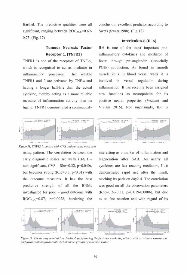
395
Barthel. The predictive qualities were all
significant, ranging between ROCAUC=0.69-
0.75. (Fig. 17)
Tumour Necrosis Factor
Receptor 1. (TNFR1)
TNFR1 is one of the receptors of TNF-α,
which is recognised to act as mediator in
inflammatory processes. The soluble
TNFR1 and 2 are activated by TNF-α and
having a longer half-life than the actual
cytokine, thereby acting as a more reliable
measure of inflammation activity than its
ligand. TNFR1 demonstrated a continuously
rising pattern. The correlation between the
early diagnostic scales are weak (H&H –
non significant, CVS – Rho=0.32, p=0.040),
but becomes strong (Rho>0.5, p<0.01) with
the outcome measures. It has the best
predictive strength of all the BNMs
investigated for poor – good outcome with
ROCAUC=0.87, p=0.0028, bordering the
conclusion: excellent predictor according to
Swets (Swets 1988). (Fig.18)
Interleukin-6 (IL-6)
IL6 is one of the most important pro-
inflammatory cytokines and mediator of
fever through prostaglandin (especially
PGE2) production. As found in smooth
muscle cells in blood vessel walls it is
involved in vessel regulation during
inflammation. It has recently been assigned
new functions as neuropoietin for its
positive neural properties (Vezzani and
Viviani 2015). Not surprisingly, IL6 is
interesting as a marker of inflammation and
regeneration after SAB. As nearly all
cytokines are fast reacting mediators, IL-6
demonstrated rapid rise after the insult,
reaching its peak on day2-4. The correlation
was good on all the observation parameters
(Rho=0.36-0.51, p=0.019-0.0006), but due
to its fast reaction and with regard of its
OR (95%CI): Area under ROC curve (95%CI):
765.8 (8.50-69038) 0 84 (0 71 0 96)
OR (95%CI): Area under ROC curve (95%CI):
287.8 (4.18-19812) 0 76 (0 60 0 91)
OR (95%CI): Area under ROC curve (95%CI):
1514.8 (12.46-184188) 0 87 (0 75 0 98)
OR (95%CI): Area under ROC curve (95%CI):
21.08(0.95-469.0)
( )
Figure 18. TNFR1’s course with CVS and outcome measures
OR (95%CI): Area under ROC curve
(95%CI): 1.03 (0.99-1.08) 0 76 (0 60
OR (95%CI): Area under ROC curve
(95%CI): 1.02 (0.98-1.05) 0 65 (0 48
OR (95%CI): Area under ROC curve
(95%CI): 1.03 (0.99-1.07) 0 75 (0 59
OR (95%CI): Area under ROC curve
(95%CI): 1.09 (1.02-1.16) 0 80 (0 65
Figure 19. The development of Interleukin-6 (IL6) during the first two weeks in patients with or without vasospasm and favourable/unfavourable dichotomous groups of outcome scales
4
gap becomes noticeable in the late course
(after day4) between these cohorts. The
correlation is nevertheless significant
between the course of DDMR and H&H
(Rho=0.77, p<0.001), CVS (Rho =0.31,
p=0.046) and all of the outcome parameters.
Even though mean values give significant
correlations to all outcome measures,
interestingly the maximum values give 25-
30% higher correlation coefficients, hence
considerably lower p values. This
characteristic is unique among BMNs.
Because of the similar patterns between
patients with poor and good outcome up
until day3-4, the DDMRs predictive value
reaches significance only to calculate focal
deficit (NIHSS; ROCAUC= 0.71, p=0.041).
(Fig.16)
D. Inflammation markers
Probably the most prominent reaction
occurring after the “early injury” of the
initial haemorrhage is inflammation. It is the
tissue’s reaction to foreign substances, and
part of the immunological processes of
healing. It is one of the most studied
pathological systems and hundreds of
mediators, substances, ligands and receptors
are described to be involved in this process.
Randox, UK-s immunological biochip-
plate has chosen four of them to be included
in their diagnostic neuro-panel.
Neutrophil Gelatinase-
Associated Lipocalin
(NGAL)
NGAL, or Lipocalin-2 has only been
previously investigated in one study in
patients with aSAH (Serra et al. 2014).
NGAL showed a weak, nevertheless
significant correlation to CVS (Rho=0.33,
p=0.032), NIHSS (Rho=0.38, p=0.014) and
Barthel (Rho=-0.32, p=0.040). The temporal
course of the marker showed a clear
difference between the worse/better patients
in regard of CVS, GOSE, NIHSS, and
OR (95%CI): Area under ROC curve
(95%CI): 1.00 (1.00-1.01) 0.75 (0.60-
OR (95%CI): Area under ROC curve
(95%CI): 1.00 (1.00-1.01) 0.68 (0.51-
OR (95%CI): Area under ROC curve
(95%CI): 1.00 (1.00-1.01) 0.74 (0.59-
OR (95%CI): Area under ROC curve
(95%CI): 1.00 (1.00-1.01) 0.71 (0.54-
Figure 16. The course of DDMR in patients in the dichotomous observation parameters (CVS, GOSE, NIHSS, Barthel)
Figure 17 The course of NGAL between the worse and better patients with regards of the scales measured
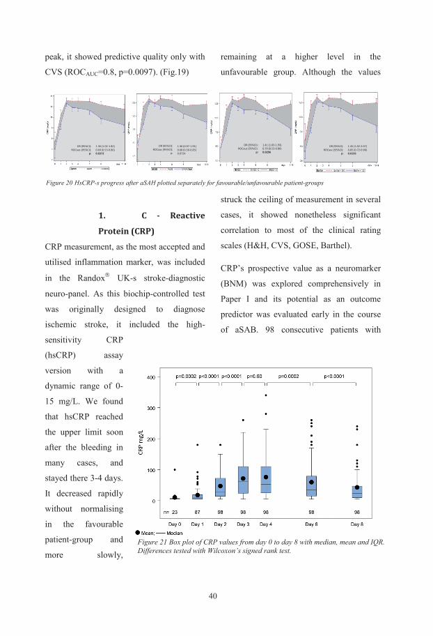
407
endovascularly treated aneurysms were
enrolled in this study. The patients’
demographics, neurological status at first
examination, radiological grading, infectious
status during observation and their CRP
values during the first week of treatment are
described in Table 1 in Paper I. We could
demonstrate a continuous rise of CRP values
from normal at day 0 (median 5 mg/L, IQR
5-7) to its peak at day 3-4, (median value 53
mg/L, IQR 24-100). The values decreased
after this peak until day 8 without
normalising. (median 24 mg/L, IQR 10-47).
The changes between each subsequent day
were significant, apart from the peak. The
course of CRP levels in the first 8 days is
depicted in Fig.21.
This change was independent from the status
of infection. We were the first to describe
this temporal course (Csajbok et al. 2005)
and were intrigued by the result, which
corresponded with our clinical experience.
We tested if there was a difference in the
course of CRP development between the
patients with unfavourable and favourable
outcome. The same boxplot, split between
these patient groups confirmed the result
(Fig.22).
The linear regression lines showed a
stronger association between the mean CRP
values in the first week and long-term
outcome, measured by GOSE, compared to
the radiological scale Fisher or initial
neurology categorised by the World
Federation of
Neurological
Surgeons Scale
(WFNS) in this
patient group.
(Fig.23) The
correlation
coefficients
(Spearman’s Rho)
confirmed the
stronger connection
between CRPmean and
CRPmax to GOSE (-
0.49, -0.45,
p<0.0001) and
NIHSS (0.45, 0.45,
Figure 22 Box plot of CRP values from day 0 to day 8 with median, mean and IQR. Differences tested with Wilcoxon’s signed rank test
6
peak, it showed predictive quality only with
CVS (ROCAUC=0.8, p=0.0097). (Fig.19)
1. C - Reactive
Protein (CRP)
CRP measurement, as the most accepted and
utilised inflammation marker, was included
in the Randox UK-s stroke-diagnostic
neuro-panel. As this biochip-controlled test
was originally designed to diagnose
ischemic stroke, it included the high-
sensitivity CRP
(hsCRP) assay
version with a
dynamic range of 0-
15 mg/L. We found
that hsCRP reached
the upper limit soon
after the bleeding in
many cases, and
stayed there 3-4 days.
It decreased rapidly
without normalising
in the favourable
patient-group and
more slowly,
remaining at a higher level in the
unfavourable group. Although the values
struck the ceiling of measurement in several
cases, it showed nonetheless significant
correlation to most of the clinical rating
scales (H&H, CVS, GOSE, Barthel).
CRP’s prospective value as a neuromarker
(BNM) was explored comprehensively in
Paper I and its potential as an outcome
predictor was evaluated early in the course
of aSAB. 98 consecutive patients with
OR (95%CI): ROCauc (95%CI):
p: OR (95%CI):
ROCauc (95%CI): p:
OR (95%CI): ROCauc (95%CI):
p: OR (95%CI):
ROCauc (95%CI): p:
1.36 (0.97-1.91) 0.68 (0.50-0.85) 0.0724
1.41 (1.03-1.93) 0.70 (0.53-0.86) 0.0296
1.45 (1.02-2.07) 0.85 (0.72-0.98) 0.0195
1.36 (1.02-1.82) 0.69 (0.53-0.86) 0.0372
Figure 20 HsCRP-s progress after aSAH plotted separately for favourable/unfavourable patient-groups
Figure 21 Box plot of CRP values from day 0 to day 8 with median, mean and IQR. Differences tested with Wilcoxon’s signed rank test.
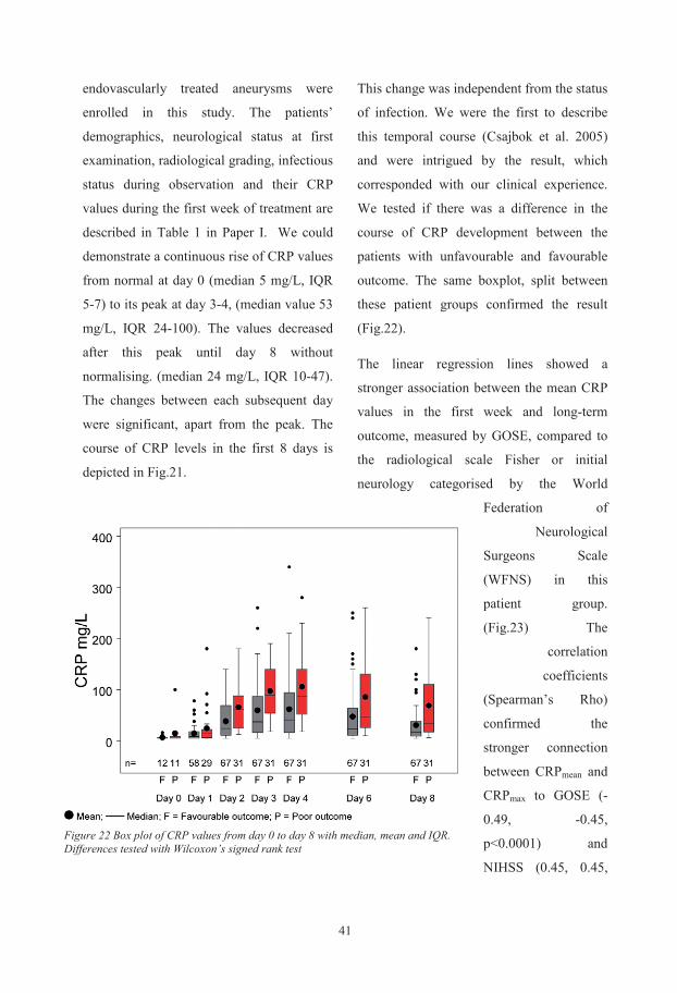
417
endovascularly treated aneurysms were
enrolled in this study. The patients’
demographics, neurological status at first
examination, radiological grading, infectious
status during observation and their CRP
values during the first week of treatment are
described in Table 1 in Paper I. We could
demonstrate a continuous rise of CRP values
from normal at day 0 (median 5 mg/L, IQR
5-7) to its peak at day 3-4, (median value 53
mg/L, IQR 24-100). The values decreased
after this peak until day 8 without
normalising. (median 24 mg/L, IQR 10-47).
The changes between each subsequent day
were significant, apart from the peak. The
course of CRP levels in the first 8 days is
depicted in Fig.21.
This change was independent from the status
of infection. We were the first to describe
this temporal course (Csajbok et al. 2005)
and were intrigued by the result, which
corresponded with our clinical experience.
We tested if there was a difference in the
course of CRP development between the
patients with unfavourable and favourable
outcome. The same boxplot, split between
these patient groups confirmed the result
(Fig.22).
The linear regression lines showed a
stronger association between the mean CRP
values in the first week and long-term
outcome, measured by GOSE, compared to
the radiological scale Fisher or initial
neurology categorised by the World
Federation of
Neurological
Surgeons Scale
(WFNS) in this
patient group.
(Fig.23) The
correlation
coefficients
(Spearman’s Rho)
confirmed the
stronger connection
between CRPmean and
CRPmax to GOSE (-
0.49, -0.45,
p<0.0001) and
NIHSS (0.45, 0.45,
Figure 22 Box plot of CRP values from day 0 to day 8 with median, mean and IQR. Differences tested with Wilcoxon’s signed rank test
6
peak, it showed predictive quality only with
CVS (ROCAUC=0.8, p=0.0097). (Fig.19)
1. C - Reactive
Protein (CRP)
CRP measurement, as the most accepted and
utilised inflammation marker, was included
in the Randox UK-s stroke-diagnostic
neuro-panel. As this biochip-controlled test
was originally designed to diagnose
ischemic stroke, it included the high-
sensitivity CRP
(hsCRP) assay
version with a
dynamic range of 0-
15 mg/L. We found
that hsCRP reached
the upper limit soon
after the bleeding in
many cases, and
stayed there 3-4 days.
It decreased rapidly
without normalising
in the favourable
patient-group and
more slowly,
remaining at a higher level in the
unfavourable group. Although the values
struck the ceiling of measurement in several
cases, it showed nonetheless significant
correlation to most of the clinical rating
scales (H&H, CVS, GOSE, Barthel).
CRP’s prospective value as a neuromarker
(BNM) was explored comprehensively in
Paper I and its potential as an outcome
predictor was evaluated early in the course
of aSAB. 98 consecutive patients with
OR (95%CI): ROCauc (95%CI):
p: OR (95%CI):
ROCauc (95%CI): p:
OR (95%CI): ROCauc (95%CI):
p: OR (95%CI):
ROCauc (95%CI): p:
1.36 (0.97-1.91) 0.68 (0.50-0.85) 0.0724
1.41 (1.03-1.93) 0.70 (0.53-0.86) 0.0296
1.45 (1.02-2.07) 0.85 (0.72-0.98) 0.0195
1.36 (1.02-1.82) 0.69 (0.53-0.86) 0.0372
Figure 20 HsCRP-s progress after aSAH plotted separately for favourable/unfavourable patient-groups
Figure 21 Box plot of CRP values from day 0 to day 8 with median, mean and IQR. Differences tested with Wilcoxon’s signed rank test.
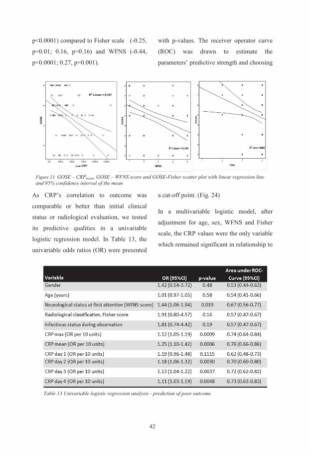
429
outcome.
The area under the
ROC curve
(AUCROC) that can
describe accuracy of
the test (Swets 1988)
showed moderate
accuracy for CRPmean
and CRPmax (0.76,
p<0.001; 0.74,
p<0.001), poor
accuracy for WFNS
(0.67, p=0.008) and
no predictive value
for Fisher scale (0.57, p=0.14), rendering
WFNS and Fisher scale inapplicable for
outcome prediction. In our endeavour to find
even earlier prognostics, we added CRP
values on day 2 and 3 to our ROC analysis
and found surprisingly, that already CRPday2
provided better prognostic accuracy
(AUCROC =0.7, p=0.002) than WFNS and
Fisher scale.
The logistic
regression
analysis could
offer us a
probability curve
to predict poor
outcome for the
best predictors in
relation to the
CRP values
(Fig.25). This is
the first time a
practical
Figure 24 ROC curve of CRPmean, CRPmax, CRP∆, WFNS score and Fisher scale for prediction of outcome
Fugure 25 Probability of poor outcome expressed as a measurement of CRP on day 2 and day 3, CRPmean and CRPmax during the first week after aSAH calculated from the logistic regression model
8
p<0.0001) compared to Fisher scale (-0.25,
p=0.01; 0.16, p=0.16) and WFNS (-0.44,
p<0.0001; 0.27, p=0.001).
As CRP’s correlation to outcome was
comparable or better than initial clinical
status or radiological evaluation, we tested
its predictive qualities in a univariable
logistic regression model. In Table 13, the
univariable odds ratios (OR) were presented
with p-values. The receiver operator curve
(ROC) was drawn to estimate the
parameters’ predictive strength and choosing
a cut-off point. (Fig. 24)
In a multivariable logistic model, after
adjustment for age, sex, WFNS and Fisher
scale, the CRP values were the only variable
which remained significant in relationship to
Figure 23 GOSE – CRPmean, GOSE – WFNS score and GOSE-Fisher scatter plot with linear regression line and 95% confidence interval of the mean
Table 13 Univariable logistic regression analysis - prediction of poor outcome
439
outcome.
The area under the
ROC curve
(AUCROC) that can
describe accuracy of
the test (Swets 1988)
showed moderate
accuracy for CRPmean
and CRPmax (0.76,
p<0.001; 0.74,
p<0.001), poor
accuracy for WFNS
(0.67, p=0.008) and
no predictive value
for Fisher scale (0.57, p=0.14), rendering
WFNS and Fisher scale inapplicable for
outcome prediction. In our endeavour to find
even earlier prognostics, we added CRP
values on day 2 and 3 to our ROC analysis
and found surprisingly, that already CRPday2
provided better prognostic accuracy
(AUCROC =0.7, p=0.002) than WFNS and
Fisher scale.
The logistic
regression
analysis could
offer us a
probability curve
to predict poor
outcome for the
best predictors in
relation to the
CRP values
(Fig.25). This is
the first time a
practical
Figure 24 ROC curve of CRPmean, CRPmax, CRP∆, WFNS score and Fisher scale for prediction of outcome
Fugure 25 Probability of poor outcome expressed as a measurement of CRP on day 2 and day 3, CRPmean and CRPmax during the first week after aSAH calculated from the logistic regression model
8
p<0.0001) compared to Fisher scale (-0.25,
p=0.01; 0.16, p=0.16) and WFNS (-0.44,
p<0.0001; 0.27, p=0.001).
As CRP’s correlation to outcome was
comparable or better than initial clinical
status or radiological evaluation, we tested
its predictive qualities in a univariable
logistic regression model. In Table 13, the
univariable odds ratios (OR) were presented
with p-values. The receiver operator curve
(ROC) was drawn to estimate the
parameters’ predictive strength and choosing
a cut-off point. (Fig. 24)
In a multivariable logistic model, after
adjustment for age, sex, WFNS and Fisher
scale, the CRP values were the only variable
which remained significant in relationship to
Figure 23 GOSE – CRPmean, GOSE – WFNS score and GOSE-Fisher scatter plot with linear regression line and 95% confidence interval of the mean
Table 13 Univariable logistic regression analysis - prediction of poor outcome
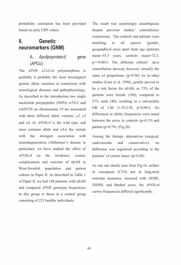
4411
B. Chromosome 9p21 As we did not find an association between
subarachnoid haemorrhage’s
pathophysiology and the well-known,
previously described potential susceptibility
gene on chromosome 19q13.2, we turned to
the Department of Genetics at the
Sahlgrenska University Hospital to identify
other possible genetic markers.
In Paper III, we described a genetic study
where variants of a gene in a particular
chromosome-segment were investigated. We
examined whether an association could be
identified between single nucleotide
polymorphisms (SNPs) and subarachnoid
haemorrhage by
comparing the
patients’ and
controls’ genetic
configuration at those
particular base-pairs.
With genome wide
association studies
(GWAS) in European
and Japanese
populations
chromosome 9p21
was linked to
intracranial
aneurysms (IA)
(Bilguvar et al. 2008).
A 44 kbp (kilo base-
pair) long DNA sequence was searched with
the help of the HapMap database and the
HaploView program. This exercise resulted
in six tagged SNPs (Fig.27).
The genotype frequencies at those base-pars
were analysed both in patients and controls
and the uncommon allele determined. One
SNP, rs10757278 emerged already at this
stage as the only SNP showing significant
difference between controls and patients.
(p=0.02) The uncommon alleles were
subsequently inserted in a univariable
regression analysis and ORs were
calculated. Two of the alleles showed
significant ORs to develop aSAH.
Figure 26 Proportion of APOE ε4 and nonAPOE ε4 allele carriers in healthy controls and SAH patients, in SAH patients with CVS and in SAH patient with poor outcome after one year. (SAH – subarachnoid haemorrhage, CVS – cerebral vasospasm (Spasm), Poor outcome – Glasgow Outcome Scale Extended (GOSE) 1-4, Focal deficit - National Institute of Health Stroke Scale (NIHSS) = 0 APOE ε4 – Apolipoprotein E gen, allele 4)
10
probability estimation has been provided
based on early CRP values.
II. Genetic neuromarkers (GNM)
A. ApolipoproteinΕ gene (APOε)
The APOE ε2/ε3/ε4 polymorphism is
probably is probably the most investigated
genetic allele variation in connection with
neurological diseases and pathophysiology.
As described in the introduction two single
nucleotide polypeptides (SNPs) rs7412 and
rs429358 on chromosome 19 are associated
with three different allele variants: ε2, ε3
and ε4. As APOEε3 is the wild type, and
most common allele and ε4 is the variant
with the strongest association with
neurodegeneration (Alzheimer’s disease in
particular), we have studied the effect of
APOEε4 on the incidence, course,
complications and outcome of aSAH in
West-Swedish population and patient
cohorts in Paper II. As described in Table 1
of Paper II, we had 148 patients, with aSAH
and compared APOE genotype frequencies
in this group to those in a control group
consisting of 221 healthy individuals.
The result was surprisingly unambiguous
despite previous studies’ contradictory
conclusions. The controls and patients were
matching in all aspects (gender,
geographical area) apart from age (patients
mean=55.3 years, controls mean=72.3,
p=<0.001). The different cohorts’ Apoε
constellation showed, however virtually the
same ε4 proportions. (p=0.96) As in other
studies (Linn et al. 1996), gender proved to
be a risk factor for aSAH, as 73% of the
patients were female (108), compared to
27% male (40), resulting in a univariable
OR of 1.86 (1.19-2.92, p<0.001). No
differences in allelic frequencies were noted
between the sexes in controls (p=0.15) and
patient (p=0.79). (Fig.26)
Among the therapy alternatives (surgical,
endovascular and conservative), no
difference was registered according to the
patients’ ε4 carrier status. (p=0.68)
As one can clearly note from Fig.16, neither
in vasospasm (CVS) nor in long-term
outcome measures, assessed with GOSE,
NIHSS, and Barthel score, the APOEε4
carrier frequencies differed significantly.
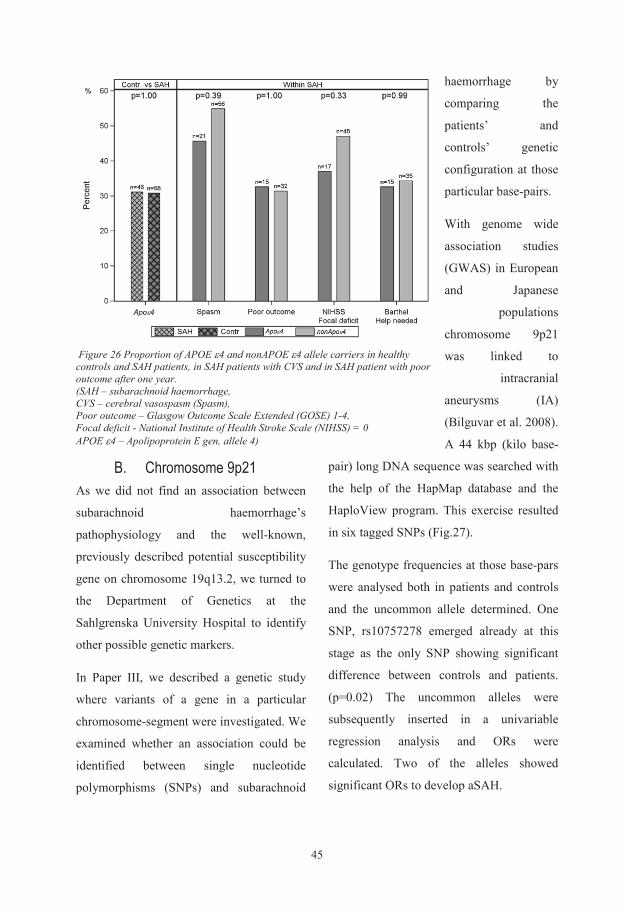
4511
B. Chromosome 9p21 As we did not find an association between
subarachnoid haemorrhage’s
pathophysiology and the well-known,
previously described potential susceptibility
gene on chromosome 19q13.2, we turned to
the Department of Genetics at the
Sahlgrenska University Hospital to identify
other possible genetic markers.
In Paper III, we described a genetic study
where variants of a gene in a particular
chromosome-segment were investigated. We
examined whether an association could be
identified between single nucleotide
polymorphisms (SNPs) and subarachnoid
haemorrhage by
comparing the
patients’ and
controls’ genetic
configuration at those
particular base-pairs.
With genome wide
association studies
(GWAS) in European
and Japanese
populations
chromosome 9p21
was linked to
intracranial
aneurysms (IA)
(Bilguvar et al. 2008).
A 44 kbp (kilo base-
pair) long DNA sequence was searched with
the help of the HapMap database and the
HaploView program. This exercise resulted
in six tagged SNPs (Fig.27).
The genotype frequencies at those base-pars
were analysed both in patients and controls
and the uncommon allele determined. One
SNP, rs10757278 emerged already at this
stage as the only SNP showing significant
difference between controls and patients.
(p=0.02) The uncommon alleles were
subsequently inserted in a univariable
regression analysis and ORs were
calculated. Two of the alleles showed
significant ORs to develop aSAH.
Figure 26 Proportion of APOE ε4 and nonAPOE ε4 allele carriers in healthy controls and SAH patients, in SAH patients with CVS and in SAH patient with poor outcome after one year. (SAH – subarachnoid haemorrhage, CVS – cerebral vasospasm (Spasm), Poor outcome – Glasgow Outcome Scale Extended (GOSE) 1-4, Focal deficit - National Institute of Health Stroke Scale (NIHSS) = 0 APOE ε4 – Apolipoprotein E gen, allele 4)
10
probability estimation has been provided
based on early CRP values.
II. Genetic neuromarkers (GNM)
A. ApolipoproteinΕ gene (APOε)
The APOE ε2/ε3/ε4 polymorphism is
probably is probably the most investigated
genetic allele variation in connection with
neurological diseases and pathophysiology.
As described in the introduction two single
nucleotide polypeptides (SNPs) rs7412 and
rs429358 on chromosome 19 are associated
with three different allele variants: ε2, ε3
and ε4. As APOEε3 is the wild type, and
most common allele and ε4 is the variant
with the strongest association with
neurodegeneration (Alzheimer’s disease in
particular), we have studied the effect of
APOEε4 on the incidence, course,
complications and outcome of aSAH in
West-Swedish population and patient
cohorts in Paper II. As described in Table 1
of Paper II, we had 148 patients, with aSAH
and compared APOE genotype frequencies
in this group to those in a control group
consisting of 221 healthy individuals.
The result was surprisingly unambiguous
despite previous studies’ contradictory
conclusions. The controls and patients were
matching in all aspects (gender,
geographical area) apart from age (patients
mean=55.3 years, controls mean=72.3,
p=<0.001). The different cohorts’ Apoε
constellation showed, however virtually the
same ε4 proportions. (p=0.96) As in other
studies (Linn et al. 1996), gender proved to
be a risk factor for aSAH, as 73% of the
patients were female (108), compared to
27% male (40), resulting in a univariable
OR of 1.86 (1.19-2.92, p<0.001). No
differences in allelic frequencies were noted
between the sexes in controls (p=0.15) and
patient (p=0.79). (Fig.26)
Among the therapy alternatives (surgical,
endovascular and conservative), no
difference was registered according to the
patients’ ε4 carrier status. (p=0.68)
As one can clearly note from Fig.16, neither
in vasospasm (CVS) nor in long-term
outcome measures, assessed with GOSE,
NIHSS, and Barthel score, the APOEε4
carrier frequencies differed significantly.
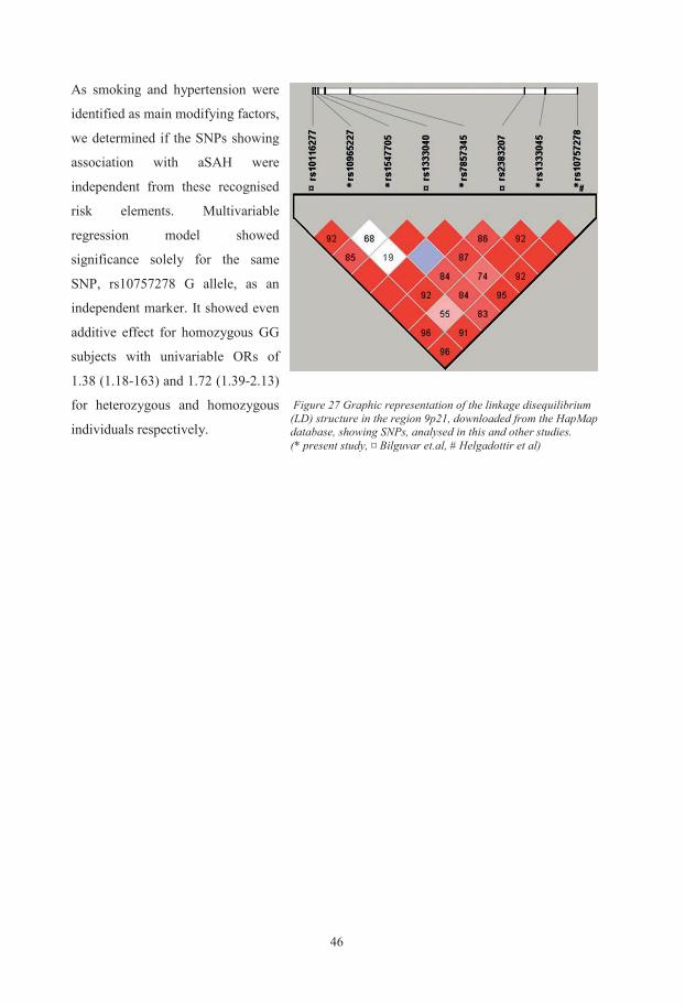
461
DISCUSSION
I. General considerations In our studies, we have focused on a patient -
group with aneurysmal subarachnoid
haemorrhage. We have explored different
biochemical and genetic markers’ relation to
the incidence of the bleeding, the course
during the disease development, the
complication frequency and the different
measures of outcome (Papers I-III). In the
final study, we evaluated the usefulness of an
innovative biochip-array for monitoring and
prediction of outcome in this patient cohort
(Paper IV). Our main findings were the
associations or the absence of associatiations,
between these neuromarkers and one or more
aspects of this devastating haemorrhagic
stroke entity. Some of the links we have
discovered were strong enough to enable us
to make predictions about general outcome.
Our overall aim was to look at these results
from an intensive care physician’s point of
view but with a neurologist’s sense for
details and a neurosurgeon’s focus on the
whole picture. Our tools, to achieve this were
a prospective cohort patient population with
a complete inclusion, to gather so many
patients as possible, daily or almost daily
serum sampling, noting all details of the
condition and treatment of the patients and
finally a meticulous long-term follow-up.
We lost a mere 0.6 % (12) of the identified,
eligible patients from inclusion during the
two-year study period, which stretched over
three years from inclusion to final outcome
assessment. The study borders feasibility for
a single-centre in this respect. Retrospective,
multi-centre or meta-analysis studies are the
next step to increase the number of
participants with other types of
methodological problems to consider. We
had a general unfavourable outcome, i.e.
death or dependency of 32% among the
aSAH patients which is in line with other
large studies (the ISAT study) (Molyneux et
al., 2002; Rosen and Macdonald, 2004) and
even the proportion of poor grade
subarachnoid haemorrhage was
corresponding with 22% as in Rosen’s study
with 3567 participants. This indicates that
our study-population has similar distribution
as larger studies in the world, which makes
our conclusions generally applicable for
other subarachnoid patient groups in similar
hospital environment.
These studies were not designed to compare
therapy alternatives. The figures were merely
described as methodological information for
parallel interpretation with other studies in
the literature. The majority, 71% of our
patients were endovascularly treated; surgical
12
As smoking and hypertension were
identified as main modifying factors,
we determined if the SNPs showing
association with aSAH were
independent from these recognised
risk elements. Multivariable
regression model showed
significance solely for the same
SNP, rs10757278 G allele, as an
independent marker. It showed even
additive effect for homozygous GG
subjects with univariable ORs of
1.38 (1.18-163) and 1.72 (1.39-2.13)
for heterozygous and homozygous
individuals respectively.
Figure 27 Graphic representation of the linkage disequilibrium (LD) structure in the region 9p21, downloaded from the HapMap database, showing SNPs, analysed in this and other studies. (* present study, ¤ Bilguvar et.al, # Helgadottir et al)
471
DISCUSSION
I. General considerations In our studies, we have focused on a patient -
group with aneurysmal subarachnoid
haemorrhage. We have explored different
biochemical and genetic markers’ relation to
the incidence of the bleeding, the course
during the disease development, the
complication frequency and the different
measures of outcome (Papers I-III). In the
final study, we evaluated the usefulness of an
innovative biochip-array for monitoring and
prediction of outcome in this patient cohort
(Paper IV). Our main findings were the
associations or the absence of associatiations,
between these neuromarkers and one or more
aspects of this devastating haemorrhagic
stroke entity. Some of the links we have
discovered were strong enough to enable us
to make predictions about general outcome.
Our overall aim was to look at these results
from an intensive care physician’s point of
view but with a neurologist’s sense for
details and a neurosurgeon’s focus on the
whole picture. Our tools, to achieve this were
a prospective cohort patient population with
a complete inclusion, to gather so many
patients as possible, daily or almost daily
serum sampling, noting all details of the
condition and treatment of the patients and
finally a meticulous long-term follow-up.
We lost a mere 0.6 % (12) of the identified,
eligible patients from inclusion during the
two-year study period, which stretched over
three years from inclusion to final outcome
assessment. The study borders feasibility for
a single-centre in this respect. Retrospective,
multi-centre or meta-analysis studies are the
next step to increase the number of
participants with other types of
methodological problems to consider. We
had a general unfavourable outcome, i.e.
death or dependency of 32% among the
aSAH patients which is in line with other
large studies (the ISAT study) (Molyneux et
al., 2002; Rosen and Macdonald, 2004) and
even the proportion of poor grade
subarachnoid haemorrhage was
corresponding with 22% as in Rosen’s study
with 3567 participants. This indicates that
our study-population has similar distribution
as larger studies in the world, which makes
our conclusions generally applicable for
other subarachnoid patient groups in similar
hospital environment.
These studies were not designed to compare
therapy alternatives. The figures were merely
described as methodological information for
parallel interpretation with other studies in
the literature. The majority, 71% of our
patients were endovascularly treated; surgical
12
As smoking and hypertension were
identified as main modifying factors,
we determined if the SNPs showing
association with aSAH were
independent from these recognised
risk elements. Multivariable
regression model showed
significance solely for the same
SNP, rs10757278 G allele, as an
independent marker. It showed even
additive effect for homozygous GG
subjects with univariable ORs of
1.38 (1.18-163) and 1.72 (1.39-2.13)
for heterozygous and homozygous
individuals respectively.
Figure 27 Graphic representation of the linkage disequilibrium (LD) structure in the region 9p21, downloaded from the HapMap database, showing SNPs, analysed in this and other studies. (* present study, ¤ Bilguvar et.al, # Helgadottir et al)
483
Seizures present an extremely complicating
dimension to initial assessment. Nearly one
in ten patients was found having early-onset
seizures (EOS) at admission (Fung et al.,
2015), of whom 66% were graded poor on
clinical scales. 68% of these patients
achieved nevertheless good outcome at 6
month. Furthermore, patients having seizures
are treated on site with anti-epileptics (often
benzodiazepines) which affect the level of
consciousness, resulting in worse clinical
scores than the bleeding would have
warranted. This fact adds to the explanation
why initial neurology fails to predict
outcome reliably. (Paper I) These seizures
may even proceed to non-convulsive seizures
(NCSs) that have been implicated in the
aetiology of secondary brain injury. Claassen
and co-workers observed 479 SAH patients
and found 11% having NCSs and could
directly associate their condition to
inflammatory state with markers like TNFR1
and hs-CRP. (Claassen et al., 2014)
The clinical appearance of aSAH is highly
altered by the patients’ co-morbidity status.
Severe congestive heart failure, COPD, pre-
existing neurological disease and psychiatric
conditions may complicate the initial
assessment and definitely affect long-term
outcome to the worse. It led to development
of Charlson Co-morbidity Index (CCI) and
its application on aSAH patients. (Boogaarts
et al., 2014) If this index had been available
at the time of study design, it would have
been easier to adjust the outcome for co-
morbidity.
Finally two facts need to be mentioned,
which might have confounded some aspects
of the results. A difference might have
emerged between patients presenting directly
at the University Hospital compared to those,
being referred from other health facilities
with the ambulance services. In the letter
case we were compelled to seek secondary
information on patients’ initial status by
consulting local hospital journals and
ambulance records. These patients came
frequently sedated, sometimes anaesthetised
and in respirator as recommended by inter-
hospital transport protocol based on current
knowledge (Uren et al., 2009). Neurological
investigations were seriously influenced,
often impeded on these patients.
Technical circumstances compelled us to
exclude 12 patients, 3 of whom were foreign
citizens, presumably not available for
outcome investigations and in 9 cases,
informed consents could not be obtained or
were withdrawn. These cases might have
affected outcome, as the main reason for
failure to obtain consent was that these
patients were severely affected by their
illness and could not sign it themselves and
relatives could not be traced. It might have
been however compensated by those few,
who withdrew their consent because they
2
clipping was therapy alternative in 26% and
3% were treated conservatively i.e. basic
intensive care. These figures vary both
geographically and in time in the literature.
In a Chinese centre all good-grade aSAH
patients (H&H 1-2) were operated in 2003,
the rest were conservatively treated (Tang et
al., 2003), while in a Scottish centre 11% of
the patients operated and 89 treated with
coiling (Teo et al., 2015). At the same
department, the therapy trend changed in
favour of coiling from 11% in 1995 to 89%
in 2003 and has continued on that level since
then. This paradigm shift happened after the
ISAT study, although the difference in RR
(relative risk) reduction between the
endovascular vs. neurosurgical treatment
groups was only 15% with 2-3% higher risk
of re-bleeding in the coiling group. The 18-
year follow-up of this material has been
published this year (Molyneux et al., 2015)
and it showed that the difference in
dependency is not significant, but the chance
for being alive 10 years after treatment is
higher in the coil-group (OR: 1.34), however
with 1,7% re-bleeding risk from the target
aneurysm.
II. Patient considerations There is one weakness in all third-level
referral hospital driven clinical trials; they all
have flawed inclusion, however thoroughly
they try to avoid it. There is an important
group of patients in poor condition, who
never reach these university hospitals,
thereby the public impact of the diseases is
much larger than the actual figures and the
outcome is worse than claimed (Nylen,
2007). Unfortunately it is only disease
demographic studies, based on records and
data-bases which can put these figures right.
The positive fact is that different data exist to
assess the initial status of SAH patients and
the severity of the haemorrhage: clinical-,
radiological-, laboratory examination, lumbar
puncture and not the least the experience of
the clinician. They complement one-another
and finally they form a basis for decision
with quite high sensitivity and specificity. To
have the ambition that one sole modality, let
alone a single marker would substitute this
complex decision-making system is over-
pretentious.
One of the more challenging facts is the
diversity of clinical picture the subarachnoid
haemorrhage patients present. There is a
large dispersion in the time-frame they
appear in the emergency department, from a
few minutes to a couple of days or even
weeks. There were some patients, presenting
late on the medical or neurological wards
with mild symptoms, which later could be
related to aSAH. Most of these patients were
unfortunately ineligible for our studies,
elapsing more than 2 days from the bleeding.
493
Seizures present an extremely complicating
dimension to initial assessment. Nearly one
in ten patients was found having early-onset
seizures (EOS) at admission (Fung et al.,
2015), of whom 66% were graded poor on
clinical scales. 68% of these patients
achieved nevertheless good outcome at 6
month. Furthermore, patients having seizures
are treated on site with anti-epileptics (often
benzodiazepines) which affect the level of
consciousness, resulting in worse clinical
scores than the bleeding would have
warranted. This fact adds to the explanation
why initial neurology fails to predict
outcome reliably. (Paper I) These seizures
may even proceed to non-convulsive seizures
(NCSs) that have been implicated in the
aetiology of secondary brain injury. Claassen
and co-workers observed 479 SAH patients
and found 11% having NCSs and could
directly associate their condition to
inflammatory state with markers like TNFR1
and hs-CRP. (Claassen et al., 2014)
The clinical appearance of aSAH is highly
altered by the patients’ co-morbidity status.
Severe congestive heart failure, COPD, pre-
existing neurological disease and psychiatric
conditions may complicate the initial
assessment and definitely affect long-term
outcome to the worse. It led to development
of Charlson Co-morbidity Index (CCI) and
its application on aSAH patients. (Boogaarts
et al., 2014) If this index had been available
at the time of study design, it would have
been easier to adjust the outcome for co-
morbidity.
Finally two facts need to be mentioned,
which might have confounded some aspects
of the results. A difference might have
emerged between patients presenting directly
at the University Hospital compared to those,
being referred from other health facilities
with the ambulance services. In the letter
case we were compelled to seek secondary
information on patients’ initial status by
consulting local hospital journals and
ambulance records. These patients came
frequently sedated, sometimes anaesthetised
and in respirator as recommended by inter-
hospital transport protocol based on current
knowledge (Uren et al., 2009). Neurological
investigations were seriously influenced,
often impeded on these patients.
Technical circumstances compelled us to
exclude 12 patients, 3 of whom were foreign
citizens, presumably not available for
outcome investigations and in 9 cases,
informed consents could not be obtained or
were withdrawn. These cases might have
affected outcome, as the main reason for
failure to obtain consent was that these
patients were severely affected by their
illness and could not sign it themselves and
relatives could not be traced. It might have
been however compensated by those few,
who withdrew their consent because they
2
clipping was therapy alternative in 26% and
3% were treated conservatively i.e. basic
intensive care. These figures vary both
geographically and in time in the literature.
In a Chinese centre all good-grade aSAH
patients (H&H 1-2) were operated in 2003,
the rest were conservatively treated (Tang et
al., 2003), while in a Scottish centre 11% of
the patients operated and 89 treated with
coiling (Teo et al., 2015). At the same
department, the therapy trend changed in
favour of coiling from 11% in 1995 to 89%
in 2003 and has continued on that level since
then. This paradigm shift happened after the
ISAT study, although the difference in RR
(relative risk) reduction between the
endovascular vs. neurosurgical treatment
groups was only 15% with 2-3% higher risk
of re-bleeding in the coiling group. The 18-
year follow-up of this material has been
published this year (Molyneux et al., 2015)
and it showed that the difference in
dependency is not significant, but the chance
for being alive 10 years after treatment is
higher in the coil-group (OR: 1.34), however
with 1,7% re-bleeding risk from the target
aneurysm.
II. Patient considerations There is one weakness in all third-level
referral hospital driven clinical trials; they all
have flawed inclusion, however thoroughly
they try to avoid it. There is an important
group of patients in poor condition, who
never reach these university hospitals,
thereby the public impact of the diseases is
much larger than the actual figures and the
outcome is worse than claimed (Nylen,
2007). Unfortunately it is only disease
demographic studies, based on records and
data-bases which can put these figures right.
The positive fact is that different data exist to
assess the initial status of SAH patients and
the severity of the haemorrhage: clinical-,
radiological-, laboratory examination, lumbar
puncture and not the least the experience of
the clinician. They complement one-another
and finally they form a basis for decision
with quite high sensitivity and specificity. To
have the ambition that one sole modality, let
alone a single marker would substitute this
complex decision-making system is over-
pretentious.
One of the more challenging facts is the
diversity of clinical picture the subarachnoid
haemorrhage patients present. There is a
large dispersion in the time-frame they
appear in the emergency department, from a
few minutes to a couple of days or even
weeks. There were some patients, presenting
late on the medical or neurological wards
with mild symptoms, which later could be
related to aSAH. Most of these patients were
unfortunately ineligible for our studies,
elapsing more than 2 days from the bleeding.
505
Before performing patient selection for study
resulting in Paper I (CRP), we had some
methodological discussions how we should
relate to indwelling ventricular catheter or
parenchymal pressure transducer insertion as
a confounding factor in CRP elevation. We
came to the conclusion that we confirm the
fact that catheter-insertion procedures initiate
an inflammatory reaction (Alhadad et al.,
2007; Almagor et al., 2003) but not in the
magnitude as in major operations inclusive
craniotomy for cortical tumour operations
(Mirzayan et al., 2007). Although there is no
literature support, it is plausible, that skull-
base surgery as clipping an aneurysm results
in an even larger inflammatory process than
craniotomy for cortical tumours, thereby
constituting an even more important
confounder for CRP elevation. This fact is
the most important criticism for previous
CRP studies in SAH patients. Ventricular
catheter or parenchymal pressure transducer
were placed in patients, either in need of
invasive intracranial pressure monitoring,
CSF drainage, or both because of a SAH
complication (hydrocephalus, DCI, CI). We
decided that exclusion of patients with
indwelling ventricular or parenchymal
catheter (42% of study I. population) would
result in a skewed inclusion of aSAH patients
(only the very best of patients and the
moribund ones) and that the results could no
longer be applied for this patient group.
In Paper III, performing qPCR (quantitative
real-time polymerase chain reaction) assay
for rs1333040 SNP, the test failed and due to
probe C_8766795_10 failure it could not be
repeated. This SNP which is part of the
cyclin-dependent kinase inhibitor 2 B-
antisense RNA1 (CDKN2B-AS1) a non-
coding RNA sequence, better known as
ANRIL (antisense non-coding RNA in the
INK4 locus), which has a function of
silencing other genes in the proximity.
(Popov and Gil, 2010). It is rather
unfortunate that this particular assay failed as
it was associated with atherosclerosis,
cardiovascular diseases, diabetes type 2, and
most of all intracranial aneurysms. (Abrantes
et al., 2015; Alg et al., 2013).
Our follow-up, as I described in the
introduction, is our pride, but one may argue
that even longer monitoring would have been
desirable (Molyneux et al., 2015). Wilson
and co-workers showed, however, that in the
time-course of recovery, following poor-
grade aneurysm in 88 patients, the largest
improvement occurred between hospital
discharge and 6 month (23.5%, which proved
to be significant). Further recovery measured
by modified Rankin scale, was 18% between
6 months and 1 year and further 19%
between 1 year and 3 years. They did not
differ significantly between each-other
(Wilson et al., 2013). Nylén pointed out the
risks associated with extended follow-ups,
4
were simply not interested in further blood
samples, neurological investigations or/and
an impending LP in an adjoining study.
III. Methodological considerations When we designed these studies no data
existed on many of the neuromarkers’ kinetic
properties. Some of them are fast markers
(FABP, TNFR1, DDMR) increasing within
hours, some even within minutes, others take
days to rise (CRP). Even circadian rhythm is
described in some of the markers (FABP)
(Pelsers et al., 1999). The admission
sampling, which could fall on day0, day1 or
day2 could be taken at any hour of the day,
the following samples were taken
meticulously the same hour every day. This
fact could indicate a wider technical
dispersion of the data on the initial days.
As in all patient related clinical studies, we
also have data samples missing. In our case,
the blood-sample collection’s adherence to
study protocol was around 87%, depending
on the patient selection for the study,
meaning that 13% of the blood values never
reached the study register (not taken, sample
foul, laboratory error). This led to the
exclusion of 13 patients from Paper I due to
difficulty in calculating the area under curve
(AUC) in serial data. After publication, we
have solved this problem, by using weighted
(geometric) mean values for the time -series.
After recalculating the figures for the whole
sample, it led to the same conclusion, only
changing the results by the second decimal.
These studies were not aimed to parallel
treatment strategies (surgical clipping vs.
endovascular coiling) and these data were
presented of pure descriptive reasons only, so
it can be viewed in relation to other studies in
the literature. However, out-of-study
comparison showed similar results as
published in much larger multicentre
investigations, e.g. the ISAT or BRAT study.
(Molyneux et al., 2002; Spetzler et al., 2015)
For the sake of interest, data taken from
Paper II show 28% and 30% dead or
dependency, 46% and 41% detectable
neurological deficit and 28% and 33% ADL
assistance necessity for operated and coiled
patients respectively at one-year follow up.
None of the figures showed statistically
secured difference, only a slight tendency to
more focal deficit after surgery and slightly
more assistance requirement after coiling.
One could argue that the more severe the
SAH (volume effect, intra-parenchymal
component, herniation risk), the larger is the
possibility for neurosurgical intervention
(clipping), thus it would skew the outcome.
However, this point is usually counteracted
by the fact that the most elderly and patients
with most severe co-morbidity tended to be
treated endovascularly (coiling).
515
Before performing patient selection for study
resulting in Paper I (CRP), we had some
methodological discussions how we should
relate to indwelling ventricular catheter or
parenchymal pressure transducer insertion as
a confounding factor in CRP elevation. We
came to the conclusion that we confirm the
fact that catheter-insertion procedures initiate
an inflammatory reaction (Alhadad et al.,
2007; Almagor et al., 2003) but not in the
magnitude as in major operations inclusive
craniotomy for cortical tumour operations
(Mirzayan et al., 2007). Although there is no
literature support, it is plausible, that skull-
base surgery as clipping an aneurysm results
in an even larger inflammatory process than
craniotomy for cortical tumours, thereby
constituting an even more important
confounder for CRP elevation. This fact is
the most important criticism for previous
CRP studies in SAH patients. Ventricular
catheter or parenchymal pressure transducer
were placed in patients, either in need of
invasive intracranial pressure monitoring,
CSF drainage, or both because of a SAH
complication (hydrocephalus, DCI, CI). We
decided that exclusion of patients with
indwelling ventricular or parenchymal
catheter (42% of study I. population) would
result in a skewed inclusion of aSAH patients
(only the very best of patients and the
moribund ones) and that the results could no
longer be applied for this patient group.
In Paper III, performing qPCR (quantitative
real-time polymerase chain reaction) assay
for rs1333040 SNP, the test failed and due to
probe C_8766795_10 failure it could not be
repeated. This SNP which is part of the
cyclin-dependent kinase inhibitor 2 B-
antisense RNA1 (CDKN2B-AS1) a non-
coding RNA sequence, better known as
ANRIL (antisense non-coding RNA in the
INK4 locus), which has a function of
silencing other genes in the proximity.
(Popov and Gil, 2010). It is rather
unfortunate that this particular assay failed as
it was associated with atherosclerosis,
cardiovascular diseases, diabetes type 2, and
most of all intracranial aneurysms. (Abrantes
et al., 2015; Alg et al., 2013).
Our follow-up, as I described in the
introduction, is our pride, but one may argue
that even longer monitoring would have been
desirable (Molyneux et al., 2015). Wilson
and co-workers showed, however, that in the
time-course of recovery, following poor-
grade aneurysm in 88 patients, the largest
improvement occurred between hospital
discharge and 6 month (23.5%, which proved
to be significant). Further recovery measured
by modified Rankin scale, was 18% between
6 months and 1 year and further 19%
between 1 year and 3 years. They did not
differ significantly between each-other
(Wilson et al., 2013). Nylén pointed out the
risks associated with extended follow-ups,
4
were simply not interested in further blood
samples, neurological investigations or/and
an impending LP in an adjoining study.
III. Methodological considerationsWhen we designed these studies no data
existed on many of the neuromarkers’ kinetic
properties. Some of them are fast markers
(FABP, TNFR1, DDMR) increasing within
hours, some even within minutes, others take
days to rise (CRP). Even circadian rhythm is
described in some of the markers (FABP)
(Pelsers et al., 1999). The admission
sampling, which could fall on day0, day1 or
day2 could be taken at any hour of the day,
the following samples were taken
meticulously the same hour every day. This
fact could indicate a wider technical
dispersion of the data on the initial days.
As in all patient related clinical studies, we
also have data samples missing. In our case,
the blood-sample collection’s adherence to
study protocol was around 87%, depending
on the patient selection for the study,
meaning that 13% of the blood values never
reached the study register (not taken, sample
foul, laboratory error). This led to the
exclusion of 13 patients from Paper I due to
difficulty in calculating the area under curve
(AUC) in serial data. After publication, we
have solved this problem, by using weighted
(geometric) mean values for the time -series.
After recalculating the figures for the whole
sample, it led to the same conclusion, only
changing the results by the second decimal.
These studies were not aimed to parallel
treatment strategies (surgical clipping vs.
endovascular coiling) and these data were
presented of pure descriptive reasons only, so
it can be viewed in relation to other studies in
the literature. However, out-of-study
comparison showed similar results as
published in much larger multicentre
investigations, e.g. the ISAT or BRAT study.
(Molyneux et al., 2002; Spetzler et al., 2015)
For the sake of interest, data taken from
Paper II show 28% and 30% dead or
dependency, 46% and 41% detectable
neurological deficit and 28% and 33% ADL
assistance necessity for operated and coiled
patients respectively at one-year follow up.
None of the figures showed statistically
secured difference, only a slight tendency to
more focal deficit after surgery and slightly
more assistance requirement after coiling.
One could argue that the more severe the
SAH (volume effect, intra-parenchymal
component, herniation risk), the larger is the
possibility for neurosurgical intervention
(clipping), thus it would skew the outcome.
However, this point is usually counteracted
by the fact that the most elderly and patients
with most severe co-morbidity tended to be
treated endovascularly (coiling).
527
based on a five-criterion system, awarding 0
or 1 point. The criteria were age (<50, >50
years), H&H (no coma I-III, coma IV-V),
Fisher scale (1-2, 3-4), aneurysm size (<10,
>10 mm), giant posterior circulation lesion
(no, yes). Scores are from 0-5 (Ogilvy and
Carter, 1998).
In his modified MGH-score (m-MGH),
Lagares changed the level of consciousness
to dichotomised WFNS scale (I, II-III, IV-V),
otherwise similar to above and claimed to
have better performance (Lagares et al.,
2005).
GCS grading system (GCS-GS) is
compressed version of the GCS system, there
GCS points is put into a 5-scale system. (15,
14-12, 11-9, 8-6, 5-3). It preformed
reportedly as well as WFNS and GCS.
NIS-SSS, Nationwide Inpatient Sample
(American ICD-9 based administrative
database) SAH Severity Score is developed
for database demographic studies to control
for SAH severity. Patient selection with the
diagnosis codes for coma, stupor,
hydrocephalus, paresis/plegia, aphasia and
cranial-nerve defect as well as treatment
codes for ventriculostomi, CSF shunt and
mechanical ventilation were collected and
univariate logistic regression coefficient
calculated. NIS-SSS was derived from a
weighted sum of these coefficients. It was
validated in more than 100,000 patients and
then performance tested. It performed better
than H&H for outcome prediction
(Washington et al., 2014).
The FOUR score, full outline of
unresponsiveness score was developed after
realising the shortcomings of the GCS
system on brainstem reflexes. It gives 0-4
points in four functions; eye movement,
motor response, brainstem reflexes (pupil,
cornea, cough), and respiration. According to
records, it performs similar to GCS (Chen et
al., 2013).
The HAIR score is a prediction model
comprising of 4 attributes of SAH. It gives
different points in different severity on a non-
linear scale. H&H (I-III0, IV1, V4),
Age (<600, 60-801, >802), Intra-
ventricular haemorrhage (IVH) (no0,
yes1) and Re-bleeding (no0, yes1).
Scores 0 –7 predict in-hospital mortality
between 0.9 – 83% evenly distributed (Lee et
al., 2014).
Takagi et al. had an entirely mathematical
approach of the allocation of grading based
on the GCS. He was using combinatorial
methods to divide the 15 points into 5
categories. The integrated differences led to
the Grading Scale based on GCS (GSbGCS):
I –15, II –14-11, III –10-8, IV –7-4, V –3.
They claimed more symmetrical patient -
distribution and better prediction
performance then WFNS and they promised
an easy adjustment if therapy results
improved (Takagi et al., 1999).
6
including increased number of drop-outs,
changed social and financial status and other
confounding circumstances which would
cloud the disease-dependent outcome (Nylen,
2007). On the other hand, one cannot deny
the fact, that the 37% progress in
neurological recovery between 6 month and
3 years in the Wilson study, is a more
substantial improvement than that of from
discharge to 6 month (and statistically
significant, if one after-calculates the
figures). This is one of the reasons why very
long-time (10-15 years) follow-up is under
way in our aSAH patient -group. Another
reason is to confirm a recent finding from
Lindgren et al. that aSAH patients, even long
time after their ictus, still die of
cerebrovascular reasons, unlike the general
population, where cardiovascular
explanations are the most frequent causes of
death (Lindgren et al., 2015).
IV. Classification considerations The diagnostic and severity scales detailed in
the introduction (RLS85, H&H, GCS,
WFNS, Fisher scale) are still valid today,
although many of them are modified and
numerous new ones enrich the collection. I
give a brief summary of the proposed
changes and the potentially new scales to
use. The reason for this passionate interest in
modifying the old scales or inventing new
ones lies in the fact that nearly all of them
perform poorly in outcome predictions.
There is an inherent conflict between
designing and using a diagnostic scale. The
easier, more user-friendly a scale is (less
categories) the less mathematical chance it
has for good outcome prediction. Conversely,
the more detailed, more points / categories it
has, the less popular it becomes but has the
potential (at least mathematically) for better
prediction. This ambiguity may contribute to
the fact that new prediction models appear
frequently in the literature, although none of
them transferred to clinical practice (Jaja et
al., 2013).
A. Diagnostic scales
One of the modifications to the WFNS (m-
WFNS) scale was suggested by Sano et al.
by moving GCS 14 to WFNS II and GCS 13
to WFNS III, regardless of focal deficit. It
eliminates the assessment on double axis,
which makes the scale easier to use (Sano et
al., 2015).
SAH score from Naval and co-workers
proposed a summary point system from GCS
(1-4 points), age (1– 4 points), medical
comorbidities (1– 3 points), which resulted in
9 categories and giving, according to the
authors, superior prediction to WFNS
(AUC=0.821 vs. 0.771). (Naval et al., 2014).
MGH-score was developed in the
Massachusetts General Hospital and was
537
based on a five-criterion system, awarding 0
or 1 point. The criteria were age (<50, >50
years), H&H (no coma I-III, coma IV-V),
Fisher scale (1-2, 3-4), aneurysm size (<10,
>10 mm), giant posterior circulation lesion
(no, yes). Scores are from 0-5 (Ogilvy and
Carter, 1998).
In his modified MGH-score (m-MGH),
Lagares changed the level of consciousness
to dichotomised WFNS scale (I, II-III, IV-V),
otherwise similar to above and claimed to
have better performance (Lagares et al.,
2005).
GCS grading system (GCS-GS) is
compressed version of the GCS system, there
GCS points is put into a 5-scale system. (15,
14-12, 11-9, 8-6, 5-3). It preformed
reportedly as well as WFNS and GCS.
NIS-SSS, Nationwide Inpatient Sample
(American ICD-9 based administrative
database) SAH Severity Score is developed
for database demographic studies to control
for SAH severity. Patient selection with the
diagnosis codes for coma, stupor,
hydrocephalus, paresis/plegia, aphasia and
cranial-nerve defect as well as treatment
codes for ventriculostomi, CSF shunt and
mechanical ventilation were collected and
univariate logistic regression coefficient
calculated. NIS-SSS was derived from a
weighted sum of these coefficients. It was
validated in more than 100,000 patients and
then performance tested. It performed better
than H&H for outcome prediction
(Washington et al., 2014).
The FOUR score, full outline of
unresponsiveness score was developed after
realising the shortcomings of the GCS
system on brainstem reflexes. It gives 0-4
points in four functions; eye movement,
motor response, brainstem reflexes (pupil,
cornea, cough), and respiration. According to
records, it performs similar to GCS (Chen et
al., 2013).
The HAIR score is a prediction model
comprising of 4 attributes of SAH. It gives
different points in different severity on a non-
linear scale. H&H (I-III0, IV1, V4),
Age (<600, 60-801, >802), Intra-
ventricular haemorrhage (IVH) (no0,
yes1) and Re-bleeding (no0, yes1).
Scores 0 –7 predict in-hospital mortality
between 0.9 – 83% evenly distributed (Lee et
al., 2014).
Takagi et al. had an entirely mathematical
approach of the allocation of grading based
on the GCS. He was using combinatorial
methods to divide the 15 points into 5
categories. The integrated differences led to
the Grading Scale based on GCS (GSbGCS):
I –15, II –14-11, III –10-8, IV –7-4, V –3.
They claimed more symmetrical patient -
distribution and better prediction
performance then WFNS and they promised
an easy adjustment if therapy results
improved (Takagi et al., 1999).
6
including increased number of drop-outs,
changed social and financial status and other
confounding circumstances which would
cloud the disease-dependent outcome (Nylen,
2007). On the other hand, one cannot deny
the fact, that the 37% progress in
neurological recovery between 6 month and
3 years in the Wilson study, is a more
substantial improvement than that of from
discharge to 6 month (and statistically
significant, if one after-calculates the
figures). This is one of the reasons why very
long-time (10-15 years) follow-up is under
way in our aSAH patient -group. Another
reason is to confirm a recent finding from
Lindgren et al. that aSAH patients, even long
time after their ictus, still die of
cerebrovascular reasons, unlike the general
population, where cardiovascular
explanations are the most frequent causes of
death (Lindgren et al., 2015).
IV. Classification considerations The diagnostic and severity scales detailed in
the introduction (RLS85, H&H, GCS,
WFNS, Fisher scale) are still valid today,
although many of them are modified and
numerous new ones enrich the collection. I
give a brief summary of the proposed
changes and the potentially new scales to
use. The reason for this passionate interest in
modifying the old scales or inventing new
ones lies in the fact that nearly all of them
perform poorly in outcome predictions.
There is an inherent conflict between
designing and using a diagnostic scale. The
easier, more user-friendly a scale is (less
categories) the less mathematical chance it
has for good outcome prediction. Conversely,
the more detailed, more points / categories it
has, the less popular it becomes but has the
potential (at least mathematically) for better
prediction. This ambiguity may contribute to
the fact that new prediction models appear
frequently in the literature, although none of
them transferred to clinical practice (Jaja et
al., 2013).
A. Diagnostic scales
One of the modifications to the WFNS (m-
WFNS) scale was suggested by Sano et al.
by moving GCS 14 to WFNS II and GCS 13
to WFNS III, regardless of focal deficit. It
eliminates the assessment on double axis,
which makes the scale easier to use (Sano et
al., 2015).
SAH score from Naval and co-workers
proposed a summary point system from GCS
(1-4 points), age (1– 4 points), medical
comorbidities (1– 3 points), which resulted in
9 categories and giving, according to the
authors, superior prediction to WFNS
(AUC=0.821 vs. 0.771). (Naval et al., 2014).
MGH-score was developed in the
Massachusetts General Hospital and was
549
some symptoms, 2 – slight disability (able
not all, but the majority of activities), 3 –
moderate disability (some help, able to
walk), 4 – moderately severe disability
(unable to walk or take care of body needs),
5 – severe disability (bedridden, incontinent,
constant nursing). The scale is often
dichotomised to favourable outcome (mRS 0-
2) and dependent (mRs 3-5) (van Swieten et
al., 1988).
V. Remarks on genetic markers
A. ApoE
Apolipoprotein E is a major cholesterol
transporter and an active regulator of
lipoprotein metabolism in the body (Siest et
al., 1995), and it is understandable that
genetical variants coding different protein
alternates have different effectivity in these
mechanisms. Apart from role as a transporter
in the periphery, it also assists neural
transmission (Mauch et al., 2001) and in
pathological conditions ApoE has been
suggested to promote aggregation of amyloid
β (Aβ) into plaques, which are characteristic
of Alzheimer’s disease.
The findings in our study in Paper III are
fairly clear-cut. In our clinical material, we
found no association of the variant allele
APOEε4, either with the incidence of aSAH
compared in a case control study or in an
aSAH patient cohort study on CVS and long
term-outcome. The results were so
unambiguous, that if we 10-doubled the
patient number in our material with the same
dispersion of data, it would still lead to the
same conclusion. As this question has been
extensively researched even at this diagnostic
entity, why is it still interesting?
The answer lies on at least two levels. Firstly,
the published studies demonstrate a
surprisingly unequivocal conclusion, with
approximately the same number presenting
data for a negative effect of the altered allele
on the outcome (Niskakangas et al., 2001;
Tang et al., 2003; Leung et al., 2002) as
demonstrating non-significant effect or no
influence at all (Morris et al., 2004;
Fontanella et al., 2007). One would think that
meta-analysis could solve this discrepancy by
collecting all data and calculating
summarised-odds for this allele’s effect on
several hundreds or thousands of patients.
Unfortunately it is not always true, as those
two recently published meta-analyses on this
subject demonstrate (Lanterna et al., 2007;
McColgan et al., 2010). They present equally
conflicting conclusions, on the basis of, and
it is similarly intriguing, only partially
overlapping study-material. My
interpretation of the current scientific opinion
is that it is rather unclear on this matter, so I
hope that I pushed the debate a little further.
8
The radiological assessment scales have
evolved as well over the years. Claassen
suggested a modification of the Fisher scale
(mod Fisher scale) after showing the
importance of bilateral intra-ventricular
hematoma in the risk of DCI development.
His suggestion was: 0 – no SAH, 1 – thin
SAH, no IVH in both ventr., 2 – thin SAH,
IVH in both ventr., 3 – thick SAH(>1mm),
no IVH in both ventr., 4 – thick SAH, IVH in
both ventr (Claassen et al., 2001).
A new scale of radiological classification
was proposed by Wilson, the Barrow
Neurological Institute SAH scale (BNI-SAH
Scale). It abandoned the importance of intra-
ventricular hematoma as a prognostic factor
and concentrated entirely on the thickness of
the SAH, measured perpendicular to the
direction of cistern or fissure. The scale
classified the SAH in 5 categories: 1 – No
SAH, 2 – ≤ 5.0mm, 3 – >5-10 mm, 4 – > 10-
15 mm, 5 – >15 mm. The simplicity and
Gauss distribution of patients in the scale are
a huge advantage, but the scale needs
validation in prospective studies (Wilson et
al., 2012).
It was finally relieving to read that the
Charlson Comorbidity Index, where they
allocate points by different coexisting
diseases added no extra information in a
SAH prognostic model (Boogaarts et al.,
2014).
B. Outcome scales
GOS, GOSE, NIHSS (although not strictly
an outcome scale) and the Barthel Index,
detailed in the Introduction, continue to serve
as useful tools in outcome assessment. These
scales can, and were in our research group
(Nylen et al., 2007), supplemented by the
Mini Mental State Examination (MMSE),
which can subdivide the patients with
favourable outcome by testing their higher
cognitive functions. It requires speech or
communication and is designed to test
orientation, memory and attention (Folstein
et al., 1975).
NIS-SOM, as in Nationwide Inpatient
Sample SAH Outcome Measure, as its sister
NIS-SSS mentioned above is an
administrative tool to control demographic
and economic studies’ outcome and disease
severity. It is derived from ICD-9 diagnose-
and therapy codes and calculates regression
coefficient-based measures of outcome. It is
not for clinical use, but can be applied for
hundreds of thousands of patients in
databases (Washington et al., 2014).
Finally, there is an outcome evaluation tool,
which has gained wide acceptance in
literature, and it might be the preferred
assessment measure in the future; the
modified Rankin scale (mRS). It reflects
general disability in the sense of handicap
rather than impairment. It is a six graded
scale: 0 – no symptoms, 1 – no disability
559
some symptoms, 2 – slight disability (able
not all, but the majority of activities), 3 –
moderate disability (some help, able to
walk), 4 – moderately severe disability
(unable to walk or take care of body needs),
5 – severe disability (bedridden, incontinent,
constant nursing). The scale is often
dichotomised to favourable outcome (mRS 0-
2) and dependent (mRs 3-5) (van Swieten et
al., 1988).
V. Remarks on genetic markers
A. ApoE
Apolipoprotein E is a major cholesterol
transporter and an active regulator of
lipoprotein metabolism in the body (Siest et
al., 1995), and it is understandable that
genetical variants coding different protein
alternates have different effectivity in these
mechanisms. Apart from role as a transporter
in the periphery, it also assists neural
transmission (Mauch et al., 2001) and in
pathological conditions ApoE has been
suggested to promote aggregation of amyloid
β (Aβ) into plaques, which are characteristic
of Alzheimer’s disease.
The findings in our study in Paper III are
fairly clear-cut. In our clinical material, we
found no association of the variant allele
APOEε4, either with the incidence of aSAH
compared in a case control study or in an
aSAH patient cohort study on CVS and long
term-outcome. The results were so
unambiguous, that if we 10-doubled the
patient number in our material with the same
dispersion of data, it would still lead to the
same conclusion. As this question has been
extensively researched even at this diagnostic
entity, why is it still interesting?
The answer lies on at least two levels. Firstly,
the published studies demonstrate a
surprisingly unequivocal conclusion, with
approximately the same number presenting
data for a negative effect of the altered allele
on the outcome (Niskakangas et al., 2001;
Tang et al., 2003; Leung et al., 2002) as
demonstrating non-significant effect or no
influence at all (Morris et al., 2004;
Fontanella et al., 2007). One would think that
meta-analysis could solve this discrepancy by
collecting all data and calculating
summarised-odds for this allele’s effect on
several hundreds or thousands of patients.
Unfortunately it is not always true, as those
two recently published meta-analyses on this
subject demonstrate (Lanterna et al., 2007;
McColgan et al., 2010). They present equally
conflicting conclusions, on the basis of, and
it is similarly intriguing, only partially
overlapping study-material. My
interpretation of the current scientific opinion
is that it is rather unclear on this matter, so I
hope that I pushed the debate a little further.
8
The radiological assessment scales have
evolved as well over the years. Claassen
suggested a modification of the Fisher scale
(mod Fisher scale) after showing the
importance of bilateral intra-ventricular
hematoma in the risk of DCI development.
His suggestion was: 0 – no SAH, 1 – thin
SAH, no IVH in both ventr., 2 – thin SAH,
IVH in both ventr., 3 – thick SAH(>1mm),
no IVH in both ventr., 4 – thick SAH, IVH in
both ventr (Claassen et al., 2001).
A new scale of radiological classification
was proposed by Wilson, the Barrow
Neurological Institute SAH scale (BNI-SAH
Scale). It abandoned the importance of intra-
ventricular hematoma as a prognostic factor
and concentrated entirely on the thickness of
the SAH, measured perpendicular to the
direction of cistern or fissure. The scale
classified the SAH in 5 categories: 1 – No
SAH, 2 – ≤ 5.0mm, 3 – >5-10 mm, 4 – > 10-
15 mm, 5 – >15 mm. The simplicity and
Gauss distribution of patients in the scale are
a huge advantage, but the scale needs
validation in prospective studies (Wilson et
al., 2012).
It was finally relieving to read that the
Charlson Comorbidity Index, where they
allocate points by different coexisting
diseases added no extra information in a
SAH prognostic model (Boogaarts et al.,
2014).
B. Outcome scales
GOS, GOSE, NIHSS (although not strictly
an outcome scale) and the Barthel Index,
detailed in the Introduction, continue to serve
as useful tools in outcome assessment. These
scales can, and were in our research group
(Nylen et al., 2007), supplemented by the
Mini Mental State Examination (MMSE),
which can subdivide the patients with
favourable outcome by testing their higher
cognitive functions. It requires speech or
communication and is designed to test
orientation, memory and attention (Folstein
et al., 1975).
NIS-SOM, as in Nationwide Inpatient
Sample SAH Outcome Measure, as its sister
NIS-SSS mentioned above is an
administrative tool to control demographic
and economic studies’ outcome and disease
severity. It is derived from ICD-9 diagnose-
and therapy codes and calculates regression
coefficient-based measures of outcome. It is
not for clinical use, but can be applied for
hundreds of thousands of patients in
databases (Washington et al., 2014).
Finally, there is an outcome evaluation tool,
which has gained wide acceptance in
literature, and it might be the preferred
assessment measure in the future; the
modified Rankin scale (mRS). It reflects
general disability in the sense of handicap
rather than impairment. It is a six graded
scale: 0 – no symptoms, 1 – no disability
5610
Secondly, the frequency of genetic variation
on APOE allele demonstrate a wide
geographical dispersion on the whole planet
(Gerdes, 2003) in Europe (Fontanella et al.,
2007) and especially in Northern-Europe
(Lindekleiv et al., 2010; Juvela et al., 2009;
Kern et al., 2015). The allele frequencies
vary from the highest, 41% among pygmies
in Africa to the lowest 5% in Sardinians. In
Europe, there is clear South-North
distribution from 5-6% in Sardinians and
Greeks to 31% in the Lapps in the Northern
regions of Scandinavia (Corbo and Scacchi,
1999). This fact may influence how we
interpret the results, found in another part of
the world or even a neighbouring country and
how we can apply this knowledge on our
patients.
One new aspect has emerged recently in the
literature. Apart from the shear existence of
an altered allele there might be other factors
influencing its expression, transcription and
action. Two Chinese studies looked at the
polymorphism of promoter gen to APOE, as
a probable factor influencing apoE protein
expression and found that −219T allele,
compared to −219G was negatively
associated with both CVS and re-bleeding
after aSAH (Wu et al., 2010; Yin et al.,
2015).
B. 9p21
The approximately 400 kbp large nucleotide
sequence found on the short arm of the 9th
chromosome in the human genome has been
in the focus of genetic studies for three and a
half decade and particularly genetic
association studies, investigating vascular
anomalies for the past 10 years. The intensity
of research can be described with the hit
result of 1831 articles published with the
single search word: 9p21.
In the investigated region we could identify
six single nucleotide polymorphisms, SNPs,
which had the potential to correlate to
aneurysm rupture between an aSAH patient
cohort and controls. We added an extra SNP,
(rs1537378) which had been associated
previously with ischemic stroke variant; large
vessel disease (Gschwendtner et al., 2009).
Unfortunately one SNP’s (rs133340) assay
failed, as discussed above. One SNPs
(rs10757278) distribution showed significant
correlation between samples and controls.
The univariable regression analysis showed
significant ORs with two SNPs, but only the
above mentioned SNP stayed significant,
when we controlled the model for
hypertension and smoking as further risk
factors for aSAH.
The identified SNP is included in a DNA
code sequence, determining non-coding RNA
structure in proximity of cyclin-dependent
kinas inhibitor 2A and B (CDKN2A & B)
11
coding allele. It has been associated with
most of all myocardial infarction (Fan et al.,
2013), but as the main function of this RNA
sequence is to suppress other gene-
transcriptions, its attenuated function favours
different tumour growth (Holdt et al., 2013).
Subsequently many tumour types
(melanoma, lymphatic leukaemia,
oesophagus carcinoma, lung cancer, bladder
cancer) have been associated with this
anomaly (Lee et al., 2015).
What is more interesting in an intracranial
vessel-rupture point of view is that it is
strongly associated with arterial stiffness
(Phababpha et al., 2013), carotid artery
stenosis (Bayoglu et al., 2014), intracranial
arteriovenous vessel-malformation
(Bendjilali et al., 2014) and through CARD8
expression ANRIL increases stroke risk by
promoting atherosclerosis (Bai et al., 2014;
Kojima et al., 2014).
The risk allele in the SNP is the G variant,
the homozygote wild type, AA reduces risk
to MI and intracranial aneurysm formation to
OR: 0.77, heterozygote altered gene, AG
increases risk to OR: 1.3 and homozygote,
GG to OR: 1.6 (Helgadottir et al., 2008). A
recent study from Japan explored the
predisposing genes to aneurysm rupture
(Nakaoka et al., 2014). They compared the
gene expression profiles from 8 ruptured, 5
unruptured IAs and 10 normal intracranial
vessel-walls. They found 2 distinctive
clusters in the ruptured IA group (early-late).
In comparison with the normal cluster, they
described 430 genes which were upregulated,
mostly inflammatory, immuno-response an
phagocytosis regulating genes (S100,
calgranulin) and 617 genes which were
down-regulated. Among these, they
described ANRIL and Kruppel-like family of
transcription factors which were interpreted
as mechanical weakness in the vessel wall, as
these genes had anti-inflammatory properties.
If one thinks that our life is destined and our
risk profile to diseases is genetically
predetermined, one can find some
consolation in Hindy’s study. He
investigated if an SNP, known to have close
association to cardio-vascular diseases
(CVD) in the 9p21 region, can be affected by
environmental factors. He tested in a large
cohort, whether wine and vegetable diet can
influence the expression of rs4977574 allele
and thereby alter disease incidence. He did
find a reduction of ORs to CVD and an
interaction of the SNP’s expression,
modifying HDL cholesterol levels.
VI. Remarks on biochemical neuromarkers Biomarkers are qualitative and quantitative
biological substances or characteristics,
which define pathological conditions and
may give indication on disease severity and
type of therapy (Mouhieddine et al., 2015). I
5710
Secondly, the frequency of genetic variation
on APOE allele demonstrate a wide
geographical dispersion on the whole planet
(Gerdes, 2003) in Europe (Fontanella et al.,
2007) and especially in Northern-Europe
(Lindekleiv et al., 2010; Juvela et al., 2009;
Kern et al., 2015). The allele frequencies
vary from the highest, 41% among pygmies
in Africa to the lowest 5% in Sardinians. In
Europe, there is clear South-North
distribution from 5-6% in Sardinians and
Greeks to 31% in the Lapps in the Northern
regions of Scandinavia (Corbo and Scacchi,
1999). This fact may influence how we
interpret the results, found in another part of
the world or even a neighbouring country and
how we can apply this knowledge on our
patients.
One new aspect has emerged recently in the
literature. Apart from the shear existence of
an altered allele there might be other factors
influencing its expression, transcription and
action. Two Chinese studies looked at the
polymorphism of promoter gen to APOE, as
a probable factor influencing apoE protein
expression and found that −219T allele,
compared to −219G was negatively
associated with both CVS and re-bleeding
after aSAH (Wu et al., 2010; Yin et al.,
2015).
B. 9p21
The approximately 400 kbp large nucleotide
sequence found on the short arm of the 9th
chromosome in the human genome has been
in the focus of genetic studies for three and a
half decade and particularly genetic
association studies, investigating vascular
anomalies for the past 10 years. The intensity
of research can be described with the hit
result of 1831 articles published with the
single search word: 9p21.
In the investigated region we could identify
six single nucleotide polymorphisms, SNPs,
which had the potential to correlate to
aneurysm rupture between an aSAH patient
cohort and controls. We added an extra SNP,
(rs1537378) which had been associated
previously with ischemic stroke variant; large
vessel disease (Gschwendtner et al., 2009).
Unfortunately one SNP’s (rs133340) assay
failed, as discussed above. One SNPs
(rs10757278) distribution showed significant
correlation between samples and controls.
The univariable regression analysis showed
significant ORs with two SNPs, but only the
above mentioned SNP stayed significant,
when we controlled the model for
hypertension and smoking as further risk
factors for aSAH.
The identified SNP is included in a DNA
code sequence, determining non-coding RNA
structure in proximity of cyclin-dependent
kinas inhibitor 2A and B (CDKN2A & B)
11
coding allele. It has been associated with
most of all myocardial infarction (Fan et al.,
2013), but as the main function of this RNA
sequence is to suppress other gene-
transcriptions, its attenuated function favours
different tumour growth (Holdt et al., 2013).
Subsequently many tumour types
(melanoma, lymphatic leukaemia,
oesophagus carcinoma, lung cancer, bladder
cancer) have been associated with this
anomaly (Lee et al., 2015).
What is more interesting in an intracranial
vessel-rupture point of view is that it is
strongly associated with arterial stiffness
(Phababpha et al., 2013), carotid artery
stenosis (Bayoglu et al., 2014), intracranial
arteriovenous vessel-malformation
(Bendjilali et al., 2014) and through CARD8
expression ANRIL increases stroke risk by
promoting atherosclerosis (Bai et al., 2014;
Kojima et al., 2014).
The risk allele in the SNP is the G variant,
the homozygote wild type, AA reduces risk
to MI and intracranial aneurysm formation to
OR: 0.77, heterozygote altered gene, AG
increases risk to OR: 1.3 and homozygote,
GG to OR: 1.6 (Helgadottir et al., 2008). A
recent study from Japan explored the
predisposing genes to aneurysm rupture
(Nakaoka et al., 2014). They compared the
gene expression profiles from 8 ruptured, 5
unruptured IAs and 10 normal intracranial
vessel-walls. They found 2 distinctive
clusters in the ruptured IA group (early-late).
In comparison with the normal cluster, they
described 430 genes which were upregulated,
mostly inflammatory, immuno-response an
phagocytosis regulating genes (S100,
calgranulin) and 617 genes which were
down-regulated. Among these, they
described ANRIL and Kruppel-like family of
transcription factors which were interpreted
as mechanical weakness in the vessel wall, as
these genes had anti-inflammatory properties.
If one thinks that our life is destined and our
risk profile to diseases is genetically
predetermined, one can find some
consolation in Hindy’s study. He
investigated if an SNP, known to have close
association to cardio-vascular diseases
(CVD) in the 9p21 region, can be affected by
environmental factors. He tested in a large
cohort, whether wine and vegetable diet can
influence the expression of rs4977574 allele
and thereby alter disease incidence. He did
find a reduction of ORs to CVD and an
interaction of the SNP’s expression,
modifying HDL cholesterol levels.
VI. Remarks on biochemical neuromarkers Biomarkers are qualitative and quantitative
biological substances or characteristics,
which define pathological conditions and
may give indication on disease severity and
type of therapy (Mouhieddine et al., 2015). I
5813
2012; Jung et al., 2013). Furthermore, the
study of Paper IV was not designed or
powered to investigate any particular marker.
It was rather a feasibility study to test a new
array technique in a new patient population.
B. CRP
We have proved in Paper I that CRP
increases after subarachnoid haemorrhage in
endovascularly treated patients with a
continuously rising pattern, independently
from confounding factors. The course of
CRP showed stronger correlation to outcome
parameters than initial neurology or
radiological evaluation. CRP’s reaction on
SAH is not novel as it has been demonstrated
since 2003 (Di Napoli and Papa, 2003) and
repeated in different patient populations ever
since (Feigin and Findlay, 2006; Rothoerl et
al., 2006; Fountas et al., 2009; Jeon et al.,
2012; Juvela et al., 2012; Romero et al.,
2012; Hwang et al., 2013; Romero et al.,
2014; Badjatia et al., 2015). The unique
aspect in our study is that we could eliminate
one major confounding factor (craniotomy)
and could ascertain a strong enough
association between an early reaction of the
marker and outcome to provide a prognostic
curve. There are thousands of articles to
prove that inflammatory processes are
initiated immediately after the bleeding and
most likely these reactions are more
important to follow than neural damage
mechanisms, as the inflammation always
precedes neural injury and could potentially
be reversed (Chalouhi et al., 2012).
The initial theory was to correlate the course
of CRP during the first 8 days (weighted
mean) to outcome and it was a surprise, that
not only the maximum value, but as early as
on day 2, CRP could be used as independent
predictor, with nearly the strength of the
entire series. This finding could be clinically
applicable, especially in unconscious,
sedated patients who are not available for
neurological examination.
In Paper IV, we tested CRP again in a
selection of patients as part of a validating
process for a novel multi-array system. As
the array was primarily designed for
ischemic stroke detection, they employed
high-sensitivity CRP, which proved to be too
sensitive for the intense inflammatory
reaction after SAH. As we described in Paper
I, CRP increased to a median level of 53
mg/ml, which exceeded the upper level of the
hsCRP test in many samples. Despite this
fact, it showed good prediction values in
accordance with the results of Paper I. (Some
samples however, may have appeared in both
studies.)
C. IL-6, TNFR1, NGAL
All the inflammation markers on this
neuropanel showed significant correlation to
outcome parameters, CVS development and
except TNFR1 even to admission neurology
12
endeavour to summarise below, if and how
the substances we analysed fulfil the above
criteria.
A. BDNF, NSE, GFAP
Neuromarkers above, closely described in the
Introduction, have surprisingly not shown
association with either CVS or any of the
outcome parameters. There could be several
reasons for the negative results, as all three
markers have been described to increase in
stroke (Siironen et al., 2007; Anand and
Stead, 2005; Nylen et al., 2007) and after
traumatic brain injury (Failla et al., 2015;
Cheng et al., 2014; Nylen et al., 2006).
We could see a negative association
(although not significant) between BDNF
and initial neurology, as is conceivable with
a neurotrophic protein. It could be explained
by the fact, that milder injury initiates
neuronal reparation and apoptotic
mechanisms, while a large haemorrhage
instigates necrotic processes. BDNF is
described to bind to specific Trkβ membrane
receptors and the non-specific soluble p75
receptors. Via both mechanisms it can
activate NFκβ enzyme-system which leads to
apoptosis.
NSE is a promising biomarker, as it not only
can reflect the extent of neuronal injury, but
also persevere on a higher level mirroring a
persisting secondary ischemia. (Cheng et al.,
2014) However there are several limitations
in its use. NSE is affected profoundly by
haemolysis, as erythrocytes contain a large
amount of it. Furthermore, trauma patients,
without head injury exhibited NSE increase
not leading to neurological symptoms.
Ischemia in abdominal organs in rodents has
been documented to increase NSE (Pelinka et
al., 2005). These facts have to be taken into
consideration when interpreting NSE results,
especially from TBI.
It was rather dissatisfying to detect the lack
of association to outcome in GFAP samples,
as GFAP is one of the most encouraging
neuro-injury markers with astrocyte/glia
specificity. We encountered generally low
levels in the whole sample, not unknown in
literature (Mayer et al., 2013), nevertheless
after scrutinising the data we found that the
initial levels of GFAP showed a sharp rise,
which normalised until day 3. The worse the
patient’s initial neurology was, the higher the
GFAP mean values reached underlining the
fast-reacting character of this neuromarker. It
even showed a secondary rise in moribund
patients, corresponding to earlier findings in
our group (Nylen et al., 2007).
The reason we could not find an association
with these markers may include the fact, that
the disruption of the blood-brain barrier may
have been different even within groups or
that the markers leaked out to serum in an
inconsistent pattern and/or inadequate
concentration (Olivecrona and Koskinen,
5913
2012; Jung et al., 2013). Furthermore, the
study of Paper IV was not designed or
powered to investigate any particular marker.
It was rather a feasibility study to test a new
array technique in a new patient population.
B. CRP
We have proved in Paper I that CRP
increases after subarachnoid haemorrhage in
endovascularly treated patients with a
continuously rising pattern, independently
from confounding factors. The course of
CRP showed stronger correlation to outcome
parameters than initial neurology or
radiological evaluation. CRP’s reaction on
SAH is not novel as it has been demonstrated
since 2003 (Di Napoli and Papa, 2003) and
repeated in different patient populations ever
since (Feigin and Findlay, 2006; Rothoerl et
al., 2006; Fountas et al., 2009; Jeon et al.,
2012; Juvela et al., 2012; Romero et al.,
2012; Hwang et al., 2013; Romero et al.,
2014; Badjatia et al., 2015). The unique
aspect in our study is that we could eliminate
one major confounding factor (craniotomy)
and could ascertain a strong enough
association between an early reaction of the
marker and outcome to provide a prognostic
curve. There are thousands of articles to
prove that inflammatory processes are
initiated immediately after the bleeding and
most likely these reactions are more
important to follow than neural damage
mechanisms, as the inflammation always
precedes neural injury and could potentially
be reversed (Chalouhi et al., 2012).
The initial theory was to correlate the course
of CRP during the first 8 days (weighted
mean) to outcome and it was a surprise, that
not only the maximum value, but as early as
on day 2, CRP could be used as independent
predictor, with nearly the strength of the
entire series. This finding could be clinically
applicable, especially in unconscious,
sedated patients who are not available for
neurological examination.
In Paper IV, we tested CRP again in a
selection of patients as part of a validating
process for a novel multi-array system. As
the array was primarily designed for
ischemic stroke detection, they employed
high-sensitivity CRP, which proved to be too
sensitive for the intense inflammatory
reaction after SAH. As we described in Paper
I, CRP increased to a median level of 53
mg/ml, which exceeded the upper level of the
hsCRP test in many samples. Despite this
fact, it showed good prediction values in
accordance with the results of Paper I. (Some
samples however, may have appeared in both
studies.)
C. IL-6, TNFR1, NGAL
All the inflammation markers on this
neuropanel showed significant correlation to
outcome parameters, CVS development and
except TNFR1 even to admission neurology
12
endeavour to summarise below, if and how
the substances we analysed fulfil the above
criteria.
A. BDNF, NSE, GFAP
Neuromarkers above, closely described in the
Introduction, have surprisingly not shown
association with either CVS or any of the
outcome parameters. There could be several
reasons for the negative results, as all three
markers have been described to increase in
stroke (Siironen et al., 2007; Anand and
Stead, 2005; Nylen et al., 2007) and after
traumatic brain injury (Failla et al., 2015;
Cheng et al., 2014; Nylen et al., 2006).
We could see a negative association
(although not significant) between BDNF
and initial neurology, as is conceivable with
a neurotrophic protein. It could be explained
by the fact, that milder injury initiates
neuronal reparation and apoptotic
mechanisms, while a large haemorrhage
instigates necrotic processes. BDNF is
described to bind to specific Trkβ membrane
receptors and the non-specific soluble p75
receptors. Via both mechanisms it can
activate NFκβ enzyme-system which leads to
apoptosis.
NSE is a promising biomarker, as it not only
can reflect the extent of neuronal injury, but
also persevere on a higher level mirroring a
persisting secondary ischemia. (Cheng et al.,
2014) However there are several limitations
in its use. NSE is affected profoundly by
haemolysis, as erythrocytes contain a large
amount of it. Furthermore, trauma patients,
without head injury exhibited NSE increase
not leading to neurological symptoms.
Ischemia in abdominal organs in rodents has
been documented to increase NSE (Pelinka et
al., 2005). These facts have to be taken into
consideration when interpreting NSE results,
especially from TBI.
It was rather dissatisfying to detect the lack
of association to outcome in GFAP samples,
as GFAP is one of the most encouraging
neuro-injury markers with astrocyte/glia
specificity. We encountered generally low
levels in the whole sample, not unknown in
literature (Mayer et al., 2013), nevertheless
after scrutinising the data we found that the
initial levels of GFAP showed a sharp rise,
which normalised until day 3. The worse the
patient’s initial neurology was, the higher the
GFAP mean values reached underlining the
fast-reacting character of this neuromarker. It
even showed a secondary rise in moribund
patients, corresponding to earlier findings in
our group (Nylen et al., 2007).
The reason we could not find an association
with these markers may include the fact, that
the disruption of the blood-brain barrier may
have been different even within groups or
that the markers leaked out to serum in an
inconsistent pattern and/or inadequate
concentration (Olivecrona and Koskinen,
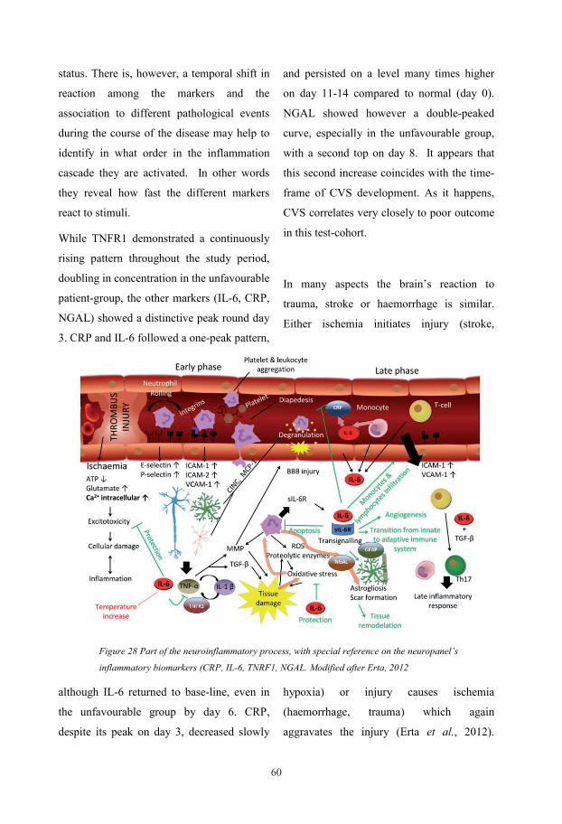
6015
Hypoxia induces a biochemical cascade,
involving exitotoxicity, oxidative stress,
leading to apoptosis. (Fig. 28) Initially the
neutrophil leucocytes extravasate after
engaging in rolling, activation and
transmigration. They release freely soluble
IL-6 receptors (sIL-6R), which attract their
ligands produced by damaged cells, together
with TNFα and its soluble receptor TNFR1
and IL-1β (Probert et al., 2000). These
cytokines attract leucocytes to the damaged
area and facilitate their degranulation,
contributing to cell necrosis. These relatively
small inflammatory substances (20-30 kDa)
can easily penetrate to the circulation, either
through a disrupted BBB (sharp and
pronounced rise) or through the glymphatic
system (delayed and prolonged expression)
and activate other immuno-inflammatory
mechanism, like CRP excretion (Muroi et al.,
2011). IL-6, among others has a negative
feed-back role in the necrotic and apoptotic
processes by down-regulating TGFβ, and
metalloproteases, inhibiting neutrophil
diapedesis and converting the innate
immunological processes to adaptive,
through T-cell recruitment (Erta et al.,
2012). It even promotes astrogliosis via
NGAL, which contributes to microglia
proliferation and transforming the astrocytes’
GFAP structure towards tissue remodelling
and endothelial cells to stimulate
angiogenesis (Yang and Wang, 2015).
D. FABP, DDMR
Both FABP and DDMR had excellent
correlation from the initial neurological
condition through the CVS development and
finally to all the outcome measurements. It
indicates that both of them are part of highly
sensitive and fast-reacting mechanisms and
are interesting candidates for further
neuromarker investigations. This rapid
response quality is supported by previous
findings (Fassbender et al., 1997; Peltonen et
al., 1997).
Fatty-acid transporting protein (FABP) in our
samples had high initial values, decreasing
rapidly and reaching its nadir on day 2 post
ictal. It is contradictory to findings of others,
as it was described in 22 patients with stroke
that values peak after 24-48 h (Zimmermann-
Ivol et al., 2004). It cannot be explained with
a wash out effect during an operation, as only
7% of the poor-grade SAH patients were
operated in this cohort. The rest of the
patients were treated by coil-embolisation.
Thereafter the FABP serum levels increased
to a second peak on day 8, nonetheless
merely in the unfavourable group, leaving the
patients with favourable properties with
normal values. As CVS correlates, as
mentioned earlier closely to all the outcome
measures, FABP separates the poor and
favourable patient groups distinctively. This
assigns bordering excellent prognostic
properties both for DCI development and
14
status. There is, however, a temporal shift in
reaction among the markers and the
association to different pathological events
during the course of the disease may help to
identify in what order in the inflammation
cascade they are activated. In other words
they reveal how fast the different markers
react to stimuli.
While TNFR1 demonstrated a continuously
rising pattern throughout the study period,
doubling in concentration in the unfavourable
patient-group, the other markers (IL-6, CRP,
NGAL) showed a distinctive peak round day
3. CRP and IL-6 followed a one-peak pattern,
although IL-6 returned to base-line, even in
the unfavourable group by day 6. CRP,
despite its peak on day 3, decreased slowly
and persisted on a level many times higher
on day 11-14 compared to normal (day 0).
NGAL showed however a double-peaked
curve, especially in the unfavourable group,
with a second top on day 8. It appears that
this second increase coincides with the time-
frame of CVS development. As it happens,
CVS correlates very closely to poor outcome
in this test-cohort.
In many aspects the brain’s reaction to
trauma, stroke or haemorrhage is similar.
Either ischemia initiates injury (stroke,
hypoxia) or injury causes ischemia
(haemorrhage, trauma) which again
aggravates the injury (Erta et al., 2012).
Figure 28 Part of the neuroinflammatory process, with special reference on the neuropanel’s
inflammatory biomarkers (CRP, IL-6, TNRF1, NGAL. Modified after Erta, 2012
6115
Hypoxia induces a biochemical cascade,
involving exitotoxicity, oxidative stress,
leading to apoptosis. (Fig. 28) Initially the
neutrophil leucocytes extravasate after
engaging in rolling, activation and
transmigration. They release freely soluble
IL-6 receptors (sIL-6R), which attract their
ligands produced by damaged cells, together
with TNFα and its soluble receptor TNFR1
and IL-1β (Probert et al., 2000). These
cytokines attract leucocytes to the damaged
area and facilitate their degranulation,
contributing to cell necrosis. These relatively
small inflammatory substances (20-30 kDa)
can easily penetrate to the circulation, either
through a disrupted BBB (sharp and
pronounced rise) or through the glymphatic
system (delayed and prolonged expression)
and activate other immuno-inflammatory
mechanism, like CRP excretion (Muroi et al.,
2011). IL-6, among others has a negative
feed-back role in the necrotic and apoptotic
processes by down-regulating TGFβ, and
metalloproteases, inhibiting neutrophil
diapedesis and converting the innate
immunological processes to adaptive,
through T-cell recruitment (Erta et al.,
2012). It even promotes astrogliosis via
NGAL, which contributes to microglia
proliferation and transforming the astrocytes’
GFAP structure towards tissue remodelling
and endothelial cells to stimulate
angiogenesis (Yang and Wang, 2015).
D. FABP, DDMR
Both FABP and DDMR had excellent
correlation from the initial neurological
condition through the CVS development and
finally to all the outcome measurements. It
indicates that both of them are part of highly
sensitive and fast-reacting mechanisms and
are interesting candidates for further
neuromarker investigations. This rapid
response quality is supported by previous
findings (Fassbender et al., 1997; Peltonen et
al., 1997).
Fatty-acid transporting protein (FABP) in our
samples had high initial values, decreasing
rapidly and reaching its nadir on day 2 post
ictal. It is contradictory to findings of others,
as it was described in 22 patients with stroke
that values peak after 24-48 h (Zimmermann-
Ivol et al., 2004). It cannot be explained with
a wash out effect during an operation, as only
7% of the poor-grade SAH patients were
operated in this cohort. The rest of the
patients were treated by coil-embolisation.
Thereafter the FABP serum levels increased
to a second peak on day 8, nonetheless
merely in the unfavourable group, leaving the
patients with favourable properties with
normal values. As CVS correlates, as
mentioned earlier closely to all the outcome
measures, FABP separates the poor and
favourable patient groups distinctively. This
assigns bordering excellent prognostic
properties both for DCI development and
14
status. There is, however, a temporal shift in
reaction among the markers and the
association to different pathological events
during the course of the disease may help to
identify in what order in the inflammation
cascade they are activated. In other words
they reveal how fast the different markers
react to stimuli.
While TNFR1 demonstrated a continuously
rising pattern throughout the study period,
doubling in concentration in the unfavourable
patient-group, the other markers (IL-6, CRP,
NGAL) showed a distinctive peak round day
3. CRP and IL-6 followed a one-peak pattern,
although IL-6 returned to base-line, even in
the unfavourable group by day 6. CRP,
despite its peak on day 3, decreased slowly
and persisted on a level many times higher
on day 11-14 compared to normal (day 0).
NGAL showed however a double-peaked
curve, especially in the unfavourable group,
with a second top on day 8. It appears that
this second increase coincides with the time-
frame of CVS development. As it happens,
CVS correlates very closely to poor outcome
in this test-cohort.
In many aspects the brain’s reaction to
trauma, stroke or haemorrhage is similar.
Either ischemia initiates injury (stroke,
hypoxia) or injury causes ischemia
(haemorrhage, trauma) which again
aggravates the injury (Erta et al., 2012).
Figure 28 Part of the neuroinflammatory process, with special reference on the neuropanel’s
inflammatory biomarkers (CRP, IL-6, TNRF1, NGAL. Modified after Erta, 2012
6216
outcome by all the scales (AUCROC 0.80 -
0.85). It is interesting to note the co-
variation of the nadir of this biomarker with
that of DDMR, which I discuss below.
DDMR, a coagulation remnant is the other
biomarker with excellent correlation to
admission neurological status, demonstrating
that the coagulo-fibrinolytic system is,
actually the most rapid responsive
mechanism in the body (Gruen et al., 2012).
It is particularly important as it was
suggested that microthrombosis was
responsible for DCI the most traitorous
complication of aSAH (Vergouwen et al.,
2008). In a previous study, the elevated level
of DDMR measured on the first day after
aSAH, was found in good correlation to 6
month outcome (Peltonen et al., 1997). In
our study, the DDMR’s initial values were
four times the normal values, corresponding
to the previously mentioned study. It fell
rapidly to normal level until day 2, when it
reached its nadir. Thereafter the level
increased again to a plateau circa six times
higher in the unfavourable group and three
times above normal level in the favourable
group where it stayed during the rest of the
observation period of 11-14 days. Because of
the clear difference between the two groups,
DDMR is proved capable of predicting
outcome.
The concavity in the course of DDMR may
be explained by the local routine use of iv.
fibrinolytic agent, tranexamic acid, which is
administered from the time of establishing a
diagnosis until the aneurysm is secured
(often within 24 h from admission) (Hillman
et al., 2002). After the drug effect diminishes
the fibrinolytic activity returns.
Unsurprisingly, the DDMR level reflects the
amount of blood in the haemorrhage, thereby
the assumed mass-effect, although as it is
coagulation specific, it cannot differentiate
between localisation and thence the
neurological consequence. As there are both
positive studies (Fujii et al., 1997; Juvela and
Siironen, 2006) and negative findings (Tseng
et al., 2007) in the literature, a recent review
article considers the evidence for using
DDMR as a biomarker after aSAH is weak
and inconsistent (Boluijt et al., 2015).
CONCLUSION
We have established a pattern of CRP development during the first week after an
aSAH, which was independent from the patients’ infectious status.
We could demonstrate a difference in this pattern between the patients with favourable
and unfavourable disease development (complication, outcome).
We could correlate this difference in CRP development to outcome measures and
build a prognostic model to predict long-term outcome.
In search for an even earlier predictor, we could identify CRP values already on day 2
with nearly the same predictive strength as the whole weeks CRP development and in
parity with initial neurology and superior to initial radiological assessment in
endovascularly treated patients.
We demonstrated that Apolipoprotein E’s genetical polymorphism had no influence
either on the incidence of aSAH, the complication development or the outcome after
the disease in West Sweden.
We identified however, a single nucleotide polymorphism (SNP) on the 9th
chromosome, which does seem to influence the incidence of intracranial aneurysm
rupture in the West-Swedish population.
Finally, we have tested a novel biochip array neuropanel in a selection of aSAH
patients and found that six of the nine neuromarkers correlated to cerebral vasospasm
and outcome. Four of them could be used for prediction purposes, thereby proving this
methods usefulness in aSAH patient monitoring.
6316
outcome by all the scales (AUCROC 0.80 -
0.85). It is interesting to note the co-
variation of the nadir of this biomarker with
that of DDMR, which I discuss below.
DDMR, a coagulation remnant is the other
biomarker with excellent correlation to
admission neurological status, demonstrating
that the coagulo-fibrinolytic system is,
actually the most rapid responsive
mechanism in the body (Gruen et al., 2012).
It is particularly important as it was
suggested that microthrombosis was
responsible for DCI the most traitorous
complication of aSAH (Vergouwen et al.,
2008). In a previous study, the elevated level
of DDMR measured on the first day after
aSAH, was found in good correlation to 6
month outcome (Peltonen et al., 1997). In
our study, the DDMR’s initial values were
four times the normal values, corresponding
to the previously mentioned study. It fell
rapidly to normal level until day 2, when it
reached its nadir. Thereafter the level
increased again to a plateau circa six times
higher in the unfavourable group and three
times above normal level in the favourable
group where it stayed during the rest of the
observation period of 11-14 days. Because of
the clear difference between the two groups,
DDMR is proved capable of predicting
outcome.
The concavity in the course of DDMR may
be explained by the local routine use of iv.
fibrinolytic agent, tranexamic acid, which is
administered from the time of establishing a
diagnosis until the aneurysm is secured
(often within 24 h from admission) (Hillman
et al., 2002). After the drug effect diminishes
the fibrinolytic activity returns.
Unsurprisingly, the DDMR level reflects the
amount of blood in the haemorrhage, thereby
the assumed mass-effect, although as it is
coagulation specific, it cannot differentiate
between localisation and thence the
neurological consequence. As there are both
positive studies (Fujii et al., 1997; Juvela and
Siironen, 2006) and negative findings (Tseng
et al., 2007) in the literature, a recent review
article considers the evidence for using
DDMR as a biomarker after aSAH is weak
and inconsistent (Boluijt et al., 2015).
CONCLUSION
We have established a pattern of CRP development during the first week after an
aSAH, which was independent from the patients’ infectious status.
We could demonstrate a difference in this pattern between the patients with favourable
and unfavourable disease development (complication, outcome).
We could correlate this difference in CRP development to outcome measures and
build a prognostic model to predict long-term outcome.
In search for an even earlier predictor, we could identify CRP values already on day 2
with nearly the same predictive strength as the whole weeks CRP development and in
parity with initial neurology and superior to initial radiological assessment in
endovascularly treated patients.
We demonstrated that Apolipoprotein E’s genetical polymorphism had no influence
either on the incidence of aSAH, the complication development or the outcome after
the disease in West Sweden.
We identified however, a single nucleotide polymorphism (SNP) on the 9th
chromosome, which does seem to influence the incidence of intracranial aneurysm
rupture in the West-Swedish population.
Finally, we have tested a novel biochip array neuropanel in a selection of aSAH
patients and found that six of the nine neuromarkers correlated to cerebral vasospasm
and outcome. Four of them could be used for prediction purposes, thereby proving this
methods usefulness in aSAH patient monitoring.
64
FUTURE PERSPECTIVES These studies were aimed to increase our knowledge on some biochemical markers and
genetical predispositions during the course of subarachnoid haemorrhage and test their
usefulness in clinical settings.
We have demonstrated the applicability of inflammation markers in general and CRP in
particular, not only as a marker of infection, but more as an assessment of disease severity.
These markers, together with specialised tissue factors (here neuromarkers) may help guide
clinicians in decision making on adjusting monitoring, choosing between therapy options or
planning rehabilitation alternatives.
We have found a need for sequential sample-taking to follow the course of the disease or
detect secondary events. It increases the credibility of the marker and reduces the possibility
for confounding errors. With some markers, continuous or near-continuous sample-taking and
measurements are available (e.g. microdialysis), with others it is on its way.
Multiple biochip-array technique is one of these techniques which can provide custom-made
investigating tools to diagnose and to follow different kinds of pathological processes,
including aSAH, using a drop of blood, liquor or any other body fluid.
Finally, we have added a piece to the mounting evidence demonstrating the connections
between genetic inheritance and risk for aSAH. Mapping our genetical risk background is in
the near future and it may affect our life-style, diet and prevention efforts to overcome our
genetical fate and to modify this predisposition. It is already a reality with some oncological
therapy decisions. The genetical predisposition may determine our preference in future
therapy-choices in many more diseases.
ACKNOWLEDGEMENTS
I wish to express my sincere gratitude to all of you, who have contributed, supported,
encouraged and assisted me during the long way of finishing this thesis. My special thanks go
to:
Doc Bengt Nellgård, my supervisor and scientific mentor, who has, with his never-ending
enthusiasm, positive attitude and passion for this project not only established neuro-research
in our Institution, but managed to create a scientific cooperation with neurologists,
neurosurgeons, neurochemists, neurophysiologists and geneticists. During these long years he
became more than a research tutor, a true friend.
Prof Kai Blennow my co-superviser, and his close associate Prof Henrik Zetterberg, for
sharing their vast knowledge on the field of neuromarkers and biochemical research and their
never-ceasing efforts to improve my manuscripts.
Med dr Karin Nylén, my co-worker, co-fellow in research, co-author and hopefully, I may call
her a friend, who not only organised our research project, but organised us in it as well. She
put in an immense work in the follow-up of more than 300 patients and allowed us an insight
of the neurologists’ meticulous work and sense for details. She had most valuable comments
on all of my manuscripts.
Prof Sven-Eric Ricksten, the head of our Institution and before him his predecessors Profs
Björn Biber and Hengo Häljamae, who have started med in my research carrier and supported
me in all possible ways in my efforts, sometimes struggle, to achieve this thesis.
Doc Hans Sonander, my clinical raw-model, who has lifted me up from the deepest well of
my research carrier by giving me an invaluable push in writing my first own manuscript. I am
ever indebted for your support during these years.
Prof Christina Jern, who has allowed me a glimpse into an entirely different world, the world
of genetics, and for answering my often unintelligible questions on the subject.
My co-authors Martin Öst, Sandra Olsson, Katarina Jood and Per Nellgård, all distinguished
researchers, for contributing substantially to this work and supporting the composition of this
thesis.
My clinical directors during the years Lars Sahlman, Helene Seeman Lodding, Johan Snygg
and especially to my present boss Doc. Elisabet Wennberg for allowing me the leave of
65
FUTURE PERSPECTIVES These studies were aimed to increase our knowledge on some biochemical markers and
genetical predispositions during the course of subarachnoid haemorrhage and test their
usefulness in clinical settings.
We have demonstrated the applicability of inflammation markers in general and CRP in
particular, not only as a marker of infection, but more as an assessment of disease severity.
These markers, together with specialised tissue factors (here neuromarkers) may help guide
clinicians in decision making on adjusting monitoring, choosing between therapy options or
planning rehabilitation alternatives.
We have found a need for sequential sample-taking to follow the course of the disease or
detect secondary events. It increases the credibility of the marker and reduces the possibility
for confounding errors. With some markers, continuous or near-continuous sample-taking and
measurements are available (e.g. microdialysis), with others it is on its way.
Multiple biochip-array technique is one of these techniques which can provide custom-made
investigating tools to diagnose and to follow different kinds of pathological processes,
including aSAH, using a drop of blood, liquor or any other body fluid.
Finally, we have added a piece to the mounting evidence demonstrating the connections
between genetic inheritance and risk for aSAH. Mapping our genetical risk background is in
the near future and it may affect our life-style, diet and prevention efforts to overcome our
genetical fate and to modify this predisposition. It is already a reality with some oncological
therapy decisions. The genetical predisposition may determine our preference in future
therapy-choices in many more diseases.
ACKNOWLEDGEMENTS
I wish to express my sincere gratitude to all of you, who have contributed, supported,
encouraged and assisted me during the long way of finishing this thesis. My special thanks go
to:
Doc Bengt Nellgård, my supervisor and scientific mentor, who has, with his never-ending
enthusiasm, positive attitude and passion for this project not only established neuro-research
in our Institution, but managed to create a scientific cooperation with neurologists,
neurosurgeons, neurochemists, neurophysiologists and geneticists. During these long years he
became more than a research tutor, a true friend.
Prof Kai Blennow my co-superviser, and his close associate Prof Henrik Zetterberg, for
sharing their vast knowledge on the field of neuromarkers and biochemical research and their
never-ceasing efforts to improve my manuscripts.
Med dr Karin Nylén, my co-worker, co-fellow in research, co-author and hopefully, I may call
her a friend, who not only organised our research project, but organised us in it as well. She
put in an immense work in the follow-up of more than 300 patients and allowed us an insight
of the neurologists’ meticulous work and sense for details. She had most valuable comments
on all of my manuscripts.
Prof Sven-Eric Ricksten, the head of our Institution and before him his predecessors Profs
Björn Biber and Hengo Häljamae, who have started med in my research carrier and supported
me in all possible ways in my efforts, sometimes struggle, to achieve this thesis.
Doc Hans Sonander, my clinical raw-model, who has lifted me up from the deepest well of
my research carrier by giving me an invaluable push in writing my first own manuscript. I am
ever indebted for your support during these years.
Prof Christina Jern, who has allowed me a glimpse into an entirely different world, the world
of genetics, and for answering my often unintelligible questions on the subject.
My co-authors Martin Öst, Sandra Olsson, Katarina Jood and Per Nellgård, all distinguished
researchers, for contributing substantially to this work and supporting the composition of this
thesis.
My clinical directors during the years Lars Sahlman, Helene Seeman Lodding, Johan Snygg
and especially to my present boss Doc. Elisabet Wennberg for allowing me the leave of
66
absence which was necessary for this research education, to perform the studies and finally to
put it all in writing.
The research nurses involved with our project Catherine Ritzén, Ingrid Petterson, Lovisa
Seleskog for their practical help, with the samples and registering all relevant data and the
staff at the Neurointensive Care Unit at Sahlgrenska Hospital (NIVA) for coping with the
inclusion and management of the study patents. Without them, there would not be any
research or theses originating from this Unit and they have continued ever since. Special
thoughts go to the chief consultant of NIVA and personal friend of mine, Christina Grivans
for her commitment and devotion to patient care and to the research-projects conducted in her
unit, in addition to Prof Bertil Rydenhag, senior consultant neurosurgeon for his support and
understanding for our neurosurgical intensive-care research.
Ingrid Eiving, previous research assistant and intensive-care nurse, who has helped me
beyond the scope of her job and during the years, became a true friend, the best of friends.
Without her dedicated assistance and friendship I would have struggled to endure many of
those difficulties the life in general can expedite.
Prof Stefan Lundin for his economical contribution to replace my saltwater-damaged
computer and the friends in the VBN group for their support and encouragement.
All my colleagues and friends at the Depts. of Anaesthesiology and Intensive Care at both
Sahlgrenska University Hospital and Kungälvs General Hospital and the Kungälv Hospital’s
Pain Clinic for making my work so joyful and stimulating and most of all for tolerating my
absence to complete this thesis.
To all the patients and healthy individuals, who participated in these studies accepting extra
trouble and inconvenience in giving blood for sampling, being questioned and examined and
travelling extra miles for follow-up investigations. Without their involvement, there would
have been no study.
Last but not least I would like to thank my family and friends for not forgetting me and
allowing me to dedicate time and effort to this thesis instead of being a friend, uncle, god-
father, brother, father and son. Thank you all!
This thesis is supported by grants from the Gothenburg Medical Society, Swedish Medical Research Council, Swedish State LUA/ALF grants, Torsten Söderberg Foundation, Hjärnfonden, Mattsson’s Foundation, Heart-Lung Foundation, Knut and Alice Wallenberg Foundation, The Swedish Stroke Association, Rune and Ulla Amlövs Foundation for Neurological Research and Yngve Land Foundation.
50
REFERENCES: Abrantes, P., M. M. Santos, I. Sousa, J. M. Xavier, V. Francisco, T. Krug, . . . S. A. Oliveira.
2015. 'Genetic Variants Underlying Risk of Intracranial Aneurysms: Insights from a GWAS in Portugal', PLoS One, 10: e0133422.
Agosta, F., K. A. Vossel, B. L. Miller, R. Migliaccio, S. J. Bonasera, M. Filippi, . . . M. L. Gorno-Tempini. 2009. 'Apolipoprotein E epsilon4 is associated with disease-specific effects on brain atrophy in Alzheimer's disease and frontotemporal dementia', Proc Natl Acad Sci U S A, 106: 2018-22.
Alg, V. S., R. Sofat, H. Houlden, and D. J. Werring. 2013. 'Genetic risk factors for intracranial aneurysms: a meta-analysis in more than 116,000 individuals', Neurology, 80: 2154-65.
Alhadad, A., G. Guron, E. Fortuna-Nowakowska, A. Saeed, I. Mattiasson, G. Jensen, . . . H. Herlitz. 2007. 'Renal angioplasty causes a rapid transient increase in inflammatory biomarkers, but reduced levels of interleukin-6 and endothelin-1 1 month after intervention', J Hypertens, 25: 1907-14.
Almagor, M., A. Keren, and S. Banai. 2003. 'Increased C-reactive protein level after coronary stent implantation in patients with stable coronary artery disease', Am Heart J, 145: 248-53.
Almeida, R. D., B. J. Manadas, C. V. Melo, J. R. Gomes, C. S. Mendes, M. M. Graos, . . . C. B. Duarte. 2005. 'Neuroprotection by BDNF against glutamate-induced apoptotic cell death is mediated by ERK and PI3-kinase pathways', Cell Death Differ, 12: 1329-43.
Anand, N., and L. G. Stead. 2005. 'Neuron-specific enolase as a marker for acute ischemic stroke: a systematic review', Cerebrovasc Dis, 20: 213-9.
Anderson, S. I., A. M. Housley, P. A. Jones, J. Slattery, and J. D. Miller. 1993. 'Glasgow Outcome Scale: an inter-rater reliability study', Brain Inj, 7: 309-17.
Backes, D., G. J. Rinkel, H. Kemperman, F. H. Linn, and M. D. Vergouwen. 2012. 'Time-dependent test characteristics of head computed tomography in patients suspected of nontraumatic subarachnoid hemorrhage', Stroke, 43: 2115-9.
Badjatia, N., A. Monahan, A. Carpenter, J. Zimmerman, J. M. Schmidt, J. Claassen, . . . D. Seres. 2015. 'Inflammation, negative nitrogen balance, and outcome after aneurysmal subarachnoid hemorrhage', Neurology, 84: 680-7.
Bai, Y., S. Nie, G. Jiang, Y. Zhou, M. Zhou, Y. Zhao, . . . Q. K. Wang. 2014. 'Regulation of CARD8 expression by ANRIL and association of CARD8 single nucleotide polymorphism rs2043211 (p.C10X) with ischemic stroke', Stroke, 45: 383-8.
Bayoglu, B., C. Arslan, S. Gode, F. Kaya Dagistanli, B. Arapi, S. Burc Deser, . . . M. Cengiz. 2014. 'The severity of internal carotid artery stenosis is associated with the cyclin-dependent kinase inhibitor 2A gene expression', J Atheroscler Thromb, 21: 659-71.
Bendjilali, N., J. Nelson, S. Weinsheimer, S. Sidney, J. G. Zaroff, S. W. Hetts, . . . H. Kim. 2014. 'Common variants on 9p21.3 are associated with brain arteriovenous malformations with accompanying arterial aneurysms', J Neurol Neurosurg Psychiatry, 85: 1280-3.
Beutler, B. A., I. W. Milsark, and A. Cerami. 1985. 'Cachectin/tumor necrosis factor: production, distribution, and metabolic fate in vivo', J Immunol, 135: 3972-7.
Bhardwaj, A. 2003. 'SAH-induced cerebral vasospasm: unraveling molecular mechanisms of a complex disease', Stroke, 34: 427-33.
Bilguvar, K., K. Yasuno, M. Niemela, Y. M. Ruigrok, M. von Und Zu Fraunberg, C. M. van Duijn, . . . M. Gunel. 2008. 'Susceptibility loci for intracranial aneurysm in European and Japanese populations', Nat Genet, 40: 1472-7.
67
absence which was necessary for this research education, to perform the studies and finally to
put it all in writing.
The research nurses involved with our project Catherine Ritzén, Ingrid Petterson, Lovisa
Seleskog for their practical help, with the samples and registering all relevant data and the
staff at the Neurointensive Care Unit at Sahlgrenska Hospital (NIVA) for coping with the
inclusion and management of the study patents. Without them, there would not be any
research or theses originating from this Unit and they have continued ever since. Special
thoughts go to the chief consultant of NIVA and personal friend of mine, Christina Grivans
for her commitment and devotion to patient care and to the research-projects conducted in her
unit, in addition to Prof Bertil Rydenhag, senior consultant neurosurgeon for his support and
understanding for our neurosurgical intensive-care research.
Ingrid Eiving, previous research assistant and intensive-care nurse, who has helped me
beyond the scope of her job and during the years, became a true friend, the best of friends.
Without her dedicated assistance and friendship I would have struggled to endure many of
those difficulties the life in general can expedite.
Prof Stefan Lundin for his economical contribution to replace my saltwater-damaged
computer and the friends in the VBN group for their support and encouragement.
All my colleagues and friends at the Depts. of Anaesthesiology and Intensive Care at both
Sahlgrenska University Hospital and Kungälvs General Hospital and the Kungälv Hospital’s
Pain Clinic for making my work so joyful and stimulating and most of all for tolerating my
absence to complete this thesis.
To all the patients and healthy individuals, who participated in these studies accepting extra
trouble and inconvenience in giving blood for sampling, being questioned and examined and
travelling extra miles for follow-up investigations. Without their involvement, there would
have been no study.
Last but not least I would like to thank my family and friends for not forgetting me and
allowing me to dedicate time and effort to this thesis instead of being a friend, uncle, god-
father, brother, father and son. Thank you all!
This thesis is supported by grants from the Gothenburg Medical Society, Swedish Medical Research Council, Swedish State LUA/ALF grants, Torsten Söderberg Foundation, Hjärnfonden, Mattsson’s Foundation, Heart-Lung Foundation, Knut and Alice Wallenberg Foundation, The Swedish Stroke Association, Rune and Ulla Amlövs Foundation for Neurological Research and Yngve Land Foundation.
50
REFERENCES: Abrantes, P., M. M. Santos, I. Sousa, J. M. Xavier, V. Francisco, T. Krug, . . . S. A. Oliveira.
2015. 'Genetic Variants Underlying Risk of Intracranial Aneurysms: Insights from a GWAS in Portugal', PLoS One, 10: e0133422.
Agosta, F., K. A. Vossel, B. L. Miller, R. Migliaccio, S. J. Bonasera, M. Filippi, . . . M. L. Gorno-Tempini. 2009. 'Apolipoprotein E epsilon4 is associated with disease-specific effects on brain atrophy in Alzheimer's disease and frontotemporal dementia', Proc Natl Acad Sci U S A, 106: 2018-22.
Alg, V. S., R. Sofat, H. Houlden, and D. J. Werring. 2013. 'Genetic risk factors for intracranial aneurysms: a meta-analysis in more than 116,000 individuals', Neurology, 80: 2154-65.
Alhadad, A., G. Guron, E. Fortuna-Nowakowska, A. Saeed, I. Mattiasson, G. Jensen, . . . H. Herlitz. 2007. 'Renal angioplasty causes a rapid transient increase in inflammatory biomarkers, but reduced levels of interleukin-6 and endothelin-1 1 month after intervention', J Hypertens, 25: 1907-14.
Almagor, M., A. Keren, and S. Banai. 2003. 'Increased C-reactive protein level after coronary stent implantation in patients with stable coronary artery disease', Am Heart J, 145: 248-53.
Almeida, R. D., B. J. Manadas, C. V. Melo, J. R. Gomes, C. S. Mendes, M. M. Graos, . . . C. B. Duarte. 2005. 'Neuroprotection by BDNF against glutamate-induced apoptotic cell death is mediated by ERK and PI3-kinase pathways', Cell Death Differ, 12: 1329-43.
Anand, N., and L. G. Stead. 2005. 'Neuron-specific enolase as a marker for acute ischemic stroke: a systematic review', Cerebrovasc Dis, 20: 213-9.
Anderson, S. I., A. M. Housley, P. A. Jones, J. Slattery, and J. D. Miller. 1993. 'Glasgow Outcome Scale: an inter-rater reliability study', Brain Inj, 7: 309-17.
Backes, D., G. J. Rinkel, H. Kemperman, F. H. Linn, and M. D. Vergouwen. 2012. 'Time-dependent test characteristics of head computed tomography in patients suspected of nontraumatic subarachnoid hemorrhage', Stroke, 43: 2115-9.
Badjatia, N., A. Monahan, A. Carpenter, J. Zimmerman, J. M. Schmidt, J. Claassen, . . . D. Seres. 2015. 'Inflammation, negative nitrogen balance, and outcome after aneurysmal subarachnoid hemorrhage', Neurology, 84: 680-7.
Bai, Y., S. Nie, G. Jiang, Y. Zhou, M. Zhou, Y. Zhao, . . . Q. K. Wang. 2014. 'Regulation of CARD8 expression by ANRIL and association of CARD8 single nucleotide polymorphism rs2043211 (p.C10X) with ischemic stroke', Stroke, 45: 383-8.
Bayoglu, B., C. Arslan, S. Gode, F. Kaya Dagistanli, B. Arapi, S. Burc Deser, . . . M. Cengiz. 2014. 'The severity of internal carotid artery stenosis is associated with the cyclin-dependent kinase inhibitor 2A gene expression', J Atheroscler Thromb, 21: 659-71.
Bendjilali, N., J. Nelson, S. Weinsheimer, S. Sidney, J. G. Zaroff, S. W. Hetts, . . . H. Kim. 2014. 'Common variants on 9p21.3 are associated with brain arteriovenous malformations with accompanying arterial aneurysms', J Neurol Neurosurg Psychiatry, 85: 1280-3.
Beutler, B. A., I. W. Milsark, and A. Cerami. 1985. 'Cachectin/tumor necrosis factor: production, distribution, and metabolic fate in vivo', J Immunol, 135: 3972-7.
Bhardwaj, A. 2003. 'SAH-induced cerebral vasospasm: unraveling molecular mechanisms of a complex disease', Stroke, 34: 427-33.
Bilguvar, K., K. Yasuno, M. Niemela, Y. M. Ruigrok, M. von Und Zu Fraunberg, C. M. van Duijn, . . . M. Gunel. 2008. 'Susceptibility loci for intracranial aneurysm in European and Japanese populations', Nat Genet, 40: 1472-7.
6851
Blennow, K., and R. F. Cowburn. 1996. 'The neurochemistry of Alzheimer's disease', Acta Neurol Scand Suppl, 168: 77-86.
Blennow, K., A. Ricksten, J. A. Prince, A. J. Brookes, T. Emahazion, C. Wasslavik, . . . L. Rymo. 2000. 'No association between the alpha2-macroglobulin (A2M) deletion and Alzheimer's disease, and no change in A2M mRNA, protein, or protein expression', J Neural Transm, 107: 1065-79.
Boluijt, J., J. C. Meijers, G. J. Rinkel, and M. D. Vergouwen. 2015. 'Hemostasis and fibrinolysis in delayed cerebral ischemia after aneurysmal subarachnoid hemorrhage: a systematic review', J Cereb Blood Flow Metab, 35: 724-33.
Boogaarts, H. D., M. P. Conde, E. Janssen, W. F. van Nuenen, J. de Vries, R. Donders, . . . R. H. Bartels. 2014. 'The value of the Charlson Co-morbidity Index in aneurysmal subarachnoid haemorrhage', Acta Neurochir (Wien), 156: 1663-7.
Botterell, E. H., W. M. Lougheed, J. W. Scott, and S. L. Vandewater. 1956. 'Hypothermia, and interruption of carotid, or carotid and vertebral circulation, in the surgical management of intracranial aneurysms', J Neurosurg, 13: 1-42.
Brott, T., H. P. Adams, Jr., C. P. Olinger, J. R. Marler, W. G. Barsan, J. Biller, . . . et al. 1989. 'Measurements of acute cerebral infarction: a clinical examination scale', Stroke, 20: 864-70.
Caranci, F., F. Briganti, L. Cirillo, M. Leonardi, and M. Muto. 2013. 'Epidemiology and genetics of intracranial aneurysms', Eur J Radiol, 82: 1598-605.
Chalouhi, N., M. S. Ali, P. M. Jabbour, S. I. Tjoumakaris, L. F. Gonzalez, R. H. Rosenwasser, . . . A. S. Dumont. 2012. 'Biology of intracranial aneurysms: role of inflammation', J Cereb Blood Flow Metab, 32: 1659-76.
Chen, B., C. Grothe, and K. Schaller. 2013. 'Validation of a new neurological score (FOUR Score) in the assessment of neurosurgical patients with severely impaired consciousness', Acta Neurochir (Wien), 155: 2133-9; discussion 39.
Cheng, F., Q. Yuan, J. Yang, W. Wang, and H. Liu. 2014. 'The prognostic value of serum neuron-specific enolase in traumatic brain injury: systematic review and meta-analysis', PLoS One, 9: e106680.
Claassen, J., D. Albers, J. M. Schmidt, G. M. De Marchis, D. Pugin, C. M. Falo, . . . N. Badjatia. 2014. 'Nonconvulsive seizures in subarachnoid hemorrhage link inflammation and outcome', Ann Neurol, 75: 771-81.
Claassen, J., G. L. Bernardini, K. Kreiter, J. Bates, Y. E. Du, D. Copeland, . . . S. A. Mayer. 2001. 'Effect of cisternal and ventricular blood on risk of delayed cerebral ischemia after subarachnoid hemorrhage: the Fisher scale revisited', Stroke, 32: 2012-20.
Clifton, G. L., R. L. Hayes, H. S. Levin, M. E. Michel, and S. C. Choi. 1992. 'Outcome measures for clinical trials involving traumatically brain-injured patients: report of a conference', Neurosurgery, 31: 975-8.
Corbo, R. M., and R. Scacchi. 1999. 'Apolipoprotein E (APOE) allele distribution in the world. Is APOE*4 a 'thrifty' allele?', Ann Hum Genet, 63: 301-10.
Csajbok, L. Z. , M. Öst, K. Nylen, and B. Nellgard. 2005. 'C-reactive protein is an independent prognostic factor of outcome after subarachnoid haemorrhage', Acta Anaesthesiol Scand, 49: 7.
Di Napoli, M., and F. Papa. 2003. 'Clinical use of C-reactive protein for prognostic stratification in ischemic stroke: has the time come for including it in the patient risk profile?', Stroke, 34: 375-6; author reply 75-6.
Diez-Ruiz, A., G. P. Tilz, R. Zangerle, G. Baier-Bitterlich, H. Wachter, and D. Fuchs. 1995. 'Soluble receptors for tumour necrosis factor in clinical laboratory diagnosis', Eur J Haematol, 54: 1-8.
52
Edlow, J. A., and J. Fisher. 2012. 'Diagnosis of subarachnoid hemorrhage: time to change the guidelines?', Stroke, 43: 2031-2.
Endre, Z. H., J. W. Pickering, R. J. Walker, P. Devarajan, C. L. Edelstein, J. V. Bonventre, . . . P. M. George. 2011. 'Improved performance of urinary biomarkers of acute kidney injury in the critically ill by stratification for injury duration and baseline renal function', Kidney Int, 79: 1119-30.
Erta, M., A. Quintana, and J. Hidalgo. 2012. 'Interleukin-6, a major cytokine in the central nervous system', Int J Biol Sci, 8: 1254-66.
Failla, M. D., Y. P. Conley, and A. K. Wagner. 2015. 'Brain-Derived Neurotrophic Factor(BDNF) in Traumatic Brain Injury-Related Mortality: Interrelationships Between Genetics and Acute Systemic and Central Nervous System BDNF Profiles', Neurorehabil Neural Repair.
Fan, M., S. Dandona, R. McPherson, H. Allayee, S. L. Hazen, G. A. Wells, . . . A. F. Stewart. 2013. 'Two chromosome 9p21 haplotype blocks distinguish between coronary artery disease and myocardial infarction risk', Circ Cardiovasc Genet, 6: 372-80.
Fassbender, K., R. Schmidt, A. Schreiner, M. Fatar, F. Muhlhauser, M. Daffertshofer, and M. Hennerici. 1997. 'Leakage of brain-originated proteins in peripheral blood: temporal profile and diagnostic value in early ischemic stroke', J Neurol Sci, 148: 101-5.
Fazekas, F., S. Strasser-Fuchs, H. Kollegger, T. Berger, W. Kristoferitsch, H. Schmidt, . . . R. Schmidt. 2001. 'Apolipoprotein E epsilon 4 is associated with rapid progression of multiple sclerosis', Neurology, 57: 853-7.
Feigin, V. L., and M. Findlay. 2006. 'Advances in subarachnoid hemorrhage', Stroke, 37: 305-8.
Fisher, C. M., J. P. Kistler, and J. M. Davis. 1980. 'Relation of cerebral vasospasm to subarachnoid hemorrhage visualized by computerized tomographic scanning',Neurosurgery, 6: 1-9.
Folstein, M. F., S. E. Folstein, and P. R. McHugh. 1975. '"Mini-mental state". A practicalmethod for grading the cognitive state of patients for the clinician', J Psychiatr Res, 12: 189-98.
Fontanella, M., I. Rainero, S. Gallone, E. Rubino, C. Rivoiro, W. Valfre, . . . L. Pinessi. 2007. 'Lack of association between the apolipoprotein E gene and aneurysmal subarachnoid hemorrhage in an Italian population', J Neurosurg, 106: 245-9.
Fountas, K. N., A. Tasiou, E. Z. Kapsalaki, K. N. Paterakis, A. A. Grigorian, G. P. Lee, and J. S. Robinson, Jr. 2009. 'Serum and cerebrospinal fluid C-reactive protein levels as predictors of vasospasm in aneurysmal subarachnoid hemorrhage. Clinical article', Neurosurg Focus, 26: E22.
Frontera, J. A., J. Claassen, J. M. Schmidt, K. E. Wartenberg, R. Temes, E. S. Connolly, Jr., . . . S. A. Mayer. 2006. 'Prediction of symptomatic vasospasm after subarachnoidhemorrhage: the modified fisher scale', Neurosurgery, 59: 21-7; discussion 21-7.
Fujii, Y., S. Takeuchi, O. Sasaki, T. Minakawa, T. Koike, and R. Tanaka. 1997. 'Serial changes of hemostasis in aneurysmal subarachnoid hemorrhage with special reference to delayed ischemic neurological deficits', J Neurosurg, 86: 594-602.
Fung, C., M. Balmer, M. Murek, W. J. Z'Graggen, J. Abu-Isa, C. Ozdoba, . . . J. Beck. 2015. 'Impact of early-onset seizures on grading and outcome in patients with subarachnoid hemorrhage', J Neurosurg, 122: 408-13.
Gaist, D., M. Vaeth, I. Tsiropoulos, K. Christensen, E. Corder, J. Olsen, and H. T. Sorensen. 2000. 'Risk of subarachnoid haemorrhage in first degree relatives of patients with subarachnoid haemorrhage: follow up study based on national registries in Denmark', BMJ, 320: 141-5.
6951
Blennow, K., and R. F. Cowburn. 1996. 'The neurochemistry of Alzheimer's disease', Acta Neurol Scand Suppl, 168: 77-86.
Blennow, K., A. Ricksten, J. A. Prince, A. J. Brookes, T. Emahazion, C. Wasslavik, . . . L. Rymo. 2000. 'No association between the alpha2-macroglobulin (A2M) deletion and Alzheimer's disease, and no change in A2M mRNA, protein, or protein expression', J Neural Transm, 107: 1065-79.
Boluijt, J., J. C. Meijers, G. J. Rinkel, and M. D. Vergouwen. 2015. 'Hemostasis and fibrinolysis in delayed cerebral ischemia after aneurysmal subarachnoid hemorrhage: a systematic review', J Cereb Blood Flow Metab, 35: 724-33.
Boogaarts, H. D., M. P. Conde, E. Janssen, W. F. van Nuenen, J. de Vries, R. Donders, . . . R. H. Bartels. 2014. 'The value of the Charlson Co-morbidity Index in aneurysmal subarachnoid haemorrhage', Acta Neurochir (Wien), 156: 1663-7.
Botterell, E. H., W. M. Lougheed, J. W. Scott, and S. L. Vandewater. 1956. 'Hypothermia, and interruption of carotid, or carotid and vertebral circulation, in the surgical management of intracranial aneurysms', J Neurosurg, 13: 1-42.
Brott, T., H. P. Adams, Jr., C. P. Olinger, J. R. Marler, W. G. Barsan, J. Biller, . . . et al. 1989. 'Measurements of acute cerebral infarction: a clinical examination scale', Stroke, 20: 864-70.
Caranci, F., F. Briganti, L. Cirillo, M. Leonardi, and M. Muto. 2013. 'Epidemiology and genetics of intracranial aneurysms', Eur J Radiol, 82: 1598-605.
Chalouhi, N., M. S. Ali, P. M. Jabbour, S. I. Tjoumakaris, L. F. Gonzalez, R. H. Rosenwasser, . . . A. S. Dumont. 2012. 'Biology of intracranial aneurysms: role of inflammation', J Cereb Blood Flow Metab, 32: 1659-76.
Chen, B., C. Grothe, and K. Schaller. 2013. 'Validation of a new neurological score (FOUR Score) in the assessment of neurosurgical patients with severely impaired consciousness', Acta Neurochir (Wien), 155: 2133-9; discussion 39.
Cheng, F., Q. Yuan, J. Yang, W. Wang, and H. Liu. 2014. 'The prognostic value of serum neuron-specific enolase in traumatic brain injury: systematic review and meta-analysis', PLoS One, 9: e106680.
Claassen, J., D. Albers, J. M. Schmidt, G. M. De Marchis, D. Pugin, C. M. Falo, . . . N. Badjatia. 2014. 'Nonconvulsive seizures in subarachnoid hemorrhage link inflammation and outcome', Ann Neurol, 75: 771-81.
Claassen, J., G. L. Bernardini, K. Kreiter, J. Bates, Y. E. Du, D. Copeland, . . . S. A. Mayer. 2001. 'Effect of cisternal and ventricular blood on risk of delayed cerebral ischemia after subarachnoid hemorrhage: the Fisher scale revisited', Stroke, 32: 2012-20.
Clifton, G. L., R. L. Hayes, H. S. Levin, M. E. Michel, and S. C. Choi. 1992. 'Outcome measures for clinical trials involving traumatically brain-injured patients: report of a conference', Neurosurgery, 31: 975-8.
Corbo, R. M., and R. Scacchi. 1999. 'Apolipoprotein E (APOE) allele distribution in the world. Is APOE*4 a 'thrifty' allele?', Ann Hum Genet, 63: 301-10.
Csajbok, L. Z. , M. Öst, K. Nylen, and B. Nellgard. 2005. 'C-reactive protein is an independent prognostic factor of outcome after subarachnoid haemorrhage', Acta Anaesthesiol Scand, 49: 7.
Di Napoli, M., and F. Papa. 2003. 'Clinical use of C-reactive protein for prognostic stratification in ischemic stroke: has the time come for including it in the patient risk profile?', Stroke, 34: 375-6; author reply 75-6.
Diez-Ruiz, A., G. P. Tilz, R. Zangerle, G. Baier-Bitterlich, H. Wachter, and D. Fuchs. 1995. 'Soluble receptors for tumour necrosis factor in clinical laboratory diagnosis', Eur J Haematol, 54: 1-8.
52
Edlow, J. A., and J. Fisher. 2012. 'Diagnosis of subarachnoid hemorrhage: time to change the guidelines?', Stroke, 43: 2031-2.
Endre, Z. H., J. W. Pickering, R. J. Walker, P. Devarajan, C. L. Edelstein, J. V. Bonventre, . . . P. M. George. 2011. 'Improved performance of urinary biomarkers of acute kidney injury in the critically ill by stratification for injury duration and baseline renal function', Kidney Int, 79: 1119-30.
Erta, M., A. Quintana, and J. Hidalgo. 2012. 'Interleukin-6, a major cytokine in the central nervous system', Int J Biol Sci, 8: 1254-66.
Failla, M. D., Y. P. Conley, and A. K. Wagner. 2015. 'Brain-Derived Neurotrophic Factor (BDNF) in Traumatic Brain Injury-Related Mortality: Interrelationships Between Genetics and Acute Systemic and Central Nervous System BDNF Profiles', Neurorehabil Neural Repair.
Fan, M., S. Dandona, R. McPherson, H. Allayee, S. L. Hazen, G. A. Wells, . . . A. F. Stewart. 2013. 'Two chromosome 9p21 haplotype blocks distinguish between coronary artery disease and myocardial infarction risk', Circ Cardiovasc Genet, 6: 372-80.
Fassbender, K., R. Schmidt, A. Schreiner, M. Fatar, F. Muhlhauser, M. Daffertshofer, and M. Hennerici. 1997. 'Leakage of brain-originated proteins in peripheral blood: temporal profile and diagnostic value in early ischemic stroke', J Neurol Sci, 148: 101-5.
Fazekas, F., S. Strasser-Fuchs, H. Kollegger, T. Berger, W. Kristoferitsch, H. Schmidt, . . . R. Schmidt. 2001. 'Apolipoprotein E epsilon 4 is associated with rapid progression of multiple sclerosis', Neurology, 57: 853-7.
Feigin, V. L., and M. Findlay. 2006. 'Advances in subarachnoid hemorrhage', Stroke, 37: 305-8.
Fisher, C. M., J. P. Kistler, and J. M. Davis. 1980. 'Relation of cerebral vasospasm to subarachnoid hemorrhage visualized by computerized tomographic scanning', Neurosurgery, 6: 1-9.
Folstein, M. F., S. E. Folstein, and P. R. McHugh. 1975. '"Mini-mental state". A practical method for grading the cognitive state of patients for the clinician', J Psychiatr Res, 12: 189-98.
Fontanella, M., I. Rainero, S. Gallone, E. Rubino, C. Rivoiro, W. Valfre, . . . L. Pinessi. 2007. 'Lack of association between the apolipoprotein E gene and aneurysmal subarachnoid hemorrhage in an Italian population', J Neurosurg, 106: 245-9.
Fountas, K. N., A. Tasiou, E. Z. Kapsalaki, K. N. Paterakis, A. A. Grigorian, G. P. Lee, and J. S. Robinson, Jr. 2009. 'Serum and cerebrospinal fluid C-reactive protein levels as predictors of vasospasm in aneurysmal subarachnoid hemorrhage. Clinical article', Neurosurg Focus, 26: E22.
Frontera, J. A., J. Claassen, J. M. Schmidt, K. E. Wartenberg, R. Temes, E. S. Connolly, Jr., . . . S. A. Mayer. 2006. 'Prediction of symptomatic vasospasm after subarachnoid hemorrhage: the modified fisher scale', Neurosurgery, 59: 21-7; discussion 21-7.
Fujii, Y., S. Takeuchi, O. Sasaki, T. Minakawa, T. Koike, and R. Tanaka. 1997. 'Serial changes of hemostasis in aneurysmal subarachnoid hemorrhage with special reference to delayed ischemic neurological deficits', J Neurosurg, 86: 594-602.
Fung, C., M. Balmer, M. Murek, W. J. Z'Graggen, J. Abu-Isa, C. Ozdoba, . . . J. Beck. 2015. 'Impact of early-onset seizures on grading and outcome in patients with subarachnoid hemorrhage', J Neurosurg, 122: 408-13.
Gaist, D., M. Vaeth, I. Tsiropoulos, K. Christensen, E. Corder, J. Olsen, and H. T. Sorensen. 2000. 'Risk of subarachnoid haemorrhage in first degree relatives of patients with subarachnoid haemorrhage: follow up study based on national registries in Denmark', BMJ, 320: 141-5.
7053
Genolution Pharmaceutical. 2015. 'Geno_prep All DNA kit', http://www.genolution1.com/geno_en/download_en/Geno-Prep%20All%20DNA%20Kit.pdf.
Gerdes, L. U. 2003. 'The common polymorphism of apolipoprotein E: geographical aspects and new pathophysiological relations', Clin Chem Lab Med, 41: 628-31.
Gertz, K., G. Kronenberg, R. E. Kalin, T. Baldinger, C. Werner, M. Balkaya, . . . M. Endres. 2012. 'Essential role of interleukin-6 in post-stroke angiogenesis', Brain, 135: 1964-80.
Gilsbach, J. M., A. G. Harders, H. R. Eggert, and M. E. Hornyak. 1988. 'Early aneurysm surgery: a 7 year clinical practice report', Acta Neurochir (Wien), 90: 91-102.
Gouweleeuw, L., P. J. Naude, M. Rots, M. J. DeJongste, U. L. Eisel, and R. G. Schoemaker. 2015. 'The role of neutrophil gelatinase associated lipocalin (NGAL) as biological constituent linking depression and cardiovascular disease', Brain Behav Immun, 46: 23-32.
Gradisek, P., J. Osredkar, M. Korsic, and B. Kremzar. 2012. 'Multiple indicators model of long-term mortality in traumatic brain injury', Brain Inj, 26: 1472-81.
Gruen, R. L., K. Brohi, M. Schreiber, Z. J. Balogh, V. Pitt, M. Narayan, and R. V. Maier. 2012. 'Haemorrhage control in severely injured patients', Lancet, 380: 1099-108.
Grunwald, I. Q., A. L. Kuhn, A. J. Schmitt, and J. S. Balami. 2014. 'Aneurysmal SAH: current management and complications associated with treatment and disease', J Invasive Cardiol, 26: 30-7.
Gschwendtner, A., S. Bevan, J. W. Cole, A. Plourde, M. Matarin, H. Ross-Adams, . . . Consortium International Stroke Genetics. 2009. 'Sequence variants on chromosome 9p21.3 confer risk for atherosclerotic stroke', Ann Neurol, 65: 531-9.
Guglielmi, G., F. Vinuela, G. Duckwiler, J. Dion, P. Lylyk, A. Berenstein, . . . et al. 1992. 'Endovascular treatment of posterior circulation aneurysms by electrothrombosis using electrically detachable coils', J Neurosurg, 77: 515-24.
Helgadottir, A., G. Thorleifsson, K. P. Magnusson, S. Gretarsdottir, V. Steinthorsdottir, A. Manolescu, . . . K. Stefansson. 2008. 'The same sequence variant on 9p21 associates with myocardial infarction, abdominal aortic aneurysm and intracranial aneurysm', Nat Genet, 40: 217-24.
Hillman, J., S. Fridriksson, O. Nilsson, Z. Yu, H. Saveland, and K. E. Jakobsson. 2002. 'Immediate administration of tranexamic acid and reduced incidence of early rebleeding after aneurysmal subarachnoid hemorrhage: a prospective randomized study', J Neurosurg, 97: 771-8.
Hoh, B. L., Y. Y. Chi, M. F. Lawson, J. Mocco, and F. G. Barker, 2nd. 2010. 'Length of stay and total hospital charges of clipping versus coiling for ruptured and unruptured adult cerebral aneurysms in the Nationwide Inpatient Sample database 2002 to 2006', Stroke, 41: 337-42.
Holdt, L. M., S. Hoffmann, K. Sass, D. Langenberger, M. Scholz, K. Krohn, . . . D. Teupser. 2013. 'Alu elements in ANRIL non-coding RNA at chromosome 9p21 modulate atherogenic cell functions through trans-regulation of gene networks', PLoS Genet, 9: e1003588.
Hotamisligil, G. S., and D. A. Bernlohr. 2015. 'Metabolic functions of FABPs-mechanisms and therapeutic implications', Nat Rev Endocrinol.
Hunt, W. E., and R. M. Hess. 1968. 'Surgical risk as related to time of intervention in the repair of intracranial aneurysms', J Neurosurg, 28: 14-20.
Hwang, S. H., Y. S. Park, J. T. Kwon, T. K. Nam, S. N. Hwang, and H. Kang. 2013. 'Significance of C-reactive protein and transcranial Doppler in cerebral vasospasm
54
following aneurysmal subarachnoid hemorrhage', J Korean Neurosurg Soc, 54: 289-95.
Jaja, B. N., M. D. Cusimano, N. Etminan, D. Hanggi, D. Hasan, D. Ilodigwe, . . . R. L. Macdonald. 2013. 'Clinical prediction models for aneurysmal subarachnoid hemorrhage: a systematic review', Neurocrit Care, 18: 143-53.
Jakovljevic, D., C. Sarti, J. Sivenius, J. Torppa, M. Mahonen, P. Immonen-Raiha, . . . V. Salomaa. 2001. 'Socioeconomic status and ischemic stroke: The FINMONICA Stroke Register', Stroke, 32: 1492-8.
Jennett, B., J. Snoek, M. R. Bond, and N. Brooks. 1981. 'Disability after severe head injury: observations on the use of the Glasgow Outcome Scale', J Neurol Neurosurg Psychiatry, 44: 285-93.
Jeon, Y. T., J. H. Lee, H. Lee, H. K. Lee, J. W. Hwang, Y. J. Lim, and H. P. Park. 2012. 'The postoperative C-reactive protein level can be a useful prognostic factor for poor outcome and symptomatic vasospasm in patients with aneurysmal subarachnoid hemorrhage', J Neurosurg Anesthesiol, 24: 317-24.
Joo, H., M. G. George, J. Fang, and G. Wang. 2014. 'A literature review of indirect costs associated with stroke', J Stroke Cerebrovasc Dis, 23: 1753-63.
Jung, C. S., B. Lange, M. Zimmermann, and V. Seifert. 2013. 'CSF and Serum Biomarkers Focusing on Cerebral Vasospasm and Ischemia after Subarachnoid Hemorrhage', Stroke Res Treat, 2013: 560305.
Juvela, S., J. Kuhmonen, and J. Siironen. 2012. 'C-reactive protein as predictor for poor outcome after aneurysmal subarachnoid haemorrhage', Acta Neurochir (Wien), 154: 397-404.
Juvela, S., and J. Siironen. 2006. 'D-dimer as an independent predictor for poor outcome after aneurysmal subarachnoid hemorrhage', Stroke, 37: 1451-6.
Juvela, S., J. Siironen, and J. Lappalainen. 2009. 'Apolipoprotein E genotype and outcome after aneurysmal subarachnoid hemorrhage', J Neurosurg, 110: 989-95.
Kern, S., K. Mehlig, J. Kern, H. Zetterberg, D. Thelle, I. Skoog, . . . A. Borjesson-Hanson. 2015. 'The distribution of apolipoprotein E genotype over the adult lifespan and in relation to country of birth', Am J Epidemiol, 181: 214-7.
Kim, J., J. M. Basak, and D. M. Holtzman. 2009. 'The role of apolipoprotein E in Alzheimer's disease', Neuron, 63: 287-303.
Kojima, Y., K. Downing, R. Kundu, C. Miller, F. Dewey, H. Lancero, . . . N. J. Leeper. 2014. 'Cyclin-dependent kinase inhibitor 2B regulates efferocytosis and atherosclerosis', J Clin Invest, 124: 1083-97.
Kreuzer, K. A., J. K. Rockstroh, T. Sauerbruch, and U. Spengler. 1996. 'A comparative study of different enzyme immunosorbent assays for human tumor necrosis factor-alpha', J Immunol Methods, 195: 49-54.
Krings, T., P. L. Lasjaunias, S. Geibprasert, V. Pereira, and F. J. Hans. 2008. 'The aneurysmal wall. The key to a subclassification of intracranial arterial aneurysm vasculopathies?', Interv Neuroradiol, 14 Suppl 1: 39-47.
Krishnamurthi, R. V., A. E. Moran, M. H. Forouzanfar, D. A. Bennett, G. A. Mensah, C. M. Lawes, . . . Group Risk Factors Study Stroke Expert. 2014. 'The global burden of hemorrhagic stroke: a summary of findings from the GBD 2010 study', Glob Heart, 9: 101-6.
Lagares, A., P. A. Gomez, J. F. Alen, R. D. Lobato, J. J. Rivas, R. Alday, . . . A. G. de la Camara. 2005. 'A comparison of different grading scales for predicting outcome after subarachnoid haemorrhage', Acta Neurochir (Wien), 147: 5-16; discussion 16.
7153
Genolution Pharmaceutical. 2015. 'Geno_prep All DNA kit', http://www.genolution1.com/geno_en/download_en/Geno-Prep%20All%20DNA%20Kit.pdf.
Gerdes, L. U. 2003. 'The common polymorphism of apolipoprotein E: geographical aspects and new pathophysiological relations', Clin Chem Lab Med, 41: 628-31.
Gertz, K., G. Kronenberg, R. E. Kalin, T. Baldinger, C. Werner, M. Balkaya, . . . M. Endres. 2012. 'Essential role of interleukin-6 in post-stroke angiogenesis', Brain, 135: 1964-80.
Gilsbach, J. M., A. G. Harders, H. R. Eggert, and M. E. Hornyak. 1988. 'Early aneurysm surgery: a 7 year clinical practice report', Acta Neurochir (Wien), 90: 91-102.
Gouweleeuw, L., P. J. Naude, M. Rots, M. J. DeJongste, U. L. Eisel, and R. G. Schoemaker. 2015. 'The role of neutrophil gelatinase associated lipocalin (NGAL) as biological constituent linking depression and cardiovascular disease', Brain Behav Immun, 46: 23-32.
Gradisek, P., J. Osredkar, M. Korsic, and B. Kremzar. 2012. 'Multiple indicators model of long-term mortality in traumatic brain injury', Brain Inj, 26: 1472-81.
Gruen, R. L., K. Brohi, M. Schreiber, Z. J. Balogh, V. Pitt, M. Narayan, and R. V. Maier. 2012. 'Haemorrhage control in severely injured patients', Lancet, 380: 1099-108.
Grunwald, I. Q., A. L. Kuhn, A. J. Schmitt, and J. S. Balami. 2014. 'Aneurysmal SAH: current management and complications associated with treatment and disease', J Invasive Cardiol, 26: 30-7.
Gschwendtner, A., S. Bevan, J. W. Cole, A. Plourde, M. Matarin, H. Ross-Adams, . . . Consortium International Stroke Genetics. 2009. 'Sequence variants on chromosome 9p21.3 confer risk for atherosclerotic stroke', Ann Neurol, 65: 531-9.
Guglielmi, G., F. Vinuela, G. Duckwiler, J. Dion, P. Lylyk, A. Berenstein, . . . et al. 1992. 'Endovascular treatment of posterior circulation aneurysms by electrothrombosis using electrically detachable coils', J Neurosurg, 77: 515-24.
Helgadottir, A., G. Thorleifsson, K. P. Magnusson, S. Gretarsdottir, V. Steinthorsdottir, A. Manolescu, . . . K. Stefansson. 2008. 'The same sequence variant on 9p21 associates with myocardial infarction, abdominal aortic aneurysm and intracranial aneurysm', Nat Genet, 40: 217-24.
Hillman, J., S. Fridriksson, O. Nilsson, Z. Yu, H. Saveland, and K. E. Jakobsson. 2002. 'Immediate administration of tranexamic acid and reduced incidence of early rebleeding after aneurysmal subarachnoid hemorrhage: a prospective randomized study', J Neurosurg, 97: 771-8.
Hoh, B. L., Y. Y. Chi, M. F. Lawson, J. Mocco, and F. G. Barker, 2nd. 2010. 'Length of stay and total hospital charges of clipping versus coiling for ruptured and unruptured adult cerebral aneurysms in the Nationwide Inpatient Sample database 2002 to 2006', Stroke, 41: 337-42.
Holdt, L. M., S. Hoffmann, K. Sass, D. Langenberger, M. Scholz, K. Krohn, . . . D. Teupser. 2013. 'Alu elements in ANRIL non-coding RNA at chromosome 9p21 modulate atherogenic cell functions through trans-regulation of gene networks', PLoS Genet, 9: e1003588.
Hotamisligil, G. S., and D. A. Bernlohr. 2015. 'Metabolic functions of FABPs-mechanisms and therapeutic implications', Nat Rev Endocrinol.
Hunt, W. E., and R. M. Hess. 1968. 'Surgical risk as related to time of intervention in the repair of intracranial aneurysms', J Neurosurg, 28: 14-20.
Hwang, S. H., Y. S. Park, J. T. Kwon, T. K. Nam, S. N. Hwang, and H. Kang. 2013. 'Significance of C-reactive protein and transcranial Doppler in cerebral vasospasm
54
following aneurysmal subarachnoid hemorrhage', J Korean Neurosurg Soc, 54: 289-95.
Jaja, B. N., M. D. Cusimano, N. Etminan, D. Hanggi, D. Hasan, D. Ilodigwe, . . . R. L. Macdonald. 2013. 'Clinical prediction models for aneurysmal subarachnoid hemorrhage: a systematic review', Neurocrit Care, 18: 143-53.
Jakovljevic, D., C. Sarti, J. Sivenius, J. Torppa, M. Mahonen, P. Immonen-Raiha, . . . V. Salomaa. 2001. 'Socioeconomic status and ischemic stroke: The FINMONICA Stroke Register', Stroke, 32: 1492-8.
Jennett, B., J. Snoek, M. R. Bond, and N. Brooks. 1981. 'Disability after severe head injury: observations on the use of the Glasgow Outcome Scale', J Neurol Neurosurg Psychiatry, 44: 285-93.
Jeon, Y. T., J. H. Lee, H. Lee, H. K. Lee, J. W. Hwang, Y. J. Lim, and H. P. Park. 2012. 'The postoperative C-reactive protein level can be a useful prognostic factor for poor outcome and symptomatic vasospasm in patients with aneurysmal subarachnoid hemorrhage', J Neurosurg Anesthesiol, 24: 317-24.
Joo, H., M. G. George, J. Fang, and G. Wang. 2014. 'A literature review of indirect costs associated with stroke', J Stroke Cerebrovasc Dis, 23: 1753-63.
Jung, C. S., B. Lange, M. Zimmermann, and V. Seifert. 2013. 'CSF and Serum Biomarkers Focusing on Cerebral Vasospasm and Ischemia after Subarachnoid Hemorrhage', Stroke Res Treat, 2013: 560305.
Juvela, S., J. Kuhmonen, and J. Siironen. 2012. 'C-reactive protein as predictor for poor outcome after aneurysmal subarachnoid haemorrhage', Acta Neurochir (Wien), 154: 397-404.
Juvela, S., and J. Siironen. 2006. 'D-dimer as an independent predictor for poor outcome after aneurysmal subarachnoid hemorrhage', Stroke, 37: 1451-6.
Juvela, S., J. Siironen, and J. Lappalainen. 2009. 'Apolipoprotein E genotype and outcome after aneurysmal subarachnoid hemorrhage', J Neurosurg, 110: 989-95.
Kern, S., K. Mehlig, J. Kern, H. Zetterberg, D. Thelle, I. Skoog, . . . A. Borjesson-Hanson. 2015. 'The distribution of apolipoprotein E genotype over the adult lifespan and in relation to country of birth', Am J Epidemiol, 181: 214-7.
Kim, J., J. M. Basak, and D. M. Holtzman. 2009. 'The role of apolipoprotein E in Alzheimer's disease', Neuron, 63: 287-303.
Kojima, Y., K. Downing, R. Kundu, C. Miller, F. Dewey, H. Lancero, . . . N. J. Leeper. 2014. 'Cyclin-dependent kinase inhibitor 2B regulates efferocytosis and atherosclerosis', J Clin Invest, 124: 1083-97.
Kreuzer, K. A., J. K. Rockstroh, T. Sauerbruch, and U. Spengler. 1996. 'A comparative study of different enzyme immunosorbent assays for human tumor necrosis factor-alpha', J Immunol Methods, 195: 49-54.
Krings, T., P. L. Lasjaunias, S. Geibprasert, V. Pereira, and F. J. Hans. 2008. 'The aneurysmal wall. The key to a subclassification of intracranial arterial aneurysm vasculopathies?', Interv Neuroradiol, 14 Suppl 1: 39-47.
Krishnamurthi, R. V., A. E. Moran, M. H. Forouzanfar, D. A. Bennett, G. A. Mensah, C. M. Lawes, . . . Group Risk Factors Study Stroke Expert. 2014. 'The global burden of hemorrhagic stroke: a summary of findings from the GBD 2010 study', Glob Heart, 9: 101-6.
Lagares, A., P. A. Gomez, J. F. Alen, R. D. Lobato, J. J. Rivas, R. Alday, . . . A. G. de la Camara. 2005. 'A comparison of different grading scales for predicting outcome after subarachnoid haemorrhage', Acta Neurochir (Wien), 147: 5-16; discussion 16.
7255
Lai, P. M., and R. Du. 2015. 'Role of genetic polymorphisms in predicting delayed cerebral ischemia and radiographic vasospasm after aneurysmal subarachnoid hemorrhage: a meta-analysis', World Neurosurg.
Lanterna, L. A., Y. Ruigrok, S. Alexander, J. Tang, F. Biroli, L. T. Dunn, and W. S. Poon. 2007. 'Meta-analysis of APOE genotype and subarachnoid hemorrhage: clinical outcome and delayed ischemia', Neurology, 69: 766-75.
Lee, B., K. Yoon, S. Lee, J. M. Kang, J. Kim, S. H. Shim, . . . S. J. Kim. 2015. 'Homozygous deletions at 3p22, 5p14, 6q15, and 9p21 result in aberrant expression of tumor suppressor genes in gastric cancer', Genes Chromosomes Cancer, 54: 142-55.
Lee, V. H., B. Ouyang, S. John, J. J. Conners, R. Garg, T. P. Bleck, . . . S. Prabhakaran. 2014. 'Risk stratification for the in-hospital mortality in subarachnoid hemorrhage: the HAIR score', Neurocrit Care, 21: 14-9.
Leung, C. H., W. S. Poon, L. M. Yu, G. K. Wong, and H. K. Ng. 2002. 'Apolipoprotein e genotype and outcome in aneurysmal subarachnoid hemorrhage', Stroke, 33: 548-52.
Lindekleiv, H. M., I. Njolstad, T. Ingebrigtsen, and E. B. Mathiesen. 2010. 'Incidence of aneurysmal subarachnoid hemorrhage in Norway, 1999-2007', Acta Neurol Scand.
Lindgren, C., S. Soderberg, L. O. Koskinen, M. Hultin, L. Edvardsson, and S. Naredi. 2015. 'Long-term subarachnoid haemorrhage survivors still die due to cerebrovascular causes', Acta Neurol Scand.
Linn, F. H., G. J. Rinkel, A. Algra, and J. van Gijn. 1996. 'Incidence of subarachnoid hemorrhage: role of region, year, and rate of computed tomography: a meta-analysis', Stroke, 27: 625-9.
Lu, B. 2003. 'BDNF and activity-dependent synaptic modulation', Learn Mem, 10: 86-98. Macdonald, R. L. 2013. 'Subarachnoid hemorrhage and outcome', J Neurosurg, 119: 603-4. Magni, F., M. Pozzi, M. Rota, A. Vargiolu, and G. Citerio. 2015. 'High-Resolution
Intracranial Pressure Burden and Outcome in Subarachnoid Hemorrhage', Stroke, DOI: 10.1161/STROKEAHA.115.010219.
Mahley, R. W. 1988. 'Apolipoprotein E: cholesterol transport protein with expanding role in cell biology', Science, 240: 622-30.
Mahley, R. W., and Y. Huang. 2012. 'Small-molecule structure correctors target abnormal protein structure and function: structure corrector rescue of apolipoprotein E4-associated neuropathology', J Med Chem, 55: 8997-9008.
Mahoney, F. I., and D. W. Barthel. 1965. 'Functional Evaluation: The Barthel Index', Md State Med J, 14: 61-5.
Marschall, L.F. 1987. 'Neurobehavioral outcome: the neurosurgeons's perspective.' in H.S. Levin, Grafman J., Eisenberg H.M. (ed.), Neurobehavioral Recovery from Head Injury (Oxford University Press: Oxford).
Martinez, M., A. Brice, J. R. Vaughan, A. Zimprich, M. M. Breteler, G. Meco, . . . A. Durr. 2005. 'Apolipoprotein E4 is probably responsible for the chromosome 19 linkage peak for Parkinson's disease', Am J Med Genet B Neuropsychiatr Genet, 136B: 72-4.
Mauch, D. H., K. Nagler, S. Schumacher, C. Goritz, E. C. Muller, A. Otto, and F. W. Pfrieger. 2001. 'CNS synaptogenesis promoted by glia-derived cholesterol', Science, 294: 1354-7.
Mayer, C. A., R. Brunkhorst, M. Niessner, W. Pfeilschifter, H. Steinmetz, and C. Foerch. 2013. 'Blood levels of glial fibrillary acidic protein (GFAP) in patients with neurological diseases', PLoS One, 8: e62101.
McCarron, M. O., D. Delong, and M. J. Alberts. 1999. 'APOE genotype as a risk factor for ischemic cerebrovascular disease: a meta-analysis', Neurology, 53: 1308-11.
56
McColgan, P., K. Z. Thant, and P. Sharma. 2010. 'The genetics of sporadic ruptured and unruptured intracranial aneurysms: a genetic meta-analysis of 8 genes and 13 polymorphisms in approximately 20,000 individuals', J Neurosurg, 112: 714-21.
Mirzayan, M. J., A. Gharabaghi, M. Samii, M. Tatagiba, J. K. Krauss, and S. K. Rosahl. 2007. 'Response of C-reactive protein after craniotomy for microsurgery of intracranial tumors', Neurosurgery, 60: 621-5; discussion 25.
Molyneux, A. J., J. Birks, A. Clarke, M. Sneade, and R. S. Kerr. 2015. 'The durability of endovascular coiling versus neurosurgical clipping of ruptured cerebral aneurysms: 18 year follow-up of the UK cohort of the International Subarachnoid Aneurysm Trial (ISAT)', Lancet, 385: 691-7.
Molyneux, A., R. Kerr, I. Stratton, P. Sandercock, M. Clarke, J. Shrimpton, . . . Group International Subarachnoid Aneurysm Trial Collaborative. 2002. 'International Subarachnoid Aneurysm Trial (ISAT) of neurosurgical clipping versus endovascular coiling in 2143 patients with ruptured intracranial aneurysms: a randomised trial', Lancet, 360: 1267-74.
Moore, B. W., and D. McGregor. 1965. 'Chromatographic and Electrophoretic Fractionation of Soluble Proteins of Brain and Liver', J Biol Chem, 240: 1647-53.
Morris, P. G., J. T. Wilson, L. T. Dunn, and J. A. Nicoll. 2004. 'Apolipoprotein E polymorphism and neuropsychological outcome following subarachnoid haemorrhage', Acta Neurol Scand, 109: 205-9.
Mouhieddine, T. H., L. El Houjeiri, M. Sabra, R. L. Hayes, and S. Mondello. 2015. 'CNS Trauma Biomarkers and Surrogate Endpoints Pipeline from Bench to Bedside: A Translational Perspective.' in F. H. PhD Kobeissy (ed.), Brain Neurotrauma: Molecular, Neuropsychological, and Rehabilitation Aspects (Boca Raton (FL)).
Muroi, C., S. Mink, M. Seule, D. Bellut, J. Fandino, and E. Keller. 2011. 'Monitoring of the inflammatory response after aneurysmal subarachnoid haemorrhage in the clinical setting: review of literature and report of preliminary clinical experience', Acta Neurochir Suppl, 110: 191-6.
Nagy, K., I. Skagervik, H. Tumani, A. Petzold, M. Wick, H. J. Kuhn, . . . N. Mattsson. 2013. 'Cerebrospinal fluid analyses for the diagnosis of subarachnoid haemorrhage and experience from a Swedish study. What method is preferable when diagnosing a subarachnoid haemorrhage?', Clin Chem Lab Med, 51: 2073-86.
Nakaoka, H., A. Tajima, T. Yoneyama, K. Hosomichi, H. Kasuya, T. Mizutani, and I. Inoue. 2014. 'Gene expression profiling reveals distinct molecular signatures associated with the rupture of intracranial aneurysm', Stroke, 45: 2239-45.
Naude, P. J., U. L. Eisel, H. C. Comijs, N. A. Groenewold, P. P. De Deyn, F. J. Bosker, . . . R. C. Oude Voshaar. 2013. 'Neutrophil gelatinase-associated lipocalin: a novel inflammatory marker associated with late-life depression', J Psychosom Res, 75: 444-50.
Naude, P. J., C. Nyakas, L. E. Eiden, D. Ait-Ali, R. van der Heide, S. Engelborghs, . . . U. L. Eisel. 2012. 'Lipocalin 2: novel component of proinflammatory signaling in Alzheimer's disease', FASEB J, 26: 2811-23.
Naval, N. S., T. Chang, F. Caserta, R. G. Kowalski, J. R. Carhuapoma, and R. J. Tamargo. 2013. 'Improved aneurysmal subarachnoid hemorrhage outcomes: a comparison of 2 decades at an academic center', J Crit Care, 28: 182-8.
Naval, N. S., R. G. Kowalski, T. R. Chang, F. Caserta, J. R. Carhuapoma, and R. J. Tamargo. 2014. 'The SAH Score: a comprehensive communication tool', J Stroke Cerebrovasc Dis, 23: 902-9.
7355
Lai, P. M., and R. Du. 2015. 'Role of genetic polymorphisms in predicting delayed cerebral ischemia and radiographic vasospasm after aneurysmal subarachnoid hemorrhage: a meta-analysis', World Neurosurg.
Lanterna, L. A., Y. Ruigrok, S. Alexander, J. Tang, F. Biroli, L. T. Dunn, and W. S. Poon. 2007. 'Meta-analysis of APOE genotype and subarachnoid hemorrhage: clinical outcome and delayed ischemia', Neurology, 69: 766-75.
Lee, B., K. Yoon, S. Lee, J. M. Kang, J. Kim, S. H. Shim, . . . S. J. Kim. 2015. 'Homozygous deletions at 3p22, 5p14, 6q15, and 9p21 result in aberrant expression of tumor suppressor genes in gastric cancer', Genes Chromosomes Cancer, 54: 142-55.
Lee, V. H., B. Ouyang, S. John, J. J. Conners, R. Garg, T. P. Bleck, . . . S. Prabhakaran. 2014. 'Risk stratification for the in-hospital mortality in subarachnoid hemorrhage: the HAIR score', Neurocrit Care, 21: 14-9.
Leung, C. H., W. S. Poon, L. M. Yu, G. K. Wong, and H. K. Ng. 2002. 'Apolipoprotein e genotype and outcome in aneurysmal subarachnoid hemorrhage', Stroke, 33: 548-52.
Lindekleiv, H. M., I. Njolstad, T. Ingebrigtsen, and E. B. Mathiesen. 2010. 'Incidence of aneurysmal subarachnoid hemorrhage in Norway, 1999-2007', Acta Neurol Scand.
Lindgren, C., S. Soderberg, L. O. Koskinen, M. Hultin, L. Edvardsson, and S. Naredi. 2015. 'Long-term subarachnoid haemorrhage survivors still die due to cerebrovascular causes', Acta Neurol Scand.
Linn, F. H., G. J. Rinkel, A. Algra, and J. van Gijn. 1996. 'Incidence of subarachnoid hemorrhage: role of region, year, and rate of computed tomography: a meta-analysis', Stroke, 27: 625-9.
Lu, B. 2003. 'BDNF and activity-dependent synaptic modulation', Learn Mem, 10: 86-98. Macdonald, R. L. 2013. 'Subarachnoid hemorrhage and outcome', J Neurosurg, 119: 603-4. Magni, F., M. Pozzi, M. Rota, A. Vargiolu, and G. Citerio. 2015. 'High-Resolution
Intracranial Pressure Burden and Outcome in Subarachnoid Hemorrhage', Stroke, DOI: 10.1161/STROKEAHA.115.010219.
Mahley, R. W. 1988. 'Apolipoprotein E: cholesterol transport protein with expanding role in cell biology', Science, 240: 622-30.
Mahley, R. W., and Y. Huang. 2012. 'Small-molecule structure correctors target abnormal protein structure and function: structure corrector rescue of apolipoprotein E4-associated neuropathology', J Med Chem, 55: 8997-9008.
Mahoney, F. I., and D. W. Barthel. 1965. 'Functional Evaluation: The Barthel Index', Md State Med J, 14: 61-5.
Marschall, L.F. 1987. 'Neurobehavioral outcome: the neurosurgeons's perspective.' in H.S. Levin, Grafman J., Eisenberg H.M. (ed.), Neurobehavioral Recovery from Head Injury (Oxford University Press: Oxford).
Martinez, M., A. Brice, J. R. Vaughan, A. Zimprich, M. M. Breteler, G. Meco, . . . A. Durr. 2005. 'Apolipoprotein E4 is probably responsible for the chromosome 19 linkage peak for Parkinson's disease', Am J Med Genet B Neuropsychiatr Genet, 136B: 72-4.
Mauch, D. H., K. Nagler, S. Schumacher, C. Goritz, E. C. Muller, A. Otto, and F. W.Pfrieger. 2001. 'CNS synaptogenesis promoted by glia-derived cholesterol', Science, 294: 1354-7.
Mayer, C. A., R. Brunkhorst, M. Niessner, W. Pfeilschifter, H. Steinmetz, and C. Foerch. 2013. 'Blood levels of glial fibrillary acidic protein (GFAP) in patients with neurological diseases', PLoS One, 8: e62101.
McCarron, M. O., D. Delong, and M. J. Alberts. 1999. 'APOE genotype as a risk factor for ischemic cerebrovascular disease: a meta-analysis', Neurology, 53: 1308-11.
56
McColgan, P., K. Z. Thant, and P. Sharma. 2010. 'The genetics of sporadic ruptured and unruptured intracranial aneurysms: a genetic meta-analysis of 8 genes and 13 polymorphisms in approximately 20,000 individuals', J Neurosurg, 112: 714-21.
Mirzayan, M. J., A. Gharabaghi, M. Samii, M. Tatagiba, J. K. Krauss, and S. K. Rosahl. 2007. 'Response of C-reactive protein after craniotomy for microsurgery of intracranial tumors', Neurosurgery, 60: 621-5; discussion 25.
Molyneux, A. J., J. Birks, A. Clarke, M. Sneade, and R. S. Kerr. 2015. 'The durability of endovascular coiling versus neurosurgical clipping of ruptured cerebral aneurysms: 18 year follow-up of the UK cohort of the International Subarachnoid Aneurysm Trial (ISAT)', Lancet, 385: 691-7.
Molyneux, A., R. Kerr, I. Stratton, P. Sandercock, M. Clarke, J. Shrimpton, . . . Group International Subarachnoid Aneurysm Trial Collaborative. 2002. 'International Subarachnoid Aneurysm Trial (ISAT) of neurosurgical clipping versus endovascular coiling in 2143 patients with ruptured intracranial aneurysms: a randomised trial', Lancet, 360: 1267-74.
Moore, B. W., and D. McGregor. 1965. 'Chromatographic and Electrophoretic Fractionation of Soluble Proteins of Brain and Liver', J Biol Chem, 240: 1647-53.
Morris, P. G., J. T. Wilson, L. T. Dunn, and J. A. Nicoll. 2004. 'Apolipoprotein E polymorphism and neuropsychological outcome following subarachnoid haemorrhage', Acta Neurol Scand, 109: 205-9.
Mouhieddine, T. H., L. El Houjeiri, M. Sabra, R. L. Hayes, and S. Mondello. 2015. 'CNS Trauma Biomarkers and Surrogate Endpoints Pipeline from Bench to Bedside: A Translational Perspective.' in F. H. PhD Kobeissy (ed.), Brain Neurotrauma: Molecular, Neuropsychological, and Rehabilitation Aspects (Boca Raton (FL)).
Muroi, C., S. Mink, M. Seule, D. Bellut, J. Fandino, and E. Keller. 2011. 'Monitoring of the inflammatory response after aneurysmal subarachnoid haemorrhage in the clinical setting: review of literature and report of preliminary clinical experience', Acta Neurochir Suppl, 110: 191-6.
Nagy, K., I. Skagervik, H. Tumani, A. Petzold, M. Wick, H. J. Kuhn, . . . N. Mattsson. 2013. 'Cerebrospinal fluid analyses for the diagnosis of subarachnoid haemorrhage and experience from a Swedish study. What method is preferable when diagnosing a subarachnoid haemorrhage?', Clin Chem Lab Med, 51: 2073-86.
Nakaoka, H., A. Tajima, T. Yoneyama, K. Hosomichi, H. Kasuya, T. Mizutani, and I. Inoue. 2014. 'Gene expression profiling reveals distinct molecular signatures associated with the rupture of intracranial aneurysm', Stroke, 45: 2239-45.
Naude, P. J., U. L. Eisel, H. C. Comijs, N. A. Groenewold, P. P. De Deyn, F. J. Bosker, . . . R. C. Oude Voshaar. 2013. 'Neutrophil gelatinase-associated lipocalin: a novel inflammatory marker associated with late-life depression', J Psychosom Res, 75: 444-50.
Naude, P. J., C. Nyakas, L. E. Eiden, D. Ait-Ali, R. van der Heide, S. Engelborghs, . . . U. L. Eisel. 2012. 'Lipocalin 2: novel component of proinflammatory signaling in Alzheimer's disease', FASEB J, 26: 2811-23.
Naval, N. S., T. Chang, F. Caserta, R. G. Kowalski, J. R. Carhuapoma, and R. J. Tamargo. 2013. 'Improved aneurysmal subarachnoid hemorrhage outcomes: a comparison of 2 decades at an academic center', J Crit Care, 28: 182-8.
Naval, N. S., R. G. Kowalski, T. R. Chang, F. Caserta, J. R. Carhuapoma, and R. J. Tamargo. 2014. 'The SAH Score: a comprehensive communication tool', J Stroke Cerebrovasc Dis, 23: 902-9.
7457
Niskakangas, T., J. Ohman, M. Niemela, E. Ilveskoski, T. A. Kunnas, and P. J. Karhunen. 2001. 'Association of apolipoprotein E polymorphism with outcome after aneurysmal subarachnoid hemorrhage: a preliminary study', Stroke, 32: 1181-4.
Norlen, G., and H. Olivecrona. 1953. 'The treatment of aneurysms of the circle of Willis', J Neurosurg, 10: 404-15.
Nylen, K. 2007. Studies of biochemical brain damage markers in patients at a neurointensive care unit (Academic Thesis; University of Gothenburg: Gothenburg).
Nylen, K., L. Z. Csajbok, M. Ost, A. Rashid, K. Blennow, B. Nellgard, and L. Rosengren. 2007. 'Serum glial fibrillary acidic protein is related to focal brain injury and outcome after aneurysmal subarachnoid hemorrhage', Stroke, 38: 1489-94.
Nylen, K., M. Ost, L. Z. Csajbok, I. Nilsson, K. Blennow, B. Nellgard, and L. Rosengren. 2006. 'Increased serum-GFAP in patients with severe traumatic brain injury is related to outcome', J Neurol Sci, 240: 85-91.
Ockner, R. K., J. A. Manning, R. B. Poppenhausen, and W. K. Ho. 1972. 'A binding protein for fatty acids in cytosol of intestinal mucosa, liver, myocardium, and other tissues', Science, 177: 56-8.
Ogilvy, C. S., and B. S. Carter. 1998. 'A proposed comprehensive grading system to predict outcome for surgical management of intracranial aneurysms', Neurosurgery, 42: 959-68; discussion 68-70.
Olivecrona, Z., and L. O. Koskinen. 2012. 'The release of S-100B and NSE in severe traumatic head injury is associated with APOE epsilon4', Acta Neurochir (Wien), 154: 675-80; discussion 80.
Pelinka, L. E., H. Hertz, W. Mauritz, N. Harada, M. Jafarmadar, M. Albrecht, . . . S. Bahrami. 2005. 'Nonspecific increase of systemic neuron-specific enolase after trauma: clinical and experimental findings', Shock, 24: 119-23.
Pelsers, M. M., J. P. Chapelle, M. Knapen, C. Vermeer, A. M. Muijtjens, W. T. Hermens, and J. F. Glatz. 1999. 'Influence of age and sex and day-to-day and within-day biological variation on plasma concentrations of fatty acid-binding protein and myoglobin in healthy subjects', Clin Chem, 45: 441-3.
Pelsers, M. M., and J. F. Glatz. 2005. 'Detection of brain injury by fatty acid-binding proteins', Clin Chem Lab Med, 43: 802-9.
Pelsers, M. M., T. Hanhoff, D. Van der Voort, B. Arts, M. Peters, R. Ponds, . . . J. F. Glatz. 2004. 'Brain- and heart-type fatty acid-binding proteins in the brain: tissue distribution and clinical utility', Clin Chem, 50: 1568-75.
Peltonen, S., S. Juvela, M. Kaste, and R. Lassila. 1997. 'Hemostasis and fibrinolysis activation after subarachnoid hemorrhage', J Neurosurg, 87: 207-14.
Pencea, V., K. D. Bingaman, S. J. Wiegand, and M. B. Luskin. 2001. 'Infusion of brain-derived neurotrophic factor into the lateral ventricle of the adult rat leads to new neurons in the parenchyma of the striatum, septum, thalamus, and hypothalamus', J Neurosci, 21: 6706-17.
Pereira, V. M., O. Brina, A. M. Gonzalez, A. P. Narata, R. Ouared, and L. Karl-Olof. 2013. 'Biology and hemodynamics of aneurismal vasculopathies', Eur J Radiol, 82: 1606-17.
Peschon, J. J., D. S. Torrance, K. L. Stocking, M. B. Glaccum, C. Otten, C. R. Willis, . . . K. M. Mohler. 1998. 'TNF receptor-deficient mice reveal divergent roles for p55 and p75 in several models of inflammation', J Immunol, 160: 943-52.
Phababpha, S., U. Kukongviriyapan, P. Pakdeechote, L. Senggunprai, V. Kukongviriyapan, C. Settasatian, . . . S. E. Greenwald. 2013. 'Association of arterial stiffness with single nucleotide polymorphism rs1333049 and metabolic risk factors', Cardiovasc Diabetol, 12: 93.
58
Plog, B. A., M. L. Dashnaw, E. Hitomi, W. Peng, Y. Liao, N. Lou, . . . M. Nedergaard. 2015. 'Biomarkers of traumatic injury are transported from brain to blood via the glymphatic system', J Neurosci, 35: 518-26.
Popov, N., and J. Gil. 2010. 'Epigenetic regulation of the INK4b-ARF-INK4a locus: in sickness and in health', Epigenetics, 5: 685-90.
Prince, J. A., H. Zetterberg, N. Andreasen, J. Marcusson, and K. Blennow. 2004. 'APOE epsilon4 allele is associated with reduced cerebrospinal fluid levels of Abeta42', Neurology, 62: 2116-8.
Probert, L., H. P. Eugster, K. Akassoglou, J. Bauer, K. Frei, H. Lassmann, and A. Fontana. 2000. 'TNFR1 signalling is critical for the development of demyelination and the limitation of T-cell responses during immune-mediated CNS disease', Brain, 123 ( Pt 10): 2005-19.
Roche-Diagnostic. 2001. 'BH/Hitachi 917, CRP 1929372', https://isesearch.com/document/pdf/10a4o3v/CRP/file.pdf.
Romero, F. R., F. Bertolini Ede, E. G. Figueiredo, and M. J. Teixeira. 2012. 'Serum C-reactive protein levels predict neurological outcome after aneurysmal subarachnoid hemorrhage', Arq Neuropsiquiatr, 70: 202-5.
Romero, F. R., D. C. Cataneo, and A. J. Cataneo. 2014. 'C-reactive protein and vasospasm after aneurysmal subarachnoid hemorrhage', Acta Cir Bras, 29: 340-5.
Ronkainen, A., J. Hernesniemi, M. Puranen, L. Niemitukia, R. Vanninen, M. Ryynanen, . . . G. Tromp. 1997. 'Familial intracranial aneurysms', Lancet, 349: 380-4.
Rosen, D. S., and R. L. Macdonald. 2004. 'Grading of subarachnoid hemorrhage: modification of the world World Federation of Neurosurgical Societies scale on the basis of data for a large series of patients', Neurosurgery, 54: 566-75; discussion 75-6.
Rothoerl, R. D., C. Axmann, A. L. Pina, C. Woertgen, and A. Brawanski. 2006. 'Possible role of the C-reactive protein and white blood cell count in the pathogenesis of cerebral vasospasm following aneurysmal subarachnoid hemorrhage', J Neurosurg Anesthesiol, 18: 68-72.
Rowland, M. J., G. Hadjipavlou, M. Kelly, J. Westbrook, and K. T. Pattinson. 2012. 'Delayed cerebral ischaemia after subarachnoid haemorrhage: looking beyond vasospasm', Br J Anaesth, 109: 315-29.
Sano, H., A. Satoh, Y. Murayama, Y. Kato, H. Origasa, J. Inamasu, . . . Committee Treatment. 2015. 'Modified World Federation of Neurosurgical Societies subarachnoid hemorrhage grading system', World Neurosurg, 83: 801-7.
Schievink, W. I., E. F. Wijdicks, D. G. Piepgras, D. A. Nichols, and M. J. Ebersold. 1994. 'Perimesencephalic subarachnoid hemorrhage. Additional perspectives from four cases', Stroke, 25: 1507-11.
Selmaj, K. W., and C. S. Raine. 1988. 'Tumor necrosis factor mediates myelin and oligodendrocyte damage in vitro', Ann Neurol, 23: 339-46.
Serra, R., G. Volpentesta, L. Gallelli, R. Grande, G. Buffone, A. Lavano, and S. de Franciscis. 2014. 'Metalloproteinase-9 and neutrophil gelatinase-associated lipocalin plasma and tissue levels evaluation in middle cerebral artery aneurysms', Br J Neurosurg.
Shapiro, N. I., S. Trzeciak, J. E. Hollander, R. Birkhahn, R. Otero, T. M. Osborn, . . . E. P. Rivers. 2010. 'The diagnostic accuracy of plasma neutrophil gelatinase-associated lipocalin in the prediction of acute kidney injury in emergency department patients with suspected sepsis', Ann Emerg Med, 56: 52-59 e1.
Siest, G., T. Pillot, A. Regis-Bailly, B. Leininger-Muller, J. Steinmetz, M. M. Galteau, and S. Visvikis. 1995. 'Apolipoprotein E: an important gene and protein to follow in laboratory medicine', Clin Chem, 41: 1068-86.
7557
Niskakangas, T., J. Ohman, M. Niemela, E. Ilveskoski, T. A. Kunnas, and P. J. Karhunen. 2001. 'Association of apolipoprotein E polymorphism with outcome after aneurysmal subarachnoid hemorrhage: a preliminary study', Stroke, 32: 1181-4.
Norlen, G., and H. Olivecrona. 1953. 'The treatment of aneurysms of the circle of Willis', J Neurosurg, 10: 404-15.
Nylen, K. 2007. Studies of biochemical brain damage markers in patients at a neurointensive care unit (Academic Thesis; University of Gothenburg: Gothenburg).
Nylen, K., L. Z. Csajbok, M. Ost, A. Rashid, K. Blennow, B. Nellgard, and L. Rosengren. 2007. 'Serum glial fibrillary acidic protein is related to focal brain injury and outcome after aneurysmal subarachnoid hemorrhage', Stroke, 38: 1489-94.
Nylen, K., M. Ost, L. Z. Csajbok, I. Nilsson, K. Blennow, B. Nellgard, and L. Rosengren. 2006. 'Increased serum-GFAP in patients with severe traumatic brain injury is related to outcome', J Neurol Sci, 240: 85-91.
Ockner, R. K., J. A. Manning, R. B. Poppenhausen, and W. K. Ho. 1972. 'A binding protein for fatty acids in cytosol of intestinal mucosa, liver, myocardium, and other tissues', Science, 177: 56-8.
Ogilvy, C. S., and B. S. Carter. 1998. 'A proposed comprehensive grading system to predict outcome for surgical management of intracranial aneurysms', Neurosurgery, 42: 959-68; discussion 68-70.
Olivecrona, Z., and L. O. Koskinen. 2012. 'The release of S-100B and NSE in severe traumatic head injury is associated with APOE epsilon4', Acta Neurochir (Wien), 154: 675-80; discussion 80.
Pelinka, L. E., H. Hertz, W. Mauritz, N. Harada, M. Jafarmadar, M. Albrecht, . . . S. Bahrami. 2005. 'Nonspecific increase of systemic neuron-specific enolase after trauma: clinical and experimental findings', Shock, 24: 119-23.
Pelsers, M. M., J. P. Chapelle, M. Knapen, C. Vermeer, A. M. Muijtjens, W. T. Hermens, and J. F. Glatz. 1999. 'Influence of age and sex and day-to-day and within-day biological variation on plasma concentrations of fatty acid-binding protein and myoglobin in healthy subjects', Clin Chem, 45: 441-3.
Pelsers, M. M., and J. F. Glatz. 2005. 'Detection of brain injury by fatty acid-binding proteins', Clin Chem Lab Med, 43: 802-9.
Pelsers, M. M., T. Hanhoff, D. Van der Voort, B. Arts, M. Peters, R. Ponds, . . . J. F. Glatz. 2004. 'Brain- and heart-type fatty acid-binding proteins in the brain: tissue distribution and clinical utility', Clin Chem, 50: 1568-75.
Peltonen, S., S. Juvela, M. Kaste, and R. Lassila. 1997. 'Hemostasis and fibrinolysis activation after subarachnoid hemorrhage', J Neurosurg, 87: 207-14.
Pencea, V., K. D. Bingaman, S. J. Wiegand, and M. B. Luskin. 2001. 'Infusion of brain-derived neurotrophic factor into the lateral ventricle of the adult rat leads to new neurons in the parenchyma of the striatum, septum, thalamus, and hypothalamus', J Neurosci, 21: 6706-17.
Pereira, V. M., O. Brina, A. M. Gonzalez, A. P. Narata, R. Ouared, and L. Karl-Olof. 2013. 'Biology and hemodynamics of aneurismal vasculopathies', Eur J Radiol, 82: 1606-17.
Peschon, J. J., D. S. Torrance, K. L. Stocking, M. B. Glaccum, C. Otten, C. R. Willis, . . . K. M. Mohler. 1998. 'TNF receptor-deficient mice reveal divergent roles for p55 and p75 in several models of inflammation', J Immunol, 160: 943-52.
Phababpha, S., U. Kukongviriyapan, P. Pakdeechote, L. Senggunprai, V. Kukongviriyapan, C. Settasatian, . . . S. E. Greenwald. 2013. 'Association of arterial stiffness with single nucleotide polymorphism rs1333049 and metabolic risk factors', Cardiovasc Diabetol, 12: 93.
58
Plog, B. A., M. L. Dashnaw, E. Hitomi, W. Peng, Y. Liao, N. Lou, . . . M. Nedergaard. 2015. 'Biomarkers of traumatic injury are transported from brain to blood via the glymphatic system', J Neurosci, 35: 518-26.
Popov, N., and J. Gil. 2010. 'Epigenetic regulation of the INK4b-ARF-INK4a locus: in sickness and in health', Epigenetics, 5: 685-90.
Prince, J. A., H. Zetterberg, N. Andreasen, J. Marcusson, and K. Blennow. 2004. 'APOE epsilon4 allele is associated with reduced cerebrospinal fluid levels of Abeta42', Neurology, 62: 2116-8.
Probert, L., H. P. Eugster, K. Akassoglou, J. Bauer, K. Frei, H. Lassmann, and A. Fontana. 2000. 'TNFR1 signalling is critical for the development of demyelination and the limitation of T-cell responses during immune-mediated CNS disease', Brain, 123 ( Pt 10): 2005-19.
Roche-Diagnostic. 2001. 'BH/Hitachi 917, CRP 1929372', https://isesearch.com/document/pdf/10a4o3v/CRP/file.pdf.
Romero, F. R., F. Bertolini Ede, E. G. Figueiredo, and M. J. Teixeira. 2012. 'Serum C-reactive protein levels predict neurological outcome after aneurysmal subarachnoid hemorrhage', Arq Neuropsiquiatr, 70: 202-5.
Romero, F. R., D. C. Cataneo, and A. J. Cataneo. 2014. 'C-reactive protein and vasospasm after aneurysmal subarachnoid hemorrhage', Acta Cir Bras, 29: 340-5.
Ronkainen, A., J. Hernesniemi, M. Puranen, L. Niemitukia, R. Vanninen, M. Ryynanen, . . . G. Tromp. 1997. 'Familial intracranial aneurysms', Lancet, 349: 380-4.
Rosen, D. S., and R. L. Macdonald. 2004. 'Grading of subarachnoid hemorrhage: modification of the world World Federation of Neurosurgical Societies scale on the basis of data for a large series of patients', Neurosurgery, 54: 566-75; discussion 75-6.
Rothoerl, R. D., C. Axmann, A. L. Pina, C. Woertgen, and A. Brawanski. 2006. 'Possible role of the C-reactive protein and white blood cell count in the pathogenesis of cerebral vasospasm following aneurysmal subarachnoid hemorrhage', J Neurosurg Anesthesiol, 18: 68-72.
Rowland, M. J., G. Hadjipavlou, M. Kelly, J. Westbrook, and K. T. Pattinson. 2012. 'Delayed cerebral ischaemia after subarachnoid haemorrhage: looking beyond vasospasm', Br J Anaesth, 109: 315-29.
Sano, H., A. Satoh, Y. Murayama, Y. Kato, H. Origasa, J. Inamasu, . . . Committee Treatment. 2015. 'Modified World Federation of Neurosurgical Societies subarachnoid hemorrhage grading system', World Neurosurg, 83: 801-7.
Schievink, W. I., E. F. Wijdicks, D. G. Piepgras, D. A. Nichols, and M. J. Ebersold. 1994. 'Perimesencephalic subarachnoid hemorrhage. Additional perspectives from four cases', Stroke, 25: 1507-11.
Selmaj, K. W., and C. S. Raine. 1988. 'Tumor necrosis factor mediates myelin and oligodendrocyte damage in vitro', Ann Neurol, 23: 339-46.
Serra, R., G. Volpentesta, L. Gallelli, R. Grande, G. Buffone, A. Lavano, and S. de Franciscis. 2014. 'Metalloproteinase-9 and neutrophil gelatinase-associated lipocalin plasma and tissue levels evaluation in middle cerebral artery aneurysms', Br J Neurosurg.
Shapiro, N. I., S. Trzeciak, J. E. Hollander, R. Birkhahn, R. Otero, T. M. Osborn, . . . E. P. Rivers. 2010. 'The diagnostic accuracy of plasma neutrophil gelatinase-associated lipocalin in the prediction of acute kidney injury in emergency department patients with suspected sepsis', Ann Emerg Med, 56: 52-59 e1.
Siest, G., T. Pillot, A. Regis-Bailly, B. Leininger-Muller, J. Steinmetz, M. M. Galteau, and S. Visvikis. 1995. 'Apolipoprotein E: an important gene and protein to follow in laboratory medicine', Clin Chem, 41: 1068-86.
7659
Siironen, J., S. Juvela, K. Kanarek, J. Vilkki, J. Hernesniemi, and J. Lappalainen. 2007. 'The Met allele of the BDNF Val66Met polymorphism predicts poor outcome among survivors of aneurysmal subarachnoid hemorrhage', Stroke, 38: 2858-60.
Smith, M. 2007. 'Intensive care management of patients with subarachnoid haemorrhage', Curr Opin Anaesthesiol, 20: 400-7.
Smith, M. L., J. M. Abrahams, S. Chandela, M. J. Smith, R. W. Hurst, and P. D. Le Roux. 2005. 'Subarachnoid hemorrhage on computed tomography scanning and the development of cerebral vasospasm: the Fisher grade revisited', Surg Neurol, 63: 229-34; discussion 34-5.
Spetzler, R. F., C. G. McDougall, J. M. Zabramski, F. C. Albuquerque, N. K. Hills, J. J. Russin, . . . R. C. Wallace. 2015. 'The Barrow Ruptured Aneurysm Trial: 6-year results', J Neurosurg, 123: 609-17.
Starmark, J. E., D. Stalhammar, and E. Holmgren. 1988. 'The Reaction Level Scale (RLS85). Manual and guidelines', Acta Neurochir (Wien), 91: 12-20.
Steiner, T., S. Juvela, A. Unterberg, C. Jung, M. Forsting, G. Rinkel, and Organization European Stroke. 2013. 'European Stroke Organization guidelines for the management of intracranial aneurysms and subarachnoid haemorrhage', Cerebrovasc Dis, 35: 93-112.
Strittmatter, W. J., A. M. Saunders, D. Schmechel, M. Pericak-Vance, J. Enghild, G. S. Salvesen, and A. D. Roses. 1993. 'Apolipoprotein E: high-avidity binding to beta-amyloid and increased frequency of type 4 allele in late-onset familial Alzheimer disease', Proc Natl Acad Sci U S A, 90: 1977-81.
Sugita, K., S. Kobayashi, T. Inoue, and T. Takemae. 1984. 'Characteristics and use of ultra-long aneurysm clips', J Neurosurg, 60: 145-50.
Swets, J. A. 1988. 'Measuring the accuracy of diagnostic systems', Science, 240: 1285-93. Takagi, K., A. Tamura, T. Nakagomi, H. Nakayama, O. Gotoh, K. Kawai, . . . K. Sano. 1999.
'How should a subarachnoid hemorrhage grading scale be determined? A combinatorial approach based solely on the Glasgow Coma Scale', J Neurosurg, 90: 680-7.
Tang, J., J. Zhao, Y. Zhao, S. Wang, B. Chen, and W. Zeng. 2003. 'Apolipoprotein E epsilon4 and the risk of unfavorable outcome after aneurysmal subarachnoid hemorrhage', Surg Neurol, 60: 391-6; discussion 96-7.
Teasdale, G., and B. Jennett. 1974. 'Assessment of coma and impaired consciousness. A practical scale', Lancet, 2: 81-4.
Teasdale, G. M., C. G. Drake, W. Hunt, N. Kassell, K. Sano, B. Pertuiset, and J. C. De Villiers. 1988. 'A universal subarachnoid hemorrhage scale: report of a committee of the World Federation of Neurosurgical Societies', J Neurol Neurosurg Psychiatry, 51: 1457.
Teo, M., S. Martin, A. Ponweera, A. Macey, N. Suttner, J. Brown, and J. St George. 2015. 'Results of surgical clipping in a neurointerventional dominant department', Br J Neurosurg: 1-7.
Tillett, W. S., and T. Francis. 1930. 'Serological Reactions in Pneumonia with a Non-Protein Somatic Fraction of Pneumococcus', J Exp Med, 52: 561-71.
Tregouet, D. A., and V. Garelle. 2007. 'A new JAVA interface implementation of THESIAS: testing haplotype effects in association studies', Bioinformatics, 23: 1038-9.
Tseng, M. Y., P. J. Hutchinson, C. L. Turner, M. Czosnyka, H. Richards, J. D. Pickard, and P. J. Kirkpatrick. 2007. 'Biological effects of acute pravastatin treatment in patients after aneurysmal subarachnoid hemorrhage: a double-blind, placebo-controlled trial', J Neurosurg, 107: 1092-100.
60
Tulamo, R., J. Frosen, E. Laaksamo, M. Niemela, A. Laakso, and J. Hernesniemi. 2011. 'Why does the cerebral artery aneurysm rupture?', Duodecim, 127: 244-52.
Turjman, F., T. F. Massoud, C. Ji, G. Guglielmi, F. Vinuela, and J. Robert. 1994. 'Combined stent implantation and endosaccular coil placement for treatment of experimental wide-necked aneurysms: a feasibility study in swine', AJNR Am J Neuroradiol, 15: 1087-90.
Uren, B., M. J. Lowell, and R. Silbergleit. 2009. 'Critical care transport of patients who have acute neurological emergencies', Emerg Med Clin North Am, 27: 17-26, vii.
Wallach, D., M. Boldin, E. Varfolomeev, R. Beyaert, P. Vandenabeele, and W. Fiers. 1997. 'Cell death induction by receptors of the TNF family: towards a molecular understanding', FEBS Lett, 410: 96-106.
Walton, J. N. 1952. 'The late prognosis of subarachnoid haemorrhage', Br Med J, 2: 802-8. van Swieten, J. C., P. J. Koudstaal, M. C. Visser, H. J. Schouten, and J. van Gijn. 1988.
'Interobserver agreement for the assessment of handicap in stroke patients', Stroke, 19: 604-7.
Wartenberg, K. E. 2011. 'Critical care of poor-grade subarachnoid hemorrhage', Curr Opin Crit Care, 17: 85-93.
Washington, C. W., C. P. Derdeyn, R. G. Dacey, Jr., R. Dhar, and G. J. Zipfel. 2014. 'Analysis of subarachnoid hemorrhage using the Nationwide Inpatient Sample: the NIS-SAH Severity Score and Outcome Measure', J Neurosurg, 121: 482-9.
Wellcome Trust Case Control, Consortium. 2007. 'Genome-wide association study of 14,000 cases of seven common diseases and 3,000 shared controls', Nature, 447: 661-78.
Vergouwen, M. D., M. Vermeulen, B. A. Coert, E. S. Stroes, and Y. B. Roos. 2008. 'Microthrombosis after aneurysmal subarachnoid hemorrhage: an additional explanation for delayed cerebral ischemia', J Cereb Blood Flow Metab, 28: 1761-70.
Vergouwen, M. D., M. Vermeulen, J. van Gijn, G. J. Rinkel, E. F. Wijdicks, J. P. Muizelaar, . . . Y. B. Roos. 2010. 'Definition of delayed cerebral ischemia after aneurysmal subarachnoid hemorrhage as an outcome event in clinical trials and observationalstudies: proposal of a multidisciplinary research group', Stroke, 41: 2391-5.
Vezzani, A., and B. Viviani. 2015. 'Neuromodulatory properties of inflammatory cytokines and their impact on neuronal excitability', Neuropharmacology, 96: 70-82.
Wiebers, D. O., J. P. Whisnant, J. Huston, 3rd, I. Meissner, R. D. Brown, Jr., D. G. Piepgras, . . . Investigators International Study of Unruptured Intracranial Aneurysms. 2003. 'Unruptured intracranial aneurysms: natural history, clinical outcome, and risks of surgical and endovascular treatment', Lancet, 362: 103-10.
Wilhelmsen, L., S. Johansson, A. Rosengren, I. Wallin, A. Dotevall, and G. Lappas. 1997. 'Risk factors for cardiovascular disease during the period 1985-1995 in Goteborg, Sweden. The GOT-MONICA Project', J Intern Med, 242: 199-211.
Wilson, D. A., P. Nakaji, A. A. Abla, T. D. Uschold, D. J. Fusco, M. E. Oppenlander, . . . R. F. Spetzler. 2012. 'A simple and quantitative method to predict symptomatic vasospasm after subarachnoid hemorrhage based on computed tomography: beyond the Fisher scale', Neurosurgery, 71: 869-75.
Wilson, D. A., P. Nakaji, F. C. Albuquerque, C. G. McDougall, J. M. Zabramski, and R. F. Spetzler. 2013. 'Time course of recovery following poor-grade SAH: the incidence of delayed improvement and implications for SAH outcome study design', J Neurosurg, 119: 606-12.
Wilson, J. T., L. E. Pettigrew, and G. M. Teasdale. 1998. 'Structured interviews for the Glasgow Outcome Scale and the extended Glasgow Outcome Scale: guidelines for their use', J Neurotrauma, 15: 573-85.
7759
Siironen, J., S. Juvela, K. Kanarek, J. Vilkki, J. Hernesniemi, and J. Lappalainen. 2007. 'The Met allele of the BDNF Val66Met polymorphism predicts poor outcome among survivors of aneurysmal subarachnoid hemorrhage', Stroke, 38: 2858-60.
Smith, M. 2007. 'Intensive care management of patients with subarachnoid haemorrhage', Curr Opin Anaesthesiol, 20: 400-7.
Smith, M. L., J. M. Abrahams, S. Chandela, M. J. Smith, R. W. Hurst, and P. D. Le Roux. 2005. 'Subarachnoid hemorrhage on computed tomography scanning and the development of cerebral vasospasm: the Fisher grade revisited', Surg Neurol, 63: 229-34; discussion 34-5.
Spetzler, R. F., C. G. McDougall, J. M. Zabramski, F. C. Albuquerque, N. K. Hills, J. J. Russin, . . . R. C. Wallace. 2015. 'The Barrow Ruptured Aneurysm Trial: 6-year results', J Neurosurg, 123: 609-17.
Starmark, J. E., D. Stalhammar, and E. Holmgren. 1988. 'The Reaction Level Scale (RLS85). Manual and guidelines', Acta Neurochir (Wien), 91: 12-20.
Steiner, T., S. Juvela, A. Unterberg, C. Jung, M. Forsting, G. Rinkel, and Organization European Stroke. 2013. 'European Stroke Organization guidelines for the management of intracranial aneurysms and subarachnoid haemorrhage', Cerebrovasc Dis, 35: 93-112.
Strittmatter, W. J., A. M. Saunders, D. Schmechel, M. Pericak-Vance, J. Enghild, G. S. Salvesen, and A. D. Roses. 1993. 'Apolipoprotein E: high-avidity binding to beta-amyloid and increased frequency of type 4 allele in late-onset familial Alzheimer disease', Proc Natl Acad Sci U S A, 90: 1977-81.
Sugita, K., S. Kobayashi, T. Inoue, and T. Takemae. 1984. 'Characteristics and use of ultra-long aneurysm clips', J Neurosurg, 60: 145-50.
Swets, J. A. 1988. 'Measuring the accuracy of diagnostic systems', Science, 240: 1285-93. Takagi, K., A. Tamura, T. Nakagomi, H. Nakayama, O. Gotoh, K. Kawai, . . . K. Sano. 1999.
'How should a subarachnoid hemorrhage grading scale be determined? A combinatorial approach based solely on the Glasgow Coma Scale', J Neurosurg, 90: 680-7.
Tang, J., J. Zhao, Y. Zhao, S. Wang, B. Chen, and W. Zeng. 2003. 'Apolipoprotein E epsilon4 and the risk of unfavorable outcome after aneurysmal subarachnoid hemorrhage', Surg Neurol, 60: 391-6; discussion 96-7.
Teasdale, G., and B. Jennett. 1974. 'Assessment of coma and impaired consciousness. A practical scale', Lancet, 2: 81-4.
Teasdale, G. M., C. G. Drake, W. Hunt, N. Kassell, K. Sano, B. Pertuiset, and J. C. De Villiers. 1988. 'A universal subarachnoid hemorrhage scale: report of a committee of the World Federation of Neurosurgical Societies', J Neurol Neurosurg Psychiatry, 51: 1457.
Teo, M., S. Martin, A. Ponweera, A. Macey, N. Suttner, J. Brown, and J. St George. 2015. 'Results of surgical clipping in a neurointerventional dominant department', Br J Neurosurg: 1-7.
Tillett, W. S., and T. Francis. 1930. 'Serological Reactions in Pneumonia with a Non-Protein Somatic Fraction of Pneumococcus', J Exp Med, 52: 561-71.
Tregouet, D. A., and V. Garelle. 2007. 'A new JAVA interface implementation of THESIAS: testing haplotype effects in association studies', Bioinformatics, 23: 1038-9.
Tseng, M. Y., P. J. Hutchinson, C. L. Turner, M. Czosnyka, H. Richards, J. D. Pickard, and P. J. Kirkpatrick. 2007. 'Biological effects of acute pravastatin treatment in patients after aneurysmal subarachnoid hemorrhage: a double-blind, placebo-controlled trial', J Neurosurg, 107: 1092-100.
60
Tulamo, R., J. Frosen, E. Laaksamo, M. Niemela, A. Laakso, and J. Hernesniemi. 2011. 'Why does the cerebral artery aneurysm rupture?', Duodecim, 127: 244-52.
Turjman, F., T. F. Massoud, C. Ji, G. Guglielmi, F. Vinuela, and J. Robert. 1994. 'Combined stent implantation and endosaccular coil placement for treatment of experimental wide-necked aneurysms: a feasibility study in swine', AJNR Am J Neuroradiol, 15: 1087-90.
Uren, B., M. J. Lowell, and R. Silbergleit. 2009. 'Critical care transport of patients who have acute neurological emergencies', Emerg Med Clin North Am, 27: 17-26, vii.
Wallach, D., M. Boldin, E. Varfolomeev, R. Beyaert, P. Vandenabeele, and W. Fiers. 1997. 'Cell death induction by receptors of the TNF family: towards a molecular understanding', FEBS Lett, 410: 96-106.
Walton, J. N. 1952. 'The late prognosis of subarachnoid haemorrhage', Br Med J, 2: 802-8. van Swieten, J. C., P. J. Koudstaal, M. C. Visser, H. J. Schouten, and J. van Gijn. 1988.
'Interobserver agreement for the assessment of handicap in stroke patients', Stroke, 19: 604-7.
Wartenberg, K. E. 2011. 'Critical care of poor-grade subarachnoid hemorrhage', Curr Opin Crit Care, 17: 85-93.
Washington, C. W., C. P. Derdeyn, R. G. Dacey, Jr., R. Dhar, and G. J. Zipfel. 2014. 'Analysis of subarachnoid hemorrhage using the Nationwide Inpatient Sample: the NIS-SAH Severity Score and Outcome Measure', J Neurosurg, 121: 482-9.
Wellcome Trust Case Control, Consortium. 2007. 'Genome-wide association study of 14,000 cases of seven common diseases and 3,000 shared controls', Nature, 447: 661-78.
Vergouwen, M. D., M. Vermeulen, B. A. Coert, E. S. Stroes, and Y. B. Roos. 2008. 'Microthrombosis after aneurysmal subarachnoid hemorrhage: an additional explanation for delayed cerebral ischemia', J Cereb Blood Flow Metab, 28: 1761-70.
Vergouwen, M. D., M. Vermeulen, J. van Gijn, G. J. Rinkel, E. F. Wijdicks, J. P. Muizelaar, . . . Y. B. Roos. 2010. 'Definition of delayed cerebral ischemia after aneurysmal subarachnoid hemorrhage as an outcome event in clinical trials and observational studies: proposal of a multidisciplinary research group', Stroke, 41: 2391-5.
Vezzani, A., and B. Viviani. 2015. 'Neuromodulatory properties of inflammatory cytokines and their impact on neuronal excitability', Neuropharmacology, 96: 70-82.
Wiebers, D. O., J. P. Whisnant, J. Huston, 3rd, I. Meissner, R. D. Brown, Jr., D. G. Piepgras, . . . Investigators International Study of Unruptured Intracranial Aneurysms. 2003. 'Unruptured intracranial aneurysms: natural history, clinical outcome, and risks of surgical and endovascular treatment', Lancet, 362: 103-10.
Wilhelmsen, L., S. Johansson, A. Rosengren, I. Wallin, A. Dotevall, and G. Lappas. 1997. 'Risk factors for cardiovascular disease during the period 1985-1995 in Goteborg, Sweden. The GOT-MONICA Project', J Intern Med, 242: 199-211.
Wilson, D. A., P. Nakaji, A. A. Abla, T. D. Uschold, D. J. Fusco, M. E. Oppenlander, . . . R. F. Spetzler. 2012. 'A simple and quantitative method to predict symptomatic vasospasm after subarachnoid hemorrhage based on computed tomography: beyond the Fisher scale', Neurosurgery, 71: 869-75.
Wilson, D. A., P. Nakaji, F. C. Albuquerque, C. G. McDougall, J. M. Zabramski, and R. F. Spetzler. 2013. 'Time course of recovery following poor-grade SAH: the incidence of delayed improvement and implications for SAH outcome study design', J Neurosurg, 119: 606-12.
Wilson, J. T., L. E. Pettigrew, and G. M. Teasdale. 1998. 'Structured interviews for the Glasgow Outcome Scale and the extended Glasgow Outcome Scale: guidelines for their use', J Neurotrauma, 15: 573-85.
7861
Vlak, M. H., G. J. Rinkel, P. Greebe, and A. Algra. 2013. 'Independent risk factors for intracranial aneurysms and their joint effect: a case-control study', Stroke, 44: 984-7.
Wu, H. T., J. Ruan, X. D. Zhang, H. J. Xia, Y. Jiang, and X. C. Sun. 2010. 'Association of promoter polymorphism of apolipoprotein E gene with cerebral vasospasm after spontaneous SAH', Brain Res, 1362: 112-6.
Yang, J., D. Goetz, J. Y. Li, W. Wang, K. Mori, D. Setlik, . . . J. Barasch. 2002. 'An iron delivery pathway mediated by a lipocalin', Mol Cell, 10: 1045-56.
Yang, Z., and K. K. Wang. 2015. 'Glial fibrillary acidic protein: from intermediate filament assembly and gliosis to neurobiomarker', Trends Neurosci, 38: 364-74.
Yin, C., G. F. Huang, J. Ruan, Z. Z. He, and X. C. Sun. 2015. 'The APOE promoter polymorphism is associated with rebleeding after spontaneous SAH in a Chinese population', Gene, 563: 52-5.
Zimmermann-Ivol, C. G., P. R. Burkhard, J. Le Floch-Rohr, L. Allard, D. F. Hochstrasser, and J. C. Sanchez. 2004. 'Fatty acid binding protein as a serum marker for the early diagnosis of stroke: a pilot study', Mol Cell Proteomics, 3: 66-72.