Embed Size (px)
Citation preview
ENDORSED BY GOVERNANCE COMMITTEE
SCancer NetworkGuidelinesGuidelines and Pathways by SpecialityChemotherapyCurrent Approved Versions (Word amp PDF)Management of Extravasation version 20doc
Page 1 of 21
Guideline for the Management of Extravasation
Version History
Version
Date Issued
Brief Summary of Change
10 190307 Endorsed by the Governance Committee
11 210808 Prepared for review
12 090209 Changes made following review by Andrew Stanley
13 041010 Discussion at Chemotherapy Network Site Specific Group
14 141110 With comments from Andrew Stanley
15 310111 Discussion at Chemotherapy Network Site Specific Group and updated by Andrew Stanley
16 ndash 18
01 ndash 04 11
Various versions for consideration ndash sent to NSSG April 2011
19 050511 Final version by Andrew Stanley for review by the Chemotherapy NSSG and Jeanette Hawkins
20 140611 Endorsed by the Governance Committee
Date Approved by Network Governance June 2011
Date for Review June 2014
Changes since version 1 Part 1 has been added to describe the use of dexrazoxane The updated version of the Royal Marsden Hospital Manual has been added
ENDORSED BY GOVERNANCE COMMITTEE
SCancer NetworkGuidelinesGuidelines and Pathways by SpecialityChemotherapyCurrent Approved Versions (Word amp PDF)Management of Extravasation version 20doc
Page 2 of 21
1 Scope of the Guideline This guidance has been produced to support the following
The prevention of the extravasation of intravenous anti-cancer drugs
The early detection of the extravasation of intravenous anti-cancer drugs
The treatment of the extravasation of intravenous anti-cancer drugs 2 Guideline Statement Statement 2 The Network Site Specific Group has agreed to adopt the Royal Marsden Hospital Manual of Clinical Nursing Procedures 7th Edition Blackwell Publishing (2008) chapter on extravasation with the addition of a section on dexrazoxane Part 1 is Detailed guidance on the use of dexrazoxane Part 2 is The Royal Marsden Hospital Manual of Clinical Nursing Procedures 7th
Edition Blackwell Publishing (2008) chapter on extravasation These have been adopted as guidelines for the management of extravasation of anti-cancer drugs used in the cancer care setting Statement 2 There are a number of drugs included which normally have a three year review This guideline will be reviewed in between times if new drugs become available
Part 1 - Detailed Guidance on the Use of dexrazoxane
3 Dexrazoxane 31 DEXRAZOXANE IS NOT RECOMMENDED FOR USE IN CHILDREN FOR
MANAGEMENT OF ANTHRACYCLINE EXTRAVASATION IN CHILDREN USE COOLING AND DMSO
32 Dexrazoxane (Savene) is the first systemic antidote for the treatment of
anthracycline extravasation it is a cytotoxic drug Dexrazoxane has two major mechanisms of action
a It inhibits DNA topoisomerase II b It acts as an iron chelator and thereby reduces iron-dependant free
radical oxidative stress associated with anthracycline induced toxicity
33 As a cytotoxic drug disposal and safe handling requirements should be as any cytotoxic drug Reconstitution needs to be carried out by an oncology pharmacist It is administered as an intravenous infusion over 1-2 hours once
ENDORSED BY GOVERNANCE COMMITTEE
SCancer NetworkGuidelinesGuidelines and Pathways by SpecialityChemotherapyCurrent Approved Versions (Word amp PDF)Management of Extravasation version 20doc
Page 3 of 21
a day for 3 days following extravasation The first infusion needs to be administered within the first 6 hrs of extravasation Dosing schedule is as follows
Day one dexrazoxane 1000mgm2
Day two dexrazoxane 1000mgm2
Day three dexrazoxane 500mgm2
Maximum single dose is 2000mg 34 Site for Administration Administration should be through a large vein away
from the site of extravasation so as not to cause any further tissue damage due to leakage of another drug If the other arm cannot be used a site proximal to the area of extravasation should be used If the extravasation is from a central venous access device (CVAD) the antidote should be given peripherally If the extravasation is from a PICC line then the opposite arm should be used
35 Prior to Administration Discontinue any cooling procedures 15 minutes before
administration Do not use DMSO in conjunction with dexrazoxane 36 When to Administer Dexrazoxane
a Peripheral extravasations dexrazoxane should only be administered when a positive diagnosis of an anthracycline extravasation has occurred This is most likely when sufficient volume has extravasated to allow the attending practitioner to palpate the area and feel the lsquospongeyrsquo nature of the tissues around the site of the cannular tip The current view is that this represents an extravasated volume of approximately 3mls Volumes below 3mls or where the attending practitioner is unsure whether an extravasation has occurred should be treated with the application of DMSO and cold compression Extravasation diagnosed substantially beyond the 6 hour lsquowindowrsquo for treatment recommended by the manufacturer ie up to 9 hours should be treated with DMSO
b Dexrazoxane is also recommended for any suspected extravasations of anthracyclines from central catheters
37 Subsequent Chemotherapy Treatments As dexrazoxane is an antidote to
anthracyclines and blocks their action the MDT will need to decide whether the course of chemotherapy that the extravasation occurred on needs to be repeated This will depend on when the extravasation occurred during the administration of the cytotoxic drug ie if not much had been administered the treatment may be repeated if most of the anthracycline had been administered the patient may progress onto the next course although dexrazoxane may have blocked the action of that particular dose Details of what drugs had been successfully delivered must be available for discussion with the oncologist
ENDORSED BY GOVERNANCE COMMITTEE
SCancer NetworkGuidelinesGuidelines and Pathways by SpecialityChemotherapyCurrent Approved Versions (Word amp PDF)Management of Extravasation version 20doc
Page 4 of 21
38 Side Effects Results from clinical trials and case studies indicate that dexrazoxane is a safe and well-tolerated drug Side effects include those common to most cytotoxic drugs Those to note are neutropenia infection phlebitis stomatitis nausea and vomiting Dexrazoxane is excreted via the kidneys therefore decreased renal function may increase serum concentrations Following administration haematological and biochemical monitoring should take place the regularity will depend on the patient and will need to be clarified with the medical team The solution contains potassium therefore potassium levels should be monitored in patients who are at risk of hyperkalaemia
Reporting see page 14 number 16 of RMH guideline These incidents
should be reported in accordance with the Trust clinical incident reporting procedure for each organisation
39 Where is it Stored Dexrazoxane is stored in pharmacy It is the
responsibility of individual units to ensure access to dexrazoxane is possible within 6 hours of an extravasation
ENDORSED BY GOVERNANCE COMMITTEE
SCancer NetworkGuidelinesGuidelines and Pathways by SpecialityChemotherapyCurrent Approved Versions (Word amp PDF)Management of Extravasation version 20doc
Page 5 of 21
4 Royal Marsden Hospital Guidance THE NUMBERING FOR THIS SECTION RESTARTS AS IT IS TAKEN
DIRECTLY FROM THE RMH MANUAL
Part 2 - The Royal Marsden Hospital Manual of Clinical Nursing Procedures 7th Edition Blackwell Publishing (2008) chapter on extravasation
Extravasation of Vesicant Drugs - Definition Extravasation is a well-recognised complication of intravenous (IV) chemotherapy administration but in general is a condition that is often under-diagnosed undertreated and under-reported (Stanley 2002) The incidence of extravasation is estimated to be between 05 and 60 of cytotoxic drug administration (Kassner 2000 Khan amp Holmes 2002 Lawson 2003 Masoorli 2003 Goolsby amp Lombardo 2006) with some estimates for peripheral extravasation between 23 and 25 (Roth 2003) CVADs have decreased the incidence of extravasation but it can still occur usually as a result of a leaking or damaged catheter fibrin sheath formation (Mayo 1998) or a port needle dislodgement (Schulmeister 1998) The incidence estimated is up to 6 with ports (Masoorli 2003) However whilst the incidence is lower the severity of the injuries is far greater as detection tends to occur later (Kassner 2000 Stanley 2002 Polovich et al 2005) Even when practitioners have many years of experience extravasation of vesicant agents can occur and is an extremely stressful event but is not in itself an act of negligence (Weinstein 2007) Early detection and treatment are crucial if the consequences of an untreated or poorly managed extravasation are to be avoided (Figure 123) These may include
Pain from necrotic areas
Physical defect
The cost of hospitalization and plastic surgery
Delay in the treatment of disease
Psychological distress Litigation nurses are now being named in malpractice allegations and extravasation injuries are an area for concern (Dougherty 2003 Masoorli 2003 Roth 2003 Weinstein 2007) 1 Prevention of Extravasation The nurses focus should be on safe intravenous technique and implementing strategies to minimize the risk (Weinstein 2007) This includes the following strategies 2 Monitoring the Site Confirming venous patency by flushing with 09 sodium chloride solution with at least 5ndash10 ml prior to administration of vesicants and frequent monitoring thereafter (Goolsby amp Lombardo 2006 Weinstein 2007) Checking blood return after every 2ndash5 ml is recommended but cannot be relied upon as the key sign when giving a bolus
ENDORSED BY GOVERNANCE COMMITTEE
SCancer NetworkGuidelinesGuidelines and Pathways by SpecialityChemotherapyCurrent Approved Versions (Word amp PDF)Management of Extravasation version 20doc
Page 6 of 21
injection and monitoring the site every 5ndash10 minutes for any swelling (Weinstein 2007) 3 Location of the Device The most appropriate site for the location of a peripheral cannula is considered to be the forearm (Schrijvers 2003 Weinstein 2007) However a large straight vein over the dorsum of the hand is preferable to a smaller vein in the forearm (Weinstein 2007) Siting over joints should be avoided as tissue damage in this area may limit joint movement in the future It is also recommended that the antecubital fossa should never be used for the administration of vesicants because of the risk of damage to local structures such as nerves and tendons (Hayden amp Goodman 2005 Weinstein 2007 Gabriel 2008) Avoid venepuncture sites in limbs with impaired circulation sclerosis thrombosis or scar formation Also avoid cannulation below a recent venepuncture site (Goolsby amp Lombardo 2006) 4 Patients at Risk Patients who are at increased risk of extravasation (Box 122) should be observed more closely and cared for with extra caution Box 122 Patients at Risk of Extravasation
Infants and young children
Elderly patients
Those who are unable to communicate eg sedated unconscious confused language issues
Those with chronic diseases eg cancer peripheral vascular disease superior vena cava (SVC) syndrome lymphoedema
Those on medications anticoagulants steroids
Those who have undergone repeated intravenous cannulationvenepuncture
Those with fragile veins or who are thrombocytopenic Polovich et al 2005 Hayden amp Goodman 2005 Sauerland et al 2006 Sequence of Drugs (Table 123) Vesicants should be given first (Kassner 2000 Hayden amp Goodman 2005 Goolsby amp Lombardo 2006) Reasons for this include
1 Vascular integrity decreases over time 2 Vein is most stable and least irritated at start of treatment 3 Initial assessment of vein patency is most accurate 4 Patients awareness of changes more acute (Weinstein 2007)
ENDORSED BY GOVERNANCE COMMITTEE
SCancer NetworkGuidelinesGuidelines and Pathways by SpecialityChemotherapyCurrent Approved Versions (Word amp PDF)Management of Extravasation version 20doc
Page 7 of 21
Table 123 Drug sequencing ndash rationale for administering vesicant drugs first or last (Stanley 2002 Weinstein 2007)
Vesicants First Vesicants Last
1 Vascular integrity decreases over time
1 Vesicants are irritating and increase vein fragility
2 Vein is most stable and least irritated at start of treatment
2 Venous spasm may occur and mask signs of extravasation
3 Initial assessment of vein patency is most accurate
4 Patients awareness of changes more acute
5 Types of Devices The use of steel needles is associated with a greater risk of extravasation and should be discouraged and a plastic cannula should be used instead (Schrijvers 2003 Polovich et al 2005 Sauerland et al 2006) Vesicants should be given via a newly established cannula wherever possible (Dougherty 2005 Goolsby amp Lombardo 2006) and consideration should be given to changing the cannula site after 24 hours (Hayden amp Goodman 2005) However if the fluid runs freely there is good blood return and there are no signs of erythema pain or swelling at the site there is no reason to inflict a second cannulation on the patient (Weinstein 2007) Consideration should be given to a CVAD if peripheral access is difficult 6 Method of Administration Many vesicants must be given as a slow bolus injection often via the side arm of a fast-running intravenous infusion of a compatible solution eg doxorubicin or epirubicin via an infusion of 09 sodium chloride If repeated infusions are to be given then a CVAD may be more appropriate (Stanley 2002 Weinstein 2007) 7 Skill of the Practitioner Correct choice of device and location the ability to use the most appropriate vasodilatation techniques early recognition of extravasation and prompt action come from ensuring only skilled and knowledgeable practitioners administer vesicant drugs andor insert the vascular access device (Schrijvers 2003 Dougherty 2005 Goolsby amp Lombardo 2006 Sauerland et al 2006) Successful cannulation at the first attempt is ideal as vesicants have been known to seep into tissues at a vein entry site of a previous cannulation (Gault amp Challands 1997 Perdue 2001) This also includes accessing a port as it is vital that the correct selection of needle is made and that the device is secured adequately (Camp Sorrell 2005)
ENDORSED BY GOVERNANCE COMMITTEE
SCancer NetworkGuidelinesGuidelines and Pathways by SpecialityChemotherapyCurrent Approved Versions (Word amp PDF)Management of Extravasation version 20doc
Page 8 of 21
8 Patient Information Patients should be informed of the potential problems of administering vesicants and the possible consequences of extravasation (Stanley 2002 Sauerland et al 2006 Weinstein 2007) Adequate information given to patients will ensure early recognition and co-operation as patients are the first to notice pain The patient should be urged to report immediately any change in sensation such as burning or stinging (Goolsby amp Lombardo 2006) 9 Drugs Capable of Causing Tissue Necrosis Before administration of vesicant cytotoxic drugs the nurse should know which agents are capable of producing tissue necrosis The following is a list of examples of those in common use
Group A Drugs Group B Drugs
Vinca alkaloids Amsacrine
Vinblastine Carmustine (concentrated solution)
Vindesine Dacarbazine (concentrated solution)
Vinorelbine Dactinomycin
Vincristine Daunorubicin
Vinflunine Doxorubicin
Paclitaxel Epirubicin
Idarubicin
Mithramycin
Mitomycin C
Mechlorethamine
Streptozocin
If in any doubt the drug data sheet should be consulted or reference made to a research trial protocol Drugs should not be reconstituted to give solutions which are higher than the manufacturers recommended concentration and the method of administration should be checked eg infusion injection
ENDORSED BY GOVERNANCE COMMITTEE
SCancer NetworkGuidelinesGuidelines and Pathways by SpecialityChemotherapyCurrent Approved Versions (Word amp PDF)Management of Extravasation version 20doc
Page 9 of 21
A variety of vesicant non-cytotoxic agents in frequent use are also capable of causing severe tissue damage if extravasated Examples include
Group A Drugs Group B Drugs
Calcium chloride Aciclovir
Calcium gluconate Amphotericin
Phenytoin Cefotaxime
Hypertonic solutions eg sodium chloride gt09
Diazepam
Ganciclovir
Sodium bicarbonate (gt5) Potassium chloride (gt40 mmoll)
Glucose 50 Mannitol
This potential hazard should always be remembered The actions listed in this procedure may not be appropriate in all these instances Drug data sheets should always be checked and the pharmacy departments should be consulted if the information is insufficient regarding action to take if a vesicant drug extravasates 10 Signs and Symptoms of Extravasation (see Table 124) Extravasation should be suspected if
1 The patient complains of burning stinging pain or any other acute change at the injection site although this is not always present (Hayden amp Goodman 2005) This should be distinguished from a feeling of cold which may occur with some drugs or venous spasm which can be caused by irritation usually accompanied by pain described as an achiness or tightness (Hayden amp Goodman 2005) Any change of sensation warrants further investigation (Goolsby amp Lombardo 2006)
2 Swelling is the most common symptom (Polovich et al 2005) Induration or leakage may also occur at the injection site Swelling may not always be immediately obvious if the patient has the cannula sited in an area of deep subcutaneous fat in a deep vein or if the leak is via the posterior vein wall (Dougherty 2005)
3 Blanching of the skin occurs (Comerford et al 2002) Erythema can occur around the injection site but this is not usually present immediately (Hayden amp Goodman 2005) It is important that this is distinguished from a lsquoflarersquo reaction (Polovich et al 2005)
4 Blood return is one of the most misleading of all signs particularly related to peripheral devices In peripheral devices if blood return is sluggish or absent this may indicate lack of patency or incorrect position of the device However if no other signs are apparent this should not be regarded as an indication of a non-patent vein as a vein may not bleed back for a number of reasons and extravasation may occur even in the event of good blood return (Hayden amp Goodman 2005) Any change in blood flow should be investigated (Hayden amp Goodman 2005 Weinstein 2007) In CVADs there should always be blood return and if this is absent steps should be followed in order to be able to verify correct tip and needle position or resolve a fibrin sheath (see Figure 451)
ENDORSED BY GOVERNANCE COMMITTEE
SCancer NetworkGuidelinesGuidelines and Pathways by SpecialityChemotherapyCurrent Approved Versions (Word amp PDF)Management of Extravasation version 20doc
Page 10 of 21
5 A resistance is felt on the plunger of the syringe if drugs are given by bolus (Vandergrift 2001 Stanley 2002)
6 There is absence of free flow when administration is by infusion once other reasons have been excluded eg position (Vandergrift 2001 Stanley 2002)
Note one or more of the above may be present If extravasation is suspected or confirmed the injection or infusion must be stopped immediately and action must be taken (Polovich et al 2005 Infusion Nurses Society 2006 Weinstein 2007)
ENDORSED BY GOVERNANCE COMMITTEE
SCancer NetworkGuidelinesGuidelines and Pathways by SpecialityChemotherapyCurrent Approved Versions (Word amp PDF)Management of Extravasation version 20doc Page 11 of 21
Table 124 Nursing assessment of extravasation versus other reactions (Polovich et al 2005 Weinstein 2007)
Extravasation
Assessment parameter
Flare reaction Venous irritation Immediate manifestations ie during drug administration
Delayed manifestations ie from 24 hours after
extravasation
Pain None Aching throbbing sensation along vein and in the limb
Severe stinging or burning pain (not always present) This can last from minutes to hours and will eventually subside Occurs during drug administration at the device site and surrounding areas
Can continue following extravasation or start within 48 hours
Redness Immediate blotches or tracking along the vein This will subside within 30ndash45 minutes with or without treatment (usually steroid cream)
Vein may become red or darkened
Not always present immediately more likely to see blanching of the skin As area becomes inflamed redness will appear around the device site
Later occurrence
Swelling Unlikely Unlikely May occur immediately but may not always be easy to identify immediately
Usually within 48 hours
Blood return Usually present Usually present but may require application of heat to improve blood return
Inability to obtain blood return (peripheral or central) but blood return may be present throughout
Ulceration Unlikely Unlikely Unlikely Can occur within 48ndash96 hours but may take 3ndash4 weeks to develop
Others Urticaria None Change in quality of the infusion or pressure on the syringe
Local tingling and sensory deficits
ENDORSED BY GOVERNANCE COMMITTEE
SCancer NetworkGuidelinesGuidelines and Pathways by SpecialityChemotherapyCurrent Approved Versions (Word amp PDF)Management of Extravasation version 20doc
Page 12 of 21
11 Management of Extravasation The management of extravasation of chemotherapy agents is controversial and there is little documented evidence of efficacy Until recently no antidote has received clear validation in controlled clinical trials and no randomized trials managing cytotoxic extravasation in humans have been completed (Bertolli 1995) Some studies performed on animals have demonstrated both effective and ineffective treatments but extrapolation from animals to humans is limited (Polovich et al 2005 Wickham et al 2006) Other studies have been small with low numbers of patients Another problem is that it is often difficult to ascertain whether any extravasation has actually occurred (Weinstein 2007) Therefore recommendations are based on more consistent experimental evidence cumulative clinical experience from available case reports and uncontrolled studies and empirical guidelines (Bertolli 1995) Drugs where there is evidence of effective management include anthracyclines (Rudolph amp Larson 1987) vinca alkaloids (Bertolli 1995) computed tomography (CT) contrast media (Federle et al 1998) and paclitaxel (Bertolli et al 1997) The management of extravasation involves several stages including the following Stage 1 stopping infusioninjection and aspirating the drug It appears that most authors are agreed that aspirating as much of the drug as possible as soon as extravasation is suspected is beneficial (Rudolph amp Larson 1987 Vandergrift 2001 Polovich et al 2005 Weinstein 2007) and can help lower the concentration of the drug in the area (Goolsby amp Lombardo 2006) However withdrawal is only immediately possible during bolus injections as an infusion would need to be stopped and a syringe attached in an attempt to aspirate Aspiration may be successful if extravasation presents itself as a raised blister but may be unsuccessful if tissue is soft and soggy (CP Pharmaceuticals 1999 Stanley 2002) It may help to reduce the size of the lesion (Vandergrift 2001) In practice it may achieve little and often distresses the patient (Gault amp Challands 1997) The likelihood of withdrawing blood (as suggested by Ignoffo amp Friedman 1980) is small and the practitioner may waste valuable time attempting this which could lead to delay in the rest of the management procedure Stage 2 removing device Some clinicians advocate that the peripheral vascular access device be left in situ in order to instil the antidote via the device and into the affected tissues (Kassner 2000 Stanley 2002 Weinstein 2007) However others recommend that the peripheral device should be removed to prevent any injected solution increasing the size of the affected area (Rudolph amp Larson 1987 CP Pharmaceuticals 1999 Vandergrift 2001) There appears to be no research evidence to support either practice Stage 3 applying hot or cold packs Cooling appears to be a better choice with the exception of the vinca alkaloids and some non-cytotoxic drugs than warming (Bertolli 1995 CP Pharmaceuticals 1999) Cold causes vasoconstriction localizing the extravasation and perhaps allowing time for local vascular and lymphatic systems to contain the drug It should be applied for
ENDORSED BY GOVERNANCE COMMITTEE
SCancer NetworkGuidelinesGuidelines and Pathways by SpecialityChemotherapyCurrent Approved Versions (Word amp PDF)Management of Extravasation version 20doc
Page 13 of 21
15ndash20 minutes 3ndash4 times a day for up to 3 days (Gault amp Challands 1997 CP Pharmaceuticals 1999 Polovich et al 2005) Heat promotes healing after the first 24 hours by increasing the blood supply (Polovich et al 2005 Weinstein 2007) It also decreases local drug concentration increasing the blood flow which results in enhanced resolution of pain and reabsorption of local swelling Stage 4 use of antidotes A number of antidotes are available but again there is a lack of scientific evidence to demonstrate their value and so the role of antidotes is still not clear (Polovich et al 2005) There appear to be two main methods (i) localize and neutralize (using hyaluronidase) (CP Pharmaceuticals 1999) and (ii) spread and dilute (using an antidote) (Stanley 2002) Administration of antidotes if not via the cannula is by the pincushion technique that is instilling small volumes around and over the areas affected using a small gauge (25) needle towards the centre of a clock face The procedure causes considerable discomfort to patients and if large areas are to be tackled analgesia should be considered (Stanley 2002) Hyaluronidase is an enzyme which breaks down hyaluronic acid a normal component of tissue lsquocementrsquo and helps to reduce or prevent tissue damage by allowing rapid diffusion of the extravasated fluid and promoting drug absorption (Few 1987) The usual dose is 1500 IU (Bertolli 1995 Vandergrift 2001) It should be injected within 1 hour of extravasation ideally through the intravenous device delivering the enzyme to the same tissue (Gault amp Challands 1997 Vandergrift 2001 Stanley 2002 Weinstein 2007) NB Hyaluronidase increases the absorption of local anaesthetic Therefore if local anaesthetic has been applied to the area eg Ametop gel prior to cannulation within 6 hours of extravasation then the patient should be monitored for signs and symptoms of systemic anaesthesia such as increased pulse rate and decreased respirations and the doctor informed immediately (BMARPSGB 2008) Corticosteroids have long been advocated as a treatment for anthracycline extravasation in reducing inflammatory components although inflammation is not a prominent feature of tissue necrosis (Camp Sorrell 1998) and they appear to have little benefit Data now discourages the use of locally injected steroids as there is little evidence to support their use (Bertolli 1995 Gault amp Challands 1997 Wickham et al 2006) However given as a cream they can help to reduce local trauma and irritation (Stanley 2002) Dimethyl sulfoxide (DMSO) is a potent free radical scavanger that rapidly penetrates tissues when applied topically (Bertolli 1995) Reports on the clinical use of topical DMSO show it is effective and well tolerated in extravasation (Bertolli 1995) However this is based on a high dose (95) and only 50 is easily available in the UK (Stanley 2002) Side-effects from DMSO include itching erythema mild burning and a characteristic breath odour (Bertolli 1995) Recently dexrazoxane a topoisomerase II catalytic inhibitor used clinically to minimize the cardiotoxicity of doxorubicin has been tested in animal models and a small number of patients for its use in extravasation It is given IV 3ndash6 hours after the extravasation and it appears to reduce the wound size and duration with
ENDORSED BY GOVERNANCE COMMITTEE
SCancer NetworkGuidelinesGuidelines and Pathways by SpecialityChemotherapyCurrent Approved Versions (Word amp PDF)Management of Extravasation version 20doc
Page 14 of 21
anthracyclines The triple dosage appears to be more effective than a single dose (Langer et al 2000 El Saghir et al 2004) A consensus group (Jackson et al 2006) have developed recommendations for the use of dexrazoxane
1 For anthracycline extravasations resulting from peripheral administration the site expert or team should be consulted in order to determine whether the use of dexrazoxane is indicated
2 Absolute indications are if the peripherally extravasated volume exceeds 15 ml and in the event of a central venous extravasation
Finally granulocyte macrophage-colony stimulating factor (GM-CSF) is a growth factor and has been found effective in accelerating wound healing and inducing formation of granulation of tissue (Ulutin et al 2000 El Saghir et al 2004) Stage 5 elevation of limb This is recommended as it minimizes swelling (Rudolph amp Larson 1987) and movement should be encouraged to prevent adhesion of damaged areas to underlying tissue (Gabriel 2008) Stage 6 surgical techniques Some centres suggest that a plastic surgery consultation be performed as part of the management procedure in order to remove the tissue containing the drug Surgical intervention is recommended especially if the lesion is greater than 2 cm there is significant residual pain 1ndash2 weeks after extravasation or there is minimal healing 2ndash3 weeks after injury despite local therapeutic measures (Goolsby amp Lombardo 2006) Liposuction or a flush-out technique can remove extravasated drug without resorting to excision and skin grafting A liposuction cannula can be used to aspirate extravasated material and subcutaneous fat If there is little subcutaneous fat eg preterm infants then the saline flush-out technique is recommended particularly if done within the first 24 hours It has been suggested as a less traumatic and cheaper procedure than surgery Four small stab incisions are made and large volumes of 09 sodium chloride are administered which flush out the extravasated drug (Gault amp Challands 1997) Management of large extravasations from CVADs is usually by surgical intervention and washout of affected tissues 12 Extravasation Kits The use of extravasation kits has been recommended in order to provide immediate management (Khan amp Holmes 2002 Hayden amp Goodman 2005) Kits should be assembled according to the particular needs of individual institutions They should be kept in all areas where staff are regularly administering vesicant drugs so staff have immediate access to equipment (Gabriel 2008) The kit should be simple to avoid confusion but comprehensive enough to meet all reasonable needs (Stanley 2002) (see Procedure guidelines Extravasation management peripheral cannula below) Instructions should be clear and easy to follow and the use of a flow chart enables staff to follow the management procedure in easy steps (Figure 124)
ENDORSED BY GOVERNANCE COMMITTEE
SCancer NetworkGuidelinesGuidelines and Pathways by SpecialityChemotherapyCurrent Approved Versions (Word amp PDF)Management of Extravasation version 20doc
Page 15 of 21
13 Mixed Vesicant Extravasation Consideration should be given to the management of mixed vesicant drug extravasation in terms of which drug to treat with which antidote It has been recommended to act in accordance with the drug which possesses the most deleterious properties (How amp Brown 1998) 14 Informing the Patient Patients should always be informed when an extravasation has occurred and be given an explanation of what has happened and what management has been carried out (McCaffrey Boyle amp Engelking 1995) An information sheet should be given to patients with instructions of what symptoms to look out for and when to contact the hospital during the follow-up period (Gabriel 2008) 15 Wound Management Damage will be affected by the site amount of drug concentration of the agent and if it binds to DNA or not (Polovich et al 2005) Ulceration may occur over a period of days to weeks and extravasation wounds may be complicated by tissue ischaemia related to endothelial damage (Naylor 2005) The type of injury will dictate the type of dressing Assessment of the wound should include position and size of the wound amount and type of tissue present amount and type of exudate and extent and spread of erythema (Naylor 2005) 16 Documentation and Follow Up An extravasation must be reported and fully documented as it is an accident and the patient may require follow-up care (NMC 2005 RCN 2005) Information may also be used for statistical purposes for example collation and analysis using the green card scheme devised by St Chads Hospital Birmingham (Stanley 2002) Statistics on the incidence degree causes and corrective action should be monitored and analysed (Infusion Nurses Society 2006) Finally it may be required in case of litigation which is now on the increase (Dougherty 2003 Masoorli 2003)
ENDORSED BY GOVERNANCE COMMITTEE
SCancer NetworkGuidelinesGuidelines and Pathways by SpecialityChemotherapyCurrent Approved Versions (Word amp PDF)Management of Extravasation version 20doc
Page 16 of 21
Figure
ENDORSED BY GOVERNANCE COMMITTEE
SCancer NetworkGuidelinesGuidelines and Pathways by SpecialityChemotherapyCurrent Approved Versions (Word amp PDF)Management of Extravasation version 20doc
Page 17 of 21
Equipment To assist the nurse an extravasation kit should be assembled and should be readily available in each wardunit It contains
1 Gel packs times 2 one to be kept in the fridge and one available for heating (an electric heating blanket can be used whilst pack is heating)
2 Hyaluronidase 1500 IU2 ml sterile water 3 Hydrocortisone cream 1 15 g tube times 1 4 2 ml syringes times 1 5 25 G needles times 2 6 Alcohol swabs 7 Documentation forms 8 Copy of extravasation management procedure 9 Patient information leaflet
Procedure
Action Rationale
1 Explain and discuss the procedure with the patient
To ensure that the patient understands the procedure and gives hisher valid consent (DH 2002a C NMC 2008b C)
2 Stop injection or infusion immediately leaving the cannula in place
To minimize local injury To allow aspiration of the drug to be attempted (Polovich et al 2005 C RCN 2005 C)
3 Aspirate any residual drug from the device and suspected extravasation site
To minimise local injury by removing as much drug as possible and only attempt if appropriate Subsequent damage is related to the volume of the extravasation in addition to other factors (Polovich et al 2005 C RCN 2005 C)
4 Remove the cannula To prevent the device from being used for antidote administration (Rudolph amp Larson 1987 E)
5 Collect the extravasation pack and take it to the patient
It contains all the equipment necessary for managing extravasation (Stanley 2002 E Dougherty 2005 E)
ENDORSED BY GOVERNANCE COMMITTEE
SCancer NetworkGuidelinesGuidelines and Pathways by SpecialityChemotherapyCurrent Approved Versions (Word amp PDF)Management of Extravasation version 20doc
Page 18 of 21
Group A Drugs
Draw up hyaluronidase 1500 IU in 1 ml water for injection and inject volumes of 01ndash02 ml subcutaneously at points of the compass around the circumference of the area of extravasation
This is the recommended agent for group A drugs The warm pack speeds up absorption of the drug by the tissues (Bertolli 1995 C)
Apply warm pack
Group B Drugs
Apply cold pack or ice instantly To localize the area of extravasation slow cell metabolism and decrease the area of tissue destruction To reduce local pain (Polovich et al 2005 C)
However if extravasation is with any of the following category B drugs mitomycin C doxorubicin idarubicin epirubicin actinomycin D then
This is the recommended agent for these anthracyclines and helps to reduce local tissue damage (Bertolli 1995 C)
Draw around area of extravasation with indelible pen
Put on gloves
Apply thin layer of DMSO topically to the marked area using the small plastic spatula in lid of the bottle
Allow it to dry
Apply gauze
This should be applied within 10ndash25 minutes
Where possible elevate the extremity andor encourage movement
To minimize swelling and to prevent adhesion of damaged area to underlying tissue which could result in restriction of movement (Rudolph amp Larson 1987 E)
Inform a member of the medical staff at the earliest opportunity and administer any other prescribed antidotes eg dexrazoxane
To enable actions differing from agreed policy to be taken if considered in the best interests of the patient To notify the doctor of the need to prescribe any other drugs (E)
Apply hydrocortisone cream 1 twice daily and instruct the patient how to do this Continue as long as erythema persists
To reduce local inflammation and promote patient comfort (Stanley 2002 E)
Where appropriate apply DMSO every 2 hours on day 1 and then every 6 hours for up to 7 days (patients will need to have this prescribed as a to take out [TTO] and continue treatment at home where necessary)
To help reduce local tissue damage (Bertolli 1995 C)
ENDORSED BY GOVERNANCE COMMITTEE
SCancer NetworkGuidelinesGuidelines and Pathways by SpecialityChemotherapyCurrent Approved Versions (Word amp PDF)Management of Extravasation version 20doc
Page 19 of 21
Heat packs (group A drugs) should be reapplied after initial management for 2ndash4 hours Cold packs (group B drugs) should be applied for 15ndash20 minutes 3ndash4 times a day for up to 3 days
To localize the steroid effect in the area of extravasation To reduce local pain and promote patient comfort (Bertolli 1995 C)
Provide analgesia as required To promote patient comfort To encourage movement of the limb as advised (E)
Document the following details in duplicate on the form provided
a) Patients namenumber
b) Wardunit
c) Date time
d) Signs and symptoms
e) Venepuncture site (on diagram)
f) Drug sequence
g) Drug administration technique ie lsquobolusrsquo or infusion
h) Approximate amount of the drug extravasated
i) Diameter length and width of extravasation area
j) Appearance of the area
k) Step-by-step management with date and time of each step performed and medical officer notification
To provide an immediate full record of all details of the incident which may be referred to if necessary To provide a baseline for future observation and monitoring of patients condition To comply with NMC guidelines (Kassner 2000 E NMC 2005 C RCN 2005 C Weinstein 2007 E)
l) Patients complaints comments statements
m) Indication that patients information sheet given to patient
n) Follow-up section
o) Whether photograph was taken
p) If required when patient referred to plastic surgeon
q) Signature of the nurse
Explain to the patient that the site may remain sore for several days
To reduce anxiety and ensure continued co-operation (P E)
Observe the area regularly for erythema induration blistering or necrosis Inpatients monitor daily Where appropriate take photograph
To detect any changes at the earliest possible moment (RCN 2005 C)
ENDORSED BY GOVERNANCE COMMITTEE
SCancer NetworkGuidelinesGuidelines and Pathways by SpecialityChemotherapyCurrent Approved Versions (Word amp PDF)Management of Extravasation version 20doc
Page 20 of 21
All patients should receive written information explaining what has occurred what management has been carried out what they need to look for at the site and when to report any changes For example increased discomfort peeling or blistering of the skin should be reported immediately
To detect any changes as early as possible and allow for a review of future management This may include referral to a plastic surgeon (Gault amp Challands 1997 E Polovich et al 2005 C RCN 2005 C)
If blistering or tissue breakdown occurs begin dressing techniques and seek advice regarding wound management
To minimize the risk of a superimposed infection and sterile increase healing (Naylor 2005 E)
Depending on size of lesion degree of pain type of drug refer to plastic surgeon
To prevent further pain or other complications as chemically induced ulcers rarely heal spontaneously (Polovich et al 2005 C Dougherty 2005 E)
Monitoring of the Guideline Implementation of the guidance will be considered as a topic for audit by the NSSG in and reviewed in Authors Andrew Stanley Network Pharmacist References
The Royal Marsden Hospital Manual of Clinical Nursing Procedures 7th Edition Blackwell Publishing (2008)
ENDORSED BY GOVERNANCE COMMITTEE
SCancer NetworkGuidelinesGuidelines and Pathways by SpecialityChemotherapyCurrent Approved Versions (Word amp PDF)Management of Extravasation version 20doc
Page 21 of 21
Approval Signatures Pan Birmingham Cancer Network Governance Committee Chair Name Doug Wullf
Signature Date July 2011 Pan Birmingham Cancer Network Manager Name Karen Metcalf
Signature Date July 2011 Network Site Specific Group Clinical Chair Name Frances Shaw
Signature Date July 2011
ENDORSED BY GOVERNANCE COMMITTEE
SCancer NetworkGuidelinesGuidelines and Pathways by SpecialityChemotherapyCurrent Approved Versions (Word amp PDF)Management of Extravasation version 20doc
Page 2 of 21
1 Scope of the Guideline This guidance has been produced to support the following
The prevention of the extravasation of intravenous anti-cancer drugs
The early detection of the extravasation of intravenous anti-cancer drugs
The treatment of the extravasation of intravenous anti-cancer drugs 2 Guideline Statement Statement 2 The Network Site Specific Group has agreed to adopt the Royal Marsden Hospital Manual of Clinical Nursing Procedures 7th Edition Blackwell Publishing (2008) chapter on extravasation with the addition of a section on dexrazoxane Part 1 is Detailed guidance on the use of dexrazoxane Part 2 is The Royal Marsden Hospital Manual of Clinical Nursing Procedures 7th
Edition Blackwell Publishing (2008) chapter on extravasation These have been adopted as guidelines for the management of extravasation of anti-cancer drugs used in the cancer care setting Statement 2 There are a number of drugs included which normally have a three year review This guideline will be reviewed in between times if new drugs become available
Part 1 - Detailed Guidance on the Use of dexrazoxane
3 Dexrazoxane 31 DEXRAZOXANE IS NOT RECOMMENDED FOR USE IN CHILDREN FOR
MANAGEMENT OF ANTHRACYCLINE EXTRAVASATION IN CHILDREN USE COOLING AND DMSO
32 Dexrazoxane (Savene) is the first systemic antidote for the treatment of
anthracycline extravasation it is a cytotoxic drug Dexrazoxane has two major mechanisms of action
a It inhibits DNA topoisomerase II b It acts as an iron chelator and thereby reduces iron-dependant free
radical oxidative stress associated with anthracycline induced toxicity
33 As a cytotoxic drug disposal and safe handling requirements should be as any cytotoxic drug Reconstitution needs to be carried out by an oncology pharmacist It is administered as an intravenous infusion over 1-2 hours once
ENDORSED BY GOVERNANCE COMMITTEE
SCancer NetworkGuidelinesGuidelines and Pathways by SpecialityChemotherapyCurrent Approved Versions (Word amp PDF)Management of Extravasation version 20doc
Page 3 of 21
a day for 3 days following extravasation The first infusion needs to be administered within the first 6 hrs of extravasation Dosing schedule is as follows
Day one dexrazoxane 1000mgm2
Day two dexrazoxane 1000mgm2
Day three dexrazoxane 500mgm2
Maximum single dose is 2000mg 34 Site for Administration Administration should be through a large vein away
from the site of extravasation so as not to cause any further tissue damage due to leakage of another drug If the other arm cannot be used a site proximal to the area of extravasation should be used If the extravasation is from a central venous access device (CVAD) the antidote should be given peripherally If the extravasation is from a PICC line then the opposite arm should be used
35 Prior to Administration Discontinue any cooling procedures 15 minutes before
administration Do not use DMSO in conjunction with dexrazoxane 36 When to Administer Dexrazoxane
a Peripheral extravasations dexrazoxane should only be administered when a positive diagnosis of an anthracycline extravasation has occurred This is most likely when sufficient volume has extravasated to allow the attending practitioner to palpate the area and feel the lsquospongeyrsquo nature of the tissues around the site of the cannular tip The current view is that this represents an extravasated volume of approximately 3mls Volumes below 3mls or where the attending practitioner is unsure whether an extravasation has occurred should be treated with the application of DMSO and cold compression Extravasation diagnosed substantially beyond the 6 hour lsquowindowrsquo for treatment recommended by the manufacturer ie up to 9 hours should be treated with DMSO
b Dexrazoxane is also recommended for any suspected extravasations of anthracyclines from central catheters
37 Subsequent Chemotherapy Treatments As dexrazoxane is an antidote to
anthracyclines and blocks their action the MDT will need to decide whether the course of chemotherapy that the extravasation occurred on needs to be repeated This will depend on when the extravasation occurred during the administration of the cytotoxic drug ie if not much had been administered the treatment may be repeated if most of the anthracycline had been administered the patient may progress onto the next course although dexrazoxane may have blocked the action of that particular dose Details of what drugs had been successfully delivered must be available for discussion with the oncologist
ENDORSED BY GOVERNANCE COMMITTEE
SCancer NetworkGuidelinesGuidelines and Pathways by SpecialityChemotherapyCurrent Approved Versions (Word amp PDF)Management of Extravasation version 20doc
Page 4 of 21
38 Side Effects Results from clinical trials and case studies indicate that dexrazoxane is a safe and well-tolerated drug Side effects include those common to most cytotoxic drugs Those to note are neutropenia infection phlebitis stomatitis nausea and vomiting Dexrazoxane is excreted via the kidneys therefore decreased renal function may increase serum concentrations Following administration haematological and biochemical monitoring should take place the regularity will depend on the patient and will need to be clarified with the medical team The solution contains potassium therefore potassium levels should be monitored in patients who are at risk of hyperkalaemia
Reporting see page 14 number 16 of RMH guideline These incidents
should be reported in accordance with the Trust clinical incident reporting procedure for each organisation
39 Where is it Stored Dexrazoxane is stored in pharmacy It is the
responsibility of individual units to ensure access to dexrazoxane is possible within 6 hours of an extravasation
ENDORSED BY GOVERNANCE COMMITTEE
SCancer NetworkGuidelinesGuidelines and Pathways by SpecialityChemotherapyCurrent Approved Versions (Word amp PDF)Management of Extravasation version 20doc
Page 5 of 21
4 Royal Marsden Hospital Guidance THE NUMBERING FOR THIS SECTION RESTARTS AS IT IS TAKEN
DIRECTLY FROM THE RMH MANUAL
Part 2 - The Royal Marsden Hospital Manual of Clinical Nursing Procedures 7th Edition Blackwell Publishing (2008) chapter on extravasation
Extravasation of Vesicant Drugs - Definition Extravasation is a well-recognised complication of intravenous (IV) chemotherapy administration but in general is a condition that is often under-diagnosed undertreated and under-reported (Stanley 2002) The incidence of extravasation is estimated to be between 05 and 60 of cytotoxic drug administration (Kassner 2000 Khan amp Holmes 2002 Lawson 2003 Masoorli 2003 Goolsby amp Lombardo 2006) with some estimates for peripheral extravasation between 23 and 25 (Roth 2003) CVADs have decreased the incidence of extravasation but it can still occur usually as a result of a leaking or damaged catheter fibrin sheath formation (Mayo 1998) or a port needle dislodgement (Schulmeister 1998) The incidence estimated is up to 6 with ports (Masoorli 2003) However whilst the incidence is lower the severity of the injuries is far greater as detection tends to occur later (Kassner 2000 Stanley 2002 Polovich et al 2005) Even when practitioners have many years of experience extravasation of vesicant agents can occur and is an extremely stressful event but is not in itself an act of negligence (Weinstein 2007) Early detection and treatment are crucial if the consequences of an untreated or poorly managed extravasation are to be avoided (Figure 123) These may include
Pain from necrotic areas
Physical defect
The cost of hospitalization and plastic surgery
Delay in the treatment of disease
Psychological distress Litigation nurses are now being named in malpractice allegations and extravasation injuries are an area for concern (Dougherty 2003 Masoorli 2003 Roth 2003 Weinstein 2007) 1 Prevention of Extravasation The nurses focus should be on safe intravenous technique and implementing strategies to minimize the risk (Weinstein 2007) This includes the following strategies 2 Monitoring the Site Confirming venous patency by flushing with 09 sodium chloride solution with at least 5ndash10 ml prior to administration of vesicants and frequent monitoring thereafter (Goolsby amp Lombardo 2006 Weinstein 2007) Checking blood return after every 2ndash5 ml is recommended but cannot be relied upon as the key sign when giving a bolus
ENDORSED BY GOVERNANCE COMMITTEE
SCancer NetworkGuidelinesGuidelines and Pathways by SpecialityChemotherapyCurrent Approved Versions (Word amp PDF)Management of Extravasation version 20doc
Page 6 of 21
injection and monitoring the site every 5ndash10 minutes for any swelling (Weinstein 2007) 3 Location of the Device The most appropriate site for the location of a peripheral cannula is considered to be the forearm (Schrijvers 2003 Weinstein 2007) However a large straight vein over the dorsum of the hand is preferable to a smaller vein in the forearm (Weinstein 2007) Siting over joints should be avoided as tissue damage in this area may limit joint movement in the future It is also recommended that the antecubital fossa should never be used for the administration of vesicants because of the risk of damage to local structures such as nerves and tendons (Hayden amp Goodman 2005 Weinstein 2007 Gabriel 2008) Avoid venepuncture sites in limbs with impaired circulation sclerosis thrombosis or scar formation Also avoid cannulation below a recent venepuncture site (Goolsby amp Lombardo 2006) 4 Patients at Risk Patients who are at increased risk of extravasation (Box 122) should be observed more closely and cared for with extra caution Box 122 Patients at Risk of Extravasation
Infants and young children
Elderly patients
Those who are unable to communicate eg sedated unconscious confused language issues
Those with chronic diseases eg cancer peripheral vascular disease superior vena cava (SVC) syndrome lymphoedema
Those on medications anticoagulants steroids
Those who have undergone repeated intravenous cannulationvenepuncture
Those with fragile veins or who are thrombocytopenic Polovich et al 2005 Hayden amp Goodman 2005 Sauerland et al 2006 Sequence of Drugs (Table 123) Vesicants should be given first (Kassner 2000 Hayden amp Goodman 2005 Goolsby amp Lombardo 2006) Reasons for this include
1 Vascular integrity decreases over time 2 Vein is most stable and least irritated at start of treatment 3 Initial assessment of vein patency is most accurate 4 Patients awareness of changes more acute (Weinstein 2007)
ENDORSED BY GOVERNANCE COMMITTEE
SCancer NetworkGuidelinesGuidelines and Pathways by SpecialityChemotherapyCurrent Approved Versions (Word amp PDF)Management of Extravasation version 20doc
Page 7 of 21
Table 123 Drug sequencing ndash rationale for administering vesicant drugs first or last (Stanley 2002 Weinstein 2007)
Vesicants First Vesicants Last
1 Vascular integrity decreases over time
1 Vesicants are irritating and increase vein fragility
2 Vein is most stable and least irritated at start of treatment
2 Venous spasm may occur and mask signs of extravasation
3 Initial assessment of vein patency is most accurate
4 Patients awareness of changes more acute
5 Types of Devices The use of steel needles is associated with a greater risk of extravasation and should be discouraged and a plastic cannula should be used instead (Schrijvers 2003 Polovich et al 2005 Sauerland et al 2006) Vesicants should be given via a newly established cannula wherever possible (Dougherty 2005 Goolsby amp Lombardo 2006) and consideration should be given to changing the cannula site after 24 hours (Hayden amp Goodman 2005) However if the fluid runs freely there is good blood return and there are no signs of erythema pain or swelling at the site there is no reason to inflict a second cannulation on the patient (Weinstein 2007) Consideration should be given to a CVAD if peripheral access is difficult 6 Method of Administration Many vesicants must be given as a slow bolus injection often via the side arm of a fast-running intravenous infusion of a compatible solution eg doxorubicin or epirubicin via an infusion of 09 sodium chloride If repeated infusions are to be given then a CVAD may be more appropriate (Stanley 2002 Weinstein 2007) 7 Skill of the Practitioner Correct choice of device and location the ability to use the most appropriate vasodilatation techniques early recognition of extravasation and prompt action come from ensuring only skilled and knowledgeable practitioners administer vesicant drugs andor insert the vascular access device (Schrijvers 2003 Dougherty 2005 Goolsby amp Lombardo 2006 Sauerland et al 2006) Successful cannulation at the first attempt is ideal as vesicants have been known to seep into tissues at a vein entry site of a previous cannulation (Gault amp Challands 1997 Perdue 2001) This also includes accessing a port as it is vital that the correct selection of needle is made and that the device is secured adequately (Camp Sorrell 2005)
ENDORSED BY GOVERNANCE COMMITTEE
SCancer NetworkGuidelinesGuidelines and Pathways by SpecialityChemotherapyCurrent Approved Versions (Word amp PDF)Management of Extravasation version 20doc
Page 8 of 21
8 Patient Information Patients should be informed of the potential problems of administering vesicants and the possible consequences of extravasation (Stanley 2002 Sauerland et al 2006 Weinstein 2007) Adequate information given to patients will ensure early recognition and co-operation as patients are the first to notice pain The patient should be urged to report immediately any change in sensation such as burning or stinging (Goolsby amp Lombardo 2006) 9 Drugs Capable of Causing Tissue Necrosis Before administration of vesicant cytotoxic drugs the nurse should know which agents are capable of producing tissue necrosis The following is a list of examples of those in common use
Group A Drugs Group B Drugs
Vinca alkaloids Amsacrine
Vinblastine Carmustine (concentrated solution)
Vindesine Dacarbazine (concentrated solution)
Vinorelbine Dactinomycin
Vincristine Daunorubicin
Vinflunine Doxorubicin
Paclitaxel Epirubicin
Idarubicin
Mithramycin
Mitomycin C
Mechlorethamine
Streptozocin
If in any doubt the drug data sheet should be consulted or reference made to a research trial protocol Drugs should not be reconstituted to give solutions which are higher than the manufacturers recommended concentration and the method of administration should be checked eg infusion injection
ENDORSED BY GOVERNANCE COMMITTEE
SCancer NetworkGuidelinesGuidelines and Pathways by SpecialityChemotherapyCurrent Approved Versions (Word amp PDF)Management of Extravasation version 20doc
Page 9 of 21
A variety of vesicant non-cytotoxic agents in frequent use are also capable of causing severe tissue damage if extravasated Examples include
Group A Drugs Group B Drugs
Calcium chloride Aciclovir
Calcium gluconate Amphotericin
Phenytoin Cefotaxime
Hypertonic solutions eg sodium chloride gt09
Diazepam
Ganciclovir
Sodium bicarbonate (gt5) Potassium chloride (gt40 mmoll)
Glucose 50 Mannitol
This potential hazard should always be remembered The actions listed in this procedure may not be appropriate in all these instances Drug data sheets should always be checked and the pharmacy departments should be consulted if the information is insufficient regarding action to take if a vesicant drug extravasates 10 Signs and Symptoms of Extravasation (see Table 124) Extravasation should be suspected if
1 The patient complains of burning stinging pain or any other acute change at the injection site although this is not always present (Hayden amp Goodman 2005) This should be distinguished from a feeling of cold which may occur with some drugs or venous spasm which can be caused by irritation usually accompanied by pain described as an achiness or tightness (Hayden amp Goodman 2005) Any change of sensation warrants further investigation (Goolsby amp Lombardo 2006)
2 Swelling is the most common symptom (Polovich et al 2005) Induration or leakage may also occur at the injection site Swelling may not always be immediately obvious if the patient has the cannula sited in an area of deep subcutaneous fat in a deep vein or if the leak is via the posterior vein wall (Dougherty 2005)
3 Blanching of the skin occurs (Comerford et al 2002) Erythema can occur around the injection site but this is not usually present immediately (Hayden amp Goodman 2005) It is important that this is distinguished from a lsquoflarersquo reaction (Polovich et al 2005)
4 Blood return is one of the most misleading of all signs particularly related to peripheral devices In peripheral devices if blood return is sluggish or absent this may indicate lack of patency or incorrect position of the device However if no other signs are apparent this should not be regarded as an indication of a non-patent vein as a vein may not bleed back for a number of reasons and extravasation may occur even in the event of good blood return (Hayden amp Goodman 2005) Any change in blood flow should be investigated (Hayden amp Goodman 2005 Weinstein 2007) In CVADs there should always be blood return and if this is absent steps should be followed in order to be able to verify correct tip and needle position or resolve a fibrin sheath (see Figure 451)
ENDORSED BY GOVERNANCE COMMITTEE
SCancer NetworkGuidelinesGuidelines and Pathways by SpecialityChemotherapyCurrent Approved Versions (Word amp PDF)Management of Extravasation version 20doc
Page 10 of 21
5 A resistance is felt on the plunger of the syringe if drugs are given by bolus (Vandergrift 2001 Stanley 2002)
6 There is absence of free flow when administration is by infusion once other reasons have been excluded eg position (Vandergrift 2001 Stanley 2002)
Note one or more of the above may be present If extravasation is suspected or confirmed the injection or infusion must be stopped immediately and action must be taken (Polovich et al 2005 Infusion Nurses Society 2006 Weinstein 2007)
ENDORSED BY GOVERNANCE COMMITTEE
SCancer NetworkGuidelinesGuidelines and Pathways by SpecialityChemotherapyCurrent Approved Versions (Word amp PDF)Management of Extravasation version 20doc Page 11 of 21
Table 124 Nursing assessment of extravasation versus other reactions (Polovich et al 2005 Weinstein 2007)
Extravasation
Assessment parameter
Flare reaction Venous irritation Immediate manifestations ie during drug administration
Delayed manifestations ie from 24 hours after
extravasation
Pain None Aching throbbing sensation along vein and in the limb
Severe stinging or burning pain (not always present) This can last from minutes to hours and will eventually subside Occurs during drug administration at the device site and surrounding areas
Can continue following extravasation or start within 48 hours
Redness Immediate blotches or tracking along the vein This will subside within 30ndash45 minutes with or without treatment (usually steroid cream)
Vein may become red or darkened
Not always present immediately more likely to see blanching of the skin As area becomes inflamed redness will appear around the device site
Later occurrence
Swelling Unlikely Unlikely May occur immediately but may not always be easy to identify immediately
Usually within 48 hours
Blood return Usually present Usually present but may require application of heat to improve blood return
Inability to obtain blood return (peripheral or central) but blood return may be present throughout
Ulceration Unlikely Unlikely Unlikely Can occur within 48ndash96 hours but may take 3ndash4 weeks to develop
Others Urticaria None Change in quality of the infusion or pressure on the syringe
Local tingling and sensory deficits
ENDORSED BY GOVERNANCE COMMITTEE
SCancer NetworkGuidelinesGuidelines and Pathways by SpecialityChemotherapyCurrent Approved Versions (Word amp PDF)Management of Extravasation version 20doc
Page 12 of 21
11 Management of Extravasation The management of extravasation of chemotherapy agents is controversial and there is little documented evidence of efficacy Until recently no antidote has received clear validation in controlled clinical trials and no randomized trials managing cytotoxic extravasation in humans have been completed (Bertolli 1995) Some studies performed on animals have demonstrated both effective and ineffective treatments but extrapolation from animals to humans is limited (Polovich et al 2005 Wickham et al 2006) Other studies have been small with low numbers of patients Another problem is that it is often difficult to ascertain whether any extravasation has actually occurred (Weinstein 2007) Therefore recommendations are based on more consistent experimental evidence cumulative clinical experience from available case reports and uncontrolled studies and empirical guidelines (Bertolli 1995) Drugs where there is evidence of effective management include anthracyclines (Rudolph amp Larson 1987) vinca alkaloids (Bertolli 1995) computed tomography (CT) contrast media (Federle et al 1998) and paclitaxel (Bertolli et al 1997) The management of extravasation involves several stages including the following Stage 1 stopping infusioninjection and aspirating the drug It appears that most authors are agreed that aspirating as much of the drug as possible as soon as extravasation is suspected is beneficial (Rudolph amp Larson 1987 Vandergrift 2001 Polovich et al 2005 Weinstein 2007) and can help lower the concentration of the drug in the area (Goolsby amp Lombardo 2006) However withdrawal is only immediately possible during bolus injections as an infusion would need to be stopped and a syringe attached in an attempt to aspirate Aspiration may be successful if extravasation presents itself as a raised blister but may be unsuccessful if tissue is soft and soggy (CP Pharmaceuticals 1999 Stanley 2002) It may help to reduce the size of the lesion (Vandergrift 2001) In practice it may achieve little and often distresses the patient (Gault amp Challands 1997) The likelihood of withdrawing blood (as suggested by Ignoffo amp Friedman 1980) is small and the practitioner may waste valuable time attempting this which could lead to delay in the rest of the management procedure Stage 2 removing device Some clinicians advocate that the peripheral vascular access device be left in situ in order to instil the antidote via the device and into the affected tissues (Kassner 2000 Stanley 2002 Weinstein 2007) However others recommend that the peripheral device should be removed to prevent any injected solution increasing the size of the affected area (Rudolph amp Larson 1987 CP Pharmaceuticals 1999 Vandergrift 2001) There appears to be no research evidence to support either practice Stage 3 applying hot or cold packs Cooling appears to be a better choice with the exception of the vinca alkaloids and some non-cytotoxic drugs than warming (Bertolli 1995 CP Pharmaceuticals 1999) Cold causes vasoconstriction localizing the extravasation and perhaps allowing time for local vascular and lymphatic systems to contain the drug It should be applied for
ENDORSED BY GOVERNANCE COMMITTEE
SCancer NetworkGuidelinesGuidelines and Pathways by SpecialityChemotherapyCurrent Approved Versions (Word amp PDF)Management of Extravasation version 20doc
Page 13 of 21
15ndash20 minutes 3ndash4 times a day for up to 3 days (Gault amp Challands 1997 CP Pharmaceuticals 1999 Polovich et al 2005) Heat promotes healing after the first 24 hours by increasing the blood supply (Polovich et al 2005 Weinstein 2007) It also decreases local drug concentration increasing the blood flow which results in enhanced resolution of pain and reabsorption of local swelling Stage 4 use of antidotes A number of antidotes are available but again there is a lack of scientific evidence to demonstrate their value and so the role of antidotes is still not clear (Polovich et al 2005) There appear to be two main methods (i) localize and neutralize (using hyaluronidase) (CP Pharmaceuticals 1999) and (ii) spread and dilute (using an antidote) (Stanley 2002) Administration of antidotes if not via the cannula is by the pincushion technique that is instilling small volumes around and over the areas affected using a small gauge (25) needle towards the centre of a clock face The procedure causes considerable discomfort to patients and if large areas are to be tackled analgesia should be considered (Stanley 2002) Hyaluronidase is an enzyme which breaks down hyaluronic acid a normal component of tissue lsquocementrsquo and helps to reduce or prevent tissue damage by allowing rapid diffusion of the extravasated fluid and promoting drug absorption (Few 1987) The usual dose is 1500 IU (Bertolli 1995 Vandergrift 2001) It should be injected within 1 hour of extravasation ideally through the intravenous device delivering the enzyme to the same tissue (Gault amp Challands 1997 Vandergrift 2001 Stanley 2002 Weinstein 2007) NB Hyaluronidase increases the absorption of local anaesthetic Therefore if local anaesthetic has been applied to the area eg Ametop gel prior to cannulation within 6 hours of extravasation then the patient should be monitored for signs and symptoms of systemic anaesthesia such as increased pulse rate and decreased respirations and the doctor informed immediately (BMARPSGB 2008) Corticosteroids have long been advocated as a treatment for anthracycline extravasation in reducing inflammatory components although inflammation is not a prominent feature of tissue necrosis (Camp Sorrell 1998) and they appear to have little benefit Data now discourages the use of locally injected steroids as there is little evidence to support their use (Bertolli 1995 Gault amp Challands 1997 Wickham et al 2006) However given as a cream they can help to reduce local trauma and irritation (Stanley 2002) Dimethyl sulfoxide (DMSO) is a potent free radical scavanger that rapidly penetrates tissues when applied topically (Bertolli 1995) Reports on the clinical use of topical DMSO show it is effective and well tolerated in extravasation (Bertolli 1995) However this is based on a high dose (95) and only 50 is easily available in the UK (Stanley 2002) Side-effects from DMSO include itching erythema mild burning and a characteristic breath odour (Bertolli 1995) Recently dexrazoxane a topoisomerase II catalytic inhibitor used clinically to minimize the cardiotoxicity of doxorubicin has been tested in animal models and a small number of patients for its use in extravasation It is given IV 3ndash6 hours after the extravasation and it appears to reduce the wound size and duration with
ENDORSED BY GOVERNANCE COMMITTEE
SCancer NetworkGuidelinesGuidelines and Pathways by SpecialityChemotherapyCurrent Approved Versions (Word amp PDF)Management of Extravasation version 20doc
Page 14 of 21
anthracyclines The triple dosage appears to be more effective than a single dose (Langer et al 2000 El Saghir et al 2004) A consensus group (Jackson et al 2006) have developed recommendations for the use of dexrazoxane
1 For anthracycline extravasations resulting from peripheral administration the site expert or team should be consulted in order to determine whether the use of dexrazoxane is indicated
2 Absolute indications are if the peripherally extravasated volume exceeds 15 ml and in the event of a central venous extravasation
Finally granulocyte macrophage-colony stimulating factor (GM-CSF) is a growth factor and has been found effective in accelerating wound healing and inducing formation of granulation of tissue (Ulutin et al 2000 El Saghir et al 2004) Stage 5 elevation of limb This is recommended as it minimizes swelling (Rudolph amp Larson 1987) and movement should be encouraged to prevent adhesion of damaged areas to underlying tissue (Gabriel 2008) Stage 6 surgical techniques Some centres suggest that a plastic surgery consultation be performed as part of the management procedure in order to remove the tissue containing the drug Surgical intervention is recommended especially if the lesion is greater than 2 cm there is significant residual pain 1ndash2 weeks after extravasation or there is minimal healing 2ndash3 weeks after injury despite local therapeutic measures (Goolsby amp Lombardo 2006) Liposuction or a flush-out technique can remove extravasated drug without resorting to excision and skin grafting A liposuction cannula can be used to aspirate extravasated material and subcutaneous fat If there is little subcutaneous fat eg preterm infants then the saline flush-out technique is recommended particularly if done within the first 24 hours It has been suggested as a less traumatic and cheaper procedure than surgery Four small stab incisions are made and large volumes of 09 sodium chloride are administered which flush out the extravasated drug (Gault amp Challands 1997) Management of large extravasations from CVADs is usually by surgical intervention and washout of affected tissues 12 Extravasation Kits The use of extravasation kits has been recommended in order to provide immediate management (Khan amp Holmes 2002 Hayden amp Goodman 2005) Kits should be assembled according to the particular needs of individual institutions They should be kept in all areas where staff are regularly administering vesicant drugs so staff have immediate access to equipment (Gabriel 2008) The kit should be simple to avoid confusion but comprehensive enough to meet all reasonable needs (Stanley 2002) (see Procedure guidelines Extravasation management peripheral cannula below) Instructions should be clear and easy to follow and the use of a flow chart enables staff to follow the management procedure in easy steps (Figure 124)
ENDORSED BY GOVERNANCE COMMITTEE
SCancer NetworkGuidelinesGuidelines and Pathways by SpecialityChemotherapyCurrent Approved Versions (Word amp PDF)Management of Extravasation version 20doc
Page 15 of 21
13 Mixed Vesicant Extravasation Consideration should be given to the management of mixed vesicant drug extravasation in terms of which drug to treat with which antidote It has been recommended to act in accordance with the drug which possesses the most deleterious properties (How amp Brown 1998) 14 Informing the Patient Patients should always be informed when an extravasation has occurred and be given an explanation of what has happened and what management has been carried out (McCaffrey Boyle amp Engelking 1995) An information sheet should be given to patients with instructions of what symptoms to look out for and when to contact the hospital during the follow-up period (Gabriel 2008) 15 Wound Management Damage will be affected by the site amount of drug concentration of the agent and if it binds to DNA or not (Polovich et al 2005) Ulceration may occur over a period of days to weeks and extravasation wounds may be complicated by tissue ischaemia related to endothelial damage (Naylor 2005) The type of injury will dictate the type of dressing Assessment of the wound should include position and size of the wound amount and type of tissue present amount and type of exudate and extent and spread of erythema (Naylor 2005) 16 Documentation and Follow Up An extravasation must be reported and fully documented as it is an accident and the patient may require follow-up care (NMC 2005 RCN 2005) Information may also be used for statistical purposes for example collation and analysis using the green card scheme devised by St Chads Hospital Birmingham (Stanley 2002) Statistics on the incidence degree causes and corrective action should be monitored and analysed (Infusion Nurses Society 2006) Finally it may be required in case of litigation which is now on the increase (Dougherty 2003 Masoorli 2003)
ENDORSED BY GOVERNANCE COMMITTEE
SCancer NetworkGuidelinesGuidelines and Pathways by SpecialityChemotherapyCurrent Approved Versions (Word amp PDF)Management of Extravasation version 20doc
Page 16 of 21
Figure
ENDORSED BY GOVERNANCE COMMITTEE
SCancer NetworkGuidelinesGuidelines and Pathways by SpecialityChemotherapyCurrent Approved Versions (Word amp PDF)Management of Extravasation version 20doc
Page 17 of 21
Equipment To assist the nurse an extravasation kit should be assembled and should be readily available in each wardunit It contains
1 Gel packs times 2 one to be kept in the fridge and one available for heating (an electric heating blanket can be used whilst pack is heating)
2 Hyaluronidase 1500 IU2 ml sterile water 3 Hydrocortisone cream 1 15 g tube times 1 4 2 ml syringes times 1 5 25 G needles times 2 6 Alcohol swabs 7 Documentation forms 8 Copy of extravasation management procedure 9 Patient information leaflet
Procedure
Action Rationale
1 Explain and discuss the procedure with the patient
To ensure that the patient understands the procedure and gives hisher valid consent (DH 2002a C NMC 2008b C)
2 Stop injection or infusion immediately leaving the cannula in place
To minimize local injury To allow aspiration of the drug to be attempted (Polovich et al 2005 C RCN 2005 C)
3 Aspirate any residual drug from the device and suspected extravasation site
To minimise local injury by removing as much drug as possible and only attempt if appropriate Subsequent damage is related to the volume of the extravasation in addition to other factors (Polovich et al 2005 C RCN 2005 C)
4 Remove the cannula To prevent the device from being used for antidote administration (Rudolph amp Larson 1987 E)
5 Collect the extravasation pack and take it to the patient
It contains all the equipment necessary for managing extravasation (Stanley 2002 E Dougherty 2005 E)
ENDORSED BY GOVERNANCE COMMITTEE
SCancer NetworkGuidelinesGuidelines and Pathways by SpecialityChemotherapyCurrent Approved Versions (Word amp PDF)Management of Extravasation version 20doc
Page 18 of 21
Group A Drugs
Draw up hyaluronidase 1500 IU in 1 ml water for injection and inject volumes of 01ndash02 ml subcutaneously at points of the compass around the circumference of the area of extravasation
This is the recommended agent for group A drugs The warm pack speeds up absorption of the drug by the tissues (Bertolli 1995 C)
Apply warm pack
Group B Drugs
Apply cold pack or ice instantly To localize the area of extravasation slow cell metabolism and decrease the area of tissue destruction To reduce local pain (Polovich et al 2005 C)
However if extravasation is with any of the following category B drugs mitomycin C doxorubicin idarubicin epirubicin actinomycin D then
This is the recommended agent for these anthracyclines and helps to reduce local tissue damage (Bertolli 1995 C)
Draw around area of extravasation with indelible pen
Put on gloves
Apply thin layer of DMSO topically to the marked area using the small plastic spatula in lid of the bottle
Allow it to dry
Apply gauze
This should be applied within 10ndash25 minutes
Where possible elevate the extremity andor encourage movement
To minimize swelling and to prevent adhesion of damaged area to underlying tissue which could result in restriction of movement (Rudolph amp Larson 1987 E)
Inform a member of the medical staff at the earliest opportunity and administer any other prescribed antidotes eg dexrazoxane
To enable actions differing from agreed policy to be taken if considered in the best interests of the patient To notify the doctor of the need to prescribe any other drugs (E)
Apply hydrocortisone cream 1 twice daily and instruct the patient how to do this Continue as long as erythema persists
To reduce local inflammation and promote patient comfort (Stanley 2002 E)
Where appropriate apply DMSO every 2 hours on day 1 and then every 6 hours for up to 7 days (patients will need to have this prescribed as a to take out [TTO] and continue treatment at home where necessary)
To help reduce local tissue damage (Bertolli 1995 C)
ENDORSED BY GOVERNANCE COMMITTEE
SCancer NetworkGuidelinesGuidelines and Pathways by SpecialityChemotherapyCurrent Approved Versions (Word amp PDF)Management of Extravasation version 20doc
Page 19 of 21
Heat packs (group A drugs) should be reapplied after initial management for 2ndash4 hours Cold packs (group B drugs) should be applied for 15ndash20 minutes 3ndash4 times a day for up to 3 days
To localize the steroid effect in the area of extravasation To reduce local pain and promote patient comfort (Bertolli 1995 C)
Provide analgesia as required To promote patient comfort To encourage movement of the limb as advised (E)
Document the following details in duplicate on the form provided
a) Patients namenumber
b) Wardunit
c) Date time
d) Signs and symptoms
e) Venepuncture site (on diagram)
f) Drug sequence
g) Drug administration technique ie lsquobolusrsquo or infusion
h) Approximate amount of the drug extravasated
i) Diameter length and width of extravasation area
j) Appearance of the area
k) Step-by-step management with date and time of each step performed and medical officer notification
To provide an immediate full record of all details of the incident which may be referred to if necessary To provide a baseline for future observation and monitoring of patients condition To comply with NMC guidelines (Kassner 2000 E NMC 2005 C RCN 2005 C Weinstein 2007 E)
l) Patients complaints comments statements
m) Indication that patients information sheet given to patient
n) Follow-up section
o) Whether photograph was taken
p) If required when patient referred to plastic surgeon
q) Signature of the nurse
Explain to the patient that the site may remain sore for several days
To reduce anxiety and ensure continued co-operation (P E)
Observe the area regularly for erythema induration blistering or necrosis Inpatients monitor daily Where appropriate take photograph
To detect any changes at the earliest possible moment (RCN 2005 C)
ENDORSED BY GOVERNANCE COMMITTEE
SCancer NetworkGuidelinesGuidelines and Pathways by SpecialityChemotherapyCurrent Approved Versions (Word amp PDF)Management of Extravasation version 20doc
Page 20 of 21
All patients should receive written information explaining what has occurred what management has been carried out what they need to look for at the site and when to report any changes For example increased discomfort peeling or blistering of the skin should be reported immediately
To detect any changes as early as possible and allow for a review of future management This may include referral to a plastic surgeon (Gault amp Challands 1997 E Polovich et al 2005 C RCN 2005 C)
If blistering or tissue breakdown occurs begin dressing techniques and seek advice regarding wound management
To minimize the risk of a superimposed infection and sterile increase healing (Naylor 2005 E)
Depending on size of lesion degree of pain type of drug refer to plastic surgeon
To prevent further pain or other complications as chemically induced ulcers rarely heal spontaneously (Polovich et al 2005 C Dougherty 2005 E)
Monitoring of the Guideline Implementation of the guidance will be considered as a topic for audit by the NSSG in and reviewed in Authors Andrew Stanley Network Pharmacist References
The Royal Marsden Hospital Manual of Clinical Nursing Procedures 7th Edition Blackwell Publishing (2008)
ENDORSED BY GOVERNANCE COMMITTEE
SCancer NetworkGuidelinesGuidelines and Pathways by SpecialityChemotherapyCurrent Approved Versions (Word amp PDF)Management of Extravasation version 20doc
Page 21 of 21
Approval Signatures Pan Birmingham Cancer Network Governance Committee Chair Name Doug Wullf
Signature Date July 2011 Pan Birmingham Cancer Network Manager Name Karen Metcalf
Signature Date July 2011 Network Site Specific Group Clinical Chair Name Frances Shaw
Signature Date July 2011
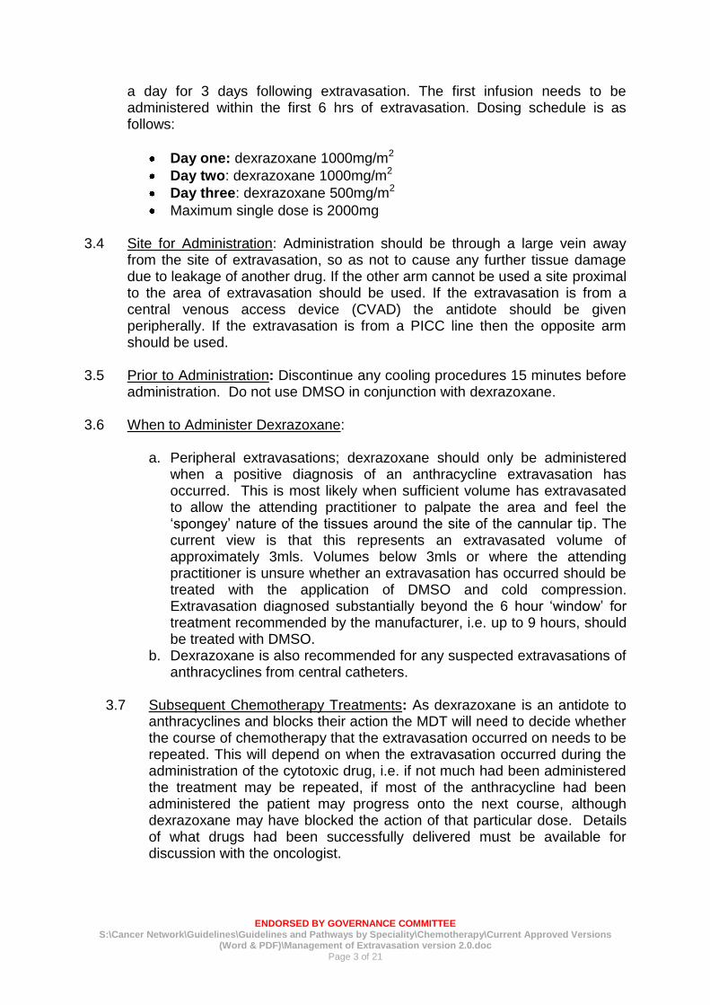
ENDORSED BY GOVERNANCE COMMITTEE
SCancer NetworkGuidelinesGuidelines and Pathways by SpecialityChemotherapyCurrent Approved Versions (Word amp PDF)Management of Extravasation version 20doc
Page 3 of 21
a day for 3 days following extravasation The first infusion needs to be administered within the first 6 hrs of extravasation Dosing schedule is as follows
Day one dexrazoxane 1000mgm2
Day two dexrazoxane 1000mgm2
Day three dexrazoxane 500mgm2
Maximum single dose is 2000mg 34 Site for Administration Administration should be through a large vein away
from the site of extravasation so as not to cause any further tissue damage due to leakage of another drug If the other arm cannot be used a site proximal to the area of extravasation should be used If the extravasation is from a central venous access device (CVAD) the antidote should be given peripherally If the extravasation is from a PICC line then the opposite arm should be used
35 Prior to Administration Discontinue any cooling procedures 15 minutes before
administration Do not use DMSO in conjunction with dexrazoxane 36 When to Administer Dexrazoxane
a Peripheral extravasations dexrazoxane should only be administered when a positive diagnosis of an anthracycline extravasation has occurred This is most likely when sufficient volume has extravasated to allow the attending practitioner to palpate the area and feel the lsquospongeyrsquo nature of the tissues around the site of the cannular tip The current view is that this represents an extravasated volume of approximately 3mls Volumes below 3mls or where the attending practitioner is unsure whether an extravasation has occurred should be treated with the application of DMSO and cold compression Extravasation diagnosed substantially beyond the 6 hour lsquowindowrsquo for treatment recommended by the manufacturer ie up to 9 hours should be treated with DMSO
b Dexrazoxane is also recommended for any suspected extravasations of anthracyclines from central catheters
37 Subsequent Chemotherapy Treatments As dexrazoxane is an antidote to
anthracyclines and blocks their action the MDT will need to decide whether the course of chemotherapy that the extravasation occurred on needs to be repeated This will depend on when the extravasation occurred during the administration of the cytotoxic drug ie if not much had been administered the treatment may be repeated if most of the anthracycline had been administered the patient may progress onto the next course although dexrazoxane may have blocked the action of that particular dose Details of what drugs had been successfully delivered must be available for discussion with the oncologist
ENDORSED BY GOVERNANCE COMMITTEE
SCancer NetworkGuidelinesGuidelines and Pathways by SpecialityChemotherapyCurrent Approved Versions (Word amp PDF)Management of Extravasation version 20doc
Page 4 of 21
38 Side Effects Results from clinical trials and case studies indicate that dexrazoxane is a safe and well-tolerated drug Side effects include those common to most cytotoxic drugs Those to note are neutropenia infection phlebitis stomatitis nausea and vomiting Dexrazoxane is excreted via the kidneys therefore decreased renal function may increase serum concentrations Following administration haematological and biochemical monitoring should take place the regularity will depend on the patient and will need to be clarified with the medical team The solution contains potassium therefore potassium levels should be monitored in patients who are at risk of hyperkalaemia
Reporting see page 14 number 16 of RMH guideline These incidents
should be reported in accordance with the Trust clinical incident reporting procedure for each organisation
39 Where is it Stored Dexrazoxane is stored in pharmacy It is the
responsibility of individual units to ensure access to dexrazoxane is possible within 6 hours of an extravasation
ENDORSED BY GOVERNANCE COMMITTEE
SCancer NetworkGuidelinesGuidelines and Pathways by SpecialityChemotherapyCurrent Approved Versions (Word amp PDF)Management of Extravasation version 20doc
Page 5 of 21
4 Royal Marsden Hospital Guidance THE NUMBERING FOR THIS SECTION RESTARTS AS IT IS TAKEN
DIRECTLY FROM THE RMH MANUAL
Part 2 - The Royal Marsden Hospital Manual of Clinical Nursing Procedures 7th Edition Blackwell Publishing (2008) chapter on extravasation
Extravasation of Vesicant Drugs - Definition Extravasation is a well-recognised complication of intravenous (IV) chemotherapy administration but in general is a condition that is often under-diagnosed undertreated and under-reported (Stanley 2002) The incidence of extravasation is estimated to be between 05 and 60 of cytotoxic drug administration (Kassner 2000 Khan amp Holmes 2002 Lawson 2003 Masoorli 2003 Goolsby amp Lombardo 2006) with some estimates for peripheral extravasation between 23 and 25 (Roth 2003) CVADs have decreased the incidence of extravasation but it can still occur usually as a result of a leaking or damaged catheter fibrin sheath formation (Mayo 1998) or a port needle dislodgement (Schulmeister 1998) The incidence estimated is up to 6 with ports (Masoorli 2003) However whilst the incidence is lower the severity of the injuries is far greater as detection tends to occur later (Kassner 2000 Stanley 2002 Polovich et al 2005) Even when practitioners have many years of experience extravasation of vesicant agents can occur and is an extremely stressful event but is not in itself an act of negligence (Weinstein 2007) Early detection and treatment are crucial if the consequences of an untreated or poorly managed extravasation are to be avoided (Figure 123) These may include
Pain from necrotic areas
Physical defect
The cost of hospitalization and plastic surgery
Delay in the treatment of disease
Psychological distress Litigation nurses are now being named in malpractice allegations and extravasation injuries are an area for concern (Dougherty 2003 Masoorli 2003 Roth 2003 Weinstein 2007) 1 Prevention of Extravasation The nurses focus should be on safe intravenous technique and implementing strategies to minimize the risk (Weinstein 2007) This includes the following strategies 2 Monitoring the Site Confirming venous patency by flushing with 09 sodium chloride solution with at least 5ndash10 ml prior to administration of vesicants and frequent monitoring thereafter (Goolsby amp Lombardo 2006 Weinstein 2007) Checking blood return after every 2ndash5 ml is recommended but cannot be relied upon as the key sign when giving a bolus
ENDORSED BY GOVERNANCE COMMITTEE
SCancer NetworkGuidelinesGuidelines and Pathways by SpecialityChemotherapyCurrent Approved Versions (Word amp PDF)Management of Extravasation version 20doc
Page 6 of 21
injection and monitoring the site every 5ndash10 minutes for any swelling (Weinstein 2007) 3 Location of the Device The most appropriate site for the location of a peripheral cannula is considered to be the forearm (Schrijvers 2003 Weinstein 2007) However a large straight vein over the dorsum of the hand is preferable to a smaller vein in the forearm (Weinstein 2007) Siting over joints should be avoided as tissue damage in this area may limit joint movement in the future It is also recommended that the antecubital fossa should never be used for the administration of vesicants because of the risk of damage to local structures such as nerves and tendons (Hayden amp Goodman 2005 Weinstein 2007 Gabriel 2008) Avoid venepuncture sites in limbs with impaired circulation sclerosis thrombosis or scar formation Also avoid cannulation below a recent venepuncture site (Goolsby amp Lombardo 2006) 4 Patients at Risk Patients who are at increased risk of extravasation (Box 122) should be observed more closely and cared for with extra caution Box 122 Patients at Risk of Extravasation
Infants and young children
Elderly patients
Those who are unable to communicate eg sedated unconscious confused language issues
Those with chronic diseases eg cancer peripheral vascular disease superior vena cava (SVC) syndrome lymphoedema
Those on medications anticoagulants steroids
Those who have undergone repeated intravenous cannulationvenepuncture
Those with fragile veins or who are thrombocytopenic Polovich et al 2005 Hayden amp Goodman 2005 Sauerland et al 2006 Sequence of Drugs (Table 123) Vesicants should be given first (Kassner 2000 Hayden amp Goodman 2005 Goolsby amp Lombardo 2006) Reasons for this include
1 Vascular integrity decreases over time 2 Vein is most stable and least irritated at start of treatment 3 Initial assessment of vein patency is most accurate 4 Patients awareness of changes more acute (Weinstein 2007)
ENDORSED BY GOVERNANCE COMMITTEE
SCancer NetworkGuidelinesGuidelines and Pathways by SpecialityChemotherapyCurrent Approved Versions (Word amp PDF)Management of Extravasation version 20doc
Page 7 of 21
Table 123 Drug sequencing ndash rationale for administering vesicant drugs first or last (Stanley 2002 Weinstein 2007)
Vesicants First Vesicants Last
1 Vascular integrity decreases over time
1 Vesicants are irritating and increase vein fragility
2 Vein is most stable and least irritated at start of treatment
2 Venous spasm may occur and mask signs of extravasation
3 Initial assessment of vein patency is most accurate
4 Patients awareness of changes more acute
5 Types of Devices The use of steel needles is associated with a greater risk of extravasation and should be discouraged and a plastic cannula should be used instead (Schrijvers 2003 Polovich et al 2005 Sauerland et al 2006) Vesicants should be given via a newly established cannula wherever possible (Dougherty 2005 Goolsby amp Lombardo 2006) and consideration should be given to changing the cannula site after 24 hours (Hayden amp Goodman 2005) However if the fluid runs freely there is good blood return and there are no signs of erythema pain or swelling at the site there is no reason to inflict a second cannulation on the patient (Weinstein 2007) Consideration should be given to a CVAD if peripheral access is difficult 6 Method of Administration Many vesicants must be given as a slow bolus injection often via the side arm of a fast-running intravenous infusion of a compatible solution eg doxorubicin or epirubicin via an infusion of 09 sodium chloride If repeated infusions are to be given then a CVAD may be more appropriate (Stanley 2002 Weinstein 2007) 7 Skill of the Practitioner Correct choice of device and location the ability to use the most appropriate vasodilatation techniques early recognition of extravasation and prompt action come from ensuring only skilled and knowledgeable practitioners administer vesicant drugs andor insert the vascular access device (Schrijvers 2003 Dougherty 2005 Goolsby amp Lombardo 2006 Sauerland et al 2006) Successful cannulation at the first attempt is ideal as vesicants have been known to seep into tissues at a vein entry site of a previous cannulation (Gault amp Challands 1997 Perdue 2001) This also includes accessing a port as it is vital that the correct selection of needle is made and that the device is secured adequately (Camp Sorrell 2005)
ENDORSED BY GOVERNANCE COMMITTEE
SCancer NetworkGuidelinesGuidelines and Pathways by SpecialityChemotherapyCurrent Approved Versions (Word amp PDF)Management of Extravasation version 20doc
Page 8 of 21
8 Patient Information Patients should be informed of the potential problems of administering vesicants and the possible consequences of extravasation (Stanley 2002 Sauerland et al 2006 Weinstein 2007) Adequate information given to patients will ensure early recognition and co-operation as patients are the first to notice pain The patient should be urged to report immediately any change in sensation such as burning or stinging (Goolsby amp Lombardo 2006) 9 Drugs Capable of Causing Tissue Necrosis Before administration of vesicant cytotoxic drugs the nurse should know which agents are capable of producing tissue necrosis The following is a list of examples of those in common use
Group A Drugs Group B Drugs
Vinca alkaloids Amsacrine
Vinblastine Carmustine (concentrated solution)
Vindesine Dacarbazine (concentrated solution)
Vinorelbine Dactinomycin
Vincristine Daunorubicin
Vinflunine Doxorubicin
Paclitaxel Epirubicin
Idarubicin
Mithramycin
Mitomycin C
Mechlorethamine
Streptozocin
If in any doubt the drug data sheet should be consulted or reference made to a research trial protocol Drugs should not be reconstituted to give solutions which are higher than the manufacturers recommended concentration and the method of administration should be checked eg infusion injection
ENDORSED BY GOVERNANCE COMMITTEE
SCancer NetworkGuidelinesGuidelines and Pathways by SpecialityChemotherapyCurrent Approved Versions (Word amp PDF)Management of Extravasation version 20doc
Page 9 of 21
A variety of vesicant non-cytotoxic agents in frequent use are also capable of causing severe tissue damage if extravasated Examples include
Group A Drugs Group B Drugs
Calcium chloride Aciclovir
Calcium gluconate Amphotericin
Phenytoin Cefotaxime
Hypertonic solutions eg sodium chloride gt09
Diazepam
Ganciclovir
Sodium bicarbonate (gt5) Potassium chloride (gt40 mmoll)
Glucose 50 Mannitol
This potential hazard should always be remembered The actions listed in this procedure may not be appropriate in all these instances Drug data sheets should always be checked and the pharmacy departments should be consulted if the information is insufficient regarding action to take if a vesicant drug extravasates 10 Signs and Symptoms of Extravasation (see Table 124) Extravasation should be suspected if
1 The patient complains of burning stinging pain or any other acute change at the injection site although this is not always present (Hayden amp Goodman 2005) This should be distinguished from a feeling of cold which may occur with some drugs or venous spasm which can be caused by irritation usually accompanied by pain described as an achiness or tightness (Hayden amp Goodman 2005) Any change of sensation warrants further investigation (Goolsby amp Lombardo 2006)
2 Swelling is the most common symptom (Polovich et al 2005) Induration or leakage may also occur at the injection site Swelling may not always be immediately obvious if the patient has the cannula sited in an area of deep subcutaneous fat in a deep vein or if the leak is via the posterior vein wall (Dougherty 2005)
3 Blanching of the skin occurs (Comerford et al 2002) Erythema can occur around the injection site but this is not usually present immediately (Hayden amp Goodman 2005) It is important that this is distinguished from a lsquoflarersquo reaction (Polovich et al 2005)
4 Blood return is one of the most misleading of all signs particularly related to peripheral devices In peripheral devices if blood return is sluggish or absent this may indicate lack of patency or incorrect position of the device However if no other signs are apparent this should not be regarded as an indication of a non-patent vein as a vein may not bleed back for a number of reasons and extravasation may occur even in the event of good blood return (Hayden amp Goodman 2005) Any change in blood flow should be investigated (Hayden amp Goodman 2005 Weinstein 2007) In CVADs there should always be blood return and if this is absent steps should be followed in order to be able to verify correct tip and needle position or resolve a fibrin sheath (see Figure 451)
ENDORSED BY GOVERNANCE COMMITTEE
SCancer NetworkGuidelinesGuidelines and Pathways by SpecialityChemotherapyCurrent Approved Versions (Word amp PDF)Management of Extravasation version 20doc
Page 10 of 21
5 A resistance is felt on the plunger of the syringe if drugs are given by bolus (Vandergrift 2001 Stanley 2002)
6 There is absence of free flow when administration is by infusion once other reasons have been excluded eg position (Vandergrift 2001 Stanley 2002)
Note one or more of the above may be present If extravasation is suspected or confirmed the injection or infusion must be stopped immediately and action must be taken (Polovich et al 2005 Infusion Nurses Society 2006 Weinstein 2007)
ENDORSED BY GOVERNANCE COMMITTEE
SCancer NetworkGuidelinesGuidelines and Pathways by SpecialityChemotherapyCurrent Approved Versions (Word amp PDF)Management of Extravasation version 20doc Page 11 of 21
Table 124 Nursing assessment of extravasation versus other reactions (Polovich et al 2005 Weinstein 2007)
Extravasation
Assessment parameter
Flare reaction Venous irritation Immediate manifestations ie during drug administration
Delayed manifestations ie from 24 hours after
extravasation
Pain None Aching throbbing sensation along vein and in the limb
Severe stinging or burning pain (not always present) This can last from minutes to hours and will eventually subside Occurs during drug administration at the device site and surrounding areas
Can continue following extravasation or start within 48 hours
Redness Immediate blotches or tracking along the vein This will subside within 30ndash45 minutes with or without treatment (usually steroid cream)
Vein may become red or darkened
Not always present immediately more likely to see blanching of the skin As area becomes inflamed redness will appear around the device site
Later occurrence
Swelling Unlikely Unlikely May occur immediately but may not always be easy to identify immediately
Usually within 48 hours
Blood return Usually present Usually present but may require application of heat to improve blood return
Inability to obtain blood return (peripheral or central) but blood return may be present throughout
Ulceration Unlikely Unlikely Unlikely Can occur within 48ndash96 hours but may take 3ndash4 weeks to develop
Others Urticaria None Change in quality of the infusion or pressure on the syringe
Local tingling and sensory deficits
ENDORSED BY GOVERNANCE COMMITTEE
SCancer NetworkGuidelinesGuidelines and Pathways by SpecialityChemotherapyCurrent Approved Versions (Word amp PDF)Management of Extravasation version 20doc
Page 12 of 21
11 Management of Extravasation The management of extravasation of chemotherapy agents is controversial and there is little documented evidence of efficacy Until recently no antidote has received clear validation in controlled clinical trials and no randomized trials managing cytotoxic extravasation in humans have been completed (Bertolli 1995) Some studies performed on animals have demonstrated both effective and ineffective treatments but extrapolation from animals to humans is limited (Polovich et al 2005 Wickham et al 2006) Other studies have been small with low numbers of patients Another problem is that it is often difficult to ascertain whether any extravasation has actually occurred (Weinstein 2007) Therefore recommendations are based on more consistent experimental evidence cumulative clinical experience from available case reports and uncontrolled studies and empirical guidelines (Bertolli 1995) Drugs where there is evidence of effective management include anthracyclines (Rudolph amp Larson 1987) vinca alkaloids (Bertolli 1995) computed tomography (CT) contrast media (Federle et al 1998) and paclitaxel (Bertolli et al 1997) The management of extravasation involves several stages including the following Stage 1 stopping infusioninjection and aspirating the drug It appears that most authors are agreed that aspirating as much of the drug as possible as soon as extravasation is suspected is beneficial (Rudolph amp Larson 1987 Vandergrift 2001 Polovich et al 2005 Weinstein 2007) and can help lower the concentration of the drug in the area (Goolsby amp Lombardo 2006) However withdrawal is only immediately possible during bolus injections as an infusion would need to be stopped and a syringe attached in an attempt to aspirate Aspiration may be successful if extravasation presents itself as a raised blister but may be unsuccessful if tissue is soft and soggy (CP Pharmaceuticals 1999 Stanley 2002) It may help to reduce the size of the lesion (Vandergrift 2001) In practice it may achieve little and often distresses the patient (Gault amp Challands 1997) The likelihood of withdrawing blood (as suggested by Ignoffo amp Friedman 1980) is small and the practitioner may waste valuable time attempting this which could lead to delay in the rest of the management procedure Stage 2 removing device Some clinicians advocate that the peripheral vascular access device be left in situ in order to instil the antidote via the device and into the affected tissues (Kassner 2000 Stanley 2002 Weinstein 2007) However others recommend that the peripheral device should be removed to prevent any injected solution increasing the size of the affected area (Rudolph amp Larson 1987 CP Pharmaceuticals 1999 Vandergrift 2001) There appears to be no research evidence to support either practice Stage 3 applying hot or cold packs Cooling appears to be a better choice with the exception of the vinca alkaloids and some non-cytotoxic drugs than warming (Bertolli 1995 CP Pharmaceuticals 1999) Cold causes vasoconstriction localizing the extravasation and perhaps allowing time for local vascular and lymphatic systems to contain the drug It should be applied for
ENDORSED BY GOVERNANCE COMMITTEE
SCancer NetworkGuidelinesGuidelines and Pathways by SpecialityChemotherapyCurrent Approved Versions (Word amp PDF)Management of Extravasation version 20doc
Page 13 of 21
15ndash20 minutes 3ndash4 times a day for up to 3 days (Gault amp Challands 1997 CP Pharmaceuticals 1999 Polovich et al 2005) Heat promotes healing after the first 24 hours by increasing the blood supply (Polovich et al 2005 Weinstein 2007) It also decreases local drug concentration increasing the blood flow which results in enhanced resolution of pain and reabsorption of local swelling Stage 4 use of antidotes A number of antidotes are available but again there is a lack of scientific evidence to demonstrate their value and so the role of antidotes is still not clear (Polovich et al 2005) There appear to be two main methods (i) localize and neutralize (using hyaluronidase) (CP Pharmaceuticals 1999) and (ii) spread and dilute (using an antidote) (Stanley 2002) Administration of antidotes if not via the cannula is by the pincushion technique that is instilling small volumes around and over the areas affected using a small gauge (25) needle towards the centre of a clock face The procedure causes considerable discomfort to patients and if large areas are to be tackled analgesia should be considered (Stanley 2002) Hyaluronidase is an enzyme which breaks down hyaluronic acid a normal component of tissue lsquocementrsquo and helps to reduce or prevent tissue damage by allowing rapid diffusion of the extravasated fluid and promoting drug absorption (Few 1987) The usual dose is 1500 IU (Bertolli 1995 Vandergrift 2001) It should be injected within 1 hour of extravasation ideally through the intravenous device delivering the enzyme to the same tissue (Gault amp Challands 1997 Vandergrift 2001 Stanley 2002 Weinstein 2007) NB Hyaluronidase increases the absorption of local anaesthetic Therefore if local anaesthetic has been applied to the area eg Ametop gel prior to cannulation within 6 hours of extravasation then the patient should be monitored for signs and symptoms of systemic anaesthesia such as increased pulse rate and decreased respirations and the doctor informed immediately (BMARPSGB 2008) Corticosteroids have long been advocated as a treatment for anthracycline extravasation in reducing inflammatory components although inflammation is not a prominent feature of tissue necrosis (Camp Sorrell 1998) and they appear to have little benefit Data now discourages the use of locally injected steroids as there is little evidence to support their use (Bertolli 1995 Gault amp Challands 1997 Wickham et al 2006) However given as a cream they can help to reduce local trauma and irritation (Stanley 2002) Dimethyl sulfoxide (DMSO) is a potent free radical scavanger that rapidly penetrates tissues when applied topically (Bertolli 1995) Reports on the clinical use of topical DMSO show it is effective and well tolerated in extravasation (Bertolli 1995) However this is based on a high dose (95) and only 50 is easily available in the UK (Stanley 2002) Side-effects from DMSO include itching erythema mild burning and a characteristic breath odour (Bertolli 1995) Recently dexrazoxane a topoisomerase II catalytic inhibitor used clinically to minimize the cardiotoxicity of doxorubicin has been tested in animal models and a small number of patients for its use in extravasation It is given IV 3ndash6 hours after the extravasation and it appears to reduce the wound size and duration with
ENDORSED BY GOVERNANCE COMMITTEE
SCancer NetworkGuidelinesGuidelines and Pathways by SpecialityChemotherapyCurrent Approved Versions (Word amp PDF)Management of Extravasation version 20doc
Page 14 of 21
anthracyclines The triple dosage appears to be more effective than a single dose (Langer et al 2000 El Saghir et al 2004) A consensus group (Jackson et al 2006) have developed recommendations for the use of dexrazoxane
1 For anthracycline extravasations resulting from peripheral administration the site expert or team should be consulted in order to determine whether the use of dexrazoxane is indicated
2 Absolute indications are if the peripherally extravasated volume exceeds 15 ml and in the event of a central venous extravasation
Finally granulocyte macrophage-colony stimulating factor (GM-CSF) is a growth factor and has been found effective in accelerating wound healing and inducing formation of granulation of tissue (Ulutin et al 2000 El Saghir et al 2004) Stage 5 elevation of limb This is recommended as it minimizes swelling (Rudolph amp Larson 1987) and movement should be encouraged to prevent adhesion of damaged areas to underlying tissue (Gabriel 2008) Stage 6 surgical techniques Some centres suggest that a plastic surgery consultation be performed as part of the management procedure in order to remove the tissue containing the drug Surgical intervention is recommended especially if the lesion is greater than 2 cm there is significant residual pain 1ndash2 weeks after extravasation or there is minimal healing 2ndash3 weeks after injury despite local therapeutic measures (Goolsby amp Lombardo 2006) Liposuction or a flush-out technique can remove extravasated drug without resorting to excision and skin grafting A liposuction cannula can be used to aspirate extravasated material and subcutaneous fat If there is little subcutaneous fat eg preterm infants then the saline flush-out technique is recommended particularly if done within the first 24 hours It has been suggested as a less traumatic and cheaper procedure than surgery Four small stab incisions are made and large volumes of 09 sodium chloride are administered which flush out the extravasated drug (Gault amp Challands 1997) Management of large extravasations from CVADs is usually by surgical intervention and washout of affected tissues 12 Extravasation Kits The use of extravasation kits has been recommended in order to provide immediate management (Khan amp Holmes 2002 Hayden amp Goodman 2005) Kits should be assembled according to the particular needs of individual institutions They should be kept in all areas where staff are regularly administering vesicant drugs so staff have immediate access to equipment (Gabriel 2008) The kit should be simple to avoid confusion but comprehensive enough to meet all reasonable needs (Stanley 2002) (see Procedure guidelines Extravasation management peripheral cannula below) Instructions should be clear and easy to follow and the use of a flow chart enables staff to follow the management procedure in easy steps (Figure 124)
ENDORSED BY GOVERNANCE COMMITTEE
SCancer NetworkGuidelinesGuidelines and Pathways by SpecialityChemotherapyCurrent Approved Versions (Word amp PDF)Management of Extravasation version 20doc
Page 15 of 21
13 Mixed Vesicant Extravasation Consideration should be given to the management of mixed vesicant drug extravasation in terms of which drug to treat with which antidote It has been recommended to act in accordance with the drug which possesses the most deleterious properties (How amp Brown 1998) 14 Informing the Patient Patients should always be informed when an extravasation has occurred and be given an explanation of what has happened and what management has been carried out (McCaffrey Boyle amp Engelking 1995) An information sheet should be given to patients with instructions of what symptoms to look out for and when to contact the hospital during the follow-up period (Gabriel 2008) 15 Wound Management Damage will be affected by the site amount of drug concentration of the agent and if it binds to DNA or not (Polovich et al 2005) Ulceration may occur over a period of days to weeks and extravasation wounds may be complicated by tissue ischaemia related to endothelial damage (Naylor 2005) The type of injury will dictate the type of dressing Assessment of the wound should include position and size of the wound amount and type of tissue present amount and type of exudate and extent and spread of erythema (Naylor 2005) 16 Documentation and Follow Up An extravasation must be reported and fully documented as it is an accident and the patient may require follow-up care (NMC 2005 RCN 2005) Information may also be used for statistical purposes for example collation and analysis using the green card scheme devised by St Chads Hospital Birmingham (Stanley 2002) Statistics on the incidence degree causes and corrective action should be monitored and analysed (Infusion Nurses Society 2006) Finally it may be required in case of litigation which is now on the increase (Dougherty 2003 Masoorli 2003)
ENDORSED BY GOVERNANCE COMMITTEE
SCancer NetworkGuidelinesGuidelines and Pathways by SpecialityChemotherapyCurrent Approved Versions (Word amp PDF)Management of Extravasation version 20doc
Page 16 of 21
Figure
ENDORSED BY GOVERNANCE COMMITTEE
SCancer NetworkGuidelinesGuidelines and Pathways by SpecialityChemotherapyCurrent Approved Versions (Word amp PDF)Management of Extravasation version 20doc
Page 17 of 21
Equipment To assist the nurse an extravasation kit should be assembled and should be readily available in each wardunit It contains
1 Gel packs times 2 one to be kept in the fridge and one available for heating (an electric heating blanket can be used whilst pack is heating)
2 Hyaluronidase 1500 IU2 ml sterile water 3 Hydrocortisone cream 1 15 g tube times 1 4 2 ml syringes times 1 5 25 G needles times 2 6 Alcohol swabs 7 Documentation forms 8 Copy of extravasation management procedure 9 Patient information leaflet
Procedure
Action Rationale
1 Explain and discuss the procedure with the patient
To ensure that the patient understands the procedure and gives hisher valid consent (DH 2002a C NMC 2008b C)
2 Stop injection or infusion immediately leaving the cannula in place
To minimize local injury To allow aspiration of the drug to be attempted (Polovich et al 2005 C RCN 2005 C)
3 Aspirate any residual drug from the device and suspected extravasation site
To minimise local injury by removing as much drug as possible and only attempt if appropriate Subsequent damage is related to the volume of the extravasation in addition to other factors (Polovich et al 2005 C RCN 2005 C)
4 Remove the cannula To prevent the device from being used for antidote administration (Rudolph amp Larson 1987 E)
5 Collect the extravasation pack and take it to the patient
It contains all the equipment necessary for managing extravasation (Stanley 2002 E Dougherty 2005 E)
ENDORSED BY GOVERNANCE COMMITTEE
SCancer NetworkGuidelinesGuidelines and Pathways by SpecialityChemotherapyCurrent Approved Versions (Word amp PDF)Management of Extravasation version 20doc
Page 18 of 21
Group A Drugs
Draw up hyaluronidase 1500 IU in 1 ml water for injection and inject volumes of 01ndash02 ml subcutaneously at points of the compass around the circumference of the area of extravasation
This is the recommended agent for group A drugs The warm pack speeds up absorption of the drug by the tissues (Bertolli 1995 C)
Apply warm pack
Group B Drugs
Apply cold pack or ice instantly To localize the area of extravasation slow cell metabolism and decrease the area of tissue destruction To reduce local pain (Polovich et al 2005 C)
However if extravasation is with any of the following category B drugs mitomycin C doxorubicin idarubicin epirubicin actinomycin D then
This is the recommended agent for these anthracyclines and helps to reduce local tissue damage (Bertolli 1995 C)
Draw around area of extravasation with indelible pen
Put on gloves
Apply thin layer of DMSO topically to the marked area using the small plastic spatula in lid of the bottle
Allow it to dry
Apply gauze
This should be applied within 10ndash25 minutes
Where possible elevate the extremity andor encourage movement
To minimize swelling and to prevent adhesion of damaged area to underlying tissue which could result in restriction of movement (Rudolph amp Larson 1987 E)
Inform a member of the medical staff at the earliest opportunity and administer any other prescribed antidotes eg dexrazoxane
To enable actions differing from agreed policy to be taken if considered in the best interests of the patient To notify the doctor of the need to prescribe any other drugs (E)
Apply hydrocortisone cream 1 twice daily and instruct the patient how to do this Continue as long as erythema persists
To reduce local inflammation and promote patient comfort (Stanley 2002 E)
Where appropriate apply DMSO every 2 hours on day 1 and then every 6 hours for up to 7 days (patients will need to have this prescribed as a to take out [TTO] and continue treatment at home where necessary)
To help reduce local tissue damage (Bertolli 1995 C)
ENDORSED BY GOVERNANCE COMMITTEE
SCancer NetworkGuidelinesGuidelines and Pathways by SpecialityChemotherapyCurrent Approved Versions (Word amp PDF)Management of Extravasation version 20doc
Page 19 of 21
Heat packs (group A drugs) should be reapplied after initial management for 2ndash4 hours Cold packs (group B drugs) should be applied for 15ndash20 minutes 3ndash4 times a day for up to 3 days
To localize the steroid effect in the area of extravasation To reduce local pain and promote patient comfort (Bertolli 1995 C)
Provide analgesia as required To promote patient comfort To encourage movement of the limb as advised (E)
Document the following details in duplicate on the form provided
a) Patients namenumber
b) Wardunit
c) Date time
d) Signs and symptoms
e) Venepuncture site (on diagram)
f) Drug sequence
g) Drug administration technique ie lsquobolusrsquo or infusion
h) Approximate amount of the drug extravasated
i) Diameter length and width of extravasation area
j) Appearance of the area
k) Step-by-step management with date and time of each step performed and medical officer notification
To provide an immediate full record of all details of the incident which may be referred to if necessary To provide a baseline for future observation and monitoring of patients condition To comply with NMC guidelines (Kassner 2000 E NMC 2005 C RCN 2005 C Weinstein 2007 E)
l) Patients complaints comments statements
m) Indication that patients information sheet given to patient
n) Follow-up section
o) Whether photograph was taken
p) If required when patient referred to plastic surgeon
q) Signature of the nurse
Explain to the patient that the site may remain sore for several days
To reduce anxiety and ensure continued co-operation (P E)
Observe the area regularly for erythema induration blistering or necrosis Inpatients monitor daily Where appropriate take photograph
To detect any changes at the earliest possible moment (RCN 2005 C)
ENDORSED BY GOVERNANCE COMMITTEE
SCancer NetworkGuidelinesGuidelines and Pathways by SpecialityChemotherapyCurrent Approved Versions (Word amp PDF)Management of Extravasation version 20doc
Page 20 of 21
All patients should receive written information explaining what has occurred what management has been carried out what they need to look for at the site and when to report any changes For example increased discomfort peeling or blistering of the skin should be reported immediately
To detect any changes as early as possible and allow for a review of future management This may include referral to a plastic surgeon (Gault amp Challands 1997 E Polovich et al 2005 C RCN 2005 C)
If blistering or tissue breakdown occurs begin dressing techniques and seek advice regarding wound management
To minimize the risk of a superimposed infection and sterile increase healing (Naylor 2005 E)
Depending on size of lesion degree of pain type of drug refer to plastic surgeon
To prevent further pain or other complications as chemically induced ulcers rarely heal spontaneously (Polovich et al 2005 C Dougherty 2005 E)
Monitoring of the Guideline Implementation of the guidance will be considered as a topic for audit by the NSSG in and reviewed in Authors Andrew Stanley Network Pharmacist References
The Royal Marsden Hospital Manual of Clinical Nursing Procedures 7th Edition Blackwell Publishing (2008)
ENDORSED BY GOVERNANCE COMMITTEE
SCancer NetworkGuidelinesGuidelines and Pathways by SpecialityChemotherapyCurrent Approved Versions (Word amp PDF)Management of Extravasation version 20doc
Page 21 of 21
Approval Signatures Pan Birmingham Cancer Network Governance Committee Chair Name Doug Wullf
Signature Date July 2011 Pan Birmingham Cancer Network Manager Name Karen Metcalf
Signature Date July 2011 Network Site Specific Group Clinical Chair Name Frances Shaw
Signature Date July 2011
ENDORSED BY GOVERNANCE COMMITTEE
SCancer NetworkGuidelinesGuidelines and Pathways by SpecialityChemotherapyCurrent Approved Versions (Word amp PDF)Management of Extravasation version 20doc
Page 6 of 21
injection and monitoring the site every 5ndash10 minutes for any swelling (Weinstein 2007) 3 Location of the Device The most appropriate site for the location of a peripheral cannula is considered to be the forearm (Schrijvers 2003 Weinstein 2007) However a large straight vein over the dorsum of the hand is preferable to a smaller vein in the forearm (Weinstein 2007) Siting over joints should be avoided as tissue damage in this area may limit joint movement in the future It is also recommended that the antecubital fossa should never be used for the administration of vesicants because of the risk of damage to local structures such as nerves and tendons (Hayden amp Goodman 2005 Weinstein 2007 Gabriel 2008) Avoid venepuncture sites in limbs with impaired circulation sclerosis thrombosis or scar formation Also avoid cannulation below a recent venepuncture site (Goolsby amp Lombardo 2006) 4 Patients at Risk Patients who are at increased risk of extravasation (Box 122) should be observed more closely and cared for with extra caution Box 122 Patients at Risk of Extravasation
Infants and young children
Elderly patients
Those who are unable to communicate eg sedated unconscious confused language issues
Those with chronic diseases eg cancer peripheral vascular disease superior vena cava (SVC) syndrome lymphoedema
Those on medications anticoagulants steroids
Those who have undergone repeated intravenous cannulationvenepuncture
Those with fragile veins or who are thrombocytopenic Polovich et al 2005 Hayden amp Goodman 2005 Sauerland et al 2006 Sequence of Drugs (Table 123) Vesicants should be given first (Kassner 2000 Hayden amp Goodman 2005 Goolsby amp Lombardo 2006) Reasons for this include
1 Vascular integrity decreases over time 2 Vein is most stable and least irritated at start of treatment 3 Initial assessment of vein patency is most accurate 4 Patients awareness of changes more acute (Weinstein 2007)
ENDORSED BY GOVERNANCE COMMITTEE
SCancer NetworkGuidelinesGuidelines and Pathways by SpecialityChemotherapyCurrent Approved Versions (Word amp PDF)Management of Extravasation version 20doc
Page 7 of 21
Table 123 Drug sequencing ndash rationale for administering vesicant drugs first or last (Stanley 2002 Weinstein 2007)
Vesicants First Vesicants Last
1 Vascular integrity decreases over time
1 Vesicants are irritating and increase vein fragility
2 Vein is most stable and least irritated at start of treatment
2 Venous spasm may occur and mask signs of extravasation
3 Initial assessment of vein patency is most accurate
4 Patients awareness of changes more acute
5 Types of Devices The use of steel needles is associated with a greater risk of extravasation and should be discouraged and a plastic cannula should be used instead (Schrijvers 2003 Polovich et al 2005 Sauerland et al 2006) Vesicants should be given via a newly established cannula wherever possible (Dougherty 2005 Goolsby amp Lombardo 2006) and consideration should be given to changing the cannula site after 24 hours (Hayden amp Goodman 2005) However if the fluid runs freely there is good blood return and there are no signs of erythema pain or swelling at the site there is no reason to inflict a second cannulation on the patient (Weinstein 2007) Consideration should be given to a CVAD if peripheral access is difficult 6 Method of Administration Many vesicants must be given as a slow bolus injection often via the side arm of a fast-running intravenous infusion of a compatible solution eg doxorubicin or epirubicin via an infusion of 09 sodium chloride If repeated infusions are to be given then a CVAD may be more appropriate (Stanley 2002 Weinstein 2007) 7 Skill of the Practitioner Correct choice of device and location the ability to use the most appropriate vasodilatation techniques early recognition of extravasation and prompt action come from ensuring only skilled and knowledgeable practitioners administer vesicant drugs andor insert the vascular access device (Schrijvers 2003 Dougherty 2005 Goolsby amp Lombardo 2006 Sauerland et al 2006) Successful cannulation at the first attempt is ideal as vesicants have been known to seep into tissues at a vein entry site of a previous cannulation (Gault amp Challands 1997 Perdue 2001) This also includes accessing a port as it is vital that the correct selection of needle is made and that the device is secured adequately (Camp Sorrell 2005)
ENDORSED BY GOVERNANCE COMMITTEE
SCancer NetworkGuidelinesGuidelines and Pathways by SpecialityChemotherapyCurrent Approved Versions (Word amp PDF)Management of Extravasation version 20doc
Page 8 of 21
8 Patient Information Patients should be informed of the potential problems of administering vesicants and the possible consequences of extravasation (Stanley 2002 Sauerland et al 2006 Weinstein 2007) Adequate information given to patients will ensure early recognition and co-operation as patients are the first to notice pain The patient should be urged to report immediately any change in sensation such as burning or stinging (Goolsby amp Lombardo 2006) 9 Drugs Capable of Causing Tissue Necrosis Before administration of vesicant cytotoxic drugs the nurse should know which agents are capable of producing tissue necrosis The following is a list of examples of those in common use
Group A Drugs Group B Drugs
Vinca alkaloids Amsacrine
Vinblastine Carmustine (concentrated solution)
Vindesine Dacarbazine (concentrated solution)
Vinorelbine Dactinomycin
Vincristine Daunorubicin
Vinflunine Doxorubicin
Paclitaxel Epirubicin
Idarubicin
Mithramycin
Mitomycin C
Mechlorethamine
Streptozocin
If in any doubt the drug data sheet should be consulted or reference made to a research trial protocol Drugs should not be reconstituted to give solutions which are higher than the manufacturers recommended concentration and the method of administration should be checked eg infusion injection
ENDORSED BY GOVERNANCE COMMITTEE
SCancer NetworkGuidelinesGuidelines and Pathways by SpecialityChemotherapyCurrent Approved Versions (Word amp PDF)Management of Extravasation version 20doc
Page 9 of 21
A variety of vesicant non-cytotoxic agents in frequent use are also capable of causing severe tissue damage if extravasated Examples include
Group A Drugs Group B Drugs
Calcium chloride Aciclovir
Calcium gluconate Amphotericin
Phenytoin Cefotaxime
Hypertonic solutions eg sodium chloride gt09
Diazepam
Ganciclovir
Sodium bicarbonate (gt5) Potassium chloride (gt40 mmoll)
Glucose 50 Mannitol
This potential hazard should always be remembered The actions listed in this procedure may not be appropriate in all these instances Drug data sheets should always be checked and the pharmacy departments should be consulted if the information is insufficient regarding action to take if a vesicant drug extravasates 10 Signs and Symptoms of Extravasation (see Table 124) Extravasation should be suspected if
1 The patient complains of burning stinging pain or any other acute change at the injection site although this is not always present (Hayden amp Goodman 2005) This should be distinguished from a feeling of cold which may occur with some drugs or venous spasm which can be caused by irritation usually accompanied by pain described as an achiness or tightness (Hayden amp Goodman 2005) Any change of sensation warrants further investigation (Goolsby amp Lombardo 2006)
2 Swelling is the most common symptom (Polovich et al 2005) Induration or leakage may also occur at the injection site Swelling may not always be immediately obvious if the patient has the cannula sited in an area of deep subcutaneous fat in a deep vein or if the leak is via the posterior vein wall (Dougherty 2005)
3 Blanching of the skin occurs (Comerford et al 2002) Erythema can occur around the injection site but this is not usually present immediately (Hayden amp Goodman 2005) It is important that this is distinguished from a lsquoflarersquo reaction (Polovich et al 2005)
4 Blood return is one of the most misleading of all signs particularly related to peripheral devices In peripheral devices if blood return is sluggish or absent this may indicate lack of patency or incorrect position of the device However if no other signs are apparent this should not be regarded as an indication of a non-patent vein as a vein may not bleed back for a number of reasons and extravasation may occur even in the event of good blood return (Hayden amp Goodman 2005) Any change in blood flow should be investigated (Hayden amp Goodman 2005 Weinstein 2007) In CVADs there should always be blood return and if this is absent steps should be followed in order to be able to verify correct tip and needle position or resolve a fibrin sheath (see Figure 451)
ENDORSED BY GOVERNANCE COMMITTEE
SCancer NetworkGuidelinesGuidelines and Pathways by SpecialityChemotherapyCurrent Approved Versions (Word amp PDF)Management of Extravasation version 20doc
Page 10 of 21
5 A resistance is felt on the plunger of the syringe if drugs are given by bolus (Vandergrift 2001 Stanley 2002)
6 There is absence of free flow when administration is by infusion once other reasons have been excluded eg position (Vandergrift 2001 Stanley 2002)
Note one or more of the above may be present If extravasation is suspected or confirmed the injection or infusion must be stopped immediately and action must be taken (Polovich et al 2005 Infusion Nurses Society 2006 Weinstein 2007)
ENDORSED BY GOVERNANCE COMMITTEE
SCancer NetworkGuidelinesGuidelines and Pathways by SpecialityChemotherapyCurrent Approved Versions (Word amp PDF)Management of Extravasation version 20doc Page 11 of 21
Table 124 Nursing assessment of extravasation versus other reactions (Polovich et al 2005 Weinstein 2007)
Extravasation
Assessment parameter
Flare reaction Venous irritation Immediate manifestations ie during drug administration
Delayed manifestations ie from 24 hours after
extravasation
Pain None Aching throbbing sensation along vein and in the limb
Severe stinging or burning pain (not always present) This can last from minutes to hours and will eventually subside Occurs during drug administration at the device site and surrounding areas
Can continue following extravasation or start within 48 hours
Redness Immediate blotches or tracking along the vein This will subside within 30ndash45 minutes with or without treatment (usually steroid cream)
Vein may become red or darkened
Not always present immediately more likely to see blanching of the skin As area becomes inflamed redness will appear around the device site
Later occurrence
Swelling Unlikely Unlikely May occur immediately but may not always be easy to identify immediately
Usually within 48 hours
Blood return Usually present Usually present but may require application of heat to improve blood return
Inability to obtain blood return (peripheral or central) but blood return may be present throughout
Ulceration Unlikely Unlikely Unlikely Can occur within 48ndash96 hours but may take 3ndash4 weeks to develop
Others Urticaria None Change in quality of the infusion or pressure on the syringe
Local tingling and sensory deficits
ENDORSED BY GOVERNANCE COMMITTEE
SCancer NetworkGuidelinesGuidelines and Pathways by SpecialityChemotherapyCurrent Approved Versions (Word amp PDF)Management of Extravasation version 20doc
Page 12 of 21
11 Management of Extravasation The management of extravasation of chemotherapy agents is controversial and there is little documented evidence of efficacy Until recently no antidote has received clear validation in controlled clinical trials and no randomized trials managing cytotoxic extravasation in humans have been completed (Bertolli 1995) Some studies performed on animals have demonstrated both effective and ineffective treatments but extrapolation from animals to humans is limited (Polovich et al 2005 Wickham et al 2006) Other studies have been small with low numbers of patients Another problem is that it is often difficult to ascertain whether any extravasation has actually occurred (Weinstein 2007) Therefore recommendations are based on more consistent experimental evidence cumulative clinical experience from available case reports and uncontrolled studies and empirical guidelines (Bertolli 1995) Drugs where there is evidence of effective management include anthracyclines (Rudolph amp Larson 1987) vinca alkaloids (Bertolli 1995) computed tomography (CT) contrast media (Federle et al 1998) and paclitaxel (Bertolli et al 1997) The management of extravasation involves several stages including the following Stage 1 stopping infusioninjection and aspirating the drug It appears that most authors are agreed that aspirating as much of the drug as possible as soon as extravasation is suspected is beneficial (Rudolph amp Larson 1987 Vandergrift 2001 Polovich et al 2005 Weinstein 2007) and can help lower the concentration of the drug in the area (Goolsby amp Lombardo 2006) However withdrawal is only immediately possible during bolus injections as an infusion would need to be stopped and a syringe attached in an attempt to aspirate Aspiration may be successful if extravasation presents itself as a raised blister but may be unsuccessful if tissue is soft and soggy (CP Pharmaceuticals 1999 Stanley 2002) It may help to reduce the size of the lesion (Vandergrift 2001) In practice it may achieve little and often distresses the patient (Gault amp Challands 1997) The likelihood of withdrawing blood (as suggested by Ignoffo amp Friedman 1980) is small and the practitioner may waste valuable time attempting this which could lead to delay in the rest of the management procedure Stage 2 removing device Some clinicians advocate that the peripheral vascular access device be left in situ in order to instil the antidote via the device and into the affected tissues (Kassner 2000 Stanley 2002 Weinstein 2007) However others recommend that the peripheral device should be removed to prevent any injected solution increasing the size of the affected area (Rudolph amp Larson 1987 CP Pharmaceuticals 1999 Vandergrift 2001) There appears to be no research evidence to support either practice Stage 3 applying hot or cold packs Cooling appears to be a better choice with the exception of the vinca alkaloids and some non-cytotoxic drugs than warming (Bertolli 1995 CP Pharmaceuticals 1999) Cold causes vasoconstriction localizing the extravasation and perhaps allowing time for local vascular and lymphatic systems to contain the drug It should be applied for
ENDORSED BY GOVERNANCE COMMITTEE
SCancer NetworkGuidelinesGuidelines and Pathways by SpecialityChemotherapyCurrent Approved Versions (Word amp PDF)Management of Extravasation version 20doc
Page 13 of 21
15ndash20 minutes 3ndash4 times a day for up to 3 days (Gault amp Challands 1997 CP Pharmaceuticals 1999 Polovich et al 2005) Heat promotes healing after the first 24 hours by increasing the blood supply (Polovich et al 2005 Weinstein 2007) It also decreases local drug concentration increasing the blood flow which results in enhanced resolution of pain and reabsorption of local swelling Stage 4 use of antidotes A number of antidotes are available but again there is a lack of scientific evidence to demonstrate their value and so the role of antidotes is still not clear (Polovich et al 2005) There appear to be two main methods (i) localize and neutralize (using hyaluronidase) (CP Pharmaceuticals 1999) and (ii) spread and dilute (using an antidote) (Stanley 2002) Administration of antidotes if not via the cannula is by the pincushion technique that is instilling small volumes around and over the areas affected using a small gauge (25) needle towards the centre of a clock face The procedure causes considerable discomfort to patients and if large areas are to be tackled analgesia should be considered (Stanley 2002) Hyaluronidase is an enzyme which breaks down hyaluronic acid a normal component of tissue lsquocementrsquo and helps to reduce or prevent tissue damage by allowing rapid diffusion of the extravasated fluid and promoting drug absorption (Few 1987) The usual dose is 1500 IU (Bertolli 1995 Vandergrift 2001) It should be injected within 1 hour of extravasation ideally through the intravenous device delivering the enzyme to the same tissue (Gault amp Challands 1997 Vandergrift 2001 Stanley 2002 Weinstein 2007) NB Hyaluronidase increases the absorption of local anaesthetic Therefore if local anaesthetic has been applied to the area eg Ametop gel prior to cannulation within 6 hours of extravasation then the patient should be monitored for signs and symptoms of systemic anaesthesia such as increased pulse rate and decreased respirations and the doctor informed immediately (BMARPSGB 2008) Corticosteroids have long been advocated as a treatment for anthracycline extravasation in reducing inflammatory components although inflammation is not a prominent feature of tissue necrosis (Camp Sorrell 1998) and they appear to have little benefit Data now discourages the use of locally injected steroids as there is little evidence to support their use (Bertolli 1995 Gault amp Challands 1997 Wickham et al 2006) However given as a cream they can help to reduce local trauma and irritation (Stanley 2002) Dimethyl sulfoxide (DMSO) is a potent free radical scavanger that rapidly penetrates tissues when applied topically (Bertolli 1995) Reports on the clinical use of topical DMSO show it is effective and well tolerated in extravasation (Bertolli 1995) However this is based on a high dose (95) and only 50 is easily available in the UK (Stanley 2002) Side-effects from DMSO include itching erythema mild burning and a characteristic breath odour (Bertolli 1995) Recently dexrazoxane a topoisomerase II catalytic inhibitor used clinically to minimize the cardiotoxicity of doxorubicin has been tested in animal models and a small number of patients for its use in extravasation It is given IV 3ndash6 hours after the extravasation and it appears to reduce the wound size and duration with
ENDORSED BY GOVERNANCE COMMITTEE
SCancer NetworkGuidelinesGuidelines and Pathways by SpecialityChemotherapyCurrent Approved Versions (Word amp PDF)Management of Extravasation version 20doc
Page 14 of 21
anthracyclines The triple dosage appears to be more effective than a single dose (Langer et al 2000 El Saghir et al 2004) A consensus group (Jackson et al 2006) have developed recommendations for the use of dexrazoxane
1 For anthracycline extravasations resulting from peripheral administration the site expert or team should be consulted in order to determine whether the use of dexrazoxane is indicated
2 Absolute indications are if the peripherally extravasated volume exceeds 15 ml and in the event of a central venous extravasation
Finally granulocyte macrophage-colony stimulating factor (GM-CSF) is a growth factor and has been found effective in accelerating wound healing and inducing formation of granulation of tissue (Ulutin et al 2000 El Saghir et al 2004) Stage 5 elevation of limb This is recommended as it minimizes swelling (Rudolph amp Larson 1987) and movement should be encouraged to prevent adhesion of damaged areas to underlying tissue (Gabriel 2008) Stage 6 surgical techniques Some centres suggest that a plastic surgery consultation be performed as part of the management procedure in order to remove the tissue containing the drug Surgical intervention is recommended especially if the lesion is greater than 2 cm there is significant residual pain 1ndash2 weeks after extravasation or there is minimal healing 2ndash3 weeks after injury despite local therapeutic measures (Goolsby amp Lombardo 2006) Liposuction or a flush-out technique can remove extravasated drug without resorting to excision and skin grafting A liposuction cannula can be used to aspirate extravasated material and subcutaneous fat If there is little subcutaneous fat eg preterm infants then the saline flush-out technique is recommended particularly if done within the first 24 hours It has been suggested as a less traumatic and cheaper procedure than surgery Four small stab incisions are made and large volumes of 09 sodium chloride are administered which flush out the extravasated drug (Gault amp Challands 1997) Management of large extravasations from CVADs is usually by surgical intervention and washout of affected tissues 12 Extravasation Kits The use of extravasation kits has been recommended in order to provide immediate management (Khan amp Holmes 2002 Hayden amp Goodman 2005) Kits should be assembled according to the particular needs of individual institutions They should be kept in all areas where staff are regularly administering vesicant drugs so staff have immediate access to equipment (Gabriel 2008) The kit should be simple to avoid confusion but comprehensive enough to meet all reasonable needs (Stanley 2002) (see Procedure guidelines Extravasation management peripheral cannula below) Instructions should be clear and easy to follow and the use of a flow chart enables staff to follow the management procedure in easy steps (Figure 124)
ENDORSED BY GOVERNANCE COMMITTEE
SCancer NetworkGuidelinesGuidelines and Pathways by SpecialityChemotherapyCurrent Approved Versions (Word amp PDF)Management of Extravasation version 20doc
Page 15 of 21
13 Mixed Vesicant Extravasation Consideration should be given to the management of mixed vesicant drug extravasation in terms of which drug to treat with which antidote It has been recommended to act in accordance with the drug which possesses the most deleterious properties (How amp Brown 1998) 14 Informing the Patient Patients should always be informed when an extravasation has occurred and be given an explanation of what has happened and what management has been carried out (McCaffrey Boyle amp Engelking 1995) An information sheet should be given to patients with instructions of what symptoms to look out for and when to contact the hospital during the follow-up period (Gabriel 2008) 15 Wound Management Damage will be affected by the site amount of drug concentration of the agent and if it binds to DNA or not (Polovich et al 2005) Ulceration may occur over a period of days to weeks and extravasation wounds may be complicated by tissue ischaemia related to endothelial damage (Naylor 2005) The type of injury will dictate the type of dressing Assessment of the wound should include position and size of the wound amount and type of tissue present amount and type of exudate and extent and spread of erythema (Naylor 2005) 16 Documentation and Follow Up An extravasation must be reported and fully documented as it is an accident and the patient may require follow-up care (NMC 2005 RCN 2005) Information may also be used for statistical purposes for example collation and analysis using the green card scheme devised by St Chads Hospital Birmingham (Stanley 2002) Statistics on the incidence degree causes and corrective action should be monitored and analysed (Infusion Nurses Society 2006) Finally it may be required in case of litigation which is now on the increase (Dougherty 2003 Masoorli 2003)
ENDORSED BY GOVERNANCE COMMITTEE
SCancer NetworkGuidelinesGuidelines and Pathways by SpecialityChemotherapyCurrent Approved Versions (Word amp PDF)Management of Extravasation version 20doc
Page 16 of 21
Figure
ENDORSED BY GOVERNANCE COMMITTEE
SCancer NetworkGuidelinesGuidelines and Pathways by SpecialityChemotherapyCurrent Approved Versions (Word amp PDF)Management of Extravasation version 20doc
Page 17 of 21
Equipment To assist the nurse an extravasation kit should be assembled and should be readily available in each wardunit It contains
1 Gel packs times 2 one to be kept in the fridge and one available for heating (an electric heating blanket can be used whilst pack is heating)
2 Hyaluronidase 1500 IU2 ml sterile water 3 Hydrocortisone cream 1 15 g tube times 1 4 2 ml syringes times 1 5 25 G needles times 2 6 Alcohol swabs 7 Documentation forms 8 Copy of extravasation management procedure 9 Patient information leaflet
Procedure
Action Rationale
1 Explain and discuss the procedure with the patient
To ensure that the patient understands the procedure and gives hisher valid consent (DH 2002a C NMC 2008b C)
2 Stop injection or infusion immediately leaving the cannula in place
To minimize local injury To allow aspiration of the drug to be attempted (Polovich et al 2005 C RCN 2005 C)
3 Aspirate any residual drug from the device and suspected extravasation site
To minimise local injury by removing as much drug as possible and only attempt if appropriate Subsequent damage is related to the volume of the extravasation in addition to other factors (Polovich et al 2005 C RCN 2005 C)
4 Remove the cannula To prevent the device from being used for antidote administration (Rudolph amp Larson 1987 E)
5 Collect the extravasation pack and take it to the patient
It contains all the equipment necessary for managing extravasation (Stanley 2002 E Dougherty 2005 E)
ENDORSED BY GOVERNANCE COMMITTEE
SCancer NetworkGuidelinesGuidelines and Pathways by SpecialityChemotherapyCurrent Approved Versions (Word amp PDF)Management of Extravasation version 20doc
Page 18 of 21
Group A Drugs
Draw up hyaluronidase 1500 IU in 1 ml water for injection and inject volumes of 01ndash02 ml subcutaneously at points of the compass around the circumference of the area of extravasation
This is the recommended agent for group A drugs The warm pack speeds up absorption of the drug by the tissues (Bertolli 1995 C)
Apply warm pack
Group B Drugs
Apply cold pack or ice instantly To localize the area of extravasation slow cell metabolism and decrease the area of tissue destruction To reduce local pain (Polovich et al 2005 C)
However if extravasation is with any of the following category B drugs mitomycin C doxorubicin idarubicin epirubicin actinomycin D then
This is the recommended agent for these anthracyclines and helps to reduce local tissue damage (Bertolli 1995 C)
Draw around area of extravasation with indelible pen
Put on gloves
Apply thin layer of DMSO topically to the marked area using the small plastic spatula in lid of the bottle
Allow it to dry
Apply gauze
This should be applied within 10ndash25 minutes
Where possible elevate the extremity andor encourage movement
To minimize swelling and to prevent adhesion of damaged area to underlying tissue which could result in restriction of movement (Rudolph amp Larson 1987 E)
Inform a member of the medical staff at the earliest opportunity and administer any other prescribed antidotes eg dexrazoxane
To enable actions differing from agreed policy to be taken if considered in the best interests of the patient To notify the doctor of the need to prescribe any other drugs (E)
Apply hydrocortisone cream 1 twice daily and instruct the patient how to do this Continue as long as erythema persists
To reduce local inflammation and promote patient comfort (Stanley 2002 E)
Where appropriate apply DMSO every 2 hours on day 1 and then every 6 hours for up to 7 days (patients will need to have this prescribed as a to take out [TTO] and continue treatment at home where necessary)
To help reduce local tissue damage (Bertolli 1995 C)
ENDORSED BY GOVERNANCE COMMITTEE
SCancer NetworkGuidelinesGuidelines and Pathways by SpecialityChemotherapyCurrent Approved Versions (Word amp PDF)Management of Extravasation version 20doc
Page 19 of 21
Heat packs (group A drugs) should be reapplied after initial management for 2ndash4 hours Cold packs (group B drugs) should be applied for 15ndash20 minutes 3ndash4 times a day for up to 3 days
To localize the steroid effect in the area of extravasation To reduce local pain and promote patient comfort (Bertolli 1995 C)
Provide analgesia as required To promote patient comfort To encourage movement of the limb as advised (E)
Document the following details in duplicate on the form provided
a) Patients namenumber
b) Wardunit
c) Date time
d) Signs and symptoms
e) Venepuncture site (on diagram)
f) Drug sequence
g) Drug administration technique ie lsquobolusrsquo or infusion
h) Approximate amount of the drug extravasated
i) Diameter length and width of extravasation area
j) Appearance of the area
k) Step-by-step management with date and time of each step performed and medical officer notification
To provide an immediate full record of all details of the incident which may be referred to if necessary To provide a baseline for future observation and monitoring of patients condition To comply with NMC guidelines (Kassner 2000 E NMC 2005 C RCN 2005 C Weinstein 2007 E)
l) Patients complaints comments statements
m) Indication that patients information sheet given to patient
n) Follow-up section
o) Whether photograph was taken
p) If required when patient referred to plastic surgeon
q) Signature of the nurse
Explain to the patient that the site may remain sore for several days
To reduce anxiety and ensure continued co-operation (P E)
Observe the area regularly for erythema induration blistering or necrosis Inpatients monitor daily Where appropriate take photograph
To detect any changes at the earliest possible moment (RCN 2005 C)
ENDORSED BY GOVERNANCE COMMITTEE
SCancer NetworkGuidelinesGuidelines and Pathways by SpecialityChemotherapyCurrent Approved Versions (Word amp PDF)Management of Extravasation version 20doc
Page 20 of 21
All patients should receive written information explaining what has occurred what management has been carried out what they need to look for at the site and when to report any changes For example increased discomfort peeling or blistering of the skin should be reported immediately
To detect any changes as early as possible and allow for a review of future management This may include referral to a plastic surgeon (Gault amp Challands 1997 E Polovich et al 2005 C RCN 2005 C)
If blistering or tissue breakdown occurs begin dressing techniques and seek advice regarding wound management
To minimize the risk of a superimposed infection and sterile increase healing (Naylor 2005 E)
Depending on size of lesion degree of pain type of drug refer to plastic surgeon
To prevent further pain or other complications as chemically induced ulcers rarely heal spontaneously (Polovich et al 2005 C Dougherty 2005 E)
Monitoring of the Guideline Implementation of the guidance will be considered as a topic for audit by the NSSG in and reviewed in Authors Andrew Stanley Network Pharmacist References
The Royal Marsden Hospital Manual of Clinical Nursing Procedures 7th Edition Blackwell Publishing (2008)
ENDORSED BY GOVERNANCE COMMITTEE
SCancer NetworkGuidelinesGuidelines and Pathways by SpecialityChemotherapyCurrent Approved Versions (Word amp PDF)Management of Extravasation version 20doc
Page 21 of 21
Approval Signatures Pan Birmingham Cancer Network Governance Committee Chair Name Doug Wullf
Signature Date July 2011 Pan Birmingham Cancer Network Manager Name Karen Metcalf
Signature Date July 2011 Network Site Specific Group Clinical Chair Name Frances Shaw
Signature Date July 2011
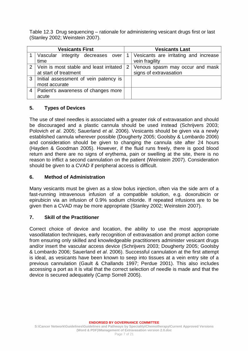
ENDORSED BY GOVERNANCE COMMITTEE
SCancer NetworkGuidelinesGuidelines and Pathways by SpecialityChemotherapyCurrent Approved Versions (Word amp PDF)Management of Extravasation version 20doc
Page 7 of 21
Table 123 Drug sequencing ndash rationale for administering vesicant drugs first or last (Stanley 2002 Weinstein 2007)
Vesicants First Vesicants Last
1 Vascular integrity decreases over time
1 Vesicants are irritating and increase vein fragility
2 Vein is most stable and least irritated at start of treatment
2 Venous spasm may occur and mask signs of extravasation
3 Initial assessment of vein patency is most accurate
4 Patients awareness of changes more acute
5 Types of Devices The use of steel needles is associated with a greater risk of extravasation and should be discouraged and a plastic cannula should be used instead (Schrijvers 2003 Polovich et al 2005 Sauerland et al 2006) Vesicants should be given via a newly established cannula wherever possible (Dougherty 2005 Goolsby amp Lombardo 2006) and consideration should be given to changing the cannula site after 24 hours (Hayden amp Goodman 2005) However if the fluid runs freely there is good blood return and there are no signs of erythema pain or swelling at the site there is no reason to inflict a second cannulation on the patient (Weinstein 2007) Consideration should be given to a CVAD if peripheral access is difficult 6 Method of Administration Many vesicants must be given as a slow bolus injection often via the side arm of a fast-running intravenous infusion of a compatible solution eg doxorubicin or epirubicin via an infusion of 09 sodium chloride If repeated infusions are to be given then a CVAD may be more appropriate (Stanley 2002 Weinstein 2007) 7 Skill of the Practitioner Correct choice of device and location the ability to use the most appropriate vasodilatation techniques early recognition of extravasation and prompt action come from ensuring only skilled and knowledgeable practitioners administer vesicant drugs andor insert the vascular access device (Schrijvers 2003 Dougherty 2005 Goolsby amp Lombardo 2006 Sauerland et al 2006) Successful cannulation at the first attempt is ideal as vesicants have been known to seep into tissues at a vein entry site of a previous cannulation (Gault amp Challands 1997 Perdue 2001) This also includes accessing a port as it is vital that the correct selection of needle is made and that the device is secured adequately (Camp Sorrell 2005)
ENDORSED BY GOVERNANCE COMMITTEE
SCancer NetworkGuidelinesGuidelines and Pathways by SpecialityChemotherapyCurrent Approved Versions (Word amp PDF)Management of Extravasation version 20doc
Page 8 of 21
8 Patient Information Patients should be informed of the potential problems of administering vesicants and the possible consequences of extravasation (Stanley 2002 Sauerland et al 2006 Weinstein 2007) Adequate information given to patients will ensure early recognition and co-operation as patients are the first to notice pain The patient should be urged to report immediately any change in sensation such as burning or stinging (Goolsby amp Lombardo 2006) 9 Drugs Capable of Causing Tissue Necrosis Before administration of vesicant cytotoxic drugs the nurse should know which agents are capable of producing tissue necrosis The following is a list of examples of those in common use
Group A Drugs Group B Drugs
Vinca alkaloids Amsacrine
Vinblastine Carmustine (concentrated solution)
Vindesine Dacarbazine (concentrated solution)
Vinorelbine Dactinomycin
Vincristine Daunorubicin
Vinflunine Doxorubicin
Paclitaxel Epirubicin
Idarubicin
Mithramycin
Mitomycin C
Mechlorethamine
Streptozocin
If in any doubt the drug data sheet should be consulted or reference made to a research trial protocol Drugs should not be reconstituted to give solutions which are higher than the manufacturers recommended concentration and the method of administration should be checked eg infusion injection
ENDORSED BY GOVERNANCE COMMITTEE
SCancer NetworkGuidelinesGuidelines and Pathways by SpecialityChemotherapyCurrent Approved Versions (Word amp PDF)Management of Extravasation version 20doc
Page 9 of 21
A variety of vesicant non-cytotoxic agents in frequent use are also capable of causing severe tissue damage if extravasated Examples include
Group A Drugs Group B Drugs
Calcium chloride Aciclovir
Calcium gluconate Amphotericin
Phenytoin Cefotaxime
Hypertonic solutions eg sodium chloride gt09
Diazepam
Ganciclovir
Sodium bicarbonate (gt5) Potassium chloride (gt40 mmoll)
Glucose 50 Mannitol
This potential hazard should always be remembered The actions listed in this procedure may not be appropriate in all these instances Drug data sheets should always be checked and the pharmacy departments should be consulted if the information is insufficient regarding action to take if a vesicant drug extravasates 10 Signs and Symptoms of Extravasation (see Table 124) Extravasation should be suspected if
1 The patient complains of burning stinging pain or any other acute change at the injection site although this is not always present (Hayden amp Goodman 2005) This should be distinguished from a feeling of cold which may occur with some drugs or venous spasm which can be caused by irritation usually accompanied by pain described as an achiness or tightness (Hayden amp Goodman 2005) Any change of sensation warrants further investigation (Goolsby amp Lombardo 2006)
2 Swelling is the most common symptom (Polovich et al 2005) Induration or leakage may also occur at the injection site Swelling may not always be immediately obvious if the patient has the cannula sited in an area of deep subcutaneous fat in a deep vein or if the leak is via the posterior vein wall (Dougherty 2005)
3 Blanching of the skin occurs (Comerford et al 2002) Erythema can occur around the injection site but this is not usually present immediately (Hayden amp Goodman 2005) It is important that this is distinguished from a lsquoflarersquo reaction (Polovich et al 2005)
4 Blood return is one of the most misleading of all signs particularly related to peripheral devices In peripheral devices if blood return is sluggish or absent this may indicate lack of patency or incorrect position of the device However if no other signs are apparent this should not be regarded as an indication of a non-patent vein as a vein may not bleed back for a number of reasons and extravasation may occur even in the event of good blood return (Hayden amp Goodman 2005) Any change in blood flow should be investigated (Hayden amp Goodman 2005 Weinstein 2007) In CVADs there should always be blood return and if this is absent steps should be followed in order to be able to verify correct tip and needle position or resolve a fibrin sheath (see Figure 451)
ENDORSED BY GOVERNANCE COMMITTEE
SCancer NetworkGuidelinesGuidelines and Pathways by SpecialityChemotherapyCurrent Approved Versions (Word amp PDF)Management of Extravasation version 20doc
Page 10 of 21
5 A resistance is felt on the plunger of the syringe if drugs are given by bolus (Vandergrift 2001 Stanley 2002)
6 There is absence of free flow when administration is by infusion once other reasons have been excluded eg position (Vandergrift 2001 Stanley 2002)
Note one or more of the above may be present If extravasation is suspected or confirmed the injection or infusion must be stopped immediately and action must be taken (Polovich et al 2005 Infusion Nurses Society 2006 Weinstein 2007)
ENDORSED BY GOVERNANCE COMMITTEE
SCancer NetworkGuidelinesGuidelines and Pathways by SpecialityChemotherapyCurrent Approved Versions (Word amp PDF)Management of Extravasation version 20doc Page 11 of 21
Table 124 Nursing assessment of extravasation versus other reactions (Polovich et al 2005 Weinstein 2007)
Extravasation
Assessment parameter
Flare reaction Venous irritation Immediate manifestations ie during drug administration
Delayed manifestations ie from 24 hours after
extravasation
Pain None Aching throbbing sensation along vein and in the limb
Severe stinging or burning pain (not always present) This can last from minutes to hours and will eventually subside Occurs during drug administration at the device site and surrounding areas
Can continue following extravasation or start within 48 hours
Redness Immediate blotches or tracking along the vein This will subside within 30ndash45 minutes with or without treatment (usually steroid cream)
Vein may become red or darkened
Not always present immediately more likely to see blanching of the skin As area becomes inflamed redness will appear around the device site
Later occurrence
Swelling Unlikely Unlikely May occur immediately but may not always be easy to identify immediately
Usually within 48 hours
Blood return Usually present Usually present but may require application of heat to improve blood return
Inability to obtain blood return (peripheral or central) but blood return may be present throughout
Ulceration Unlikely Unlikely Unlikely Can occur within 48ndash96 hours but may take 3ndash4 weeks to develop
Others Urticaria None Change in quality of the infusion or pressure on the syringe
Local tingling and sensory deficits
ENDORSED BY GOVERNANCE COMMITTEE
SCancer NetworkGuidelinesGuidelines and Pathways by SpecialityChemotherapyCurrent Approved Versions (Word amp PDF)Management of Extravasation version 20doc
Page 12 of 21
11 Management of Extravasation The management of extravasation of chemotherapy agents is controversial and there is little documented evidence of efficacy Until recently no antidote has received clear validation in controlled clinical trials and no randomized trials managing cytotoxic extravasation in humans have been completed (Bertolli 1995) Some studies performed on animals have demonstrated both effective and ineffective treatments but extrapolation from animals to humans is limited (Polovich et al 2005 Wickham et al 2006) Other studies have been small with low numbers of patients Another problem is that it is often difficult to ascertain whether any extravasation has actually occurred (Weinstein 2007) Therefore recommendations are based on more consistent experimental evidence cumulative clinical experience from available case reports and uncontrolled studies and empirical guidelines (Bertolli 1995) Drugs where there is evidence of effective management include anthracyclines (Rudolph amp Larson 1987) vinca alkaloids (Bertolli 1995) computed tomography (CT) contrast media (Federle et al 1998) and paclitaxel (Bertolli et al 1997) The management of extravasation involves several stages including the following Stage 1 stopping infusioninjection and aspirating the drug It appears that most authors are agreed that aspirating as much of the drug as possible as soon as extravasation is suspected is beneficial (Rudolph amp Larson 1987 Vandergrift 2001 Polovich et al 2005 Weinstein 2007) and can help lower the concentration of the drug in the area (Goolsby amp Lombardo 2006) However withdrawal is only immediately possible during bolus injections as an infusion would need to be stopped and a syringe attached in an attempt to aspirate Aspiration may be successful if extravasation presents itself as a raised blister but may be unsuccessful if tissue is soft and soggy (CP Pharmaceuticals 1999 Stanley 2002) It may help to reduce the size of the lesion (Vandergrift 2001) In practice it may achieve little and often distresses the patient (Gault amp Challands 1997) The likelihood of withdrawing blood (as suggested by Ignoffo amp Friedman 1980) is small and the practitioner may waste valuable time attempting this which could lead to delay in the rest of the management procedure Stage 2 removing device Some clinicians advocate that the peripheral vascular access device be left in situ in order to instil the antidote via the device and into the affected tissues (Kassner 2000 Stanley 2002 Weinstein 2007) However others recommend that the peripheral device should be removed to prevent any injected solution increasing the size of the affected area (Rudolph amp Larson 1987 CP Pharmaceuticals 1999 Vandergrift 2001) There appears to be no research evidence to support either practice Stage 3 applying hot or cold packs Cooling appears to be a better choice with the exception of the vinca alkaloids and some non-cytotoxic drugs than warming (Bertolli 1995 CP Pharmaceuticals 1999) Cold causes vasoconstriction localizing the extravasation and perhaps allowing time for local vascular and lymphatic systems to contain the drug It should be applied for
ENDORSED BY GOVERNANCE COMMITTEE
SCancer NetworkGuidelinesGuidelines and Pathways by SpecialityChemotherapyCurrent Approved Versions (Word amp PDF)Management of Extravasation version 20doc
Page 13 of 21
15ndash20 minutes 3ndash4 times a day for up to 3 days (Gault amp Challands 1997 CP Pharmaceuticals 1999 Polovich et al 2005) Heat promotes healing after the first 24 hours by increasing the blood supply (Polovich et al 2005 Weinstein 2007) It also decreases local drug concentration increasing the blood flow which results in enhanced resolution of pain and reabsorption of local swelling Stage 4 use of antidotes A number of antidotes are available but again there is a lack of scientific evidence to demonstrate their value and so the role of antidotes is still not clear (Polovich et al 2005) There appear to be two main methods (i) localize and neutralize (using hyaluronidase) (CP Pharmaceuticals 1999) and (ii) spread and dilute (using an antidote) (Stanley 2002) Administration of antidotes if not via the cannula is by the pincushion technique that is instilling small volumes around and over the areas affected using a small gauge (25) needle towards the centre of a clock face The procedure causes considerable discomfort to patients and if large areas are to be tackled analgesia should be considered (Stanley 2002) Hyaluronidase is an enzyme which breaks down hyaluronic acid a normal component of tissue lsquocementrsquo and helps to reduce or prevent tissue damage by allowing rapid diffusion of the extravasated fluid and promoting drug absorption (Few 1987) The usual dose is 1500 IU (Bertolli 1995 Vandergrift 2001) It should be injected within 1 hour of extravasation ideally through the intravenous device delivering the enzyme to the same tissue (Gault amp Challands 1997 Vandergrift 2001 Stanley 2002 Weinstein 2007) NB Hyaluronidase increases the absorption of local anaesthetic Therefore if local anaesthetic has been applied to the area eg Ametop gel prior to cannulation within 6 hours of extravasation then the patient should be monitored for signs and symptoms of systemic anaesthesia such as increased pulse rate and decreased respirations and the doctor informed immediately (BMARPSGB 2008) Corticosteroids have long been advocated as a treatment for anthracycline extravasation in reducing inflammatory components although inflammation is not a prominent feature of tissue necrosis (Camp Sorrell 1998) and they appear to have little benefit Data now discourages the use of locally injected steroids as there is little evidence to support their use (Bertolli 1995 Gault amp Challands 1997 Wickham et al 2006) However given as a cream they can help to reduce local trauma and irritation (Stanley 2002) Dimethyl sulfoxide (DMSO) is a potent free radical scavanger that rapidly penetrates tissues when applied topically (Bertolli 1995) Reports on the clinical use of topical DMSO show it is effective and well tolerated in extravasation (Bertolli 1995) However this is based on a high dose (95) and only 50 is easily available in the UK (Stanley 2002) Side-effects from DMSO include itching erythema mild burning and a characteristic breath odour (Bertolli 1995) Recently dexrazoxane a topoisomerase II catalytic inhibitor used clinically to minimize the cardiotoxicity of doxorubicin has been tested in animal models and a small number of patients for its use in extravasation It is given IV 3ndash6 hours after the extravasation and it appears to reduce the wound size and duration with
ENDORSED BY GOVERNANCE COMMITTEE
SCancer NetworkGuidelinesGuidelines and Pathways by SpecialityChemotherapyCurrent Approved Versions (Word amp PDF)Management of Extravasation version 20doc
Page 14 of 21
anthracyclines The triple dosage appears to be more effective than a single dose (Langer et al 2000 El Saghir et al 2004) A consensus group (Jackson et al 2006) have developed recommendations for the use of dexrazoxane
1 For anthracycline extravasations resulting from peripheral administration the site expert or team should be consulted in order to determine whether the use of dexrazoxane is indicated
2 Absolute indications are if the peripherally extravasated volume exceeds 15 ml and in the event of a central venous extravasation
Finally granulocyte macrophage-colony stimulating factor (GM-CSF) is a growth factor and has been found effective in accelerating wound healing and inducing formation of granulation of tissue (Ulutin et al 2000 El Saghir et al 2004) Stage 5 elevation of limb This is recommended as it minimizes swelling (Rudolph amp Larson 1987) and movement should be encouraged to prevent adhesion of damaged areas to underlying tissue (Gabriel 2008) Stage 6 surgical techniques Some centres suggest that a plastic surgery consultation be performed as part of the management procedure in order to remove the tissue containing the drug Surgical intervention is recommended especially if the lesion is greater than 2 cm there is significant residual pain 1ndash2 weeks after extravasation or there is minimal healing 2ndash3 weeks after injury despite local therapeutic measures (Goolsby amp Lombardo 2006) Liposuction or a flush-out technique can remove extravasated drug without resorting to excision and skin grafting A liposuction cannula can be used to aspirate extravasated material and subcutaneous fat If there is little subcutaneous fat eg preterm infants then the saline flush-out technique is recommended particularly if done within the first 24 hours It has been suggested as a less traumatic and cheaper procedure than surgery Four small stab incisions are made and large volumes of 09 sodium chloride are administered which flush out the extravasated drug (Gault amp Challands 1997) Management of large extravasations from CVADs is usually by surgical intervention and washout of affected tissues 12 Extravasation Kits The use of extravasation kits has been recommended in order to provide immediate management (Khan amp Holmes 2002 Hayden amp Goodman 2005) Kits should be assembled according to the particular needs of individual institutions They should be kept in all areas where staff are regularly administering vesicant drugs so staff have immediate access to equipment (Gabriel 2008) The kit should be simple to avoid confusion but comprehensive enough to meet all reasonable needs (Stanley 2002) (see Procedure guidelines Extravasation management peripheral cannula below) Instructions should be clear and easy to follow and the use of a flow chart enables staff to follow the management procedure in easy steps (Figure 124)
ENDORSED BY GOVERNANCE COMMITTEE
SCancer NetworkGuidelinesGuidelines and Pathways by SpecialityChemotherapyCurrent Approved Versions (Word amp PDF)Management of Extravasation version 20doc
Page 15 of 21
13 Mixed Vesicant Extravasation Consideration should be given to the management of mixed vesicant drug extravasation in terms of which drug to treat with which antidote It has been recommended to act in accordance with the drug which possesses the most deleterious properties (How amp Brown 1998) 14 Informing the Patient Patients should always be informed when an extravasation has occurred and be given an explanation of what has happened and what management has been carried out (McCaffrey Boyle amp Engelking 1995) An information sheet should be given to patients with instructions of what symptoms to look out for and when to contact the hospital during the follow-up period (Gabriel 2008) 15 Wound Management Damage will be affected by the site amount of drug concentration of the agent and if it binds to DNA or not (Polovich et al 2005) Ulceration may occur over a period of days to weeks and extravasation wounds may be complicated by tissue ischaemia related to endothelial damage (Naylor 2005) The type of injury will dictate the type of dressing Assessment of the wound should include position and size of the wound amount and type of tissue present amount and type of exudate and extent and spread of erythema (Naylor 2005) 16 Documentation and Follow Up An extravasation must be reported and fully documented as it is an accident and the patient may require follow-up care (NMC 2005 RCN 2005) Information may also be used for statistical purposes for example collation and analysis using the green card scheme devised by St Chads Hospital Birmingham (Stanley 2002) Statistics on the incidence degree causes and corrective action should be monitored and analysed (Infusion Nurses Society 2006) Finally it may be required in case of litigation which is now on the increase (Dougherty 2003 Masoorli 2003)
ENDORSED BY GOVERNANCE COMMITTEE
SCancer NetworkGuidelinesGuidelines and Pathways by SpecialityChemotherapyCurrent Approved Versions (Word amp PDF)Management of Extravasation version 20doc
Page 16 of 21
Figure
ENDORSED BY GOVERNANCE COMMITTEE
SCancer NetworkGuidelinesGuidelines and Pathways by SpecialityChemotherapyCurrent Approved Versions (Word amp PDF)Management of Extravasation version 20doc
Page 17 of 21
Equipment To assist the nurse an extravasation kit should be assembled and should be readily available in each wardunit It contains
1 Gel packs times 2 one to be kept in the fridge and one available for heating (an electric heating blanket can be used whilst pack is heating)
2 Hyaluronidase 1500 IU2 ml sterile water 3 Hydrocortisone cream 1 15 g tube times 1 4 2 ml syringes times 1 5 25 G needles times 2 6 Alcohol swabs 7 Documentation forms 8 Copy of extravasation management procedure 9 Patient information leaflet
Procedure
Action Rationale
1 Explain and discuss the procedure with the patient
To ensure that the patient understands the procedure and gives hisher valid consent (DH 2002a C NMC 2008b C)
2 Stop injection or infusion immediately leaving the cannula in place
To minimize local injury To allow aspiration of the drug to be attempted (Polovich et al 2005 C RCN 2005 C)
3 Aspirate any residual drug from the device and suspected extravasation site
To minimise local injury by removing as much drug as possible and only attempt if appropriate Subsequent damage is related to the volume of the extravasation in addition to other factors (Polovich et al 2005 C RCN 2005 C)
4 Remove the cannula To prevent the device from being used for antidote administration (Rudolph amp Larson 1987 E)
5 Collect the extravasation pack and take it to the patient
It contains all the equipment necessary for managing extravasation (Stanley 2002 E Dougherty 2005 E)
ENDORSED BY GOVERNANCE COMMITTEE
SCancer NetworkGuidelinesGuidelines and Pathways by SpecialityChemotherapyCurrent Approved Versions (Word amp PDF)Management of Extravasation version 20doc
Page 18 of 21
Group A Drugs
Draw up hyaluronidase 1500 IU in 1 ml water for injection and inject volumes of 01ndash02 ml subcutaneously at points of the compass around the circumference of the area of extravasation
This is the recommended agent for group A drugs The warm pack speeds up absorption of the drug by the tissues (Bertolli 1995 C)
Apply warm pack
Group B Drugs
Apply cold pack or ice instantly To localize the area of extravasation slow cell metabolism and decrease the area of tissue destruction To reduce local pain (Polovich et al 2005 C)
However if extravasation is with any of the following category B drugs mitomycin C doxorubicin idarubicin epirubicin actinomycin D then
This is the recommended agent for these anthracyclines and helps to reduce local tissue damage (Bertolli 1995 C)
Draw around area of extravasation with indelible pen
Put on gloves
Apply thin layer of DMSO topically to the marked area using the small plastic spatula in lid of the bottle
Allow it to dry
Apply gauze
This should be applied within 10ndash25 minutes
Where possible elevate the extremity andor encourage movement
To minimize swelling and to prevent adhesion of damaged area to underlying tissue which could result in restriction of movement (Rudolph amp Larson 1987 E)
Inform a member of the medical staff at the earliest opportunity and administer any other prescribed antidotes eg dexrazoxane
To enable actions differing from agreed policy to be taken if considered in the best interests of the patient To notify the doctor of the need to prescribe any other drugs (E)
Apply hydrocortisone cream 1 twice daily and instruct the patient how to do this Continue as long as erythema persists
To reduce local inflammation and promote patient comfort (Stanley 2002 E)
Where appropriate apply DMSO every 2 hours on day 1 and then every 6 hours for up to 7 days (patients will need to have this prescribed as a to take out [TTO] and continue treatment at home where necessary)
To help reduce local tissue damage (Bertolli 1995 C)
ENDORSED BY GOVERNANCE COMMITTEE
SCancer NetworkGuidelinesGuidelines and Pathways by SpecialityChemotherapyCurrent Approved Versions (Word amp PDF)Management of Extravasation version 20doc
Page 19 of 21
Heat packs (group A drugs) should be reapplied after initial management for 2ndash4 hours Cold packs (group B drugs) should be applied for 15ndash20 minutes 3ndash4 times a day for up to 3 days
To localize the steroid effect in the area of extravasation To reduce local pain and promote patient comfort (Bertolli 1995 C)
Provide analgesia as required To promote patient comfort To encourage movement of the limb as advised (E)
Document the following details in duplicate on the form provided
a) Patients namenumber
b) Wardunit
c) Date time
d) Signs and symptoms
e) Venepuncture site (on diagram)
f) Drug sequence
g) Drug administration technique ie lsquobolusrsquo or infusion
h) Approximate amount of the drug extravasated
i) Diameter length and width of extravasation area
j) Appearance of the area
k) Step-by-step management with date and time of each step performed and medical officer notification
To provide an immediate full record of all details of the incident which may be referred to if necessary To provide a baseline for future observation and monitoring of patients condition To comply with NMC guidelines (Kassner 2000 E NMC 2005 C RCN 2005 C Weinstein 2007 E)
l) Patients complaints comments statements
m) Indication that patients information sheet given to patient
n) Follow-up section
o) Whether photograph was taken
p) If required when patient referred to plastic surgeon
q) Signature of the nurse
Explain to the patient that the site may remain sore for several days
To reduce anxiety and ensure continued co-operation (P E)
Observe the area regularly for erythema induration blistering or necrosis Inpatients monitor daily Where appropriate take photograph
To detect any changes at the earliest possible moment (RCN 2005 C)
ENDORSED BY GOVERNANCE COMMITTEE
SCancer NetworkGuidelinesGuidelines and Pathways by SpecialityChemotherapyCurrent Approved Versions (Word amp PDF)Management of Extravasation version 20doc
Page 20 of 21
All patients should receive written information explaining what has occurred what management has been carried out what they need to look for at the site and when to report any changes For example increased discomfort peeling or blistering of the skin should be reported immediately
To detect any changes as early as possible and allow for a review of future management This may include referral to a plastic surgeon (Gault amp Challands 1997 E Polovich et al 2005 C RCN 2005 C)
If blistering or tissue breakdown occurs begin dressing techniques and seek advice regarding wound management
To minimize the risk of a superimposed infection and sterile increase healing (Naylor 2005 E)
Depending on size of lesion degree of pain type of drug refer to plastic surgeon
To prevent further pain or other complications as chemically induced ulcers rarely heal spontaneously (Polovich et al 2005 C Dougherty 2005 E)
Monitoring of the Guideline Implementation of the guidance will be considered as a topic for audit by the NSSG in and reviewed in Authors Andrew Stanley Network Pharmacist References
The Royal Marsden Hospital Manual of Clinical Nursing Procedures 7th Edition Blackwell Publishing (2008)
ENDORSED BY GOVERNANCE COMMITTEE
SCancer NetworkGuidelinesGuidelines and Pathways by SpecialityChemotherapyCurrent Approved Versions (Word amp PDF)Management of Extravasation version 20doc
Page 21 of 21
Approval Signatures Pan Birmingham Cancer Network Governance Committee Chair Name Doug Wullf
Signature Date July 2011 Pan Birmingham Cancer Network Manager Name Karen Metcalf
Signature Date July 2011 Network Site Specific Group Clinical Chair Name Frances Shaw
Signature Date July 2011
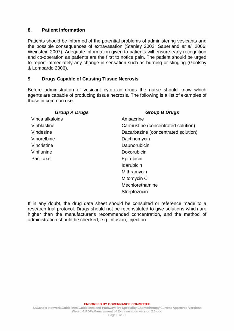
ENDORSED BY GOVERNANCE COMMITTEE
SCancer NetworkGuidelinesGuidelines and Pathways by SpecialityChemotherapyCurrent Approved Versions (Word amp PDF)Management of Extravasation version 20doc
Page 8 of 21
8 Patient Information Patients should be informed of the potential problems of administering vesicants and the possible consequences of extravasation (Stanley 2002 Sauerland et al 2006 Weinstein 2007) Adequate information given to patients will ensure early recognition and co-operation as patients are the first to notice pain The patient should be urged to report immediately any change in sensation such as burning or stinging (Goolsby amp Lombardo 2006) 9 Drugs Capable of Causing Tissue Necrosis Before administration of vesicant cytotoxic drugs the nurse should know which agents are capable of producing tissue necrosis The following is a list of examples of those in common use
Group A Drugs Group B Drugs
Vinca alkaloids Amsacrine
Vinblastine Carmustine (concentrated solution)
Vindesine Dacarbazine (concentrated solution)
Vinorelbine Dactinomycin
Vincristine Daunorubicin
Vinflunine Doxorubicin
Paclitaxel Epirubicin
Idarubicin
Mithramycin
Mitomycin C
Mechlorethamine
Streptozocin
If in any doubt the drug data sheet should be consulted or reference made to a research trial protocol Drugs should not be reconstituted to give solutions which are higher than the manufacturers recommended concentration and the method of administration should be checked eg infusion injection
ENDORSED BY GOVERNANCE COMMITTEE
SCancer NetworkGuidelinesGuidelines and Pathways by SpecialityChemotherapyCurrent Approved Versions (Word amp PDF)Management of Extravasation version 20doc
Page 9 of 21
A variety of vesicant non-cytotoxic agents in frequent use are also capable of causing severe tissue damage if extravasated Examples include
Group A Drugs Group B Drugs
Calcium chloride Aciclovir
Calcium gluconate Amphotericin
Phenytoin Cefotaxime
Hypertonic solutions eg sodium chloride gt09
Diazepam
Ganciclovir
Sodium bicarbonate (gt5) Potassium chloride (gt40 mmoll)
Glucose 50 Mannitol
This potential hazard should always be remembered The actions listed in this procedure may not be appropriate in all these instances Drug data sheets should always be checked and the pharmacy departments should be consulted if the information is insufficient regarding action to take if a vesicant drug extravasates 10 Signs and Symptoms of Extravasation (see Table 124) Extravasation should be suspected if
1 The patient complains of burning stinging pain or any other acute change at the injection site although this is not always present (Hayden amp Goodman 2005) This should be distinguished from a feeling of cold which may occur with some drugs or venous spasm which can be caused by irritation usually accompanied by pain described as an achiness or tightness (Hayden amp Goodman 2005) Any change of sensation warrants further investigation (Goolsby amp Lombardo 2006)
2 Swelling is the most common symptom (Polovich et al 2005) Induration or leakage may also occur at the injection site Swelling may not always be immediately obvious if the patient has the cannula sited in an area of deep subcutaneous fat in a deep vein or if the leak is via the posterior vein wall (Dougherty 2005)
3 Blanching of the skin occurs (Comerford et al 2002) Erythema can occur around the injection site but this is not usually present immediately (Hayden amp Goodman 2005) It is important that this is distinguished from a lsquoflarersquo reaction (Polovich et al 2005)
4 Blood return is one of the most misleading of all signs particularly related to peripheral devices In peripheral devices if blood return is sluggish or absent this may indicate lack of patency or incorrect position of the device However if no other signs are apparent this should not be regarded as an indication of a non-patent vein as a vein may not bleed back for a number of reasons and extravasation may occur even in the event of good blood return (Hayden amp Goodman 2005) Any change in blood flow should be investigated (Hayden amp Goodman 2005 Weinstein 2007) In CVADs there should always be blood return and if this is absent steps should be followed in order to be able to verify correct tip and needle position or resolve a fibrin sheath (see Figure 451)
ENDORSED BY GOVERNANCE COMMITTEE
SCancer NetworkGuidelinesGuidelines and Pathways by SpecialityChemotherapyCurrent Approved Versions (Word amp PDF)Management of Extravasation version 20doc
Page 10 of 21
5 A resistance is felt on the plunger of the syringe if drugs are given by bolus (Vandergrift 2001 Stanley 2002)
6 There is absence of free flow when administration is by infusion once other reasons have been excluded eg position (Vandergrift 2001 Stanley 2002)
Note one or more of the above may be present If extravasation is suspected or confirmed the injection or infusion must be stopped immediately and action must be taken (Polovich et al 2005 Infusion Nurses Society 2006 Weinstein 2007)
ENDORSED BY GOVERNANCE COMMITTEE
SCancer NetworkGuidelinesGuidelines and Pathways by SpecialityChemotherapyCurrent Approved Versions (Word amp PDF)Management of Extravasation version 20doc Page 11 of 21
Table 124 Nursing assessment of extravasation versus other reactions (Polovich et al 2005 Weinstein 2007)
Extravasation
Assessment parameter
Flare reaction Venous irritation Immediate manifestations ie during drug administration
Delayed manifestations ie from 24 hours after
extravasation
Pain None Aching throbbing sensation along vein and in the limb
Severe stinging or burning pain (not always present) This can last from minutes to hours and will eventually subside Occurs during drug administration at the device site and surrounding areas
Can continue following extravasation or start within 48 hours
Redness Immediate blotches or tracking along the vein This will subside within 30ndash45 minutes with or without treatment (usually steroid cream)
Vein may become red or darkened
Not always present immediately more likely to see blanching of the skin As area becomes inflamed redness will appear around the device site
Later occurrence
Swelling Unlikely Unlikely May occur immediately but may not always be easy to identify immediately
Usually within 48 hours
Blood return Usually present Usually present but may require application of heat to improve blood return
Inability to obtain blood return (peripheral or central) but blood return may be present throughout
Ulceration Unlikely Unlikely Unlikely Can occur within 48ndash96 hours but may take 3ndash4 weeks to develop
Others Urticaria None Change in quality of the infusion or pressure on the syringe
Local tingling and sensory deficits
ENDORSED BY GOVERNANCE COMMITTEE
SCancer NetworkGuidelinesGuidelines and Pathways by SpecialityChemotherapyCurrent Approved Versions (Word amp PDF)Management of Extravasation version 20doc
Page 12 of 21
11 Management of Extravasation The management of extravasation of chemotherapy agents is controversial and there is little documented evidence of efficacy Until recently no antidote has received clear validation in controlled clinical trials and no randomized trials managing cytotoxic extravasation in humans have been completed (Bertolli 1995) Some studies performed on animals have demonstrated both effective and ineffective treatments but extrapolation from animals to humans is limited (Polovich et al 2005 Wickham et al 2006) Other studies have been small with low numbers of patients Another problem is that it is often difficult to ascertain whether any extravasation has actually occurred (Weinstein 2007) Therefore recommendations are based on more consistent experimental evidence cumulative clinical experience from available case reports and uncontrolled studies and empirical guidelines (Bertolli 1995) Drugs where there is evidence of effective management include anthracyclines (Rudolph amp Larson 1987) vinca alkaloids (Bertolli 1995) computed tomography (CT) contrast media (Federle et al 1998) and paclitaxel (Bertolli et al 1997) The management of extravasation involves several stages including the following Stage 1 stopping infusioninjection and aspirating the drug It appears that most authors are agreed that aspirating as much of the drug as possible as soon as extravasation is suspected is beneficial (Rudolph amp Larson 1987 Vandergrift 2001 Polovich et al 2005 Weinstein 2007) and can help lower the concentration of the drug in the area (Goolsby amp Lombardo 2006) However withdrawal is only immediately possible during bolus injections as an infusion would need to be stopped and a syringe attached in an attempt to aspirate Aspiration may be successful if extravasation presents itself as a raised blister but may be unsuccessful if tissue is soft and soggy (CP Pharmaceuticals 1999 Stanley 2002) It may help to reduce the size of the lesion (Vandergrift 2001) In practice it may achieve little and often distresses the patient (Gault amp Challands 1997) The likelihood of withdrawing blood (as suggested by Ignoffo amp Friedman 1980) is small and the practitioner may waste valuable time attempting this which could lead to delay in the rest of the management procedure Stage 2 removing device Some clinicians advocate that the peripheral vascular access device be left in situ in order to instil the antidote via the device and into the affected tissues (Kassner 2000 Stanley 2002 Weinstein 2007) However others recommend that the peripheral device should be removed to prevent any injected solution increasing the size of the affected area (Rudolph amp Larson 1987 CP Pharmaceuticals 1999 Vandergrift 2001) There appears to be no research evidence to support either practice Stage 3 applying hot or cold packs Cooling appears to be a better choice with the exception of the vinca alkaloids and some non-cytotoxic drugs than warming (Bertolli 1995 CP Pharmaceuticals 1999) Cold causes vasoconstriction localizing the extravasation and perhaps allowing time for local vascular and lymphatic systems to contain the drug It should be applied for
ENDORSED BY GOVERNANCE COMMITTEE
SCancer NetworkGuidelinesGuidelines and Pathways by SpecialityChemotherapyCurrent Approved Versions (Word amp PDF)Management of Extravasation version 20doc
Page 13 of 21
15ndash20 minutes 3ndash4 times a day for up to 3 days (Gault amp Challands 1997 CP Pharmaceuticals 1999 Polovich et al 2005) Heat promotes healing after the first 24 hours by increasing the blood supply (Polovich et al 2005 Weinstein 2007) It also decreases local drug concentration increasing the blood flow which results in enhanced resolution of pain and reabsorption of local swelling Stage 4 use of antidotes A number of antidotes are available but again there is a lack of scientific evidence to demonstrate their value and so the role of antidotes is still not clear (Polovich et al 2005) There appear to be two main methods (i) localize and neutralize (using hyaluronidase) (CP Pharmaceuticals 1999) and (ii) spread and dilute (using an antidote) (Stanley 2002) Administration of antidotes if not via the cannula is by the pincushion technique that is instilling small volumes around and over the areas affected using a small gauge (25) needle towards the centre of a clock face The procedure causes considerable discomfort to patients and if large areas are to be tackled analgesia should be considered (Stanley 2002) Hyaluronidase is an enzyme which breaks down hyaluronic acid a normal component of tissue lsquocementrsquo and helps to reduce or prevent tissue damage by allowing rapid diffusion of the extravasated fluid and promoting drug absorption (Few 1987) The usual dose is 1500 IU (Bertolli 1995 Vandergrift 2001) It should be injected within 1 hour of extravasation ideally through the intravenous device delivering the enzyme to the same tissue (Gault amp Challands 1997 Vandergrift 2001 Stanley 2002 Weinstein 2007) NB Hyaluronidase increases the absorption of local anaesthetic Therefore if local anaesthetic has been applied to the area eg Ametop gel prior to cannulation within 6 hours of extravasation then the patient should be monitored for signs and symptoms of systemic anaesthesia such as increased pulse rate and decreased respirations and the doctor informed immediately (BMARPSGB 2008) Corticosteroids have long been advocated as a treatment for anthracycline extravasation in reducing inflammatory components although inflammation is not a prominent feature of tissue necrosis (Camp Sorrell 1998) and they appear to have little benefit Data now discourages the use of locally injected steroids as there is little evidence to support their use (Bertolli 1995 Gault amp Challands 1997 Wickham et al 2006) However given as a cream they can help to reduce local trauma and irritation (Stanley 2002) Dimethyl sulfoxide (DMSO) is a potent free radical scavanger that rapidly penetrates tissues when applied topically (Bertolli 1995) Reports on the clinical use of topical DMSO show it is effective and well tolerated in extravasation (Bertolli 1995) However this is based on a high dose (95) and only 50 is easily available in the UK (Stanley 2002) Side-effects from DMSO include itching erythema mild burning and a characteristic breath odour (Bertolli 1995) Recently dexrazoxane a topoisomerase II catalytic inhibitor used clinically to minimize the cardiotoxicity of doxorubicin has been tested in animal models and a small number of patients for its use in extravasation It is given IV 3ndash6 hours after the extravasation and it appears to reduce the wound size and duration with
ENDORSED BY GOVERNANCE COMMITTEE
SCancer NetworkGuidelinesGuidelines and Pathways by SpecialityChemotherapyCurrent Approved Versions (Word amp PDF)Management of Extravasation version 20doc
Page 14 of 21
anthracyclines The triple dosage appears to be more effective than a single dose (Langer et al 2000 El Saghir et al 2004) A consensus group (Jackson et al 2006) have developed recommendations for the use of dexrazoxane
1 For anthracycline extravasations resulting from peripheral administration the site expert or team should be consulted in order to determine whether the use of dexrazoxane is indicated
2 Absolute indications are if the peripherally extravasated volume exceeds 15 ml and in the event of a central venous extravasation
Finally granulocyte macrophage-colony stimulating factor (GM-CSF) is a growth factor and has been found effective in accelerating wound healing and inducing formation of granulation of tissue (Ulutin et al 2000 El Saghir et al 2004) Stage 5 elevation of limb This is recommended as it minimizes swelling (Rudolph amp Larson 1987) and movement should be encouraged to prevent adhesion of damaged areas to underlying tissue (Gabriel 2008) Stage 6 surgical techniques Some centres suggest that a plastic surgery consultation be performed as part of the management procedure in order to remove the tissue containing the drug Surgical intervention is recommended especially if the lesion is greater than 2 cm there is significant residual pain 1ndash2 weeks after extravasation or there is minimal healing 2ndash3 weeks after injury despite local therapeutic measures (Goolsby amp Lombardo 2006) Liposuction or a flush-out technique can remove extravasated drug without resorting to excision and skin grafting A liposuction cannula can be used to aspirate extravasated material and subcutaneous fat If there is little subcutaneous fat eg preterm infants then the saline flush-out technique is recommended particularly if done within the first 24 hours It has been suggested as a less traumatic and cheaper procedure than surgery Four small stab incisions are made and large volumes of 09 sodium chloride are administered which flush out the extravasated drug (Gault amp Challands 1997) Management of large extravasations from CVADs is usually by surgical intervention and washout of affected tissues 12 Extravasation Kits The use of extravasation kits has been recommended in order to provide immediate management (Khan amp Holmes 2002 Hayden amp Goodman 2005) Kits should be assembled according to the particular needs of individual institutions They should be kept in all areas where staff are regularly administering vesicant drugs so staff have immediate access to equipment (Gabriel 2008) The kit should be simple to avoid confusion but comprehensive enough to meet all reasonable needs (Stanley 2002) (see Procedure guidelines Extravasation management peripheral cannula below) Instructions should be clear and easy to follow and the use of a flow chart enables staff to follow the management procedure in easy steps (Figure 124)
ENDORSED BY GOVERNANCE COMMITTEE
SCancer NetworkGuidelinesGuidelines and Pathways by SpecialityChemotherapyCurrent Approved Versions (Word amp PDF)Management of Extravasation version 20doc
Page 15 of 21
13 Mixed Vesicant Extravasation Consideration should be given to the management of mixed vesicant drug extravasation in terms of which drug to treat with which antidote It has been recommended to act in accordance with the drug which possesses the most deleterious properties (How amp Brown 1998) 14 Informing the Patient Patients should always be informed when an extravasation has occurred and be given an explanation of what has happened and what management has been carried out (McCaffrey Boyle amp Engelking 1995) An information sheet should be given to patients with instructions of what symptoms to look out for and when to contact the hospital during the follow-up period (Gabriel 2008) 15 Wound Management Damage will be affected by the site amount of drug concentration of the agent and if it binds to DNA or not (Polovich et al 2005) Ulceration may occur over a period of days to weeks and extravasation wounds may be complicated by tissue ischaemia related to endothelial damage (Naylor 2005) The type of injury will dictate the type of dressing Assessment of the wound should include position and size of the wound amount and type of tissue present amount and type of exudate and extent and spread of erythema (Naylor 2005) 16 Documentation and Follow Up An extravasation must be reported and fully documented as it is an accident and the patient may require follow-up care (NMC 2005 RCN 2005) Information may also be used for statistical purposes for example collation and analysis using the green card scheme devised by St Chads Hospital Birmingham (Stanley 2002) Statistics on the incidence degree causes and corrective action should be monitored and analysed (Infusion Nurses Society 2006) Finally it may be required in case of litigation which is now on the increase (Dougherty 2003 Masoorli 2003)
ENDORSED BY GOVERNANCE COMMITTEE
SCancer NetworkGuidelinesGuidelines and Pathways by SpecialityChemotherapyCurrent Approved Versions (Word amp PDF)Management of Extravasation version 20doc
Page 16 of 21
Figure
ENDORSED BY GOVERNANCE COMMITTEE
SCancer NetworkGuidelinesGuidelines and Pathways by SpecialityChemotherapyCurrent Approved Versions (Word amp PDF)Management of Extravasation version 20doc
Page 17 of 21
Equipment To assist the nurse an extravasation kit should be assembled and should be readily available in each wardunit It contains
1 Gel packs times 2 one to be kept in the fridge and one available for heating (an electric heating blanket can be used whilst pack is heating)
2 Hyaluronidase 1500 IU2 ml sterile water 3 Hydrocortisone cream 1 15 g tube times 1 4 2 ml syringes times 1 5 25 G needles times 2 6 Alcohol swabs 7 Documentation forms 8 Copy of extravasation management procedure 9 Patient information leaflet
Procedure
Action Rationale
1 Explain and discuss the procedure with the patient
To ensure that the patient understands the procedure and gives hisher valid consent (DH 2002a C NMC 2008b C)
2 Stop injection or infusion immediately leaving the cannula in place
To minimize local injury To allow aspiration of the drug to be attempted (Polovich et al 2005 C RCN 2005 C)
3 Aspirate any residual drug from the device and suspected extravasation site
To minimise local injury by removing as much drug as possible and only attempt if appropriate Subsequent damage is related to the volume of the extravasation in addition to other factors (Polovich et al 2005 C RCN 2005 C)
4 Remove the cannula To prevent the device from being used for antidote administration (Rudolph amp Larson 1987 E)
5 Collect the extravasation pack and take it to the patient
It contains all the equipment necessary for managing extravasation (Stanley 2002 E Dougherty 2005 E)
ENDORSED BY GOVERNANCE COMMITTEE
SCancer NetworkGuidelinesGuidelines and Pathways by SpecialityChemotherapyCurrent Approved Versions (Word amp PDF)Management of Extravasation version 20doc
Page 18 of 21
Group A Drugs
Draw up hyaluronidase 1500 IU in 1 ml water for injection and inject volumes of 01ndash02 ml subcutaneously at points of the compass around the circumference of the area of extravasation
This is the recommended agent for group A drugs The warm pack speeds up absorption of the drug by the tissues (Bertolli 1995 C)
Apply warm pack
Group B Drugs
Apply cold pack or ice instantly To localize the area of extravasation slow cell metabolism and decrease the area of tissue destruction To reduce local pain (Polovich et al 2005 C)
However if extravasation is with any of the following category B drugs mitomycin C doxorubicin idarubicin epirubicin actinomycin D then
This is the recommended agent for these anthracyclines and helps to reduce local tissue damage (Bertolli 1995 C)
Draw around area of extravasation with indelible pen
Put on gloves
Apply thin layer of DMSO topically to the marked area using the small plastic spatula in lid of the bottle
Allow it to dry
Apply gauze
This should be applied within 10ndash25 minutes
Where possible elevate the extremity andor encourage movement
To minimize swelling and to prevent adhesion of damaged area to underlying tissue which could result in restriction of movement (Rudolph amp Larson 1987 E)
Inform a member of the medical staff at the earliest opportunity and administer any other prescribed antidotes eg dexrazoxane
To enable actions differing from agreed policy to be taken if considered in the best interests of the patient To notify the doctor of the need to prescribe any other drugs (E)
Apply hydrocortisone cream 1 twice daily and instruct the patient how to do this Continue as long as erythema persists
To reduce local inflammation and promote patient comfort (Stanley 2002 E)
Where appropriate apply DMSO every 2 hours on day 1 and then every 6 hours for up to 7 days (patients will need to have this prescribed as a to take out [TTO] and continue treatment at home where necessary)
To help reduce local tissue damage (Bertolli 1995 C)
ENDORSED BY GOVERNANCE COMMITTEE
SCancer NetworkGuidelinesGuidelines and Pathways by SpecialityChemotherapyCurrent Approved Versions (Word amp PDF)Management of Extravasation version 20doc
Page 19 of 21
Heat packs (group A drugs) should be reapplied after initial management for 2ndash4 hours Cold packs (group B drugs) should be applied for 15ndash20 minutes 3ndash4 times a day for up to 3 days
To localize the steroid effect in the area of extravasation To reduce local pain and promote patient comfort (Bertolli 1995 C)
Provide analgesia as required To promote patient comfort To encourage movement of the limb as advised (E)
Document the following details in duplicate on the form provided
a) Patients namenumber
b) Wardunit
c) Date time
d) Signs and symptoms
e) Venepuncture site (on diagram)
f) Drug sequence
g) Drug administration technique ie lsquobolusrsquo or infusion
h) Approximate amount of the drug extravasated
i) Diameter length and width of extravasation area
j) Appearance of the area
k) Step-by-step management with date and time of each step performed and medical officer notification
To provide an immediate full record of all details of the incident which may be referred to if necessary To provide a baseline for future observation and monitoring of patients condition To comply with NMC guidelines (Kassner 2000 E NMC 2005 C RCN 2005 C Weinstein 2007 E)
l) Patients complaints comments statements
m) Indication that patients information sheet given to patient
n) Follow-up section
o) Whether photograph was taken
p) If required when patient referred to plastic surgeon
q) Signature of the nurse
Explain to the patient that the site may remain sore for several days
To reduce anxiety and ensure continued co-operation (P E)
Observe the area regularly for erythema induration blistering or necrosis Inpatients monitor daily Where appropriate take photograph
To detect any changes at the earliest possible moment (RCN 2005 C)
ENDORSED BY GOVERNANCE COMMITTEE
SCancer NetworkGuidelinesGuidelines and Pathways by SpecialityChemotherapyCurrent Approved Versions (Word amp PDF)Management of Extravasation version 20doc
Page 20 of 21
All patients should receive written information explaining what has occurred what management has been carried out what they need to look for at the site and when to report any changes For example increased discomfort peeling or blistering of the skin should be reported immediately
To detect any changes as early as possible and allow for a review of future management This may include referral to a plastic surgeon (Gault amp Challands 1997 E Polovich et al 2005 C RCN 2005 C)
If blistering or tissue breakdown occurs begin dressing techniques and seek advice regarding wound management
To minimize the risk of a superimposed infection and sterile increase healing (Naylor 2005 E)
Depending on size of lesion degree of pain type of drug refer to plastic surgeon
To prevent further pain or other complications as chemically induced ulcers rarely heal spontaneously (Polovich et al 2005 C Dougherty 2005 E)
Monitoring of the Guideline Implementation of the guidance will be considered as a topic for audit by the NSSG in and reviewed in Authors Andrew Stanley Network Pharmacist References
The Royal Marsden Hospital Manual of Clinical Nursing Procedures 7th Edition Blackwell Publishing (2008)
ENDORSED BY GOVERNANCE COMMITTEE
SCancer NetworkGuidelinesGuidelines and Pathways by SpecialityChemotherapyCurrent Approved Versions (Word amp PDF)Management of Extravasation version 20doc
Page 21 of 21
Approval Signatures Pan Birmingham Cancer Network Governance Committee Chair Name Doug Wullf
Signature Date July 2011 Pan Birmingham Cancer Network Manager Name Karen Metcalf
Signature Date July 2011 Network Site Specific Group Clinical Chair Name Frances Shaw
Signature Date July 2011
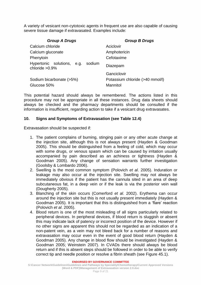
ENDORSED BY GOVERNANCE COMMITTEE
SCancer NetworkGuidelinesGuidelines and Pathways by SpecialityChemotherapyCurrent Approved Versions (Word amp PDF)Management of Extravasation version 20doc
Page 9 of 21
A variety of vesicant non-cytotoxic agents in frequent use are also capable of causing severe tissue damage if extravasated Examples include
Group A Drugs Group B Drugs
Calcium chloride Aciclovir
Calcium gluconate Amphotericin
Phenytoin Cefotaxime
Hypertonic solutions eg sodium chloride gt09
Diazepam
Ganciclovir
Sodium bicarbonate (gt5) Potassium chloride (gt40 mmoll)
Glucose 50 Mannitol
This potential hazard should always be remembered The actions listed in this procedure may not be appropriate in all these instances Drug data sheets should always be checked and the pharmacy departments should be consulted if the information is insufficient regarding action to take if a vesicant drug extravasates 10 Signs and Symptoms of Extravasation (see Table 124) Extravasation should be suspected if
1 The patient complains of burning stinging pain or any other acute change at the injection site although this is not always present (Hayden amp Goodman 2005) This should be distinguished from a feeling of cold which may occur with some drugs or venous spasm which can be caused by irritation usually accompanied by pain described as an achiness or tightness (Hayden amp Goodman 2005) Any change of sensation warrants further investigation (Goolsby amp Lombardo 2006)
2 Swelling is the most common symptom (Polovich et al 2005) Induration or leakage may also occur at the injection site Swelling may not always be immediately obvious if the patient has the cannula sited in an area of deep subcutaneous fat in a deep vein or if the leak is via the posterior vein wall (Dougherty 2005)
3 Blanching of the skin occurs (Comerford et al 2002) Erythema can occur around the injection site but this is not usually present immediately (Hayden amp Goodman 2005) It is important that this is distinguished from a lsquoflarersquo reaction (Polovich et al 2005)
4 Blood return is one of the most misleading of all signs particularly related to peripheral devices In peripheral devices if blood return is sluggish or absent this may indicate lack of patency or incorrect position of the device However if no other signs are apparent this should not be regarded as an indication of a non-patent vein as a vein may not bleed back for a number of reasons and extravasation may occur even in the event of good blood return (Hayden amp Goodman 2005) Any change in blood flow should be investigated (Hayden amp Goodman 2005 Weinstein 2007) In CVADs there should always be blood return and if this is absent steps should be followed in order to be able to verify correct tip and needle position or resolve a fibrin sheath (see Figure 451)
ENDORSED BY GOVERNANCE COMMITTEE
SCancer NetworkGuidelinesGuidelines and Pathways by SpecialityChemotherapyCurrent Approved Versions (Word amp PDF)Management of Extravasation version 20doc
Page 10 of 21
5 A resistance is felt on the plunger of the syringe if drugs are given by bolus (Vandergrift 2001 Stanley 2002)
6 There is absence of free flow when administration is by infusion once other reasons have been excluded eg position (Vandergrift 2001 Stanley 2002)
Note one or more of the above may be present If extravasation is suspected or confirmed the injection or infusion must be stopped immediately and action must be taken (Polovich et al 2005 Infusion Nurses Society 2006 Weinstein 2007)
ENDORSED BY GOVERNANCE COMMITTEE
SCancer NetworkGuidelinesGuidelines and Pathways by SpecialityChemotherapyCurrent Approved Versions (Word amp PDF)Management of Extravasation version 20doc Page 11 of 21
Table 124 Nursing assessment of extravasation versus other reactions (Polovich et al 2005 Weinstein 2007)
Extravasation
Assessment parameter
Flare reaction Venous irritation Immediate manifestations ie during drug administration
Delayed manifestations ie from 24 hours after
extravasation
Pain None Aching throbbing sensation along vein and in the limb
Severe stinging or burning pain (not always present) This can last from minutes to hours and will eventually subside Occurs during drug administration at the device site and surrounding areas
Can continue following extravasation or start within 48 hours
Redness Immediate blotches or tracking along the vein This will subside within 30ndash45 minutes with or without treatment (usually steroid cream)
Vein may become red or darkened
Not always present immediately more likely to see blanching of the skin As area becomes inflamed redness will appear around the device site
Later occurrence
Swelling Unlikely Unlikely May occur immediately but may not always be easy to identify immediately
Usually within 48 hours
Blood return Usually present Usually present but may require application of heat to improve blood return
Inability to obtain blood return (peripheral or central) but blood return may be present throughout
Ulceration Unlikely Unlikely Unlikely Can occur within 48ndash96 hours but may take 3ndash4 weeks to develop
Others Urticaria None Change in quality of the infusion or pressure on the syringe
Local tingling and sensory deficits
ENDORSED BY GOVERNANCE COMMITTEE
SCancer NetworkGuidelinesGuidelines and Pathways by SpecialityChemotherapyCurrent Approved Versions (Word amp PDF)Management of Extravasation version 20doc
Page 12 of 21
11 Management of Extravasation The management of extravasation of chemotherapy agents is controversial and there is little documented evidence of efficacy Until recently no antidote has received clear validation in controlled clinical trials and no randomized trials managing cytotoxic extravasation in humans have been completed (Bertolli 1995) Some studies performed on animals have demonstrated both effective and ineffective treatments but extrapolation from animals to humans is limited (Polovich et al 2005 Wickham et al 2006) Other studies have been small with low numbers of patients Another problem is that it is often difficult to ascertain whether any extravasation has actually occurred (Weinstein 2007) Therefore recommendations are based on more consistent experimental evidence cumulative clinical experience from available case reports and uncontrolled studies and empirical guidelines (Bertolli 1995) Drugs where there is evidence of effective management include anthracyclines (Rudolph amp Larson 1987) vinca alkaloids (Bertolli 1995) computed tomography (CT) contrast media (Federle et al 1998) and paclitaxel (Bertolli et al 1997) The management of extravasation involves several stages including the following Stage 1 stopping infusioninjection and aspirating the drug It appears that most authors are agreed that aspirating as much of the drug as possible as soon as extravasation is suspected is beneficial (Rudolph amp Larson 1987 Vandergrift 2001 Polovich et al 2005 Weinstein 2007) and can help lower the concentration of the drug in the area (Goolsby amp Lombardo 2006) However withdrawal is only immediately possible during bolus injections as an infusion would need to be stopped and a syringe attached in an attempt to aspirate Aspiration may be successful if extravasation presents itself as a raised blister but may be unsuccessful if tissue is soft and soggy (CP Pharmaceuticals 1999 Stanley 2002) It may help to reduce the size of the lesion (Vandergrift 2001) In practice it may achieve little and often distresses the patient (Gault amp Challands 1997) The likelihood of withdrawing blood (as suggested by Ignoffo amp Friedman 1980) is small and the practitioner may waste valuable time attempting this which could lead to delay in the rest of the management procedure Stage 2 removing device Some clinicians advocate that the peripheral vascular access device be left in situ in order to instil the antidote via the device and into the affected tissues (Kassner 2000 Stanley 2002 Weinstein 2007) However others recommend that the peripheral device should be removed to prevent any injected solution increasing the size of the affected area (Rudolph amp Larson 1987 CP Pharmaceuticals 1999 Vandergrift 2001) There appears to be no research evidence to support either practice Stage 3 applying hot or cold packs Cooling appears to be a better choice with the exception of the vinca alkaloids and some non-cytotoxic drugs than warming (Bertolli 1995 CP Pharmaceuticals 1999) Cold causes vasoconstriction localizing the extravasation and perhaps allowing time for local vascular and lymphatic systems to contain the drug It should be applied for
ENDORSED BY GOVERNANCE COMMITTEE
SCancer NetworkGuidelinesGuidelines and Pathways by SpecialityChemotherapyCurrent Approved Versions (Word amp PDF)Management of Extravasation version 20doc
Page 13 of 21
15ndash20 minutes 3ndash4 times a day for up to 3 days (Gault amp Challands 1997 CP Pharmaceuticals 1999 Polovich et al 2005) Heat promotes healing after the first 24 hours by increasing the blood supply (Polovich et al 2005 Weinstein 2007) It also decreases local drug concentration increasing the blood flow which results in enhanced resolution of pain and reabsorption of local swelling Stage 4 use of antidotes A number of antidotes are available but again there is a lack of scientific evidence to demonstrate their value and so the role of antidotes is still not clear (Polovich et al 2005) There appear to be two main methods (i) localize and neutralize (using hyaluronidase) (CP Pharmaceuticals 1999) and (ii) spread and dilute (using an antidote) (Stanley 2002) Administration of antidotes if not via the cannula is by the pincushion technique that is instilling small volumes around and over the areas affected using a small gauge (25) needle towards the centre of a clock face The procedure causes considerable discomfort to patients and if large areas are to be tackled analgesia should be considered (Stanley 2002) Hyaluronidase is an enzyme which breaks down hyaluronic acid a normal component of tissue lsquocementrsquo and helps to reduce or prevent tissue damage by allowing rapid diffusion of the extravasated fluid and promoting drug absorption (Few 1987) The usual dose is 1500 IU (Bertolli 1995 Vandergrift 2001) It should be injected within 1 hour of extravasation ideally through the intravenous device delivering the enzyme to the same tissue (Gault amp Challands 1997 Vandergrift 2001 Stanley 2002 Weinstein 2007) NB Hyaluronidase increases the absorption of local anaesthetic Therefore if local anaesthetic has been applied to the area eg Ametop gel prior to cannulation within 6 hours of extravasation then the patient should be monitored for signs and symptoms of systemic anaesthesia such as increased pulse rate and decreased respirations and the doctor informed immediately (BMARPSGB 2008) Corticosteroids have long been advocated as a treatment for anthracycline extravasation in reducing inflammatory components although inflammation is not a prominent feature of tissue necrosis (Camp Sorrell 1998) and they appear to have little benefit Data now discourages the use of locally injected steroids as there is little evidence to support their use (Bertolli 1995 Gault amp Challands 1997 Wickham et al 2006) However given as a cream they can help to reduce local trauma and irritation (Stanley 2002) Dimethyl sulfoxide (DMSO) is a potent free radical scavanger that rapidly penetrates tissues when applied topically (Bertolli 1995) Reports on the clinical use of topical DMSO show it is effective and well tolerated in extravasation (Bertolli 1995) However this is based on a high dose (95) and only 50 is easily available in the UK (Stanley 2002) Side-effects from DMSO include itching erythema mild burning and a characteristic breath odour (Bertolli 1995) Recently dexrazoxane a topoisomerase II catalytic inhibitor used clinically to minimize the cardiotoxicity of doxorubicin has been tested in animal models and a small number of patients for its use in extravasation It is given IV 3ndash6 hours after the extravasation and it appears to reduce the wound size and duration with
ENDORSED BY GOVERNANCE COMMITTEE
SCancer NetworkGuidelinesGuidelines and Pathways by SpecialityChemotherapyCurrent Approved Versions (Word amp PDF)Management of Extravasation version 20doc
Page 14 of 21
anthracyclines The triple dosage appears to be more effective than a single dose (Langer et al 2000 El Saghir et al 2004) A consensus group (Jackson et al 2006) have developed recommendations for the use of dexrazoxane
1 For anthracycline extravasations resulting from peripheral administration the site expert or team should be consulted in order to determine whether the use of dexrazoxane is indicated
2 Absolute indications are if the peripherally extravasated volume exceeds 15 ml and in the event of a central venous extravasation
Finally granulocyte macrophage-colony stimulating factor (GM-CSF) is a growth factor and has been found effective in accelerating wound healing and inducing formation of granulation of tissue (Ulutin et al 2000 El Saghir et al 2004) Stage 5 elevation of limb This is recommended as it minimizes swelling (Rudolph amp Larson 1987) and movement should be encouraged to prevent adhesion of damaged areas to underlying tissue (Gabriel 2008) Stage 6 surgical techniques Some centres suggest that a plastic surgery consultation be performed as part of the management procedure in order to remove the tissue containing the drug Surgical intervention is recommended especially if the lesion is greater than 2 cm there is significant residual pain 1ndash2 weeks after extravasation or there is minimal healing 2ndash3 weeks after injury despite local therapeutic measures (Goolsby amp Lombardo 2006) Liposuction or a flush-out technique can remove extravasated drug without resorting to excision and skin grafting A liposuction cannula can be used to aspirate extravasated material and subcutaneous fat If there is little subcutaneous fat eg preterm infants then the saline flush-out technique is recommended particularly if done within the first 24 hours It has been suggested as a less traumatic and cheaper procedure than surgery Four small stab incisions are made and large volumes of 09 sodium chloride are administered which flush out the extravasated drug (Gault amp Challands 1997) Management of large extravasations from CVADs is usually by surgical intervention and washout of affected tissues 12 Extravasation Kits The use of extravasation kits has been recommended in order to provide immediate management (Khan amp Holmes 2002 Hayden amp Goodman 2005) Kits should be assembled according to the particular needs of individual institutions They should be kept in all areas where staff are regularly administering vesicant drugs so staff have immediate access to equipment (Gabriel 2008) The kit should be simple to avoid confusion but comprehensive enough to meet all reasonable needs (Stanley 2002) (see Procedure guidelines Extravasation management peripheral cannula below) Instructions should be clear and easy to follow and the use of a flow chart enables staff to follow the management procedure in easy steps (Figure 124)
ENDORSED BY GOVERNANCE COMMITTEE
SCancer NetworkGuidelinesGuidelines and Pathways by SpecialityChemotherapyCurrent Approved Versions (Word amp PDF)Management of Extravasation version 20doc
Page 15 of 21
13 Mixed Vesicant Extravasation Consideration should be given to the management of mixed vesicant drug extravasation in terms of which drug to treat with which antidote It has been recommended to act in accordance with the drug which possesses the most deleterious properties (How amp Brown 1998) 14 Informing the Patient Patients should always be informed when an extravasation has occurred and be given an explanation of what has happened and what management has been carried out (McCaffrey Boyle amp Engelking 1995) An information sheet should be given to patients with instructions of what symptoms to look out for and when to contact the hospital during the follow-up period (Gabriel 2008) 15 Wound Management Damage will be affected by the site amount of drug concentration of the agent and if it binds to DNA or not (Polovich et al 2005) Ulceration may occur over a period of days to weeks and extravasation wounds may be complicated by tissue ischaemia related to endothelial damage (Naylor 2005) The type of injury will dictate the type of dressing Assessment of the wound should include position and size of the wound amount and type of tissue present amount and type of exudate and extent and spread of erythema (Naylor 2005) 16 Documentation and Follow Up An extravasation must be reported and fully documented as it is an accident and the patient may require follow-up care (NMC 2005 RCN 2005) Information may also be used for statistical purposes for example collation and analysis using the green card scheme devised by St Chads Hospital Birmingham (Stanley 2002) Statistics on the incidence degree causes and corrective action should be monitored and analysed (Infusion Nurses Society 2006) Finally it may be required in case of litigation which is now on the increase (Dougherty 2003 Masoorli 2003)
ENDORSED BY GOVERNANCE COMMITTEE
SCancer NetworkGuidelinesGuidelines and Pathways by SpecialityChemotherapyCurrent Approved Versions (Word amp PDF)Management of Extravasation version 20doc
Page 16 of 21
Figure
ENDORSED BY GOVERNANCE COMMITTEE
SCancer NetworkGuidelinesGuidelines and Pathways by SpecialityChemotherapyCurrent Approved Versions (Word amp PDF)Management of Extravasation version 20doc
Page 17 of 21
Equipment To assist the nurse an extravasation kit should be assembled and should be readily available in each wardunit It contains
1 Gel packs times 2 one to be kept in the fridge and one available for heating (an electric heating blanket can be used whilst pack is heating)
2 Hyaluronidase 1500 IU2 ml sterile water 3 Hydrocortisone cream 1 15 g tube times 1 4 2 ml syringes times 1 5 25 G needles times 2 6 Alcohol swabs 7 Documentation forms 8 Copy of extravasation management procedure 9 Patient information leaflet
Procedure
Action Rationale
1 Explain and discuss the procedure with the patient
To ensure that the patient understands the procedure and gives hisher valid consent (DH 2002a C NMC 2008b C)
2 Stop injection or infusion immediately leaving the cannula in place
To minimize local injury To allow aspiration of the drug to be attempted (Polovich et al 2005 C RCN 2005 C)
3 Aspirate any residual drug from the device and suspected extravasation site
To minimise local injury by removing as much drug as possible and only attempt if appropriate Subsequent damage is related to the volume of the extravasation in addition to other factors (Polovich et al 2005 C RCN 2005 C)
4 Remove the cannula To prevent the device from being used for antidote administration (Rudolph amp Larson 1987 E)
5 Collect the extravasation pack and take it to the patient
It contains all the equipment necessary for managing extravasation (Stanley 2002 E Dougherty 2005 E)
ENDORSED BY GOVERNANCE COMMITTEE
SCancer NetworkGuidelinesGuidelines and Pathways by SpecialityChemotherapyCurrent Approved Versions (Word amp PDF)Management of Extravasation version 20doc
Page 18 of 21
Group A Drugs
Draw up hyaluronidase 1500 IU in 1 ml water for injection and inject volumes of 01ndash02 ml subcutaneously at points of the compass around the circumference of the area of extravasation
This is the recommended agent for group A drugs The warm pack speeds up absorption of the drug by the tissues (Bertolli 1995 C)
Apply warm pack
Group B Drugs
Apply cold pack or ice instantly To localize the area of extravasation slow cell metabolism and decrease the area of tissue destruction To reduce local pain (Polovich et al 2005 C)
However if extravasation is with any of the following category B drugs mitomycin C doxorubicin idarubicin epirubicin actinomycin D then
This is the recommended agent for these anthracyclines and helps to reduce local tissue damage (Bertolli 1995 C)
Draw around area of extravasation with indelible pen
Put on gloves
Apply thin layer of DMSO topically to the marked area using the small plastic spatula in lid of the bottle
Allow it to dry
Apply gauze
This should be applied within 10ndash25 minutes
Where possible elevate the extremity andor encourage movement
To minimize swelling and to prevent adhesion of damaged area to underlying tissue which could result in restriction of movement (Rudolph amp Larson 1987 E)
Inform a member of the medical staff at the earliest opportunity and administer any other prescribed antidotes eg dexrazoxane
To enable actions differing from agreed policy to be taken if considered in the best interests of the patient To notify the doctor of the need to prescribe any other drugs (E)
Apply hydrocortisone cream 1 twice daily and instruct the patient how to do this Continue as long as erythema persists
To reduce local inflammation and promote patient comfort (Stanley 2002 E)
Where appropriate apply DMSO every 2 hours on day 1 and then every 6 hours for up to 7 days (patients will need to have this prescribed as a to take out [TTO] and continue treatment at home where necessary)
To help reduce local tissue damage (Bertolli 1995 C)
ENDORSED BY GOVERNANCE COMMITTEE
SCancer NetworkGuidelinesGuidelines and Pathways by SpecialityChemotherapyCurrent Approved Versions (Word amp PDF)Management of Extravasation version 20doc
Page 19 of 21
Heat packs (group A drugs) should be reapplied after initial management for 2ndash4 hours Cold packs (group B drugs) should be applied for 15ndash20 minutes 3ndash4 times a day for up to 3 days
To localize the steroid effect in the area of extravasation To reduce local pain and promote patient comfort (Bertolli 1995 C)
Provide analgesia as required To promote patient comfort To encourage movement of the limb as advised (E)
Document the following details in duplicate on the form provided
a) Patients namenumber
b) Wardunit
c) Date time
d) Signs and symptoms
e) Venepuncture site (on diagram)
f) Drug sequence
g) Drug administration technique ie lsquobolusrsquo or infusion
h) Approximate amount of the drug extravasated
i) Diameter length and width of extravasation area
j) Appearance of the area
k) Step-by-step management with date and time of each step performed and medical officer notification
To provide an immediate full record of all details of the incident which may be referred to if necessary To provide a baseline for future observation and monitoring of patients condition To comply with NMC guidelines (Kassner 2000 E NMC 2005 C RCN 2005 C Weinstein 2007 E)
l) Patients complaints comments statements
m) Indication that patients information sheet given to patient
n) Follow-up section
o) Whether photograph was taken
p) If required when patient referred to plastic surgeon
q) Signature of the nurse
Explain to the patient that the site may remain sore for several days
To reduce anxiety and ensure continued co-operation (P E)
Observe the area regularly for erythema induration blistering or necrosis Inpatients monitor daily Where appropriate take photograph
To detect any changes at the earliest possible moment (RCN 2005 C)
ENDORSED BY GOVERNANCE COMMITTEE
SCancer NetworkGuidelinesGuidelines and Pathways by SpecialityChemotherapyCurrent Approved Versions (Word amp PDF)Management of Extravasation version 20doc
Page 20 of 21
All patients should receive written information explaining what has occurred what management has been carried out what they need to look for at the site and when to report any changes For example increased discomfort peeling or blistering of the skin should be reported immediately
To detect any changes as early as possible and allow for a review of future management This may include referral to a plastic surgeon (Gault amp Challands 1997 E Polovich et al 2005 C RCN 2005 C)
If blistering or tissue breakdown occurs begin dressing techniques and seek advice regarding wound management
To minimize the risk of a superimposed infection and sterile increase healing (Naylor 2005 E)
Depending on size of lesion degree of pain type of drug refer to plastic surgeon
To prevent further pain or other complications as chemically induced ulcers rarely heal spontaneously (Polovich et al 2005 C Dougherty 2005 E)
Monitoring of the Guideline Implementation of the guidance will be considered as a topic for audit by the NSSG in and reviewed in Authors Andrew Stanley Network Pharmacist References
The Royal Marsden Hospital Manual of Clinical Nursing Procedures 7th Edition Blackwell Publishing (2008)
ENDORSED BY GOVERNANCE COMMITTEE
SCancer NetworkGuidelinesGuidelines and Pathways by SpecialityChemotherapyCurrent Approved Versions (Word amp PDF)Management of Extravasation version 20doc
Page 21 of 21
Approval Signatures Pan Birmingham Cancer Network Governance Committee Chair Name Doug Wullf
Signature Date July 2011 Pan Birmingham Cancer Network Manager Name Karen Metcalf
Signature Date July 2011 Network Site Specific Group Clinical Chair Name Frances Shaw
Signature Date July 2011
ENDORSED BY GOVERNANCE COMMITTEE
SCancer NetworkGuidelinesGuidelines and Pathways by SpecialityChemotherapyCurrent Approved Versions (Word amp PDF)Management of Extravasation version 20doc
Page 10 of 21
5 A resistance is felt on the plunger of the syringe if drugs are given by bolus (Vandergrift 2001 Stanley 2002)
6 There is absence of free flow when administration is by infusion once other reasons have been excluded eg position (Vandergrift 2001 Stanley 2002)
Note one or more of the above may be present If extravasation is suspected or confirmed the injection or infusion must be stopped immediately and action must be taken (Polovich et al 2005 Infusion Nurses Society 2006 Weinstein 2007)
ENDORSED BY GOVERNANCE COMMITTEE
SCancer NetworkGuidelinesGuidelines and Pathways by SpecialityChemotherapyCurrent Approved Versions (Word amp PDF)Management of Extravasation version 20doc Page 11 of 21
Table 124 Nursing assessment of extravasation versus other reactions (Polovich et al 2005 Weinstein 2007)
Extravasation
Assessment parameter
Flare reaction Venous irritation Immediate manifestations ie during drug administration
Delayed manifestations ie from 24 hours after
extravasation
Pain None Aching throbbing sensation along vein and in the limb
Severe stinging or burning pain (not always present) This can last from minutes to hours and will eventually subside Occurs during drug administration at the device site and surrounding areas
Can continue following extravasation or start within 48 hours
Redness Immediate blotches or tracking along the vein This will subside within 30ndash45 minutes with or without treatment (usually steroid cream)
Vein may become red or darkened
Not always present immediately more likely to see blanching of the skin As area becomes inflamed redness will appear around the device site
Later occurrence
Swelling Unlikely Unlikely May occur immediately but may not always be easy to identify immediately
Usually within 48 hours
Blood return Usually present Usually present but may require application of heat to improve blood return
Inability to obtain blood return (peripheral or central) but blood return may be present throughout
Ulceration Unlikely Unlikely Unlikely Can occur within 48ndash96 hours but may take 3ndash4 weeks to develop
Others Urticaria None Change in quality of the infusion or pressure on the syringe
Local tingling and sensory deficits
ENDORSED BY GOVERNANCE COMMITTEE
SCancer NetworkGuidelinesGuidelines and Pathways by SpecialityChemotherapyCurrent Approved Versions (Word amp PDF)Management of Extravasation version 20doc
Page 12 of 21
11 Management of Extravasation The management of extravasation of chemotherapy agents is controversial and there is little documented evidence of efficacy Until recently no antidote has received clear validation in controlled clinical trials and no randomized trials managing cytotoxic extravasation in humans have been completed (Bertolli 1995) Some studies performed on animals have demonstrated both effective and ineffective treatments but extrapolation from animals to humans is limited (Polovich et al 2005 Wickham et al 2006) Other studies have been small with low numbers of patients Another problem is that it is often difficult to ascertain whether any extravasation has actually occurred (Weinstein 2007) Therefore recommendations are based on more consistent experimental evidence cumulative clinical experience from available case reports and uncontrolled studies and empirical guidelines (Bertolli 1995) Drugs where there is evidence of effective management include anthracyclines (Rudolph amp Larson 1987) vinca alkaloids (Bertolli 1995) computed tomography (CT) contrast media (Federle et al 1998) and paclitaxel (Bertolli et al 1997) The management of extravasation involves several stages including the following Stage 1 stopping infusioninjection and aspirating the drug It appears that most authors are agreed that aspirating as much of the drug as possible as soon as extravasation is suspected is beneficial (Rudolph amp Larson 1987 Vandergrift 2001 Polovich et al 2005 Weinstein 2007) and can help lower the concentration of the drug in the area (Goolsby amp Lombardo 2006) However withdrawal is only immediately possible during bolus injections as an infusion would need to be stopped and a syringe attached in an attempt to aspirate Aspiration may be successful if extravasation presents itself as a raised blister but may be unsuccessful if tissue is soft and soggy (CP Pharmaceuticals 1999 Stanley 2002) It may help to reduce the size of the lesion (Vandergrift 2001) In practice it may achieve little and often distresses the patient (Gault amp Challands 1997) The likelihood of withdrawing blood (as suggested by Ignoffo amp Friedman 1980) is small and the practitioner may waste valuable time attempting this which could lead to delay in the rest of the management procedure Stage 2 removing device Some clinicians advocate that the peripheral vascular access device be left in situ in order to instil the antidote via the device and into the affected tissues (Kassner 2000 Stanley 2002 Weinstein 2007) However others recommend that the peripheral device should be removed to prevent any injected solution increasing the size of the affected area (Rudolph amp Larson 1987 CP Pharmaceuticals 1999 Vandergrift 2001) There appears to be no research evidence to support either practice Stage 3 applying hot or cold packs Cooling appears to be a better choice with the exception of the vinca alkaloids and some non-cytotoxic drugs than warming (Bertolli 1995 CP Pharmaceuticals 1999) Cold causes vasoconstriction localizing the extravasation and perhaps allowing time for local vascular and lymphatic systems to contain the drug It should be applied for
ENDORSED BY GOVERNANCE COMMITTEE
SCancer NetworkGuidelinesGuidelines and Pathways by SpecialityChemotherapyCurrent Approved Versions (Word amp PDF)Management of Extravasation version 20doc
Page 13 of 21
15ndash20 minutes 3ndash4 times a day for up to 3 days (Gault amp Challands 1997 CP Pharmaceuticals 1999 Polovich et al 2005) Heat promotes healing after the first 24 hours by increasing the blood supply (Polovich et al 2005 Weinstein 2007) It also decreases local drug concentration increasing the blood flow which results in enhanced resolution of pain and reabsorption of local swelling Stage 4 use of antidotes A number of antidotes are available but again there is a lack of scientific evidence to demonstrate their value and so the role of antidotes is still not clear (Polovich et al 2005) There appear to be two main methods (i) localize and neutralize (using hyaluronidase) (CP Pharmaceuticals 1999) and (ii) spread and dilute (using an antidote) (Stanley 2002) Administration of antidotes if not via the cannula is by the pincushion technique that is instilling small volumes around and over the areas affected using a small gauge (25) needle towards the centre of a clock face The procedure causes considerable discomfort to patients and if large areas are to be tackled analgesia should be considered (Stanley 2002) Hyaluronidase is an enzyme which breaks down hyaluronic acid a normal component of tissue lsquocementrsquo and helps to reduce or prevent tissue damage by allowing rapid diffusion of the extravasated fluid and promoting drug absorption (Few 1987) The usual dose is 1500 IU (Bertolli 1995 Vandergrift 2001) It should be injected within 1 hour of extravasation ideally through the intravenous device delivering the enzyme to the same tissue (Gault amp Challands 1997 Vandergrift 2001 Stanley 2002 Weinstein 2007) NB Hyaluronidase increases the absorption of local anaesthetic Therefore if local anaesthetic has been applied to the area eg Ametop gel prior to cannulation within 6 hours of extravasation then the patient should be monitored for signs and symptoms of systemic anaesthesia such as increased pulse rate and decreased respirations and the doctor informed immediately (BMARPSGB 2008) Corticosteroids have long been advocated as a treatment for anthracycline extravasation in reducing inflammatory components although inflammation is not a prominent feature of tissue necrosis (Camp Sorrell 1998) and they appear to have little benefit Data now discourages the use of locally injected steroids as there is little evidence to support their use (Bertolli 1995 Gault amp Challands 1997 Wickham et al 2006) However given as a cream they can help to reduce local trauma and irritation (Stanley 2002) Dimethyl sulfoxide (DMSO) is a potent free radical scavanger that rapidly penetrates tissues when applied topically (Bertolli 1995) Reports on the clinical use of topical DMSO show it is effective and well tolerated in extravasation (Bertolli 1995) However this is based on a high dose (95) and only 50 is easily available in the UK (Stanley 2002) Side-effects from DMSO include itching erythema mild burning and a characteristic breath odour (Bertolli 1995) Recently dexrazoxane a topoisomerase II catalytic inhibitor used clinically to minimize the cardiotoxicity of doxorubicin has been tested in animal models and a small number of patients for its use in extravasation It is given IV 3ndash6 hours after the extravasation and it appears to reduce the wound size and duration with
ENDORSED BY GOVERNANCE COMMITTEE
SCancer NetworkGuidelinesGuidelines and Pathways by SpecialityChemotherapyCurrent Approved Versions (Word amp PDF)Management of Extravasation version 20doc
Page 14 of 21
anthracyclines The triple dosage appears to be more effective than a single dose (Langer et al 2000 El Saghir et al 2004) A consensus group (Jackson et al 2006) have developed recommendations for the use of dexrazoxane
1 For anthracycline extravasations resulting from peripheral administration the site expert or team should be consulted in order to determine whether the use of dexrazoxane is indicated
2 Absolute indications are if the peripherally extravasated volume exceeds 15 ml and in the event of a central venous extravasation
Finally granulocyte macrophage-colony stimulating factor (GM-CSF) is a growth factor and has been found effective in accelerating wound healing and inducing formation of granulation of tissue (Ulutin et al 2000 El Saghir et al 2004) Stage 5 elevation of limb This is recommended as it minimizes swelling (Rudolph amp Larson 1987) and movement should be encouraged to prevent adhesion of damaged areas to underlying tissue (Gabriel 2008) Stage 6 surgical techniques Some centres suggest that a plastic surgery consultation be performed as part of the management procedure in order to remove the tissue containing the drug Surgical intervention is recommended especially if the lesion is greater than 2 cm there is significant residual pain 1ndash2 weeks after extravasation or there is minimal healing 2ndash3 weeks after injury despite local therapeutic measures (Goolsby amp Lombardo 2006) Liposuction or a flush-out technique can remove extravasated drug without resorting to excision and skin grafting A liposuction cannula can be used to aspirate extravasated material and subcutaneous fat If there is little subcutaneous fat eg preterm infants then the saline flush-out technique is recommended particularly if done within the first 24 hours It has been suggested as a less traumatic and cheaper procedure than surgery Four small stab incisions are made and large volumes of 09 sodium chloride are administered which flush out the extravasated drug (Gault amp Challands 1997) Management of large extravasations from CVADs is usually by surgical intervention and washout of affected tissues 12 Extravasation Kits The use of extravasation kits has been recommended in order to provide immediate management (Khan amp Holmes 2002 Hayden amp Goodman 2005) Kits should be assembled according to the particular needs of individual institutions They should be kept in all areas where staff are regularly administering vesicant drugs so staff have immediate access to equipment (Gabriel 2008) The kit should be simple to avoid confusion but comprehensive enough to meet all reasonable needs (Stanley 2002) (see Procedure guidelines Extravasation management peripheral cannula below) Instructions should be clear and easy to follow and the use of a flow chart enables staff to follow the management procedure in easy steps (Figure 124)
ENDORSED BY GOVERNANCE COMMITTEE
SCancer NetworkGuidelinesGuidelines and Pathways by SpecialityChemotherapyCurrent Approved Versions (Word amp PDF)Management of Extravasation version 20doc
Page 15 of 21
13 Mixed Vesicant Extravasation Consideration should be given to the management of mixed vesicant drug extravasation in terms of which drug to treat with which antidote It has been recommended to act in accordance with the drug which possesses the most deleterious properties (How amp Brown 1998) 14 Informing the Patient Patients should always be informed when an extravasation has occurred and be given an explanation of what has happened and what management has been carried out (McCaffrey Boyle amp Engelking 1995) An information sheet should be given to patients with instructions of what symptoms to look out for and when to contact the hospital during the follow-up period (Gabriel 2008) 15 Wound Management Damage will be affected by the site amount of drug concentration of the agent and if it binds to DNA or not (Polovich et al 2005) Ulceration may occur over a period of days to weeks and extravasation wounds may be complicated by tissue ischaemia related to endothelial damage (Naylor 2005) The type of injury will dictate the type of dressing Assessment of the wound should include position and size of the wound amount and type of tissue present amount and type of exudate and extent and spread of erythema (Naylor 2005) 16 Documentation and Follow Up An extravasation must be reported and fully documented as it is an accident and the patient may require follow-up care (NMC 2005 RCN 2005) Information may also be used for statistical purposes for example collation and analysis using the green card scheme devised by St Chads Hospital Birmingham (Stanley 2002) Statistics on the incidence degree causes and corrective action should be monitored and analysed (Infusion Nurses Society 2006) Finally it may be required in case of litigation which is now on the increase (Dougherty 2003 Masoorli 2003)
ENDORSED BY GOVERNANCE COMMITTEE
SCancer NetworkGuidelinesGuidelines and Pathways by SpecialityChemotherapyCurrent Approved Versions (Word amp PDF)Management of Extravasation version 20doc
Page 16 of 21
Figure
ENDORSED BY GOVERNANCE COMMITTEE
SCancer NetworkGuidelinesGuidelines and Pathways by SpecialityChemotherapyCurrent Approved Versions (Word amp PDF)Management of Extravasation version 20doc
Page 17 of 21
Equipment To assist the nurse an extravasation kit should be assembled and should be readily available in each wardunit It contains
1 Gel packs times 2 one to be kept in the fridge and one available for heating (an electric heating blanket can be used whilst pack is heating)
2 Hyaluronidase 1500 IU2 ml sterile water 3 Hydrocortisone cream 1 15 g tube times 1 4 2 ml syringes times 1 5 25 G needles times 2 6 Alcohol swabs 7 Documentation forms 8 Copy of extravasation management procedure 9 Patient information leaflet
Procedure
Action Rationale
1 Explain and discuss the procedure with the patient
To ensure that the patient understands the procedure and gives hisher valid consent (DH 2002a C NMC 2008b C)
2 Stop injection or infusion immediately leaving the cannula in place
To minimize local injury To allow aspiration of the drug to be attempted (Polovich et al 2005 C RCN 2005 C)
3 Aspirate any residual drug from the device and suspected extravasation site
To minimise local injury by removing as much drug as possible and only attempt if appropriate Subsequent damage is related to the volume of the extravasation in addition to other factors (Polovich et al 2005 C RCN 2005 C)
4 Remove the cannula To prevent the device from being used for antidote administration (Rudolph amp Larson 1987 E)
5 Collect the extravasation pack and take it to the patient
It contains all the equipment necessary for managing extravasation (Stanley 2002 E Dougherty 2005 E)
ENDORSED BY GOVERNANCE COMMITTEE
SCancer NetworkGuidelinesGuidelines and Pathways by SpecialityChemotherapyCurrent Approved Versions (Word amp PDF)Management of Extravasation version 20doc
Page 18 of 21
Group A Drugs
Draw up hyaluronidase 1500 IU in 1 ml water for injection and inject volumes of 01ndash02 ml subcutaneously at points of the compass around the circumference of the area of extravasation
This is the recommended agent for group A drugs The warm pack speeds up absorption of the drug by the tissues (Bertolli 1995 C)
Apply warm pack
Group B Drugs
Apply cold pack or ice instantly To localize the area of extravasation slow cell metabolism and decrease the area of tissue destruction To reduce local pain (Polovich et al 2005 C)
However if extravasation is with any of the following category B drugs mitomycin C doxorubicin idarubicin epirubicin actinomycin D then
This is the recommended agent for these anthracyclines and helps to reduce local tissue damage (Bertolli 1995 C)
Draw around area of extravasation with indelible pen
Put on gloves
Apply thin layer of DMSO topically to the marked area using the small plastic spatula in lid of the bottle
Allow it to dry
Apply gauze
This should be applied within 10ndash25 minutes
Where possible elevate the extremity andor encourage movement
To minimize swelling and to prevent adhesion of damaged area to underlying tissue which could result in restriction of movement (Rudolph amp Larson 1987 E)
Inform a member of the medical staff at the earliest opportunity and administer any other prescribed antidotes eg dexrazoxane
To enable actions differing from agreed policy to be taken if considered in the best interests of the patient To notify the doctor of the need to prescribe any other drugs (E)
Apply hydrocortisone cream 1 twice daily and instruct the patient how to do this Continue as long as erythema persists
To reduce local inflammation and promote patient comfort (Stanley 2002 E)
Where appropriate apply DMSO every 2 hours on day 1 and then every 6 hours for up to 7 days (patients will need to have this prescribed as a to take out [TTO] and continue treatment at home where necessary)
To help reduce local tissue damage (Bertolli 1995 C)
ENDORSED BY GOVERNANCE COMMITTEE
SCancer NetworkGuidelinesGuidelines and Pathways by SpecialityChemotherapyCurrent Approved Versions (Word amp PDF)Management of Extravasation version 20doc
Page 19 of 21
Heat packs (group A drugs) should be reapplied after initial management for 2ndash4 hours Cold packs (group B drugs) should be applied for 15ndash20 minutes 3ndash4 times a day for up to 3 days
To localize the steroid effect in the area of extravasation To reduce local pain and promote patient comfort (Bertolli 1995 C)
Provide analgesia as required To promote patient comfort To encourage movement of the limb as advised (E)
Document the following details in duplicate on the form provided
a) Patients namenumber
b) Wardunit
c) Date time
d) Signs and symptoms
e) Venepuncture site (on diagram)
f) Drug sequence
g) Drug administration technique ie lsquobolusrsquo or infusion
h) Approximate amount of the drug extravasated
i) Diameter length and width of extravasation area
j) Appearance of the area
k) Step-by-step management with date and time of each step performed and medical officer notification
To provide an immediate full record of all details of the incident which may be referred to if necessary To provide a baseline for future observation and monitoring of patients condition To comply with NMC guidelines (Kassner 2000 E NMC 2005 C RCN 2005 C Weinstein 2007 E)
l) Patients complaints comments statements
m) Indication that patients information sheet given to patient
n) Follow-up section
o) Whether photograph was taken
p) If required when patient referred to plastic surgeon
q) Signature of the nurse
Explain to the patient that the site may remain sore for several days
To reduce anxiety and ensure continued co-operation (P E)
Observe the area regularly for erythema induration blistering or necrosis Inpatients monitor daily Where appropriate take photograph
To detect any changes at the earliest possible moment (RCN 2005 C)
ENDORSED BY GOVERNANCE COMMITTEE
SCancer NetworkGuidelinesGuidelines and Pathways by SpecialityChemotherapyCurrent Approved Versions (Word amp PDF)Management of Extravasation version 20doc
Page 20 of 21
All patients should receive written information explaining what has occurred what management has been carried out what they need to look for at the site and when to report any changes For example increased discomfort peeling or blistering of the skin should be reported immediately
To detect any changes as early as possible and allow for a review of future management This may include referral to a plastic surgeon (Gault amp Challands 1997 E Polovich et al 2005 C RCN 2005 C)
If blistering or tissue breakdown occurs begin dressing techniques and seek advice regarding wound management
To minimize the risk of a superimposed infection and sterile increase healing (Naylor 2005 E)
Depending on size of lesion degree of pain type of drug refer to plastic surgeon
To prevent further pain or other complications as chemically induced ulcers rarely heal spontaneously (Polovich et al 2005 C Dougherty 2005 E)
Monitoring of the Guideline Implementation of the guidance will be considered as a topic for audit by the NSSG in and reviewed in Authors Andrew Stanley Network Pharmacist References
The Royal Marsden Hospital Manual of Clinical Nursing Procedures 7th Edition Blackwell Publishing (2008)
ENDORSED BY GOVERNANCE COMMITTEE
SCancer NetworkGuidelinesGuidelines and Pathways by SpecialityChemotherapyCurrent Approved Versions (Word amp PDF)Management of Extravasation version 20doc
Page 21 of 21
Approval Signatures Pan Birmingham Cancer Network Governance Committee Chair Name Doug Wullf
Signature Date July 2011 Pan Birmingham Cancer Network Manager Name Karen Metcalf
Signature Date July 2011 Network Site Specific Group Clinical Chair Name Frances Shaw
Signature Date July 2011
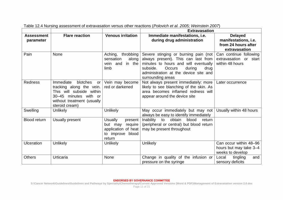
ENDORSED BY GOVERNANCE COMMITTEE
SCancer NetworkGuidelinesGuidelines and Pathways by SpecialityChemotherapyCurrent Approved Versions (Word amp PDF)Management of Extravasation version 20doc Page 11 of 21
Table 124 Nursing assessment of extravasation versus other reactions (Polovich et al 2005 Weinstein 2007)
Extravasation
Assessment parameter
Flare reaction Venous irritation Immediate manifestations ie during drug administration
Delayed manifestations ie from 24 hours after
extravasation
Pain None Aching throbbing sensation along vein and in the limb
Severe stinging or burning pain (not always present) This can last from minutes to hours and will eventually subside Occurs during drug administration at the device site and surrounding areas
Can continue following extravasation or start within 48 hours
Redness Immediate blotches or tracking along the vein This will subside within 30ndash45 minutes with or without treatment (usually steroid cream)
Vein may become red or darkened
Not always present immediately more likely to see blanching of the skin As area becomes inflamed redness will appear around the device site
Later occurrence
Swelling Unlikely Unlikely May occur immediately but may not always be easy to identify immediately
Usually within 48 hours
Blood return Usually present Usually present but may require application of heat to improve blood return
Inability to obtain blood return (peripheral or central) but blood return may be present throughout
Ulceration Unlikely Unlikely Unlikely Can occur within 48ndash96 hours but may take 3ndash4 weeks to develop
Others Urticaria None Change in quality of the infusion or pressure on the syringe
Local tingling and sensory deficits
ENDORSED BY GOVERNANCE COMMITTEE
SCancer NetworkGuidelinesGuidelines and Pathways by SpecialityChemotherapyCurrent Approved Versions (Word amp PDF)Management of Extravasation version 20doc
Page 12 of 21
11 Management of Extravasation The management of extravasation of chemotherapy agents is controversial and there is little documented evidence of efficacy Until recently no antidote has received clear validation in controlled clinical trials and no randomized trials managing cytotoxic extravasation in humans have been completed (Bertolli 1995) Some studies performed on animals have demonstrated both effective and ineffective treatments but extrapolation from animals to humans is limited (Polovich et al 2005 Wickham et al 2006) Other studies have been small with low numbers of patients Another problem is that it is often difficult to ascertain whether any extravasation has actually occurred (Weinstein 2007) Therefore recommendations are based on more consistent experimental evidence cumulative clinical experience from available case reports and uncontrolled studies and empirical guidelines (Bertolli 1995) Drugs where there is evidence of effective management include anthracyclines (Rudolph amp Larson 1987) vinca alkaloids (Bertolli 1995) computed tomography (CT) contrast media (Federle et al 1998) and paclitaxel (Bertolli et al 1997) The management of extravasation involves several stages including the following Stage 1 stopping infusioninjection and aspirating the drug It appears that most authors are agreed that aspirating as much of the drug as possible as soon as extravasation is suspected is beneficial (Rudolph amp Larson 1987 Vandergrift 2001 Polovich et al 2005 Weinstein 2007) and can help lower the concentration of the drug in the area (Goolsby amp Lombardo 2006) However withdrawal is only immediately possible during bolus injections as an infusion would need to be stopped and a syringe attached in an attempt to aspirate Aspiration may be successful if extravasation presents itself as a raised blister but may be unsuccessful if tissue is soft and soggy (CP Pharmaceuticals 1999 Stanley 2002) It may help to reduce the size of the lesion (Vandergrift 2001) In practice it may achieve little and often distresses the patient (Gault amp Challands 1997) The likelihood of withdrawing blood (as suggested by Ignoffo amp Friedman 1980) is small and the practitioner may waste valuable time attempting this which could lead to delay in the rest of the management procedure Stage 2 removing device Some clinicians advocate that the peripheral vascular access device be left in situ in order to instil the antidote via the device and into the affected tissues (Kassner 2000 Stanley 2002 Weinstein 2007) However others recommend that the peripheral device should be removed to prevent any injected solution increasing the size of the affected area (Rudolph amp Larson 1987 CP Pharmaceuticals 1999 Vandergrift 2001) There appears to be no research evidence to support either practice Stage 3 applying hot or cold packs Cooling appears to be a better choice with the exception of the vinca alkaloids and some non-cytotoxic drugs than warming (Bertolli 1995 CP Pharmaceuticals 1999) Cold causes vasoconstriction localizing the extravasation and perhaps allowing time for local vascular and lymphatic systems to contain the drug It should be applied for
ENDORSED BY GOVERNANCE COMMITTEE
SCancer NetworkGuidelinesGuidelines and Pathways by SpecialityChemotherapyCurrent Approved Versions (Word amp PDF)Management of Extravasation version 20doc
Page 13 of 21
15ndash20 minutes 3ndash4 times a day for up to 3 days (Gault amp Challands 1997 CP Pharmaceuticals 1999 Polovich et al 2005) Heat promotes healing after the first 24 hours by increasing the blood supply (Polovich et al 2005 Weinstein 2007) It also decreases local drug concentration increasing the blood flow which results in enhanced resolution of pain and reabsorption of local swelling Stage 4 use of antidotes A number of antidotes are available but again there is a lack of scientific evidence to demonstrate their value and so the role of antidotes is still not clear (Polovich et al 2005) There appear to be two main methods (i) localize and neutralize (using hyaluronidase) (CP Pharmaceuticals 1999) and (ii) spread and dilute (using an antidote) (Stanley 2002) Administration of antidotes if not via the cannula is by the pincushion technique that is instilling small volumes around and over the areas affected using a small gauge (25) needle towards the centre of a clock face The procedure causes considerable discomfort to patients and if large areas are to be tackled analgesia should be considered (Stanley 2002) Hyaluronidase is an enzyme which breaks down hyaluronic acid a normal component of tissue lsquocementrsquo and helps to reduce or prevent tissue damage by allowing rapid diffusion of the extravasated fluid and promoting drug absorption (Few 1987) The usual dose is 1500 IU (Bertolli 1995 Vandergrift 2001) It should be injected within 1 hour of extravasation ideally through the intravenous device delivering the enzyme to the same tissue (Gault amp Challands 1997 Vandergrift 2001 Stanley 2002 Weinstein 2007) NB Hyaluronidase increases the absorption of local anaesthetic Therefore if local anaesthetic has been applied to the area eg Ametop gel prior to cannulation within 6 hours of extravasation then the patient should be monitored for signs and symptoms of systemic anaesthesia such as increased pulse rate and decreased respirations and the doctor informed immediately (BMARPSGB 2008) Corticosteroids have long been advocated as a treatment for anthracycline extravasation in reducing inflammatory components although inflammation is not a prominent feature of tissue necrosis (Camp Sorrell 1998) and they appear to have little benefit Data now discourages the use of locally injected steroids as there is little evidence to support their use (Bertolli 1995 Gault amp Challands 1997 Wickham et al 2006) However given as a cream they can help to reduce local trauma and irritation (Stanley 2002) Dimethyl sulfoxide (DMSO) is a potent free radical scavanger that rapidly penetrates tissues when applied topically (Bertolli 1995) Reports on the clinical use of topical DMSO show it is effective and well tolerated in extravasation (Bertolli 1995) However this is based on a high dose (95) and only 50 is easily available in the UK (Stanley 2002) Side-effects from DMSO include itching erythema mild burning and a characteristic breath odour (Bertolli 1995) Recently dexrazoxane a topoisomerase II catalytic inhibitor used clinically to minimize the cardiotoxicity of doxorubicin has been tested in animal models and a small number of patients for its use in extravasation It is given IV 3ndash6 hours after the extravasation and it appears to reduce the wound size and duration with
ENDORSED BY GOVERNANCE COMMITTEE
SCancer NetworkGuidelinesGuidelines and Pathways by SpecialityChemotherapyCurrent Approved Versions (Word amp PDF)Management of Extravasation version 20doc
Page 14 of 21
anthracyclines The triple dosage appears to be more effective than a single dose (Langer et al 2000 El Saghir et al 2004) A consensus group (Jackson et al 2006) have developed recommendations for the use of dexrazoxane
1 For anthracycline extravasations resulting from peripheral administration the site expert or team should be consulted in order to determine whether the use of dexrazoxane is indicated
2 Absolute indications are if the peripherally extravasated volume exceeds 15 ml and in the event of a central venous extravasation
Finally granulocyte macrophage-colony stimulating factor (GM-CSF) is a growth factor and has been found effective in accelerating wound healing and inducing formation of granulation of tissue (Ulutin et al 2000 El Saghir et al 2004) Stage 5 elevation of limb This is recommended as it minimizes swelling (Rudolph amp Larson 1987) and movement should be encouraged to prevent adhesion of damaged areas to underlying tissue (Gabriel 2008) Stage 6 surgical techniques Some centres suggest that a plastic surgery consultation be performed as part of the management procedure in order to remove the tissue containing the drug Surgical intervention is recommended especially if the lesion is greater than 2 cm there is significant residual pain 1ndash2 weeks after extravasation or there is minimal healing 2ndash3 weeks after injury despite local therapeutic measures (Goolsby amp Lombardo 2006) Liposuction or a flush-out technique can remove extravasated drug without resorting to excision and skin grafting A liposuction cannula can be used to aspirate extravasated material and subcutaneous fat If there is little subcutaneous fat eg preterm infants then the saline flush-out technique is recommended particularly if done within the first 24 hours It has been suggested as a less traumatic and cheaper procedure than surgery Four small stab incisions are made and large volumes of 09 sodium chloride are administered which flush out the extravasated drug (Gault amp Challands 1997) Management of large extravasations from CVADs is usually by surgical intervention and washout of affected tissues 12 Extravasation Kits The use of extravasation kits has been recommended in order to provide immediate management (Khan amp Holmes 2002 Hayden amp Goodman 2005) Kits should be assembled according to the particular needs of individual institutions They should be kept in all areas where staff are regularly administering vesicant drugs so staff have immediate access to equipment (Gabriel 2008) The kit should be simple to avoid confusion but comprehensive enough to meet all reasonable needs (Stanley 2002) (see Procedure guidelines Extravasation management peripheral cannula below) Instructions should be clear and easy to follow and the use of a flow chart enables staff to follow the management procedure in easy steps (Figure 124)
ENDORSED BY GOVERNANCE COMMITTEE
SCancer NetworkGuidelinesGuidelines and Pathways by SpecialityChemotherapyCurrent Approved Versions (Word amp PDF)Management of Extravasation version 20doc
Page 15 of 21
13 Mixed Vesicant Extravasation Consideration should be given to the management of mixed vesicant drug extravasation in terms of which drug to treat with which antidote It has been recommended to act in accordance with the drug which possesses the most deleterious properties (How amp Brown 1998) 14 Informing the Patient Patients should always be informed when an extravasation has occurred and be given an explanation of what has happened and what management has been carried out (McCaffrey Boyle amp Engelking 1995) An information sheet should be given to patients with instructions of what symptoms to look out for and when to contact the hospital during the follow-up period (Gabriel 2008) 15 Wound Management Damage will be affected by the site amount of drug concentration of the agent and if it binds to DNA or not (Polovich et al 2005) Ulceration may occur over a period of days to weeks and extravasation wounds may be complicated by tissue ischaemia related to endothelial damage (Naylor 2005) The type of injury will dictate the type of dressing Assessment of the wound should include position and size of the wound amount and type of tissue present amount and type of exudate and extent and spread of erythema (Naylor 2005) 16 Documentation and Follow Up An extravasation must be reported and fully documented as it is an accident and the patient may require follow-up care (NMC 2005 RCN 2005) Information may also be used for statistical purposes for example collation and analysis using the green card scheme devised by St Chads Hospital Birmingham (Stanley 2002) Statistics on the incidence degree causes and corrective action should be monitored and analysed (Infusion Nurses Society 2006) Finally it may be required in case of litigation which is now on the increase (Dougherty 2003 Masoorli 2003)
ENDORSED BY GOVERNANCE COMMITTEE
SCancer NetworkGuidelinesGuidelines and Pathways by SpecialityChemotherapyCurrent Approved Versions (Word amp PDF)Management of Extravasation version 20doc
Page 16 of 21
Figure
ENDORSED BY GOVERNANCE COMMITTEE
SCancer NetworkGuidelinesGuidelines and Pathways by SpecialityChemotherapyCurrent Approved Versions (Word amp PDF)Management of Extravasation version 20doc
Page 17 of 21
Equipment To assist the nurse an extravasation kit should be assembled and should be readily available in each wardunit It contains
1 Gel packs times 2 one to be kept in the fridge and one available for heating (an electric heating blanket can be used whilst pack is heating)
2 Hyaluronidase 1500 IU2 ml sterile water 3 Hydrocortisone cream 1 15 g tube times 1 4 2 ml syringes times 1 5 25 G needles times 2 6 Alcohol swabs 7 Documentation forms 8 Copy of extravasation management procedure 9 Patient information leaflet
Procedure
Action Rationale
1 Explain and discuss the procedure with the patient
To ensure that the patient understands the procedure and gives hisher valid consent (DH 2002a C NMC 2008b C)
2 Stop injection or infusion immediately leaving the cannula in place
To minimize local injury To allow aspiration of the drug to be attempted (Polovich et al 2005 C RCN 2005 C)
3 Aspirate any residual drug from the device and suspected extravasation site
To minimise local injury by removing as much drug as possible and only attempt if appropriate Subsequent damage is related to the volume of the extravasation in addition to other factors (Polovich et al 2005 C RCN 2005 C)
4 Remove the cannula To prevent the device from being used for antidote administration (Rudolph amp Larson 1987 E)
5 Collect the extravasation pack and take it to the patient
It contains all the equipment necessary for managing extravasation (Stanley 2002 E Dougherty 2005 E)
ENDORSED BY GOVERNANCE COMMITTEE
SCancer NetworkGuidelinesGuidelines and Pathways by SpecialityChemotherapyCurrent Approved Versions (Word amp PDF)Management of Extravasation version 20doc
Page 18 of 21
Group A Drugs
Draw up hyaluronidase 1500 IU in 1 ml water for injection and inject volumes of 01ndash02 ml subcutaneously at points of the compass around the circumference of the area of extravasation
This is the recommended agent for group A drugs The warm pack speeds up absorption of the drug by the tissues (Bertolli 1995 C)
Apply warm pack
Group B Drugs
Apply cold pack or ice instantly To localize the area of extravasation slow cell metabolism and decrease the area of tissue destruction To reduce local pain (Polovich et al 2005 C)
However if extravasation is with any of the following category B drugs mitomycin C doxorubicin idarubicin epirubicin actinomycin D then
This is the recommended agent for these anthracyclines and helps to reduce local tissue damage (Bertolli 1995 C)
Draw around area of extravasation with indelible pen
Put on gloves
Apply thin layer of DMSO topically to the marked area using the small plastic spatula in lid of the bottle
Allow it to dry
Apply gauze
This should be applied within 10ndash25 minutes
Where possible elevate the extremity andor encourage movement
To minimize swelling and to prevent adhesion of damaged area to underlying tissue which could result in restriction of movement (Rudolph amp Larson 1987 E)
Inform a member of the medical staff at the earliest opportunity and administer any other prescribed antidotes eg dexrazoxane
To enable actions differing from agreed policy to be taken if considered in the best interests of the patient To notify the doctor of the need to prescribe any other drugs (E)
Apply hydrocortisone cream 1 twice daily and instruct the patient how to do this Continue as long as erythema persists
To reduce local inflammation and promote patient comfort (Stanley 2002 E)
Where appropriate apply DMSO every 2 hours on day 1 and then every 6 hours for up to 7 days (patients will need to have this prescribed as a to take out [TTO] and continue treatment at home where necessary)
To help reduce local tissue damage (Bertolli 1995 C)
ENDORSED BY GOVERNANCE COMMITTEE
SCancer NetworkGuidelinesGuidelines and Pathways by SpecialityChemotherapyCurrent Approved Versions (Word amp PDF)Management of Extravasation version 20doc
Page 19 of 21
Heat packs (group A drugs) should be reapplied after initial management for 2ndash4 hours Cold packs (group B drugs) should be applied for 15ndash20 minutes 3ndash4 times a day for up to 3 days
To localize the steroid effect in the area of extravasation To reduce local pain and promote patient comfort (Bertolli 1995 C)
Provide analgesia as required To promote patient comfort To encourage movement of the limb as advised (E)
Document the following details in duplicate on the form provided
a) Patients namenumber
b) Wardunit
c) Date time
d) Signs and symptoms
e) Venepuncture site (on diagram)
f) Drug sequence
g) Drug administration technique ie lsquobolusrsquo or infusion
h) Approximate amount of the drug extravasated
i) Diameter length and width of extravasation area
j) Appearance of the area
k) Step-by-step management with date and time of each step performed and medical officer notification
To provide an immediate full record of all details of the incident which may be referred to if necessary To provide a baseline for future observation and monitoring of patients condition To comply with NMC guidelines (Kassner 2000 E NMC 2005 C RCN 2005 C Weinstein 2007 E)
l) Patients complaints comments statements
m) Indication that patients information sheet given to patient
n) Follow-up section
o) Whether photograph was taken
p) If required when patient referred to plastic surgeon
q) Signature of the nurse
Explain to the patient that the site may remain sore for several days
To reduce anxiety and ensure continued co-operation (P E)
Observe the area regularly for erythema induration blistering or necrosis Inpatients monitor daily Where appropriate take photograph
To detect any changes at the earliest possible moment (RCN 2005 C)
ENDORSED BY GOVERNANCE COMMITTEE
SCancer NetworkGuidelinesGuidelines and Pathways by SpecialityChemotherapyCurrent Approved Versions (Word amp PDF)Management of Extravasation version 20doc
Page 20 of 21
All patients should receive written information explaining what has occurred what management has been carried out what they need to look for at the site and when to report any changes For example increased discomfort peeling or blistering of the skin should be reported immediately
To detect any changes as early as possible and allow for a review of future management This may include referral to a plastic surgeon (Gault amp Challands 1997 E Polovich et al 2005 C RCN 2005 C)
If blistering or tissue breakdown occurs begin dressing techniques and seek advice regarding wound management
To minimize the risk of a superimposed infection and sterile increase healing (Naylor 2005 E)
Depending on size of lesion degree of pain type of drug refer to plastic surgeon
To prevent further pain or other complications as chemically induced ulcers rarely heal spontaneously (Polovich et al 2005 C Dougherty 2005 E)
Monitoring of the Guideline Implementation of the guidance will be considered as a topic for audit by the NSSG in and reviewed in Authors Andrew Stanley Network Pharmacist References
The Royal Marsden Hospital Manual of Clinical Nursing Procedures 7th Edition Blackwell Publishing (2008)
ENDORSED BY GOVERNANCE COMMITTEE
SCancer NetworkGuidelinesGuidelines and Pathways by SpecialityChemotherapyCurrent Approved Versions (Word amp PDF)Management of Extravasation version 20doc
Page 21 of 21
Approval Signatures Pan Birmingham Cancer Network Governance Committee Chair Name Doug Wullf
Signature Date July 2011 Pan Birmingham Cancer Network Manager Name Karen Metcalf
Signature Date July 2011 Network Site Specific Group Clinical Chair Name Frances Shaw
Signature Date July 2011
ENDORSED BY GOVERNANCE COMMITTEE
SCancer NetworkGuidelinesGuidelines and Pathways by SpecialityChemotherapyCurrent Approved Versions (Word amp PDF)Management of Extravasation version 20doc
Page 15 of 21
13 Mixed Vesicant Extravasation Consideration should be given to the management of mixed vesicant drug extravasation in terms of which drug to treat with which antidote It has been recommended to act in accordance with the drug which possesses the most deleterious properties (How amp Brown 1998) 14 Informing the Patient Patients should always be informed when an extravasation has occurred and be given an explanation of what has happened and what management has been carried out (McCaffrey Boyle amp Engelking 1995) An information sheet should be given to patients with instructions of what symptoms to look out for and when to contact the hospital during the follow-up period (Gabriel 2008) 15 Wound Management Damage will be affected by the site amount of drug concentration of the agent and if it binds to DNA or not (Polovich et al 2005) Ulceration may occur over a period of days to weeks and extravasation wounds may be complicated by tissue ischaemia related to endothelial damage (Naylor 2005) The type of injury will dictate the type of dressing Assessment of the wound should include position and size of the wound amount and type of tissue present amount and type of exudate and extent and spread of erythema (Naylor 2005) 16 Documentation and Follow Up An extravasation must be reported and fully documented as it is an accident and the patient may require follow-up care (NMC 2005 RCN 2005) Information may also be used for statistical purposes for example collation and analysis using the green card scheme devised by St Chads Hospital Birmingham (Stanley 2002) Statistics on the incidence degree causes and corrective action should be monitored and analysed (Infusion Nurses Society 2006) Finally it may be required in case of litigation which is now on the increase (Dougherty 2003 Masoorli 2003)
ENDORSED BY GOVERNANCE COMMITTEE
SCancer NetworkGuidelinesGuidelines and Pathways by SpecialityChemotherapyCurrent Approved Versions (Word amp PDF)Management of Extravasation version 20doc
Page 16 of 21
Figure
ENDORSED BY GOVERNANCE COMMITTEE
SCancer NetworkGuidelinesGuidelines and Pathways by SpecialityChemotherapyCurrent Approved Versions (Word amp PDF)Management of Extravasation version 20doc
Page 17 of 21
Equipment To assist the nurse an extravasation kit should be assembled and should be readily available in each wardunit It contains
1 Gel packs times 2 one to be kept in the fridge and one available for heating (an electric heating blanket can be used whilst pack is heating)
2 Hyaluronidase 1500 IU2 ml sterile water 3 Hydrocortisone cream 1 15 g tube times 1 4 2 ml syringes times 1 5 25 G needles times 2 6 Alcohol swabs 7 Documentation forms 8 Copy of extravasation management procedure 9 Patient information leaflet
Procedure
Action Rationale
1 Explain and discuss the procedure with the patient
To ensure that the patient understands the procedure and gives hisher valid consent (DH 2002a C NMC 2008b C)
2 Stop injection or infusion immediately leaving the cannula in place
To minimize local injury To allow aspiration of the drug to be attempted (Polovich et al 2005 C RCN 2005 C)
3 Aspirate any residual drug from the device and suspected extravasation site
To minimise local injury by removing as much drug as possible and only attempt if appropriate Subsequent damage is related to the volume of the extravasation in addition to other factors (Polovich et al 2005 C RCN 2005 C)
4 Remove the cannula To prevent the device from being used for antidote administration (Rudolph amp Larson 1987 E)
5 Collect the extravasation pack and take it to the patient
It contains all the equipment necessary for managing extravasation (Stanley 2002 E Dougherty 2005 E)
ENDORSED BY GOVERNANCE COMMITTEE
SCancer NetworkGuidelinesGuidelines and Pathways by SpecialityChemotherapyCurrent Approved Versions (Word amp PDF)Management of Extravasation version 20doc
Page 18 of 21
Group A Drugs
Draw up hyaluronidase 1500 IU in 1 ml water for injection and inject volumes of 01ndash02 ml subcutaneously at points of the compass around the circumference of the area of extravasation
This is the recommended agent for group A drugs The warm pack speeds up absorption of the drug by the tissues (Bertolli 1995 C)
Apply warm pack
Group B Drugs
Apply cold pack or ice instantly To localize the area of extravasation slow cell metabolism and decrease the area of tissue destruction To reduce local pain (Polovich et al 2005 C)
However if extravasation is with any of the following category B drugs mitomycin C doxorubicin idarubicin epirubicin actinomycin D then
This is the recommended agent for these anthracyclines and helps to reduce local tissue damage (Bertolli 1995 C)
Draw around area of extravasation with indelible pen
Put on gloves
Apply thin layer of DMSO topically to the marked area using the small plastic spatula in lid of the bottle
Allow it to dry
Apply gauze
This should be applied within 10ndash25 minutes
Where possible elevate the extremity andor encourage movement
To minimize swelling and to prevent adhesion of damaged area to underlying tissue which could result in restriction of movement (Rudolph amp Larson 1987 E)
Inform a member of the medical staff at the earliest opportunity and administer any other prescribed antidotes eg dexrazoxane
To enable actions differing from agreed policy to be taken if considered in the best interests of the patient To notify the doctor of the need to prescribe any other drugs (E)
Apply hydrocortisone cream 1 twice daily and instruct the patient how to do this Continue as long as erythema persists
To reduce local inflammation and promote patient comfort (Stanley 2002 E)
Where appropriate apply DMSO every 2 hours on day 1 and then every 6 hours for up to 7 days (patients will need to have this prescribed as a to take out [TTO] and continue treatment at home where necessary)
To help reduce local tissue damage (Bertolli 1995 C)
ENDORSED BY GOVERNANCE COMMITTEE
SCancer NetworkGuidelinesGuidelines and Pathways by SpecialityChemotherapyCurrent Approved Versions (Word amp PDF)Management of Extravasation version 20doc
Page 19 of 21
Heat packs (group A drugs) should be reapplied after initial management for 2ndash4 hours Cold packs (group B drugs) should be applied for 15ndash20 minutes 3ndash4 times a day for up to 3 days
To localize the steroid effect in the area of extravasation To reduce local pain and promote patient comfort (Bertolli 1995 C)
Provide analgesia as required To promote patient comfort To encourage movement of the limb as advised (E)
Document the following details in duplicate on the form provided
a) Patients namenumber
b) Wardunit
c) Date time
d) Signs and symptoms
e) Venepuncture site (on diagram)
f) Drug sequence
g) Drug administration technique ie lsquobolusrsquo or infusion
h) Approximate amount of the drug extravasated
i) Diameter length and width of extravasation area
j) Appearance of the area
k) Step-by-step management with date and time of each step performed and medical officer notification
To provide an immediate full record of all details of the incident which may be referred to if necessary To provide a baseline for future observation and monitoring of patients condition To comply with NMC guidelines (Kassner 2000 E NMC 2005 C RCN 2005 C Weinstein 2007 E)
l) Patients complaints comments statements
m) Indication that patients information sheet given to patient
n) Follow-up section
o) Whether photograph was taken
p) If required when patient referred to plastic surgeon
q) Signature of the nurse
Explain to the patient that the site may remain sore for several days
To reduce anxiety and ensure continued co-operation (P E)
Observe the area regularly for erythema induration blistering or necrosis Inpatients monitor daily Where appropriate take photograph
To detect any changes at the earliest possible moment (RCN 2005 C)
ENDORSED BY GOVERNANCE COMMITTEE
SCancer NetworkGuidelinesGuidelines and Pathways by SpecialityChemotherapyCurrent Approved Versions (Word amp PDF)Management of Extravasation version 20doc
Page 20 of 21
All patients should receive written information explaining what has occurred what management has been carried out what they need to look for at the site and when to report any changes For example increased discomfort peeling or blistering of the skin should be reported immediately
To detect any changes as early as possible and allow for a review of future management This may include referral to a plastic surgeon (Gault amp Challands 1997 E Polovich et al 2005 C RCN 2005 C)
If blistering or tissue breakdown occurs begin dressing techniques and seek advice regarding wound management
To minimize the risk of a superimposed infection and sterile increase healing (Naylor 2005 E)
Depending on size of lesion degree of pain type of drug refer to plastic surgeon
To prevent further pain or other complications as chemically induced ulcers rarely heal spontaneously (Polovich et al 2005 C Dougherty 2005 E)
Monitoring of the Guideline Implementation of the guidance will be considered as a topic for audit by the NSSG in and reviewed in Authors Andrew Stanley Network Pharmacist References
The Royal Marsden Hospital Manual of Clinical Nursing Procedures 7th Edition Blackwell Publishing (2008)
ENDORSED BY GOVERNANCE COMMITTEE
SCancer NetworkGuidelinesGuidelines and Pathways by SpecialityChemotherapyCurrent Approved Versions (Word amp PDF)Management of Extravasation version 20doc
Page 21 of 21
Approval Signatures Pan Birmingham Cancer Network Governance Committee Chair Name Doug Wullf
Signature Date July 2011 Pan Birmingham Cancer Network Manager Name Karen Metcalf
Signature Date July 2011 Network Site Specific Group Clinical Chair Name Frances Shaw
Signature Date July 2011
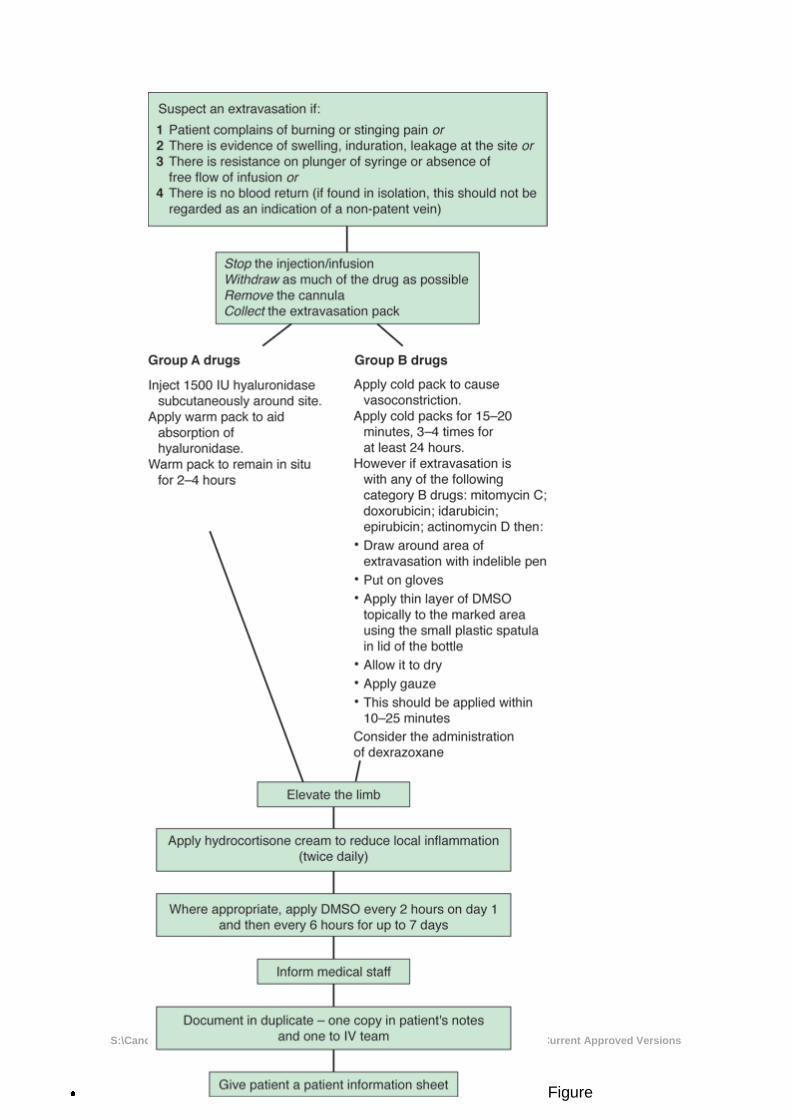
ENDORSED BY GOVERNANCE COMMITTEE
SCancer NetworkGuidelinesGuidelines and Pathways by SpecialityChemotherapyCurrent Approved Versions (Word amp PDF)Management of Extravasation version 20doc
Page 16 of 21
Figure
ENDORSED BY GOVERNANCE COMMITTEE
SCancer NetworkGuidelinesGuidelines and Pathways by SpecialityChemotherapyCurrent Approved Versions (Word amp PDF)Management of Extravasation version 20doc
Page 17 of 21
Equipment To assist the nurse an extravasation kit should be assembled and should be readily available in each wardunit It contains
1 Gel packs times 2 one to be kept in the fridge and one available for heating (an electric heating blanket can be used whilst pack is heating)
2 Hyaluronidase 1500 IU2 ml sterile water 3 Hydrocortisone cream 1 15 g tube times 1 4 2 ml syringes times 1 5 25 G needles times 2 6 Alcohol swabs 7 Documentation forms 8 Copy of extravasation management procedure 9 Patient information leaflet
Procedure
Action Rationale
1 Explain and discuss the procedure with the patient
To ensure that the patient understands the procedure and gives hisher valid consent (DH 2002a C NMC 2008b C)
2 Stop injection or infusion immediately leaving the cannula in place
To minimize local injury To allow aspiration of the drug to be attempted (Polovich et al 2005 C RCN 2005 C)
3 Aspirate any residual drug from the device and suspected extravasation site
To minimise local injury by removing as much drug as possible and only attempt if appropriate Subsequent damage is related to the volume of the extravasation in addition to other factors (Polovich et al 2005 C RCN 2005 C)
4 Remove the cannula To prevent the device from being used for antidote administration (Rudolph amp Larson 1987 E)
5 Collect the extravasation pack and take it to the patient
It contains all the equipment necessary for managing extravasation (Stanley 2002 E Dougherty 2005 E)
ENDORSED BY GOVERNANCE COMMITTEE
SCancer NetworkGuidelinesGuidelines and Pathways by SpecialityChemotherapyCurrent Approved Versions (Word amp PDF)Management of Extravasation version 20doc
Page 18 of 21
Group A Drugs
Draw up hyaluronidase 1500 IU in 1 ml water for injection and inject volumes of 01ndash02 ml subcutaneously at points of the compass around the circumference of the area of extravasation
This is the recommended agent for group A drugs The warm pack speeds up absorption of the drug by the tissues (Bertolli 1995 C)
Apply warm pack
Group B Drugs
Apply cold pack or ice instantly To localize the area of extravasation slow cell metabolism and decrease the area of tissue destruction To reduce local pain (Polovich et al 2005 C)
However if extravasation is with any of the following category B drugs mitomycin C doxorubicin idarubicin epirubicin actinomycin D then
This is the recommended agent for these anthracyclines and helps to reduce local tissue damage (Bertolli 1995 C)
Draw around area of extravasation with indelible pen
Put on gloves
Apply thin layer of DMSO topically to the marked area using the small plastic spatula in lid of the bottle
Allow it to dry
Apply gauze
This should be applied within 10ndash25 minutes
Where possible elevate the extremity andor encourage movement
To minimize swelling and to prevent adhesion of damaged area to underlying tissue which could result in restriction of movement (Rudolph amp Larson 1987 E)
Inform a member of the medical staff at the earliest opportunity and administer any other prescribed antidotes eg dexrazoxane
To enable actions differing from agreed policy to be taken if considered in the best interests of the patient To notify the doctor of the need to prescribe any other drugs (E)
Apply hydrocortisone cream 1 twice daily and instruct the patient how to do this Continue as long as erythema persists
To reduce local inflammation and promote patient comfort (Stanley 2002 E)
Where appropriate apply DMSO every 2 hours on day 1 and then every 6 hours for up to 7 days (patients will need to have this prescribed as a to take out [TTO] and continue treatment at home where necessary)
To help reduce local tissue damage (Bertolli 1995 C)
ENDORSED BY GOVERNANCE COMMITTEE
SCancer NetworkGuidelinesGuidelines and Pathways by SpecialityChemotherapyCurrent Approved Versions (Word amp PDF)Management of Extravasation version 20doc
Page 19 of 21
Heat packs (group A drugs) should be reapplied after initial management for 2ndash4 hours Cold packs (group B drugs) should be applied for 15ndash20 minutes 3ndash4 times a day for up to 3 days
To localize the steroid effect in the area of extravasation To reduce local pain and promote patient comfort (Bertolli 1995 C)
Provide analgesia as required To promote patient comfort To encourage movement of the limb as advised (E)
Document the following details in duplicate on the form provided
a) Patients namenumber
b) Wardunit
c) Date time
d) Signs and symptoms
e) Venepuncture site (on diagram)
f) Drug sequence
g) Drug administration technique ie lsquobolusrsquo or infusion
h) Approximate amount of the drug extravasated
i) Diameter length and width of extravasation area
j) Appearance of the area
k) Step-by-step management with date and time of each step performed and medical officer notification
To provide an immediate full record of all details of the incident which may be referred to if necessary To provide a baseline for future observation and monitoring of patients condition To comply with NMC guidelines (Kassner 2000 E NMC 2005 C RCN 2005 C Weinstein 2007 E)
l) Patients complaints comments statements
m) Indication that patients information sheet given to patient
n) Follow-up section
o) Whether photograph was taken
p) If required when patient referred to plastic surgeon
q) Signature of the nurse
Explain to the patient that the site may remain sore for several days
To reduce anxiety and ensure continued co-operation (P E)
Observe the area regularly for erythema induration blistering or necrosis Inpatients monitor daily Where appropriate take photograph
To detect any changes at the earliest possible moment (RCN 2005 C)
ENDORSED BY GOVERNANCE COMMITTEE
SCancer NetworkGuidelinesGuidelines and Pathways by SpecialityChemotherapyCurrent Approved Versions (Word amp PDF)Management of Extravasation version 20doc
Page 20 of 21
All patients should receive written information explaining what has occurred what management has been carried out what they need to look for at the site and when to report any changes For example increased discomfort peeling or blistering of the skin should be reported immediately
To detect any changes as early as possible and allow for a review of future management This may include referral to a plastic surgeon (Gault amp Challands 1997 E Polovich et al 2005 C RCN 2005 C)
If blistering or tissue breakdown occurs begin dressing techniques and seek advice regarding wound management
To minimize the risk of a superimposed infection and sterile increase healing (Naylor 2005 E)
Depending on size of lesion degree of pain type of drug refer to plastic surgeon
To prevent further pain or other complications as chemically induced ulcers rarely heal spontaneously (Polovich et al 2005 C Dougherty 2005 E)
Monitoring of the Guideline Implementation of the guidance will be considered as a topic for audit by the NSSG in and reviewed in Authors Andrew Stanley Network Pharmacist References
The Royal Marsden Hospital Manual of Clinical Nursing Procedures 7th Edition Blackwell Publishing (2008)
ENDORSED BY GOVERNANCE COMMITTEE
SCancer NetworkGuidelinesGuidelines and Pathways by SpecialityChemotherapyCurrent Approved Versions (Word amp PDF)Management of Extravasation version 20doc
Page 21 of 21
Approval Signatures Pan Birmingham Cancer Network Governance Committee Chair Name Doug Wullf
Signature Date July 2011 Pan Birmingham Cancer Network Manager Name Karen Metcalf
Signature Date July 2011 Network Site Specific Group Clinical Chair Name Frances Shaw
Signature Date July 2011
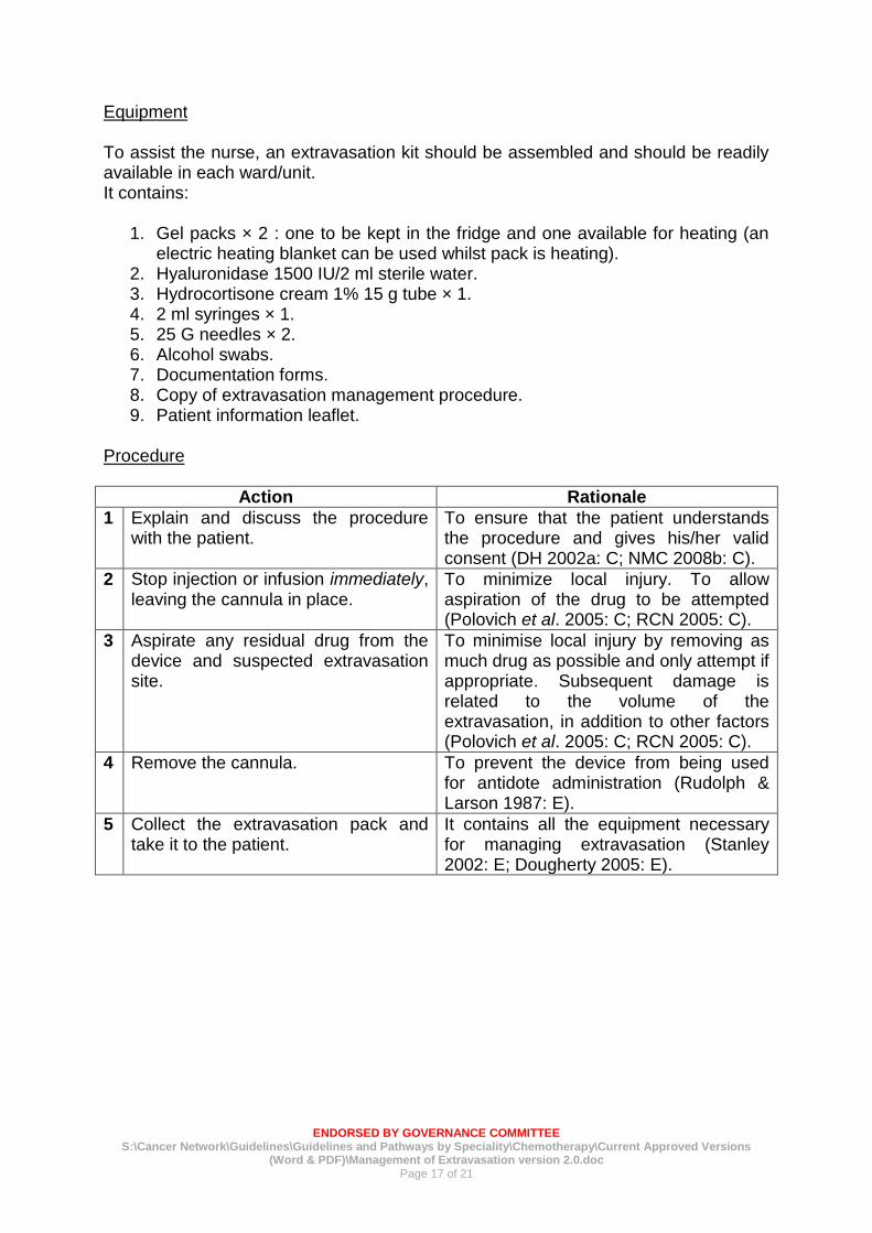
ENDORSED BY GOVERNANCE COMMITTEE
SCancer NetworkGuidelinesGuidelines and Pathways by SpecialityChemotherapyCurrent Approved Versions (Word amp PDF)Management of Extravasation version 20doc
Page 17 of 21
Equipment To assist the nurse an extravasation kit should be assembled and should be readily available in each wardunit It contains
1 Gel packs times 2 one to be kept in the fridge and one available for heating (an electric heating blanket can be used whilst pack is heating)
2 Hyaluronidase 1500 IU2 ml sterile water 3 Hydrocortisone cream 1 15 g tube times 1 4 2 ml syringes times 1 5 25 G needles times 2 6 Alcohol swabs 7 Documentation forms 8 Copy of extravasation management procedure 9 Patient information leaflet
Procedure
Action Rationale
1 Explain and discuss the procedure with the patient
To ensure that the patient understands the procedure and gives hisher valid consent (DH 2002a C NMC 2008b C)
2 Stop injection or infusion immediately leaving the cannula in place
To minimize local injury To allow aspiration of the drug to be attempted (Polovich et al 2005 C RCN 2005 C)
3 Aspirate any residual drug from the device and suspected extravasation site
To minimise local injury by removing as much drug as possible and only attempt if appropriate Subsequent damage is related to the volume of the extravasation in addition to other factors (Polovich et al 2005 C RCN 2005 C)
4 Remove the cannula To prevent the device from being used for antidote administration (Rudolph amp Larson 1987 E)
5 Collect the extravasation pack and take it to the patient
It contains all the equipment necessary for managing extravasation (Stanley 2002 E Dougherty 2005 E)
ENDORSED BY GOVERNANCE COMMITTEE
SCancer NetworkGuidelinesGuidelines and Pathways by SpecialityChemotherapyCurrent Approved Versions (Word amp PDF)Management of Extravasation version 20doc
Page 18 of 21
Group A Drugs
Draw up hyaluronidase 1500 IU in 1 ml water for injection and inject volumes of 01ndash02 ml subcutaneously at points of the compass around the circumference of the area of extravasation
This is the recommended agent for group A drugs The warm pack speeds up absorption of the drug by the tissues (Bertolli 1995 C)
Apply warm pack
Group B Drugs
Apply cold pack or ice instantly To localize the area of extravasation slow cell metabolism and decrease the area of tissue destruction To reduce local pain (Polovich et al 2005 C)
However if extravasation is with any of the following category B drugs mitomycin C doxorubicin idarubicin epirubicin actinomycin D then
This is the recommended agent for these anthracyclines and helps to reduce local tissue damage (Bertolli 1995 C)
Draw around area of extravasation with indelible pen
Put on gloves
Apply thin layer of DMSO topically to the marked area using the small plastic spatula in lid of the bottle
Allow it to dry
Apply gauze
This should be applied within 10ndash25 minutes
Where possible elevate the extremity andor encourage movement
To minimize swelling and to prevent adhesion of damaged area to underlying tissue which could result in restriction of movement (Rudolph amp Larson 1987 E)
Inform a member of the medical staff at the earliest opportunity and administer any other prescribed antidotes eg dexrazoxane
To enable actions differing from agreed policy to be taken if considered in the best interests of the patient To notify the doctor of the need to prescribe any other drugs (E)
Apply hydrocortisone cream 1 twice daily and instruct the patient how to do this Continue as long as erythema persists
To reduce local inflammation and promote patient comfort (Stanley 2002 E)
Where appropriate apply DMSO every 2 hours on day 1 and then every 6 hours for up to 7 days (patients will need to have this prescribed as a to take out [TTO] and continue treatment at home where necessary)
To help reduce local tissue damage (Bertolli 1995 C)
ENDORSED BY GOVERNANCE COMMITTEE
SCancer NetworkGuidelinesGuidelines and Pathways by SpecialityChemotherapyCurrent Approved Versions (Word amp PDF)Management of Extravasation version 20doc
Page 19 of 21
Heat packs (group A drugs) should be reapplied after initial management for 2ndash4 hours Cold packs (group B drugs) should be applied for 15ndash20 minutes 3ndash4 times a day for up to 3 days
To localize the steroid effect in the area of extravasation To reduce local pain and promote patient comfort (Bertolli 1995 C)
Provide analgesia as required To promote patient comfort To encourage movement of the limb as advised (E)
Document the following details in duplicate on the form provided
a) Patients namenumber
b) Wardunit
c) Date time
d) Signs and symptoms
e) Venepuncture site (on diagram)
f) Drug sequence
g) Drug administration technique ie lsquobolusrsquo or infusion
h) Approximate amount of the drug extravasated
i) Diameter length and width of extravasation area
j) Appearance of the area
k) Step-by-step management with date and time of each step performed and medical officer notification
To provide an immediate full record of all details of the incident which may be referred to if necessary To provide a baseline for future observation and monitoring of patients condition To comply with NMC guidelines (Kassner 2000 E NMC 2005 C RCN 2005 C Weinstein 2007 E)
l) Patients complaints comments statements
m) Indication that patients information sheet given to patient
n) Follow-up section
o) Whether photograph was taken
p) If required when patient referred to plastic surgeon
q) Signature of the nurse
Explain to the patient that the site may remain sore for several days
To reduce anxiety and ensure continued co-operation (P E)
Observe the area regularly for erythema induration blistering or necrosis Inpatients monitor daily Where appropriate take photograph
To detect any changes at the earliest possible moment (RCN 2005 C)
ENDORSED BY GOVERNANCE COMMITTEE
SCancer NetworkGuidelinesGuidelines and Pathways by SpecialityChemotherapyCurrent Approved Versions (Word amp PDF)Management of Extravasation version 20doc
Page 20 of 21
All patients should receive written information explaining what has occurred what management has been carried out what they need to look for at the site and when to report any changes For example increased discomfort peeling or blistering of the skin should be reported immediately
To detect any changes as early as possible and allow for a review of future management This may include referral to a plastic surgeon (Gault amp Challands 1997 E Polovich et al 2005 C RCN 2005 C)
If blistering or tissue breakdown occurs begin dressing techniques and seek advice regarding wound management
To minimize the risk of a superimposed infection and sterile increase healing (Naylor 2005 E)
Depending on size of lesion degree of pain type of drug refer to plastic surgeon
To prevent further pain or other complications as chemically induced ulcers rarely heal spontaneously (Polovich et al 2005 C Dougherty 2005 E)
Monitoring of the Guideline Implementation of the guidance will be considered as a topic for audit by the NSSG in and reviewed in Authors Andrew Stanley Network Pharmacist References
The Royal Marsden Hospital Manual of Clinical Nursing Procedures 7th Edition Blackwell Publishing (2008)
ENDORSED BY GOVERNANCE COMMITTEE
SCancer NetworkGuidelinesGuidelines and Pathways by SpecialityChemotherapyCurrent Approved Versions (Word amp PDF)Management of Extravasation version 20doc
Page 21 of 21
Approval Signatures Pan Birmingham Cancer Network Governance Committee Chair Name Doug Wullf
Signature Date July 2011 Pan Birmingham Cancer Network Manager Name Karen Metcalf
Signature Date July 2011 Network Site Specific Group Clinical Chair Name Frances Shaw
Signature Date July 2011
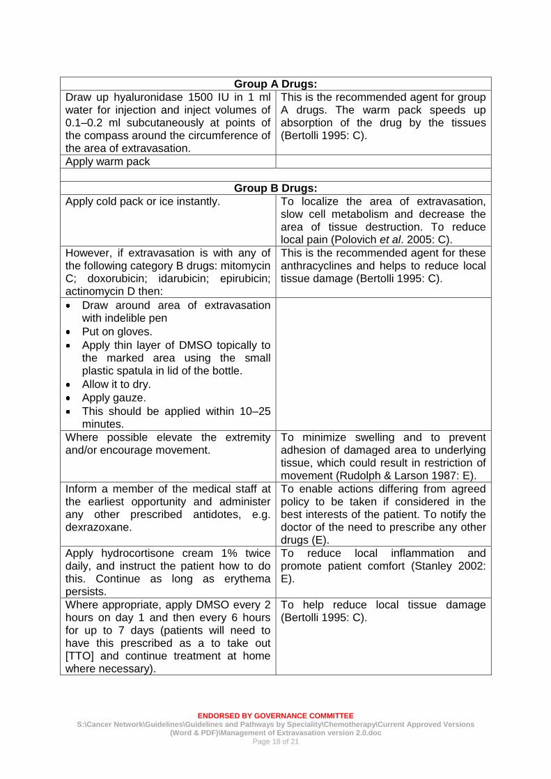
ENDORSED BY GOVERNANCE COMMITTEE
SCancer NetworkGuidelinesGuidelines and Pathways by SpecialityChemotherapyCurrent Approved Versions (Word amp PDF)Management of Extravasation version 20doc
Page 18 of 21
Group A Drugs
Draw up hyaluronidase 1500 IU in 1 ml water for injection and inject volumes of 01ndash02 ml subcutaneously at points of the compass around the circumference of the area of extravasation
This is the recommended agent for group A drugs The warm pack speeds up absorption of the drug by the tissues (Bertolli 1995 C)
Apply warm pack
Group B Drugs
Apply cold pack or ice instantly To localize the area of extravasation slow cell metabolism and decrease the area of tissue destruction To reduce local pain (Polovich et al 2005 C)
However if extravasation is with any of the following category B drugs mitomycin C doxorubicin idarubicin epirubicin actinomycin D then
This is the recommended agent for these anthracyclines and helps to reduce local tissue damage (Bertolli 1995 C)
Draw around area of extravasation with indelible pen
Put on gloves
Apply thin layer of DMSO topically to the marked area using the small plastic spatula in lid of the bottle
Allow it to dry
Apply gauze
This should be applied within 10ndash25 minutes
Where possible elevate the extremity andor encourage movement
To minimize swelling and to prevent adhesion of damaged area to underlying tissue which could result in restriction of movement (Rudolph amp Larson 1987 E)
Inform a member of the medical staff at the earliest opportunity and administer any other prescribed antidotes eg dexrazoxane
To enable actions differing from agreed policy to be taken if considered in the best interests of the patient To notify the doctor of the need to prescribe any other drugs (E)
Apply hydrocortisone cream 1 twice daily and instruct the patient how to do this Continue as long as erythema persists
To reduce local inflammation and promote patient comfort (Stanley 2002 E)
Where appropriate apply DMSO every 2 hours on day 1 and then every 6 hours for up to 7 days (patients will need to have this prescribed as a to take out [TTO] and continue treatment at home where necessary)
To help reduce local tissue damage (Bertolli 1995 C)
ENDORSED BY GOVERNANCE COMMITTEE
SCancer NetworkGuidelinesGuidelines and Pathways by SpecialityChemotherapyCurrent Approved Versions (Word amp PDF)Management of Extravasation version 20doc
Page 19 of 21
Heat packs (group A drugs) should be reapplied after initial management for 2ndash4 hours Cold packs (group B drugs) should be applied for 15ndash20 minutes 3ndash4 times a day for up to 3 days
To localize the steroid effect in the area of extravasation To reduce local pain and promote patient comfort (Bertolli 1995 C)
Provide analgesia as required To promote patient comfort To encourage movement of the limb as advised (E)
Document the following details in duplicate on the form provided
a) Patients namenumber
b) Wardunit
c) Date time
d) Signs and symptoms
e) Venepuncture site (on diagram)
f) Drug sequence
g) Drug administration technique ie lsquobolusrsquo or infusion
h) Approximate amount of the drug extravasated
i) Diameter length and width of extravasation area
j) Appearance of the area
k) Step-by-step management with date and time of each step performed and medical officer notification
To provide an immediate full record of all details of the incident which may be referred to if necessary To provide a baseline for future observation and monitoring of patients condition To comply with NMC guidelines (Kassner 2000 E NMC 2005 C RCN 2005 C Weinstein 2007 E)
l) Patients complaints comments statements
m) Indication that patients information sheet given to patient
n) Follow-up section
o) Whether photograph was taken
p) If required when patient referred to plastic surgeon
q) Signature of the nurse
Explain to the patient that the site may remain sore for several days
To reduce anxiety and ensure continued co-operation (P E)
Observe the area regularly for erythema induration blistering or necrosis Inpatients monitor daily Where appropriate take photograph
To detect any changes at the earliest possible moment (RCN 2005 C)
ENDORSED BY GOVERNANCE COMMITTEE
SCancer NetworkGuidelinesGuidelines and Pathways by SpecialityChemotherapyCurrent Approved Versions (Word amp PDF)Management of Extravasation version 20doc
Page 20 of 21
All patients should receive written information explaining what has occurred what management has been carried out what they need to look for at the site and when to report any changes For example increased discomfort peeling or blistering of the skin should be reported immediately
To detect any changes as early as possible and allow for a review of future management This may include referral to a plastic surgeon (Gault amp Challands 1997 E Polovich et al 2005 C RCN 2005 C)
If blistering or tissue breakdown occurs begin dressing techniques and seek advice regarding wound management
To minimize the risk of a superimposed infection and sterile increase healing (Naylor 2005 E)
Depending on size of lesion degree of pain type of drug refer to plastic surgeon
To prevent further pain or other complications as chemically induced ulcers rarely heal spontaneously (Polovich et al 2005 C Dougherty 2005 E)
Monitoring of the Guideline Implementation of the guidance will be considered as a topic for audit by the NSSG in and reviewed in Authors Andrew Stanley Network Pharmacist References
The Royal Marsden Hospital Manual of Clinical Nursing Procedures 7th Edition Blackwell Publishing (2008)
ENDORSED BY GOVERNANCE COMMITTEE
SCancer NetworkGuidelinesGuidelines and Pathways by SpecialityChemotherapyCurrent Approved Versions (Word amp PDF)Management of Extravasation version 20doc
Page 21 of 21
Approval Signatures Pan Birmingham Cancer Network Governance Committee Chair Name Doug Wullf
Signature Date July 2011 Pan Birmingham Cancer Network Manager Name Karen Metcalf
Signature Date July 2011 Network Site Specific Group Clinical Chair Name Frances Shaw
Signature Date July 2011
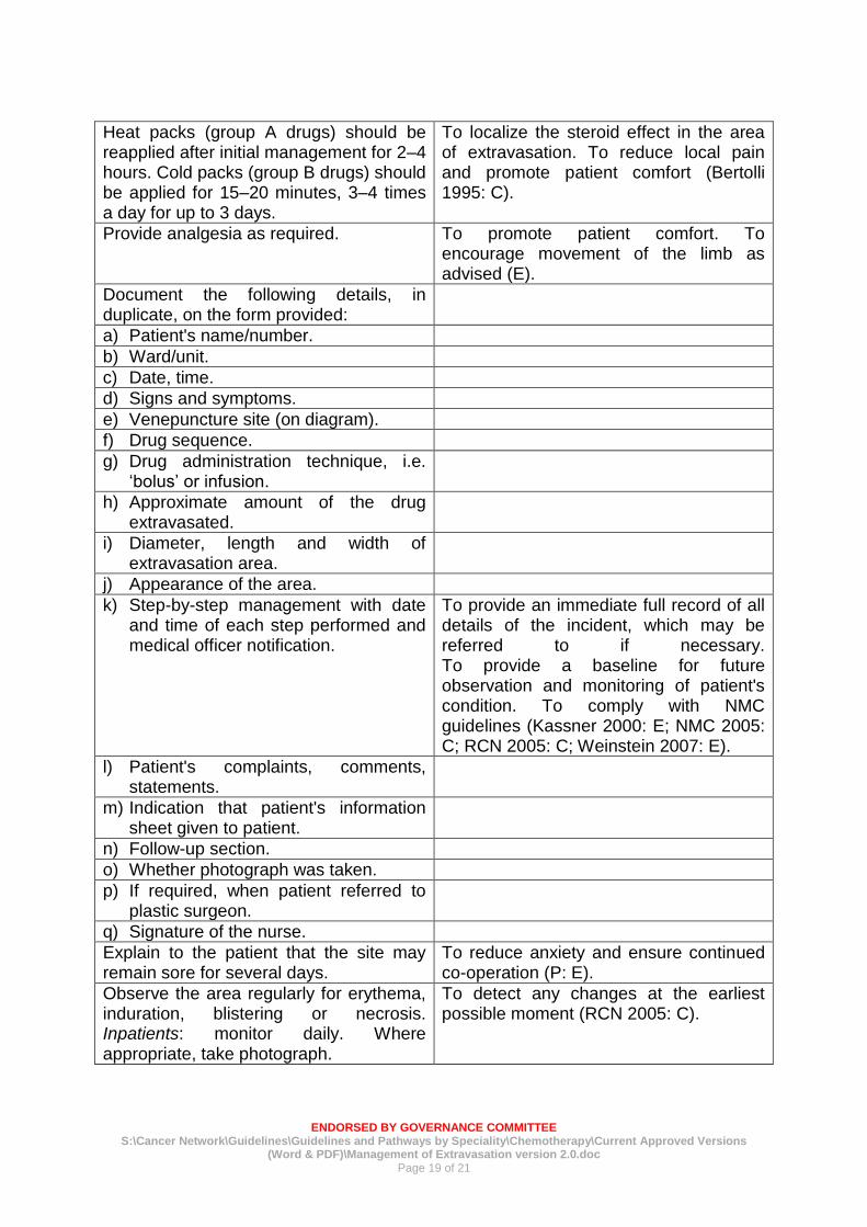
ENDORSED BY GOVERNANCE COMMITTEE
SCancer NetworkGuidelinesGuidelines and Pathways by SpecialityChemotherapyCurrent Approved Versions (Word amp PDF)Management of Extravasation version 20doc
Page 19 of 21
Heat packs (group A drugs) should be reapplied after initial management for 2ndash4 hours Cold packs (group B drugs) should be applied for 15ndash20 minutes 3ndash4 times a day for up to 3 days
To localize the steroid effect in the area of extravasation To reduce local pain and promote patient comfort (Bertolli 1995 C)
Provide analgesia as required To promote patient comfort To encourage movement of the limb as advised (E)
Document the following details in duplicate on the form provided
a) Patients namenumber
b) Wardunit
c) Date time
d) Signs and symptoms
e) Venepuncture site (on diagram)
f) Drug sequence
g) Drug administration technique ie lsquobolusrsquo or infusion
h) Approximate amount of the drug extravasated
i) Diameter length and width of extravasation area
j) Appearance of the area
k) Step-by-step management with date and time of each step performed and medical officer notification
To provide an immediate full record of all details of the incident which may be referred to if necessary To provide a baseline for future observation and monitoring of patients condition To comply with NMC guidelines (Kassner 2000 E NMC 2005 C RCN 2005 C Weinstein 2007 E)
l) Patients complaints comments statements
m) Indication that patients information sheet given to patient
n) Follow-up section
o) Whether photograph was taken
p) If required when patient referred to plastic surgeon
q) Signature of the nurse
Explain to the patient that the site may remain sore for several days
To reduce anxiety and ensure continued co-operation (P E)
Observe the area regularly for erythema induration blistering or necrosis Inpatients monitor daily Where appropriate take photograph
To detect any changes at the earliest possible moment (RCN 2005 C)
ENDORSED BY GOVERNANCE COMMITTEE
SCancer NetworkGuidelinesGuidelines and Pathways by SpecialityChemotherapyCurrent Approved Versions (Word amp PDF)Management of Extravasation version 20doc
Page 20 of 21
All patients should receive written information explaining what has occurred what management has been carried out what they need to look for at the site and when to report any changes For example increased discomfort peeling or blistering of the skin should be reported immediately
To detect any changes as early as possible and allow for a review of future management This may include referral to a plastic surgeon (Gault amp Challands 1997 E Polovich et al 2005 C RCN 2005 C)
If blistering or tissue breakdown occurs begin dressing techniques and seek advice regarding wound management
To minimize the risk of a superimposed infection and sterile increase healing (Naylor 2005 E)
Depending on size of lesion degree of pain type of drug refer to plastic surgeon
To prevent further pain or other complications as chemically induced ulcers rarely heal spontaneously (Polovich et al 2005 C Dougherty 2005 E)
Monitoring of the Guideline Implementation of the guidance will be considered as a topic for audit by the NSSG in and reviewed in Authors Andrew Stanley Network Pharmacist References
The Royal Marsden Hospital Manual of Clinical Nursing Procedures 7th Edition Blackwell Publishing (2008)
ENDORSED BY GOVERNANCE COMMITTEE
SCancer NetworkGuidelinesGuidelines and Pathways by SpecialityChemotherapyCurrent Approved Versions (Word amp PDF)Management of Extravasation version 20doc
Page 21 of 21
Approval Signatures Pan Birmingham Cancer Network Governance Committee Chair Name Doug Wullf
Signature Date July 2011 Pan Birmingham Cancer Network Manager Name Karen Metcalf
Signature Date July 2011 Network Site Specific Group Clinical Chair Name Frances Shaw
Signature Date July 2011
ENDORSED BY GOVERNANCE COMMITTEE
SCancer NetworkGuidelinesGuidelines and Pathways by SpecialityChemotherapyCurrent Approved Versions (Word amp PDF)Management of Extravasation version 20doc
Page 21 of 21
Approval Signatures Pan Birmingham Cancer Network Governance Committee Chair Name Doug Wullf
Signature Date July 2011 Pan Birmingham Cancer Network Manager Name Karen Metcalf
Signature Date July 2011 Network Site Specific Group Clinical Chair Name Frances Shaw
Signature Date July 2011