Embed Size (px)
Citation preview

Growth, Interaction, and Positioning of Microtubule Astersin Extremely Large Vertebrate Embryo Cells
Timothy Mitchison, Martin Wuhr, Phuong Nguyen, Keisuke Ishihara,Aaron Groen, and Christine M. Field*Department of Systems Biology, Harvard Medical School and Marine Biological Laboratory, Woods Hole, Massachusetts
Received 1 April 2012; Revised 27 June 2012; Accepted 28 June 2012Monitoring Editor: Douglas Robinson
Ray Rappaport spent many years studying microtu-bule asters, and how they induce cleavage furrows.Here, we review recent progress on aster structureand dynamics in zygotes and early blastomeres ofXenopus laevis and Zebrafish, where cells areextremely large. Mitotic and interphase asters differmarkedly in size, and only interphase asters span thecell. Growth of interphase asters occurs by a mecha-nism that allows microtubule density at the aster pe-riphery to remain approximately constant as radiusincreases. We discuss models for aster growth, andfavor a branching nucleation process. Neighboringasters that grow into each other interact to blockfurther growth at the shared boundary. We comparethe morphology of interaction zones formed betweenpairs of asters that grow out from the poles of thesame mitotic spindle (sister asters) and between pairsnot related by mitosis (non-sister asters) that meetfollowing polyspermic fertilization. We argue grow-ing asters recognize each other by interactionbetween antiparallel microtubules at the mutualboundary, and discuss models for molecular organi-zation of interaction zones. Finally, we discuss mod-els for how asters, and the centrosomes within them,are positioned by dynein-mediated pulling forces soas to generate stereotyped cleavage patterns. Studyingthese problems in extremely large cells is starting toreveal how general principles of cell organizationscale with cell size. VC 2012 Wiley Periodicals, Inc
KeyWords: aster, embryo, microtubule, cleavage, centrosome
Introduction
Microtubule asters—radial arrays of microtubulesradiating from centrosomes—play a central organiz-
ing role in early embryos. Ray Rappaport was fascinatedby the question of how asters, in particular pairs of asters,induce cleavage furrows. One of his most celebrated dis-coveries [Rappaport, 1961] was that neighboring pairs ofmicrotubule asters can induce cleavage furrows in echino-derm embryos whether the asters arise from the poles ofthe same mitotic spindle (which we will call sisters) orfrom juxtaposed poles of two different spindles (which wewill call non-sisters). This discovery had a profound influ-ence on subsequent thinking in the cytokinesis field. Howmicrotubules communicate with the cortex is the subjectof other articles in this volume. Here, we will take a moremicrotubule-centric perspective, and ask: how do astersgrow, how do they interact with other asters, and how arethey positioned in the cytoplasm? These processes deter-mine where aster pairs will interact with the cortex, andthus define cleavage plane geometry. We will discuss howthese processes occur in zygotes and early blastomeres ofamphibians and Zebrafish, which provide convenient ex-perimental systems, but also represent extremely largecells. Comparison with similar processes in smaller cellswill reveal how conserved, microtubules-based spatialorganizing mechanisms scale with cell size.
The amphibian Xenopus laevis and the fish Danio rerio(Zebrafish) are easy to rear in the laboratory, and offercomplementary technical advantages. Xenopus eggs cleavecompletely and are easy to fertilize with one or multiplesperm and to microinject. They are opaque, which pre-cludes live imaging of internal events, but fixed embryoscan be cleared for immunofluorescence imaging byimmersion in a high refractive index medium [Klymkow-sky and Hanken, 1991; Becker and Gard, 2006]. Impor-tantly for us, essentially undiluted cell-free extracts can beprepared from Xenopus eggs which recapitulate much ofthe biology of the early embryo and are highly tractablefor biochemical manipulation, physical manipulation, and
*Address correspondence to: Christine M. Field, Department ofSystems Biology, Harvard Medical School and Marine BiologicalLaboratory, Woods Hole, Massachusetts.E-mail: [email protected]
Published online 20 August 2012 in Wiley Online Library(wileyonlinelibrary.com).
REVIEWARTICLECytoskeleton, October 2012 69:738–750 (doi: 10.1002/cm.21050)VC 2012 Wiley Periodicals, Inc.
n 738

live imaging [Desai et al., 1999; Chan and Forbes, 2006;Maresca and Heald, 2006]. Early Zebrafish embryos aremeroblastic, that is, they do not cleave completely. Theiranimal pole region is yolk-free and transparent, whichallows live imaging. Zebrafish are highly tractable for clas-sic genetics, and transgenic lines that stably express greenfluorescent protein (GFP)-tagged proteins can be gener-ated easily. The mechanisms we discuss are broadly con-served in evolution, and important comparison systemswith smaller cells include embryos of marine invertebrates,C. elegans and Drosophila as well as somatic cells. Drosoph-ila offers an interesting biological twist in that early divi-sions are syncytial, so aster growth and interactions areuncoupled from cytokinesis for the first 12 cell cycles.Xenopus and Zebrafish zygotes and early blastomeres are
extremely large cells, with zygotes �1200 lm and �600lm in diameter, respectively. They are also unusually fastcompared to somatic cells, in the sense that the cell cycletakes 20–30 min to complete at room temperature (thefirst cell cycles are longer). These sizes and speeds repre-sent physical extremes compared to typical somatic cells,which may require special adaptations of conserved cellorganizing mechanisms, and/or reveal underappreciatedintrinsic capabilities of those mechanisms. One well-stud-ied example is adaptation of replication origins for veryfast genome duplication [Blow, 2001]. Here, we will focuson adaptations of aster growth and interaction mecha-nisms that allow rapid and accurate spatial organizationon a scale of hundreds of lm. This is much larger thanthe molecular length scale, and may even be larger thanthe microtubule length scale inside the aster.
Aster Growth in Large Cells
The question of how microtubule asters grow in extremelylarge embryo cells has received little attention, but webelieve that answering it will reveal principles of size scal-ing and unexpected molecular mechanisms. Figures 1 and2 illustrate aster morphology and growth during the firstand second cell cycle in frog and fish embryos. Inspectionof these and similar images [Wuhr et al., 2009, 2010,2011] suggests that the structure of large interphase astersin these embryos does not conform to the standardmodel, where all microtubules radiate as straight linesfrom a single point at the centrosome. Rather, the micro-tubule network in the asters appears bushy and singlemicrotubules or thin bundles at the aster periphery appearwavy or curved (Fig. 2 last panel). Least consistent withthe conventional model, microtubule density at the asterperiphery appears to remain constant, or increase, with as-ter radius (Fig. 1) and time (Fig. 2). These images forcedus to reconsider the standard model for aster growth, andhow it scales with cell size.
Figure 3A illustrates the standard model for astergrowth, which we call the radial elongation model. Cen-
trosomes nucleate microtubules and hold on to minusends, while plus ends elongate in a liner trajectory byaddition of GTP-tubulin. This mechanism was inferredfrom analysis of nucleation and elongation in culturedmammalian cells and isolated centrosomes [Bergen et al.,1980; Brinkley et al., 1981; Brinkley, 1985]. It remainsthe standard model for animal cells, though many instan-ces are known where microtubules nucleate from locationsother than centrosomes [reviewed in Luders and Stearns,2007]. We believe there is a fundamental problem in scal-ing the radial elongation model to large cells. Microtubuledensity at the periphery must decrease with aster radius inthis model. In extremely large cells, the density of micro-tubules at the periphery would become so low that signal-ing to the cortex to initiate cytokinesis might becomeimpossible. This problem could be solved, theoretically,by increasing centrosome size. Centrosome size doesindeed scale with cell size in C. elegans embryos [Deckeret al., 2011], and centrosomes in marine invertebrateembryos can be many microns in diameter in the first cellcycle (Fig. 4) [Asnes and Schroeder, 1979; Stricklandet al., 2004; Foe and von Dassow, 2008]. It is difficult toprecisely define interphase centrosome size in images ofearly frog and fish embryos. However, by both microtu-bule (Figs. 1 and 2) and c-tubulin staining (Fig. 1D0) cen-trosomes appear small compared to cell size, suggestingthere may be an upper limit to centrosome size, as thereis to spindle length [Wuhr et al., 2008]. It thus appearsthat the problem of scaling aster size to cell size in verylarge cells is not solved by scaling centrosome size. Figures3B–3D illustrate various candidate additions or alterna-tives to the radial elongation model to solve the size scal-ing problem and account for observed aster morphology.Whatever the molecules involved, the mechanism bywhich a growing aster adds new microtubules as it’s radiusincreases must generate an approximately constant densityat the periphery, with plus ends pointing on averageoutward.
Figure 3B illustrates a model in which microtubules arenucleated within the aster as it grows, but away from thecentrosome. Nucleation could occur from the side ofexisting microtubules like Arp2/3 nucleation of actin [Pol-lard and Borisy, 2003], from Golgi membranes within theaster [Efimov et al., 2007; Rivero et al., 2009], or fromsome other location. We currently favor this class ofmodel based on morphology and precedent from othersystems. Microtubule nucleation from the sides of preex-isting microtubules has been observed in several systems,including the cortex of higher plant cells [Murata et al.,2005; Chan et al., 2009; Kirik et al., 2012], and in cyto-plasmic bundles in S. pombe [Samejima et al., 2006]. Inboth cases, c-tubulin complex is recruited to the side ofpreexisting microtubules where it nucleates and holds onto a new minus end. The regulators are best characterizedin S. pombe, where the Mto1.Mto2 complex recruits
CYTOSKELETON Growth, Interaction, and Positioning of Microtubule Asters 739 n

c-tubulin to the side of preexisting microtubules, and acti-vates it to nucleate [Samejima et al., 2006]. A similarfunction was proposed for the Augmin/Haus complex inthe mitotic spindle and cytokinesis midzone complex ofanimal cells [Uehara et al., 2009]. Structural studies of c-tubulin ring complexes predict that the complex needs toundergo a conformational change to become active innucleation [Kollman et al., 2011]. Microtubule nucleationis, in general, poorly understood at a biophysical level,and pure proteins reconstitution studies are needed to elu-cidate the mechanisms by which c-tubulin is recruitedand activated in any system. A recent study showed that acentrosomal c-tubulin recruitment factor, CDK5RAP2,could stimulate nucleation by isolated c-tubulin com-plexes [Choi et al., 2010], an encouraging step toward fullreconstitution. In favorable immunofluorescence images,
we observe a diffuse glow of c-tubulin in large asters inXenopus embryos (Fig. 1D0), but its significance is unclear.
Figure 3C illustrates a model in which microtubulescontinually release from the centrosome and slide out-ward. This mechanism was posited to account for non-centrosomal microtubules in neurons [Ahmad and Baas,1995], and astral microtubules have been shown to detachand slide outward during anaphase–telophase in tissue cul-ture cells [Rusan et al., 2002]. We know that dyneinexerts outward pulling force on astral microtubules duringanaphase–telophase in early embryos (discussed below).The same force might well cause outward microtubulesliding, so this model is plausible.
Figure 3D illustrates a model in which the growing as-ter captures microtubules that were nucleated outside theaster, for example, by the cortex or distributed organelles.
Fig. 1. Growth and interaction of sister asters in the first two divisions in X. laevis. Fertilized eggs were fixed, stained for tubu-lin (A, A0 upper, B, C, C0, C00, D, D00) DNA (A0 lower) and c-tubulin (D0,D00 0) as described [Wuhr et al., 2010]. The animal half ofthe egg was cut off and imaged from the cut surface, so the z-axis is parallel to the animal-vegetal axis of the zygote. One letter isused to designate each different embryo. A, A0: metaphase of first mitosis. Note small asters. B: anaphase–telophase of first mitosis.Note aster growth and formation of an interaction zone between sister asters at mid-cell. C, C0, C00: later telophase. Note the dense,bushy appearance of microtubules at the aster periphery, low microtubule density, and probable antiparallel bundles in the interactionzone. D, D0, D00, D000 telophase of 2nd mitosis, with the 1st cleavage plane oriented North-South. The presumptive 2nd cleavageplane will cut each blastomere between sister asters, at �90� to the 1st cleavage plane. Note bushy asters and interaction zones. c-tubulin staining is brightest at points corresponding to centrosomes, but dimmer staining is evident throughout the aster.
n 740 Mitchison et al. CYTOSKELETON

Possibly consistent with this model, prometaphase astersin tissue culture cells capture and orient non-centrosomalmicrotubules using dynein [Rusan et al., 2002], thoughthis activity probably requires a mitotic state of the cyto-plasm where dynein acts to cluster minus ends [Gatlinet al., 2009]. We currently disfavor this model as a majoraster growth mechanism in early frog and fish embryosbecause we do not observe many microtubules in thecytoplasm between the expanding aster periphery and thecortex, but we cannot rule it out.
An important question is whether the unusual astergrowth mechanisms illustrated in Fig. 3 apply in othercells. In echinoderm zygotes (�50–200 lm in diameter),astral microtubules appear radial in metaphase, but morecomplex in anaphase–telophase [Strickland et al., 2004;Foe and von Dassow, 2008]. In Fig. 4, we show images ofmicrotubules in metaphase and anaphase in zygotes ofCerebratulus, a nemertean ribbon worm with eggs �100lm in diameter, kindly provided by George von Dassow(Oregon Institute of Marine Biology). At 1st metaphase,most microtubules appear to radiate from large centro-somes, and the density clearly drops with radius (Fig. 4A).These images are consistent with all microtubule initiatingat centrosomes and growing out to the cortex, as has beenshown in C. elegans embryos by EB1 tracking [Sraykoet al., 2005]. At anaphase–telophase, the microtubule dis-
tribution appears to change. Microtubule density no lon-ger decreases with radius as strongly, and some degree ofbushiness is evident toward the periphery (Fig. 4B).Microtubules also appear more bundled. This kind ofimage suggests that one or more of the mechanism illus-trated in Figs. 3B and 3C may operate at telophase inzygotes that are smaller than Xenopus and Zebrafish, butstill large compared to somatic cells.
Aster size in frog and fish embryos is temporally con-trolled by the cell cycle, with important implications forgrowth mechanisms and embryo organization. Aster radiusat the poles of the first metaphase spindle is �30–40 lmin Xenopus [Figs. 1A and 1B, Wuhr et al., 2008] and simi-lar in Zebrafish [Fig. 2, 4 min, Wuhr et al., 2010]. Inboth cases, this is much smaller than the zygote radius.Asters grow dramatically at anaphase onset, presumablydue to decreased activity of Cdk1 (Cdc2.Cyclin B) kinase.In mitosis, Cdk1 acts on a complex network of microtu-bule interacting proteins to promote catastrophes (growingto shrinking transitions) and limit length [Belmont et al.,1990; Verde et al., 1992; Niethammer et al., 2007].Microtubule growth in this regime was termed‘‘bounded,’’ because while the lengths of individual micro-tubules fluctuate by dynamic instability, the average lengthdoes not increase with time [Verde et al., 1992]. Asterradius at metaphase, when Cdk1 activity is high, is
Fig. 2. Growth and interaction of sister asters in Zebrafish 1st mitosis. Fertilized eggs from fish stably expressing the microtubulebinding domain of ensconsin fused to 3�GFP were imaged live by confocal microscopy [Wuhr et al., 2010, 2011]. The cell isimaged with the animal pole next to the �20 immersion objective lens. 0 min: prophase, note the intact nucleus. 4 min: metaphase,note the small aster radius at this stage. 8–20 min: after anaphase onset, the paired sister asters rapidly grow, and they meet andinteract at the midplane of the cell. Note that the density of microtubules at the aster periphery, which is artificially highlighted usingthe ensconsin probe, remains approximately constant. Note also that microtubules at the aster periphery often appear curved andsomewhat disorganized. The nucleus (n) and centrosomes (c) are highlighted in the last panel. Note the centrosomes inside one asterseparate on the north–south axis as they move away from the interaction zones. The 2nd mitotic spindles will later assemble on thisnorth–south axis.
CYTOSKELETON Growth, Interaction, and Positioning of Microtubule Asters 741 n

presumably limited by the length distribution of microtu-bules in this bounded regime. Cdk1 levels drop shortly af-ter fertilization, and at anaphase onset [reviewed inMorgan, 2006]. This lowers the catastrophe rate, andallows microtubules to grow longer. Experiments in Xeno-pus extract suggest growth of individual microtubule maybecome unbounded in interphase [Verde et al., 1992], soin principle single microtubules might elongate continu-ously from the centrosome to the cortex, hundreds of lmin early frog and fish embryos. The actual length of indi-vidual microtubules in large interphase asters is currentlyunknown. If microtubules are in fact short compared toaster radius, as is the case for Xenopus meiosis-II spindles[Burbank et al., 2006], a serious rethink of aster mechan-ics will be required.
Cell cycle regulation of aster size has important implica-tions for spatial organization of the early embryo. In earlyfrog or fish blastomeres metaphase spindles are centrallylocated, and their short astral microtubules do not reachthe cortex (Fig. 1B) [Wuhr et al., 2009, 2010]. Thus,metaphase spindles cannot position themselves relative tothe cell cortex using their astral microtubules as usuallyproposed. In smaller embryos, such as sea urchin, C. ele-gans and Cerebratulus (Fig. 4A) astral microtubules reachthe cortex in metaphase, and metaphase spindle can posi-tion themselves. In early divisions in frog and fish, thecentrosome pairs that initiate the spindles are preposi-tioned by astral microtubules during the preceding inter-phase [Wuhr et al., 2010]. Following anaphase onset,asters rapidly grow to span the whole cell, and touch thecortex, by one or more of the mechanisms illustrated inFig. 3. Touching the cortex is presumably required forasters to position cleavage furrows [reviewed in Rappaport,1996], though von Dassow et al. [2009] recently sug-gested that asters can communicate to the cortex withoutphysical contact. How asters communicate with the cortexis addressed by other articles in this volume.
Aster–Aster Interactions
What happens when two neighboring asters grow to toucheach other? This question was of great interest to Rappa-port, since cleavage furrows are typically induced whereand when microtubules growing from aster pairs meet atthe cortex. Figure 5 shows three possibilities drawn fromthe literature, where the consequence of aster–aster inter-action depends strongly on the system and cell cycle state.When asters grow from nearby centrosomes in pure tubu-lin, their microtubules simply interpenetrate (Fig. 5A) [seeexamples in Brinkley et al., 1981]. This is expectedbecause the microtubules are too far apart in three dimen-sions to physically bump into each other. When two astersmeet in mitotic Xenopus egg extract, they adhere, thenmove together and fuse [Fig. 5B, Gatlin et al., 2009].Movement and fusion are driven by dynein in this system,
presumably cross-bridging between two microtubules.When two asters meet during interphase in frog and fishembryos, their microtubules do not interpenetrate and theasters tend to move apart [Fig. 5C, Wuhr et al., 2010].Aster movement is again driven by dynein, but in thiscase the dynein is presumably anchored in the cytoplasm(discussed below), rather than to another microtubule, soit produces force in the opposite direction. An effect ofcell cycle state on aster–aster interaction was also noted inechinoderm embryos using electron microscopy (EM)[Asnes and Schroeder, 1979]. Microtubules from the twoasters of one spindle interpenetrated at the equator duringmetaphase. During anaphase–telophase there was no inter-penetration at the equator, despite the fact that astralmicrotubules radiating away from the equator were longeron average. This classic observation suggests that somefactor blocks aster interpenetration specifically during ana-phase–telophase. More recent immunofluorescence imagessuggest some astral microtubule do cross the equator atanaphase–telophase (Fig. 4B), though this may be system-dependent.
Asters grow into each other in early embryos under dif-ferent circumstances. Two asters grow out from the polesof each mitotic spindle at anaphase, and meet each otherat the midplane of the cell (Figs. 1 and 2). We will callthese ‘‘sister asters.’’ The name traditional name for a pairof sister asters is the ‘‘amphiaster’’ [Wilson, 1925], fromthe Greek ‘‘amphi-’’ meaning ‘‘on both sides.’’ The mid-plane between the asters has been called the ‘‘diastem’’[Wilson, 1925] or ‘‘diastema’’ [e.g., Wakabayashi and Shi-nagawa, 2001], meaning space or gap between two struc-tures. More recently, the term ‘‘telophase disc’’ was coinedto describe the analogous location in somatic cells[Andreassen et al., 1991]. We will use the term ‘‘aster–as-ter interaction zone’’ for this midplane, to emphasize theprocess by which it forms. Non-sister asters meet in thezygote following polyspermic fertilization, when eachsperm centrosome nucleates an aster, and after anaphasefollowing polyspermy or cytokinesis failure, when astersgrowing from the poles of separate spindles meet. Exam-ples of interactions between non-sister asters followingpolyspermic fertilization, before and after 1st mitosis, areshown in Fig. 6. Polyspermy is abnormal in Xenopus (andmost frogs) and Zebrafish, but normal in many pleuro-deles (newts and salamanders) [Fankhauser, 1948; Iwao,1989], including axolotl (Fig. 6F, F0). In naturally polysper-mic pleurodele zygotes, the excess male pronuclei and cen-trosomes that do not capture the single female pronucleusare destroyed by an unknown mechanism around prophaseof 1st mitosis [Fankhauser, 1948]. This mechanism foreliminating excess nuclei and centrosomes is missing infrogs, so forced polyspermic fertilization leads to multiplespindles and multiple cleavage furrows (Fig. 6).
The most characteristic consequence of aster–aster inter-action in interphase frog and fish embryos, seen for both
n 742 Mitchison et al. CYTOSKELETON
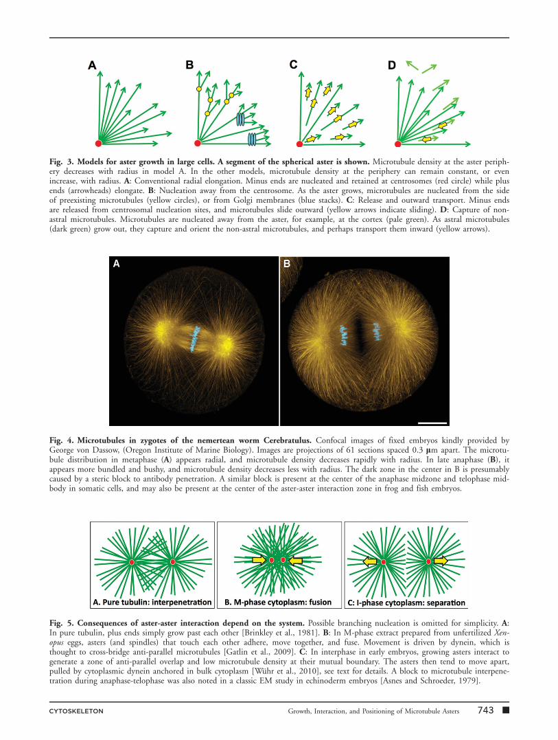
Fig. 4. Microtubules in zygotes of the nemertean worm Cerebratulus. Confocal images of fixed embryos kindly provided byGeorge von Dassow, (Oregon Institute of Marine Biology). Images are projections of 61 sections spaced 0.3 lm apart. The microtu-bule distribution in metaphase (A) appears radial, and microtubule density decreases rapidly with radius. In late anaphase (B), itappears more bundled and bushy, and microtubule density decreases less with radius. The dark zone in the center in B is presumablycaused by a steric block to antibody penetration. A similar block is present at the center of the anaphase midzone and telophase mid-body in somatic cells, and may also be present at the center of the aster-aster interaction zone in frog and fish embryos.
Fig. 3. Models for aster growth in large cells. A segment of the spherical aster is shown. Microtubule density at the aster periph-ery decreases with radius in model A. In the other models, microtubule density at the periphery can remain constant, or evenincrease, with radius. A: Conventional radial elongation. Minus ends are nucleated and retained at centrosomes (red circle) while plusends (arrowheads) elongate. B: Nucleation away from the centrosome. As the aster grows, microtubules are nucleated from the sideof preexisting microtubules (yellow circles), or from Golgi membranes (blue stacks). C: Release and outward transport. Minus endsare released from centrosomal nucleation sites, and microtubules slide outward (yellow arrows indicate sliding). D: Capture of non-astral microtubules. Microtubules are nucleated away from the aster, for example, at the cortex (pale green). As astral microtubules(dark green) grow out, they capture and orient the non-astral microtubules, and perhaps transport them inward (yellow arrows).
Fig. 5. Consequences of aster-aster interaction depend on the system. Possible branching nucleation is omitted for simplicity. A:In pure tubulin, plus ends simply grow past each other [Brinkley et al., 1981]. B: In M-phase extract prepared from unfertilized Xen-opus eggs, asters (and spindles) that touch each other adhere, move together, and fuse. Movement is driven by dynein, which isthought to cross-bridge anti-parallel microtubules [Gatlin et al., 2009]. C: In interphase in early embryos, growing asters interact togenerate a zone of anti-parallel overlap and low microtubule density at their mutual boundary. The asters then tend to move apart,pulled by cytoplasmic dynein anchored in bulk cytoplasm [Wuhr et al., 2010], see text for details. A block to microtubule interpene-tration during anaphase-telophase was also noted in a classic EM study in echinoderm embryos [Asnes and Schroeder, 1979].
CYTOSKELETON Growth, Interaction, and Positioning of Microtubule Asters 743 n
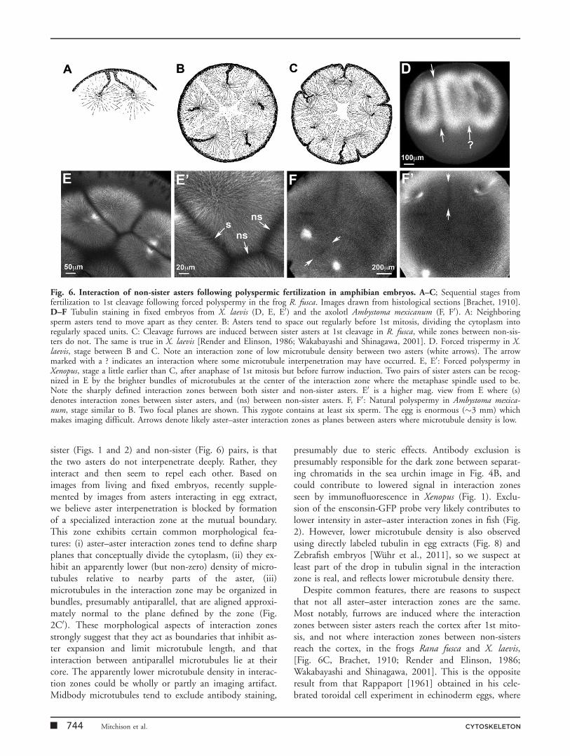
sister (Figs. 1 and 2) and non-sister (Fig. 6) pairs, is thatthe two asters do not interpenetrate deeply. Rather, theyinteract and then seem to repel each other. Based onimages from living and fixed embryos, recently supple-mented by images from asters interacting in egg extract,we believe aster interpenetration is blocked by formationof a specialized interaction zone at the mutual boundary.This zone exhibits certain common morphological fea-tures: (i) aster–aster interaction zones tend to define sharpplanes that conceptually divide the cytoplasm, (ii) they ex-hibit an apparently lower (but non-zero) density of micro-tubules relative to nearby parts of the aster, (iii)microtubules in the interaction zone may be organized inbundles, presumably antiparallel, that are aligned approxi-mately normal to the plane defined by the zone (Fig.2C0). These morphological aspects of interaction zonesstrongly suggest that they act as boundaries that inhibit as-ter expansion and limit microtubule length, and thatinteraction between antiparallel microtubules lie at theircore. The apparently lower microtubule density in interac-tion zones could be wholly or partly an imaging artifact.Midbody microtubules tend to exclude antibody staining,
presumably due to steric effects. Antibody exclusion ispresumably responsible for the dark zone between separat-ing chromatids in the sea urchin image in Fig. 4B, andcould contribute to lowered signal in interaction zonesseen by immunofluorescence in Xenopus (Fig. 1). Exclu-sion of the ensconsin-GFP probe very likely contributes tolower intensity in aster–aster interaction zones in fish (Fig.2). However, lower microtubule density is also observedusing directly labeled tubulin in egg extracts (Fig. 8) andZebrafish embryos [Wuhr et al., 2011], so we suspect atleast part of the drop in tubulin signal in the interactionzone is real, and reflects lower microtubule density there.
Despite common features, there are reasons to suspectthat not all aster–aster interaction zones are the same.Most notably, furrows are induced where the interactionzones between sister asters reach the cortex after 1st mito-sis, and not where interaction zones between non-sistersreach the cortex, in the frogs Rana fusca and X. laevis,[Fig. 6C, Brachet, 1910; Render and Elinson, 1986;Wakabayashi and Shinagawa, 2001]. This is the oppositeresult from that Rappaport [1961] obtained in his cele-brated toroidal cell experiment in echinoderm eggs, where
Fig. 6. Interaction of non-sister asters following polyspermic fertilization in amphibian embryos. A–C; Sequential stages fromfertilization to 1st cleavage following forced polyspermy in the frog R. fusca. Images drawn from histological sections [Brachet, 1910].D–F Tubulin staining in fixed embryos from X. laevis (D, E, E0) and the axolotl Ambystoma mexicanum (F, F0). A: Neighboringsperm asters tend to move apart as they center. B: Asters tend to space out regularly before 1st mitosis, dividing the cytoplasm intoregularly spaced units. C: Cleavage furrows are induced between sister asters at 1st cleavage in R. fusca, while zones between non-sis-ters do not. The same is true in X. laevis [Render and Elinson, 1986; Wakabayashi and Shinagawa, 2001]. D. Forced trispermy in X.laevis, stage between B and C. Note an interaction zone of low microtubule density between two asters (white arrows). The arrowmarked with a ? indicates an interaction where some microtubule interpenetration may have occurred. E, E0: Forced polyspermy inXenopus, stage a little earlier than C, after anaphase of 1st mitosis but before furrow induction. Two pairs of sister asters can be recog-nized in E by the brighter bundles of microtubules at the center of the interaction zone where the metaphase spindle used to be.Note the sharply defined interaction zones between both sister and non-sister asters. E0 is a higher mag. view from E where (s)denotes interaction zones between sister asters, and (ns) between non-sister asters. F, F0: Natural polyspermy in Ambystoma mexica-num, stage similar to B. Two focal planes are shown. This zygote contains at least six sperm. The egg is enormous (�3 mm) whichmakes imaging difficult. Arrows denote likely aster–aster interaction zones as planes between asters where microtubule density is low.
n 744 Mitchison et al. CYTOSKELETON

the interaction between non-sister asters from two differ-ent spindles efficiently induced furrows if they were suffi-ciently close together. Non-sister asters also generatedfurrows at their interaction zone in tissue culture cells[Savoian et al., 1999] and C. elegans embryos [Baruniet al., 2008]. It is unclear why non-sister asters fail to ini-tiate furrows in frogs, and this system may be useful fordiscriminating interaction zone molecules that are, and arenot, required for furrow induction.
What molecules are likely to mediate aster–aster inter-actions in early frog and fish embryos? To our knowledge,no molecule has been specifically localized to aster–asterinteraction zones in frog or fish embryos, but one logicalset of candidates are molecules that organize cytokinesismidzone complexes in smaller cells [reviewed in Glotzer,2005; Eggert et al., 2006]. Midzones, also called ‘‘centralspindles,’’ are barrel-shaped assemblies composed of anti-parallel microtubule bundles that form between separatingchromosomes in anaphase. Later, in telophase, theymature into midbodies, and this maturation is accompa-nied by relocalization of the microtubule organizing pro-teins [Hu et al., 2012]. Midzones in small animal cellsand interaction zones between sister asters in large embryocells are functionally analogous, and share key features of(i) antiparallel microtubule overlap at their center, (ii) lim-ited polymerization at plus ends in the overlap region,and (iii) assembly in a cytoplasm where Cdk1 activityrecently dropped, but Aurora B and Plk1 kinases are stillactive. Phragmoplasts, antiparallel microtubule arrays thatdirect formation of a new cell wall during cytokines inhigher plants, also share some or all of these attributes.These similarities suggest organization by similar mole-cules. Consistent with this possibility, the midzone in C.elegans zygotes, which is larger than a typical somatic mid-zone, though still much smaller than aster interactionzones in frogs and fish, is organized by essentially thesame molecules as somatic midzones [Glotzer, 2005].
Midzones are organized by three conserved proteinmodules or complexes [Glotzer, 2005, Eggert et al.,2006]: (i) Aurora B kinase complex or ‘‘chromosome pas-senger complex’’ [Ruchaud et al., 2007], (ii) Kif4/PRC1,and (iii) Kif23/RAPGAP1. Kif23 is also called CHO1and MKLP1, RAPGAP1 is also called MgcRacGap andCyk4, and the complex between them is also called Cen-tralspindlin. We currently hypothesize that overlapbetween antiparallel microtubules (in the appropriate cellcycle state) is the feature that allows plus ends at the pe-riphery of the two asters to recognize each other, thoughalternative models discussed in Wuhr et al. [2009] havenot been ruled out. At least two of the conserved mid-zone-organizing proteins (PRC1 and Kif23) are known tomediate interactions between antiparallel microtubules[Nislow et al., 1992, Mishima et al., 2002, Mollinariet al., 2002, Subramanian et al., 2010], and are thus can-didates to mediate recognition between the peripheries of
two growing asters. Once formed, interaction zones mustsomehow inhibit aster growth and deep interpenetrationby growing plus ends. A candidate for this function isKif4, a kinesin with plus end directed motor activity thatalso inhibits plus ends polymerization in vitro [Bringmannet al., 2004] and in somatic midzones [Hu et al., 2011].Kif4 can be targeted to antiparallel overlaps by interactionwith PRC1 [Bieling et al., 2010]. It will be interesting totest which midzone proteins localize to sister and non-sis-ter interaction zones in frog and fish embryos. Aurora Blocalized to antiparallel microtubule bundles close thecleavage furrow in Zebrafish, and was required for normalmicrotubule organization and furrowing, but was notclearly recruited to interaction zones [Yabe et al., 2009].We expect aster–aster interaction zones will share impor-tant molecular and organizational mechanisms with so-matic midzones, but that additional mechanisms may berequired to adapt a conserved organizing principle-interac-tion between antiparallel microtubules coupled to localrecruitment of polymerization inhibitors - to very largelength scales.
Aster and Centrosome Positioning
How asters, and the centrosomes at their centers, positionthemselves within embryos was also of great interest toRappaport. Aster movement in large embryo cells isdriven mainly by cytoplasmic dynein pulling on microtu-bules [G€onczy et al., 1999; Grill and Hyman, 2005;Wuhr et al., 2010; Kimura and Kimura, 2011]. Pushingforces can also contribute to aster centering, and maydominate in small cells [Tran et al., 2001; Howard,2006], but pulling by dynein is thought to dominate inlarge cells. Aster movements can be broadly classified intothose that tend to center centrosomes within the cell, andthose that tend to move them away from the center. Thecanonical example of centering is migration of the spermcentrosome, and its associated pronucleus, to the center ofthe zygote [Wilson, 1925; Hamaguchi and Hiramoto,1986]. The canonical example of decentering is movementof one spindle pole toward the posterior cortex duringmetaphase–anaphase in C. elegans zygotes [Hyman, 1989].The mechanistic distinction may hinge on whether dyneinpulls from bulk cytoplasm (centering), or from localizedregions on the cortex (decentering). Complex cleavage pat-terns, such as spiral cleavage [Wilson, 1925], may dependon a complex interplay of centering and decenteringmovements. Here, we will focus on centering, which pre-dominate in early Xenopus and Zebrafish cleavagedivisions.
Hamaguchi and Hiramoto [1986] postulated that thesperm centrosome centers in the zygote due to length-de-pendent pulling forces on astral microtubules, combinedwith limitation of microtubule length by interaction withthe cortex. Their model was based on elegant experiments
CYTOSKELETON Growth, Interaction, and Positioning of Microtubule Asters 745 n

where local inactivation of colcemid with UV light wasused to artificially control microtubule length distributionin echinoderm embryos. Recent mathematical models sup-port the concept that asters center by pulling forces thatincrease with microtubule length [Kimura and Onami,2005; Minc et al., 2011; Shinar et al., 2011]. Hiramotoet al suggested that motor-dependent forces that movevesicles and polystyrene beads toward centrosomes gener-ate counter-forces that pull microtubules in the oppositedirection [Hamaguchi et al., 1986; Hamaguchi and Hira-moto, 1986]. In C. elegans, dynein centers the sperm asterby pulling from anchor sites on endosomes and lysosomes,and a candidate anchoring protein has been identified[Kimura and Kimura, 2011], which confirms and extendsHiramoto’s hypothesis.
We hypothesized that Hiramoto’s basic idea, which iscartooned in Fig. 7A, can be extended to all centrosomemovements in early frog and fish embryos simply by add-ing length-limitation by aster–aster interaction zones[Wuhr et al., 2010]. Asters grow longer on the side awayfrom the zone (Figs. 1, 2, and 6), so they should engagemore dynein on that side, and thus move away from thezone. An example of centrosome movement away from aninteraction zone is shown in the last panel in Fig. 2. In
this model interaction zones and the cell cortex influenceasters in the same way, by limiting their growth, and thustotal microtubule length projecting in a particular direc-tion. Accordingly, asters will naturally space out betweeneach other and the cortex, as is seen for aster spacing inpolyspermy [Brachet, 1910; Herlant, 1911] (Fig. 6). Morecomplex models are possible, of course. For example,interaction zones might accumulate specific molecules thatlocally influence motor proteins.
Perhaps the most intriguing unexplained aspect of asterand centrosome movement is orthogonal orientation ofsuccessive cleavage planes in embryos with an orthoradialcleavage pattern [Wilson, 1925]. In Xenopus, the first threecleavage planes are approximately orthogonal to eachother in three dimensions in most embryos. Figure 1Dshows an example of the orthogonal relationship betweenthe 1st cleavage plane (running north–south) and the pre-sumptive 2nd planes which will run east–west, cuttingthrough the interactions zones between sister asters ineach blastomere. The first plane cuts through the spermentrance point [Black and Vincent, 1988], and the firsttwo planes both cut parallel to the animal-vegetal axis.Early Zebrafish blastomeres (which do not cleave com-pletely) are confined to two dimensions, presumably
Fig. 7. Model for centrosome movement and orientation of the axis between centrosomes. A: Dynein in bulk cytoplasm,presumably anchored to organelles, generates a pulling force on centrosomes that increases with microtubule length [Hamaguchiand Hiramoto, 1986; Kimura and Kimura, 2011]. B: Orientation of centrosome pairs orthogonal to the aster–aster interactionzone. Sister asters are dome shaped at telophase due to the interaction zone. This results in microtubule lengths, and net forceon each centrosome, as indicated. In response, centrosome pairs move away from the interaction zone, while the centrosomeswithin each pair separate and orient on an axis parallel to the interaction zone. The axis between the centrosomes will laterbecome the axis of 2nd mitotic spindle [Wuhr et al 2010]. C,C0: Example of centrosome separation within telophase asters fol-lowing 1st mitosis in Xenopus. C0 is a high mag. view from C highlighting centrosomes. D: Model for forces on centrosomes ina compressed egg. Compression forces cleavage to orient across the short axis of the egg [Pfluger, 1884; Hertwig, 1893]. SeeMinc et al. [2011] for a mathematical model of this situation. E, E0: Recent repeat of Hertwig’s classic egg compression experi-ment [Wuhr, 2010]. This compressed egg was fixed in prophase and stained for tubulin (green) and c-tubulin (red). The centro-some pair has already oriented along the long axis of the cell, presumably in response to compression of the sperm aster asindicated in D.
n 746 Mitchison et al. CYTOSKELETON
because the yolk-free cytoplasm is laid down as a sheet inthe oocyte. Successive cleavage planes are approximatelyorthogonal in two dimensions.
Although cleavage plane geometry is stereotyped in frogeggs, it is not rigidly prespecified. Changing the shape ofthe egg, or the distribution of yolk within it [Chunget al., 1994], can override intrinsic tendencies to cleavewith a particular geometry. In a classic example, frog eggssqueezed into elongated shapes cleave normal to theimposed long axis [Pfluger, 1884; Hertwig, 1893], over-riding the intrinsic tendency of the first cleavage furrow tobisect the sperm entrance point. Rappaport [1996]explored may variations of this kind of experiment inechinoderm eggs. The plasticity in cleavage plane geome-try revealed by this kind of experiment suggests that theorthogonal orientation of successive planes is not a conse-quence of some hard-wired property of the system, suchas the angle between mother and daughter centrioles, butis rather an emergent property that depends on aster shapeand forces within asters.
Figure 7B presents a model for orthogonal orientationof successive cleavage planes based on Hiramoto-pulling(Fig. 7A) combined with microtubule length limitation byaster–aster interactions [Wuhr et al., 2010]. Key to thismodel is our observations that the two centrosomes withineach aster split apart as the dome-shaped aster grows dur-ing telophase (examples shown in Fig. 7C0 for frog andthe last panel in Fig. 2 for fish). The next metaphase spin-dle will set up on the axis defined by centrosome splittingat this stage [Wuhr et al., 2010]. We hypothesize that thedome shape of the aster causes asymmetries in microtu-
bule lengths, and therefore in dynein pulling forces pull-ing on centrosomes. These pull the centrosome pair apartalong the longest axis of the aster, which is parallel to theinteraction zone (Fig. 7B). Some additional asymmetry isrequired to orient centrosomes relative the z-axis in Fig.7B, equivalent to the animal-vegetal axis in the zygote.We do not understand this aspect, though we suspect itdepends on the gradient of yolk in the z-axis. Figure 7Dcartoons the equivalent model for orientation of the firstcleavage plane by compression of the egg [Pfluger, 1884;Hertwig, 1893], along with images from a recent repeatof this experiment where we demonstrated that the axisbetween centrosomes is already orientated in prophase,implying the geometry of the compressed egg is sensed bythe sperm aster before 1st mitosis [Wuhr et al., 2010].Minc et al. [2011] built a mathematical model of situa-tions similar to Fig. 7D in sea urchin zygotes, and testedit by systematically deforming the egg into differentshapes. The agreement they found between experimentand model is encouraging, but it is important to realizethat Figs. 7B and 7D, Kimura and Onami [2005], Wuhret al. [2010] and Minc et al. [2011] all make untestedassumptions about the spatial distribution of microtu-bules, and the forces acting on them. It is also notablethat these models fail to predict or explain the wavy,somewhat disorganized appearance of individual microtu-bules and bundles at the aster periphery (evident inFig. 2). It is not clear if pulling force-per-unit-lengthmodels are still valid if individual microtubules are shorterthan the aster radius. Many questions remain as to howasters and centrosomes are positioned in early embryos.
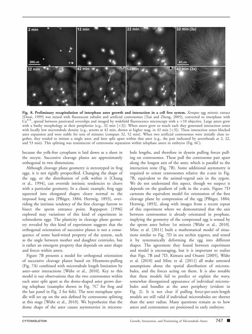
Fig. 8. Preliminary recapitulation of interphase aster growth and interaction in a cell free system. Xenopus egg mitotic extract[Desai, 1999] was mixed with fluorescent tubulin and artificial centrosomes [Tsai and Zheng, 2005], converted to interphase withCa11, spread between passivated coverslips and imaged by widefield fluorescence microscopy with a �10 objective. Large asters grewwith a bushy morphology at their peripheries (e.g., 32 min [�3]). When asters grew to touch each they generated interaction zoneswith locally low microtubule density (e.g., arrows at 42 min, shown at higher mag. in 42 min [�3]). These interaction zones blockedaster expansion and were stable for tens of minutes (compare 32, 52 min). When two artificial centrosomes were initially close to-gether, they tended to initiate a single aster, and later split apart within that aster (e.g., the pair indicated by arrowheads at 2, 22,and 53 min). This splitting was reminiscent of centrosome separation within telophase asters in embryos (Fig. 6C).
CYTOSKELETON Growth, Interaction, and Positioning of Microtubule Asters 747 n

Cell-Free Reconstitution ofInterphase Aster Growth andInteraction
It will be difficult to elucidate the molecular and biophysi-cal mechanisms involved in aster dynamics using whole,living embryos as the only experimental system, especiallyin Xenopus where the egg is opaque. The related problemof meiosis-II spindle assembly in Xenopus eggs was tackledusing cell-free extracts that accurately recapitulated the as-sembly process and greatly facilitated imaging and pertur-bation experiments [Desai et al., 1999; Maresca andHeald, 2006]. Xenopus egg extract is essentially undilutedcytoplasm with abundant organelles and vigorous energymetabolism [Niethammer et al., 2008]. We recently modi-fied this system to mimic polyspermic fertilization (Fig. 8).We observed rapid growth of large interphase asters and for-mation of aster–aster interaction zones when asters grew intoeach other (e.g., arrows in Fig. 8, 42 min). Aster morphol-ogy in this system recapitulated key aspects noted in embryosby immunofluorescence and live imaging, including ‘‘bushy’’peripheries, locally low density of microtubules in interactionzones, and a tendency of closely spaced nucleating sites tosplit apart within single asters. This system should facilitateprogress on molecular and biophysical mechanisms thatunderlie aster dynamics in large embryo cells.
Questions and Directions
In closing, we will highlight key questions from each sec-tion of this review where we need to uncover new molecu-lar and biophysical mechanisms. (i) Aster growth: what isthe mechanism for keeping microtubule density constantas aster radius expands? If microtubules nucleate awayfrom the centrosome as we suspect, what is the mecha-nism? (ii) Aster-aster interaction: how do growing astersrecognize each other when they touch, and how does thisrecognition lead to inhibition of aster growth? To whatextent are interaction zones between sister and non-sisterasters similar at the molecular level, and why do only theformer induce furrows in frog zygotes at 1st mitosis? (iii)Aster positioning: can we find further experimental valida-tion for the Hiramoto model for aster centering? How docentrosomes split apart within growing asters, and whatdetermines the axis on which they separate?
Answering these questions will surely require interdisci-plinary approaches that combine imaging, biochemistry,genetics, physical perturbation, force measurement, andcomputational modeling. Different biological systems havecomplementary advantages for these approaches, and weexpect that Xenopus egg extract will prove particularly ver-satile. Vertebrate embryos with extremely large cells, whereaster dynamics operate at a physical extreme, will help elu-cidate not only general principles of physical organizationof cells, but also how these principles scale with cell size.
Acknowledgments
This work was supported by NIH GM39565. The authorsthank Nikon Inc. for support of microscopy at the HMSNikon Imaging Center and MBL. Work at MBL was sup-ported by fellowships from the Evans Foundation, MBLAssociates, and the Laura and Authur Colwin SummerResearch Fund. They thank Sean Megason (HMS) andAngela DePace (HMS) for advice and time on their confo-cal microscopes. They thank Elly Tanaka (TU Dresden)and Mirjam Mayer (Whitehead Institute) for providingfixed axolotl zygotes. They also thank George von Dassow(Oregon Institute for Marine Biology) for providingunpublished data (Fig. 4) as well as stimulating discussion.
References
Ahmad FJ, Baas PW. 1995. Microtubules released from the neuro-nal centrosome are transported into the axon. J Cell Sci 108:2761–2769.
Andreassen PR, Palmer DK, Wener MH, Margolis RL. 1991. Telo-phase disc: a new mammalian mitotic organelle that bisects telo-phase cells with a possible function in cytokinesis. J Cell Sci 99:523–534.
Asnes CF, Schroeder TE. 1979. Cell cleavage. Ultrastructural evi-dence against equatorial stimulation by aster microtubules. Exp CellRes 122:327–38.
Baruni JR, Munro EM, von Dassow G. 2008. Cytokinetic furrow-ing in toroidal, binucleate and anucleate cells in C. elegansembryos. J Cell Sci 121:306–316.
Becker BE, Gard DL. 2006. Visualization of the cytoskeleton inXenopus oocytes and eggs by confocal immunofluorescence micros-copy. Methods Mol Biol. 322:69–86.
Belmont LD, Hyman AA, Sawin KE, Mitchison TJ. 1990. Real-time visualization of cell cycle-dependent changes in microtubuledynamics in cytoplasmic extracts. Cell 62:579–589.
Bergen LG, Kuriyama R, Borisy GG. 1980. Polarity of microtu-bules nucleated by centrosomes and chromosomes of Chinese ham-ster ovary cells in vitro. J Cell Biol 84:151–159.
Bieling P, Telley IA, Surrey T. 2010. A minimal midzone proteinmodule controls formation and length of antiparallel microtubuleoverlaps. Cell 142:420–432.
Black SD, Vincent JP. 1988. The first cleavage plane and the em-bryonic axis are determined by separate mechanisms in Xenopuslaevis. II. Experimental dissociation by lateral compression of theegg. Dev Biol 128:65–71.
Blow JJ. 2001. Control of chromosomal DNA replication in theearly Xenopus embryo. EMBO J 20:3293–3297
Brachet A. 1910. La polyspermie experimental comme moyend’analyse de la fecondacion. Arch Entwicklungsmech Org 30:261–303.
Bringmann H, Skiniotis G, Spilker A, Kandels-Lewis S, Vernos I,Surrey T. 2004. A kinesin-like motor inhibits microtubule dynamicinstability. Science 303:1519–1522.
Brinkley BR. 1985. Microtubule organizing centers. Annu Rev CellBiol 1:145–172.
Brinkley BR, Cox SM, Pepper DA, Wible L, Brenner SL, PardueRL. 1981. Tubulin assembly sites and the organization of cytoplas-mic microtubules in cultured mammalian cells. J Cell Biol 90:554–562.
n 748 Mitchison et al. CYTOSKELETON

Burbank KS, Groen AC, Perlman ZE, Fisher DS, Mitchison TJ.2006. A new method reveals microtubule minus ends throughoutthe meiotic spindle. J Cell Biol 175:369–375.
Chan RC, Forbes DI. 2006. In vitro study of nuclear assembly andnuclear import using Xenopus egg extracts. Methods Mol Biol 322:289–300.
Chan J, Sambade A, Calder G, Lloyd C. 2009. Arabidopsis corticalmicrotubules are initiated along, as well as branching from, existingmicrotubules. Plant Cell 21:2298–2306.
Choi YK, Liu P, Sze SK, Dai C, Qi RZ. 2010. CDK5RAP2 stimu-lates microtubule nucleation by the gamma-tubulin ring complex. JCell Biol 13;191(6):1089–1095.
Chung HM, Yokota H, Dent A, Malacinski GM, Neff AW. 1994. Thelocation of the third cleavage plane of Xenopus embryos partitionsmorphogenetic information in animal quartets. Int J Dev Biol 38:421–428
Decker M, Jaensch S, Pozniakovsky A, Zinke A, O’Connell KF,Zachariae W, Myers E, Hyman AA. 2011. Limiting amounts ofcentrosome material set centrosome size in C. elegans embryos.Curr Biol 21:1259–1267.
Desai A, Murray A, Mitchison TJ, Walczak CE. 1999. The use ofXenopus egg extracts to study mitotic spindle assembly and func-tion in vitro. Methods Cell Biol 61:385–412.
Efimov A, Kharitonov A, Efimova N, Loncarek J, Miller PM,Andreyeva N, Gleeson P, Galjart N, Maia AR, McLeod IX, YatesJR, III, Maiato H, Khodjakov A, Akhmanova A, Kaverina I. 2007.Asymmetric CLASP-dependent nucleation of noncentrosomalmicrotubules at the trans-Golgi network. Dev Cell 12:917–930.
Eggert US, Mitchison TJ, Field CM. 2006. Animal cytokinesis:from parts list to mechanisms. Annu Rev Biochem 75:543–566.
Fankhauser G. 1948. The organization of the amphibian egg duringfertilization and cleavage. Ann N Y Acad Sci 49(Art 5):684–708
Foe VE, von Dassow G. 2008. Stable and dynamic microtubulescoordinately shape the myosin activation zone during cytokineticfurrow formation. J Cell Biol 183:457–470.
Gatlin JC, Matov A, Groen AC, Needleman DJ, Maresca TJ, Dan-user G, Mitchison TJ, Salmon ED. 2009. Spindle fusion requiresdynein-mediated sliding of oppositely oriented microtubules. CurrBiol 19:287–296.
Glotzer M. 2005. The molecular requirements for cytokinesis. Sci-ence 307:1735–1739.
G€onczy P, Pichler S, Kirkham M, Hyman AA. 1999 Cytoplasmicdynein is required for distinct aspects of MTOC positioning,including centrosome separation, in the one cell stage Caenorhabdi-tis elegans embryo. J Cell Biol 147:135–150
Grill SW, Hyman AA. 2005. Spindle positioning by cortical pullingforces. Dev Cell 8:461–465.
Hamaguchi MS, Hiramoto Y. 1986. Analysis of the role of astralrays in pronuclear migration in sand dollar eggs by the colcemid-UV method. Dev Growth Differ 28:143–156
Hamaguchi MS, Hamaguchi Y, Hiramoto Y. 1986. Polystyrenebeads move along astral rays in sand dollar eggs. Dev Growth Dif-fer 28:461–470.
Herlant, M. 1911. Recherches sur les oeufs di-et-trispermiques degrenouille. Archs Biol 26:103–328.
Hertwig O. 1893. Ueber den Werth der ersten Furchungszellen fuerdie Organbildung des Embryo. Experimentelle Studien am Frosch-und Tritonei. Arch mikr Anat xlii 662–807.
Howard J. 2006. Elastic and damping forces generated by confinedarrays of dynamic microtubules. Phys Biol 3:54–66.
Hu CK, Coughlin M, Field CM, Mitchison TJ. 2011. KIF4 regu-lates midzone length during cytokinesis. Curr Biol 21:815–824.
Hu CK, Coughlin M, Mitchison TJ. 2012. Midbody assembly andits regulation during cytokinesis. Mol Biol Cell 23:1024–1034.
Hyman AA. 1989. Centrosome movement in the early divisions ofCaenorhabditis elegans: a cortical site determining centrosome posi-tion. J Cell Biol 109:1185–1193.
Iwao Y. 1989. An electrically mediated block to polyspermy in theprimitive urodele Hynobis nebulous and phylogenetic comparisonwith other amphibians. Dev Biol 134:438–445.
Kimura A, Onami S. 2005. Computer simulations and image proc-essing reveal length-dependent pulling force as the primary mecha-nism for C. elegans male pronuclear migration. Dev Cell 8:765–775.
Kimura K, Kimura A. 2011. Intracellular organelles mediate cyto-plasmic pulling force for centrosome centration in the Caenorhabdi-tis elegans early embryo. Proc Natl Acad Sci USA 108(1):137–142.
Kirik A, Ehrhardt DW, Kirik V. 2012. TONNEAU2/FASS regu-lates the geometry of microtubule nucleation and cortical array orga-nization in interphase Arabidopsis cells. Plant Cell 24:1158–1170.
Klymkowsky MW, Hanken J. 1991. Whole-mount staining of Xen-opus and other vertebrates. Methods Cell Biol 36:419–441
Kollman JM, Merdes A, Mourey L, Agard DA. 2011. Microtubulenucleation by c-tubulin complexes. Nat Rev Mol Cell Biol 2(11):709–721
Luders J, Stearns T. 2007. Microtubule-organizing centres: a re-eval-uation. Nat Rev Mol Cell Biol 8:161–167.
Maresca TJ, Heald R. 2006. Methods for studying spindle assemblyand chromosome condensation in Xenopus egg extracts. MethodsMol Biol 322:459–474.
Minc N, Burgess D, Chang F. 2011. Influence of cell geometry ondivision-plane positioning. Cell 144:414–426.
Mishima M, Kaitna S, Glotzer M. 2002. Central spindle assemblyand cytokinesis require a kinesin-like protein/RhoGAP complexwith microtubule bundling activity. Dev Cell 2:41–54.
Mollinari C, Kleman JP, Jiang W, Schoehn G, Hunter T, MargolisRL. 2002. PRC1 is a microtubule binding and bundling proteinessential to maintain the mitotic spindle midzone. J Cell Biol 157:1175–1186.
Morgan DO. 2006. The Cell Cycle. Principles of Control. Orby,Northants, UK: Oxford University Press.
Murata T, Sonobe S, Baskin TI, Hyodo S, Hasezawa S, Nagata T,Horio T, Hasebe M. 2005. Microtubule-dependent microtubulenucleation based on recruitment of gamma-tubulin in higher plants.Nat Cell Biol 7(10):961–968.
Niethammer P, Kronja I, Kandels-Lewis S, Rybina S, Bastiaens P,Karsenti E. 2007. Discrete states of a protein interaction networkgovern interphase and mitotic microtubule dynamics. PLoS Biol5(2):e29.
Niethammer P, Kueh HY, Mitchison TJ. 2008. Spatial patterningof metabolism by mitochondria, oxygen, and energy sinks in amodel cytoplasm. Curr Biol 18:586–591.
Nislow C, Lombillo VA, Kuriyama R, McIntosh JR. 1992. A plus-end-directed motor enzyme that moves antiparallel microtubules invitro localizes to the interzone of mitotic spindles. Nature 359:543–547.
Pfluger E. 1884. Ueber die Einwirkung der Schwerkraft undanderer Bedingungen auf die Richtung der Zelltheilung. PflugersArchiv. 34:607–616.
Pollard TD, Borisy GG. 2003. Cellular motility driven by assemblyand disassembly of actin filaments. Cell 112:453–465.
CYTOSKELETON Growth, Interaction, and Positioning of Microtubule Asters 749 n

Rappaport R 1961 Experiments concerning the cleavage stimulus inSand Dollar eggs. J Exp Zool 148:81–89.
Rappaport R. 1996. Cytokinesis in Animal Cells. UK: CambridgeUniversity Press.
Render JA, Elinson RP. 1986. Axis determination in polyspermicXenopus laevis eggs. Dev Biol 115:425–433.
Rivero S, Cardenas J, Bornens M, Rios RM. 2009. Microtubulenucleation at the cis-side of the Golgi apparatus requires AKAP450and GM130. EMBO J 28:1016–1028.
Ruchaud S, Carmena M, Earnshaw WC. 2007. Chromosomalpassengers: conducting cell division. Nat Rev Mol Cell Biol 8:798–812.
Rusan NM, Tulu US, Fagerstrom C, Wadsworth P. 2002. Reorgan-ization of the microtubule array in prophase/prometaphase requirescytoplasmic dynein-dependent microtubule transport. J Cell Biol158:997–1003.
Samejima I, Lourenco PC, Snaith HA, Sawin KE. 2006. Fissionyeast mto2p regulates microtubule nucleation by the centrosomin-related protein mto1p. Mol Biol Cell 16:3040–3051.
Savoian MS, Earnshaw WC, Khodjakov A, Rieder CL. 1999.Cleavage furrows formed between centrosomes lacking an interven-ing spindle and chromosome contain microtubule bundles,INCENP and CHO1, but not CENP-E. Mol Biol Cell 10:297–311.
Shinar T, Mana M, Piano F, Shelley MJ. 2011. A model of cytoplasmi-cally driven microtubule-based motion in the single-celled Caenorhab-ditis elegans embryo. Proc Natl Acad Sci USA 108:10508–10513.
Srayko M, Kaya A, Stamford J, Hyman AA. 2005. Identificationand characterization of factors required for microtubule growth andnucleation in the early C. elegans embryo. Dev Cell 9:223–236.
Strickland L, von Dassow G, Ellenberg J, Foe V, Lenart P, BurgessD. 2004. Light microscopy of echinoderm embryos. Methods CellBiol 74:371–409.
Subramanian R, Wilson-Kubalek EM, Arthur CP, Bick MJ, Camp-bell EA, Darst SA, Milligan RA, Kapoor TM. 2010. Insights intoantiparallel microtubule crosslinking by PRC1, a conserved nonmo-tor microtubule binding protein. Cell 142:433–443.
Tran PT, Marsh L, Doye V, Inou�e S, Chang F. 2001. A mechanismfor nuclear positioning in fission yeast based on microtubule push-ing. J Cell Biol 153:397–411.
Tsai MY, Zheng Y. 2005. Aurora A kinase-coated beads function asmicrotubule-organizing centers and enhance RanGTP-induced spin-dle assembly. Curr Biol 15:2156–2163.
Uehara R, Nozawa RS, Tomioka A, Petry S, Vale RD, Obuse C,Goshima G. 2009. The augmin complex plays a critical role inspindle microtubule generation for mitotic progression and cytoki-nesis in human cells. Proc Natl Acad Sci USA 106:6998–7003.
Verde F, Dogterom M, Stelzer E, Karsenti E, Leibler S. 1992. Con-trol of microtubule dynamics and length by cyclin A- and cyclin B-dependent kinases in Xenopus egg extracts. J Cell Biol 118:1097–1108.
von Dassow G, Verbrugghe KJ, Miller AL, Sider JR, Bement WM.2009. Action at a distance during cytokinesis. J Cell Biol 187:831–845.
Wakabayashi Y, Shinagawa A. 2001. Presence of a nucleus or nu-cleus-deriving factors is indispensable for the formation of the spin-dle, the diastema and the cleavage furrow in the blastomere of theXenopus embryo. Dev Growth Differ 43:633–646.
Wilson EB. 1925. The Cell in Development and Heredity, 3rd ed.NY: McMillan.
Wuhr M, Chen Y, Dumont S, Groen AC, Needleman DJ, Salic A,Mitchison TJ. 2008. Evidence for an upper limit to mitotic spindlelength. Curr Biol 18:1256–1261.
Wuhr M, Dumont S, Groen AC, Needleman DJ, Mitchison TJ.2009. How does a millimeter-sized cell find its center? Cell Cycle15;8(8):1115–1121.
Wuhr M, Tan ES, Parker SK, Detrich HW, III, Mitchison TJ.2010. A model for cleavage plane determination in early amphibianand fish embryos. Curr Biol 20:2040–2045.
Wuhr M, Obholzer ND, Megason SG, Detrich HW, III, MitchisonTJ. 2011. Live imaging of the cytoskeleton in early cleavage-stagezebrafish embryos. Methods Cell Biol 101:1–18
Yabe T, Ge X, Lindeman R, Nair S, Runke G, Mullins MC, PelegriF. 2009. The maternal-effect gene cellular island encodes aurora Bkinase and is essential for furrow formation in the early zebrafishembryo. PLoS Genet 5(6):e1000518
n 750 Mitchison et al. CYTOSKELETON