Embed Size (px)
Citation preview
OpenStax-CNX module: m44617 1
Groups of Protists∗
OpenStax College
This work is produced by OpenStax-CNX and licensed under the
Creative Commons Attribution License 3.0†
Abstract
By the end of this section, you will be able to:
• Describe representative protist organisms from each of the six presently recognized supergroups ofeukaryotes
• Identify the evolutionary relationships of plants, animals, and fungi within the six presently recog-nized supergroups of eukaryotes
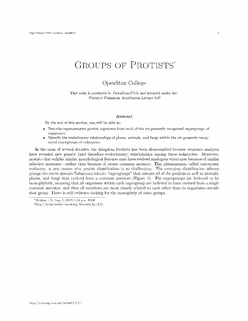
In the span of several decades, the Kingdom Protista has been disassembled because sequence analyseshave revealed new genetic (and therefore evolutionary) relationships among these eukaryotes. Moreover,protists that exhibit similar morphological features may have evolved analogous structures because of similarselective pressures�rather than because of recent common ancestry. This phenomenon, called convergentevolution, is one reason why protist classi�cation is so challenging. The emerging classi�cation schemegroups the entire domain Eukaryota into six �supergroups� that contain all of the protists as well as animals,plants, and fungi that evolved from a common ancestor (Figure 1). The supergroups are believed to bemonophyletic, meaning that all organisms within each supergroup are believed to have evolved from a singlecommon ancestor, and thus all members are most closely related to each other than to organisms outsidethat group. There is still evidence lacking for the monophyly of some groups.
∗Version 1.7: Aug 7, 2013 4:49 pm -0500†http://creativecommons.org/licenses/by/3.0/
http://cnx.org/content/m44617/1.7/
OpenStax-CNX module: m44617 2
Figure 1: This diagram shows a proposed classi�cation of the domain Eukara. Currently, the domainEukarya is divided into six supergroups. Within each supergroup are multiple kingdoms. Dotted linesindicate suggested evolutionary relationships that remain under debate.
http://cnx.org/content/m44617/1.7/
OpenStax-CNX module: m44617 3
The classi�cation of eukaryotes is still in �ux, and the six supergroups may be modi�ed or replaced by amore appropriate hierarchy as genetic, morphological, and ecological data accumulate. Keep in mind that theclassi�cation scheme presented here is just one of several hypotheses, and the true evolutionary relationshipsare still to be determined. When learning about protists, it is helpful to focus less on the nomenclature andmore on the commonalities and di�erences that de�ne the groups themselves.
1 Excavata
Many of the protist species classi�ed into the supergroup Excavata are asymmetrical, single-celled organ-isms with a feeding groove �excavated� from one side. This supergroup includes heterotrophic predators,photosynthetic species, and parasites. Its subgroups are the diplomonads, parabasalids, and euglenozoans.
1.1 Diplomonads
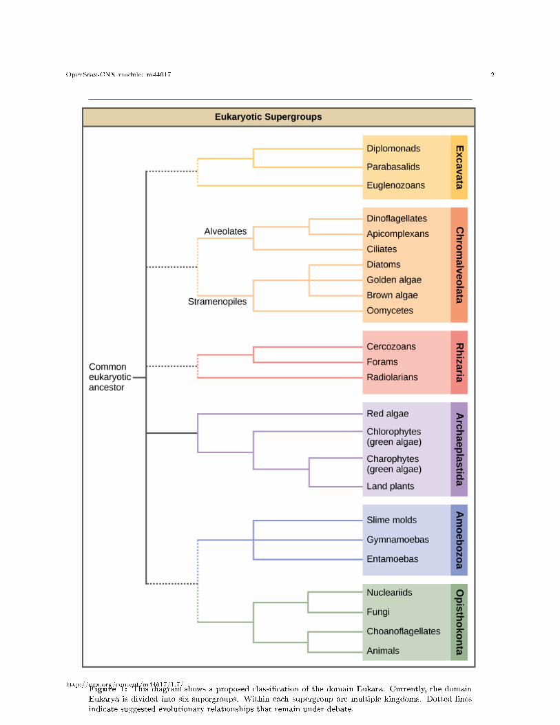
Among the Excavata are the diplomonads, which include the intestinal parasite, Giardia lamblia (Figure 2).Until recently, these protists were believed to lack mitochondria. Mitochondrial remnant organelles, calledmitosomes, have since been identi�ed in diplomonads, but these mitosomes are essentially nonfunctional.Diplomonads exist in anaerobic environments and use alternative pathways, such as glycolysis, to generateenergy. Each diplomonad cell has two identical nuclei and uses several �agella for locomotion.
Figure 2: The mammalian intestinal parasite Giardia lamblia, visualized here using scanning electronmicroscopy, is a waterborne protist that causes severe diarrhea when ingested. (credit: modi�cation ofwork by Janice Carr, CDC; scale-bar data from Matt Russell)
http://cnx.org/content/m44617/1.7/
OpenStax-CNX module: m44617 4
1.2 Parabasalids
A second Excavata subgroup, the parabasalids, also exhibits semi-functional mitochondria. In parabasalids,these structures function anaerobically and are called hydrogenosomes because they produce hydrogengas as a byproduct. Parabasalids move with �agella and membrane rippling. Trichomonas vaginalis, aparabasalid that causes a sexually transmitted disease in humans, employs these mechanisms to transitthrough the male and female urogenital tracts. T. vaginalis causes trichamoniasis, which appears in anestimated 180 million cases worldwide each year. Whereas men rarely exhibit symptoms during an infectionwith this protist, infected women may become more susceptible to secondary infection with human immun-ode�ciency virus (HIV) and may be more likely to develop cervical cancer. Pregnant women infected withT. vaginalis are at an increased risk of serious complications, such as pre-term delivery.
1.3 Euglenozoans
Euglenozoans includes parasites, heterotrophs, autotrophs, and mixotrophs, ranging in size from 10 to 500µm. Euglenoids move through their aquatic habitats using two long �agella that guide them toward lightsources sensed by a primitive ocular organ called an eyespot. The familiar genus, Euglena, encompasses somemixotrophic species that display a photosynthetic capability only when light is present. In the dark, thechloroplasts of Euglena shrink up and temporarily cease functioning, and the cells instead take up organicnutrients from their environment.
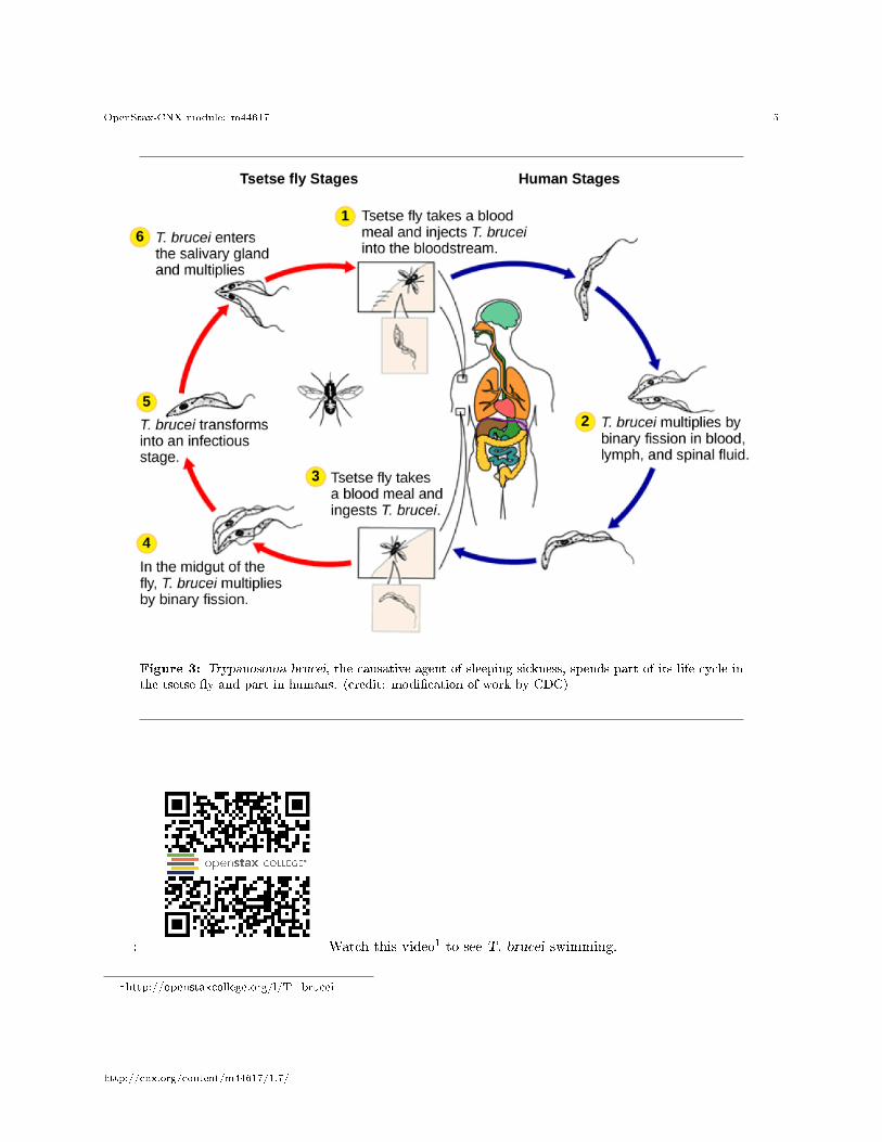
The human parasite, Trypanosoma brucei, belongs to a di�erent subgroup of Euglenozoa, the kinetoplas-tids. The kinetoplastid subgroup is named after the kinetoplast, a DNA mass carried within the single,oversized mitochondrion possessed by each of these cells. This subgroup includes several parasites, collec-tively called trypanosomes, which cause devastating human diseases and infect an insect species during aportion of their life cycle. T. brucei develops in the gut of the tsetse �y after the �y bites an infectedhuman or other mammalian host. The parasite then travels to the insect salivary glands to be transmittedto another human or other mammal when the infected tsetse �y consumes another blood meal. T. brucei iscommon in central Africa and is the causative agent of African sleeping sickness, a disease associated withsevere chronic fatigue, coma, and can be fatal if left untreated.
http://cnx.org/content/m44617/1.7/
OpenStax-CNX module: m44617 5
Figure 3: Trypanosoma brucei, the causative agent of sleeping sickness, spends part of its life cycle inthe tsetse �y and part in humans. (credit: modi�cation of work by CDC)
: Watch this video1 to see T. brucei swimming.
1http://openstaxcollege.org/l/T_brucei
http://cnx.org/content/m44617/1.7/
OpenStax-CNX module: m44617 6
2 Chromalveolata
Current evidence suggests that species classi�ed as chromalveolates are derived from a common ancestorthat engulfed a photosynthetic red algal cell, which itself had already evolved chloroplasts from an endosym-biotic relationship with a photosynthetic prokaryote. Therefore, the ancestor of chromalveolates is believedto have resulted from a secondary endosymbiotic event. However, some chromalveolates appear to havelost red alga-derived plastid organelles or lack plastid genes altogether. Therefore, this supergroup shouldbe considered a hypothesis-based working group that is subject to change. Chromalveolates include veryimportant photosynthetic organisms, such as diatoms, brown algae, and signi�cant disease agents in animalsand plants. The chromalveolates can be subdivided into alveolates and stramenopiles.
2.1 Alveolates: Dino�agellates, Apicomplexians, and Ciliates
A large body of data supports that the alveolates are derived from a shared common ancestor. The alveolatesare named for the presence of an alveolus, or membrane-enclosed sac, beneath the cell membrane. The exactfunction of the alveolus is unknown, but it may be involved in osmoregulation. The alveolates are furthercategorized into some of the better-known protists: the dino�agellates, the apicomplexans, and the ciliates.
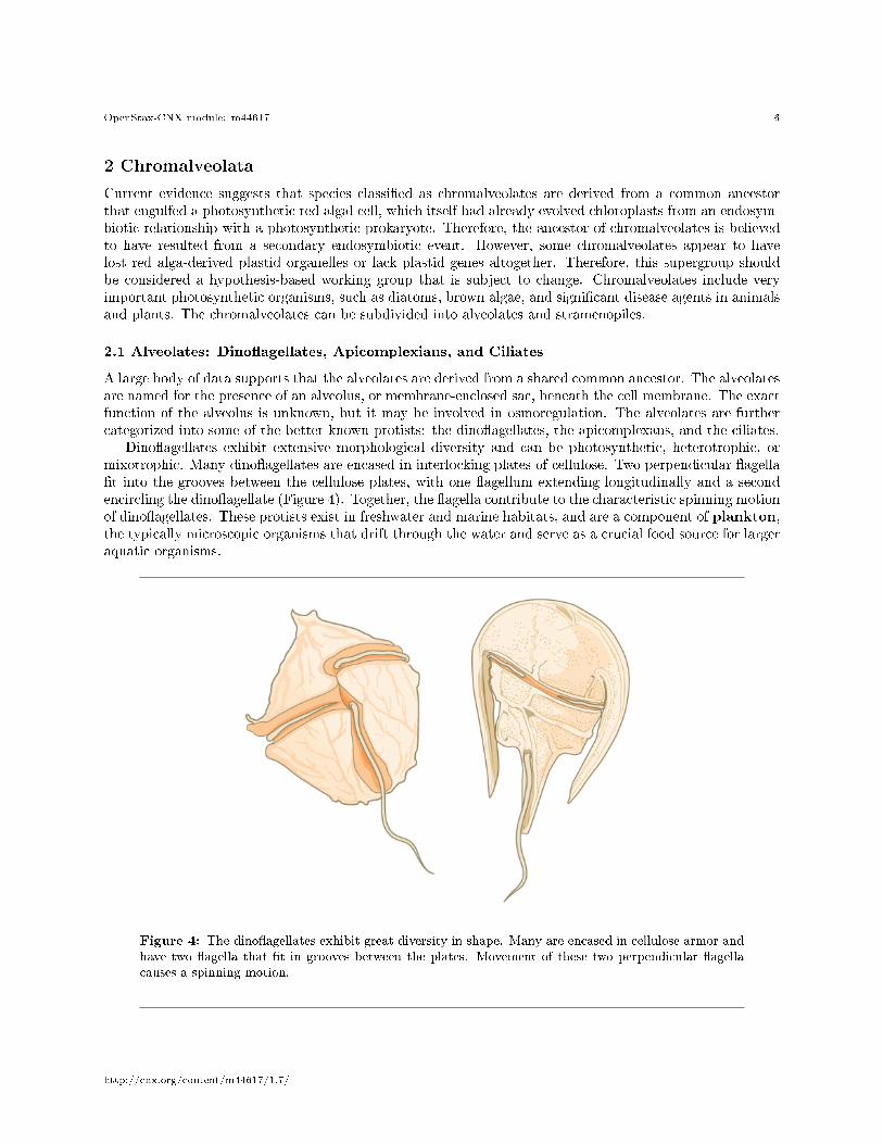
Dino�agellates exhibit extensive morphological diversity and can be photosynthetic, heterotrophic, ormixotrophic. Many dino�agellates are encased in interlocking plates of cellulose. Two perpendicular �agella�t into the grooves between the cellulose plates, with one �agellum extending longitudinally and a secondencircling the dino�agellate (Figure 4). Together, the �agella contribute to the characteristic spinning motionof dino�agellates. These protists exist in freshwater and marine habitats, and are a component of plankton,the typically microscopic organisms that drift through the water and serve as a crucial food source for largeraquatic organisms.
Figure 4: The dino�agellates exhibit great diversity in shape. Many are encased in cellulose armor andhave two �agella that �t in grooves between the plates. Movement of these two perpendicular �agellacauses a spinning motion.
http://cnx.org/content/m44617/1.7/
OpenStax-CNX module: m44617 7
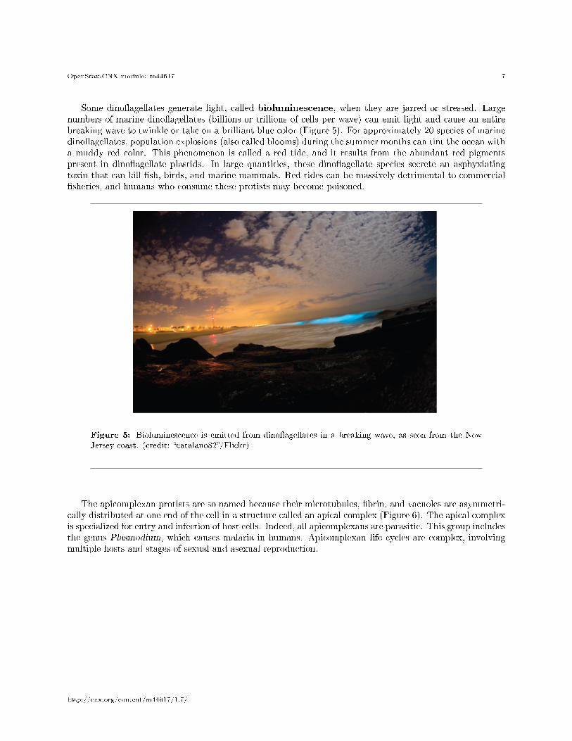
Some dino�agellates generate light, called bioluminescence, when they are jarred or stressed. Largenumbers of marine dino�agellates (billions or trillions of cells per wave) can emit light and cause an entirebreaking wave to twinkle or take on a brilliant blue color (Figure 5). For approximately 20 species of marinedino�agellates, population explosions (also called blooms) during the summer months can tint the ocean witha muddy red color. This phenomenon is called a red tide, and it results from the abundant red pigmentspresent in dino�agellate plastids. In large quantities, these dino�agellate species secrete an asphyxiatingtoxin that can kill �sh, birds, and marine mammals. Red tides can be massively detrimental to commercial�sheries, and humans who consume these protists may become poisoned.
Figure 5: Bioluminescence is emitted from dino�agellates in a breaking wave, as seen from the NewJersey coast. (credit: �catalano82�/Flickr)
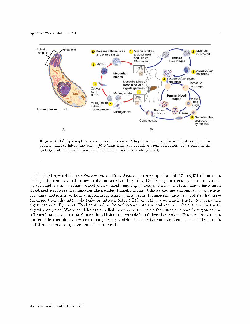
The apicomplexan protists are so named because their microtubules, �brin, and vacuoles are asymmetri-cally distributed at one end of the cell in a structure called an apical complex (Figure 6). The apical complexis specialized for entry and infection of host cells. Indeed, all apicomplexans are parasitic. This group includesthe genus Plasmodium, which causes malaria in humans. Apicomplexan life cycles are complex, involvingmultiple hosts and stages of sexual and asexual reproduction.
http://cnx.org/content/m44617/1.7/
OpenStax-CNX module: m44617 8
Figure 6: (a) Apicomplexans are parasitic protists. They have a characteristic apical complex thatenables them to infect host cells. (b) Plasmodium, the causative agent of malaria, has a complex lifecycle typical of apicomplexans. (credit b: modi�cation of work by CDC)
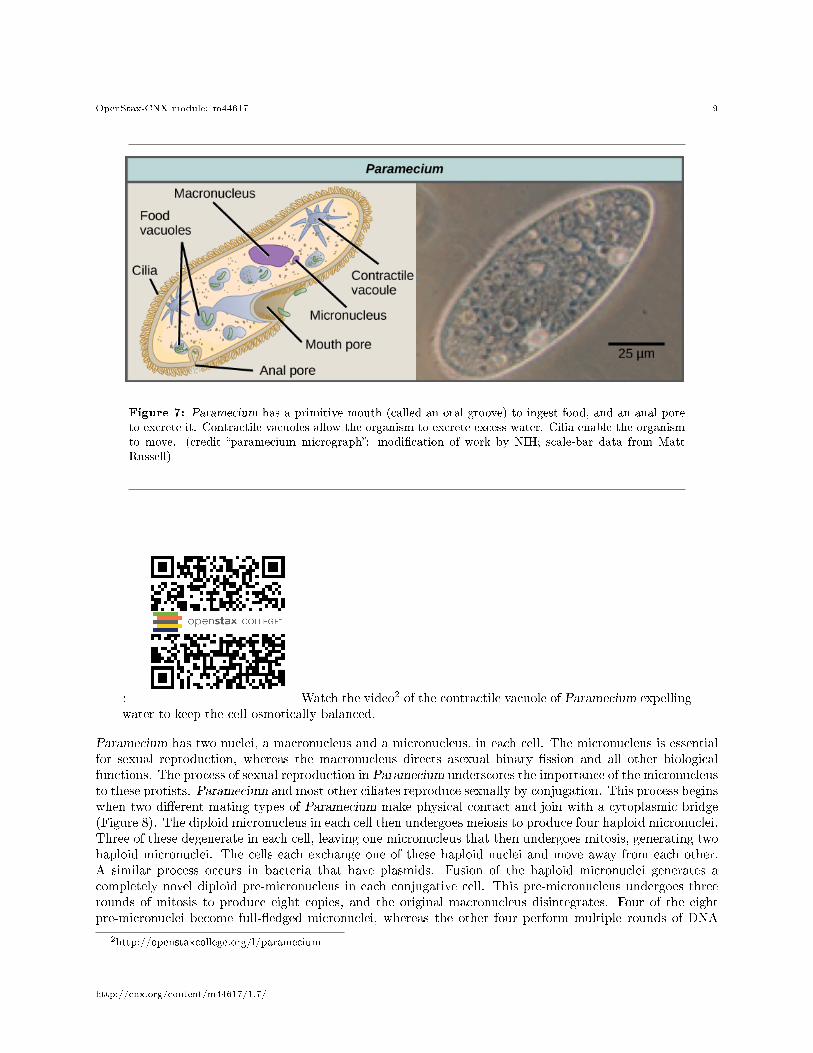
The ciliates, which include Paramecium and Tetrahymena, are a group of protists 10 to 3,000 micrometersin length that are covered in rows, tufts, or spirals of tiny cilia. By beating their cilia synchronously or inwaves, ciliates can coordinate directed movements and ingest food particles. Certain ciliates have fusedcilia-based structures that function like paddles, funnels, or �ns. Ciliates also are surrounded by a pellicle,providing protection without compromising agility. The genus Paramecium includes protists that haveorganized their cilia into a plate-like primitive mouth, called an oral groove, which is used to capture anddigest bacteria (Figure 7). Food captured in the oral groove enters a food vacuole, where it combines withdigestive enzymes. Waste particles are expelled by an exocytic vesicle that fuses at a speci�c region on thecell membrane, called the anal pore. In addition to a vacuole-based digestive system, Paramecium also usescontractile vacuoles, which are osmoregulatory vesicles that �ll with water as it enters the cell by osmosisand then contract to squeeze water from the cell.
http://cnx.org/content/m44617/1.7/
OpenStax-CNX module: m44617 9
Figure 7: Paramecium has a primitive mouth (called an oral groove) to ingest food, and an anal poreto excrete it. Contractile vacuoles allow the organism to excrete excess water. Cilia enable the organismto move. (credit �paramecium micrograph�: modi�cation of work by NIH; scale-bar data from MattRussell)
: Watch the video2 of the contractile vacuole of Paramecium expellingwater to keep the cell osmotically balanced.
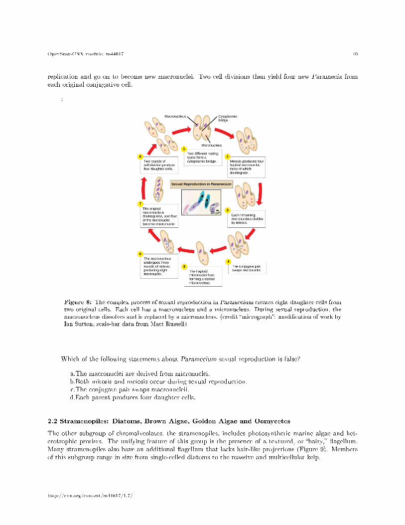
Paramecium has two nuclei, a macronucleus and a micronucleus, in each cell. The micronucleus is essentialfor sexual reproduction, whereas the macronucleus directs asexual binary �ssion and all other biologicalfunctions. The process of sexual reproduction in Paramecium underscores the importance of the micronucleusto these protists. Paramecium and most other ciliates reproduce sexually by conjugation. This process beginswhen two di�erent mating types of Paramecium make physical contact and join with a cytoplasmic bridge(Figure 8). The diploid micronucleus in each cell then undergoes meiosis to produce four haploid micronuclei.Three of these degenerate in each cell, leaving one micronucleus that then undergoes mitosis, generating twohaploid micronuclei. The cells each exchange one of these haploid nuclei and move away from each other.A similar process occurs in bacteria that have plasmids. Fusion of the haploid micronuclei generates acompletely novel diploid pre-micronucleus in each conjugative cell. This pre-micronucleus undergoes threerounds of mitosis to produce eight copies, and the original macronucleus disintegrates. Four of the eightpre-micronuclei become full-�edged micronuclei, whereas the other four perform multiple rounds of DNA
2http://openstaxcollege.org/l/paramecium
http://cnx.org/content/m44617/1.7/
OpenStax-CNX module: m44617 10
replication and go on to become new macronuclei. Two cell divisions then yield four new Paramecia fromeach original conjugative cell.
:
Figure 8: The complex process of sexual reproduction in Paramecium creates eight daughter cells fromtwo original cells. Each cell has a macronucleus and a micronucleus. During sexual reproduction, themacronucleus dissolves and is replaced by a micronucleus. (credit �micrograph�: modi�cation of work byIan Sutton; scale-bar data from Matt Russell)
Which of the following statements about Paramecium sexual reproduction is false?
a.The macronuclei are derived from micronuclei.b.Both mitosis and meiosis occur during sexual reproduction.c.The conjugate pair swaps macronucleii.d.Each parent produces four daughter cells.
2.2 Stramenopiles: Diatoms, Brown Algae, Golden Algae and Oomycetes
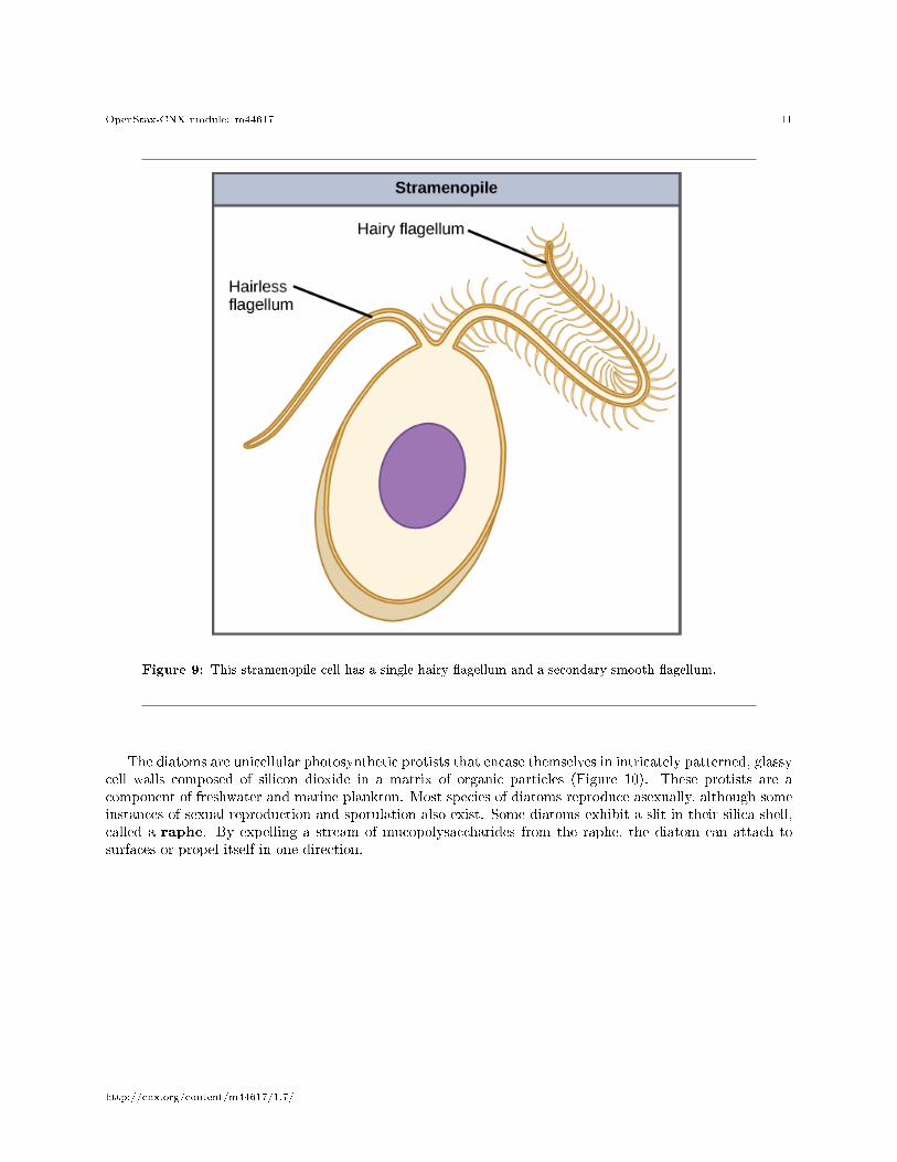
The other subgroup of chromalveolates, the stramenopiles, includes photosynthetic marine algae and het-erotrophic protists. The unifying feature of this group is the presence of a textured, or �hairy,� �agellum.Many stramenopiles also have an additional �agellum that lacks hair-like projections (Figure 9). Membersof this subgroup range in size from single-celled diatoms to the massive and multicellular kelp.
http://cnx.org/content/m44617/1.7/
OpenStax-CNX module: m44617 11
Figure 9: This stramenopile cell has a single hairy �agellum and a secondary smooth �agellum.
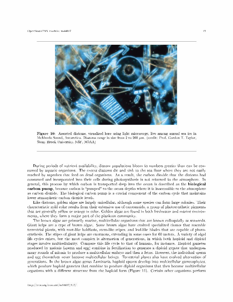
The diatoms are unicellular photosynthetic protists that encase themselves in intricately patterned, glassycell walls composed of silicon dioxide in a matrix of organic particles (Figure 10). These protists are acomponent of freshwater and marine plankton. Most species of diatoms reproduce asexually, although someinstances of sexual reproduction and sporulation also exist. Some diatoms exhibit a slit in their silica shell,called a raphe. By expelling a stream of mucopolysaccharides from the raphe, the diatom can attach tosurfaces or propel itself in one direction.
http://cnx.org/content/m44617/1.7/
OpenStax-CNX module: m44617 12
Figure 10: Assorted diatoms, visualized here using light microscopy, live among annual sea ice inMcMurdo Sound, Antarctica. Diatoms range in size from 2 to 200 µm. (credit: Prof. Gordon T. Taylor,Stony Brook University, NSF, NOAA)
During periods of nutrient availability, diatom populations bloom to numbers greater than can be con-sumed by aquatic organisms. The excess diatoms die and sink to the sea �oor where they are not easilyreached by saprobes that feed on dead organisms. As a result, the carbon dioxide that the diatoms hadconsumed and incorporated into their cells during photosynthesis is not returned to the atmosphere. Ingeneral, this process by which carbon is transported deep into the ocean is described as the biologicalcarbon pump, because carbon is �pumped� to the ocean depths where it is inaccessible to the atmosphereas carbon dioxide. The biological carbon pump is a crucial component of the carbon cycle that maintainslower atmospheric carbon dioxide levels.
Like diatoms, golden algae are largely unicellular, although some species can form large colonies. Theircharacteristic gold color results from their extensive use of carotenoids, a group of photosynthetic pigmentsthat are generally yellow or orange in color. Golden algae are found in both freshwater and marine environ-ments, where they form a major part of the plankton community.
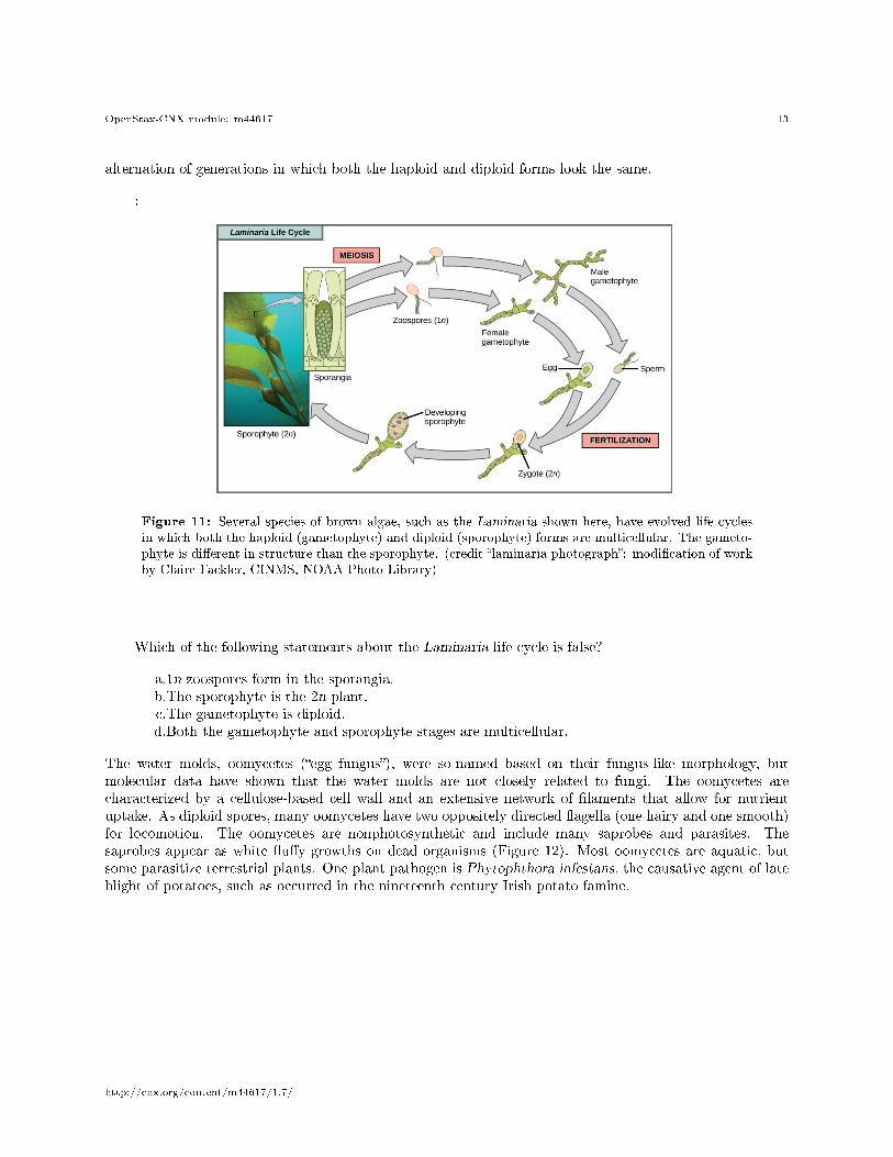
The brown algae are primarily marine, multicellular organisms that are known colloquially as seaweeds.Giant kelps are a type of brown algae. Some brown algae have evolved specialized tissues that resembleterrestrial plants, with root-like holdfasts, stem-like stipes, and leaf-like blades that are capable of photo-synthesis. The stipes of giant kelps are enormous, extending in some cases for 60 meters. A variety of algallife cycles exists, but the most complex is alternation of generations, in which both haploid and diploidstages involve multicellularity. Compare this life cycle to that of humans, for instance. Haploid gametesproduced by meiosis (sperm and egg) combine in fertilization to generate a diploid zygote that undergoesmany rounds of mitosis to produce a multicellular embryo and then a fetus. However, the individual spermand egg themselves never become multicellular beings. Terrestrial plants also have evolved alternation ofgenerations. In the brown algae genus Laminaria, haploid spores develop into multicellular gametophytes,which produce haploid gametes that combine to produce diploid organisms that then become multicellularorganisms with a di�erent structure from the haploid form (Figure 11). Certain other organisms perform
http://cnx.org/content/m44617/1.7/
OpenStax-CNX module: m44617 13
alternation of generations in which both the haploid and diploid forms look the same.
:
Figure 11: Several species of brown algae, such as the Laminaria shown here, have evolved life cyclesin which both the haploid (gametophyte) and diploid (sporophyte) forms are multicellular. The gameto-phyte is di�erent in structure than the sporophyte. (credit �laminaria photograph�: modi�cation of workby Claire Fackler, CINMS, NOAA Photo Library)
Which of the following statements about the Laminaria life cycle is false?
a.1n zoospores form in the sporangia.b.The sporophyte is the 2n plant.c.The gametophyte is diploid.d.Both the gametophyte and sporophyte stages are multicellular.
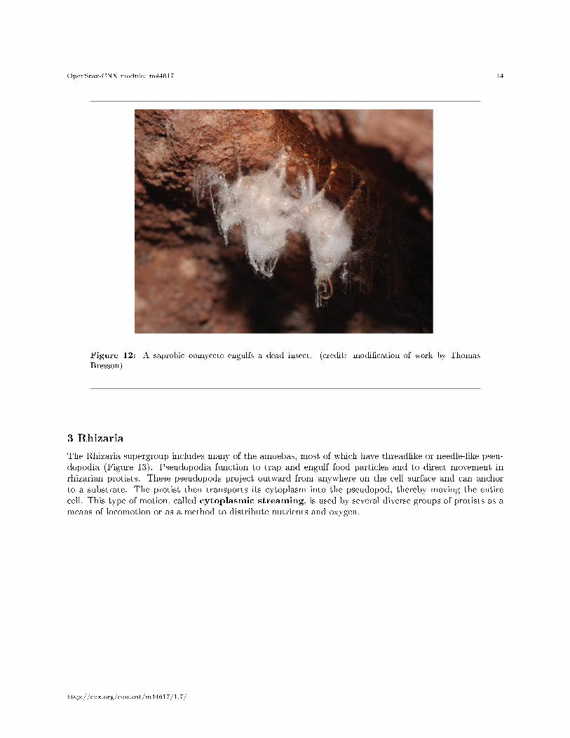
The water molds, oomycetes (�egg fungus�), were so-named based on their fungus-like morphology, butmolecular data have shown that the water molds are not closely related to fungi. The oomycetes arecharacterized by a cellulose-based cell wall and an extensive network of �laments that allow for nutrientuptake. As diploid spores, many oomycetes have two oppositely directed �agella (one hairy and one smooth)for locomotion. The oomycetes are nonphotosynthetic and include many saprobes and parasites. Thesaprobes appear as white �u�y growths on dead organisms (Figure 12). Most oomycetes are aquatic, butsome parasitize terrestrial plants. One plant pathogen is Phytophthora infestans, the causative agent of lateblight of potatoes, such as occurred in the nineteenth century Irish potato famine.
http://cnx.org/content/m44617/1.7/
OpenStax-CNX module: m44617 14
Figure 12: A saprobic oomycete engulfs a dead insect. (credit: modi�cation of work by ThomasBresson)
3 Rhizaria
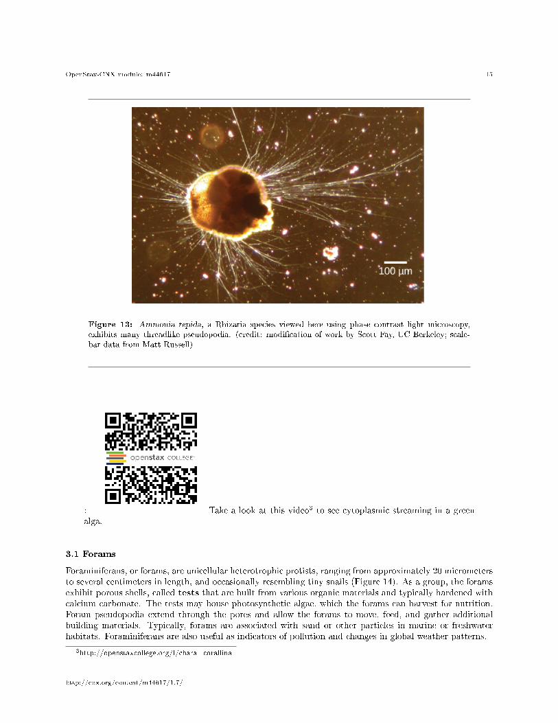
The Rhizaria supergroup includes many of the amoebas, most of which have threadlike or needle-like pseu-dopodia (Figure 13). Pseudopodia function to trap and engulf food particles and to direct movement inrhizarian protists. These pseudopods project outward from anywhere on the cell surface and can anchorto a substrate. The protist then transports its cytoplasm into the pseudopod, thereby moving the entirecell. This type of motion, called cytoplasmic streaming, is used by several diverse groups of protists as ameans of locomotion or as a method to distribute nutrients and oxygen.
http://cnx.org/content/m44617/1.7/
OpenStax-CNX module: m44617 15
Figure 13: Ammonia tepida, a Rhizaria species viewed here using phase contrast light microscopy,exhibits many threadlike pseudopodia. (credit: modi�cation of work by Scott Fay, UC Berkeley; scale-bar data from Matt Russell)
: Take a look at this video3 to see cytoplasmic streaming in a greenalga.
3.1 Forams
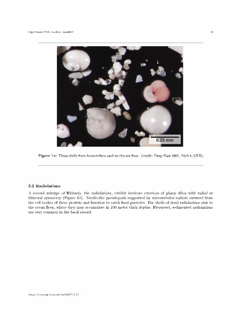
Foraminiferans, or forams, are unicellular heterotrophic protists, ranging from approximately 20 micrometersto several centimeters in length, and occasionally resembling tiny snails (Figure 14). As a group, the foramsexhibit porous shells, called tests that are built from various organic materials and typically hardened withcalcium carbonate. The tests may house photosynthetic algae, which the forams can harvest for nutrition.Foram pseudopodia extend through the pores and allow the forams to move, feed, and gather additionalbuilding materials. Typically, forams are associated with sand or other particles in marine or freshwaterhabitats. Foraminiferans are also useful as indicators of pollution and changes in global weather patterns.
3http://openstaxcollege.org/l/chara_corallina
http://cnx.org/content/m44617/1.7/
OpenStax-CNX module: m44617 16
Figure 14: These shells from foraminifera sank to the sea �oor. (credit: Deep East 2001, NOAA/OER)
3.2 Radiolarians
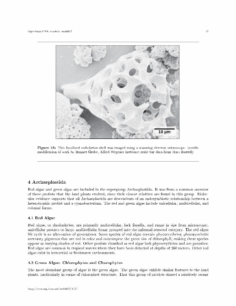
A second subtype of Rhizaria, the radiolarians, exhibit intricate exteriors of glassy silica with radial orbilateral symmetry (Figure 15). Needle-like pseudopods supported by microtubules radiate outward fromthe cell bodies of these protists and function to catch food particles. The shells of dead radiolarians sink tothe ocean �oor, where they may accumulate in 100 meter-thick depths. Preserved, sedimented radiolariansare very common in the fossil record.
http://cnx.org/content/m44617/1.7/
OpenStax-CNX module: m44617 17
Figure 15: This fossilized radiolarian shell was imaged using a scanning electron microscope. (credit:modi�cation of work by Hannes Grobe, Alfred Wegener Institute; scale-bar data from Matt Russell)
4 Archaeplastida
Red algae and green algae are included in the supergroup Archaeplastida. It was from a common ancestorof these protists that the land plants evolved, since their closest relatives are found in this group. Molec-ular evidence supports that all Archaeplastida are descendents of an endosymbiotic relationship between aheterotrophic protist and a cyanobacterium. The red and green algae include unicellular, multicellular, andcolonial forms.
4.1 Red Algae
Red algae, or rhodophytes, are primarily multicellular, lack �agella, and range in size from microscopic,unicellular protists to large, multicellular forms grouped into the informal seaweed category. The red algaelife cycle is an alternation of generations. Some species of red algae contain phycoerythrins, photosyntheticaccessory pigments that are red in color and outcompete the green tint of chlorophyll, making these speciesappear as varying shades of red. Other protists classi�ed as red algae lack phycoerythrins and are parasites.Red algae are common in tropical waters where they have been detected at depths of 260 meters. Other redalgae exist in terrestrial or freshwater environments.
4.2 Green Algae: Chlorophytes and Charophytes
The most abundant group of algae is the green algae. The green algae exhibit similar features to the landplants, particularly in terms of chloroplast structure. That this group of protists shared a relatively recent
http://cnx.org/content/m44617/1.7/
OpenStax-CNX module: m44617 18
common ancestor with land plants is well supported. The green algae are subdivided into the chlorophytesand the charophytes. The charophytes are the closest living relatives to land plants and resemble them inmorphology and reproductive strategies. Charophytes are common in wet habitats, and their presence oftensignals a healthy ecosystem.
The chlorophytes exhibit great diversity of form and function. Chlorophytes primarily inhabit freshwaterand damp soil, and are a common component of plankton. Chlamydomonas is a simple, unicellular chloro-phyte with a pear-shaped morphology and two opposing, anterior �agella that guide this protist toward lightsensed by its eyespot. More complex chlorophyte species exhibit haploid gametes and spores that resembleChlamydomonas.
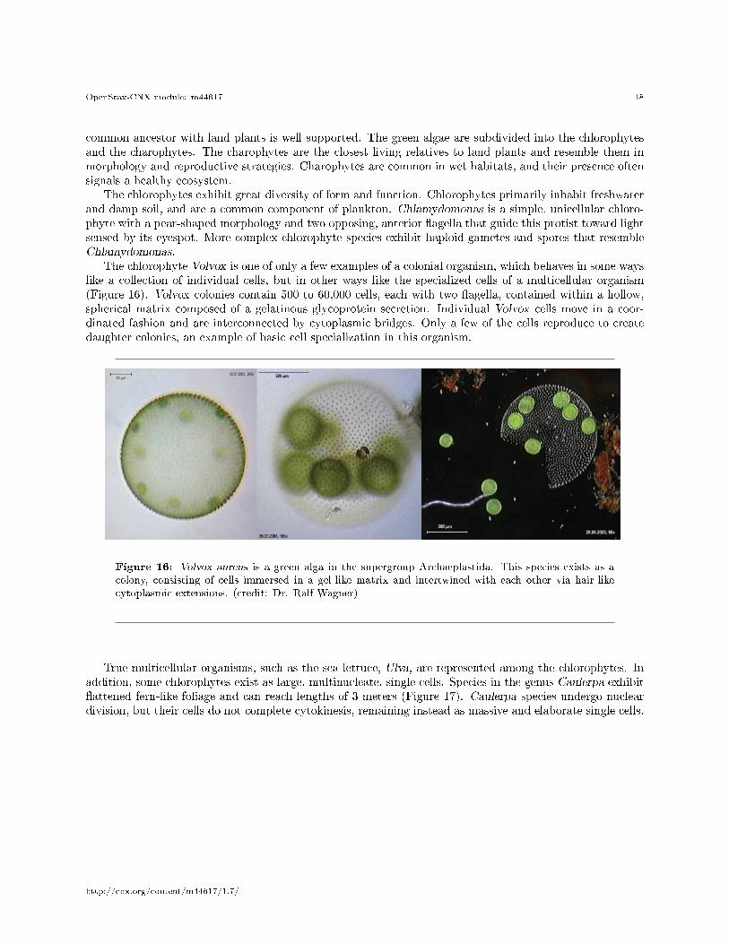
The chlorophyte Volvox is one of only a few examples of a colonial organism, which behaves in some wayslike a collection of individual cells, but in other ways like the specialized cells of a multicellular organism(Figure 16). Volvox colonies contain 500 to 60,000 cells, each with two �agella, contained within a hollow,spherical matrix composed of a gelatinous glycoprotein secretion. Individual Volvox cells move in a coor-dinated fashion and are interconnected by cytoplasmic bridges. Only a few of the cells reproduce to createdaughter colonies, an example of basic cell specialization in this organism.
Figure 16: Volvox aureus is a green alga in the supergroup Archaeplastida. This species exists as acolony, consisting of cells immersed in a gel-like matrix and intertwined with each other via hair-likecytoplasmic extensions. (credit: Dr. Ralf Wagner)
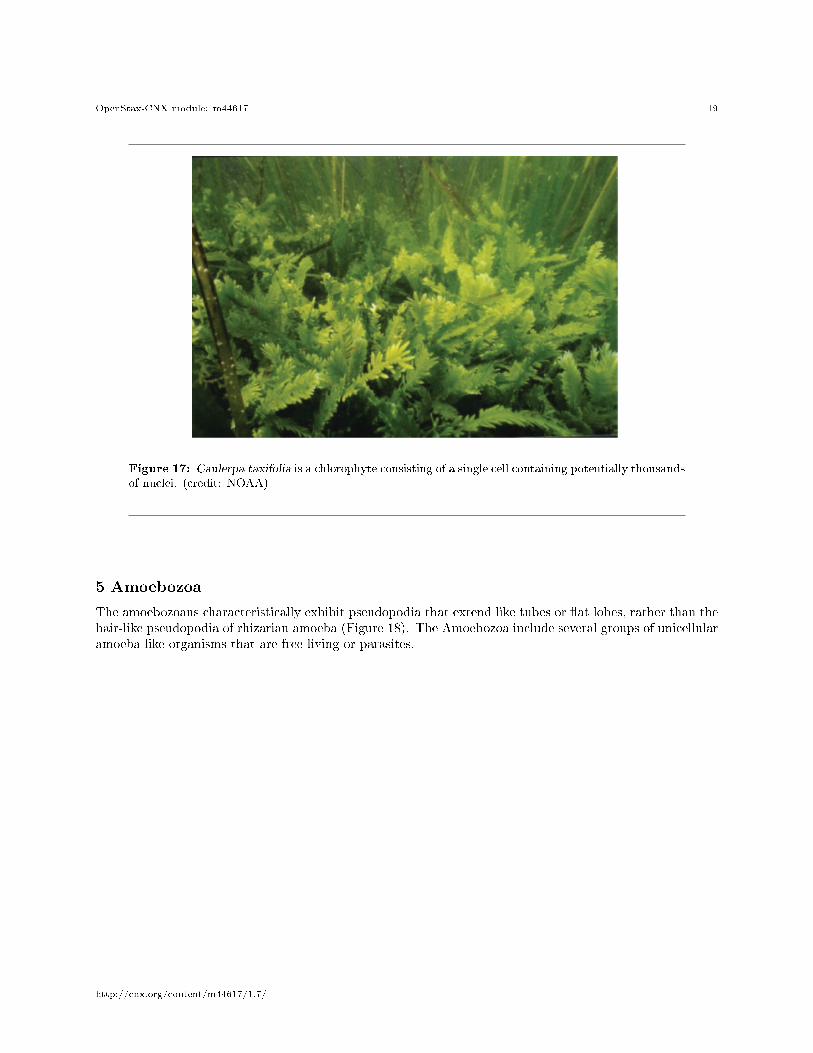
True multicellular organisms, such as the sea lettuce, Ulva, are represented among the chlorophytes. Inaddition, some chlorophytes exist as large, multinucleate, single cells. Species in the genus Caulerpa exhibit�attened fern-like foliage and can reach lengths of 3 meters (Figure 17). Caulerpa species undergo nucleardivision, but their cells do not complete cytokinesis, remaining instead as massive and elaborate single cells.
http://cnx.org/content/m44617/1.7/
OpenStax-CNX module: m44617 19
Figure 17: Caulerpa taxifolia is a chlorophyte consisting of a single cell containing potentially thousandsof nuclei. (credit: NOAA)
5 Amoebozoa
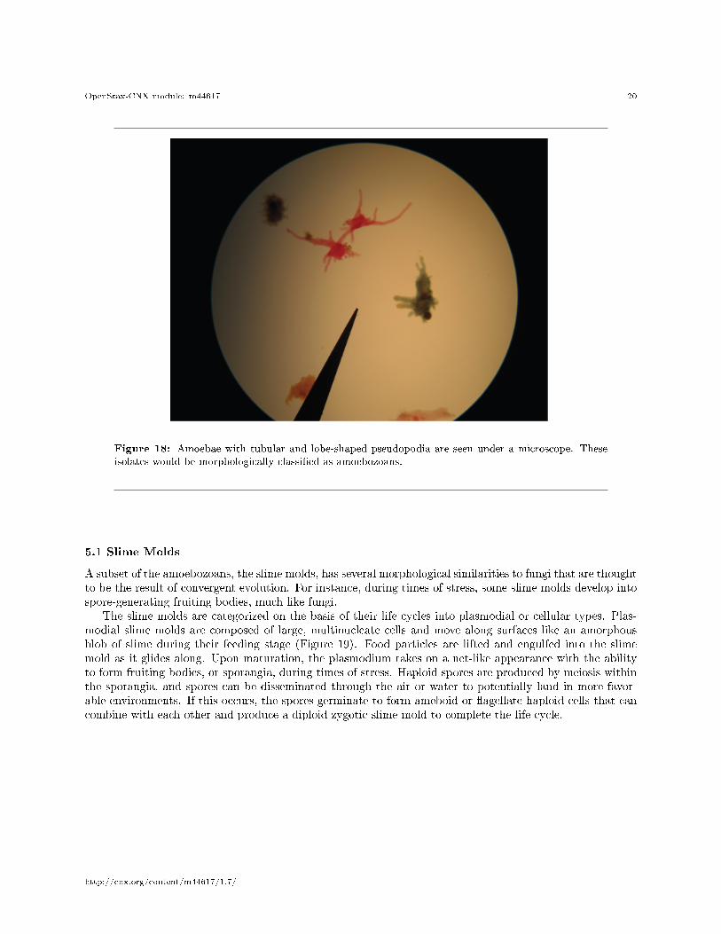
The amoebozoans characteristically exhibit pseudopodia that extend like tubes or �at lobes, rather than thehair-like pseudopodia of rhizarian amoeba (Figure 18). The Amoebozoa include several groups of unicellularamoeba-like organisms that are free-living or parasites.
http://cnx.org/content/m44617/1.7/
OpenStax-CNX module: m44617 20
Figure 18: Amoebae with tubular and lobe-shaped pseudopodia are seen under a microscope. Theseisolates would be morphologically classi�ed as amoebozoans.
5.1 Slime Molds
A subset of the amoebozoans, the slime molds, has several morphological similarities to fungi that are thoughtto be the result of convergent evolution. For instance, during times of stress, some slime molds develop intospore-generating fruiting bodies, much like fungi.
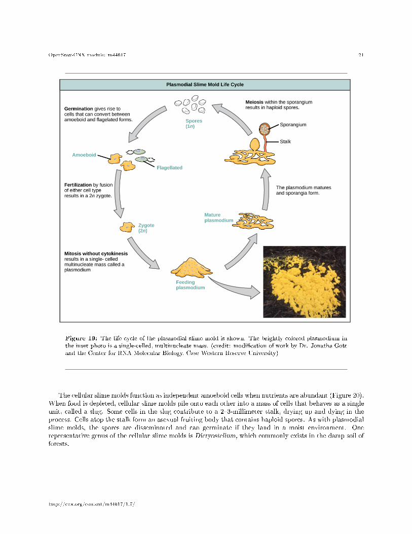
The slime molds are categorized on the basis of their life cycles into plasmodial or cellular types. Plas-modial slime molds are composed of large, multinucleate cells and move along surfaces like an amorphousblob of slime during their feeding stage (Figure 19). Food particles are lifted and engulfed into the slimemold as it glides along. Upon maturation, the plasmodium takes on a net-like appearance with the abilityto form fruiting bodies, or sporangia, during times of stress. Haploid spores are produced by meiosis withinthe sporangia, and spores can be disseminated through the air or water to potentially land in more favor-able environments. If this occurs, the spores germinate to form ameboid or �agellate haploid cells that cancombine with each other and produce a diploid zygotic slime mold to complete the life cycle.
http://cnx.org/content/m44617/1.7/
OpenStax-CNX module: m44617 21
Figure 19: The life cycle of the plasmodial slime mold is shown. The brightly colored plasmodium inthe inset photo is a single-celled, multinucleate mass. (credit: modi�cation of work by Dr. Jonatha Gottand the Center for RNA Molecular Biology, Case Western Reserve University)
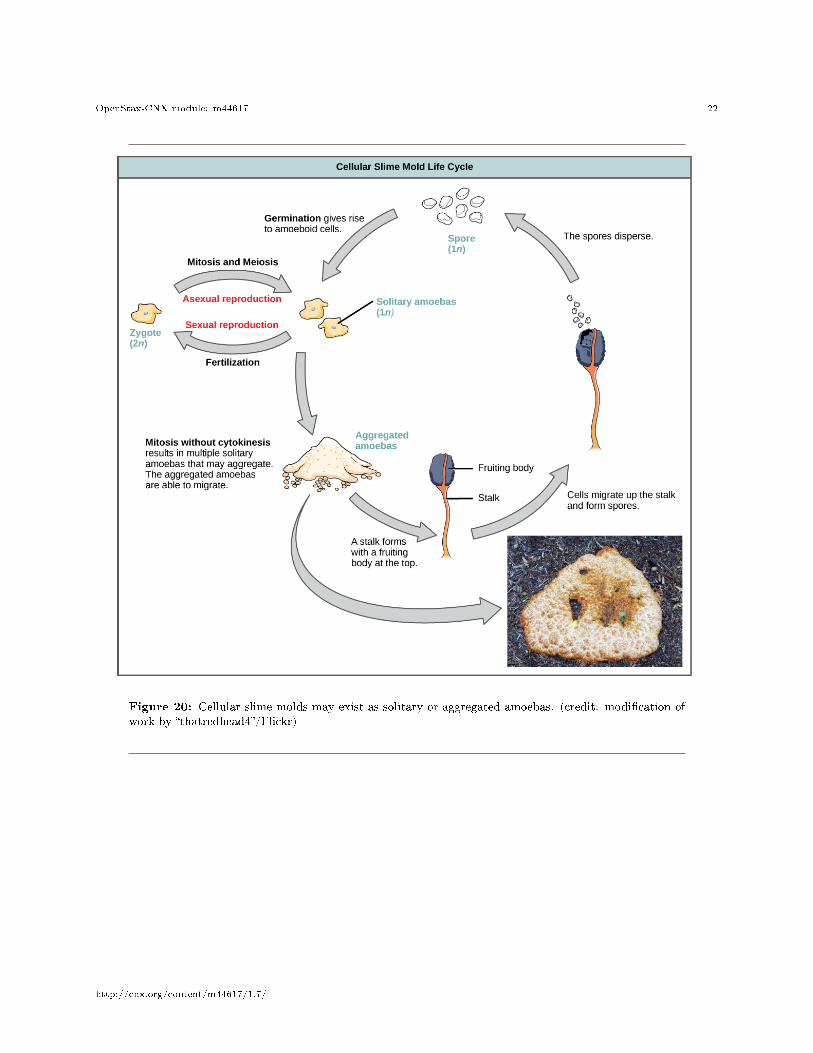
The cellular slime molds function as independent amoeboid cells when nutrients are abundant (Figure 20).When food is depleted, cellular slime molds pile onto each other into a mass of cells that behaves as a singleunit, called a slug. Some cells in the slug contribute to a 2�3-millimeter stalk, drying up and dying in theprocess. Cells atop the stalk form an asexual fruiting body that contains haploid spores. As with plasmodialslime molds, the spores are disseminated and can germinate if they land in a moist environment. Onerepresentative genus of the cellular slime molds is Dictyostelium, which commonly exists in the damp soil offorests.
http://cnx.org/content/m44617/1.7/
OpenStax-CNX module: m44617 22
Figure 20: Cellular slime molds may exist as solitary or aggregated amoebas. (credit: modi�cation ofwork by �thatredhead4�/Flickr)
http://cnx.org/content/m44617/1.7/
OpenStax-CNX module: m44617 23
: View this site4 to see the formation of a fruiting body by a cellularslime mold.
6 Opisthokonta
The opisthokonts include the animal-like choano�agellates, which are believed to resemble the commonancestor of sponges and, in fact, all animals. Choano�agellates include unicellular and colonial forms, andnumber about 244 described species. These organisms exhibit a single, apical �agellum that is surroundedby a contractile collar composed of microvilli. The collar uses a similar mechanism to sponges to �lterout bacteria for ingestion by the protist. The morphology of choano�agellates was recognized early on asresembling the collar cells of sponges, and suggesting a possible relationship to animals.
The Mesomycetozoa form a small group of parasites, primarily of �sh, and at least one form that canparasitize humans. Their life cycles are poorly understood. These organisms are of special interest, becausethey appear to be so closely related to animals. In the past, they were grouped with fungi and other protistsbased on their morphology.
7 Section Summary
The process of classifying protists into meaningful groups is ongoing, but genetic data in the past 20 yearshave clari�ed many relationships that were previously unclear or mistaken. The majority view at present is toorder all eukaryotes into six supergroups: Excavata, Chromalveolata, Rhizaria, Archaeplastida, Amoebozoa,and Opisthokonta. The goal of this classi�cation scheme is to create clusters of species that all are derivedfrom a common ancestor. At present, the monophyly of some of the supergroups are better supported bygenetic data than others. Although tremendous variation exists within the supergroups, commonalities atthe morphological, physiological, and ecological levels can be identi�ed.
8 Art Connections
Exercise 1 (Solution on p. 25.)
Figure 7 Which of the following statements about Paramecium sexual reproduction is false?
a. The macronuclei are derived from micronuclei.b. Both mitosis and meiosis occur during sexual reproduction.c. The conjugate pair swaps macronuclei.d. Each parent produces four daughter cells.
Exercise 2 (Solution on p. 25.)
Figure 10 Which of the following statements about the Laminaria life cycle is false?
a. 1n zoospores form in the sporangia.
4http://openstaxcollege.org/l/slime_mold
http://cnx.org/content/m44617/1.7/
OpenStax-CNX module: m44617 24
b. The sporophyte is the 2n plant.c. The gametophyte is diploid.d. Both the gametophyte and sporophyte stages are multicellular.
9 Review Questions
Exercise 3 (Solution on p. 25.)
Which protist group exhibits mitochondrial remnants with reduced functionality?
a. slime moldsb. diatomsc. parabasalidsd. dino�agellates
Exercise 4 (Solution on p. 25.)
Conjugation between two Paramecia produces ________ total daughter cells.
a. 2b. 4c. 8d. 16
Exercise 5 (Solution on p. 25.)
What is the function of the raphe in diatoms?
a. locomotionb. defensec. capturing foodd. photosynthesis
Exercise 6 (Solution on p. 25.)
What genus of protists appears to contradict the statement that unicellularity restricts cell size?
a. Dictyosteliumb. Ulvac. Plasmodium
d. Caulerpa
10 Free Response
Exercise 7 (Solution on p. 25.)
The chlorophyte (green algae) genera Ulva and Caulerpa both have macroscopic leaf-like andstem-like structures, but only Ulva species are considered truly multicellular. Explain why.
Exercise 8 (Solution on p. 25.)
Why might a light-sensing eyespot be ine�ective for an obligate saprobe? Suggest an alternativeorgan for a saprobic protist.
http://cnx.org/content/m44617/1.7/
OpenStax-CNX module: m44617 25
Solutions to Exercises in this Module
to Exercise (p. 23)Figure 7 Cto Exercise (p. 23)Figure 10 Cto Exercise (p. 24)Cto Exercise (p. 24)Cto Exercise (p. 24)Ato Exercise (p. 24)Dto Exercise (p. 24)Unlike Ulva, protists in the genus Caulerpa actually are large, multinucleate, single cells. Because theseorganisms undergo mitosis without cytokinesis and lack cytoplasmic divisions, they cannot be consideredtruly multicellular.to Exercise (p. 24)By de�nition, an obligate saprobe lacks the ability to perform photosynthesis, so it cannot directly obtainnutrition by searching for light. Instead, a chemotactic mechanism that senses the odors released duringdecay might be a more e�ective sensing organ for a saprobe.
Glossary
De�nition 1: biological carbon pumpprocess by which inorganic carbon is �xed by photosynthetic species that then die and fall to thesea �oor where they cannot be reached by saprobes and their carbon dioxide consumption cannotbe returned to the atmosphere
De�nition 2: bioluminescencegeneration and emission of light by an organism, as in dino�agellates
De�nition 3: contractile vacuolevesicle that �lls with water (as it enters the cell by osmosis) and then contracts to squeeze waterfrom the cell; an osmoregulatory vesicle
De�nition 4: cytoplasmic streamingmovement of cytoplasm into an extended pseudopod such that the entire cell is transported to thesite of the pseudopod
De�nition 5: hydrogenosomeorganelle carried by parabasalids (Excavata) that functions anaerobically and outputs hydrogen gasas a byproduct; likely evolved from mitochondria
De�nition 6: kinetoplastmass of DNA carried within the single, oversized mitochondrion, characteristic of kinetoplastids(phylum: Euglenozoa)
De�nition 7: mitosomenonfunctional organelle carried in the cells of diplomonads (Excavata) that likely evolved from amitochondrion
De�nition 8: planktondiverse group of mostly microscopic organisms that drift in marine and freshwater systems andserve as a food source for larger aquatic organisms
http://cnx.org/content/m44617/1.7/
OpenStax-CNX module: m44617 26
De�nition 9: rapheslit in the silica shell of diatoms through which the protist secretes a stream of mucopolysaccharidesfor locomotion and attachment to substrates
De�nition 10: testporous shell of a foram that is built from various organic materials and typically hardened withcalcium carbonate
http://cnx.org/content/m44617/1.7/