Embed Size (px)
Citation preview

CONSULT THE EXPERT h PARASITOLOGY h PEER REVIEWED
transfusion has been described in humans,9 so dogs and cats that donate blood should be tested for infection prior to donation.10 Because of the shared Ixodes spp tick vector, concurrent infection with Borrelia burgdorferi has been reported in dogs and cats and may contribute to more severe clinical manifestation than either infection alone.11-15
Geographic distribution of anaplasmosis follows that of the tick vector, and the seroprevalence of A phagocy-tophilum in dogs and cats varies by region.2,4,12,15-18 Anaplasmosis has been reported with highest incidence in the midwest, northeast, and western United States.2,16 Infection has also been reported in Canada, Europe, Asia, and South America, although more studies are needed to understand vector transmission in areas not endemic for Ixodes spp.3,4,6,17,19-24
Risk for exposure and/or infection is increased in animals that live in an Ixodes-spp endemic area, receive
Granulocytic anaplasmosis, an acute disease that affects dogs, cats, and other mammals, results from infection with the bacterium Anaplasma phagocytophilum and is transmitted primarily by the bite of an Ixodes spp tick. Exposure is most prominent in areas endemic for Ixodes spp.1-6 Most dogs and cats exposed to A phagocytophilum do not develop clinical signs, but animals that do develop clinical disease often show nonspecific signs of lethargy, fever, and decreased appetite.2,7
Although there are several species in the Anaplasmata-ceae family that can cause anaplasmosis, this article focuses on A phagocytophilum.
Background & Pathophysiology A phagocytophilum, a gram-negative intracellular bacte-rium, infects neutrophils and, occasionally, eosinophils in a variety of mammals, including dogs and cats.1,8 Although the organism is primarily vector-transmitted by the bite of an Ixodes spp tick, transmission via blood
Granulocytic AnaplasmosisChristine Savidge, DVM, DACVIMAtlantic Veterinary CollegeCharlottetown, Prince Edward Island
32 cliniciansbrief.com March 2019
March 2019 cliniciansbrief.com 33
acaricidal medications inconsistently, and have increased outdoor access, particularly to natural tick habitats. Transmission to mammals occurs within 24 to 48 hours of tick attachment.25,26 Disease prevalence corresponds with increased Ixodes spp tick activity and is therefore highest in the late spring and autumn. Seropositive preva-lence is increased in older dogs and cats, as these animals have had more years of exposure.1,7,12,19 Prevalence of exposure to A phagocytophilum and incidence of clinical disease are not equal. Most dogs that test positive for A phagocytophilum infec-tion have no history of clinical illness, and cats that test positive for A phagocytophilum serum anti- bodies may not have signs of clinical disease.12,27 This discordance complicates testing and confirma-tion of disease. HistoryDogs and cats are often presented within 2 to 3 days (range, 1-14 days) of onset of signs. Acute lethargy, malaise, and decreased appetite are the most com-mon clinical signs on presentation,2,7,19,20,28-30 with signs of lameness, circling, vomiting, and epistaxis reported less commonly.20,30 Relevant history includes inconsistent or irregular receipt of anti-parasitics or acaricides, travel history, and known tick exposure.31-33
Clinical SignsClinical presentation is variable and depends on the strain of bacterium and host variables, including immune response and coinfections. Most dogs and cats exposed to A phagocytophilum do not develop dis-ease or develop only mild self-resolving signs, as sug-gested by experimental infections and high positive serology in the absence of clinical disease.2,12,27,34,35 Animals that do develop clinical illness may show a variety of multisystemic, inflammatory signs but most commonly show nonspecific signs of lethargy, fever, and decreased appetite.1-3,7,19,20,28-30 Lameness or muscle pain, lymphadenopathy, splenomegaly, and, more rarely, neurologic signs of circling, seizures, neck pain, and epistaxis have also been reported.7,20,30
DiagnosisMaking an accurate clinical diagnosis can be challenging, as many dogs and cats that are exposed to A phagocytophilum do not develop clinical disease.12,27 Determining whether an animal’s signs are attributable to active infection requires a multimodal approach to diagnosis.2 Diagnosis of acute granulocytic anaplasmosis should include consistent history and clinical signs, as well as support from CBC results, with direct visual examination of a blood smear, serology test-ing for specific antibodies, and PCR testing.
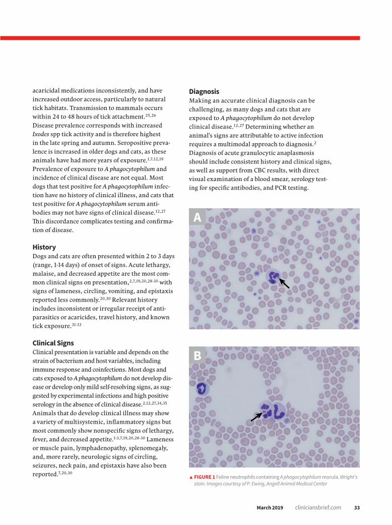
d FIGURE 1 Feline neutrophils containing A phagocytophilum morula. Wright’s stain. Images courtesy of P. Ewing, Angell Animal Medical Center
B
A

34 cliniciansbrief.com March 2019
The most common abnormalities seen on CBC in dogs are thrombocytopenia, anemia, and leukope-nia1,2,19,28,29; these changes are also exhibited in cats, although cats are more prone to platelet clumping and should not be erroneously diagnosed with thrombocytopenia in these cases.7,20 In dogs and cats with acute infection, morulae (ie, clusters of bacteria) can be observed as intracytoplasmic inclu-sions (Figure 1, previous page) on direct examina-tion of a blood smear. When identified, morulae have been reported in as few as 5% and up to 32% of neutrophils.2,7,19,20,29,34 Morulae can also be identi-fied in other affected fluids such as CSF and joint fluid (Figure 2). Serum chemistry results may show nonspecific, inconsistent changes, including increased liver enzymes, hyperbilirubinemia, hypo-albuminemia, and/or hyperglobulinemia.2,20,29
A number of commercially available serology tests (eg, ELISA, immunofluorescence assay) that assess for specific antibodies against A phagocytophilum surface proteins are readily available, accurate, and cost-effective.35-37 It is important to note that anti-bodies will be absent or delayed in an acute infec-tion, will appear 14 days postexposure, and will be consistently present 30 days after exposure.35 If a patient is suspected of having granulocytic anaplas-mosis but has a negative serology test result, addi-tional testing (eg, conventional or real-time PCR of
peripheral blood, recheck serology 4 weeks later) is warranted. A positive test result confirms exposure to the organism and should also be present with clinical signs before making a clinical diagnosis of anaplasmosis. In addition to identifying intragranu-locytic morulae on visual examination of a blood smear, identifying bacterial DNA in peripheral blood through PCR testing can confirm the pres-ence of the organism directly in the blood.1,12,38 Peripheral blood can be positive on PCR testing as early as 2 days postexposure and can persist for up to 2 months, even in the absence of clinical signs.34,35,39,40 Antibiotics administered before PCR testing can cause false-negative results for detecting morulae or bacterial DNA in peripheral blood; anti-body detection through serologic tests, however, is not affected by previous treatments.2,34,41 In addi-tion to specific testing for A phagocytophilum and a positive response to treatment,20 other causes of clinical signs and clinicopathologic abnormalities should be ruled out to complete the diagnosis of granulocytic anaplasmosis.
Treatment & ManagementTreatment is recommended in any patient that tests positive for A phagocytophilum and also has concurrent clinical signs or clinicopathologic changes that could be consistent with anaplasmosis (eg, thrombocytopenia, intracellular morulae). Treatment with doxycycline (5 mg/kg PO q12h for 14-28 days) is effective in treating most dogs and cats with acute granulocytic anaplasmosis.7,19,20,34,41 Food or water should be given to animals, particu-larly cats, after doxycycline administration to avoid esophagitis.42 Most dogs and cats respond to ther-apy within 2 to 3 days, and complete resolution of infection generally occurs in 97% of dogs and 96% of cats.8,20,29,43,44 Treatment is not recommended in patients that are only serologically positive that do not have clinical signs or clinicopathologic changes con-sistent with anaplasmosis.45 Coinciding immune-mediated conditions have been described and war-rant closer monitoring and concurrent treatment with glucocorticoids and supportive care, blood transfusions, and analgesia, as indicated by the patient’s clinical signs.29
d FIGURE 2 Canine neutrophil containing A phagocytophilum morula in inflamed synovial fluid. Wright’s stain
CONSULT THE EXPERT h PARASITOLOGY h PEER REVIEWED

March 2019 cliniciansbrief.com 35
Continues on page 64 h
Regular tick preventive use is warranted in animals with outdoor access and animals that live in an Ixodes-spp endemic area, as routine use has been shown to significantly reduce the risk for infec-tion.31-33
Prognosis & PreventionPrognosis is excellent in patients in which disease and/or coinfection is identified and treated early. Patients that have ongoing exposure to Ixodes spp ticks are at risk for re-exposure. Granulocytic anaplasmosis is best prevented by maintaining stringent tick control, reducing exposure to tick habitats, using tick repellents regularly, and having patients screened regularly for ticks after outdoor activity. No vaccines for disease prevention are currently available.
Clinical Follow-Up & MonitoringMost dogs and cats respond to treatment and have no residual signs that require follow-up. If signifi-cant hematologic abnormalities are present, a recheck examination 14 days after initiating treat-ment is warranted to assess response to therapy. Idiosyncratic hepatic injury secondary to doxy-cycline administration has been described46; there-fore, a serum liver panel obtained 5 to 7 days after initiating treatment may be warranted. If clinical signs persist or a patient is presented with a more complicated condition (eg, immune-mediated hemo-lytic anemia, immune-mediated polyarthropathy,
meningitis) or develops new signs, re-evaluation should be scheduled based on individual patient needs and concurrent condition(s).
Granulocytic anaplasmosis should be included on the differential list for any dog or cat that has been exposed to Ixodes spp ticks and has acute or intermittent signs of lethargy, anorexia, and/or fever and a history of receiving inconsistent tick prevention. n
References1. Greig B, Asanovich KM, Armstrong PJ, Dumler JS. Geographic,
clinical, serologic, and molecular evidence of granulocytic ehrlichiosis, a likely zoonotic disease, in Minnesota and Wisconsin dogs. J Clin Microbiol. 1996;34(1):44-48.
2. Carrade DD, Foley JE, Borjesson DL, Sykes JE. Canine granulocytic anaplasmosis: a review. J Vet Intern Med. 2009;23(6):1129-1141.
3. Egenvall AE, Hedhammar AA, Bjöersdorff AI. Clinical features and serology of 14 dogs affected by granulocytic ehrlichiosis in Sweden. Vet Rec. 1997;140(9):222-226.
4. Mrljak V, Kuleš J, Mihaljević Ž, et al. Prevalence and geographic distribution of vector-borne pathogens in apparently healthy dogs in Croatia. Vector Borne Zoonotic Dis. 2017;17(6):398-408.
5. Lee SH, Kim N, Kwak D. First clinical case of canine granulocytic anaplasmosis in Korea and genotypic analyses of Anaplasma phagocytophilum. Ticks Tick Borne Dis. 2017;8(4):462-465.
6. Silveira JA, Valente PC, Paes PR, Vasconcelos AV, Silvestre BT,
Ribeiro MF. The first clinical and laboratory evidence of co-infection by Anaplasma phagocytophilum and Ehrlichia canis in a Brazilian dog. Ticks Tick Borne Dis. 2015;6(3):242-245.
7. Savidge C, Ewing P, Andrews J, Aucoin D, Lappin MR, Moroff S. Anaplasma phagocytophilum infection of domestic cats: 16 cases from the northeastern USA. J Feline Med Surg. 2016;18(2):85-91.
8. Bjöersdorff A, Svendenius L, Owens JH, Massung RF. Feline granulocytic ehrlichiosis—a report of a new clinical entity and characterisation of the infectious agent. J Small Anim Pract. 1999;40(1):20-24.
9. Fine AB, Sweeney JD, Nixon CP, Knoll BM. Transfusion-transmitted anaplasmosis from a leukoreduced platelet pool. Transfusion. 2016;56(3):699-704.
10. Wardrop KJ, Birkenheuer A, Blais MC, et al. Update on canine and feline blood donor screening for blood-borne pathogens. J Vet Intern Med. 2016;30(1):15-35.
POLL
Have you ever seen clinical anaplasmosis in any of your patients?
A. YesB. NoC. Suspected but was unable to confirm
diagnosis
Scan the QR code to submit your answer and see the other responses! The poll is located at the bottom of the article.
Using QR codes from your mobile device is easy and quick!
Simply focus your phone’s camera on the QR code as if taking a picture (but don’t click!). A notification banner will pop up at the top of your screen; tap the banner to view the linked content.

64 cliniciansbrief.com March 2019
11. Jäderlund KH, Egenvall A, Bergström K, Hedhammar A. Seroprevalence of Borrelia burgdorferi sensu lato and Anaplasma phagocytophilum in dogs with neurological signs. Vet Rec. 2007;160(24):825-831.
12. Beall MJ, Chandrashekar R, Eberts MD, et al. Serological and molecular prevalence of Borrelia burgdorferi, Anaplasma phagocytophilum, and Ehrlichia species in dogs from Minnesota. Vector Borne Zoonotic Dis. 2008;(8)4:455-464.
13. Nieto NC, Foley J. Meta-analysis of coinfection and coexposure with Borrelia burgdorferi and Anaplasma phagocytophilum in humans, domestic animals, wildlife, and Ixodes ricinus-complex ticks. Vector Borne Zoonotic Dis. 2009;9(1):93-102.
14. Nyarko E, Grab DJ, Dumler JS. Anaplasma phagocytophilum-infected neutro-phils enhance transmigration of Borrelia burgdorferi across the human blood brain barrier in vitro. Int J Parasitol. 2006;36(5):601-605.
15. Hoyt K, Chandrashekar R, Breitschwerdt E, Lappin MR. Anaplasma phago-cytophilum and Borrelia burgdorferi antibodies in naturally exposed cats in Maine. Paper presented at: American College of Veterinary Internal Medicine Forum; June 3-6, 2014; Nashville, TN.
16. McMahan CS, Wang D, Beall MJ, et al. Factors associated with Anaplasma spp seroprevalence among dogs in the United States. Parasit Vectors. 2016;9:169.
17. Qurollo BA, Chandrashekar R, Hegarty BC, et al. A serological survey of tick-borne pathogens in dogs in North America and the Caribbean as assessed by Anaplasma phagocytophilum, A platys, Ehrlichia canis, E chaffeensis, E ewingii, and Borrelia burgdorferi species-specific peptides. Infect Ecol Epidemiol. 2014;4:24699.
18. Billeter SA, Spencer JA, Griffin B, Dykstra CC, Blagburn BL. Prevalence of Anaplasma phagocytophilum in domestic felines in the United States. Vet Parasitol. 2007;147(1-2):194-198.
19. Kohn B, Galke D, Beelitz P, Pfister K. Clinical features of canine granulocytic anaplasmosis in 18 naturally infected dogs. J Vet Intern Med. 2008;22(6):1289-1295.
20. Tarello W. Microscopic and clinical evidence for Anaplasma (Ehrlichia) phago-cytophilum infection in Italian cats. Vet Rec. 2005;156(24):772-774.
21. Domingos MC, Trotta M, Briend-Marchal A, Medaille C. Anaplasmosis in two dogs in France and molecular and phylogenetic characterization of Anaplas-ma phagocytophilum. Vet Clin Pathol. 2011;40(2):215-221.
22. Santos HA, Pires MS, Vilela JA, et al. Detection of Anaplasma phagocytophilum in Brazilian dogs by real-time polymerase chain reaction. J Vet Diagn Invest. 2011;23(4):770-774.
23. Lee S, Kim N, Kwak D. First clinical case of canine granulocytic anaplasmosis in Korea and genotypic analyses of Anaplasma phagocytophilum. Ticks Tick Borne Dis. 2017;8(4):462-465.
24. Fukui Y, Ohkawa S, Inokuma H. First molecular detection and phylogenetic analysis of Anaplasma phagocytophilum from a clinical case of canine granu-locytic anaplasmosis in Japan. Jpn J Infect Dis. 2018;71(4):302-305.
25. Hodzic E, Fish D, Maretzki CM, De Silva AM, Feng S, Barthold SW. Acquisition and transmission of the agent of human granulocytic ehrlichiosis by Ixodes scapularis ticks. J Clin Microbiol. 1998;36(12):3574-3578.
26. Katavolos P, Armstrong PM, Dawson JE, Telford SR 3rd. Duration of tick attachment required for transmission of granulocytic ehrlichiosis. J Infect Dis. 1998;177(5):1422-1425.
27. Galemore ER, Labato MA, O’Neil E. Prevalence of Anaplasma phago-cytophilum infection in feral cats in Massachusetts. JFMS Open Rep. 2018;4(1):2055116917753804.
28. Granick JL, Armstrong PJ, Bender JB. Anaplasma phagocytophilum infection in dogs: 34 cases (2000-2007). J Am Vet Med Assoc. 2009;234(12):1559-1565.
29. Chirek A, Silaghi C, Pfister K, Kohn B. Granulocytic anaplasmosis in 63 dogs:
clinical signs, laboratory results, therapy and course of disease. J Small Anim Pract. 2018;59(2):112-120.
30. Eberts MD, Vissotto de Paiva Diniz PP, Beall MJ, Stillman BA, Chandrashekar R, Breitschwerdt EB. Typical and atypical manifestations of Anaplasma phagocy-tophilum infection in dogs. J Am Anim Hosp Assoc. 2011;47(6):e86-94.
31. Honsberger NA, Six RH, Heinz TJ, Weber A, Mahabir SP, Berg TC. Efficacy of sarolaner in the prevention of Borrelia burgdorferi and Anaplasma phagocy-tophilum transmission from infected Ixodes scapularis to dogs. Vet Parasitol. 2016;222:67-72.
32. Wengenmayer C, Williams H, Zschiesche E, et al. The speed of kill of fluralaner (Bravecto™) against Ixodes ricinus ticks on dogs. Parasit Vectors. 2014;7:525.
33. McCall JW, Baker CF, Mather TN, et al. The ability of a topical novel combi-nation of fipronil, amitraz and (S)-methoprene to protect dogs from Borrelia burgdorferi and Anaplasma phagocytophilum infections transmitted by Ixodes scapularis. Vet Parasitol. 2011;179(4):335-342.
34. Lappin MR, Chandrashekar R, Stillman B, Liu J, Mather TN. Evidence of Ana-plasma phagocytophilum and Borrelia burgdorferi infection in cats after expo-sure to wild-caught adult Ixodes scapularis. J Vet Diagn Invest. 2015;27(4):522-525.
35. Moroff S, Sokolchik I, Woodring T, Woodruff C, Atkinson B, Lappin MR. Detection of antibodies against Anaplasma phagocytophilum in dogs using an automated fluorescence-based system. Vet J. 2014;202(2):348-352.
36. Ravnik U, Tozon N, Smrdel KS, Zupanc TA. Anaplasmosis in dogs: the relation of haematological, biochemical and clinical alterations to antibody titre and PCR confirmed infection. Vet Microbiol. 2011;149(1-2):172-176.
37. Liu J, Eberts M, Bewsey H, O’Connor TP, Chandrashekar R, Breitschwerdt EB. Sensitivity and specificity levels of two rapid assays for antibodies to Anaplas-ma spp in dogs. J Vet Diagn Invest. 2018;30(2):290-293.
38. Drazenovich N, Foley J, Brown RN. Use of real-time quantitative PCR targeting the msp2 protein gene to identify cryptic Anaplasma phagocytophilum infec-tions in wildlife and domestic animals. Vector Borne Zoonotic Dis. 2006;6(1):83-90.
39. Chandrashekar R, Beall MJ, Thatcher B, Saucier JM, Tyrrell P, Lappin MR. Serologic responses to peptides of Anaplasma phagocytophilum and Bor-relia burgdorferi in dogs infested with wild-caught Ixodes scapularis. Vet J. 2017;226:6-11.
40. Scorpio DG, Dumler JS, Barat NC, et al. Comparative strain analysis of Ana-plasma phagocytophilum infection and clinical outcomes in a canine model of granulocytic anaplasmosis. Vector Borne Zoonotic Dis. 2011;11(3):223-229.
41. Yancey CB, Diniz PPVP, Breitschwerdt EB, Hegarty BC, Wiesen C, Qurollo BA. Doxycycline treatment efficacy in dogs with naturally occurring Anaplasma phagocytophilum infection. J Small Anim Pract. 2018;59(5):286-293.
42. Frowde PE, Battersby IA, Whitley NT, Elwood CM. Oesophageal disease in 33 cats. J Feline Med Surg. 2011;13(8):564-569.
43. Lappin MR, Breitschwerdt EB, Jensen WA, et al. Molecular and serologic evidence of Anaplasma phagocytophilum infection in cats in North America. J Am Vet Med Assoc. 2004;225(6):893-896.
44. Adaszek Ł, Górna M, Skrzypczak M, Buczek K, Balicki I, Winiarczyk S. Three clinical cases of Anaplasma phagocytophilum infection in cats in Poland. J Feline Med Surg. 2012;15(4):333-337.
45. Companion Animal Parasite Council. Ehrlichia spp. and Anaplasma spp. https://capcvet.org/guidelines/ehrlichia-spp-and-anaplasma-spp. Published June 1, 2015. Accessed February 2, 2019.
46. Plumb DC. Doxycycline. In: Plumb DC. Plumb’s Veterinary Drug Handbook. 8th ed. Ames, IA: Wiley Blackwell; 2015;365-369.
CONSULT THE EXPERT h CONTINUED FROM PAGE 35