Embed Size (px)
Citation preview
|| 109 || | European Journal of General Dentistry | Vol 1 | Issue 2 | May-August 2012 |
Granular cell ameloblastoma: A diagnostic dilemma for histopathologist
ABSTrACTA rare case of granular cell ameloblastoma in the posterior mandible of a 45‑year‑old female has been studied both histologically and immunohistochemically. Histopathologically, hematoxylin and eosin and Periodic acid Schiff (PAS) prepared sections of granular cell ameloblastoma showed granular neoplastic cells residing within the tumor follicles. To characterize the granular cells in ameloblastoma, we examined the expression of low molecular weight cytokeratin and S‑100 protein using immunohistochemistry. Granular cells exhibited positivity only with cytokeratin while S‑100 protein was negative. This result allowed easy distinction of granular cell ameloblastomas from similar tumors exhibiting granular cell changes, and indicated that the granularity in ameloblastoma cells is consequent to lysosomal overload.
Key wordsAmeloblastoma, granular cells, rare
Access this article onlineQuick Response Code:
Website: www.ejgd.org
DOI: 10.4103/2278-9626.103398
vijaykumar G. Biradar, rahul G. latturiya, Surekha v. Biradar
Department of Oral and Maxillofacial Pathology, Maharashtra University of Health Sciences, MIDSR Dental College, Hospital and Research Centre, Latur, Maharashtra, India
Address for correspondence: Dr. Vijaykumar Goudappagouda Biradar,
Department of Oral and Maxillofacial Pathology, Maharashtra University
of Health Sciences, MIDSR Dental College, Hospital and Research Centre,
Latur, Maharashtra, India. E-mail: [email protected]
iNTRODUCTiON
Ameloblastoma is the most significant epithelial odontogenic tumor of concern for the oral pathologist because of its high incidence among all odontogenic tumors and true neoplastic (infiltrative and recurrent) potential, combined with its varieties of histologic patterns.[1] Six histopathologic subtypes of ameloblastoma are recognized: follicular, acanthomatous, granular cell, basal cell, desmoplastic and plexiform. [2] Granular cell ameloblastomas are uncommon lesions accounting for about 3–5% of all histologic subtypes of ameloblastoma.[3] Granular cell ameloblastoma is characterized by nests of large, eosinophilic granular cells. These latter cells have long been the subject of debate.[4] From the studies on ameloblastomas to date, it seems that the old belief that granular cell ameblastoma is the most aggressive variant of ameloblastoma is a myth, and in all probability, granular cells are just
a transitional or matured phase in the life cycle of ameloblastomas, starting with normal stellate reticulum-like cells, leading to a production of granules and finally leading to degeneration and formation of cystic areas.[5]
This report describes granular cell ameloblastoma that is a relatively rare histologic subtype and aggressive lesion with marked proclivity for recurrence unless appropriate surgical measures are instituted at first operation.
CASE rEPorT
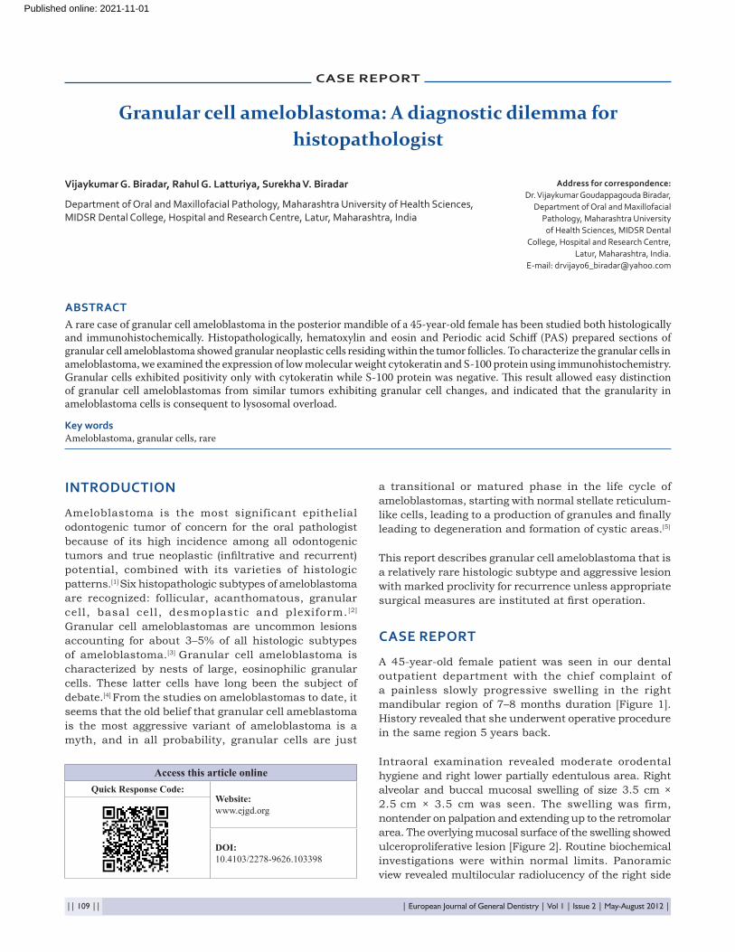
A 45-year-old female patient was seen in our dental outpatient department with the chief complaint of a painless slowly progressive swelling in the right mandibular region of 7–8 months duration [Figure 1]. History revealed that she underwent operative procedure in the same region 5 years back.
Intraoral examination revealed moderate orodental hygiene and right lower partially edentulous area. Right alveolar and buccal mucosal swelling of size 3.5 cm × 2.5 cm × 3.5 cm was seen. The swelling was firm, nontender on palpation and extending up to the retromolar area. The overlying mucosal surface of the swelling showed ulceroproliferative lesion [Figure 2]. Routine biochemical investigations were within normal limits. Panoramic view revealed multilocular radiolucency of the right side
CASE rEPorT
Published online: 2021-11-01
Biradar, et al.: Granular cell ameloblastoma
| European Journal of General Dentistry | Vol 1 | Issue 2 | May-August 2012 | || 110 ||
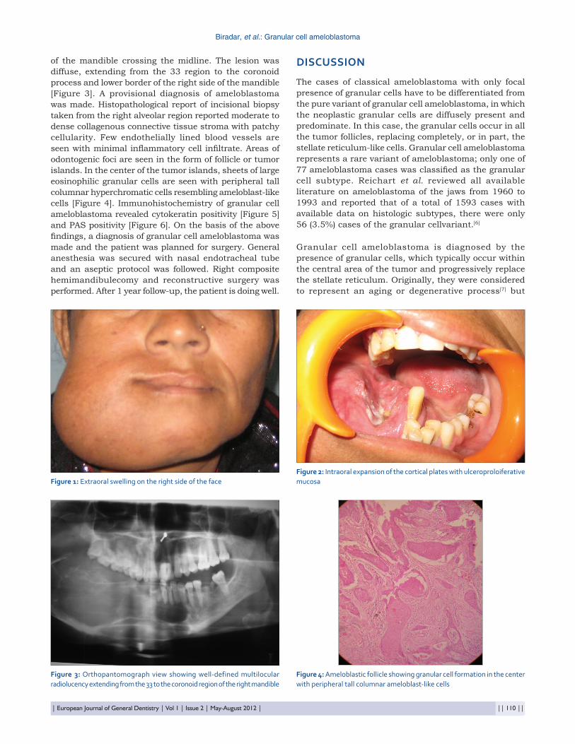
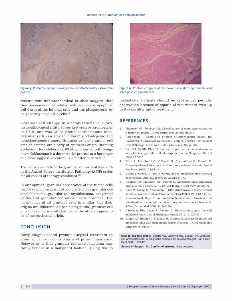
of the mandible crossing the midline. The lesion was diffuse, extending from the 33 region to the coronoid process and lower border of the right side of the mandible [Figure 3]. A provisional diagnosis of ameloblastoma was made. Histopathological report of incisional biopsy taken from the right alveolar region reported moderate to dense collagenous connective tissue stroma with patchy cellularity. Few endothelially lined blood vessels are seen with minimal inflammatory cell infiltrate. Areas of odontogenic foci are seen in the form of follicle or tumor islands. In the center of the tumor islands, sheets of large eosinophilic granular cells are seen with peripheral tall columnar hyperchromatic cells resembling ameloblast-like cells [Figure 4]. Immunohistochemistry of granular cell ameloblastoma revealed cytokeratin positivity [Figure 5] and PAS positivity [Figure 6]. On the basis of the above findings, a diagnosis of granular cell ameloblastoma was made and the patient was planned for surgery. General anesthesia was secured with nasal endotracheal tube and an aseptic protocol was followed. Right composite hemimandibulecomy and reconstructive surgery was performed. After 1 year follow-up, the patient is doing well.
DiSCUSSiON
The cases of classical ameloblastoma with only focal presence of granular cells have to be differentiated from the pure variant of granular cell ameloblastoma, in which the neoplastic granular cells are diffusely present and predominate. In this case, the granular cells occur in all the tumor follicles, replacing completely, or in part, the stellate reticulum-like cells. Granular cell ameloblastoma represents a rare variant of ameloblastoma; only one of 77 ameloblastoma cases was classified as the granular cell subtype. Reichart et al. reviewed all available literature on ameloblastoma of the jaws from 1960 to 1993 and reported that of a total of 1593 cases with available data on histologic subtypes, there were only 56 (3.5%) cases of the granular cellvariant.[6]
Granular cell ameloblastoma is diagnosed by the presence of granular cells, which typically occur within the central area of the tumor and progressively replace the stellate reticulum. Originally, they were considered to represent an aging or degenerative process[7] but
figure 1: Extraoral swelling on the right side of the facefigure 2: Intraoral expansion of the cortical plates with ulceroproloiferative mucosa
figure 3: Orthopantomograph view showing well-defined multilocular radiolucency extending from the 33 to the coronoid region of the right mandible
figure 4: Ameloblastic follicle showing granular cell formation in the center with peripheral tall columnar ameloblast-like cells
Biradar, et al.: Granular cell ameloblastoma
|| 111 || | European Journal of General Dentistry | Vol 1 | Issue 2 | May-August 2012 |
recent immunohistochemical studies suggest that this phenomenon is related with increased apoptotic cell death of the lesional cells and the phagocytosis by neighboring neoplastic cells.[8]
Granular cell change in ameloblastoma is a rare histopathological entity. It was first seen by Krompecher in 1918, and was called pseudoxanthomatous cells. Granular cells can appear in various odontogenic and nonodontogenic tumors. Granular cells of granular cell ameloblastoma are clearly of epithelial origin, staining exclusively for cytokeratin. Whether granular cell change in ameloblastoma is a degenerative process or a harbinger of a more aggressive course is a matter of debate.[9]
The recurrence rate of the granular cell variant was 73% in the Armed Forces Institute of Pathology (AFIP) series for all modes of therapy combined.[10]
In our opinion, granular appearance of the tumor cells can be seen in various oral tumors, such as granular cell ameloblastoma, granular cell myoblastoma, congenital epulis and granular cell ameloblastic fibromas. The morphology of all granular cells is similar, but their origins are different. As per histogenesis, granular cell ameloblastoma is epithelial, while the others appear to be of mesenchymal origin.
CONCLUSiON
Early diagnosis and prompt surgical treatment in granular cell ameloblastoma is of prime importance. Noteworthy is that granular cell ameloblastoma may rarely behave in a malignant fashion, giving rise to
metastasis. Patients should be kept under periodic observation because of reports of recurrences even up to 8 years after initial treatment.
rEfErEnCES
1. Philipsen HP, Reichart PA. Classification of odontogenictumours; A historical review. J Oral Pathol Med 2006;35:525-9.
2. Rajendran R. Cysts and Tumors of Odontogenic Origin. In: Rajendran R, Shivapathsundaram S, editors. Shafer’s Text book of Oral Pathology. 5thed. New Delhi: Elsevier; 2006. p. 386.
3. Siar CH, Ng KH, Chia TY. Combined granular cell ameloblastoma and plexiform granular cell odontogenictumour. Singapore Dent J 1990;15:35-7.
4. Dina R, Marchetti C, Vallania G, Corinaldesi G, Eusebi V. Granularcellameloblastoma. An immunocytochemical study. Pathol Res Pract 1996;192:541-6.
5. Gupta S, Grewal H, Sah K. Granular cell ameloblastoma showing desmoplasia. Ann Saudi Med 2012;32:537-40.
6. Reichart PA, Philipsen HP, Sonner S. Ameloblastoma: Biological profile of 3677 cases. Eur J Cancer B Oral Oncol 1995;31:86-99.
7. Nasu M, Takagi M, Yamamoto H. Ultrastructural and histochemical studies of granular-cellameloblastoma. J Oral Pathol 1984;13:448- 56.
8. Kumamoto H, Ooya K. Immunohistochemical and ultrastructural investigation of apoptotic cell death in granularcellameloblastoma. J Oral Pathol Med 2001;30:245-50.
9. Bansal A, Bhatnagar A, Saxena S. Metastasizing granular cell ameloblastoma. J Oral Maxillofac Pathol 2012;16:122-4.
10. Cranin AN, Bennett J, Solomon M, Quarcoo S. Massive Granular cell ameloblastoma with metastasis: Report of a case. J Oral Maxillofac Surg 1987;45:800-4.
figure 5: Photomicrograph showing immunohistochemistry cytokeratin positive
figure 6: Photomicrograph of low power view showing periodic acid schiff-positive granular cells
How to cite this article: Biradar VG, Latturiya RG, Biradar SV. Granular cell ameloblastoma: A diagnostic dilemma for histopathologist. Eur J Gen Dent 2012;1:109-12.
Source of Support: Nil, Conflict of Interest: None declared.