Embed Size (px)
Citation preview
GOOGLE GLASS INDIRECT OPHTHALMOSCOPY
Aaron Wang, MD, PhD1, Alex Christoff, CO, COT1, David L. Guyton, MD1, Michael X. Repka, MD1,
Mahsa Rezaei, MS1, Allen O. Eghrari, MD1
1Department of Ophthalmology, Johns Hopkins University School of Medicine, Baltimore, Maryland, USA
Corresponding Author: [email protected]
Background: Google Glass is a wearable, head-mounted computer with display, photographic andvideographic imaging capability, and connectivity to other devices through Wi-Fi and Bluetoothsignaling.
Aims: To describe for the first time the use of Google Glass for use in indirect ophthalmoscopy andmodification techniques to assist with its use.
Methods: A lightweight, portable light source was installed above the Glass aperture, a small tissuepaper used to diffuse the light, and the arm of the headset was taped to the examiner’s glasses inorder to bring the display into the right eye’s central visual field.
Results: Using a slightly modified Glass headset, the examiner documented the central andperipheral retina in a young male with ease.
Conclusion: We demonstrate for the first time that Glass, with minor modifications, can be used as asimple and effective method to perform and record a fundus examination.
Journal MTM 4:1:15�19, 2015 doi:10.7309/jmtm.4.1.4 www.journalmtm.com
IntroductionIn April 2013, Google released a beta version of theGoogle Glass for developers for $1500, termed the
Explorer version. Glass is a wearable headset
weighing 50 grams with a prismatic heads-up color
display in the superior visual field of the right eye. Itincludes a built-in 5 megapixel camera with 1280 x
720 pixel HD video at 30 frames per second
ambient light sensor, Wi-fi and Bluetooth connec-tivity, a capacitive touchpad on the temple frame,
16GB of flash memory, and is powered by a 2.1
Watt-hour lithium polymer battery. Among the
earliest adopters of this wearable technology werephysicians, who quickly integrated this tool into
medical and surgical practice.1
In health care, the application of the Glass has
largely centered on the ability to access patient-
specific medical records in a convenient manner.
Reports of its use to display radiographic images by
the bedside or intraoperatively,2 allergy information
in an emergency medicine setting,3 or medical
records by facial recognition,4 demonstrate the
unique utility of this commercially available head-
mounted tool to present physicians with needed
information quickly and effectively. Patient privacy
has been addressed by customization of the Glass to
shut off social media sharing.5
The use of imaging through Glass for health
delivery has been particularly relevant for surgery.
ORIGINAL ARTICLE
#JOURNAL OF MOBILE TECHNOLOGY IN MEDICINE VOL. 4 | ISSUE 1 | JANUARY 2015 15
Advantages include its head mounted, hands-freeuse6 and the ability to allow other physicians andobservers to view surgical techniques from thevisual perspective of the surgeon.7,8
To our understanding, however, the use of Glass forimaging of fine details or microscopy in vivo has notbeen explored. This may be due in part to the lackof manual controls in image acquisition and theinability to zoom, as well as the superiorly displaceddisplay over the right eye visual field, which maycause symptoms of strain9 for the user and preventcomfortable real-time imaging. In contrast to mostcurrent-generation smartphones, the Glass alsodoes not have an illuminating light source, andtherefore no flash photography.
Here, we describe for the first time the use of theGlass to acquire images of the retina. Throughsimple, affordable modifications of the commer-cially available headset, ‘‘Glass indirect ophthalmo-scopy’’ can assist with disease management andeducation.
MethodsMinimal modification of Glass is necessary for usein indirect ophthalmoscopy, as the headset alreadyincludes an onboard camera and display. We affixedan external light source, small enough to beattached securely to the device, on top of theaperture, as the standard indirect binocularophthalmoscopes with which ophthalmologists areaccustomed include a light source above the viewingoptics. The orientation of the light with respect tothe viewing optics, or in this case, the Glass’s
camera aperture, affects the character of the glare
and reflections for the condensing lens and cornea.
Affixing the light source above the aperture pre-
serves the familiar movements of the user’s head
and tilting of the condensing lens to optimize
fundus viewing and minimize glare and reflections.
The light itself needs to be bright enough to view
the fundus, but not too bright that it overwhelms
the autoexposure capability of the Glass. It should
be battery operated to prevent the need for addi-
tional power or obtrusive wiring.
After utilizing several portable LED lights of
various shapes and sizes, we identified a small
keychain LED light from http://meritline.com
(SKU 600-773-001) to be particularly useful, as its
switch allows the light to be continually powered.
To create a soft diffuse light, we attached a small
piece of tissue paper in front of the LED. The light
was then affixed to the Glass with Velcro. This
allows the position of the light to be easily adjusted
to ensure that the direction of the light is the same
as that of the aperture. This apparatus is demon-
strated in Figure 1.
The exam of the fundus is performed by viewing the
onboard display only, while the Glass is viewing the
aerial image of the condensing lens. This technique
ensures that what is seen is what is captured. In its
current form, the user starts video capture, taps the
Glass arm to extend the length of the video, and
taps to stop the recording after the exam. Although
the default assembly of the Glass requires the
examiner to direct gaze superotemporally, for Glass
Indirect ophthalmoscopy the user should position
Figure 1: Google Glass with LED lighting apparatus fixed directly superior to aperture. During use, tissue paper is placed in
front of the light for diffusion.
ORIGINAL ARTICLE
#JOURNAL OF MOBILE TECHNOLOGY IN MEDICINE VOL. 4 | ISSUE 1 | JANUARY 2015 16
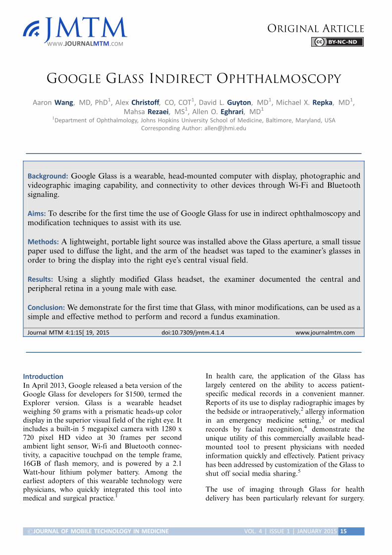
the display centrally, to be in primary gaze for
extended comfortable viewing with the Glass for the
duration of the exam. This is accomplished by
unscrewing the Google Glass frame from the main
unit and taping it to the frame of the user’s own
glasses (see Figure 2), or a blank pair.
To perform the exam, the light is toggled on and the
video capture is started. The exam is then per-
formed in the same manner as with the traditional
binocular indirect ophthalmoscope (BIO) by hold-
ing the condensing lens in one hand. Since the
camera application for the Glass currently is wide
angle and zooming options are not currently
present, the fundus will appear small in the display.
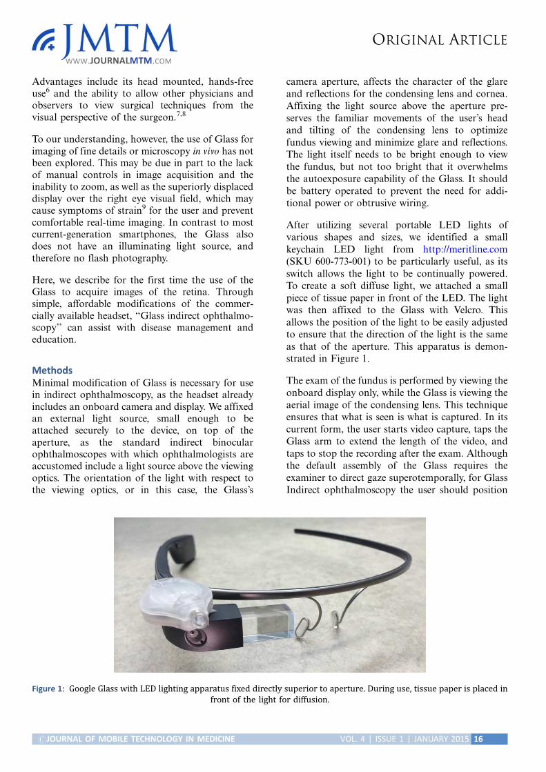
To address this issue, the examiner should position
his or her head about 25cm (rather than arm’s
length) from the patient’s eye so that the fundus can
fill the field of view of the Glass camera. This
represents the only change in examination techni-
que that differs from use of the traditional BIO, and
is demonstrated in Figure 3.
After discussion of risks and benefits, a 31-year-old
male volunteer provided informed consent to pro-
ceed with Glass indirect ophthalmoscopy and
recording, as part of a study of head-mounted
digital camera indirect ophthalmoscopy for which
Institutional Research Board approval was ob-
tained. The participant’s right eye was dilated with
2.5% phenylephrine and 1% tropicamide.
All authors have completed the Unified Competing
Interest form at www.icmje.org/coi_disclosure.pdf
and declare: no support from any organisation for
the submitted work; no financial relationships with
any organisations that might have an interest in the
submitted work in the previous 3 years; no other
relationships or activities that could appear to have
influenced the submitted work.
Figure 2: The Google Glass unit is attached to the user’s
own glasses with tape around the arm, allowing the
display to be positioned directly in the center of the
user’s field of view.
Figure 3: The examiner uses Glass on a subject for fundoscopy at a relatively close distance, positioned between the typical
near (direct ophthalmoscopy) and far (indirect ophthalmoscopy) locations encountered with standard techniques.
ORIGINAL ARTICLE
#JOURNAL OF MOBILE TECHNOLOGY IN MEDICINE VOL. 4 | ISSUE 1 | JANUARY 2015 17
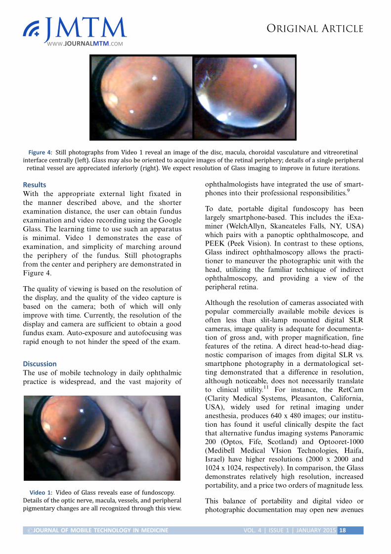
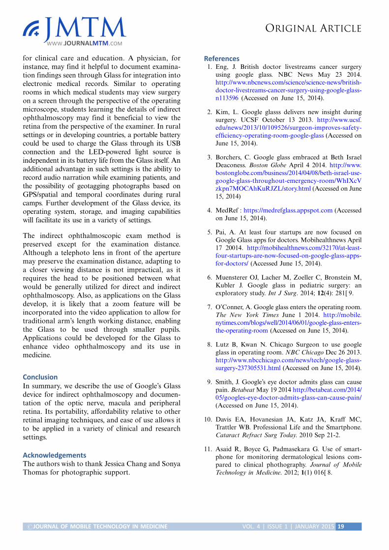
ResultsWith the appropriate external light fixated in
the manner described above, and the shorter
examination distance, the user can obtain fundus
examination and video recording using the Google
Glass. The learning time to use such an apparatus
is minimal. Video 1 demonstrates the ease of
examination, and simplicity of marching around
the periphery of the fundus. Still photographs
from the center and periphery are demonstrated in
Figure 4.
The quality of viewing is based on the resolution of
the display, and the quality of the video capture is
based on the camera; both of which will only
improve with time. Currently, the resolution of the
display and camera are sufficient to obtain a good
fundus exam. Auto-exposure and autofocusing was
rapid enough to not hinder the speed of the exam.
DiscussionThe use of mobile technology in daily ophthalmic
practice is widespread, and the vast majority of
ophthalmologists have integrated the use of smart-
phones into their professional responsibilities.9
To date, portable digital fundoscopy has been
largely smartphone-based. This includes the iExa-
miner (WelchAllyn, Skaneateles Falls, NY, USA)
which pairs with a panoptic ophthalmoscope, and
PEEK (Peek Vision). In contrast to these options,
Glass indirect ophthalmoscopy allows the practi-
tioner to maneuver the photographic unit with the
head, utilizing the familiar technique of indirect
ophthalmoscopy, and providing a view of the
peripheral retina.
Although the resolution of cameras associated with
popular commercially available mobile devices is
often less than slit-lamp mounted digital SLR
cameras, image quality is adequate for documenta-
tion of gross and, with proper magnification, fine
features of the retina. A direct head-to-head diag-
nostic comparison of images from digital SLR vs.
smartphone photography in a dermatological set-
ting demonstrated that a difference in resolution,
although noticeable, does not necessarily translate
to clinical utility.11 For instance, the RetCam
(Clarity Medical Systems, Pleasanton, California,
USA), widely used for retinal imaging under
anesthesia, produces 640 x 480 images; our institu-
tion has found it useful clinically despite the fact
that alternative fundus imaging systems Panoramic
200 (Optos, Fife, Scotland) and Optooret-1000
(Medibell Medical VIsion Technologies, Haifa,
Israel) have higher resolutions (2000 x 2000 and
1024 x 1024, respectively). In comparison, the Glass
demonstrates relatively high resolution, increased
portability, and a price two orders of magnitude less.
This balance of portability and digital video or
photographic documentation may open new avenues
Figure 4: Still photographs from Video 1 reveal an image of the disc, macula, choroidal vasculature and vitreoretinal
interface centrally (left). Glass may also be oriented to acquire images of the retinal periphery; details of a single peripheral
retinal vessel are appreciated inferiorly (right). We expect resolution of Glass imaging to improve in future iterations.
Video 1: Video of Glass reveals ease of fundoscopy.
Details of the optic nerve, macula, vessels, and peripheral
pigmentary changes are all recognized through this view.
ORIGINAL ARTICLE
#JOURNAL OF MOBILE TECHNOLOGY IN MEDICINE VOL. 4 | ISSUE 1 | JANUARY 2015 18
for clinical care and education. A physician, forinstance, may find it helpful to document examina-tion findings seen through Glass for integration intoelectronic medical records. Similar to operatingrooms in which medical students may view surgeryon a screen through the perspective of the operatingmicroscope, students learning the details of indirectophthalmoscopy may find it beneficial to view theretina from the perspective of the examiner. In ruralsettings or in developing countries, a portable batterycould be used to charge the Glass through its USBconnection and the LED-powered light source isindependent in its battery life from the Glass itself. Anadditional advantage in such settings is the ability torecord audio narration while examining patients, andthe possibility of geotagging photographs based onGPS/spatial and temporal coordinates during ruralcamps. Further development of the Glass device, itsoperating system, storage, and imaging capabilitieswill facilitate its use in a variety of settings.
The indirect ophthalmoscopic exam method ispreserved except for the examination distance.Although a telephoto lens in front of the aperturemay preserve the examination distance, adapting toa closer viewing distance is not impractical, as itrequires the head to be positioned between whatwould be generally utilized for direct and indirectophthalmoscopy. Also, as applications on the Glassdevelop, it is likely that a zoom feature will beincorporated into the video application to allow fortraditional arm’s length working distance, enablingthe Glass to be used through smaller pupils.Applications could be developed for the Glass toenhance video ophthalmoscopy and its use inmedicine.
ConclusionIn summary, we describe the use of Google’s Glassdevice for indirect ophthalmoscopy and documen-tation of the optic nerve, macula and peripheralretina. Its portability, affordability relative to otherretinal imaging techniques, and ease of use allows itto be applied in a variety of clinical and researchsettings.
AcknowledgementsThe authors wish to thank Jessica Chang and SonyaThomas for photographic support.
References1. Eng, J. British doctor livestreams cancer surgery
using google glass. NBC News May 23 2014.
http://www.nbcnews.com/science/science-news/british-
doctor-livestreams-cancer-surgery-using-google-glass-
n113596 (Accessed on June 15, 2014).
2. Kim, L. Google glasss delivers new insight during
surgery. UCSF October 13 2013. http://www.ucsf.edu/news/2013/10/109526/surgeon-improves-safety-
efficiency-operating-room-google-glass (Accessed on
June 15, 2014).
3. Borchers, C. Google glass embraced at Beth Israel
Deaconess. Boston Globe April 4 2014. http://www.
bostonglobe.com/business/2014/04/08/beth-israel-use-
google-glass-throughout-emergency-room/WhIXcVzkpn7MOCAhKuRJZL/story.html (Accessed on June
15, 2014)
4. MedRef : https://medrefglass.appspot.com (Accessed
on June 15, 2014).
5. Pai, A. At least four startups are now focused on
Google Glass apps for doctors. Mobihealthnews April
17 20014. http://mobihealthnews.com/32170/at-least-four-startups-are-now-focused-on-google-glass-apps-
for-doctors/ (Accessed June 15, 2014).
6. Muensterer OJ, Lacher M, Zoeller C, Bronstein M,
Kubler J. Google glass in pediatric surgery: an
exploratory study. Int J Surg. 2014; 12(4): 281�9.
7. O’Conner, A. Google glass enters the operating room.
The New York Times June 1 2014. http://mobile.nytimes.com/blogs/well/2014/06/01/google-glass-enters-
the-operating-room (Accessed on June 15, 2014).
8. Lutz B, Kwan N. Chicago Surgeon to use google
glass in operating room. NBC Chicago Dec 26 2013.
http://www.nbcchicago.com/news/tech/google-glass-
surgery-237305531.html (Accessed on June 15, 2014).
9. Smith, J. Google’s eye doctor admits glass can causepain. Betabeat May 19 2014 http://betabeat.com/2014/
05/googles-eye-doctor-admits-glass-can-cause-pain/
(Accessed on June 15, 2014).
10. Davis EA, Hovanesian JA, Katz JA, Kraff MC,
Trattler WB. Professional Life and the Smartphone.
Cataract Refract Surg Today. 2010 Sep 21-2.
11. Asaid R, Boyce G, Padmasekara G. Use of smart-
phone for monitoring dermatological lesions com-pared to clinical phothography. Journal of Mobile
Technology in Medicine. 2012; 1(1) 016�8.
ORIGINAL ARTICLE
#JOURNAL OF MOBILE TECHNOLOGY IN MEDICINE VOL. 4 | ISSUE 1 | JANUARY 2015 19