Embed Size (px)
Citation preview

/. Embryol. exp. Morph. 98, 269-285 (1986) 269Printed in Great Britain © The Company of Biologists Limited 1986
Gonadal development of the chick embryo followingmicrosurgically caused agenesis of the mesonephrosand using interspecific quail—chick chimaeras
EVA S. RODEMER, ALICE IHMER AND H. WARTENBERGAnatomisches Institut der Universitdt Bonn, Nufiallee 10, D-5300 Bonn 1,Federal Republic of Germany
SUMMARYMesonephric agenesis was achieved by microsurgical excision of the left Wolffian duct and the
underlying intermediate mesoderm of different regions between somites 16 and 23 in chickensafter 50-52 h of incubation (stage 14 HH). Quail-chick chimaeras were produced by transplan-tation of corresponding quail tissue in the region of somites 18-21.
A morphometrical analysis of the mesonephric and gonadal area in cross sections shows thatthe intermediate mesoderm from somites 16 to 23 develops into the mesonephros. A partialagenesis of the mesonephros brought about by removal of the intermediate mesoderm at thelevel of somites 18 to 21 at stage 14 leads to a mean reduction of the gonadal volume of 37-8 %compared to the volume of the untreated side at stage 30. Transplantation of quail intermediatemesoderm in this region of the excision results in development of a hybrid mesonephros.Consequently, the gonads are invaded and colonized by quail cells mobilized from mesonephriccorpuscles examined at stage 30, 35 and 36.
These results are discussed in terms of the origin of the gonadal stroma during this develop-mental period; they show that in the region from the third to the sixth segment the ventromedialpart of the differentiating mesonephros participates in the contribution of stromal cells to thegonad.
INTRODUCTION
The development of the gonads of vertebrates starts with the formation of thegerminal epithelium, bilateral thickenings of the coelomic epithelium that appearventral to the developing mesonephroi. The proliferation of both germinal and'nongerminal' cells leads to the formation of distinct gonadal primordia which areidentical in both sexes, the indifferent gonad (see for review Zuckerman & Weir,1977). The data on the origin of the gonadal stroma in sexually indifferent gonadsare at variance. Considered by Callebaut (1976), Dodd (1977), Kopp & Bertrand(1978), Fargeix, Didier & Didier (1981), Merchant-Larios & Villalpando (1981)and Popova & Scheib (1981) to derive from the coelomic epithelium, the stroma ofthe gonads was suggested by others to arise either from the mesonephric blastema(Dang & Fouquet, 1979; Carlon, Pizant & Stahl, 1983) or from the differentiated
Key words: chick embryo, mesonephros, agenesis, quail-chick marker system, gonad,chimaera.
270 E. S. RODEMER, A. IHMER AND H. WARTENBERG
mesonephros (Witschi, 1914, 1951; Swift, 1916; Upadhyay, Luciani & Zamboni,1979; Zamboni, Bezard & Mauleon, 1979).
In order to ascertain the dependence of the gonad on the intermediate meso-derm or the mesonephros in chick embryos, we excised the intermediate meso-derm in the presumptive mesonephric area at day two. Following subsequentincubation, we morphometrically analysed the indifferent chick gonad at day 7(end of the indifferent stage). To obtain further information about gonad-con-tributing cells of the mesonephros, we transplanted corresponding quail tissue inthe presumptive gonadal area at day 2 and examined the chick gonads at days 7, 9and 10.
MATERIALS AND METHODS
Microsurgery to cause mesonephric agenesis
Fertilized eggs of the domestic fowl (Gallus domesticus, strain White Leghorn) were acquiredfrom a local supplier and incubated at 37-8°C ± 1°C, 70 % to 80 % humidity.
After 50-52 h of incubation the position of the embryo was marked by candling and thesurface of each egg was sterilized by swabbing it with 70 % ethanol. A hole was pricked in theblunt end of the egg, a procedure that leads to air being forced out of the air space and whichresults in the embryo dropping away from the surface.
Operations were performed under aseptic conditions with the aid of a stereomicroscope, theegg being supported horizontally in a cradle of cellulose. After cutting a window (approximately0-7x0-5 cm) in the shell and its subjacent shell membrane at the marked area, the chick embryowas floated up to the level of the window by infusion of Locke solution (Hara, 1971). The stagesof development were determined according to the criteria of Hamburger & Hamilton (1951).We used embryos at stage 14.
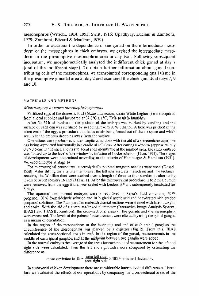
For microsurgical procedures, electrolytically pointed tungsten needles were used (Dossel,1958). After slitting the vitelline membrane, the left intermediate mesoderm and, for technicalreasons, the Wolffian duct were excised over a length of three to four somites at alternatinglevels between somites 16 and 23 (Fig. 1). After the microsurgical procedure, 1 to 2 ml albumenwere removed from the egg; it then was sealed with Leukosilk® and subsequently incubated for5 days.
The operated and normal embryos were killed, fixed in Serra's fluid containing 60%propanol, 30 % formaldehyde solution and 10 % glacial acetic acid and dehydrated with gradedpropanol solutions. The 7 jum paraffin-embedded serial sections were stained with haematoxylinand eosin. With the aid of a computer-linked planimeter (Interactive Image Analysis System,IB AS I and IB AS II, Kontron), the cross-sectional areas of the gonads and the mesonephroiwere measured. The levels of the points of measurement were elicited by using the spinal gangliaas a means of orientation.
In the region of the mesonephros at the beginning and end of each spinal ganglion thecircumference of the mesonephros was marked by a digitizer (Fig. 2). From this, IB AScalculated the cross-sectional areas in [im2. In the region of the gonad, measurements in themiddle of each spinal ganglion and at the midpoint between two ganglia were added.
In the normal embryos the average of the areas for each point of measurement for the left andright side were calculated. Then the left and right sides were compared by estimating thedifference as
j • .• • m area left side ^ 1 n r i , . , , , . ..mean deviation in % = . , . . , x 100 ± standard deviation.
area right sideIn embryonal chicken development there are considerable interindividual differences. There-
fore we evaluated the effects of our operations by comparing the cross-sectional areas of the

Avian gonadal development following quail-chick grafting
Chick embryoStaee 14
271
16
Fig. 1. Diagram showing on the right side the unilateral extirpation of the intermediatemesoderm (im) and the Wolffian duct (Wd) from a chick embryo at stage 14 over alength of three to four somites in different regions between somites 16 and 23. The leftside shows the substitution by an equal quail graft previously isolated from a quailembryo of the same stage of development. Transplantation in the region of somites18 to 21.
mesonephros and the gonad of the operated side with the corresponding areas of the untreatednormal side as
deviation in % = area left sidearea right side
xlOO
in each embryo. The percentage deviation of all points of measurement of one organ was thenused to calculate the arithmetical mean of the difference between the operated and the normalside.
Grafting procedureFertilized eggs of the Japanese quail (Coturnix coturnixjaponica), delivered by a local supplier,
were incubated together with the chick eggs. The chick embryos were operated at the levelof somites 18 to 21 at stage 14HH as described above. For the quail, whose development isprecocious relative to chicks although it does not differ by more than a few hours until the thirdday of incubation, the stages were determined by analogy with the chick.
Quail embryos were removed from the egg, placed on agar and covered with Locke solution.Left intermediate mesoderm and Wolffian duct were cut from the quail donor with fine tungstenneedles at the level of somites 18 to 21 and stained lightly with Nile blue sulphate impregnatedin agar. These were then transferred by means of a Spemann micropipette from donor to host

272 E. S. R O D E M E R , A. IHMER AND H. WARTENBERG
embryo and manoeuvred into place with tungsten needles (Fig. 1). In order to ensure a properfit and good adhesion between host and graft tissues, excess Locke solution was carefullywithdrawn with a micropipette. After removal of 1 to 2 ml albumen the sealed egg was incubatedfor 5, 7 and 8 days. Chimaeras at HH stages 30, 35 and 36 were processed for histological
Fig. 2. Transverse section of a chick embryo in the region of mesonephric agenesisat stage 30 after excision of the left intermediate mesoderm and Wolffian duct atstage 14. On the operated side the mesonephros (m) and the Wolffian duct (Wd) aremissing. Both gonads (g) are present. , measured circumference of mesonephros,—, measured circumference of gonad. Scale bar, 0-2 mm; stained with haematoxylinand eosin.
Avian gonadal development following quail-chick grafting 273
examination: they were fixed in Serra's fluid by transcardiac perfusion and subsequent immer-sion. After paraffin embedding the specimens were serially sectioned at 7 jum. As the interphasenuclei of quail cells are characterized by a large mass of nucleolus-associated heterochromaticDNA, the sections were stained for DNA (Feulgen & Rossenbeck, 1924) and counterstainedwith lightgreen, or they were stained with haematoxylin and eosin following acid hydrolysis(Hutson & Donahoe, 1984).
RESULTS
Morphometry of normal embryos
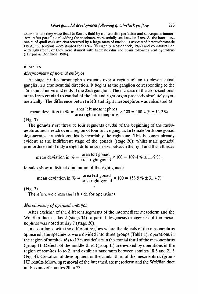
At stage 30 the mesonephros extends over a region of ten to eleven spinalganglia in a craniocaudal direction. It begins at the ganglion corresponding to the15th spinal nerve and ends at the 25th ganglion. The increase of the cross-sectionalareas from craniad to caudad of the left and right organ proceeds absolutely sym-metrically. The difference between left and right mesonephros was calculated as
A • .- -of area left mesonephros w 1ftft i n n . M , 1O « ~mean deviation m % = r-r *-. x 100 = 100-4 % ± 12-2 %
area right mesonephros(Fig. 3).
The gonads start three to four segments caudal of the beginning of the meso-nephros and stretch over a region of four to five ganglia. In female birds one gonaddegenerates; in chickens this is invariably the right one. This becomes alreadyevident at the indifferent stage of the gonads (stage 30): whilst male gonadalprimordia exhibit only a slight difference in size between the right and the left side:
mean deviation in % = a r e a left gonad x m = m ^ % ± n Q %
area right gonadfemales show a distinct diminution of the right gonad:
mean deviation in % = a r e a left gonad x m = ^ ^ % ± ^ %
area right gonad
(Fig. 3).Therefore we chose the left side for operations.
Morphometry of operated embryos
After excision of the different segments of the intermediate mesoderm and theWolffian duct at day 2 (stage 14), a partial dysgenesis or agenesis of the meso-nephros was noted at day 7 (stage 30).
In accordance with the different regions where the defects of the mesonephrosappeared, the specimens were divided into three groups (Table 1): operations inthe region of somites 16i to 19 cause defects in the cranial third of the mesonephros(group I). Defects of the middle third (group II) are evoked by operations in theregion of somites 18 to 21 and exhibit a maximum between somites 18-5 and 21-5(Fig. 4). Cessation of development of the caudal third of the mesonephros (groupIII) results following removal of the intermediate mesoderm and the Wolffian ductin the zone of somites 20 to 23.
274 E. S. RODEMER, A. IHMER AND H. WARTENBERG
0 20 40 60 80 100 0 2 4 6 8 0 2 4 6 8
0 15
Area (,um2)
17
05Spinal ganglia
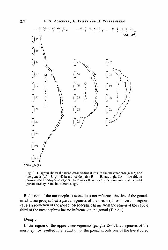
Fig. 3. Diagram shows the mean cross-sectional area of the mesonephroi (n = 7) andthe gonads (d" = 3; $ = 4) in /mi2 of the left ( • • ) and right (O O) side innormal chick embryos at stage 30. In females there is a distinct diminution of the rightgonad already in the indifferent stage.
Reduction of the mesonephros alone does not influence the size of the gonadsin all three groups. But a partial agenesis of the mesonephros in certain regionscauses a reduction of the gonad. Mesonephric tissue from the region of the caudalthird of the mesonephros has no influence on the gonad (Table 1).
Group I
In the region of the upper three segments (ganglia 15-17), an agenesis of themesonephros resulted in a reduction of the gonad in only one of the five studied
Avian gonadal development following quail-chick grafting 275
cases. If the agenesis in the two studied cases extends further caudally into theregion of ganglion 18, an effect on the gonads is observed (Table 2), namely amean reduction of 17-5 % to an average of 82-5 % as compared to the right side.
Group II
A marked influence on the size of the gonads is attributable to the absence ofthe mesonephros in the region of ganglia 18 to 20. In nine cases studied, a meanreduction of 37-8 % to an average of 62-2 % as compared with the opposite sideresults (Fig. 4). Agenesis of the mesonephros commencing from the region ofganglion 20 and further caudad has no more influence on the gonad (Table 2).The region of the reduction of the gonad is always observed caudally from thecorresponding defect of the mesonephros (Fig. 4). Only in one case did diminutionof the gonad appear at the same level as the defect of the mesonephros, but themaximum diminution nevertheless occurred further caudad.
Interspecific quail-chick chimaeras
43 out of 175 embryos operated in this way developed after subsequent in-cubation. Of these 43 specimens, 27 contained quail tissue. 21 of the latter werefixed on day 7, three on day 9 and three on day 10.
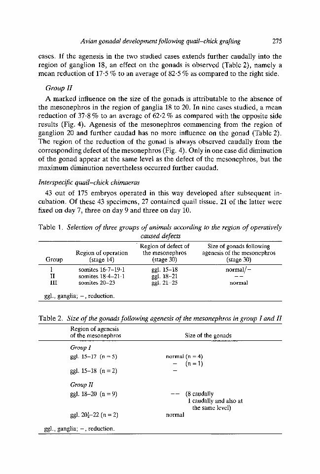
Table 1. Selection of three groups of animals according to the region of operativelycaused defectsRegion of defect of Size of gonads following
Region of operation the mesonephros agenesis of the mesonephrosGroup (stage 14) (stage 30) (stage 30)
I somites 16-7-191 ggl. 15-18 normal/-II somites 18-4-21-1 ggl. 18-21III somites 20-23 ggl. 21-25 normal
ggl., ganglia; - , reduction.
Table 2. Size of the gonads following agenesis of the mesonephros in group I and II
Region of agenesisof the mesonephros Size of the gonads
Group Iggl. 15-17 (n = 5) normal (n = 4)
- (n = l)ggl. 15-18 (n = 2)
Group II
ggl. 18-20 (n = 9) — (8 caudally1 caudally and also at
the same level)ggl. 201-22 (n = 2) normal
ggl., ganglia; - , reduction.
276 E. S. RODEMER, A. IHMER AND H. WARTENBERG
0 20 40 60 80 1001 I 1 I I 1 —
0 20 40 60 80 100-) 1 1 1 1 1 c
Area
0000*00
20
21
25
Spinal ganglia
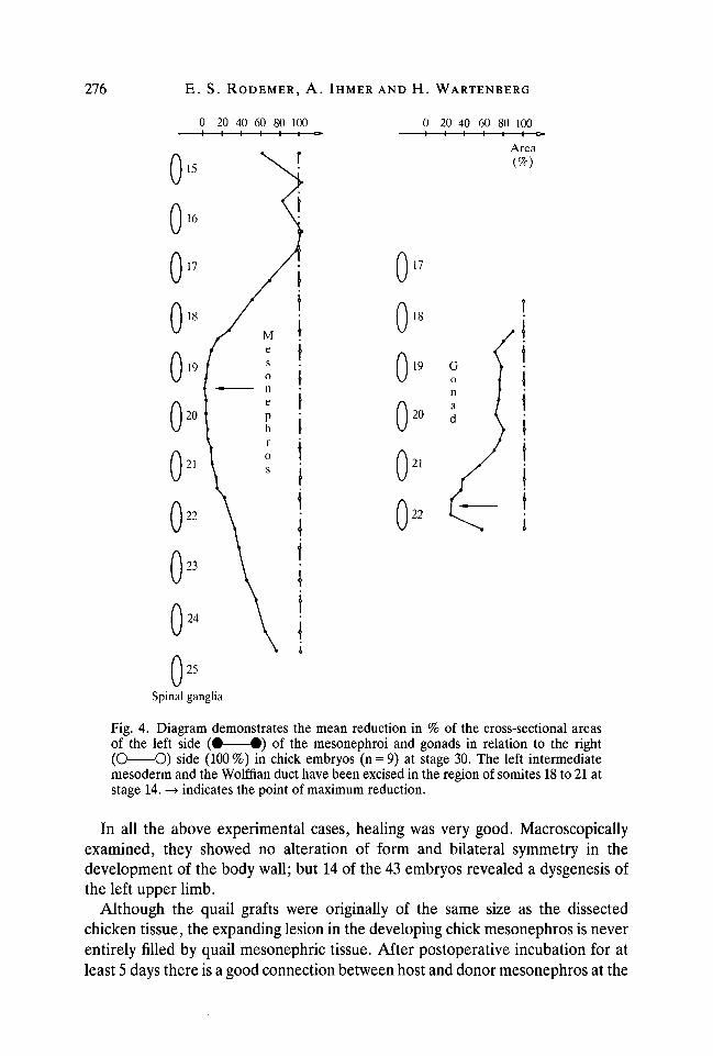
Fig. 4. Diagram demonstrates the mean reduction in % of the cross-sectional areasof the left side ( • • ) of the mesonephroi and gonads in relation to the right(O O) side (100%) in chick embryos (n = 9) at stage 30. The left intermediatemesoderm and the Wolffian duct have been excised in the region of somites 18 to 21 atstage 14. —> indicates the point of maximum reduction.
In all the above experimental cases, healing was very good. Macroscopicallyexamined, they showed no alteration of form and bilateral symmetry in thedevelopment of the body wall; but 14 of the 43 embryos revealed a dysgenesis ofthe left upper limb.
Although the quail grafts were originally of the same size as the dissectedchicken tissue, the expanding lesion in the developing chick mesonephros is neverentirely filled by quail mesonephric tissue. After postoperative incubation for atleast 5 days there is a good connection between host and donor mesonephros at the
Avian gonadal development following quail-chick grafting 277
cranial end of the transplanted tissue, but at the caudal end a distinct gap exists. Ineach case, in the region of the graft, mesonephric tubuli consisting of cells withtypical quail nuclei are found. Also hybrid tubuli are demonstrable, one part ofthe tubulus wall containing chick cells, the other part formed by quail cells; nointermingling of the different cells occurs (Fig. 5). If the quail graft develops onlytubuli and no other mesonephric structures, no quail cells are discovered in thegonad.
The quail tissue is also able to form mesonephric stromal cells and to develop aBowman's capsule. Either the Bowman's capsules circumscribe an empty space orthey contain hybrid glomeruli. These glomeruli consist of chick-derived cells withoblong, flat nuclei which we consider to be endothelial cells of the glomerularloop; they also contain cells with oval or round nuclei carrying the quail marker,which we assume to be podocytes (Fig. 6).
In nearly all chimaeras the mesonephroi are composed of alternating chick andquail tissue; in many cases the quail mesonephric tissue occupies the dorsal orlateral parts of the organs. These dorsolateral sections of the mesonephroi do notcontribute to the gonad, as no quail cells reach the gonad in such situations.
Thus, to form the gonad, the quail graft has to develop mesonephric corpusclesor stroma, and this in the ventromedial section of the mesonephros lying injuxtaposition to the gonad.
In 7-day-old embryos, quail cells egressing from the Bowman's capsules ofthe quail type had colonized the indifferent gonad in three cases. Outside theBowman's capsule the migrating cells form very delicate trabeculae (Fig. 7) which,in the serial planes of sections, can be followed all the way into the gonad. There,the colonizing quail cells do not disperse randomly throughout the inner core ofthe gonad and do not intermingle with chicken stromal cells. They occupy adistinct part of the gonadal stroma and in no case do they show contact orintermingle with the stratified surface epithelium. A band-like space beneath thelatter approximately twice as wide as the epithelium is formed by chick cells(Fig. 8). Hence, the quail cells establish no association with the primordial germcells in the deep layer of the epithelium. Germ cells arranged between the innercore and the surface epithelium are enclosed by chick and quail stromal cells; thechick cells form a crescent round the germ cells at the side apposed to theepithelium; the other side is covered by quail cells (Fig. 9).
In a 9- and 10-day-old ovary, two structural components are demonstrable: anouter cortex and an inner medulla. The medulla occupying the major portion ofthe ovary is partially formed by quail cells. Chick cells constituting the other partof the medulla hardly intermingle with the quail cells. Most of the germ cells lie inpairs or small groups in the middle or inner part of the cortex. These germ cellsare closely surrounded by chick cells. Also, the few germ cells lying in the deepmedullary part toward the hilus, constituted of quail tissue, are surrounded bychick cells (Fig. 10).
In a 10-day-old testis, germ cells show an even distribution throughout themedulla, indicating the formation of male cords. Although in this case only a few

278 E. S. RODEMER, A. IHMER AND H. WARTENBERG
1?*
-£- >'Fig. 5. Part of a transverse section through the chimaeric mesonephros of a chickembryo 8 days after exchange of the left intermediate mesoderm and Wolffian duct byan equal quail graft. The asterisk marks a hybrid tubulus. The arrows mark thebeginning of the part of the tubulus wall containing quail cells. All cells of the right sidebetween the arrows were identified as quail cells by varying the focus. The left tubulusside is formed by chick cells (arrowheads). Scale bar, 10 jwm; stained with haematoxylinand eosin following acid hydrolysis.
Fig. 6. Hybrid glomerulus 5 days after substitution of the left intermediate mesodermand the Wolffian duct by an equal quail graft. The arrowheads mark oblong, flat nucleiof chick cells. The round nuclei carry the typical quail marker (arrows). Scale bar,10 jum; stained with haematoxylin and eosin following acid hydrolysis.

Avian gonadal development following quail-chick grafting
* IS279
8
Figs 7, 8. For legends see p. 280

280 E. S. RODEMER, A. IHMER AND H. WARTENBERG
quail cells colonize the gonad in small groups, they come into contact with thegerm cells. These germ cells are consequently surrounded by cells of the chick aswell as those of the quail type (Fig. 11).
DISCUSSION
The data indicate that the cranial third of the mesonephros develops from theintermediate mesoderm of the region of somite 16 to the beginning of somite 19.The middle third is formed by material belonging to the region extending fromsomite 18 to the beginning of somite 21, and the caudal third of the mesonephrosoriginates in the intermediate mesoderm of the region comprising somites 21 to 23(Fig. 12). As shown in Fig. 12, mesonephric tissue of the region extending fromthe end of ganglion 17 to the beginning of ganglion 20 (black area) contributes tothe gonad in each case. Mesonephric material of two small regions (hatched areas)adjacent to the cranial and caudal end of this region has influence on the gonadonly in certain cases. In contradiction to authors who consider the presumptivegonadal area to be at the level of somites 9 to 14 (Romanoff, 1960) or at the level ofsomites 20 to 26 (Willier, 1933; Griinwald, 1937) or at the level of somites 24 to 29(Dantschakoff, 1931) or spread over a more extended region from somites 13 to 22(Didier, Fargeix & Bergeaud, 1980), our results of the extirpation and transplan-tation experiments suggest a presumptive gonadal area of the intermediate meso-derm extending from the beginning of somite 17 to the beginning of somite 21.
The diminution of the gonadal volume was caused by excision of the inter-mediate mesoderm; thus on the basis of our experiments alone it cannot be
Fig. 7. Section through parts of the chimaeric mesonephros and gonad 5 days aftersubstitution of the left intermediate mesoderm and the Wolffian duct by an equal quailgraft. (i) empty Bowman's capsule of quail cells, (2) gonad. The arrows demonstratedelicate trabeculae formed by quail cells migrating from the Bowman's capsule intothe gonad. Scale bar, 10 jinn; stained with haematoxylin and eosin following acidhydrolysis.
Fig. 8. Section through a part of the chimaeric mesonephros (1) and the gonad (2)5 days after exchange of the left intermediate mesoderm and the Wolffian duct by anequal quail graft. The arrows show the distinct part of the gonadal stroma formed byquail cells which do not reach the surface epithelium. Scale bar, 40jum; stained withhaematoxylin and eosin following acid hydrolysis.
Fig. 9. Section through a part of the chimaeric gonad 5 days after substitution of theleft intermediate mesoderm and the Wolffian duct by an equal quail graft. The asteriskmarks two germ cells lying between the inner core (1) and the surface epithelium (2) ofthe gonad. These germ cells are covered by chick cells (arrowheads) at the sideapposed to the surface epithelium; the other side is covered by quail cells (arrows).Scale bar, 10 /-tm; stained with haematoxylin and eosin following acid hydrolysis.Fig. 10. Section through a 10-day-old ovary 8 days after the exchange of the leftintermediate mesoderm and the Wolffian duct of an equal quail graft. Deep medullarypart constituted by quail cells (arrows). Germ cells (*) surrounded by chick cells(arrowheads). Scale bar, lOjum; stained with haematoxylin and eosin following acidhydrolysis.Fig. 11. Section through the medulla of a 10-day-old testis 8 days after transplantation.Germ cells (*) surrounded simultaneously by quail (arrows) and chick (arrowheads)cells. Scale bar, lOjum; stained with haematoxylin and eosin following acid hydrolysis.

Avian gonadal development following quail-chick grafting 281
decided whether this reduction of the chicken gonad is due to loss of themesonephric blastema or to loss of the differentiated mesonephros. However,Bishop-Calame (1966), Popova & Scheib (1981) and Merchant-Larios, Popova &

282 E. S. RODEMER, A. IHMER AND H. WARTENBERG
Somites | 16 | |17 | | l 8 | | l 9 | |20| | 2 l | |22| | 23 |
Intermediate
mesoderm
Mesonephros
ganglia —
Fig. 12. Diagram summarizing the results of extirpation of the intermediate mesodermat stage 14 (above) in three overlapping regions. At stage 30, segments of the meso-nephros (below) are missing in three corresponding regions. • = segments of themesonephros which definitely contribute to the gonad. H = region of the mesonephroswhich possibly contributes to the gonad.
Reyss-Brion (1984) observed a reduction of the gonad following interruptionof the Wolf flan duct. Since they did not excise the mesonephric blastema, butprevented mesonephric development, we attribute the diminution of the gonadalvolume in their and our experiments to the missing contribution of a differentiatedmesonephros. This is confirmed by the transplantation experiments. As theseshow, the gonads are colonized by cells originating in mesonephric corpuscles.
Several studies concerning gonadal development in mammals also postulate orshow a mesonephric origin of the somatic cells of the gonad (Witschi, 1951;Wartenberg, 1978; Fraedrich, 1979; Kinsky, 1979; Upadhyay etal. 1979; Zamboniet al. 1979). These cells originate in developing as well as in regressing or dis-integrating structures of the mesonephros. In accordance with the assumption thatmesonephric degeneration in chickens does not begin earlier than the 11th day ofincubation (Romanoff, 1960), in our examination (days 7, 9 and 10) the chickensexhibited no signs of degeneration of the mesonephros. Thus in the chicken thesegregation of cells of the mesonephros takes place during the differentiatingphase at these stages of development.
In rabbit (Kinsky, 1979), sheep, Macaca mulatto, (Zamboni etal. 1980) and man(Wartenberg, 1978) the gonad is colonized by mesonephric cells of the cranial thirdof the organ. This seems to be different in mammals from in chickens. Neither thecranial fifth of the mesonephros nor the caudal half is observed to contribute to thegonad. Only a complete agenesis of the mesonephros over a region of more thanthree segments successive to the cranial fifth causes a reduction of the gonad(Fig. 12). Likewise, the quail mesonephric tissue has to appear in the latter region

Avian gonadal development following quail-chick grafting 283
of the mesonephros in order to form gonadal stroma. Quail structures furthercraniad or caudad have no connection with the gonad.
During the operations, we took pains to avoid injuring both the dorsal aortalying directly ventral to the intermediate mesoderm and the coelomic epitheliummedial of the latter. For this reason we could not always be sure that thisventromedial part of the intermediate mesoderm was excised in toto. We supposethis incomplete removal to be the reason why in many extirpation experiments nopartial agenesis but a partially reduced mesonephros resulted in the operatedregion. If only scant mesonephric tissue appeared in the gonad-contributing areaof the mesonephros, no reduction of the gonad was observed. We consider thatthis ventromedial part of the intermediate mesoderm contributes to the formationof the ventromedial section of the mesonephros and, as the transplantationexperiments show, it is only this ventromedial section of the mesonephros thatcontributes to the gonadal stroma.
An agenesis of the mesonephros restricted to two or three segments neverresulted in agenesis of the gonad. In the same way, there was no case in whichquail cells originating from quail mesonephric corpuscles formed the entiregonadal stroma at any given level. On the one hand it is possible that the persistingcranial and caudal parts of the chicken mesonephros are extensive enough torestore the gonadal stroma and act as a regulator; for technical reasons we arenot able to excise more extended parts of the intermediate mesoderm to eliminatesuch a regulative mechanism.
On the other hand there still remains the question of a contribution of thecoelomic epithelium to the gonadal stroma. With this method we cannot excludeit, because we are not able to decide whether the chick cells in the gonad derivefrom persisting parts of the chick mesonephros or from the chick coelomicepithelium. This method only proves the mesonephros to participate, at least, inthe gonadal stroma.
The reduction of the gonad was found almost exclusively caudal to the cor-responding agenesis of the mesonephros (Fig. 4). According to observations in therabbit (Kinsky, 1979), in sheep (Zamboni et al. 1979), in the Macaca fascicularis(Dang & Fouquet, 1979) and in man (Wartenberg, 1978), confirmed by the trans-plantation experiments, the contribution of the mesonephros to the gonads wouldseem to proceed in a craniocaudal direction.
The relationship of the gonadal stromal cells of mesonephric origin to the germcells seems to differ in male and female gonads. In contradiction to findings insheep (Zamboni et al. 1979), they do not come into contact with the germ cells inchicken ovaries at this early stage of development. Male germ cells are surroundedby mesonephric quail cells and chicken cells at the same time. This result maycorrespond with the postulated dual Sertoli cell system in man and mammals(Wartenberg, 1978,1979); the possibility needs to be explored in further studies.
This work was supported by Deutsche Forschungsgemeinschaft.

284 E. S. RODEMER, A. IHMER AND H. WARTENBERG
REFERENCES
BISHOP-CALAME, S. (1966). Etude experimentale de l'organogenese du systeme urog6nitale del'embryon de poulet. Archs Anat. microsc. Morph. exp. 55, 215-309.
CALLEBAUT, M. (1976). Origin of ovarian follicle cells in birds. Experientia 32, 1337-1338.CARLON,N.,PIZANT, J. &STAHL, A. (1983). Mesonephric origin of the gonadal primitive medulla
in chick embryos. Anat. Embryol. 166, 399-414.DANG, D. C. & FOUQUET, J. P. (1979). Differentiation of the fetal gonad of Macaca fascicularis
with special reference to the testis. Annls Biol. Anim. Bioch. Biophys. 19,1197-1209.DANTSCHAKOFF, W. (1932). Keimzelle und Gonade. II b. Ganzheit des Gewebekomplexes als
Faktor in der Entwicklung der Gonade. Z. Zellforsch. mikrosk. Anat. 15, 581-644.DIDIER, E., FARGEIX, N. & BERGEAUD, Y. (1980). Age-dependent control exerted by the somatic
part of the gonad upon gonocyte proliferation in the chick embryo. Devi Biol. 77, 488-493.DODD, J. M. (1977). The structure of the ovary of nonmammalian vertebrates. In The Ovary,
vol. 1 (ed. S. Zuckerman & B. J. Weir), pp. 219-263. New York, San Francisco, London:Academic Press.
DOSSEL, W. E. (1958). Preparation of tungsten micro-needles for use in embryologic research.Lab. Invest. 7, 171-173.
FARGEIX, N., DIDIER, E. & DIDIER, P. (1981). Early sequential development in avian gonads.An ultrastructural study using selective glycogen labeling in the germ cells. Reprod. Nutr.Develop. 21, 479-496.
FEULGEN, R. &ROSSENBECK,H. (1924). Mikroskopisch-chemischerNachweiseinerNucleinsaurevom Typus der Thymonucleinsaure und die darauf beruhende elektive Farbung von Zell-kernen in mikroskopischen Praparaten. Hoppe-Seyler's Z. physiol. Chem. 135, 203-252.
FRAEDRICH, J. (1979). Licht- und elektronenmikroskopische Untersuchungen viber denZusammenhang der Mesonephros- und fruhen Gonadenentwicklung der weifien Maus.Dissertation Med. Fak. Bonn, 1-58.
GRUNWALD, P. (1937). Zur Entwicklungsmechanik des Urogenitalsystems beim Huhn. WilhelmRoux' Arch. EntwMech. Org. 136, 786-813.
HAMBURGER, V. & HAMILTON, H. L. (1951). A series of normal stages in the development of thechick embryo. /. Morph. 88, 49-92.
HARA, K. (1971). Micro-surgical operation on the chick embryo in ovo without vital staining.Mikroskopie 27, 267-270.
HUTSON, J. M. & DONAHOE, P. K. (1984). Improved histology for the chick-quail chimera. StainTechnol. 59,105-111.
JALALI, S. (1981). Histo- and cytomorphological studies on the differentiation of the male andfemale gonad during the prehatching stages of the chicken embryo. Dissertation Math.-nat.Fak. Bonn, 1-151.
KINSKY, I. (1979). Bildung des somatischen Gonadenblastems durch degenerierendeUrnierenanteile des Kaninchens. Verh. Anat. Ges., Jena 73, 403-406.
KOPP, F. & BERTRAND, M. F. (1978). Origin de la me'dullaire primitive dans la gonad del'embryon de poulet. Archs Biol. 89, 267-296.
MERCHANT-LARIOS, H. & VILLALPANDO, I. (1981). Ultrastructural events during early gonadaldevelopment in Ranapipiens and Xenopus laevis. Anat. Rec. 199, 349-360.
MERCHANT-LARIOS, H., POPOVA, L. & REYSS-BRION, M. (1984). Early morphogenesis of chickgonad in the absence of mesonephros. Dev. Growth Differ. 26, 403-417.
POPOVA, L. & SCHEIB, D. (1981). Differentiation ovarienne chez l'embryon de poulet apresagenesie experimentale du mesonephros. Archs Anat. microsc. Morph. exp. 70,189-203.
ROMANOFF, A. L. (1960). The urogenital system. In The Avian Embryo (ed. A. L. Romanoff),pp. 781-862. New York: The Macmillan Company.
SWIFT, C. H. (1916). Origin of the sex-cords and the definitive spermatogonia in the male chick.Am. J. Anat. 20, 375-410.
UPADHYAY, S., LUCIANI, L. M. & ZAMBONI, L. (1979). The role of the mesonephros in thedevelopment of indifferent gonads and ovaries of the mouse. Annls Biol. anim. Biochim.Biophys. 19,1179-1196.
WARTENBERG, H. (1978). Human testicular development and the role of the mesonephros in theorigin of a dual Sertoli cell system. Andrologia 10,1-21.

Avian gonadal development following quail-chick grafting 285
WARTENBERG, H. (1979). Der Mesonephros und die Gonadenentwicklung. Verh. anat. Ges., Jena73, 385-401.
WILLIER, B. H. (1933). Potencies of the gonad-forming area in the chick as tested in chorio-allantoic grafts. Wilhelm Rowc' Arch. EntwMech. Org. 130, 616-648.
WITSCHI, E. (1914). Experimentelle Untersuchung iiber die Entwicklungsgeschichte derKeimdriisen von Rana temporaria. Arch, mikrosk. Anat. EntwMech., Abt. 7/85, 9-113.
WITSCHI, E. (1951). Embryogenesis of the adrenal and the reproductive glands. Recent Prog.Horm. Res. 6, 1-27.
ZAMBONI, L., B£ZARD, J. & MAULEON, P. (1979). The role of the mesonephros in thedevelopment of the sheep fetal ovary. Ann. Biol. Anim. Bioch. Biophys. 19, 1153-1178.
ZAMBONI, L., UPADHYAY, S., BEZARD, J., LUCIANI, J. M. & MAUL6ON, P. (1980). The role of themesonephros in the development of the mammalian ovary. In Endocrine Physiopathology ofthe Ovary (ed. R. I. Tozzini, G. Reeves & R. L. Pineda), pp. 3-42. Amsterdam: BiomedicalPress Elsevier/North Holland.
ZUCKERMAN, S. & WEIR, B. J. (1977). The Ovary, vol. 1. New York, San Francisco, London:Academic Press.
(Accepted 12 August 1986)