Embed Size (px)
Citation preview

BiomaterialsScience
PAPER
Cite this: Biomater. Sci., 2020, 8, 506
Received 12th August 2019,Accepted 1st November 2019
DOI: 10.1039/c9bm01271k
rsc.li/biomaterials-science
Glycerylphytate as an ionic crosslinker for 3Dprinting of multi-layered scaffolds with improvedshape fidelity and biological features†
Ana Mora-Boza, a,b Małgorzata K. Włodarczyk-Biegun, c
Aránzazu del Campo, c,d Blanca Vázquez-Lasa *a,b and Julio San Román a,b
The fabrication of intricate and long-term stable 3D polymeric scaffolds by a 3D printing technique is still
a challenge. In the biomedical field, hydrogel materials are very frequently used because of their excellent
biocompatibility and biodegradability, however the improvement of their processability and mechanical
properties is still required. This paper reports the fabrication of dual crosslinked 3D scaffolds using a low
concentrated (<10 wt%) ink of gelatin methacryloyl (GelMA)/chitosan and a novel crosslinking agent, gly-
cerylphytate (G1Phy) to overcome the current limitations in the 3D printing field using hydrogels. The
applied methodology consisted of a first ultraviolet light (UV) photopolymerization followed by a post-
printing ionic crosslinking treatment with G1Phy. This crosslinker provides a robust framework and avoids
the necessity of neutralization with strong bases. The blend ink showed shear-thinning behavior and
excellent printability in the form of a straight and homogeneous filament. UV curing was undertaken sim-
ultaneously to 3D deposition, which enhanced precision and shape fidelity (resolution ≈150 µm), and
prevented the collapse of the subsequent printed layers (up to 28 layers). In the second step, the novel
G1Phy ionic crosslinker agent provided swelling and long term stability properties to the 3D scaffolds. The
multi-layered printed scaffolds were mechanically stable under physiological conditions for at least one
month. Preliminary in vitro assays using L929 fibroblasts showed very promising results in terms of
adhesion, spreading, and proliferation in comparison to other phosphate-based traditional crosslinkers
(i.e. TPP). We envision that the proposed combination of the blend ink and 3D printing approach can have
widespread applications in the regeneration of soft tissues.
Introduction
Native organs and tissues are complex and highly organizedstructures. The fabrication of three-dimensional (3D) scaffoldswhich reproduce these intricate geometries is of great interestfor tissue engineering and regenerative medicine. Traditionalfabrication techniques, such as freeze drying or solutioncasting, render random macro- and microstructures with poorcontrol over the final architecture.1 Porosity is a key parameterin the development of tissue-like structures: the pore size and
distribution determine the colonization by cells, and their distri-bution within the scaffold.2,3 3D printing has arisen as a prom-ising tool for the development of scaffolds with complex andwell defined geometries, as it allows layer-by-layer fabrication of3D constructs with flexible selection of customized geometries,sizes and materials.1,4,5 Hydrogels are preferred materials for 3Dprinting because of their excellent biocompatibility and biode-gradability. However, their high water content leads to poor pro-cessability in 3D printing methodologies.6 Moreover, theirintrinsic softness is insufficient for self-supporting of theprinted structures.1,4,7–10 A current trend to overcome theselimitations is to combine different materials that can togetherfulfil the essential requirements for good printability.11–14 Suchproperties are: (i) shear-thinning behaviour while printing, (ii)mechanical stability for maintaining shape fidelity after print-ing, (iii) good structural integrity under physiological con-ditions, and (iv) cytocompatibility.15 Extensive efforts have beenrecently made in the field to develop crosslinking processes thatcan stabilize the scaffold immediately after printing, such asphotocuring of methacrylated polymers.4,10,16
†Electronic supplementary information (ESI) available. See DOI: 10.1039/c9bm01271k
aInstitute of Polymer Science and Technology, ICTP-CSIC, Juan de la Cierva 3, 28006
Madrid, SpainbCIBER-BBN. Health Institute Carlos III, C/Monforte de Lemos 3-5, Pabellón 11,
28029 Madrid, SpaincINM – Leibniz Institute for New Materials, Campus D2 2, 66123 Saarbrücken,
GermanydChemistry Department, Saarland University, 66123 Saarbrücken, Germany
506 | Biomater. Sci., 2020, 8, 506–516 This journal is © The Royal Society of Chemistry 2020
Ope
n A
cces
s A
rtic
le. P
ublis
hed
on 0
5 N
ovem
ber
2019
. Dow
nloa
ded
on 1
0/30
/202
1 9:
36:3
9 A
M.
Thi
s ar
ticle
is li
cens
ed u
nder
a C
reat
ive
Com
mon
s A
ttrib
utio
n-N
onC
omm
erci
al 3
.0 U
npor
ted
Lic
ence
.
View Article OnlineView Journal | View Issue

Gelatin hydrogels have been widely used for 3D printing inmedical applications. Gelatin is a denatured form of collagenthat has several advantages. Gelatin shows less antigenicitycompared to collagen, but it maintains in the backbone theRGD peptide sequences for cell attachment, and the matrixmetalloproteinase-sensitive degradation domains, typical ofcollagen. Gelatin is commonly used in the tissue engineeringand regenerative medicine fields due to its low cost and easyprocessability.16 However, its gelation kinetics is too slow to beefficient for the printing process. Therefore, GelMA has beenextensively used in the last few years.6,11,13,15–20 Methacrylationallows fast covalent crosslinking in the presence of a photo-initiator and light exposure.16,21 Methacrylation does not affectthe RGD domains and allows the synthesis of materials withtunable mechanical properties.21,22 Chitosan is a natural poly-saccharide that can promote tissue regeneration through theactivation of inflammatory and fibroblast cells.23–26 Chitosansupports cell proliferation and differentiation better than algi-nate, the quintessential printable material.23 However, its usein 3D printing has been limited due to its weak mechanicalproperties.27,28 In the last few years, a few studies havereported the use of chitosan for 3D printing.5,7,23,29–31 Wuet al. studied different chitosan-based inks by dissolving thechitosan in a mixture of different acids. Gelation of printedscaffolds was achieved by post-printing immersion in NaOHsolution, which neutralized the amine groups of chitosan andreduced its solubility. However, the authors did not assess thebiological response of the printed structures.7 Demirtas et al.developed a bioprintable form of chitosan by addingβ-glycerolphosphate to the ink, which provided thermosensi-tiveness to the system. In this case, the scaffold demonstratedfavourable biological features, but the 3D printed structuresshowed poor shape fidelity.23 Therefore, the development ofsuccessful strategies to overcome current limitations in the 3Dprinting field using hydrogels is demanded. The present workproposes the use of the crosslinker glycerylphytate (G1Phy)developed by our group32 in the fabrication of dual crosslinked3D scaffolds using a low concentrated (<10 wt%) ink of GelMA/chitosan. Although other studies have been focused on thecombination of gelatin with chitosan because of their abilityto form together polyelectrolyte complexes,15,24,25,33 the blendink composed of GelMA/chitosan has not be reported so far.
G1Phy plays a key role since it provides robust networks andavoids the necessity of neutralization and washing steps.32 Theas-obtained 3D printed structures exhibit good printability,adequate mechanical properties and long-term stability. Thus,our approach involves a two-step crosslinking process thatcombined UV photopolymerization of GelMA followed by post-printing ionic crosslinking with G1Phy. Two-step 3D printingapproaches, which usually consist of the combination ofGelMA photopolymerization and ionic crosslinking processes,have been widely applied in the 3D printing field.11,34–36 Inthese studies, ionic crosslinking is commonly first applied fol-lowed by photocuring of GelMA. In the present work, simul-taneous deposition and photopolymerization of the 3D struc-tures has been performed. This approach improves resolution
and shape fidelity without the necessity of using sacrificialpolymers or template agents, which are common techniquesapplied to water-based ink solutions.11,37,38 This approach andthe incorporation of a novel crosslinker such as G1Phy for sub-sequent ionic gelation not only provides appropriate processa-bility properties to the scaffolds but also bioactive properties32
in comparison to traditional alginate–Ca2+ ionic crosslinkingsystems frequently applied in dual-step 3D printingtechnology.
In this work, 3D scaffolds printed with a pneumatic-based3D printer show excellent shape fidelity (resolution ≈150 µm).Ionic post-treatment mediated by G1Phy, a hybrid derivative ofphytic acid of reduced toxicity, provides a fast and homo-geneous ionic crosslinking between phosphate groups presentin G1Phy and amine groups of chitosan and GelMA which iscrucial for long-term stability properties of the crosslinkedpolymeric networks. Since 3D printing technology aims tomimic intricate structures and geometries with high resolu-tion, control over stability and swelling properties are essentialfor cell culture and tissue regeneration in the field of hydrogel3D printing. Finally, preliminary in vitro results of the 3Dprinted scaffolds crosslinked with G1Phy using L929 fibro-blasts display favourable biological performance in terms ofbiocompatibility, cell proliferation, and cytocompatibility.
ExperimentalMaterials
Chitosan powder (with a degree of deacetylation of 90% andMw = 300 kDa) was purchased from Altakitin (São Julião doTojal, Portugal) and used as received. Gelatin from porcineskin (type A, ∼300 bloom), methacrylic anhydride (MA), poly(ethylene glycol)dimethacrylate (PEGDMA, Mn 20 kDa),Irgacure2959, sodium tripolyphosphate (TPP), Triton, andDulbecco’s phosphate-buffered saline (DPBS) were purchasedfrom Sigma-Aldrich (St Louis, MO, USA). Bovine serumalbumin (BSA) was purchased from PAN-Biotech and parafor-maldehyde (PFA) from Electron Microscopy Sciences (Hatfield,PA, USA). The G1Phy crosslinker was synthesized in our lab aspublished elsewhere.32 The dispensing tips were purchasedfrom VIEWEG GmbH Dosier- und Mischtechnik (Kranzberg,Germany) and dispensing Optimum® cartridges fromNordson (Erkrath, Germany).
Ink design and preparation
GelMA was synthesized by adapting a previously reportedmethod.39 5 g of gelatin were dissolved in 50 mL of DPBS at50 °C and stirred for 30 min until completely dissolved. 8 mLof MA were added gradually to the solution and the reactionwas allowed to proceed for 3 h at 50 °C. The reaction wasstopped by adding 150 mL of DPBS. The final solution was dia-lyzed against distilled water (MWCO 3.5 kDa) at 40 °C for 7days. The resulting product was freeze-dried and stored at 4 °Cin a dark container. The degree of methacrylation (70%) was
Biomaterials Science Paper
This journal is © The Royal Society of Chemistry 2020 Biomater. Sci., 2020, 8, 506–516 | 507
Ope
n A
cces
s A
rtic
le. P
ublis
hed
on 0
5 N
ovem
ber
2019
. Dow
nloa
ded
on 1
0/30
/202
1 9:
36:3
9 A
M.
Thi
s ar
ticle
is li
cens
ed u
nder
a C
reat
ive
Com
mon
s A
ttrib
utio
n-N
onC
omm
erci
al 3
.0 U
npor
ted
Lic
ence
.View Article Online
calculated by 1H-nuclear magnetic resonance (NMR) in D2O at37 °C (ref. 40) (Fig. S1†).
For the preparation of the polymeric ink, GelMA was dis-solved at different concentrations (2 to 5 wt%) in distilledwater at 1% (v/v) of acetic acid and 1 wt% of PEGDMA at 40 °C.Chitosan powder was added to the solution to obtain differentconcentrations (1 to 4 wt%) in the final ink volume. Irgacure2959 was used as a photoinitiator and was added to the inksolutions at a final concentration of 0.5% (w/v). The ink solu-tions were stirred at 40 °C for 3 h under dark conditions toobtain a homogeneous solution and transferred to 10 mLvolume cartridges. Finally, the cartridges were centrifuged for5 min at 800 rpm to remove air bubbles.
The viscosity of the ink solutions was measured with arotational rheometer (DHR3, TA Instruments, USA) in oscil-latory mode by increasing the shear rate from 1 to 1000 s−1 at22 °C using a stainless-steel parallel Peltier plate geometry(12 mm diameter) with a solvent trap. The photocrosslinkingreaction was followed on the same rheometer during in situillumination using a parallel plate geometry (20 mm) at roomtemperature (22 °C), and a UV light source OmniCure S2000(Excelitas Technologies, Ontario, Canada). The UV light sourcewas previously calibrated with a UV meter. The UV intensitywas 50 mW cm−2 and a 365 nm filter was used.
3D printing methodology
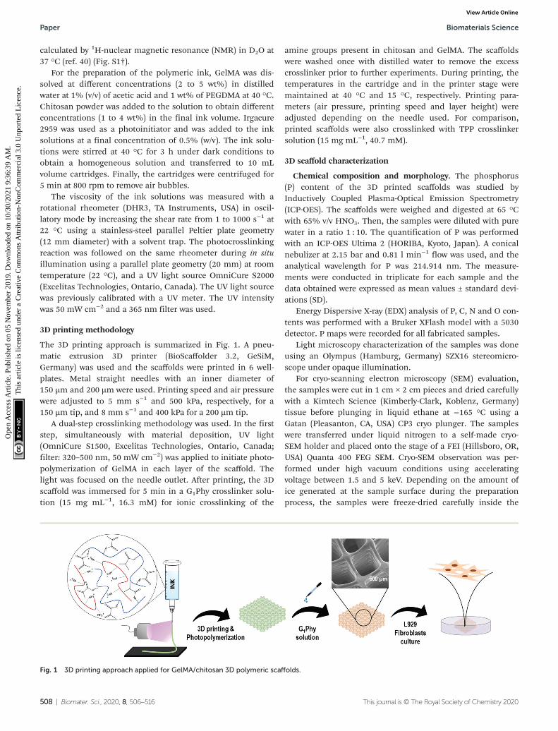
The 3D printing approach is summarized in Fig. 1. A pneu-matic extrusion 3D printer (BioScaffolder 3.2, GeSiM,Germany) was used and the scaffolds were printed in 6 well-plates. Metal straight needles with an inner diameter of150 µm and 200 µm were used. Printing speed and air pressurewere adjusted to 5 mm s−1 and 500 kPa, respectively, for a150 µm tip, and 8 mm s−1 and 400 kPa for a 200 µm tip.
A dual-step crosslinking methodology was used. In the firststep, simultaneously with material deposition, UV light(OmniCure S1500, Excelitas Technologies, Ontario, Canada;filter: 320–500 nm, 50 mW cm−2) was applied to initiate photo-polymerization of GelMA in each layer of the scaffold. Thelight was focused on the needle outlet. After printing, the 3Dscaffold was immersed for 5 min in a G1Phy crosslinker solu-tion (15 mg mL−1, 16.3 mM) for ionic crosslinking of the
amine groups present in chitosan and GelMA. The scaffoldswere washed once with distilled water to remove the excesscrosslinker prior to further experiments. During printing, thetemperatures in the cartridge and in the printer stage weremaintained at 40 °C and 15 °C, respectively. Printing para-meters (air pressure, printing speed and layer height) wereadjusted depending on the needle used. For comparison,printed scaffolds were also crosslinked with TPP crosslinkersolution (15 mg mL−1, 40.7 mM).
3D scaffold characterization
Chemical composition and morphology. The phosphorus(P) content of the 3D printed scaffolds was studied byInductively Coupled Plasma-Optical Emission Spectrometry(ICP-OES). The scaffolds were weighed and digested at 65 °Cwith 65% v/v HNO3. Then, the samples were diluted with purewater in a ratio 1 : 10. The quantification of P was performedwith an ICP-OES Ultima 2 (HORIBA, Kyoto, Japan). A conicalnebulizer at 2.15 bar and 0.81 l min−1 flow was used, and theanalytical wavelength for P was 214.914 nm. The measure-ments were conducted in triplicate for each sample and thedata obtained were expressed as mean values ± standard devi-ations (SD).
Energy Dispersive X-ray (EDX) analysis of P, C, N and O con-tents was performed with a Bruker XFlash model with a 5030detector. P maps were recorded for all fabricated samples.
Light microscopy characterization of the samples was doneusing an Olympus (Hamburg, Germany) SZX16 stereomicro-scope under opaque illumination.
For cryo-scanning electron microscopy (SEM) evaluation,the samples were cut in 1 cm × 2 cm pieces and dried carefullywith a Kimtech Science (Kimberly-Clark, Koblenz, Germany)tissue before plunging in liquid ethane at −165 °C using aGatan (Pleasanton, CA, USA) CP3 cryo plunger. The sampleswere transferred under liquid nitrogen to a self-made cryo-SEM holder and placed onto the stage of a FEI (Hillsboro, OR,USA) Quanta 400 FEG SEM. Cryo-SEM observation was per-formed under high vacuum conditions using acceleratingvoltage between 1.5 and 5 keV. Depending on the amount ofice generated at the sample surface during the preparationprocess, the samples were freeze-dried carefully inside the
Fig. 1 3D printing approach applied for GelMA/chitosan 3D polymeric scaffolds.
Paper Biomaterials Science
508 | Biomater. Sci., 2020, 8, 506–516 This journal is © The Royal Society of Chemistry 2020
Ope
n A
cces
s A
rtic
le. P
ublis
hed
on 0
5 N
ovem
ber
2019
. Dow
nloa
ded
on 1
0/30
/202
1 9:
36:3
9 A
M.
Thi
s ar
ticle
is li
cens
ed u
nder
a C
reat
ive
Com
mon
s A
ttrib
utio
n-N
onC
omm
erci
al 3
.0 U
npor
ted
Lic
ence
.View Article Online

SEM before taking secondary electron images using anEverhart–Thornley detector (ETD).
Physicochemical properties. Swelling of the printedscaffolds was evaluated by measuring the strand widths atdifferent times of incubation in PBS at 37 °C (2, 4, 7, and 10days) by imaging with a light microscope (stereomicroscopeSMZ 800N with episcopic illumination, Nikon, Germany). Themeasurements were conducted in triplicate for three differentsamples.
The degradation rate was calculated gravimetrically bydrying and weighing the scaffolds after incubation in PBS at37 °C for different time points (2, 4, 7, 10, 20 and 30 days). Themeasurements were conducted in triplicate for each sampleand the data obtained were expressed as mean values ± stan-dard deviations (SD).
Crosslinker release was followed by measuring the contentof P in the supernatant after incubating the scaffolds inmedium at increasing times. G1Phy and TPP samples wereincubated in Tris-HCl buffer at pH 7.4 and 37 °C in order toavoid interference between P in the sample and in PBS.Aliquots were taken at 2, 4, 7, 10 and 14 days of incubationand the P content was determined by ICP-OES. The measure-ments were conducted in triplicate for each sample and thedata obtained were expressed as mean values ± standard devi-ations (SD).
Mechanical properties. Viscoelastic properties of the 3Dprinted scaffolds were evaluated in a rotational rheometer(DHR3, TA Instruments, USA) in oscillatory mode at frequency1 Hz and strain 1%. 4 wt%/4 wt% GelMA/chitosan solutionswere photocrosslinked in 24 well-plates by illuminating for5 min at 50 mW cm−2, and incubated in a 15 mg mL−1 solu-tion of G1Phy or TPP for 5 min. Their storage and loss moduliwere measured using the rheometer.
Biological behaviour
Cell culture. L929 fibroblasts (ATCC) were used to evaluatethe in vitro biocompatibility of the scaffolds. The cells weregrown and maintained in RPMI 1640 medium (Gibco, 61870-010) supplemented with 20% FBS (Gibco, 10270), 200 mML-glutamine (Gibco), 100 units per mL penicillin and 100 µgmL−1 streptomycin (Invitrogen) at 37 °C in a humidified atmo-sphere of 5% CO2. Cell culture media were refreshed every48 h. The cells from passage 25–30 were used.
Live/dead assay. For live/dead assay, 10 µl microdroplets con-taining 105 cells were deposited on the surface of the scaffolds.2 mL of cell culture medium were added to each well after 1 h.The samples were incubated for 24 h at 37 °C in a humidifiedatmosphere of 5% CO2 and cell viability was assessed using afluorescein diacetate (FDA) (Sigma-Aldrich) and propidiumiodide (PI) (Sigma-Aldrich) double-staining protocol. 1 μgmL−1 PI solution and 1 μg mL−1 FDA solution were dissolvedin PBS to achieve final concentrations of 20 μg mL−1 and 6 μgmL−1, respectively. After removing culturing medium from thesamples, 100 μl of staining solution was added to each well for10 min incubation. The samples were 2× washed with PBS andimaged with a PolScope microscope (Zeiss, Germany).
Immunochemistry confocal staining. The cells were fixedwith PFA 3.7% w/v for 10 min, permeabilized with 0.5% w/vTriton-X 100 (TX) for 10 min, and blocked with 0.1% TX and5% w/v BSA for 20 min. The samples were incubated in 1 : 1000vinculin rabbit antibody (Thermo Fisher) solution for cytoskele-ton labelling and 1 : 200 Alexa fluor-546 phalloidin (Thermofisher) solution for focal adhesion staining in red color for∼1 h. Then, they were rinsed twice with PBS and incubated withsecondary antibody (Alexa flour-488 goat antirabbit, ThermoFisher) 1 : 500 solution to visualize the cytoskeleton in green.Finally, the samples were stained with 1 : 1000 DAPI (4′,6-di-amidino-2-phenylindole, dihydrochloride, Thermo Fisher) solu-tion for nuclei visualization in blue color and washed with PBS(Sigma). Imaging was performed using a Nikon Ti-Eclipse(Nikon Instruments Europe B.V., Germany) with a Sola SE 365 II(Lumencor Inc., Beaverton, USA) solid state illumination deviceand an Andor Clara CCD camera for detection.
Cytotoxicity. The evaluation of cytotoxicity of the scaffolds wasdetermined by MTT assay. The corresponding scaffold was set incell culture medium. Then, extracts of the medium were removedat different time periods (2, 4, 7 and 10 days) and replaced withfresh medium. The extracts were filtered and used for cytotoxicityassays. Thermanox® (TMX, Thermo Fisher, Waltham, MA, USA)discs were used as the negative control. First, the cells wereseeded at a density of 9 × 10⁴ cells per mL in 96 well-plates andincubated to confluence. After 24 h of incubation the mediumwas replaced with the corresponding extract and incubated at37 °C in humidified air with 5% CO2 for 24 h. Cellular viability(%) was calculated for each sample with respect to the control ateach time of incubation. The data were obtained from 3 indepen-dent series of experiments in triplicate for each sample and theywere expressed as mean values ± SD. The analysis of variance(ANOVA) of the results for G1Phy and TPP samples was performedwith respect to the control plate at each time at a significancelevel of ***p < 0.001, and for G1Phy samples with respect to theTPP sample at each time at a significance level of ##p < 0.01.
Proliferation over time. Alamar Blue (Sigma-Aldrich) assaywas used to analyze cellular proliferation at 2, 4, 7 and 10 dayson the 3D scaffolds directly printed in cellular-repellent 6 well-plates (CELLSTAR®, Greiner Bio-One, Kremsmünster, Austria).The scaffolds were sterilized with ethanol and UV light for 1 hand 30 min, respectively. The cells were seeded at a density of15 × 103 cells per mL and incubated at 37 °C. The data wereobtained from 3 independent series of experiments in tripli-cate for each sample and they were expressed as mean values ±SD. The analysis of variance (ANOVA) of the results for G1Physamples was performed with respect to TPP samples at eachtime point at a significance level of ***p < 0.001.
Results2D printability and rheological evaluation of GelMA/chitosanpolymeric inks
The printability, i.e. the extrusion of a continuous threadthrough the printer needle, of GelMA/chitosan mixtures with
Biomaterials Science Paper
This journal is © The Royal Society of Chemistry 2020 Biomater. Sci., 2020, 8, 506–516 | 509
Ope
n A
cces
s A
rtic
le. P
ublis
hed
on 0
5 N
ovem
ber
2019
. Dow
nloa
ded
on 1
0/30
/202
1 9:
36:3
9 A
M.
Thi
s ar
ticle
is li
cens
ed u
nder
a C
reat
ive
Com
mon
s A
ttrib
utio
n-N
onC
omm
erci
al 3
.0 U
npor
ted
Lic
ence
.View Article Online
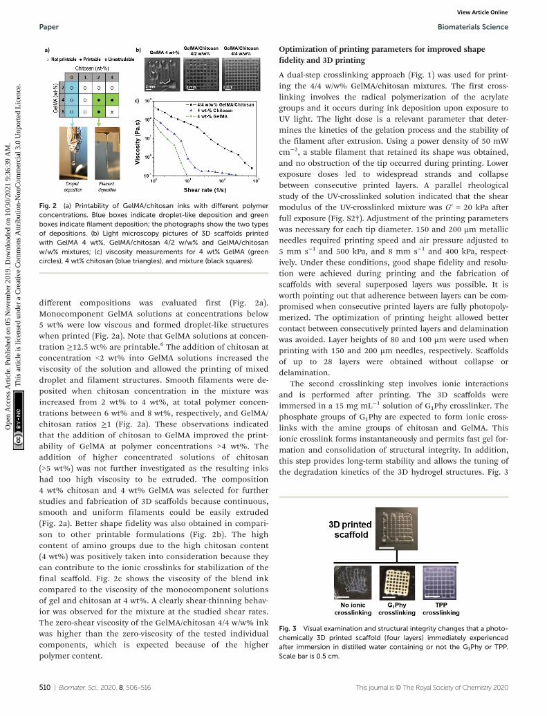
different compositions was evaluated first (Fig. 2a).Monocomponent GelMA solutions at concentrations below5 wt% were low viscous and formed droplet-like structureswhen printed (Fig. 2a). Note that GelMA solutions at concen-tration ≥12.5 wt% are printable.6 The addition of chitosan atconcentration <2 wt% into GelMA solutions increased theviscosity of the solution and allowed the printing of mixeddroplet and filament structures. Smooth filaments were de-posited when chitosan concentration in the mixture wasincreased from 2 wt% to 4 wt%, at total polymer concen-trations between 6 wt% and 8 wt%, respectively, and GelMA/chitosan ratios ≥1 (Fig. 2a). These observations indicatedthat the addition of chitosan to GelMA improved the print-ability of GelMA at polymer concentrations >4 wt%. Theaddition of higher concentrated solutions of chitosan(>5 wt%) was not further investigated as the resulting inkshad too high viscosity to be extruded. The composition4 wt% chitosan and 4 wt% GelMA was selected for furtherstudies and fabrication of 3D scaffolds because continuous,smooth and uniform filaments could be easily extruded(Fig. 2a). Better shape fidelity was also obtained in compari-son to other printable formulations (Fig. 2b). The highcontent of amino groups due to the high chitosan content(4 wt%) was positively taken into consideration because theycan contribute to the ionic crosslinks for stabilization of thefinal scaffold. Fig. 2c shows the viscosity of the blend inkcompared to the viscosity of the monocomponent solutionsof gel and chitosan at 4 wt%. A clearly shear-thinning behav-ior was observed for the mixture at the studied shear rates.The zero-shear viscosity of the GelMA/chitosan 4/4 w/w% inkwas higher than the zero-viscosity of the tested individualcomponents, which is expected because of the higherpolymer content.
Optimization of printing parameters for improved shapefidelity and 3D printing
A dual-step crosslinking approach (Fig. 1) was used for print-ing the 4/4 w/w% GelMA/chitosan mixtures. The first cross-linking involves the radical polymerization of the acrylategroups and it occurs during ink deposition upon exposure toUV light. The light dose is a relevant parameter that deter-mines the kinetics of the gelation process and the stability ofthe filament after extrusion. Using a power density of 50 mWcm−2, a stable filament that retained its shape was obtained,and no obstruction of the tip occurred during printing. Lowerexposure doses led to widespread strands and collapsebetween consecutive printed layers. A parallel rheologicalstudy of the UV-crosslinked solution indicated that the shearmodulus of the UV-crosslinked mixture was G′ = 20 kPa afterfull exposure (Fig. S2†). Adjustment of the printing parameterswas necessary for each tip diameter. 150 and 200 µm metallicneedles required printing speed and air pressure adjusted to5 mm s−1 and 500 kPa, and 8 mm s−1 and 400 kPa, respect-ively. Under these conditions, good shape fidelity and resolu-tion were achieved during printing and the fabrication ofscaffolds with several superposed layers was possible. It isworth pointing out that adherence between layers can be com-promised when consecutive printed layers are fully photopoly-merized. The optimization of printing height allowed bettercontact between consecutively printed layers and delaminationwas avoided. Layer heights of 80 and 100 µm were used whenprinting with 150 and 200 µm needles, respectively. Scaffoldsof up to 28 layers were obtained without collapse ordelamination.
The second crosslinking step involves ionic interactionsand is performed after printing. The 3D scaffolds wereimmersed in a 15 mg mL−1 solution of G1Phy crosslinker. Thephosphate groups of G1Phy are expected to form ionic cross-links with the amine groups of chitosan and GelMA. Thisionic crosslink forms instantaneously and permits fast gel for-mation and consolidation of structural integrity. In addition,this step provides long-term stability and allows the tuning ofthe degradation kinetics of the 3D hydrogel structures. Fig. 3
Fig. 2 (a) Printability of GelMA/chitosan inks with different polymerconcentrations. Blue boxes indicate droplet-like deposition and greenboxes indicate filament deposition; the photographs show the two typesof depositions. (b) Light microscopy pictures of 3D scaffolds printedwith GelMA 4 wt%, GelMA/chitosan 4/2 w/w% and GelMA/chitosanw/w% mixtures; (c) viscosity measurements for 4 wt% GelMA (greencircles), 4 wt% chitosan (blue triangles), and mixture (black squares).
Fig. 3 Visual examination and structural integrity changes that a photo-chemically 3D printed scaffold (four layers) immediately experiencedafter immersion in distilled water containing or not the G1Phy or TPP.Scale bar is 0.5 cm.
Paper Biomaterials Science
510 | Biomater. Sci., 2020, 8, 506–516 This journal is © The Royal Society of Chemistry 2020
Ope
n A
cces
s A
rtic
le. P
ublis
hed
on 0
5 N
ovem
ber
2019
. Dow
nloa
ded
on 1
0/30
/202
1 9:
36:3
9 A
M.
Thi
s ar
ticle
is li
cens
ed u
nder
a C
reat
ive
Com
mon
s A
ttrib
utio
n-N
onC
omm
erci
al 3
.0 U
npor
ted
Lic
ence
.View Article Online
shows two 3D printed scaffolds: with and without ionic treat-ment. The dual crosslinked scaffold shows higher shape fide-lity and controlled swelling, which were lost when the ionicpost-treatment was skipped. This result indicates that bothcrosslinking processes, photopolymerization and ionic cross-linking, contribute to print 3D scaffolds with structural integ-rity and long-term stability. This methodology allowed fastprinting without using supporting templates21,36,41–45 or neu-tralization and washing steps.5,7,24,28,30,46 The as-printedscaffolds were ready to be used for biological tests.
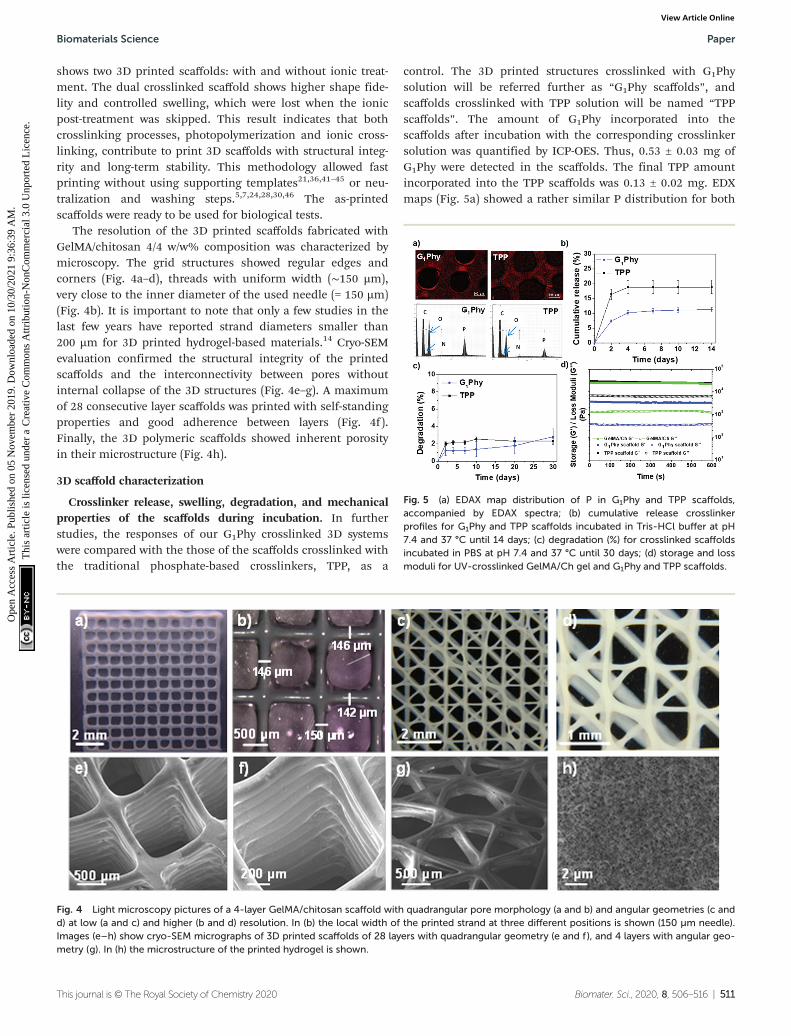
The resolution of the 3D printed scaffolds fabricated withGelMA/chitosan 4/4 w/w% composition was characterized bymicroscopy. The grid structures showed regular edges andcorners (Fig. 4a–d), threads with uniform width (∼150 µm),very close to the inner diameter of the used needle (= 150 µm)(Fig. 4b). It is important to note that only a few studies in thelast few years have reported strand diameters smaller than200 µm for 3D printed hydrogel-based materials.14 Cryo-SEMevaluation confirmed the structural integrity of the printedscaffolds and the interconnectivity between pores withoutinternal collapse of the 3D structures (Fig. 4e–g). A maximumof 28 consecutive layer scaffolds was printed with self-standingproperties and good adherence between layers (Fig. 4f).Finally, the 3D polymeric scaffolds showed inherent porosityin their microstructure (Fig. 4h).
3D scaffold characterization
Crosslinker release, swelling, degradation, and mechanicalproperties of the scaffolds during incubation. In furtherstudies, the responses of our G1Phy crosslinked 3D systemswere compared with the those of the scaffolds crosslinked withthe traditional phosphate-based crosslinkers, TPP, as a
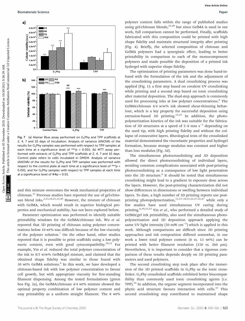
control. The 3D printed structures crosslinked with G1Physolution will be referred further as “G1Phy scaffolds”, andscaffolds crosslinked with TPP solution will be named “TPPscaffolds”. The amount of G1Phy incorporated into thescaffolds after incubation with the corresponding crosslinkersolution was quantified by ICP-OES. Thus, 0.53 ± 0.03 mg ofG1Phy were detected in the scaffolds. The final TPP amountincorporated into the TPP scaffolds was 0.13 ± 0.02 mg. EDXmaps (Fig. 5a) showed a rather similar P distribution for both
Fig. 4 Light microscopy pictures of a 4-layer GelMA/chitosan scaffold with quadrangular pore morphology (a and b) and angular geometries (c andd) at low (a and c) and higher (b and d) resolution. In (b) the local width of the printed strand at three different positions is shown (150 µm needle).Images (e–h) show cryo-SEM micrographs of 3D printed scaffolds of 28 layers with quadrangular geometry (e and f), and 4 layers with angular geo-metry (g). In (h) the microstructure of the printed hydrogel is shown.
Fig. 5 (a) EDAX map distribution of P in G1Phy and TPP scaffolds,accompanied by EDAX spectra; (b) cumulative release crosslinkerprofiles for G1Phy and TPP scaffolds incubated in Tris-HCl buffer at pH7.4 and 37 °C until 14 days; (c) degradation (%) for crosslinked scaffoldsincubated in PBS at pH 7.4 and 37 °C until 30 days; (d) storage and lossmoduli for UV-crosslinked GelMA/Ch gel and G1Phy and TPP scaffolds.
Biomaterials Science Paper
This journal is © The Royal Society of Chemistry 2020 Biomater. Sci., 2020, 8, 506–516 | 511
Ope
n A
cces
s A
rtic
le. P
ublis
hed
on 0
5 N
ovem
ber
2019
. Dow
nloa
ded
on 1
0/30
/202
1 9:
36:3
9 A
M.
Thi
s ar
ticle
is li
cens
ed u
nder
a C
reat
ive
Com
mon
s A
ttrib
utio
n-N
onC
omm
erci
al 3
.0 U
npor
ted
Lic
ence
.View Article Online

crosslinkers on the surface of the scaffolds but the distributionseemed to be somewhat more inhomogeneous in G1Physcaffolds than in TPP ones. P is accumulated in some areas ofthe former, while less P in the centre of the fiber crossingsthan in the borders is observed in the latter.
The crosslinker release of the scaffold under physiologicalconditions (pH 7.4, 37 °C) was studied (Fig. 5b). G1Phy showeda fast release (7.3%, which corresponded to 10.32 µM) duringthe first 2 days of incubation, reaching a plateau after 4 days(11.2%, which corresponded to 16.03 µM), which was main-tained until 30 days of incubation. TPP showed a burst release(16.4%, which corresponded to 14.74 µM) at 2 days. A plateauwas achieved at 4 days (18.8%, which corresponded to16.31 µM), which was sustained until the end of the incu-bation period. This different performance of the release cross-linkers can be associated with the differences in the distri-bution of the crosslinkers in the scaffold, and different ionicinteractions of the crosslinkers with the polymeric backbones.
Swelling behaviour of the scaffolds was examined bymeasuring the strand widths by light microscopy (Fig. S3†). Noswelling was observed for printed G1Phy nor TPP crosslinkedscaffolds after incubation in PBS under physiological con-ditions for 10 days. This indicated that the structures achievedtheir maximum water absorption after ionic crosslinking. Thestability of the scaffolds was followed gravimetrically bymeasuring the weight loss after different incubation times(Fig. 5c). G1Phy crosslinked scaffolds showed a weight loss of1.2 ± 0.6% during the first 2 days. The scaffolds remainedstable for the following 10 days and degraded slowly up to 2.8± 0.9% during the next 20 days. At short time points (2 days),TPP crosslinked scaffolds showed a relatively higher degra-dation rate (2.0 ± 0.2%) than G1Phy scaffolds and degradationprogressively increased until 10 days (2.5 ± 0.3%), to remainstable up to the end of the experiment (30 days). At the finaltime no significant (p < 0.5) differences between G1Phy andTPP samples were observed.
Rheological characterization of UV cured GelMA/chitosandiscs after crosslinking with G1Phy or TPP was performed tocompare the viscoelastic properties of the 3D printed struc-tures as a function of the type of ionic crosslinker. Fig. 5dshows the shear and loss moduli for crosslinked discs. G1Phycrosslinked discs exhibited lower storage modulus than TPPcrosslinked discs and GelMA/Ch photocrosslinked gel. Thestorage modulus values of TPP crosslinked scaffoldsapproached those of GelMA/Ch photocrosslinked gel but withhigher loss modulus than this last one.
Cytocompatibility results. The performance of G1Physcaffolds in biological experiments was assessed in vitro usingL929 fibroblasts which were seeded on the top of the printedscaffolds. For comparison, TPP scaffolds were evaluated in asimilar way. Live/dead staining showed high cellular viabilityand cell density attachment on the G1Phy scaffolds after 1 dayculture (Fig. 6a), indicating no visible toxicity of the materials.In contrast, live/dead staining of cells seeded on the TPPscaffolds showed that almost no cells attached to the 3D struc-ture (Fig. 6b), which migrated to the well-plate.
Immunostaining results confirmed high cell attachment onG1Phy scaffolds. The attached cells showed a spread mor-phology and formed focal adhesions stained in red (Fig. 6c).TPP scaffolds showed fewer cells attached on their surface. Thecells maintained a rounded morphology characteristic of weakcell–material interaction with the scaffold surface, coherentwith the results of the live/dead assay (Fig. 6d).
The proliferation of L929 fibroblasts on the scaffolds wasquantified using the Alamar Blue assay at days 2, 4, 7 and 10of incubation. L929 proliferation on G1Phy scaffolds increasedover time and the proliferation level was at any time higherthan that on TPP scaffolds (Fig. 7a). Finally, the possible cyto-toxic effects caused by the release of crosslinkers or low mole-cular weight residues from the 3D printed scaffolds was exam-ined by incubating L929 fibroblasts with the supernatantsfrom the scaffolds after 2, 4, 7 and 10 days of soaking in cellculture medium. No cytotoxic effects were observed from anyof the 3D printed scaffolds (Fig. 7b).
Discussion
Among all requirements that an ink must fulfil to allow suc-cessful printing homogeneity and deposition as a uniform fila-ment, shear-thinning behaviour, and appropriate viscosity arekey features.23 In addition, the 3D printed structures shouldbe biocompatible and maintain structural integrity underphysiological conditions, avoiding delamination during andafter printing.15,47 Blend inks that combine different types ofpolymers are commonly used to fulfil all the mechanical andbiological requirements.48 We decided to work with a mixtureof polysaccharide (chitosan) and protein (GelMA) hydrogel. Weexpected that the GelMA/chitosan combination provides aprinting ink with the biological advantages of both polymers,
Fig. 6 Live/dead assay on G1Phy (a) and TPP (b) scaffolds after 24 h ofincubation. Pictures were taken of scaffold strands; confocal immuno-staining assay performed on G1Phy (c) and TPP (d) scaffolds after 24 h ofincubation.
Paper Biomaterials Science
512 | Biomater. Sci., 2020, 8, 506–516 This journal is © The Royal Society of Chemistry 2020
Ope
n A
cces
s A
rtic
le. P
ublis
hed
on 0
5 N
ovem
ber
2019
. Dow
nloa
ded
on 1
0/30
/202
1 9:
36:3
9 A
M.
Thi
s ar
ticle
is li
cens
ed u
nder
a C
reat
ive
Com
mon
s A
ttrib
utio
n-N
onC
omm
erci
al 3
.0 U
npor
ted
Lic
ence
.View Article Online
and this mixture overcomes the weak mechanical properties ofchitosan.15 Previous studies have reported the use of gel/chito-san blend inks.2,15,24,25,33,49 However, the mixture of chitosanwith GelMA, which would result in superior biological pro-perties and mechanical stability, has not been explored before.
Parameter optimization was performed to identify suitableprintability windows for the GelMA/chitosan ink. Wu et al.reported that 3D printing of chitosan using polymer concen-trations below 10 wt% was difficult because of the low viscosityof the polymer solution.7 On the other hand, other studiesreported that it is possible to print scaffolds using a low poly-meric content, even with good cytocompatibility.50,51 Forexample, Yin et al. reduced the total polymer concentration ofthe ink to 8/5 w/w% GelMA/gel mixture, and claimed that theobtained shape fidelity was similar to those found with30 wt% GelMA solutions.6 In this work, we have developed achitosan-based ink with low polymer concentration to favourcell growth, but with appropriate viscosity for free-standingfilament dispensing. Among the tested formulations (greenbox Fig. 2a), the GelMA/chitosan 4/4 wt% mixture showed theoptimal property combination of low polymer content andeasy printability as a uniform straight filament. The 8 wt%
polymer content falls within the range of published studiesusing gel/chitosan blends,25,49 but since GelMA is used in ourwork, full comparison cannot be performed. Finally, scaffoldsfabricated with this composition could be printed with highshape fidelity and maintain structural integrity after printing(Fig. 4). Briefly, the selected composition of chitosan andGelMA polymers had a synergistic effect, leading to betterprintability in comparison to each of the monocomponentpolymers and made possible the deposition of a printed inkhydrogel with superior shape fidelity.
The optimization of printing parameters was done hand-in-hand with the formulation of the ink and the adjustment ofthe crosslinking parameters. A dual crosslinking process wasapplied (Fig. 1): a first step based on covalent UV crosslinkingwhile printing and a second step based on ionic crosslinkingafter material deposition. The dual-step approach is commonlyused for processing inks at low polymer concentrations.6 TheGelMA/chitosan 4/4 w/w% ink showed shear-thinning behav-iour, which is a key property for successful deposition usingextrusion-based 3D printing.15,23 In addition, the photo-polymerization kinetics of the ink was suitable for the fabrica-tion of 3D structures at a speed of 5–8 mm s−1 depending onthe used tip, with high printing fidelity and without the col-lapse of consecutive layers. Rheological tests of the crosslinkedmaterial demonstrated the viscoelastic properties and hydrogelformation, because storage modulus was constant and higherthan loss modulus (Fig. 5d).
The simultaneous photocrosslinking and 3D depositionallowed the direct photocrosslinking of individual layers,avoiding common complications associated with post-printingphotocrosslinking as a consequence of low light penetrationinto the 3D structure.6 It should be noted that simultaneouscrosslinking might lead to a gradient in exposure dose acrossthe layers. However, the post-printing characterization did notshow differences in dimensions or swelling between individuallayers. To date, a high number of 3D printing reports use post-printing photopolymerization,11,15–18,21,22,27,45,47 while only afew studies have used simultaneous UV curing duringprinting.6,39,52,53 Yin et al., who performed a detailed study ofGelMA/gel ink printability, also used the simultaneous photo-polymerization and 3D deposition approach applying thesame UV light intensity (50 mW cm−2) which is applied in thiswork. Although comparisons are difficult since 3D printingapproaches and ink composition differed somewhat, in ourwork a lower total polymer content (8 vs. 13 wt%) can beprinted with better filament resolution (150 vs. 260 µm).Nevertheless, it is important to consider that a rigorous com-parison of these results depends deeply on 3D printing para-meters and used polymers.
The second crosslinking step took place after the immer-sion of the 3D printed scaffolds in G1Phy as the ionic cross-linker. G1Phy crosslinked scaffolds exhibited better biocompat-ibility than commonly used ionic crosslinking agents (i.e.TPP).54 In addition, the organic segment incorporated into thephytic acid structure favours interaction with cells.32 Thissecond crosslinking step contributed to maintained shape
Fig. 7 (a) Alamar Blue assay performed on G1Phy and TPP scaffolds at2, 4, 7 and 10 days of incubation. Analysis of variance (ANOVA) of theresults for G1Phy samples was performed with respect to TPP samples ateach time at a significance level of ***p < 0.001; (b) MTT assay per-formed with extracts of G1Phy and TPP scaffolds at 2, 4, 7 and 10 days.Control plate refers to cells incubated in DMEM. Analysis of variance(ANOVA) of the results for G1Phy and TPP samples was performed withrespect to the control plate at each time at a significance level of ***p <0.001, and for G1Phy samples with respect to TPP samples at each timeat a significance level of ##p < 0.01.
Biomaterials Science Paper
This journal is © The Royal Society of Chemistry 2020 Biomater. Sci., 2020, 8, 506–516 | 513
Ope
n A
cces
s A
rtic
le. P
ublis
hed
on 0
5 N
ovem
ber
2019
. Dow
nloa
ded
on 1
0/30
/202
1 9:
36:3
9 A
M.
Thi
s ar
ticle
is li
cens
ed u
nder
a C
reat
ive
Com
mon
s A
ttrib
utio
n-N
onC
omm
erci
al 3
.0 U
npor
ted
Lic
ence
.View Article Online

fidelity and positively influenced swelling and long-term stabi-lity of the scaffolds (Fig. 4b). We speculate that the cross-linking of the amine groups of the chitosan and GelMA chainswith the anionic crosslinker is essential to control the wateruptake of the printed structures. This stabilization step did notrequire neutralization or washing steps, which is the commonapproach used for 3D printing of chitosan-based inks. Forexample, Wu et al. printed 10 wt% chitosan solutions obtain-ing 3D structures with high resolution (∼30 µm) and intricateshapes, but neutralization with 1 M NaOH solution for 4 h wasrequired.7 Elviri et al. were able to print lower chitosan solu-tions (6 wt%) in a cryogenic chamber followed by the sub-sequent coagulation in a KOH (8 w/v%) bath.30 Moreover, thedescribed manufacturing protocols could potentially lead toshrinking and shape deformation of the scaffolds,5,7,24,28,30,55
a post-printing phenomenon that it was not observed in ourscaffolds after ionic crosslinking (Fig. S4†). Our polymer con-centration is among the range of published ones (±2 wt%),while it avoids such sophisticated post-processing techniques,being more similar to the traditional alginate–CaCl2 systemwidely applied in 3D printing methodologies.29
The dual step approach followed in our work also avoidedthe use of sacrificial or template materials, which is a widelyused strategy to improve printing resolution withhydrogels.11,37,38 G1Phy crosslinking led to improved mechani-cal properties of the scaffolds compared to TPP crosslinking.We expect G1Phy to form less compact and softer networksbecause of the two main reasons: (i) the organic content of thiscrosslinker provides some viscoelasticity to the network, and(ii) its higher molecular weight with respect to TPP providesless dense frameworks. All these features together contributedto obtaining softer and viscoelastic gels with long-term stabilityand controlled swelling, which are essential characteristics fortissue engineering applications.1,10,56 To the best of our knowl-edge, crosslinkers derived from phytic acid have not been usedfor the development of 3D printed scaffolds so far.
The 3D structures demonstrated excellent shape fidelity.The ink did not accumulate in the edges or in the corners ofscaffolds, where printing speeds and direction changeabruptly. The scaffolds showed smooth surfaces and constantwidths. Moreover, printing of multiple consecutive layerswithout collapsing was possible, leading to the structures ofup to 28 layers height. The best reported line width for hydro-gel printing was approximately 100–200 µm, which is amongthe best resolution degree that can be currently achieved withhydrogel inks.14 This resolution is highly dependent on thediameter of the needle, and could be potentially improvedusing narrower tips. Porous and interconnected structureswere printed, which are interesting geometries for tissueengineering applications.14,57 Finally, the inherent microstruc-ture (Fig. 4h) of the polymeric scaffolds is also a decisive prop-erty for the correct distribution and diffusion of oxygen andnutrients of the ingrowth tissues.58,59 Summarizing, the devel-oped GelMA/chitosan hydrogel with a dual crosslinkingmechanism allows the printing of 3D structures with complexdesigns at high resolution.
Cytocompatibility studies indicated that G1Phy scaffoldssupported better cell attachment and proliferation than TPPcrosslinked scaffolds, which could be due to the chemicalcomposition as well as morphological properties of thesescaffolds. Nevertheless, since the initial composition (GelMA/chitosan) is the same for both crosslinked scaffolds andassuming that the chitosan surface exposure is rather similarin both types of scaffolds according to P distribution (Fig. 5a),for short time periods, the different biological responsesshould only be due to the incorporation of G1Phy or TPP intotheir structures. G1Phy has demonstrated to be a highly bio-compatible crosslinker which exhibits an organic compositionthat can enhance cellular interaction.32 In addition, thedifferent molecular weights of G1Phy and TPP could play a rolein the final mechanical properties of the ionically crosslinkednetworks. In fact, the rheological behaviour showed that theG1Phy crosslinked polymer network was softer than the TPPcrosslinked one. All this together with the sustained release ofG1Phy and the slower degradation of this scaffold can favour-ably contribute to the higher cell proliferation of the samplesin long-term periods.
Conclusions
In summary, this work shows the implementation and optim-ization of a 3D printing methodology using the novel G1Phycrosslinker. The methodology consisted of a dual-step cross-linking that allows the 3D printing of low concentrated GelMA/chitosan based-ink (total polymer concentration <10 wt%).This approach permitted the fabrication of 3D hydrogelscaffolds with excellent shape fidelity and resolution. The 3Dprinted scaffolds displayed long-term stability and excellentproperties regarding swelling behaviour, and mechanical andbiological properties. In particular, the use of the G1Phy cross-linker enhanced cell adhesion and proliferation on the 3Dscaffolds in comparison to TPP, widely used as a traditionalionic crosslinking agent. These results open a door for theextrusion of hydrogel-based inks employing phytic acidderived crosslinkers for the fabrication of complex structureswith excellent biological properties that can be used in softtissue engineering applications.
Conflicts of interest
There are no conflicts to declare.
Acknowledgements
Authors thank financial support from Ministry of Science,Innovation and Universities (Spain) (MAT2017-2017-84277-R),“La Caixa” Foundation (ID 100010434, scholarship of AnaMora-Boza, code LCF/BQ/ES16/11570018) and DAAD ResearchGrants-Short-term grants 2017. Support of the publication feeby the CSIC Open Access Publication Support Initiative
Paper Biomaterials Science
514 | Biomater. Sci., 2020, 8, 506–516 This journal is © The Royal Society of Chemistry 2020
Ope
n A
cces
s A
rtic
le. P
ublis
hed
on 0
5 N
ovem
ber
2019
. Dow
nloa
ded
on 1
0/30
/202
1 9:
36:3
9 A
M.
Thi
s ar
ticle
is li
cens
ed u
nder
a C
reat
ive
Com
mon
s A
ttrib
utio
n-N
onC
omm
erci
al 3
.0 U
npor
ted
Lic
ence
.View Article Online

through its Unit of Information Resources for Research(URICI) is also acknowledged.
The authors are indebted to Dr Marcus Koch (Leibniz-INM)for excellent technical assistance with SEM and lightmicroscopy experiments, and to Dr Claudia Fink-Straube(Leibniz-INM) for ICP-OES experiment performance.
References
1 A. V. Do, B. Khorsand, S. M. Geary and A. K. Salem, Adv.Healthcare Mater., 2015, 4, 1742–1762.
2 J. Huang, H. Fu, Z. Wang, Q. Meng, S. Liu, H. Wang,X. Zheng, J. Dai and Z. Zhang, RSC Adv., 2016, 6, 108423–108430.
3 Q. L. Loh and C. Choong, Tissue Eng., Part B, 2013, 19, 485–502.
4 M. K. Wlodarczyk-Biegun and A. Del Campo, Biomaterials,2017, 134, 180–201.
5 C. Intini, L. Elviri, J. Cabral, S. Mros, C. Bergonzi,A. Bianchera, L. Flammini, P. Govoni, E. Barocelli,R. Bettini and M. McConnell, Carbohydr. Polym., 2018, 199,593–602.
6 J. Yin, M. Yan, Y. Wang, J. Fu and H. Suo, ACS Appl. Mater.Interfaces, 2018, 10, 6849–6857.
7 Q. Wu, D. Therriault and M.-C. Heuzey, ACS Biomater. Sci.Eng., 2018, 4, 2643–2652.
8 R. Levato, W. R. Webb, I. A. Otto, A. Mensinga, Y. Zhang,M. van Rijen, R. van Weeren, I. M. Khan and J. Malda, ActaBiomater., 2017, 61, 41–53.
9 S. Stratton, N. B. Shelke, K. Hoshino, S. Rudraiah andS. G. Kumbar, Bioact. Mater., 2016, 1, 93–108.
10 S. Derakhshanfar, R. Mbeleck, K. Xu, X. Zhang, W. Zhongand M. Xing, Bioact. Mater., 2018, 3, 144–156.
11 W. Jia, P. S. Gungor-Ozkerim, Y. S. Zhang, K. Yue, K. Zhu,W. Liu, Q. Pi, B. Byambaa, M. R. Dokmeci, S. R. Shin andA. Khademhosseini, Biomaterials, 2016, 106, 58–68.
12 K. Schutz, A. M. Placht, B. Paul, S. Bruggemeier,M. Gelinsky and A. Lode, J. Tissue Eng. Regener. Med., 2017,11, 1574–1587.
13 H. Stratesteffen, M. Kopf, F. Kreimendahl, A. Blaeser,S. Jockenhoevel and H. Fischer, Biofabrication, 2017, 9,045002.
14 X. Wang, C. Wei, B. Cao, L. Jiang, Y. Hou and J. Chang, ACSAppl. Mater. Interfaces, 2018, 10, 18338–18350.
15 H. Li, Y. J. Tan, S. Liu and L. Li, ACS Appl. Mater. Interfaces,2018, 10, 11164–11174.
16 B. J. Klotz, D. Gawlitta, A. Rosenberg, J. Malda andF. P. W. Melchels, Trends Biotechnol., 2016, 34, 394–407.
17 N. Celikkin, S. Mastrogiacomo, J. Jaroszewicz,X. F. Walboomers and W. Swieszkowski, J. Biomed. Mater.Res., Part A, 2018, 106, 201–209.
18 W. Liu, M. A. Heinrich, Y. Zhou, A. Akpek, N. Hu, X. Liu,X. Guan, Z. Zhong, X. Jin, A. Khademhosseini andY. S. Zhang, Adv. Healthcare Mater., 2017, 6, 1601451.
19 N. A. Chartrain, C. B. Williams and A. R. Whittington, ActaBiomater., 2018, 74, 90–111.
20 M. Layani, X. Wang and S. Magdassi, Adv. Mater., 2018, 30,e1706344.
21 K. Yue, G. Trujillo-de Santiago, M. M. Alvarez, A. Tamayol,N. Annabi and A. Khademhosseini, Biomaterials, 2015, 73,254–271.
22 Y. L. Cheng and F. Chen, Mater. Sci. Eng., C, 2017, 81, 66–73.23 T. T. Demirtas, G. Irmak and M. Gumusderelioglu,
Biofabrication, 2017, 9, 035003.24 W. L. Ng, W. Y. Yeong and M. W. Naing, Int. J. Bioprint.,
2016, 2(1), 53–62.25 W. L. Ng, W. Y. Yeong and M. W. Naing, Procedia CIRP,
2016, 49, 105–112.26 I. C. Carvalho and H. S. Mansur, Mater. Sci. Eng., C, 2017,
78, 690–705.27 J. Radhakrishnan, A. Subramanian, U. M. Krishnan and
S. Sethuraman, Biomacromolecules, 2017, 18, 1–26.28 I. H. Liu, S. H. Chang and H. Y. Lin, Biomed. Mater., 2015,
10, 035004.29 A. R. Akkineni, T. Ahlfeld, A. Lode and M. Gelinsky,
Biofabrication, 2016, 8, 045001.30 L. Elviri, R. Foresti, C. Bergonzi, F. Zimetti, C. Marchi,
A. Bianchera, F. Bernini, M. Silvestri and R. Bettini, Biomed.Mater., 2017, 12, 045009.
31 D. Lee, J. P. Park, M. Y. Koh, P. Kim, J. Lee, M. Shin andH. Lee, Biomater. Sci., 2018, 6, 1040–1047.
32 A. Mora-Boza, M. L. López-Donaire, L. Saldaña, N. Vilaboa,B. Vazquez-Lasa and J. S. Román, Sci. Rep., 2019, 9, 11491.
33 A. Zolfagharian, A. Kaynak, S. Y. Khoo and A. Z. Kouzani,3D Print. Addit. Manuf., 2018, 5, 138–150.
34 M. M. Fares, E. Shirzaei Sani, R. Portillo Lara,R. B. Oliveira, A. Khademhosseini and N. Annabi, Biomater.Sci., 2018, 6, 2938–2950.
35 J. Visser, B. Peters, T. J. Burger, J. Boomstra, W. J. A. Dhert,F. P. W. Melchels and J. Malda, Biofabrication, 2013, 5, 035007.
36 A. Tamayol, A. H. Najafabadi, B. Aliakbarian, E. Arab-Tehrany,M. Akbari, N. Annabi, D. Juncker and A. Khademhosseini,Adv. Healthcare Mater., 2015, 4, 2146–2153.
37 P. Datta, B. Ayan and I. T. Ozbolat, Acta Biomater., 2017, 51,1–20.
38 M. Muller, J. Becher, M. Schnabelrauch and M. Zenobi-Wong, J. Visualized Exp., 2013, e50632, DOI: 10.3791/50632.
39 C. D. O’Connell, C. Di Bella, F. Thompson, C. Augustine,S. Beirne, R. Cornock, C. J. Richards, J. Chung, S. Gambhir,Z. Yue, J. Bourke, B. Zhang, A. Taylor, A. Quigley, R. Kapsa,P. Choong and G. G. Wallace, Biofabrication, 2016, 8, 015019.
40 M. Bartnikowski, R. Wellard, M. Woodruff and T. Klein,Polymers, 2015, 7, 1539.
41 Y. Zhang, Y. Yu and I. T. Ozbolat, J. Nanotechnol. Eng. Med.,2013, 4(2), 0210011–0210017.
42 Y. S. Zhang, K. Yue, J. Aleman, K. M. Moghaddam,S. M. Bakht, J. Yang, W. Jia, V. Dell’Erba, P. Assawes,S. R. Shin, M. R. Dokmeci, R. Oklu andA. Khademhosseini, Ann. Biomed. Eng., 2017, 45, 148–163.
43 I. S. Kinstlinger and J. S. Miller, Lab Chip, 2016, 16, 2025–2043.
Biomaterials Science Paper
This journal is © The Royal Society of Chemistry 2020 Biomater. Sci., 2020, 8, 506–516 | 515
Ope
n A
cces
s A
rtic
le. P
ublis
hed
on 0
5 N
ovem
ber
2019
. Dow
nloa
ded
on 1
0/30
/202
1 9:
36:3
9 A
M.
Thi
s ar
ticle
is li
cens
ed u
nder
a C
reat
ive
Com
mon
s A
ttrib
utio
n-N
onC
omm
erci
al 3
.0 U
npor
ted
Lic
ence
.View Article Online
44 J. S. Lee, J. M. Hong, J. W. Jung, J. H. Shim, J. H. Oh andD. W. Cho, Biofabrication, 2014, 6, 024103.
45 M. Costantini, J. Idaszek, K. Szöke, J. Jaroszewicz,M. Dentini, A. Barbetta, J. E. Brinchmann andW. Święszkowski, Biofabrication, 2016, 8(3), 035002.
46 J. Zhang, B. J. Allardyce, R. Rajkhowa, Y. Zhao, R. J. Dilley,S. L. Redmond, X. Wang and X. Liu, ACS Biomater. Sci. Eng.,2018, 4, 3036–3046.
47 S. Sayyar, S. Gambhir, J. Chung, D. L. Officer andG. G. Wallace, Nanoscale, 2017, 9, 2038–2050.
48 K. W. M. Boere, M. M. Blokzijl, J. Visser, J. E. A. Linssen,J. Malda, W. E. Hennink and T. Vermonden, J. Mater.Chem. B, 2015, 3, 9067–9078.
49 K. D. Roehm and S. V. Madihally, Biofabrication, 2017, 10,015002.
50 J. Malda, J. Visser, F. P. Melchels, T. Jungst, W. E. Hennink,W. J. Dhert, J. Groll and D. W. Hutmacher, Adv. Mater.,2013, 25, 5011–5028.
51 I. Pepelanova, K. Kruppa, T. Scheper and A. Lavrentieva,Bioengineering, 2018, 5(3), 55.
52 X. Cui, G. Gao, T. Yonezawa and G. Dai, J. Visualized Exp.,2014, (88), e51294.
53 G. Gao, A. F. Schilling, K. Hubbell, T. Yonezawa, D. Truong,Y. Hong, G. Dai and X. Cui, Biotechnol. Lett., 2015, 37,2349–2355.
54 HERA, 2003.55 A. Vitale and J. T. Cabral, Materials, 2016, 9(9), 760.56 K. N. Bardakova, T. S. Demina, E. A. Grebenik,
N. V. Minaev, T. A. Akopova, V. N. Bagratashvili andP. S. Timashev, IOP Conference Series: Materials Science andEngineering, 2018, 347, conference 1.
57 J. Wang, Z. Nor Hidayah, S. I. A. Razak, M. R. A. Kadir,N. H. M. Nayan, Y. Li and K. A. M. Amin, Compos.Interfaces, 2018, 1–14, DOI: 10.1080/09276440.2018.1508266.
58 P. Thangavel, B. Ramachandran and V. Muthuvijayan,J. Biomed. Mater. Res., Part B, 2016, 104, 750–760.
59 K. Zhang, Y. Fan, N. Dunne and X. Li, Regener. Biomater.,2018, 5, 115–124.
Paper Biomaterials Science
516 | Biomater. Sci., 2020, 8, 506–516 This journal is © The Royal Society of Chemistry 2020
Ope
n A
cces
s A
rtic
le. P
ublis
hed
on 0
5 N
ovem
ber
2019
. Dow
nloa
ded
on 1
0/30
/202
1 9:
36:3
9 A
M.
Thi
s ar
ticle
is li
cens
ed u
nder
a C
reat
ive
Com
mon
s A
ttrib
utio
n-N
onC
omm
erci
al 3
.0 U
npor
ted
Lic
ence
.View Article Online


![Fused filament 3D printing of ionic polymer-metal ... · jet technology was also developed based on the use of Nafion dispersions in [25]. By contrast, the 3D printing method proposed](https://img.pdfslide.us/doc/110x75/5eccf9cf7f03df4e9b7be69c/fused-filament-3d-printing-of-ionic-polymer-metal-jet-technology-was-also-developed.jpg)


![BDI Final Managing AEs part 3 - REV 10-21-10 [Read-Only] · Omniscan Differences in Chelates Non-Ionic Ionic Gadovist Non-Ionic Slide - 16 MultiHance Eovist Linear Non-Ionic Ionic](https://img.pdfslide.us/doc/110x75/5e1e62bb04363471c63c7ff4/bdi-final-managing-aes-part-3-rev-10-21-10-read-only-omniscan-differences-in.jpg)