Embed Size (px)
Citation preview

Glucocorticoids Regulate KisspeptinNeurons during Stress andContribute to Infertility and
Obesity in Leptin-Deficient MiceThe Harvard community has made this
article openly available. Please share howthis access benefits you. Your story matters
Citation Wang, Oulu. 2012. Glucocorticoids Regulate Kisspeptin Neuronsduring Stress and Contribute to Infertility and Obesity in Leptin-Deficient Mice. Doctoral dissertation, Harvard University.
Citable link http://nrs.harvard.edu/urn-3:HUL.InstRepos:9453704
Terms of Use This article was downloaded from Harvard University’s DASHrepository, and is made available under the terms and conditionsapplicable to Other Posted Material, as set forth at http://nrs.harvard.edu/urn-3:HUL.InstRepos:dash.current.terms-of-use#LAA

©2012 – Oulu Wang All rights reserved.
Dissertation Advisor: Dr. Joseph Majzoub Oulu Wang
Glucocorticoids regulate kisspeptin neurons during stress and
contribute to infertility and obesity in leptin-deficient mice
Abstract
Stressors generate adaptive responses, including transient suppression of reproductive function.
Natural selection depends on successful reproduction, but inhibition of reproduction to survive
famine or escape predation allows animals to survive to reproduce at a later time. The cellular
locations and mechanisms responsible for inhibiting and reactivating the reproductive axis
during and after stress, respectively, are not well understood.
We demonstrated that stress-induced elevation in glucocorticoids affects hypothalamic neurons
that secrete kisspeptin (KISS1), an important reproductive hormone. Stressors that stimulated
glucocorticoid secretion, as well as glucocorticoid administration itself, inhibited Kiss1 mRNA
expression, while conditions that did not change glucocorticoid secretion did not alter Kiss1
mRNA expression. In mice lacking glucocorticoid receptor specifically in kisspeptin-containing
neurons, Kiss1 mRNA expression was no longer inhibited during restraint stress despite a rise in
corticosterone, and both testosterone and copulatory behaviors showed accelerated recovery in
the post-traumatic period.
iii

We also demonstrated that increased glucocorticoid secretion contributed to infertility and
obesity in leptin-deficient mice. Leptin deficiency creates a chronic state of perceived starvation,
and leptin-deficient mice exhibit elevated plasma glucocorticoid concentrations, morbid obesity,
and infertility. Leptin-deficient, glucocorticoid-deficient mice exhibited decreased body weight
and fat composition, decreased hyperphagia, and normal fertility. When supplemented with
glucocorticoids back to the initial levels present in leptin deficiency, these mice gained weight
and became infertile. Thus, leptin is not required for fertility as previously believed, and
glucocorticoids can contribute to obesity and suppress fertility independently of leptin signaling.
Together, these findings implicate glucocorticoids in the regulation of obesity and reproductive
inhibition during stress, including perceived starvation caused by leptin deficiency. These
studies may provide novel mechanisms and molecular targets in the reproductive and metabolic
aspects of disorders characterized by glucocorticoid dysregulation, including post-traumatic
stress disorder, anorexia nervosa, and mood disorders.
iv

ABSTRACT iii
TABLE OF CONTENTS
LIST OF FIGURES vii
ACKNOWLEDGMENTS ix
1.1 STRESS 2
CHAPTER 1: INTRODUCTION 1
1.1.1 HYPOTHALAMIC-PITUITARY-ADRENAL AXIS
1.1.2 CORTICOTROPIN-RELEASING HORMONE
1.1.3 ADRENOCORTICOTROPIC HORMONE
1.1.4 GLUCOCORTICOID RECEPTOR SIGNALING
1.1.5 AFFERENTS AND EFFERENTS OF THE STRESS RESPONSE
1.2 REPRODUCTION 13
1.2.1 HYPOTHALAMIC-PITUITARY-GONADAL AXIS
1.2.2 ROLE OF KISSPEPTIN IN REPRODUCTIVE FUNCTION
1.2.3 STRESS-INDUCED INHIBITION OF REPRODUCTIVE FUNCTION
1.2.4 SEX STEROID-DEPENDENT REPRODUCTIVE BEHAVIORS
1.3 LEPTIN 23
1.3.1 LEPTIN DEFICIENCY
1.3.2 FERTILITY
1.3.3 HYPERCORTISOLEMIA
v
2.1 ABSTRACT
CHAPTER 2: STRESS-INDUCED GLUCOCORTICOID RECEPTOR SIGNALING
REGULATES KISSPEPTIN NEURONS 32
2.2 INTRODUCTION
2.3 MATERIALS AND METHODS
2.4 RESULTS
2.5 DISCUSSION
3.1 ABSTRACT
CHAPTER 3: LEPTIN IS NOT REQUIRED FOR FERTILITY 74
3.2 INTRODUCTION
3.3 MATERIALS AND METHODS
3.4 RESULTS
3.5 DISCUSSION
CHAPTER 4: CONCLUSIONS 122
REFERENCES 136
vi

FIGURE 1: SCHEMATIC OF KISSPEPTIN AND THE REPRODUCTIVE AXIS 31
LIST OF FIGURES
FIGURE 2.1: ATLAS OF KISSPEPTIN EXPRESSION 37
FIGURE 2.2: QUANTIFICATION OF KISSPEPTIN MRNA EXPRESSION 41
FIGURE 2.3: VALIDATION OF GLUCOCORTICOID RECEPTOR ANTIBODY 43
FIGURE 2.4: EFFECT OF RESTRAINT STRESS ON REPRODUCTIVE AXIS 48
FIGURE 2.5: AVPV AND AMYGDALA KISSPEPTIN EXPRESSION AFTER STRESS 49
FIGURE 2.6: WEIGHT CHANGE IN FOOD DEPRIVATION EXPERIMENTS 50
FIGURE 2.7: EFFECT OF FOOD DEPRIVATION ON REPRODUCTIVE AXIS 51
FIGURE 2.8: CORE TEMPERATURE CHANGE IN COLD EXPOSURE EXPERIMENTS 52
FIGURE 2.9: EFFECT OF COLD EXPOSURE ON REPRODUCTIVE AXIS 53
FIGURE 2.10: EFFECT OF I.P. CORTICOSTERONE ON REPRODUCTIVE AXIS 55
FIGURE 2.11: KISSPEPTIN COLOCALIZATION IN Kiss1CreBAC::R26flox-stop-tdTomato MICE 57
FIGURE 2.12: KISSPEPTIN COLOCALIZATION IN Kiss1CreBAC::R26mTomato/mGFP MICE 58
FIGURE 2.13: GLUCOCORTICOID RECEPTOR EXPRESSION IN Kiss1CreBAC::GRflox/flox MICE 59
FIGURE 2.14: CENTRAL CRE EXPRESSION IN Kiss1CreBAC MICE 60
FIGURE 2.15: PERIPHERAL CRE EXPRESSION IN Kiss1CreBAC MICE 61
FIGURE 2.16: EFFECT OF STRESS ON THE HPG AXIS IN Kiss1CreBAC::GRflox/flox MICE 64
FIGURE 2.17: COPULATORY BEHAVIORS IN Kiss1CreBAC::GRflox/flox MICE 66
FIGURE 2.18: NON-COPULATORY BEHAVIORS IN Kiss1CreBAC::GRflox/flox MICE 68
FIGURE 3.1: QUANTIFICATION OF MRNA EXPRESSION BY FILM DENSITOMETRY 82
FIGURE 3.2: PHYSIOLOGIC VALIDATION OF FILM DENSITOMETRY 84
vii
FIGURE 3.3: ADRENAL FUNCTION AND LEPTIN SECRETION IN CRHOB MICE 91
FIGURE 3.4: BODY WEIGHT IN CRHOB MICE 95
FIGURE 3.5: EFFECTS OF SURGICAL ADRENALECTOMY ON BODY WEIGHT 96
FIGURE 3.6: FOOD INTAKE AND BODY FAT COMPOSITION IN CRHOB MICE 97
FIGURE 3.7: ENERGY METABOLISM IN CRHOB MICE 98
FIGURE 3.8: REBOUND HYPERPHAGIA IN CRHOB MICE 99
FIGURE 3.9: TESTICULAR HISTOLOGY IN CRHOB MALES 101
FIGURE 3.10: OVARIAN HISTOLOGY IN CRHOB FEMALES 102
FIGURE 3.11: UTERINE HISTOLOGY IN CRHOB FEMALES 103
FIGURE 3.12: REPRODUCTIVE HORMONES IN CRHOB FEMALES 104
FIGURE 3.13: ESTROUS CYCLING IN CRHOB FEMALES 105
FIGURE 3.14: FERTILITY IN CRHOB MALES AND FEMALES 106
FIGURE 3.15: EFFECT OF SURGICAL ADRENALECTOMY ON TESTOSTERONE 107
FIGURE 3.16: ROLE OF CRH AND GLUCOCORTICOIDS ON FERTILITY 109
FIGURE 3.17: EFFECT OF GLUCOCORTICOIDS ON BODY WEIGHT IN CRHOB MICE 111
FIGURE 3.18: EFFECT OF GLUCOCORTICOIDS ON TRIGLYCERIDES IN CRHOB MICE 112
FIGURE 3.19: Kiss1CreBAC::GRflox/flox::LepOb/Ob MICE 114
FIGURE 3.20: BlbpCreBAC::GRflox/flox::LepOb/Ob FEMALES 117
FIGURE 3.21: BlbpCreBAC::GRflox/flox::LepOb/Ob MALES 118
viii

First and foremost, I would like to thank my advisor, Dr. Joseph Majzoub, who has been a
steadfast and true definition of a mentor throughout my Ph.D. Joe encouraged and taught me to
ask questions, think critically, make mistakes, and enjoy science. I am also grateful to Masato
Asai, Maria Joachim, and Stacey Carlton for welcoming me wholeheartedly into the Majzoub
lab, for being so generous with their time and energy, and for the hours of thoughtful
conversations about science, experiments, and life. I’ve also had a tremendous time working
with other members of the Majzoub lab, as well as the rest of the Division of Endocrinology at
Children’s Hospital Boston, who together created an environment of critical thinking, intellectual
stimulation, and unwavering support.
ACKNOWLEDGMENTS
I would also like to thank Dr. David Hubel for sparking my fascination with neuroscience and
Dr. Ed Kravitz for taking me in as an undergraduate and fueling my interest in research. I would
like to thank Drs. Harveen Dhillon and Brad Lowell for introducing me to neuroendocrinology –
I’ve never looked back. Drs. Rachel Wilson and Rick Born, current chairs of the Program in
Neuroscience, and Drs. Roz Segal and Gary Yellen, chairs of the Program in Neuroscience when
I enrolled, have worked tenaciously on behalf of all of us to create the kind of tight-knit
community that any scientist would be lucky to have. Karen Harmin and Gina Conquest, present
and past Program in Neuroscience administrators, made this department feel like home. Drs.
Roz Segal, Brad Lowell, and Clif Saper have provided invaluable advice month after month,
year after year on my dissertation advisory committee; I am grateful for their guidance and to
count them as my mentors. I would also like to thank my dissertation examination committee,
Drs. Clif Saper, Stephanie Seminara, Bill Carlezon, and Geert de Vries for their time and
insightful assessments of this work.
ix

Thank you to my parents, Laiyi Zhao and Steve Xiaohai Wang, for their strength and
determination in building a life for us from scratch in this country; my sister, Lucy, for growing
up with me; and my grandparents, 孟华, 王海 and 赵明琼, who helped raise me and continue to
be sources of infinite wisdom. Thank you to my friends, who are like family, for their
encouragement and support over the years. Lastly, I would like to express my gratitude to my
best friend and husband, Brendan Peltonen Lehnert, whom I met during the Program in
Neuroscience interviews in 2006 and was lucky enough to marry in 2011. He has been a
tremendous source of support and motivation throughout graduate school, and I look forward to
sharing many more adventures with him as we travel the world and grow old together.
x

CHAPTER 1
INTRODUCTION
1
1.1 STRESS
The world is full of stressors, real or perceived, that challenge self-preservation: predators, food
shortages, wounds. Such stressors disrupt homeostasis, and survival depends on the body’s
ability to generate appropriate stress responses. Adaptations include enhanced cognition,
physical strength, and analgesia, along with inhibition of nonessential functions such as
reproduction and growth. Natural selection is driven by successful reproduction; yet first and
foremost, successful reproduction depends on surviving to reproduce another day. Thus acute
stressors generate responses that improve the chances of immediate survival at the expense of
immediate reproductive fitness. Multiple stress systems coordinate these central and peripheral
adaptive responses.
1.1.1 HYPOTHALAMIC-PITUITARY-ADRENAL AXIS
One important stress response is generated by the hypothalamic-pituitary-adrenal (HPA) axis. A
subset of paraventricular nucleus (PVN) neurons in the hypothalamus produces corticotropin-
releasing hormone (CRH). Following exposure to stressors, CRH is secreted into the portal
circulation and stimulates the pituitary to produce proopiomelanocortin, which is cleaved into
adrenocorticotropic hormone (ACTH). ACTH enters the general circulation and stimulates the
zona fasciculata of the adrenal cortex to produce glucocorticoids [1]. Chromaffin cells in the
adrenal medulla produce the catecholamines epinephrine and norepinephrine, which act as
sympathetic nervous system (SNS) hormones [2]. Together, the HPA and SNS react to acute
stressors by generating fight or flight responses: increased heart rate, blood flow, oxygen flow,
and nutrient release; decreased reproductive, gastrointestinal, and immune activities.
2

Subsequently, the same hormonal outputs inhibit CRH and ACTH production in an
autoregulatory negative feedback loop.
1.1.2 CORTICOTROPIN-RELEASING HORMONE
CRH is a 41-amino acid peptide that regulates both basal and stress-induced HPA activity [3].
CRH-secreting neurons are located throughout the brain, but primarily concentrated in the PVN.
The PVN is a heterogeneous nucleus that comprises magnocellular, dorsal cap, medial
parvocellular, and submagnocellular regions. Magnocellular cells secrete vasopressin and
oxytocin and send projections to posterior pituitary gland [1]. The dorsal cap and
submagnocellular regions project to the brainstem and spinal nuclei to control sympathetic and
parasympathetic activity. Parvocellular neurons release CRH into the portal blood system, which
activates CRH type I receptors in the anterior pituitary. To date, two CRH receptor genes have
been identified in humans and other mammals, with a third additional one being described in the
catfish [4]. The CRH type 1 receptor is expressed predominantly in anterior pituitary
corticotroph cells, whereas the CRH type 2 receptor is more widely distributed in the brain and
periphery [4]. The CRH type 1 receptor mediates fear and anxiety behaviors following stressors,
even in CRH-deficient mice [5], suggesting that a CRH-related peptide can mediate fear
responses via the CRH type 1 receptor. Transient early-life CRH exposure in the forebrain
changes Crhr1 expression and induces despair-like changes in adulthood [6]. In addition to
stimulating ACTH expression and release, CRH can also directly stimulate secretion of
glucocorticoids from the adrenal gland [7]. Deletion of Crh by homologous recombination in
mice results in a phenotype largely a consequence of glucocorticoid deficiency [8] and suggests
3
that lifelong deficiency of CRH may not have important direct behavioral effects on the central
nervous system [5].
Several peptide antagonists to CRH receptors were synthesized in hopes of treating conditions
from anxiety to depression, but these peptides were all unable to pass through the blood-brain
barrier. In 1996, a CRH type 1 receptor-specific antagonist was developed [9]. CP-154,526 is
non-peptide, orally active, and features a central ring core with a basic nitrogen group that
modulates the confirmation of an agonist binding site [9]. Male rhesus macaques treated with
antalarmin, a related CRH type 1 receptor-specific antagonist, and challenged with an intense
social stressor exhibited significantly decreased ACTH and cortisol responses [10].
Additionally, behaviors typically associated with social stress, such as body tremors, grimacing,
teeth gnashing, urination, and defecation, were decreased in antalarmin-treated males.
Exploratory and sexual behaviors that are typically suppressed during stress were increased in
antalarmin-treated primates.
Arginine vasopressin (AVP) is expressed in the supraoptic nucleus and cosecreted by PVN CRH
neurons. AVP amplifies the CRH effect at the pituitary [11]. Stress, circadian rhythms, and
glucocorticoids also influence CRH release. Afferent inputs to the PVN may mediate the action
of stressors by controlling the release of CRH [12]. Sources of neuronal afferents to the
hypothalamus include the amygdala, hippocampus, and brainstem regions involved in autonomic
functions [12]. Acetylcholine, norephinephrine, angiotensin II, and possibly CRH itself, increase
CRH concentrations in the hypophyseal portal plasma. CRH, vasopressin, and glucocorticoids
4
all inhibit expression of Crhr1 mRNA, which may limit the effect of these agents during the
stress response.
1.1.3 ADRENOCORTICOTROPIC HORMONE
ACTH is derived from a 266-amino acid precursor, proopiomelanocortin (POMC), so named
because it encodes opioid, melanotropic, and corticotropic activities [13]. The human POMC
gene is a single copy gene located on chromosome 2p23, and the murine Pomc gene is located on
chromosome 12. It and the genes encoding the highly homologous opioid peptides,
preproenkephalin A and preproenkephalin B (dynorphin), are all located on different
chromosomes.
The human POMC gene is 8 kb long. It consists of a promoter of at least 400 bp at the 5' end of
the gene, followed by an untranslated 86-bp exon 1, 152-bp exon 2, 833-bp exon 3, and two
introns, 3708 and 2886 bp in length. The initiator methionine is located 20 bp into exon 2 and
followed by a 26 amino acid hydrophobic signal peptide. Except for the signal peptide and 18
amino acids of the amino-terminal glycopeptide, the majority of the POMC precursor is encoded
by exon 3 [13]. Exon 1 of the human and other mammalian POMC genes are less than 50%
identical. Exon 2 is close to 90% identical between the POMC genes of humans and other
mammals. Within exon 3 of POMC are all known peptide products of the POMC gene,
including N-terminal glycopeptide, γ-MSH, joining peptide, ACTH, α-MSH, corticotropin-like
intermediate lobe peptide (CLIP), ß-lipotropin (ß-LPH), ß-MSH, and ß-endorphin. The regions
encoding the N-terminal glycopeptide, α-MSH, ACTH, and ß-endorphin, are greater than 95%
identical between humans and other mammals. In contrast, joining peptide, the region between
5
the N-terminal glycopeptide and ACTH, is very poorly conserved among mammals, which
suggests that it does not encode a biologically important function [13].
The CRH-induced rise in cAMP is responsible for both the increase in POMC transcription and
peptide synthesis as well as for the rise in intracellular calcium which results in ACTH secretion
[14]. CRH mediates its stimulation of POMC transcription via the POMC CRH responsive
element (PCRH-RE), which binds PCRH-RE binding protein [14]. The negative effect of
glucocorticoids upon POMC gene transcription is thought to be mediated by a glucocorticoid-
glucocorticoid receptor complex binding to cis-acting DNA sequences within the POMC
promoter. The possibility exists that the glucocorticoid receptor complex does not bind directly
to the POMC gene, but instead to another protein such as a positive transcription factor, and in
this way mediates its negative effect on POMC gene expression. Glucocorticoid stimulates,
rather than inhibits, POMC gene expression in the arcuate nucleus of the hypothalamus, the site
of α-MSH production.
Release of ACTH from the corticortroph is mediated by second messengers through signal
transduction pathways, involving protein kinase A, protein kinase C, glucocorticoids, or the
Janus kinase/STAT system. These pathways result in changes in the phosphorylation pattern of
specific cellular proteins, and/or in intracellular calcium levels, impacting on ACTH synthesis
and release. Circulating ACTH then binds to the G-protein coupled type 2 melanocortin
receptors (MC2R) in the adrenal gland, leading to steroid biosynthesis [1].
6
1.1.4 GLUCOCORTICOID RECEPTOR SIGNALING
Glucocorticoids are an important output of the HPA axis, and classical actions of glucocorticoids
are exerted through glucocorticoid receptors (GRs) expressed throughout the body and brain.
GR is a steroid hormone receptor that is encoded by the Nr3c1, or nuclear receptor subfamily 3,
group C, number 1, gene. GR is part of the nuclear receptor family of transcription factors and is
related to the mineralocorticoid, androgen, estrogen, progesterone, vitamin D, and retinoic acid
steroid receptors [15]. GRs and related steroid receptors are thought to have originated from
gene duplications over 400 million years ago and are highly conserved in mammals [15].
The murine GR gene is located on chromosome 18, and the human GR gene is located on
chromosome 5. The gene contains 9 untranslated alternative first exons and 8 translated exons.
GR transcription is regulated by the 5’ untranslated region, and the multitude of possible first
exons is thought to be a mechanism for local fine-tuning of GR levels [16]. GR also has a
variable 3’ region, which encodes splice variants, including GRα, GRβ, and GR-P [16]. GRα
and GRβ are generated by two spliced 3’ exons, 9α and 9β. The predominant isoform is GRα, a
777-amino acid protein, whereas GRβ, a 742-amino acid protein, is expressed at much lower
levels [15]. GR-P lacks both exons 8 and 9, and the translated protein is a truncated ligand
binding domain that is thought to enhance GRα activity.
The endogenous ligands of GR are the glucocorticoids cortisol in humans and corticosterone in
rodents. Upon glucocorticoid binding, GR translocates to the nucleus from its inactive
cytoplasmic localization and regulates the activity of specific target genes. GR interacts with
DNA sequence-specific glucocorticoid responsive elements (GREs) and negative GREs (nGREs)
7
to cause transcriptional changes in target genes. The DNA-binding domain of GRα contains two
zinc finger motifs that bind GREs in the promoter region of target genes [17]. GREs are
characterized by a 15-bp consensus sequence 5’-AGAACAnnnTGTTCT-3’. nGREs repress the
expression of certain transcripts [18, 19]. Despite the fact that the majority of genes regulated by
glucocorticoids are repressed, very few nGREs have been identified in these genes [19]. Known
genes with nGREs include CRH, prolactin, proopiomelanocortin, and osteocalcin. In the
osteocalcin promoter, the nGRE sequence overlaps with the TATA box, and gene silencing is the
result of competition for binding sites with other transcription factors [15]. GR can exert non-
genomic effects through protein-protein interactions. Glucocorticoids also bind
mineralocorticoid receptors with higher affinity than GR, such that mineralocorticoid receptors
are occupied under basal conditions and GRs are only occupied during stress [1].
The PVN is a major site for glucocorticoid negative feedback via GR. Dexamethasone, a potent
synthetic glucocorticoid, decreases the amount of basal CRH in hypothalamic explants and CRH
responsiveness to stress. Glucocorticoids also decrease Crh mRNA expression and prevent the
rise in CRH and AVP usually seen after adrenalectomy. Additionally, glucocorticoids increase
the amount of GABA in the hypothalamus and thus inhibit CRH release. In the anterior
pituitary, glucocorticoid inhibition of ACTH secretion in vitro is mediated via GR.
In animal models, acute glucocorticoid exposure can cause chronic changes, including a decrease
in neurogenesis as a result of epigenetic changes on gene transcription [20, 21] and long-lasting
alterations in calcium influx in the hippocampus [22, 23]. Acutely, brain-specific deletion of GR
results in decreased anxiety in forced-swim and dark-light box tests [24]. Removing GR in
8
dopamine receptor-expressing neurons causes decreased motivation to self-administer cocaine
[25]. Deletion of central amygdala GR causes changes in conditioned fear behaviors [26] .
Forebrain-specific disruption of GR produces alterations in stress-induced locomotor activation
[27]. Inactivation of GR in macrophages and neutrophils abolishes downregulation of
inflammatory response [28]. GR inactivation in hepatocytes causes a reduction in body size
[29]. T-cell inactivation of GR results in significant mortality after immune activation [30].
Lung epithelial-specific GR deletion leads to impaired epithelial differentiation and reduced
viability [31]. Osteoclast-specific GR deletion enhances osteoclast survival but decreases their
bone-degrading capacity [32]. GR is essential for life, and global deletion of the gene results in
death hours after birth [33].
1.1.5 AFFERENTS AND EFFERENTS OF THE STRESS RESPONSE
Many areas of the central nervous system are involved in the regulation of stress responses.
Afferent inputs diverse mechanisms of action that can blunt or promote the stress response via
direct and indirect pathways. The HPA axis, described above, is an important stress output, and
in the hypothalamus, CRH neurons of the PVN activate this axis. The PVN receives major direct
catecholaminergic inputs from the nucleus of the solitary tract. Catecholamines activate the
HPA axis, and destroying ascending norepinephrine or epinephrine neurons reduces the HPA
axis response to physical but not psychogenic stressors [34]. Norepinephrine and epinephrine
inputs from the A2/C2 region innervate the medial parvocellular area of the PVN [35] . These
projections also release neuropeptide Y, glucagon-like peptide 1, inhibin-β, somatostatin, and
enkephalin [36-38], which can regulate HPA axis activity.
9
Serotonin stimulates the HPA axis, and serotonergic fibers from the dorsal and median raphe
nucleus project to the PVN [39]. Lesions of the raphe nuclei decrease HPA responses to restraint
stress [40]. Serotonin has been shown to stimulate ACTH and corticosterone via 5HT2A and
possibly 5HT2B receptors in the PVN. Many serotonergic fibers innervate the peri-PVN region,
which is dense in GABAergic cells, as well as forebrain stress-related structures, including the
hippocampus, prefrontal cortex, and amygdala.
Projections from the subfornical organ and lamina terminalis target the medial parvocellular
PVN neurons and convey information on blood pressure, fluid balance, and electrolyte status
[41, 42]. The system is critical in the central regulation of blood pressure by angiotensin II [34].
Direct angiotensin II-containing projections from the subfornical organ reach the medial
parvocellular PVN, where they activate the angiotensin II type I receptor [43].
Direct inputs to the PVN from the bed nucleus of the stria terminalis and parastrial nucleus
contain predominantly GABAergic neurons [44]. The anteroventral bed nucleus of the stria
terminalis activates the HPA axis, and lesions here reduce the activation of the PVN following
restraint [45, 46]. The anteroventral region also contains CRH neurons that project to the PVN,
supporting a central excitatory role on the HPA axis. By contrast, the posteromedial bed nucleus
of the stria terminalis is inhibitory for the HPA axis, and lesions to the posterior bed nucleus of
the stria terminalis enhance expression of CRH in the PVN [47]. Regulation of neurons in the
bed nucleus of the stria terminalis by dopamingergic pathways may also contribute to CRH-
dependent affective states [48].
10
The PVN may also receive inputs from the thalamic sensory nuclei, including the
subparagascicular and posterior intralaminar regions, which are thought to relay audiogenic
stressors to the medial parvocellular PVN [49, 50]. This region of the PVN is also innervated by
dopaminergic neurons from the anteromedial zona incerta [42, 51, 52], though the role of
dopaminergic neurons as a PVN afferent remains controversial. It is thought that PVN neurons
are well-positioned to receive direct information from the blood and CSF. This region is clearly
able to access blood-brain barrier permeable factors, including steroid hormones, though there is
no evidence that the capillary plexus surrounding the PVN is fenestrated.
Physical, systemic stressors are thought to be relayed directly to the PVN, while stressors
requiring interpretation by higher brain structures (e.g., psychological stress) are thought to be
channeled through the limbic system [53]. The PVN is densely surrounded by GABAergic
neurons that provide significant inhibitory tone [44, 54, 55]. These peri-PVN GABAergic
neurons receive inputs from the limbic system and modulate both autonomic and HPA axis stress
responses.
Multiple indirect pathways connect the limbic structures of the brain to the PVN. The
hippocampus is involved in inhibiting the HPA axis response via trans-synaptic mechanisms
[56]. Hippocampal lesions lead to elevated basal glucocorticoid levels [57, 58], and
hippocampal stimulation decreases glucocorticoid secretion in rats and humans [59, 60]. Both
GR and mineralocorticoid receptors are expressed abundantly in the hippocampus and allow the
region to modulate negative feedback by stress levels of glucocorticoids [56].
11
The medial prefrontal cortex (mPFC) also provides negative regulation of the HPA axis. Both c-
fos expression and glucose utilization are enhanced in this region after acute exposure to
stressors [61-64]. Lesions in the mPFC enhance ACTH and corticosterone responses [65-67].
Raphe efferents to the mPFC are activated by CRH pathways in anxiety [68]. Both the
hippocampus and mPFC regulate the duration of the HPA response but not the peak of
glucocorticoid secretion [34]. Additionally, the mPFC is thought to modulate chronic stress
responses. Chronic stress causes retraction of mPFC dendrites, reduction of prefrontal
dopamine, and sensitization to norepinephrine [56].
The amygdala activates the HPA axis. GABAergic cells engrafted into the amygdala of young
rats are anxiolytic [69]. Electrical stimulation of the amygdala increases glucocorticoid secretion
in rats, monkeys, and humans [70-72]. Ablation of the amygdala produces reduced HPA
responsiveness [58, 73]. Damage to the amygdala can suppress ACTH secretion following
adrenalectomy [74, 75]. Discrete regions within the amygdala mediate different stressor-specific
responses: the central nucleus of the amygdala is sensitive to systemic, physical stressors, like
hemorrhage and inflammation, but not restraint; the medial nucleus of the amygdala is
responsive to restraint, forced swim, and noise, but not inflammation; and the basolateral nucleus
of the amygdala is activated by psychological stressors [34]. The amygdala does not send many
projections to the PVN and is thought to affect the PVN predominantly through indirect
pathways involving the peri-PVN GABAergic neurons.
Not all stress responses are relayed through the CRH neurons of the PVN. Glucocorticoids exert
a relatively protracted secretory effect, but the autonomic nervous system is able to respond to
12

stressors within seconds. The autonomic nervous system is closely coupled with sensory
systems that monitor homeostatic disruptions. The brainstem, for example, receives information
about major homeostatic disruptions, such as blood loss, pain, and respiratory distress [34].
Sympathetic responses involve reflex arcs that communicate with the medulla and preganglionic
sympathetic neurons in the spinal cord [34]. The medulla and spinal cord systems also
communicate with higher-order autonomic sites in the raphe pallidus, parabrachial nucleus,
Kölliker-Fuse nucleus, midbrain, and forebrain. These autonomic pathways, independent of
hormonal inputs, exert integral stress responses that can generate important behavioral changes.
The autonomic nervous system, hypothalamic-pituitary-adrenal axis, and possibly other
uncharacterized sites generate stress responses, including the suppression of reproductive
function, in response to homeostatic disruptions.
1.2 REPRODUCTION
Stress is a well-documented inhibitor of reproduction. Offspring are unlikely to survive under
conditions of food shortage or heightened predation, and in supporting pregnancy, parents may
jeopardize their own chances of surviving to reproduce again under more favorable
environmental conditions. In juveniles, reproductive inhibition can manifest as delayed onset of
puberty. Even after sexual maturation, stress exerts control over reproduction, causing decreased
reproductive hormone production, abnormal estrous cycling or amenorrhea in females. How the
reproductive axis senses extrinsic stressors remains a mystery. Though it is clear that stress
correlates with decreases in every reproductive hormone, it is unclear what stressors target in the
first place and how the reproductive axis recovers in the post-traumatic period. The discovery of
13

kisspeptin, an upstream and global regulator of the reproductive axis, raises the exciting
possibility that it is through kisspeptin that stress interacts with the reproductive axis.
1.2.1 HYPOTHALAMIC-PITUITARY-GONADAL AXIS
For decades, the hypothalamic-pituitary-gonadal (HPG) reproductive axis was thought to consist
of hypothalamic gonadotropin-releasing hormone (GnRH), which is secreted via the portal
circulation to stimulate luteinizing hormone (LH) and follicle-stimulating hormone (FSH) in the
anterior pituitary. The gonadotropins LH and FSH enter general circulation and stimulate the
gonads to produce the sex steroids testosterone in males and estrogen in females. The HPG axis
is also carefully controlled by negative feedback loops: sex steroids inhibit further GnRH, LH,
and FSH release. GnRH has two modes of secretion: tonic secretion maintains follicle
development and steroidogenesis, while cyclical GnRH surges are responsible for the LH surge
that leads to ovulation. GnRH is low in childhood and begins to rise immediately before
puberty, when heightened reproductive hormone levels coincide with sexual maturation. As late
as the 1990s, experts in the field described GnRH as “the central core of the hypothalamic-
pituitary-gonadal axis” [76]. While GnRH may be a regulator in the HPG axis, it has become
clear that an upstream molecule, kisspeptin, controls GnRH.
1.2.2 ROLE OF KISSPEPTIN IN REPRODUCTIVE FUNCTION
In 1996, cancer researchers in Hershey, Pennsylvania, discovered the KiSS-1 gene, a suppressor
sequence that had anti-metastatic function in tumor cells. By subtractive hybridization, KiSS-1
was found to be upregulated nearly 10-fold in tumorigenic cells that never metastasized [77].
Herein referred to as KISS1, the human gene was mapped to chromosome 1q32 and contains 4
14
exons [78]; the transcript that derives from exons 3 and 4 encodes a 145-amino acid precursor to
the kisspeptin peptide. The precursor is cleaved into several active C-terminus products,
including kisspeptin-54, -14, -13, and -10, and the decapeptide is the most potent activator of the
HPG axis.
At the same time, independent researchers cloned a galanin receptor-like protein, GPR54 [79].
The five-exon GPR54 gene localized to chromosome 19p13.3 and encoded a 396-amino acid G-
protein coupled receptor. Despite its 45% homology to known galanin receptors, GPR54 did not
bind galanin and remained an orphan receptor until groups discovered its remarkable binding
affinity for kisspeptin [80-82]. In 2003, GPR54 (renamed KISS1R) became prominent in
reproductive biology, when two independent groups discovered hypogonadotropic individuals
with KISS1R mutations in large, consanguineous families [83, 84]. In the two families studied,
an L148S mutation and 155-base pair deletion in the KISS1R gene, respectively, rendered
affected individuals hypogonadotropic, prepubescent, and infertile. Inactivating mutations in the
KISS1 gene also rendered affected individuals infertile [85], and KISS1R gain-of-function
mutations slowed receptor desensitization and resulted in precocious puberty [86]. Kisspeptin
has also been identified as an HPG axis regulator in mice [83], rats [87], monkeys [88], fish [89],
guinea pigs [90], sheep [91], pigs [92], and cows [92].
Kiss1R knockout mice exhibited hypogonadotropic hypogonadism, failure to undergo puberty,
low reproductive hormones, small gonads, and infertility [83, 93]. Inactivating mutations in the
Kiss1 gene also caused infertility in mice [94, 95], though hypogonadism was less severe in
Kiss1 knockout than in Kiss1R knockout animals. In Kiss1 but not Kiss1R knockout mice,
15
infusion of kisspeptin restored puberty and sexual maturation [94]. In wild-type rats, kisspeptin
infusion in juvenile females caused advanced puberty [87]. While Kiss1R transcript levels were
similar in juvenile and adult mice, Kiss1 transcript levels reached maximum levels at puberty in
both males and females [87]. The number of Kiss1 mRNA-positive neurons increased
postnatally until puberty [96].
In adult men, kisspeptin caused a dose-dependent increase in plasma LH and testosterone, while
in adult women, kisspeptin caused a dose-dependent increase in plasma LH, with the greatest
kisspeptin effect observed during the preovulatory phase; [97, 98]. Likewise in adult female rats,
the maximal LH response to kisspeptin occurred at estrous [99].
In wild-type mice and rats, the stimulatory effects of kisspeptin were blocked by pretreatment
with the GnRH antagonist, acyline [100-103]. This suggests that kisspeptin acts on GnRH
neurons to stimulate LH/FSH in the HPG axis. Kiss1R is expressed in GnRH neurons [104], and
kisspeptin-positive fibers were found in close apposition to GnRH-positive neurons in the
preoptic area and median eminence [91, 96, 105]. Direct electrophysiological recordings of
GnRH neurons showed that kisspeptin causes increased firing in prepubertal males and
proestrous females [106, 107]. In juvenile mice, kisspeptin activated roughly 30% of GnRH
neurons, and this number increased during the prepubertal period; by adulthood, kisspeptin
administration elicited depolarization of >90% of GnRH neurons in both males and females
[106]. Exogenous GnRH or LH/FSH infusion increased downstream HPG output, but did not
affect kisspeptin expression or activity [108]. Continuous administration of kisspeptin-10 led to
LH peaks after 2-3 h and receptor desensitization thereafter in rhesus monkeys; desensitization
16

was associated with decreased LH and pituitary response to GnRH injection [109]. Tracing
experiments have not provided conclusive evidence as to which population(s) of kisspeptin
neurons project to GnRH neurons.
Kisspeptin transcript and protein have been found in several places in the hypothalamus,
including the anteroventral periventricular (AVPV), periventricular (PeN), anterodorsal preoptic
nucleus, and arcuate nuclei [100, 110]. In the AVPV but not arcuate nucleus, kisspeptin
expression is sexually dimorphic; females have more than 10-fold higher Kiss1 transcript [96].
The AVPV and arcuate nuclei are differentially regulated by sex steroid feedback. Castrated
males and ovariectomized females, which have absent sex steroids, had increased arcuate and
decreased AVPV nucleus Kiss1 transcript levels [111, 112]. Replacement testosterone or
estradiol decreased arcuate and increased AVPV nucleus kisspeptin expression. Taken together,
sex steroids negatively regulate the arcuate and positively regulate the AVPV nucleus. Arcuate
nucleus kisspeptin neurons are thought to mediate traditional HPG axis negative feedback.
Given the higher expression of Kiss1 mRNA in female AVPV nucleus and the positive feedback
from sex steroids, AVPV nucleus kisspeptin neurons are thought to be responsible for the
preovulatory surge in LH release [105, 113]. In mice, AVPV nucleus Kiss1 expression peaks
while arcuate levels reach a nadir during proestrous, which coincides with the estrogen- and
progesterone-induced LH surge [114]. AVPV nucleus but not arcuate kisspeptin neurons
express c-Fos during the proestrous surge. Thus, arcuate nucleus kisspeptin neurons are posited
to underlie the tonic secretion of GnRH in males and females, while AVPV nucleus kisspeptin
neurons may control the proestrous surge in females [115, 116]. Kisspeptin is the most upstream
17

regulator of the HPG axis known to date, and we hypothesize that the stress-induced elevation in
glucocorticoids during stress regulates kisspeptin neurons (Figure 1).
1.2.3 STRESS-INDUCED INHIBITION OF REPRODUCTIVE FUNCTION
Environmental factors like food availability, predator population, photoperiod, and mate
availability cause powerful reproductive changes, and even modify genetically programmed
behaviors such as the timing of puberty or ovulation. In female rodents, the presence of male
pheromones accelerated puberty and promoted lordosis behavior [117]. In males, the presence
of female pheromones led to increased testosterone, LH, and copulatory behaviors [117]. When
housed only with other males, male mice delayed pubertal onset. Mated females spontaneously
aborted pregnancies when novel males were introduced.
In humans, delayed puberty was observed in elite runners, ballet dancers, gymnasts, and girls
with anorexia nervosa, who had elevated serum cortisol [118-122]. Even in healthy girls,
elevated glucocorticoid levels in the high-normal range correlated with delayed puberty [123]. A
study of rugby players found that cortisol rose and testosterone fell during exercise and both
returned to normal after five days [124].
Corticosterone pellets implanted in neonatal rats at P3, 6, 12, or 18 caused females to exhibit
decreased lordosis behavior, prolonged estrous cyclicity, and decreased insemination by males
[125, 126]. Neonatal treatment with ACTH and hypothalamic treatment with cortisol produced
decreased sexual behaviors in adulthood [125, 126]. Treatment with a GR antagonist [127] and
adrenalectomy [128] prevented stress-induced decreases in plasma LH.
18
Following immobilization stress, intratesticular and serum testosterone levels and cAMP content
in Leydig cells fell more quickly than plasma LH levels do [129]. Inhibition of testosterone by
corticosterone was due to increased apoptosis of Leydig cells [130]. Immobilization stress
produced decreased plasma testosterone concentrations, and this effect was partially blocked by
pre-treatment with the glucocorticoid receptor antagonist, RU486 [131]. Glucocorticoids act
directly on GRs in testicular interstitial cells to suppress the testicular response to gonadotropins
in vitro [131].
CRH administration inhibited the secretion of GnRH [132] and synthesis of LH [133]. β-
endorphin inhibited GnRH secretion, and central CRH can regulate arcuate nucleus β-endorphin
release [132]. CRH neurons indirectly regulate the HPG axis via downstream glucocorticoid
production and directly regulate the HPG axis via synapses on GnRH neurons [132]. Stress-
induced reproductive inhibition by undernutrition was partially reversed by the administration of
the CRH antagonist, astressin B [134].
Throughout life, animals are confronted by stressors that inhibit the reproductive axis and must
overcome this inhibition in order to reproduce subsequently. The ability to adapt after acute
stress promotes subsequent reproductive success, while dysregulation of the response can cause
maladaptive changes, including long-term infertility. Major depression, post-traumatic stress
disorder (PTSD), and anorexia nervosa are all characterized by increased cortisol secretion,
dysregulated HPA axis feedback, and suppression of fertility [132]. Reproductive inhibition
during stress is not mediated by the HPA axis alone. Glucocorticoids are not the primary
mediator of cold stress, for example, in which norepinephrine is thought to mediate adaptive
19

thermoregulatory changes [135]. Restraint stress caused suppression of LH in rats, which was
ameliorated by lesions in the medial amygdala [136]. The relationship between stress and
reproduction is not unidirectional. Testosterone can affect basal and stress-dependent HPA
function, and several stress responses rely on the sex hormone milieu and are absent in
ovarectomized rats [133].
1.2.4 SEX STEROID-DEPENDENT REPRODUCTIVE BEHAVIORS
Males and females of many species display strikingly different behavioral repertoires, especially
in reproduction. Many areas of the brain exhibit sexually dimorphic gene expression [137-141].
Sex steroids, genetic programming, and social experience all contribute to the coordination of
these dimorphisms. Arnold Berthold was one of the earliest proponents of the hypothesis that
sex steroid hormones organize and activate male- versus female-specific sexual behaviors.
Using roosters, he demonstrated that castrated juvenile males exhibited decreased mating,
aggression, and crowing as adults. The effects of castration were reversed if the testes of another
male were implanted in the body cavity of the juvenile [117]. Female guinea pigs exposed to
perinatal androgens displayed increased male copulatory behaviors and failed to develop female
sexual behaviors [142].
The presence or absence of the Y chromosome determines gonadal sex in mammals. Sex-
specific gonadal differentiation is determined by the SRY gene on the Y chromosome, which
directs undifferentiated gonads to form testes. In mice, the sex of the gonad is specified by
E14.5, and the testes begin to secrete testosterone during the remaining days of gestation [143,
144]. Sex-specific traits in other tissues are then determined by sex steroids produced by the
20
developing gonad. The rise in testosterone is responsible for the sexual differentiation of
external genitalia in most mammals, and removing undifferentiated gonads of genetically male
rabbit embryos resulted in the birth of female offspring [145]. By contrast, replacing the
undifferentiated gonads with a testis in either genetically male or female embryos resulted in the
birth of offspring with male genitalia [145]. Investigation of SRY-independent development was
made possible by the generation of transgenic mice in which the sex of the brain is independent
of the gonadal sex. Animals that lacked the Sry gene on the Y chromosome were gonadally
female but genetically male (XY). These animals exhibited different mating and sniffing
behaviors compared to genetically XY males that also had testes [146].
Gonadectomy and sex steroid replacement in development and adulthood has been used to
determine the role of hormones in the development of sex-specific behaviors. Castrated male rat
pups treated with testosterone displayed masculine behavior as adults only if testosterone was
given during the first four days of life [147]. By contrast, males given testosterone after this
critical period failed to show male sexual behavior as adults. Female pups treated with either
testosterone or estradiol exhibited masculine adult behavior [143]. Castrated male pups treated
with estradiol showed partial restoration of normal male sexual behaviors [143]. Testosterone is
aromatized to estradiol during fetal development [148], and fetal estradiol is thought to be
responsible for sexual differentiation in the brain. Both fetal males and females have aromatase
expression in the preoptic area, a sexually dimorphic nucleus. Concentrations of aromatase
peaked with the critical period of sexual differentiation, and males had higher aromatase activity
than females [149, 150]. Estrogen exerts its effects through estrogen receptors, the manipulation
of which affects both male and females. Estrogen receptor α (ERα) knockout males and females
21

were infertile, and females exhibited no lordosis [143, 151], and males had decreased mounting
behavior, no ejaculation, and decreased preference for females in estrous [143].
Male sexual behavior is thought to be dependent on central actions of androgens acting through
the estrogen receptors, whereas peripheral androgens exert effects through androgen receptors
[143]. In castrated males, non-aromatizable androgens used in conjunction with estradiol were
more effective at restoring male-specific sexual behaviors than administration of estradiol alone
[152]. Central administration of an androgen receptor antagonist inhibited the restoration of
male copulatory behaviors after castration and testosterone replacement [153]. Castrated males
lost the preference for females in estrous, and testosterone replacement restored this preference
[154]. Not only are male-specific copulatory behaviors restored in males treated with either
estradiol or testosterone, ovariectomzied females that are treated with testosterone exhibited
male-specific mounting and thrusting behaviors [143, 155].
The medial preoptic nucleus is a site of sexually dimorphic kisspeptin expression. In rodents, the
medial preoptic nucleus is larger in adult males than in females, and progesterone receptor
expression here is higher in males than in females, which may mediate the dimorphic
development of the nucleus [137, 138]. Aromatization of testosterone into estradiol is critical for
differential progesterone receptor expression [138], and progesterone receptor signaling is
critical to the size of the adult preoptic nucleus [137]. Progesterone receptor sexual
differentiation is controlled by gonadal and not genetic sex [140]. The Bax protein is required
for cell death in developing neurons, a sexually dimorphic process, and Bax knockout mice did
not exhibit sex differences in the normally dimorphic bed nucleus of the stria terminalis and
22
preoptic area [141] Deletion of Bax did not diminish sex differences in kisspeptin expression in
the AVPV, though arcuate kisspeptin expression was significantly increased in these mice [156].
Bax knockout males exhibited normal aggression in resident/intruder tests, but did not exhibit the
same preference for female-soiled bedding as wild-type males [157]. Together, these results
demonstrate the importance of sex steroids in governing complex sexual behaviors in juveniles
and adults of both sexes.
1.3 LEPTIN
Leptin is a 167-amino acid adipokine with a four-helix bundle motif similar to that of cytokines
[158] and is produced by adipocytes in proportion to adipose mass [159]. Circulating leptin also
fluctuates acutely with caloric intake and short-term fasting [160, 161]. Leptin binds at least six
isoforms of the leptin receptor (ObR): ObRa, ObRb, ObRc, ObRd, ObRe, and ObRf. These
isoforms have homologous extracellular but different intracellular domains due to alternative
splicing [162]. The long isoform, ObRb, is primarily responsible for leptin signaling, while the
short isoforms, ObRa and ObRc, are thought to function in the transport of leptin across the
blood-brain barrier [163]. Leptin binding activates the Janus kinase 2/signal transducer and
activator of transcription 3 (JAK2/STAT3) signal transduction pathway. Activation of STAT3
stimulates POMC mRNA transcription in the arcuate nucleus of the hypothalamus. Disruption
of STAT3 signaling via the intracellular tyrosine residue Tyr1138 of ObRb caused hyperphagia
and obesity, but not infertility [164].
Leptin conveys peripheral information about fat stores to central regulators of metabolism in the
hypothalamus. The hormone targets subsets of neurons in the arcuate nucleus of the
23
hypothalamus that produce POMC, neuropeptide Y (NPY), and agouti related-peptide (AGRP).
When POMC neurons are activated by leptin, POMC is transcribed and cleaved into α-
melanocyte stimulating hormone (α-MSH), an anorexigenic hormone. NPY/AGRP neuron
activation promotes orexigenic behavior [165]. Both POMC and NPY/AGRP neurons project to
the PVN and target melanocortin 4 receptors (MC4R) , but the peptides α-MSH and AGRP have
antagonistic effects [166, 167]: α-MSH stimulates while AGRP is an inverse agonist of MC4Rs.
MC4R is a G-protein coupled receptor that mediates anorexigenic effects [168]. The MC4R
knockout was severely overweight [169], and conditional activation of MC4R in the PVN caused
decreased food intake and body weight [170].
1.3.1 LEPTIN DEFICIENCY
Naturally-occurring murine mutations in the leptin gene (LepOb/Ob) and leptin receptor gene
(LepRDb/Db) cause obesity and infertility [171, 172]. In 1949, researchers at The Jackson
Laboratories in Bar Harbor, Maine, described a naturally-occurring strain of obese mice that was
dubbed “ob,” short for obese. Two theories were postulated to explain the morbid obesity. First,
it was possible that these obese animals had an extra hormone compared to normal mice. The
extra hormone would cause the obese animal to increase its food intake and gain weight. By
contrast, it was possible the obese animals lacked a hormone that normal mice produced. This
hormone would normally inform a mouse to stop eating when it had had enough. In the 1950s
and 1960s, researchers used a technique called parabiosis to conjoin pairs of animals, and when
ob/ob mice were conjoined with WT mice, they began to eat less and eventually became lean
[173-175]. Scientists concluded the mystery mutation impeded the production of a “stop eating”
hormone in obese mice, later identified by positional cloning as leptin [171].
24
Human leptin mutations were first identified in a large, consanguineous Pakistani family [176].
One child was 190 pounds at the age of eight and had undergone liposuction at the age of seven,
while her cousin was 63 pounds at the age of two. The eight-year old patient received one
injection of leptin every morning for a year at a dose that would equal 10% the blood leptin
levels of a normal, healthy child [177]. The first meal after treatment was 42% less than the
patient’s usual food intake, and she went on to lose nearly five pounds of fat each month [177].
Leptin deficiency was not only associated with morbid obesity but with infertility and
hypercortisolemia as well. Leptin treatment restored fertility in LepOb/Ob mice and elicited
advanced puberty in WT female mice [178]. As a result, leptin was widely considered to be
required for fertility. Leptin stimulated LH secretion in vitro and in vivo, but GnRH neurons did
not express ObRb [179-182]. Some studies implicated preoptic area neurons that send afferent
projections to GnRH neurons as mediators of leptin-induced activation of the reproductive axis
[183]. Strain differences can also be important in the regulation of fertility, and leptin-deficient
BALB/cJ mice were more fertile than leptin-deficient C57BL/6J mice [184]. Lack of leptin
action causes a failure to stimulate anorexigenic α-MSH and suppress orexigenic AGRP/NPY,
and LepOb/Ob animals are understandably obese. But why are they infertile?
25
1.3.2 FERTILITY
Food availability is a dramatic regulator of fertility. When food is plentiful, both parents and
offspring have better chances of surviving the pregnancy. When food is scarce and the
energetically costly pregnancy is unlikely to result in healthy offspring, however, females inhibit
reproductive function. Anorexic women classically become amenorrheic, and pubertal delay is
seen in children who undergo severe stress. Adult female mice develop abnormal estrous
cycling and juveniles delay vaginal opening after severe stress. Thus energy metabolism is a
potent regulator of reproduction, yet the means by which energy balance is communicated to the
HPG axis remains unknown.
For many years, investigators have tried to identify mechanistic connections between energy
metabolism and reproduction. Leptin is a crucial signal of full energy stores, and many have
correlated decreased leptin with decreased fertility. Despite the insights that have been achieved
in leptin action and HPG signaling independently, no direct molecular pathways have been
implicated to connect starvation and infertility. Several groups have hypothesized a direct
interaction between kisspeptin and leptin. Food deprivation induced a decrease in rat
hypothalamic Kiss1 mRNA transcripts [185]. Exogenous kisspeptin administration did not affect
food intake after food deprivation but did elicit vaginal opening in females despite undernutrition
[185]. Others hypothesized that leptin receptors were present in kisspeptin neurons based on
evidence that fasting inhibited the HPG axis along with kisspeptin expression and that both
LepOb/Ob- and fasting-induced inhibition of gonadotropin secretion were rescued by
administration of kisspeptin [186]. Smith et al. reported coexpression of Kiss1 and ObR mRNAs
in 40% of neurons in the arcuate nucleus of the hypothalamus by double-label in situ
26
hybridization [186], but so far no other groups have repeated this finding. Additionally, these
experiments were conducted in tissues collected from gonadectomized animals, in which arcuate
nucleus Kiss1 mRNA was significantly upregulated, possibly in cells that would not express
kisspeptin under intact conditions. Decreased Kiss1 mRNA expression in these LepOb/Ob mice
was reported to be reversed by leptin infusion [186]. Selective deletion of leptin receptors in
kisspeptin neurons had no effect on fertility or body weight, and it was reported that only 4% of
kisspeptin neurons in non-gonadectomized conditions expressed leptin receptor. It is likely that
interactions between energy metabolism and reproduction are more complex than direct ObR
expression in kisspeptin neurons.
Using LepOb/Ob and diet-induced obese animals, others have found no changes in hypothalamic
Kiss1 mRNA expression; leptin infusion in LepOb/Ob mice, however, induced an increase in Kiss1
mRNA levels [187]. One group reported that Kiss1 mRNA was present in adipose tissue [188].
Given its potent role in stimulating the HPG axis, kisspeptin is still being explored as a potential
candidate for translating nutritional status to reproductive function. But is food deprivation
simple a metabolic cue, or does it represent stress in general? The LepOb/Ob mouse is not only
obese and infertile but hypercortisolemic as well. Could increased glucocorticoid secretion
cause infertility or obesity in LepOb/Ob mice?
1.3.3 HYPERCORTISOLEMIA
Cushing’s syndrome
Reproductive disturbances, including menstrual cycle abnormalities and loss of libido, often
occur in patients with Cushing’s syndrome, a disease characterized by elevated secretion of
27
cortisol. Men with Cushing’s syndrome due to adrenal hyperplasia or adenoma exhibit low
plasma testosterone, and a majority of these men complain of impotence or loss of libido [189].
Adrenalectomy restores the decreased plasma testosterone levels to normal [189]. Men with
Cushing’s syndrome have subnormal testosterone production rates, concomitant with
endogenous hypercortisolism [190]. No differences in testosterone production are observed in
women with Cushing’s syndrome, suggesting that gonadal testosterone production is suppressed
by glucocorticoids [190]. Patients with Cushing’s disease also have increased risk of developing
major depressive disorder, which may be related to elevated plasma cortisol levels [191, 192].
Chronic hypercortisolemia in Cushing’s syndrome does not appear to directly affect plasma
leptin levels [193].
Type 2 diabetes
Plasma cortisol is increased in patients with type 2 diabetes [194, 195]. Hypogonadotropic
hypogonadism is common in type 2 diabetes, and LH, FSH, and testosterone are lower in male
patients [196]. Testosterone correlates negatively with BMI, though not with cortisol [197], and
several studies have found that higher baseline levels of leptin are correlated with increased risk
of type 2 diabetes [198, 199].
Anorexia nervosa
One diagnostic criterion of anorexia nervosa, a psychiatric illness characterized by severe self-
imposed malnutrition, is amenorrhea, or the cessation of menstruation. Amenorrhea in anorexia
nervosa may be a protective adaptation that prevents pregnancy in times of insufficient nutrition.
Patients with anorexia nervosa maximize the stress response to chronic starvation and exhibit
28
high levels of glucocorticoids [200]. In its maladaptive form, anorexia-induced infertility causes
long-term infertility even after weight restoration, although most patients do regain fertility
[201].
Elite athletes
Hypogonadotropic hypogonadism is common in male and female elite athletes, and cortisol is
significantly elevated in these athletes during exercise [118]. Both LH pulse frequency and
amplitude are suppressed, and the LH response to GnRH is decreased in these athletes [118].
19% of female Olympic marathon runners are amenorrheic, and these amenorrheic runners have
higher basal serum cortisol compared to eumenorrheic marathon runners [202]. Menstrual
frequency in atheletes is negatively correlated with glucocorticoid levels [200]. Cortisol is
increased and testosterone decreased immediately after running a marathon in men, and while
cortisol returns to baseline after 24 h, testosterone only partial recovers after 24 h [203]. In non-
elite athletes monitored during the Athens marathon, serum cortisol was increased and
testosterone decreased 1 h after the race, and both returned to baseline 1 week thereafter [204].
Ultra-marathon runners monitored at the start of a race, 33 km, 75 km, and after completion of
110 km had increased cortisol and β-endorphin levels, while testosterone and LH decreased
throughout the race [205, 206]. Both chronic and acute increases in cortisol appear to suppress
reproductive axis function. Elite marathoners also exhibit very low levels of serum leptin [207].
Post-traumatic stress disorder
PTSD affects 70% of prisoner of war survivors and 30% of combat veterans, and nearly 70-80%
of these individuals report impaired libido [208]. In a study of Operation Iraqi Freedom veterans
29
with PTSD, 39 of 53 patients reported diminished libido; 26 reported erectile dysfunction; and 8
reported ejaculatory dysfunction [209]. Elite soldiers participating in psychologically stressful
exercises have lower testosterone levels immediately after training [210]. British and Australian
veterans of the first Gulf war have increased risk of infertility and longer time to conception
[211], and individuals with PTSD have lower levels of testosterone compared to healthy controls
[212]. The mechanisms by which acute stress can cause these consequences long after the initial
stressor has subsided are not well-understood.
In patient populations, it is difficult to isolate cortisol as a causal rather than correlative feature in
these disorders. Many of the diseases associated with dysregulated cortisol secretion have
complex, interrelated symptoms and etiology. Cushing’s syndrome is associated with increased
obesity; type 2 diabetes is associated with metabolic syndrome; endurance athletes have
extraordinarily low fat mass; and anorexia nervosa is a psychiatric disorder characterized by
extreme weight loss. It is clear that elevated cortisol, whether associated with weight gain or
loss, correlates with suppressed reproductive function. Using genetically engineered mouse
models to disrupt glucocorticoid secretion or signaling, we aimed to study the contribution of
glucocorticoids to these disorders, specifically the role of glucocorticoids in the regulation of
kisspeptin neurons during stress and infertility in normal mice, and of infertility and obesity in
leptin-deficient mice.
30
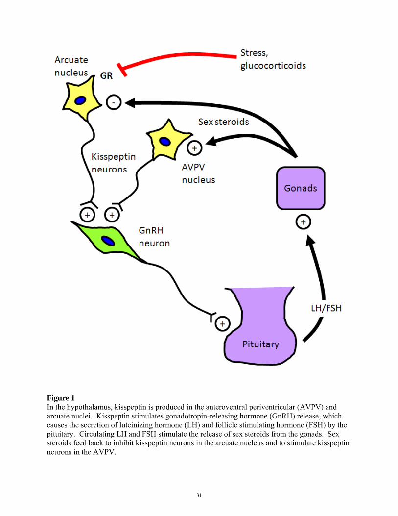
Figure 1 In the hypothalamus, kisspeptin is produced in the anteroventral periventricular (AVPV) and arcuate nuclei. Kisspeptin stimulates gonadotropin-releasing hormone (GnRH) release, which causes the secretion of luteinizing hormone (LH) and follicle stimulating hormone (FSH) by the pituitary. Circulating LH and FSH stimulate the release of sex steroids from the gonads. Sex steroids feed back to inhibit kisspeptin neurons in the arcuate nucleus and to stimulate kisspeptin neurons in the AVPV.
31
Drs. Lanjuin and Dulac generated the Kiss1CreBAC transgenic mouse. Dr. Basko-Plluska investigated the use of restraint as a psychological stressor. Dr. Muglia generated the GRflox/flox mouse.
CHAPTER 2
STRESS-INDUCED GLUCOCORTICOID RECEPTOR
SIGNALING REGULATES KISSPEPTIN NEURONS
This chapter is based on:
Oulu Wang, Anne Lanjuin, Juliana Basko-Plluska, Louis Muglia, Catherine Dulac, and Joseph Majzoub. Disruption of glucocorticoid receptor signaling in kisspeptin neurons accelerates the recovery of reproductive function in the post-traumatic stress period. In preparation, 2012.
32
2.1 ABSTRACT
Stressors generate adaptive responses to facilitate the return to homeostasis. Dysregulation of
this process can cause maladaptive responses, including cessation of reproductive function that
persists long after the stressor has subsided, through mechanisms that are not well understood.
Kisspeptin (KISS1) is required for the activation of the hypothalamic-pituitary-gonadal
reproductive axis in humans and mice. We hypothesized that acute stress in mice, acting through
the stress hormone, corticosterone, transiently inhibits kisspeptin neurons and downstream
reproductive capacity, and that the restoration of kisspeptin signaling is necessary for normal
reactivation of the reproductive axis. We examined the response of hypothalamic Kiss1 mRNA
expression and hormones of the reproductive and stress axes to different stressors. Stressors that
stimulated glucocorticoid secretion, as well as glucocorticoid administration itself, inhibited
Kiss1 mRNA expression, while conditions that did not change glucocorticoid secretion did not
alter Kiss1 mRNA expression. In mice lacking glucocorticoid receptor specifically in kisspeptin-
containing neurons, Kiss1 mRNA expression was no longer inhibited during restraint stress
despite a rise in corticosterone, and both testosterone and copulatory behaviors showed
accelerated recovery in the post-traumatic stress period. Blockade of glucocorticoid receptor
signaling in kisspeptin neurons during stress accelerates the recovery of reproductive function
during the post-traumatic stress period, a finding that may have therapeutic implications in
humans with post-traumatic stress disorders.
33
2.2 INTRODUCTION
Stress responses to acute stressors improve the chances of immediate survival, even at the
expense of immediate reproductive fitness [213]. Stressors can generate adaptive stress
responses that prepare for the return to homeostasis, but also maladaptive responses, including
the cessation of reproductive function long after the stressor has subsided. In patients with post-
traumatic stress disorder (PTSD), reproductive inhibition can persist long after the initial trauma
[208, 209]. The mechanisms by which acute stress can result in these maladaptive, post-
traumatic consequences are not well understood.
In 2003, two independent groups discovered hypogonadotropic individuals with kisspeptin
receptor mutations in large, consanguineous families [83, 84], and in humans and mouse models,
loss-of-function mutations in either the kisspeptin receptor or ligand block the onset of puberty
[83, 85, 93-95]. Conversely, gain-of-function mutations in the human receptor result in
precocious puberty [86], and administration of kisspeptin accelerates the onset of puberty in
juvenile rats [214]. Kisspeptin-secreting neurons in the arcuate and anteroventral periventricular
nuclei of the rodent hypothalamus stimulate gonadotropin-releasing hormone neurons, which
promote the release of luteinizing hormone (LH) and follicle-stimulating hormone in the anterior
pituitary [96, 100, 106, 215]. These gonadotropins stimulate the production of testosterone in
males and estrogen in females. Acute diphtheria toxin-mediated ablation of kisspeptin neurons
in adult mice inhibits fertility, indicating that kisspeptin neurons continue to regulate
reproduction in adults [216].
34
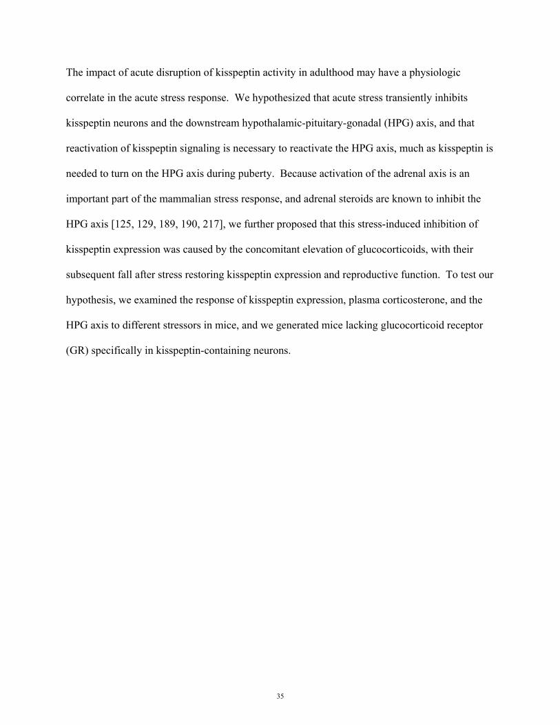
The impact of acute disruption of kisspeptin activity in adulthood may have a physiologic
correlate in the acute stress response. We hypothesized that acute stress transiently inhibits
kisspeptin neurons and the downstream hypothalamic-pituitary-gonadal (HPG) axis, and that
reactivation of kisspeptin signaling is necessary to reactivate the HPG axis, much as kisspeptin is
needed to turn on the HPG axis during puberty. Because activation of the adrenal axis is an
important part of the mammalian stress response, and adrenal steroids are known to inhibit the
HPG axis [125, 129, 189, 190, 217], we further proposed that this stress-induced inhibition of
kisspeptin expression was caused by the concomitant elevation of glucocorticoids, with their
subsequent fall after stress restoring kisspeptin expression and reproductive function. To test our
hypothesis, we examined the response of kisspeptin expression, plasma corticosterone, and the
HPG axis to different stressors in mice, and we generated mice lacking glucocorticoid receptor
(GR) specifically in kisspeptin-containing neurons.
35

2.3 MATERIALS AND METHODS
Animals and tissue preparation
All experiments were conducted in compliance with the Institutional Animal Care and Use
Committee guidelines of Children’s Hospital Boston. Adult C57BL/6J male mice were
purchased from The Jackson Laboratory (Bar Harbor, ME; 000664) and tested at 10-14 weeks of
age. All experiments concluded between 2-4PM. Animals were maintained on a 12 h light/dark
cycle with access to chow and water ad libitum and tested between 10-14 weeks of age. Before
experiments, animals were transferred to a quiet procedure room and allowed to acclimate for 7
d. Retroorbital blood samples were collected from unanesthetized animals within 1 min of cage
handling in all conditions and centrifuged at 3,000 rpm for 10 min at 4°C. Retroorbital
phlebotomy was used to collect sufficient volumes of blood in non-terminal experiments.
Animals were sacrificed by rapid decapitation without anesthesia, and dissected brains were
embedded in OCT and stored at -80°C. Brains were sectioned coronally at 20 µm from the
anteroventral periventricular nucleus to mammillary bodies (Figure 2.1) in four sets, thaw-
mounted onto 25 mm x75 mm slides, and returned to -80°C until further processing.
36
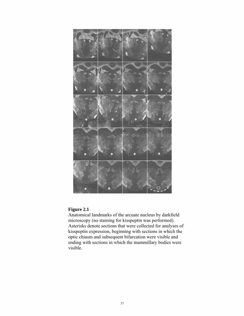
Figure 2.1 Anatomical landmarks of the arcuate nucleus by darkfield microscopy (no staining for kisspeptin was performed). Asterisks denote sections that were collected for analyses of kisspeptin expression, beginning with sections in which the optic chiasm and subsequent bifurcation were visible and ending with sections in which the mammillary bodies were visible.
37

Luteinizing hormone was assayed by the UVA Center for Research in Reproduction Ligand
Assay and Analysis Core. Plasma corticosterone and testosterone were measured by
radioimmunoassay (MP Biomedicals) with the following modifications: to minimize the amount
of plasma used in the corticosterone radioimmunoassay, we generated 1:200 dilutions using
either 1 µL of plasma with 199 µL of steroid diluent or 5 µL of plasma with 995 µL of steroid
diluent. In general, aliquots containing 1 µL were more variable in corticosterone
concentrations, likely due to pipetting error, and unless blood volume was a major constraint, we
used 5 µL of plasma for corticosterone assays. The dynamic range for the corticosterone assay
was 25 ng/mL to 1,000 ng/mL. To minimize the amount of plasma used in the testosterone
radioimmunoassay, we used 25 µL of plasma in singlet. In pilots, this volume was as effective
in identifying stress-induced testosterone suppression as 50 µL of plasma in duplicate. The
dynamic range for this assay was 0.1 ng/mL to 10 ng/mL.
Transgenic mice and breeding strategy
To study the effects of GR deletion in kisspeptin neurons, we generated Kiss1CreBAC::GRflox/flox
mice as well as Kiss1CreBAC, GRflox/flox, and WT controls. GRflox/flox animals were previously
validated and generously provided by Louis Muglia [30]. Briefly, loxP sites were targeted
upstream of exon 1C and in intron 2 of the GR gene, Nr3c1. Exon 2 contains the ATG initiation
site, and the exon and start sequence are excised by Cre-mediated recombination. GRflox/flox
animals were on a C57B background. To genetically target kisspeptin neurons, Drs. Lanjuin and
Dulac generated a Kiss1CreBAC mouse line using a BAC transgenic approach [218]. Briefly, Cre
cDNA sequences including a bGH polyA tail were recombined after the Kiss1 translational start
ATG on BAC RP23-240P23. The modified BAC was confirmed to be free of gross
38
rearrangements, linearized with Not1 to release a 102kb fragment containing the modified
Kiss1CreBAC locus (including 65kb of upstream sequences), and injected into B6/CBA oocytes.
Only one of six founder lines that we obtained showed expression in accordance with reported
sites of endogenous Kiss1 expression by double-label in situ hybridization. Kiss1CreBAC mice
were on a C57BL/CBA mixed background.
All animals used in experiments were males on a C57BL/CBA mixed background. Crosses from
Kiss1CreBAC::GRflox/+ x GRflox/+ breeders yielded Kiss1CreBAC::GRflox/flox, Kiss1CreBAC, GRflox/flox and
WT male littermates at frequencies of 6.25% each. Because these yields were insufficient to
power our study, we crossed F1 littermates, Kiss1CreBAC x WT, and Kiss1CreBAC::GRflox/flox x
GRflox/flox, to produce all the F2 littermates used in this study. Thus, in the F2 generation,
Kiss1CreBAC and WT mice were littermates, Kiss1CreBAC::GRflox/flox and GRflox/flox mice were
littermates, and all four genotypes from the F2 generation were related, because all F1 breeders
were littermates.
Stressors
Restraint causes minimal physical harm and is considered a psychological stressor [219].
Animals were placed in ventilated restraint tubes for 5 h. For food deprivation experiments,
animals were placed in cages without food for 48 h, but had access to bedding and ad libitum
water. For cold exposure experiments, animals were housed at 4°C for 24 h with bedding, food,
and water. For intraperitoneal corticosterone injections, corticosterone (Sigma C2505, St. Louis,
MO) was administered at 40 mg/kg body weight, and following decapitation, tissues were
collected 5 h post-injection for the detection of mRNA changes in the hypothalamus. In restraint
39

experiments, 14 male mice were tested per treatment (restrained or unrestrained). In food
deprivation experiments, 12 male mice were tested per treatment (food-deprived or fed). In cold
exposure experiments, 6 male mice were tested per treatment (housed at 4ºC or room
temperature). In i.p. corticosterone experiments, 7 male mice were tested per treatment
(corticosterone or saline).
In situ hybridization
The Kiss1 mRNA probe was generously provided by Robert Steiner [100]. Sense and antisense
Kiss1 probes spanning bases 76-486 of the murine Kiss1 gene were generated from a linearized
pAMP1 plasmid containing Kiss1, SP6- and T7-binding sequences. Radiolabeled probes were
synthesized using 33P-UTP, and in situ hybridization was performed as previously described
[100], with the following modifications. Briefly, tissues were washed in 4% paraformaldehyde,
acetic anhydride, 2X SSC, chloroform, and graded ethanols, then incubated in 12.7 million
dpm/mL probe for 16 h at 55°C. Slides were subsequently washed with 4X SSC, RNase, 2X
SSC, 0.5X SSC at 62°C, and graded ethanols in ammonium acetate, then dipped in
autoradiographic silver emulsion (Kodak NTB 8895666). Silver grains in the hypothalamus
were visualized by darkfield microscopy on the 10X objective of a Nikon Eclipse E800. Using
NIH ImageJ software (http://imagej.nih.gov/ij/), cells with ten-fold higher silver than
background were identified as Kiss1 mRNA-positive by a blinded observer using a semi-
automated program (Figure 2.2).
40
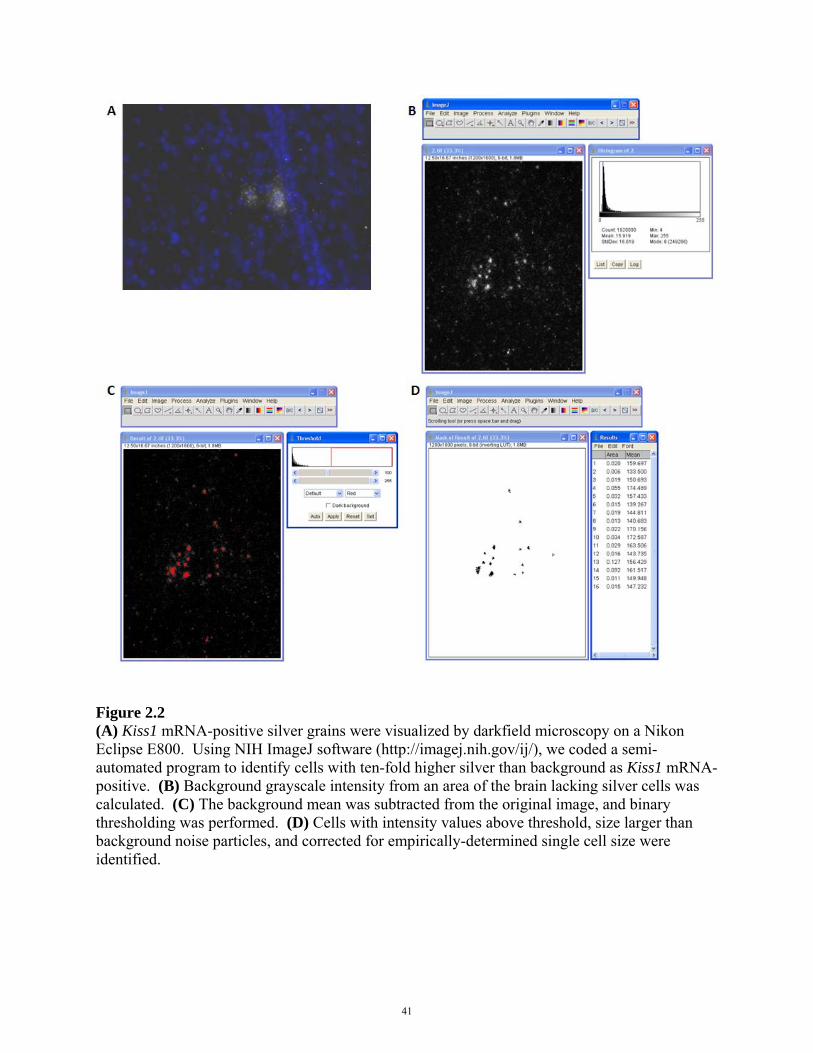
Figure 2.2 (A) Kiss1 mRNA-positive silver grains were visualized by darkfield microscopy on a Nikon Eclipse E800. Using NIH ImageJ software (http://imagej.nih.gov/ij/), we coded a semi-automated program to identify cells with ten-fold higher silver than background as Kiss1 mRNA-positive. (B) Background grayscale intensity from an area of the brain lacking silver cells was calculated. (C) The background mean was subtracted from the original image, and binary thresholding was performed. (D) Cells with intensity values above threshold, size larger than background noise particles, and corrected for empirically-determined single cell size were identified.
41

Immunohistochemical analyses
Brains from Kiss1CreBAC::R26flox-stop-tdTomato animals were fixed in 4% paraformaldehyde,
cryoprotected in 30% sucrose, and frozen in OCT. 10 µm coronal brain sections through the
anteroventral periventricular and arcuate nuclei of the hypothalamus were stained using
kisspeptin antiserum at a dilution of 1:5,000 for 7 d at 4ºC. Kiss1CreBAC::R26flox-stop-tdTomato and
Kiss1CreBAC::R26flox-stop-tdTomato::GRflox/flox brain sections treated in the same manner were stained
with rabbit antiserum for mouse/rat glucocorticoid receptor (Santa Cruz 1004) at a dilution of
1:2,000 overnight at 4ºC. We validated the antiserum for glucocorticoid receptor by using a
previously validated Cre construct to generate Sim1Cre::GRflox/flox mice, in which GR is
selectively deleted in the paraventricular nucleus. GR staining was robust in the PVN of WT
mice and absent in the PVN of Sim1Cre::GRflox/flox mice, demonstrating that the rabbit antiserum
is specific to GR (Figure 2.3). Rabbit antiserum for mouse/rat/ovine kisspeptin was previously
validated and generously provided by Alain Caraty [110]. In our experiments, kisspeptin
staining was only detected in the arcuate nucleus, AVPV, and medial amygdala. Images were
captured using Nikon Eclipse E800, Zeiss LSM700 laser scanning, and Perkin Elmer UltraVIEW
VoX spinning disk microscopes. Colocalization was analyzed by calculating the overlap
coefficient for each hypothalamic slice using NIH ImageJ software [220].
42
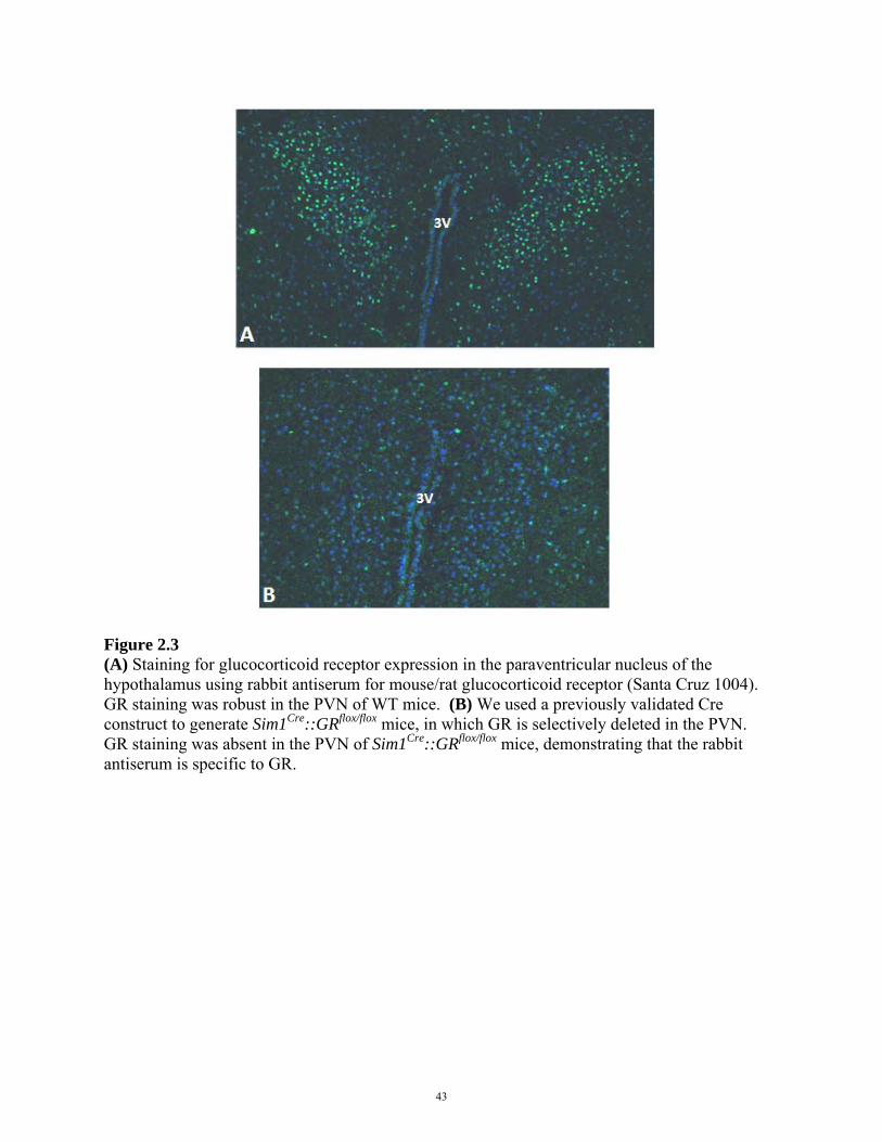
Figure 2.3 (A) Staining for glucocorticoid receptor expression in the paraventricular nucleus of the hypothalamus using rabbit antiserum for mouse/rat glucocorticoid receptor (Santa Cruz 1004). GR staining was robust in the PVN of WT mice. (B) We used a previously validated Cre construct to generate Sim1Cre::GRflox/flox mice, in which GR is selectively deleted in the PVN. GR staining was absent in the PVN of Sim1Cre::GRflox/flox mice, demonstrating that the rabbit antiserum is specific to GR.
43
Behavioral analyses
To examine copulatory behavior, each male was paired with one female with previously proven
fertility. Behavior was filmed using a Kodak Zx3 HD camera for 5 min starting at the time the
female was introduced into the home cage of the male. The amount of time the male spent
sniffing the female and the incidence of copulatory behaviors were analyzed at 30 frames/s using
VideoPad® software by a blinded observer. Open-field testing was filmed in a 45 cm x 45 cm
arena before stress, immediately after 5 h of restraint, and after 1 week of recovery. Each animal
was monitored for 5 min, and movement was tracked and analyzed using the Noldus
EthoVision® XT system.
Statistical analysis
Two-tailed two-sample t-tests were performed to compare hormone (corticosterone, LH,
testosterone) and Kiss1 mRNA expression levels between WT unstressed and restrained, food-
deprived, cold-exposed, or corticosterone-treated males. Linear mixed model analyses were
performed to compare changes in corticosterone, Kiss1 mRNA expression, and testosterone over
time. Linear mixed model analyses were performed and marginal means calculated for sniffing,
mounting, thrusting, locomotion, and open-field behaviors over time. Post-restraint, each of
these parameters was compared across genotypes by using one-way ANOVA with post hoc least
square difference (LSD) pairwise tests.
Genotyping
A list of genotyping primers and polymerase chain reaction protocols are detailed here for the
mouse strains used in these experiments: Kiss1CreBAC, GRflox/flox, tdTomato, mTomato/mGFP.
44

Kiss1CreBAC [221]
Cre forward primer 5' - CGT ACT GAC GGT GGG AGA AT - 3'
Cre reverse primer 5' - TGC ATG ATC TCC GGT ATT - 3'
94°C for 2’, (94°C for 30”, 55°C for 30”, 72°C for 1’) x 30, 72°C for 5’, 4°C
Cre band is 800 bp.
GRflox/flox [30]
GR forward primer 5' - AAT CAG AAT TGC TCA CTC ACA A - 3'
GR reverse primer 5' - CAG TGT TAC TAC TTC CAG TTC - 3'
LoxP reverse primer 5' - TGC TAT ACG AAG TTA TCA GTA C - 3'94°C for 3’, (52°C for 30”,
72°C for 30”, 95°C for 30”) x 30, 52°C for 1’, 72°C for 5’, 4°C
WT band is 200 bp, and floxed band is 290 bp.
tdTomato [222]
WT forward primer 5’ - AAG GGA GCT GCA GTG GAG TA - 3'
WT reverse primer 5’ - CCG AAA ATC TGT GGG AAG TC - 3'
tdTomato forward primer 5’ - CTG TTC CTG TAC GGC ATG G - 3'
tdTomato reverse primer 5’ - GGC ATT AAA GCA GCG TAT CC - 3'
95°C for 5’, (94°C for 30”, 56°C for 30”, 72°C for 1’) x 32, 72°C for 10’, 4°C
WT band is 300 bp, and tdTomato band is 200 bp.
45
mTomato/mGFP [223]
Because mTomato is expressed in cells throughout the body, we determined mTomato/mGFP-
positive genotypes by visualizing mTomato fluorescence in a drop of blood, collected when
obtaining tail biopsies, rather than by PCR. The following primers may be used for genotyping
by PCR:
5' - TCA ATG GGC GGG GGT CGT T - 3'
5' - CTC TGC TGC CTC CTG GCT TCT - 3'
5' - CGA GGC GGA TCA CAA GCA ATA - 3'
WT band is 330 bp, and mT/mG band is 250 bp.
46

2.4 RESULTS
Effects of stress on the reproductive axis
To test the effects of stress on Kiss1 mRNA expression, we subjected adult male mice to 5 h of
restraint, which produces psychological trauma without causing physical pain [219]. Compared
to unstressed controls, restrained animals had increased plasma corticosterone concentrations
(Figure 2.4a), concomitant with suppression of Kiss1 mRNA expression in the arcuate nucleus
of the hypothalamus (Figure 2.4b-d). LH and testosterone were inhibited following restraint
(Figure 2.4e-f). Kiss1 mRNA expression in the anteroventral periventricular nucleus and medial
amygdala remained unchanged (Figure 2.5). Food deprivation also inhibits reproductive
function [224]. Body weight was measured to confirm food deprivation in each animal (Figure
2.6). When mice were food deprived for 48 h, plasma corticosterone was increased compared to
in fed controls (Figure 2.7a), concomitant with suppression of Kiss1 mRNA expression in the
arcuate nucleus (Figure 2.7b-d), a trend towards suppression of LH, and significant suppression
of testosterone (Figure 2.7e-f). Restraint and food deprivation induced similar increases in
plasma corticosterone and decreases in Kiss1 mRNA expression.
47
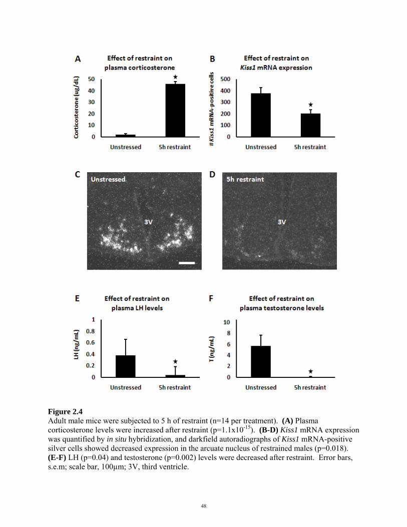
Figure 2.4 Adult male mice were subjected to 5 h of restraint (n=14 per treatment). (A) Plasma corticosterone levels were increased after restraint (p=1.1x10⁻¹⁵). (B-D) Kiss1 mRNA expression was quantified by in situ hybridization, and darkfield autoradiographs of Kiss1 mRNA-positive silver cells showed decreased expression in the arcuate nucleus of restrained males (p=0.018). (E-F) LH (p=0.04) and testosterone (p=0.002) levels were decreased after restraint. Error bars, s.e.m; scale bar, 100µm; 3V, third ventricle.
48

Figure 2.5 Adult male mice were subjected to 5 h of restraint. Kiss1 mRNA expression was quantified by in situ hybridization, and darkfield autoradiographs of Kiss1 mRNA-positive silver cells showed (A) no change in Kiss1 mRNA expression in the anteroventral periventricular nucleus (p=0.31) after restraint and (B) no change in Kiss1 mRNA expression in medial amygdala (p=0.60) after restraint.
49
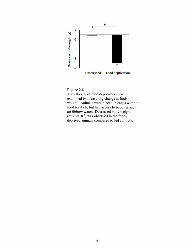
Figure 2.6 The efficacy of food deprivation was examined by measuring change in body weight. Animals were placed in cages without food for 48 h, but had access to bedding and ad libitum water. Decreased body weight (p=7.7x10⁻⁸) was observed in the food-deprived animals compared to fed controls.
50

Figure 2.7 Adult male mice were subjected to 48 h of food deprivation (n=12 per treatment). (A) Plasma corticosterone levels were elevated after food deprivation (p=6.7x10⁻¹³). (B-D) Kiss1 mRNA expression was significantly decreased following food deprivation (p=0.03). (E-F) LH trended downward (p=0.06), and testosterone was significantly suppressed (p=0.001) after food deprivation. Error bars, s.e.m; scale bar, 100µm; 3V, third ventricle.
51
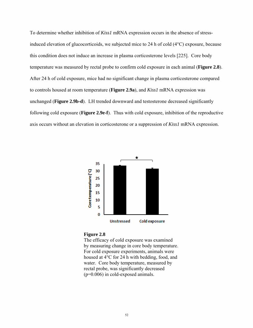
To determine whether inhibition of Kiss1 mRNA expression occurs in the absence of stress-
induced elevation of glucocorticoids, we subjected mice to 24 h of cold (4°C) exposure, because
this condition does not induce an increase in plasma corticosterone levels [225]. Core body
temperature was measured by rectal probe to confirm cold exposure in each animal (Figure 2.8).
After 24 h of cold exposure, mice had no significant change in plasma corticosterone compared
to controls housed at room temperature (Figure 2.9a), and Kiss1 mRNA expression was
unchanged (Figure 2.9b-d). LH trended downward and testosterone decreased significantly
following cold exposure (Figure 2.9e-f). Thus with cold exposure, inhibition of the reproductive
axis occurs without an elevation in corticosterone or a suppression of Kiss1 mRNA expression.
Figure 2.8 The efficacy of cold exposure was examined by measuring change in core body temperature. For cold exposure experiments, animals were housed at 4°C for 24 h with bedding, food, and water. Core body temperature, measured by rectal probe, was significantly decreased (p=0.006) in cold-exposed animals.
52
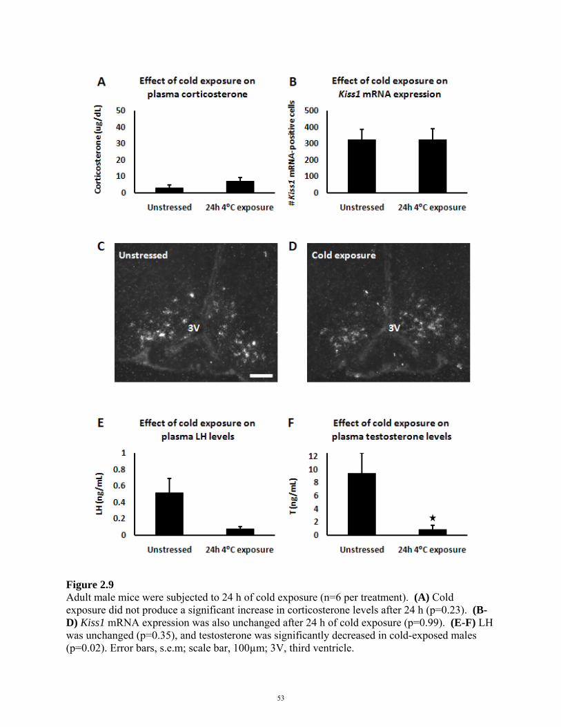
Figure 2.9 Adult male mice were subjected to 24 h of cold exposure (n=6 per treatment). (A) Cold exposure did not produce a significant increase in corticosterone levels after 24 h (p=0.23). (B-D) Kiss1 mRNA expression was also unchanged after 24 h of cold exposure (p=0.99). (E-F) LH was unchanged (p=0.35), and testosterone was significantly decreased in cold-exposed males (p=0.02). Error bars, s.e.m; scale bar, 100µm; 3V, third ventricle.
53
With the above three stressors, the consistent relationship between a rise in corticosterone and
fall in Kiss1 mRNA expression suggested that elevated corticosterone inhibits Kiss1 expression.
To test this directly, we injected adult male mice with a stress level dose of corticosterone [226]
or saline. To mimic the time course of mRNA changes in the restraint condition, we analyzed
tissues 5 h post-injection. As expected, corticosterone levels were elevated (Figure 2.10a), and
administration of corticosterone alone was sufficient to inhibit Kiss1 mRNA expression (Figure
2.10b-d). LH trended downward, and testosterone decreased following corticosterone
administration (Figure 2.10e-f). This suppression of Kiss1 mRNA expression indicates that a
rise in plasma glucocorticoid concentration can affect kisspeptin neurons, either directly or
indirectly, and may cause the inhibition of the reproductive axis after exposure to psychological
trauma and food withdrawal stressors, but not cold exposure.
54
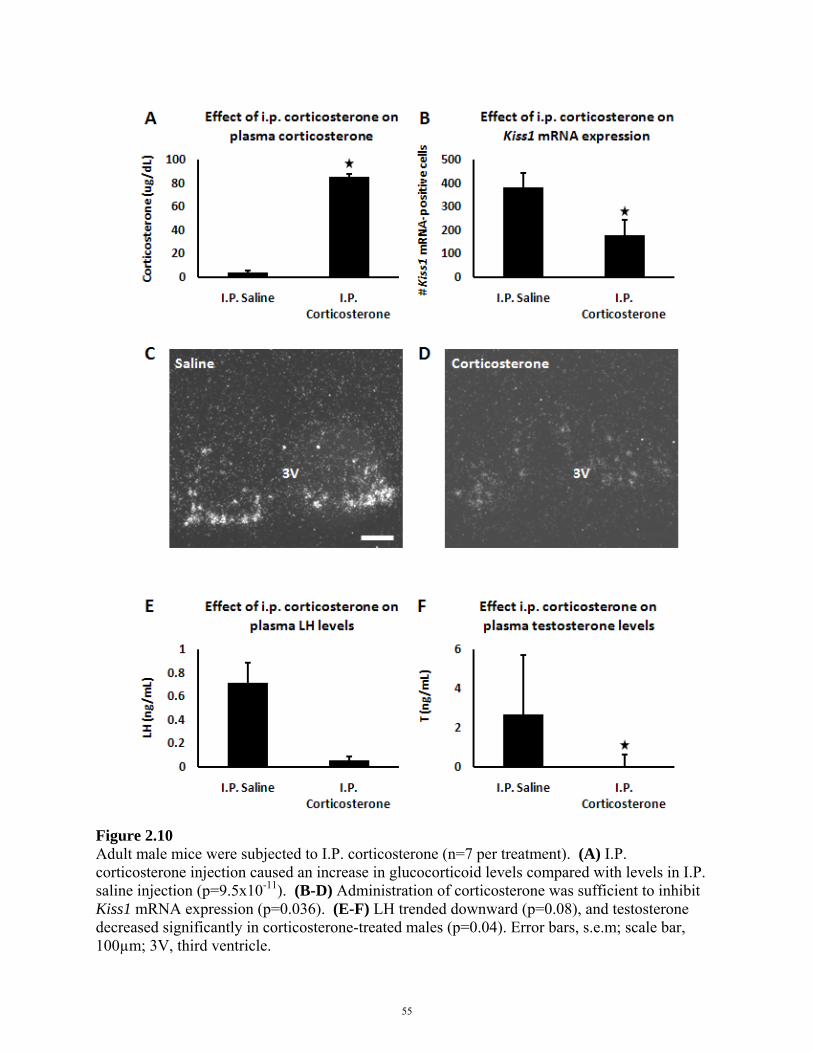
Figure 2.10 Adult male mice were subjected to I.P. corticosterone (n=7 per treatment). (A) I.P. corticosterone injection caused an increase in glucocorticoid levels compared with levels in I.P. saline injection (p=9.5x10⁻¹¹). (B-D) Administration of corticosterone was sufficient to inhibit Kiss1 mRNA expression (p=0.036). (E-F) LH trended downward (p=0.08), and testosterone decreased significantly in corticosterone-treated males (p=0.04). Error bars, s.e.m; scale bar, 100µm; 3V, third ventricle.
55
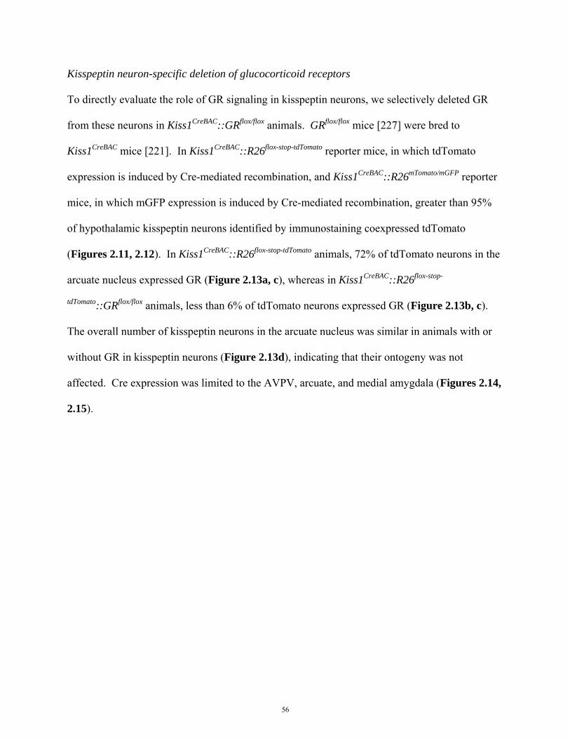
Kisspeptin neuron-specific deletion of glucocorticoid receptors
To directly evaluate the role of GR signaling in kisspeptin neurons, we selectively deleted GR
from these neurons in Kiss1CreBAC::GRflox/flox animals. GRflox/flox mice [227] were bred to
Kiss1CreBAC mice [221]. In Kiss1CreBAC::R26flox-stop-tdTomato reporter mice, in which tdTomato
expression is induced by Cre-mediated recombination, and Kiss1CreBAC::R26mTomato/mGFP reporter
mice, in which mGFP expression is induced by Cre-mediated recombination, greater than 95%
of hypothalamic kisspeptin neurons identified by immunostaining coexpressed tdTomato
(Figures 2.11, 2.12). In Kiss1CreBAC::R26flox-stop-tdTomato animals, 72% of tdTomato neurons in the
arcuate nucleus expressed GR (Figure 2.13a, c), whereas in Kiss1CreBAC::R26flox-stop-
tdTomato::GRflox/flox animals, less than 6% of tdTomato neurons expressed GR (Figure 2.13b, c).
The overall number of kisspeptin neurons in the arcuate nucleus was similar in animals with or
without GR in kisspeptin neurons (Figure 2.13d), indicating that their ontogeny was not
affected. Cre expression was limited to the AVPV, arcuate, and medial amygdala (Figures 2.14,
2.15).
56

Figure 2.11 In Kiss1CreBAC::R26flox-stop-tdTomato reporter animals, 95% (±0.8%) of kisspeptin neurons (anti-kisspeptin, green) in the hypothalamus expressed tdTomato (magenta). Arrows indicate examples of kisspeptin-positive and tdTomato-positive cells. Asterisk indicates example of kisspeptin-positive, but tdTomato-negative cell. Scale bar, 25 µm.
57

Figure 2.12 Kisspeptin staining in the arcuate nucleus preferentially stains fibers, so Kiss1CreBAC::R26mTomato/mGFP reporter animals (green) were generated to evaluate colocalization (white) with endogenous kisspeptin (magenta). Greater than 95% of kisspeptin fibers (magenta) in the arcuate nucleus expressed driven mGFP (green). Representative images of the anterior arcuate nucleus (A-D) and posterior arcuate nucleus (E-H) from two Kiss1CreBAC::R26mTomato/mGFP animals are shown. Scale bar, 100µm; 3V, third ventricle.
58
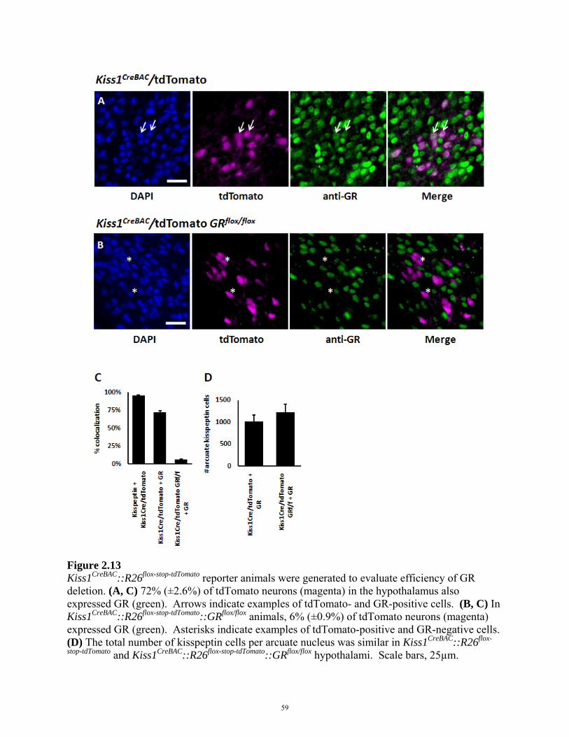
Figure 2.13 Kiss1CreBAC::R26flox-stop-tdTomato reporter animals were generated to evaluate efficiency of GR deletion. (A, C) 72% (±2.6%) of tdTomato neurons (magenta) in the hypothalamus also expressed GR (green). Arrows indicate examples of tdTomato- and GR-positive cells. (B, C) In Kiss1CreBAC::R26flox-stop-tdTomato::GRflox/flox animals, 6% (±0.9%) of tdTomato neurons (magenta) expressed GR (green). Asterisks indicate examples of tdTomato-positive and GR-negative cells. (D) The total number of kisspeptin cells per arcuate nucleus was similar in Kiss1CreBAC::R26flox-
stop-tdTomato and Kiss1CreBAC::R26flox-stop-tdTomato::GRflox/flox hypothalami. Scale bars, 25µm.
59
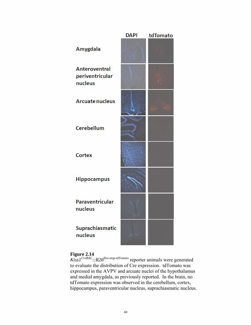
Figure 2.14 Kiss1CreBAC::R26flox-stop-tdTomato reporter animals were generated to evaluate the distribution of Cre expression. tdTomato was expressed in the AVPV and arcuate nuclei of the hypothalamus and medial amygdala, as previously reported. In the brain, no tdTomato expression was observed in the cerebellum, cortex, hippocampus, paraventricular nucleus, suprachiasmatic nucleus.
60
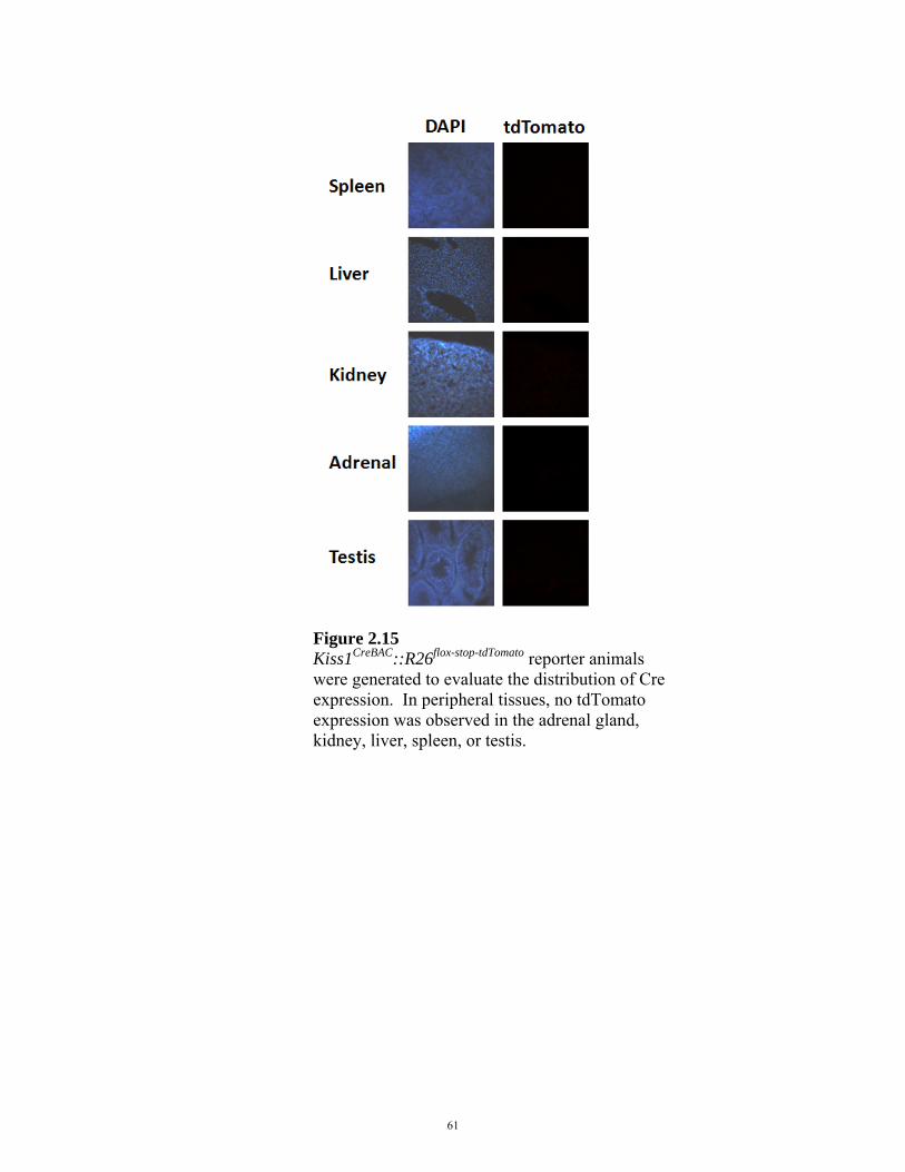
Figure 2.15 Kiss1CreBAC::R26flox-stop-tdTomato reporter animals were generated to evaluate the distribution of Cre expression. In peripheral tissues, no tdTomato expression was observed in the adrenal gland, kidney, liver, spleen, or testis.
61
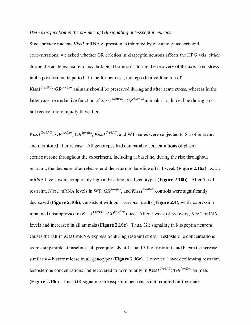
HPG axis function in the absence of GR signaling in kisspeptin neurons
Since arcuate nucleus Kiss1 mRNA expression is inhibited by elevated glucocorticoid
concentrations, we asked whether GR deletion in kisspeptin neurons affects the HPG axis, either
during the acute exposure to psychological trauma or during the recovery of the axis from stress
in the post-traumatic period. In the former case, the reproductive function of
Kiss1CreBAC::GRflox/flox animals should be preserved during and after acute stress, whereas in the
latter case, reproductive function of Kiss1CreBAC::GRflox/flox animals should decline during stress
but recover more rapidly thereafter.
Kiss1CreBAC::GRflox/flox, GRflox/flox, Kiss1CreBAC, and WT males were subjected to 5 h of restraint
and monitored after release. All genotypes had comparable concentrations of plasma
corticosterone throughout the experiment, including at baseline, during the rise throughout
restraint, the decrease after release, and the return to baseline after 1 week (Figure 2.16a). Kiss1
mRNA levels were comparably high at baseline in all genotypes (Figure 2.16b). After 5 h of
restraint, Kiss1 mRNA levels in WT, GRflox/flox, and Kiss1CreBAC controls were significantly
decreased (Figure 2.16b), consistent with our previous results (Figure 2.4), while expression
remained unsuppressed in Kiss1CreBAC::GRflox/flox mice. After 1 week of recovery, Kiss1 mRNA
levels had increased in all animals (Figure 2.16c). Thus, GR signaling in kisspeptin neurons
causes the fall in Kiss1 mRNA expression during restraint stress. Testosterone concentrations
were comparable at baseline, fell precipitously at 1 h and 5 h of restraint, and began to increase
similarly 4 h after release in all genotypes (Figure 2.16c). However, 1 week following restraint,
testosterone concentrations had recovered to normal only in Kiss1CreBAC::GRflox/flox animals
(Figure 2.16c). Thus, GR signaling in kisspeptin neurons is not required for the acute
62

suppression of the HPG axis during stress, but is required for the regulation of reproductive
recovery following stress.
63
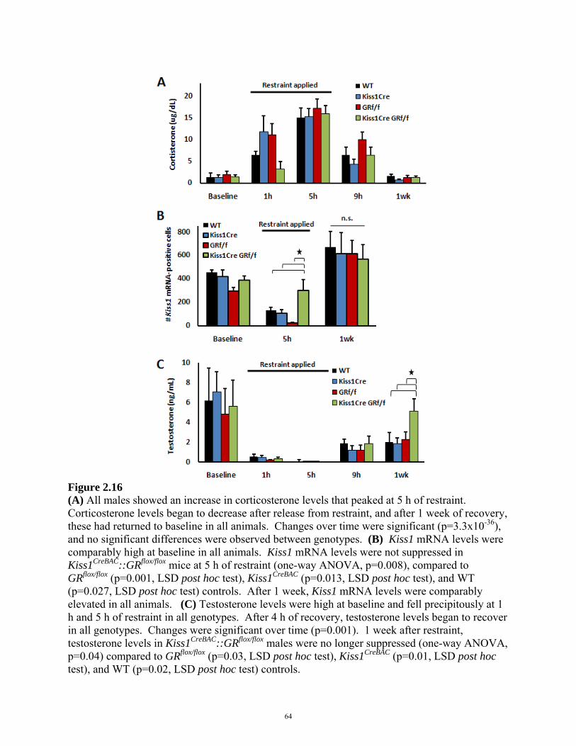
Figure 2.16 (A) All males showed an increase in corticosterone levels that peaked at 5 h of restraint. Corticosterone levels began to decrease after release from restraint, and after 1 week of recovery, these had returned to baseline in all animals. Changes over time were significant (p=3.3x10-36), and no significant differences were observed between genotypes. (B) Kiss1 mRNA levels were comparably high at baseline in all animals. Kiss1 mRNA levels were not suppressed in Kiss1CreBAC::GRflox/flox mice at 5 h of restraint (one-way ANOVA, p=0.008), compared to GRflox/flox (p=0.001, LSD post hoc test), Kiss1CreBAC (p=0.013, LSD post hoc test), and WT (p=0.027, LSD post hoc test) controls. After 1 week, Kiss1 mRNA levels were comparably elevated in all animals. (C) Testosterone levels were high at baseline and fell precipitously at 1 h and 5 h of restraint in all genotypes. After 4 h of recovery, testosterone levels began to recover in all genotypes. Changes were significant over time (p=0.001). 1 week after restraint, testosterone levels in Kiss1CreBAC::GRflox/flox males were no longer suppressed (one-way ANOVA, p=0.04) compared to GRflox/flox (p=0.03, LSD post hoc test), Kiss1CreBAC (p=0.01, LSD post hoc test), and WT (p=0.02, LSD post hoc test) controls.
64

Effects of stress on reproductive behaviors in the absence of GR signaling in kisspeptin neurons
To determine the impact of kisspeptin neuron-specific GR deletion on reproductive behaviors
during and after stress, we examined copulatory behaviors before and after 5 h of restraint.
Immediately after restraint, all genotypes exhibited comparable suppression of the copulatory
behaviors, mounting and thrusting, compared to baseline (Figure 2.17a, b), consistent with the
decrease in testosterone observed in all genotypes (Figure 2.16c). In Kiss1CreBAC::GRflox/flox
mice, however, both mounting and thrusting behaviors were fully recovered 1 week after
restraint, while copulatory activity remained low at this time in GRflox/flox, Kiss1CreBAC, and WT
controls (Figure 2.17a, b). From these studies of Kiss1CreBAC::GRflox/flox mice, we conclude that
immediately following restraint stress, GR signaling within kisspeptin neurons causes the fall in
Kiss1 mRNA, though not the inhibition of hormonal and behavioral components of the HPG
axis. GR-dependent inhibition of kisspeptin neurons during acute stress is required for regulates
the time course of recovery from stress-induced reproductive inhibition.
65

Figure 2.17 (A) Immediately after restraint, all genotypes exhibited a comparable decrease in mounting behavior (p=5.0x10⁻⁵). In Kiss1CreBAC::GRflox/flox mice, mounting was fully recovered after 1 week (p=0.017, one-way ANOVA), compared to GRflox/flox (p=0.018, LSD post hoc test), Kiss1CreBAC (p=0.007, LSD post hoc test), and WT (p=0.011, LSD post hoc test) controls in which the incidence remained low. (B) All genotypes exhibited decreased thrusting behavior after restraint (p=7.0x10⁻⁵). In Kiss1CreBAC::GRflox/flox mice, thrusting was fully recovered after 1 week (p=0.012, one-way ANOVA), compared to GRflox/flox (p=0.006, LSD post hoc test), Kiss1CreBAC (p=0.007, LSD post hoc test), and WT (p=0.011, LSD post hoc test) controls in which the incidence remained low.
66

Finally, we examined the specificity of the behavioral consequences of deleting GR from
kisspeptin neurons. While testosterone and copulatory behaviors recovered more quickly after
stress in Kiss1CreBAC::GRflox/flox males, behaviors such as sniffing, locomotor activity, and general
anxiety were unaffected by deletion of GR. Kiss1CreBAC::GRflox/flox, GRflox/flox, Kiss1CreBAC, and
WT animals spent similar amounts of time sniffing females at baseline, and all mice exhibited
decreased sniffing behavior immediately after restraint, but recovered this behavior at 1 week
(Figure 2.18a). Likewise, all genotypes exhibited decreased locomotor activity after 5 h
restraint compared to baseline and recovered after 1 week (Figure 2.18b). All animals spent less
time in the center of an open-field test immediately after restraint, a sign of increased general
anxiety [228], which was not fully recovered after 1 week (Figure 2.18c), though this may have
been due to increased time spent in the center by WT males. Thus, deletion of GR in kisspeptin
neurons selectively impacts reproductive behaviors.
67
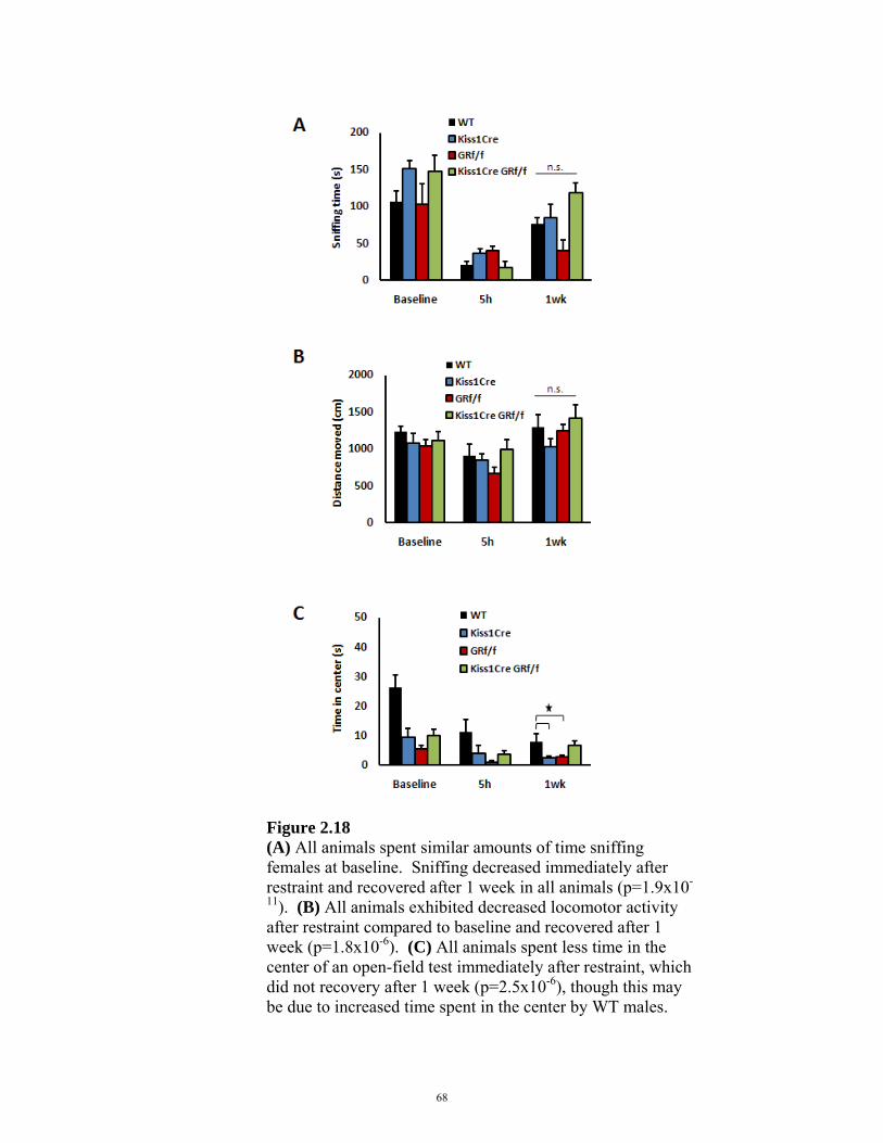
Figure 2.18 (A) All animals spent similar amounts of time sniffing females at baseline. Sniffing decreased immediately after restraint and recovered after 1 week in all animals (p=1.9x10-
11). (B) All animals exhibited decreased locomotor activity after restraint compared to baseline and recovered after 1 week (p=1.8x10⁻⁶). (C) All animals spent less time in the center of an open-field test immediately after restraint, which did not recovery after 1 week (p=2.5x10⁻⁶), though this may be due to increased time spent in the center by WT males.
68

2.5 DISCUSSION
Natural selection depends on successful reproduction, but inhibition of reproduction to survive
famine or escape predation allows animals to survive to reproduce at a later time. The cellular
locations and mechanisms responsible for inhibiting and reactivating the HPG axis during and
after stress, respectively, are not well understood. We examined the suppression of the HPG axis
to multiple stressors in male mice: restraint, food deprivation, cold exposure, and corticosterone
administration. In all cases, there was an inverse relationship between plasma corticosterone and
Kiss1 mRNA content, but even with cold exposure, when plasma corticosterone and Kiss1
mRNA were not affected, plasma testosterone was acutely inhibited.
We generated mice lacking GR specifically in kisspeptin-containing neurons. In these animals,
Kiss1 mRNA expression was no longer inhibited during restraint stress, even though
corticosterone was increased, and both testosterone and copulatory behaviors showed accelerated
recovery in the post-traumatic stress period. Neither systemic glucocorticoid levels nor general
anxiety were affected in these animals, demonstrating the hormonal and behavioral specificity of
the targeted disruption of GR signaling in kisspeptin neurons.
Stress, and glucocorticoids in particular, are known to inhibit the HPG axis. Men with Cushing’s
syndrome, a condition characterized by hypercortisolemia, and men treated with exogenous
glucocorticoid therapy experience decreased testosterone levels and loss of libido [189, 190,
217]. PTSD and other psychiatric disorders are characterized by HPA axis hyperactivity and
HPG axis suppression [208, 209]. Stress-induced elevation of corticosterone in male mice can
lead to decreased testosterone secretion [129], and male mice treated with exogenous
corticosterone exhibit decreased copulatory behaviors [125]. Food deprivation suppresses Kiss1
69
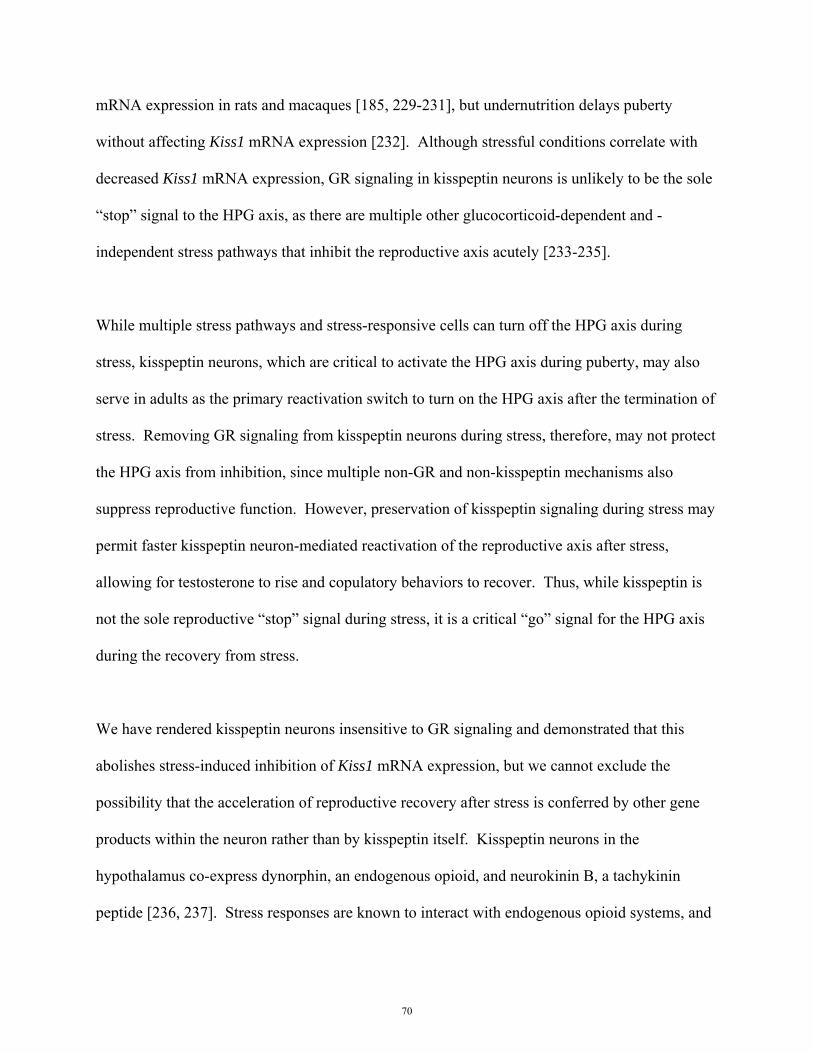
mRNA expression in rats and macaques [185, 229-231], but undernutrition delays puberty
without affecting Kiss1 mRNA expression [232]. Although stressful conditions correlate with
decreased Kiss1 mRNA expression, GR signaling in kisspeptin neurons is unlikely to be the sole
“stop” signal to the HPG axis, as there are multiple other glucocorticoid-dependent and -
independent stress pathways that inhibit the reproductive axis acutely [233-235].
While multiple stress pathways and stress-responsive cells can turn off the HPG axis during
stress, kisspeptin neurons, which are critical to activate the HPG axis during puberty, may also
serve in adults as the primary reactivation switch to turn on the HPG axis after the termination of
stress. Removing GR signaling from kisspeptin neurons during stress, therefore, may not protect
the HPG axis from inhibition, since multiple non-GR and non-kisspeptin mechanisms also
suppress reproductive function. However, preservation of kisspeptin signaling during stress may
permit faster kisspeptin neuron-mediated reactivation of the reproductive axis after stress,
allowing for testosterone to rise and copulatory behaviors to recover. Thus, while kisspeptin is
not the sole reproductive “stop” signal during stress, it is a critical “go” signal for the HPG axis
during the recovery from stress.
We have rendered kisspeptin neurons insensitive to GR signaling and demonstrated that this
abolishes stress-induced inhibition of Kiss1 mRNA expression, but we cannot exclude the
possibility that the acceleration of reproductive recovery after stress is conferred by other gene
products within the neuron rather than by kisspeptin itself. Kisspeptin neurons in the
hypothalamus co-express dynorphin, an endogenous opioid, and neurokinin B, a tachykinin
peptide [236, 237]. Stress responses are known to interact with endogenous opioid systems, and
70
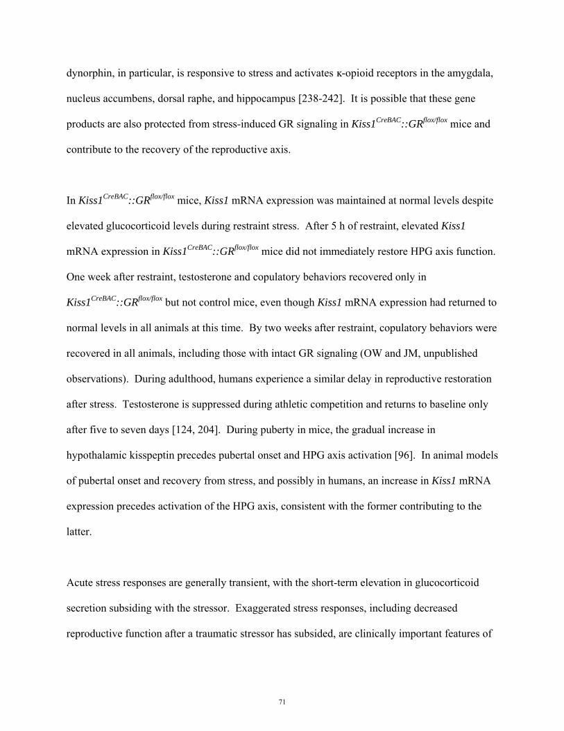
dynorphin, in particular, is responsive to stress and activates ĸ-opioid receptors in the amygdala,
nucleus accumbens, dorsal raphe, and hippocampus [238-242]. It is possible that these gene
products are also protected from stress-induced GR signaling in Kiss1CreBAC::GRflox/flox mice and
contribute to the recovery of the reproductive axis.
In Kiss1CreBAC::GRflox/flox mice, Kiss1 mRNA expression was maintained at normal levels despite
elevated glucocorticoid levels during restraint stress. After 5 h of restraint, elevated Kiss1
mRNA expression in Kiss1CreBAC::GRflox/flox mice did not immediately restore HPG axis function.
One week after restraint, testosterone and copulatory behaviors recovered only in
Kiss1CreBAC::GRflox/flox but not control mice, even though Kiss1 mRNA expression had returned to
normal levels in all animals at this time. By two weeks after restraint, copulatory behaviors were
recovered in all animals, including those with intact GR signaling (OW and JM, unpublished
observations). During adulthood, humans experience a similar delay in reproductive restoration
after stress. Testosterone is suppressed during athletic competition and returns to baseline only
after five to seven days [124, 204]. During puberty in mice, the gradual increase in
hypothalamic kisspeptin precedes pubertal onset and HPG axis activation [96]. In animal models
of pubertal onset and recovery from stress, and possibly in humans, an increase in Kiss1 mRNA
expression precedes activation of the HPG axis, consistent with the former contributing to the
latter.
Acute stress responses are generally transient, with the short-term elevation in glucocorticoid
secretion subsiding with the stressor. Exaggerated stress responses, including decreased
reproductive function after a traumatic stressor has subsided, are clinically important features of
71

PTSD [208, 209], which may be mediated by persistently elevated glucocorticoid secretion
[243]. Our findings indicate not only that kisspeptin neurons can be protected from stress by the
removal of GR signaling, but that doing so has the ability to orchestrate a repertoire of complex
behaviors leading to more rapid return of reproductive function, possibly by the downstream
regulation of sex steroid secretion. The requirement for glucocorticoid receptor signaling to
regulate kisspeptin neurons during the acute stress response suggests molecular targets for the
treatment of reproductive dysfunction in PTSD, such as the blockade of glucocorticoid receptor
signaling in kisspeptin neurons.
72
Acknowledgments
We thank We thank Drs. S.B. Seminara, W.F. Crowley, Y.M. Chan, U.B. Kaiser, J.C. Gill, C.J.
Weitz, D.T. Breault, C.B. Saper, G. J. De Vries, W.A. Carlezon, and B.B. Lowell for critical
reviews of our manuscript. We also thank J.J. LeBlanc and Dr. M. Fagiolini for providing
training and assistance with the open field test, C. Ho for helping perform experiments, and Dr.
H. Feldman for statistical assistance. This work was supported by the National Science
Foundation Graduate Research Fellowship (O.W.), Department of Defense National Defense
Science and Engineering Graduate Fellowship (O.W.), NIH T32MH20017-09 grant (O.W.), NIH
R01DC009019 (C.D.), Howard Hughes Medical Institute (C.D.), Damon Runyon Cancer
Research Foundation Award 1872-05 (A.J.), NIH MH079010 (L.M.), NIH T32DK007699-39
(J.M.), and the Timothy Murphy Fund (J.M.). The Children’s Hospital Boston IDDRC provided
microscopy services and is supported by the NIH-P30-HD-18655 grant. The UVA Ligand Core
is supported by the Eunice Kennedy Shriver NICHD/NIH (SCCPIR) grant U54-HD28932.
73
CHAPTER 3
LEPTIN IS NOT REQUIRED FOR FERTILITY
This chapter is based on:
Oulu Wang, Satoru Sakihara, Kolbein Gudmundsson, and Joseph Majzoub. Leptin is not required for fertility In preparation, 2012.
Dr. Gudmundsson created CRHOB mice on a mixed background, and Dr. Sakihara initiated studies of metabolic phenotypes in the CRHOB mouse. Oulu Wang created CRHOB mice on a B6 background and documented fertility and metabolic effects on this background.
74
3.1 ABSTRACT
Leptin is widely considered to be required for fertility. Leptin deficiency causes obesity and
infertility, and both conditions are ameliorated by leptin treatment. Leptin-deficient mice also
exhibit elevated glucocorticoid secretion, and excess glucocorticoids, independent of leptin
signaling, are associated with suppression of reproductive function. We hypothesized that
elevated glucocorticoids inhibit fertility in leptin-deficient mice. To evaluate the role of
glucocorticoids in leptin deficiency, we generated mice that were genetically leptin-deficient and
glucocorticoid-deficient. Leptin-deficient, glucocorticoid-deficient mice were fertile and
exhibited decreased body weight, fat composition, and food intake. When supplemented with
corticosterone to physiologic levels, only the leptin-deficient, glucocorticoid-deficient mice
became infertile. When corticosterone was removed, these mice regained reproductive capacity.
Rescuing hypercortisolemia in leptin-deficient mice was sufficient to restore reproductive
function, and thus, leptin is not required for fertility. These finding may provide novel
mechanisms of glucocorticoids affecting fertility, not only in leptin deficiency, but also in
multiple disorders characterized by hypercortisolemia, such as Cushing’s disease, anorexia
nervosa, and mood disorders.
75
3.2 INTRODUCTION
Leptin deficient LepOb/Ob mice are morbidly obese and infertile [171]. LepOb/Ob mice pair-fed to
achieve wild-type body weights do not regain reproductive function [244]. Leptin replacement
restores fertility in LepOb/Ob [244] and elicits advanced puberty in wild-type [178] mice, giving
rise to the tenet that leptin is required for fertility.
LepOb/Ob mice are also hypercortisolemic [245]. Leptin deficiency creates a state of perceived
starvation, and starvation is known to induce glucocorticoid secretion [246] (Figure 2.7). The
hypothalamic-pituitary-adrenal stress axis comprises corticotropin-releasing hormone (CRH)
neurons that stimulate pituitary adrenocorticotropic hormone (ACTH) release, which causes
adrenal glucocorticoid secretion. Negative feedback by glucocorticoids then suppresses CRH
and ACTH production. Excess glucocorticoid concentrations, independent of leptin signaling,
are associated with reproductive inhibition in Cushing’s disease, anorexia nervosa, exercise-
induced amenorrhea, glucocorticoid therapy, and other conditions characterized by
glucocorticoid dysregulation [132, 189, 217]. Adrenalectomy ameliorates obesity in LepOb/Ob
mice [247], a phenomenon that was ascribed to disinhibition of CRH, itself an anorectic hormone
[248]. Adrenalectomy did not reverse sterility in LepOb/Ob mice [249]. Using surgical
adrenalectomy to study the effects of glucocorticoids is confounded by upregulation of CRH,
surgical stress, and the regenerative capacity of adrenal tissue.
We hypothesized that glucocorticoids contribute to infertility in LepOb/Ob mice. We generated
LepOb/Ob mice that were genetically CRH- and glucocorticoid-deficient (CRHOB) to study the
role of glucocorticoids on fertility. Compared to LepOb/Ob controls, CRHOB mice displayed
76
normal fertility, normal reproductive organs and hormone secretion, and decreased body weight,
fat composition, and hyperphagia. When supplemented with glucocorticoids to levels present in
leptin deficiency, CRHOB but not wild-type mice became infertile, and regained reproductive
function after the termination of glucocorticoid treatment. Thus, leptin is not directly required
for fertility as previously postulated, and glucocorticoids contribute to infertility and obesity
downstream of leptin signaling. These studies may provide novel molecular targets in the
reproductive and metabolic aspects of disorders characterized by glucocorticoid dysregulation,
including leptin deficiency, Cushing’s disease, anorexia nervosa, and mood disorders.
77
3.3 MATERIAL AND METHODS
Animal husbandry and breeding strategy
All experiments were conducted in compliance with the Institutional Animal Care and Use
Committee guidelines of Children’s Hospital Boston. Animals were maintained on a 12 h
light/dark cycle with access to chow and water ad libitum and tested between 5-20 weeks of age.
Crh-/- mice were previously generated in our laboratory [8]. LepOb/+ mice were purchased from
The Jackson Laboratory (Bar Harbor, ME). Crosses from ♀ Crh+/- LepOb/+ x ♂ Crh-/- LepOb/+
breeders yielded Crh-/- LepOb/Ob and Crh-/- progeny at a frequency of 12.5% each. Crosses from
LepOb/+ x LepOb/+ breeders yielded LepOb/Ob and WT progeny at a frequency of 25% each. Thus,
Crh-/- LepOb/Ob and Crh-/- progeny were littermates, and LepOb/Ob and WT progeny were
littermates. All animals were backcrossed >10 generations onto a C57BL/6 background. 5-10
males and 5-10 females per genotype (WT, Crh-/-, LepOb/Ob, and Crh-/- LepOb/Ob) were generated
and analyzed.
Tissue preparation
Retroorbital blood samples were collected from unanesthetized animals within 1 min of cage
handling in all conditions and centrifuged at 3,000 rpm for 10 min at 4°C. At 7AM (lights on) at
6 weeks of age, blood samples were collected for analysis of corticosterone, triglycerides, and
testosterone. At 7PM (lights off) at 20 weeks of age, blood samples were collected for analysis
of corticosterone, luteinizing hormone, and estradiol. Animals were sacrificed by rapid
decapitation without anesthesia at 20 weeks, and dissected brains were embedded in OCT and
stored at -80°C. Brains were sectioned coronally at 10 µm through the hypothalamus in eight
sets, thaw-mounted onto 25 mm x75 mm slides, and returned to -80°C until further processing,
78
and slide mounted for in situ hybridization. Testes, ovaries, uteri, liver, pancreas, and adrenal
glands were fixed in 10% formalin and embedded in paraffin. 10 µm midline sections were
obtained for each tissue and stained by hematoxylin and eosin (Harvard Medical School, Rodent
Histopathology Core). Histological sections were visualized and analyzed by brightfield
microscopy on the 2X, 4X, and 10X objective of a Nikon Eclipse E800.
Luteinizing hormone was assayed by the UVA Center for Research in Reproduction Ligand
Assay and Analysis Core. Plasma triglycerides were analyzed by ELISA (Crystal Chemical).
Plasma corticosterone and testosterone were measured by radioimmunoassay (MP Biomedicals)
with the following modifications: to minimize the amount of plasma used in the corticosterone
radioimmunoassay, we generated 1:200 dilutions using either 1 µL of plasma with 199 µL of
steroid diluent or 5 µL of plasma with 995 µL of steroid diluent. In general, aliquots containing
1 µL were more variable in corticosterone concentrations, likely due to pipetting error, and
unless blood volume was a major constraint, we used 5 µL of plasma for corticosterone assays.
The dynamic range for the corticosterone assay was 25 ng/mL to 1,000 ng/mL. To minimize the
amount of plasma used in the testosterone radioimmunoassay, we used 25 µL of plasma in
singlet. In pilots, this volume was as effective in identifying stress-induced testosterone
suppression as 50 µL of plasma in duplicate. The dynamic range for this assay was 0.1 ng/mL to
10 ng/mL.
In situ hybridization
The Kiss1 mRNA probe was generously provided by Robert Steiner [100], and the Crh mRNA
probe was previously synthesized in our laboratory [5]. Radiolabeled probes were synthesized
79
using 33P-UTP, and in situ hybridization was performed as previously described [5, 100], with
the following modifications. Briefly, tissues were washed in 4% paraformaldehyde, acetic
anhydride, 2X SSC, chloroform, and graded ethanols, then incubated in 12.7 million dpm/mL
probe for 16 h at 55°C. Slides were subsequently washed with 4X SSC, RNase, 2X SSC, 0.5X
SSC at 62°C, and graded ethanols in ammonium acetate, then exposed to autoradiographic film
(Kodak XAR 1651579) or silver emulsion (Kodak NTB 8895666). Silver grains in the
hypothalamus were visualized by darkfield microscopy on the 10X objective of a Nikon Eclipse
E800. Silver-positive cell counts were analyzed by using NIH ImageJ software
(http://imagej.nih.gov/ij/), as described in Chapter 2 (Figure 2.2). Film densitometry was
analyzed using NIH ImageJ software: autoradiographic films were scanned at 2,400 dpi, and a
region-of-interest (ROI) was cropped from images of the hypothalamus (Figure 3.1a). Intensity
was maintained within a linear dynamic range (0.002 - 3.58 nCi/mg) by comparing our in situ
hybridization slides a control 14C standard slide (American Radiolabeled Chemicals 0146E) such
that low intensities were identifiable and high intensities were not oversaturated. An adjacent
section without probe labeling (“background”) was also cropped (Figure 3.1b). The mean
intensity of the background section was measured using NIH ImageJ (Figure 3.1c) and applied
uniformly to a newly generated image (Figure 3.1d). This background (Figure 3.1d) was
subtracted from the ROI (Figure 3.1a) to yield a resultant background-corrected image (Figure
3.1e). The background-corrected image can be inverted for visual contrast (Figure 3.1e’).
Pixels with ten-fold higher intensity than background were identified (Figure 3.1f). Multiple
sections from two brains were obtained (Figure 3.1g, h). Brain H had noticeably higher
background intensity than Brain G. After background subtraction, these ROIs were comparable
80

in intensity (Figure 3.1g’ , h’). Densitometric measurements were calculated based on the
[mean intensity within ROI] x [area of ROI] and summed across all brain sections.
81

Figure 3.1 To develop a method for automated analysis of film densitometry, we performed in situ hybridization for Kiss1 mRNA expression. Autoradiographic films were scanned at 2,400 dpi, and a region-of-interest (ROI) was cropped from the hypothalamus (A). An adjacent section without Kiss1 mRNA labeling (background) was also obtained (B). The mean intensity of the background section (C) was measured using NIH ImageJ and applied uniformly to a newly generated section (D). The background (D) was then subtracted from the ROI (A) to yield a resultant background-corrected image (E). The background-corrected image can be inverted for visual contrast (E’). Pixels with ten-fold higher intensity than background were identified (F). Multiple sections from two brains were obtained (G, H). (H) had noticeably higher background intensity than (G). After background subtraction, these ROIs were comparable in intensity (G’ and H’). Densitometric measurements were calculated based on the [mean intensity within ROI] x [area of ROI] and summed across all brain sections.
82
We compared quantification of mRNA expression by autoradiographic emulsion versus film.
We used Kiss1 mRNA expression to validate the densitometric quantification methodology. By
autoradiographic emulsion, Kiss1 mRNA levels were decreased 3.3-fold after stress (Figure
3.2a), and by film densitometry, Kiss1 mRNA levels were decreased 2.8-fold after stress (Figure
3.2b). We also used a physiologic system to study the validity of densitometric quantification.
The AVPV nucleus is sexually dimorphic, and the number of kisspeptin neurons in the AVPV is
higher in females than in males [96]. We performed in situ hybridization for Kiss1 mRNA in the
AVPV of male and female mice and quantified expression by autoradiographic film
densitometry. Expression of Kiss1 mRNA was significantly higher in the female compared to
male AVPV nucleus (Figure 3.2c).
83
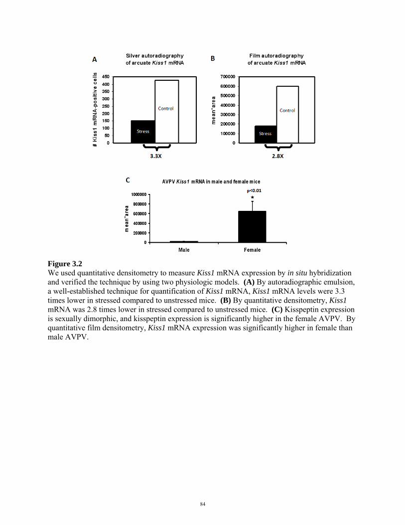
Figure 3.2 We used quantitative densitometry to measure Kiss1 mRNA expression by in situ hybridization and verified the technique by using two physiologic models. (A) By autoradiographic emulsion, a well-established technique for quantification of Kiss1 mRNA, Kiss1 mRNA levels were 3.3 times lower in stressed compared to unstressed mice. (B) By quantitative densitometry, Kiss1 mRNA was 2.8 times lower in stressed compared to unstressed mice. (C) Kisspeptin expression is sexually dimorphic, and kisspeptin expression is significantly higher in the female AVPV. By quantitative film densitometry, Kiss1 mRNA expression was significantly higher in female than male AVPV.
84
Fertility assays
Testosterone was assyed at 6 weeks of age in males. Because of the large plasma volume
required for LH and estradiol assays required terminal experiments, phlebotomies were
conducted at 20 weeks. Vaginal lavages were obtained from adult females, slide mounted, and
Geimsa stained (Fisher 22122911) for estrous cycle analysis. At 10 weeks of age, each
experimental animal was paired 1:1 with a WT mate that had previously proven fertile.
Parturition was monitored daily, and pups were euthanized at P1-3 and genotyped to confirm
parentage.
Metabolic assays
Body weight was monitored from 5-18 weeks in males and 5-10 weeks in females. Because
mating assays were conducted at 10 weeks of age and females gained weight during pregnancy
thereafter, female body weight was not analyzed after 10 weeks. We also assayed body weight
in adrenalectomized mice. We performed bilateral adrenalectomy on leptin-deficient (OBADX)
and WT mice at 4 weeks of age, obtained plasma at 6 weeks of age, and sacrificed animals at 10
weeks of age. Dual-energy X-ray absorptiometry scans were performed on mice anesthetized by
isoflurane at 5 and 10 weeks, and body fat composition was analyzed by using Lunar PIXImus
software. Food intake was measured over a 24 h period at 5 and 10 weeks of age. Rebound
hyperphagia is a phenomenon whereby an animal eats more than typical after a bout of food
deprivation. In pilot studies, we measured baseline food intake in CRHOB and OB mice for 24
h, fasted animals for 24 h, then measured food intake for 24 h in the rebound period [250].
Animals were tested at 10 weeks of age.
85
In pilot studies, we examined energy metabolism by using the Comprehensive Laboratory
Animal Monitoring System (CLAMS), which monitors oxygen consumption, carbon dioxide
release, food and water consumption, and beam break activity. Monitoring occured in single-
animal metabolic cages, and mice were acclimated to single housing in home cages for 3 d, then
acclimated to metabolic cages for 2 d, and monitored subsequently for 48 h. Measurements were
binned into and averaged as 12 h light and 12 h dark periods. We assessed energy metabolism
by indirect calorimetry based on calculations of the respiratory exchange ratio (RER) for
carbohydrate versus fatty acid oxidation:
Carbohydrate oxidation
6 O2 + C6H12O6 → 6 CO2 + 6 H2O + 38 ATP
RER = VCO2/VO2 = 6 CO2/6 O2 = 1.0
Fatty acid oxidation
23 O2 + C16H32O2 → 16 CO2 + 16 H2O + 129 ATP
RER = VCO2/VO2 = 16CO2/12O2 = 0.7
Corticosterone treatment
From 6 – 8 weeks, male mice were administered 30 µg/mL corticosterone (Sigma C2505, St.
Louis, MO) in drinking water. After 1 week of corticosterone treatment, plasma was collected
by retroorbital phlebotomy at 7AM (lights on) in unanesthetized animals and analyzed for
corticosterone and triglyceride concentrations. Body weight was measured before and after
corticosterone treatment. Males treated with corticosterone were paired 1:1 with a females that
86

had previously proven fertile. After corticosterone treatment was terminated, animals were again
paired 1:1 with a WT mate. Parturition was monitored daily, and pups were euthanized at P1-3
and genotyped to confirm parentage.
Transgenic mice and breeding strategy
To study the role of GR in kisspeptin neurons in leptin deficiency, we generated
Kiss1CreBAC::GRflox/flox::LepOb/Ob mice. Generation and validation of Kiss1CreBAC::GRflox/flox mice
were described in Chapter 2. All animals used in experiments were on a C57BL/CBA mixed
background. Crosses from Kiss1CreBAC::GRflox/flox::LepOb/+ x GRflox/flox::LepOb/+ breeders yielded
Kiss1CreBAC::GRflox/flox::LepOb/Ob males and females at frequencies of 6.25% each. 6 male
Kiss1CreBAC::GRflox/flox::LepOb/Ob and 2 female Kiss1CreBAC::GRflox/flox::LepOb/Ob mice were
generated and studied.
To study the role of GR in the brain in leptin deficiency, we generated
BlbpCre::GRflox/flox::LepOb/Ob mice. BlbpCre mice, in which Cre recombinase expression is driven
by the brain lipid binding protein promoter, were generously provided by David Gutmann [251].
All animals used in experiments were on a C57BL background. Crosses from
BlbpCre::GRflox/flox::LepOb/+ x GRflox/flox::LepOb/+ breeders yielded BlbpCre::GRflox/flox::LepOb/Ob
males and females at frequencies of 6.25% each. 4 male BlbpCre::GRflox/flox::LepOb/Ob and 6
female BlbpCre::GRflox/flox::LepOb/Ob mice were generated and studied.
87
Statistical tests
One-way ANOVA with post hoc least significant difference (LSD) pairwise testing was
performed to compare adrenal cortex width, plasma corticosterone, plasma leptin, food intake,
body fat composition, testicular weight, plasma testosterone, corpora lutea number, uterine
width, plasma LH, and plasma estradiol. Linear mixed model analyses were performed to
compare changes in body weight over time by genotype. Two-tailed two-sample t-tests were
performed to compare plasma corticosterone in corticosterone-treated versus untreated mice.
Fisher’s exact test was performed to compare fertility in males, females, and corticosterone-
treated males.
Genotyping
Kiss1CreBAC and GRflox/flox PCR protocols were described in Chapter 2. CRHKO and OB
genotyping primers and polymerase chain reaction protocols are described:
CRHKO
Crh+/+ forward primer 5’ - GAG CTT ACA CAT TTC GTC C - 3'
Crh+/+ reverse primer 5’ - GCT CAG CAA GCT CAC AGC - 3'
Crh-/- reverse primer 5’ - ATC GCC TTC TTG ACG AGT T- 3'
96°C for 1’, (96°C for 30”, 62°C for 30”, 72°C for 1’30”) x 34, 72°C for 5’, 4°C
WT band is 450 bp, and CRHKO band is 600 bp.
88
OB
Ob forward primer 5’ - ACT GGT CTG AGG CAG GGA GCA - 3'
Ob reverse primer 5’ - TGT CCA AGA TGG ACC AGA CTC - 3'
94°C for 3’, (94°C for 30”, 62°C for 1”, 72°C for 45”) x 36, 72°C for 2’, 10°C
Amplicon is digested with the restriction enzyme Dde I overnight at 37°C.
WT band is 155 bp, and OB band is 100 bp and 55 bp.
89

3.4 RESULTS
To study the role of glucocorticoids in leptin deficiency, we generated CRH-, glucocorticoid-,
and leptin-deficient Crh-/- LepOb/Ob (CRHOB) mice for comparison to wild-type (WT), Crh-/-
(CRHKO), and LepOb/Ob (OB) controls. Compared to WT mice, the adrenal cortex was
hypertrophied in OB mice (Figure 3.3a, b), consistent with hypercortisolism in these mice
(Figure 3.3c). Adrenal cortex size and plasma corticosterone concentrations were comparable in
CRHKO and CRHOB mice and significantly decreased compared to OB and WT mice (Figure
3.3a-c). Plasma leptin was undetectable in OB and CRHOB and significantly decreased
compared to WT and CRHKO mice (Figure 3.3d).
90

Figure 3.3 Adrenal development, plasma glucocorticoids, and plasma leptin were evaluated in WT, CRHKO, OB, and CRHOB mice. (A) Midline adrenal sections of the adrenal gland were stained by H&E. Scale bar, 250 µm. (B) Adrenal cortex width was significantly decreased in CRHKO (p=0.02, LSD post hoc test) and CRHOB (p=0.03, LSD post hoc test) compared to WT mice (p=6.5x10⁻⁶, one-way ANOVA) and significantly increased in OB mice (p=1.9x10⁻⁴, LSD post hoc test). Adrenal cortex width was significantly decreased in CRHOB compared to OB mice (p=2.2x10⁻⁵, LSD post hoc test). (C) Glucocorticoid concentrations were significantly decreased in CRHKO (p=0.003, LSD post hoc test) and CRHOB (p=0.01, LSD post hoc test) compared to WT mice (p=3.0x10⁻⁵, one-way ANOVA) and significantly increased in OB mice (p=0.006, LSD post hoc test). Plasma corticosterone was significantly decreased in CRHOB compared to OB mice (p=8.5x10⁻⁵, LSD post hoc test). (D) Plasma leptin was undetectable in OB (p=7.0x10⁻⁶, LSD post hoc test) and CRHOB (p=3.0x10⁻⁵, LSD post hoc test) compared to WT animals (1.2x10⁻⁵, one-way ANOVA) and not different from one another (p=1.0, LSD post hoc test).
91

Figure 3.3, continued
92

Patients with Cushing’s disease, a condition characterized by hypercortisolemia, and those
treated with exogenous glucocorticoids experience weight gain, and we hypothesized that
elevated glucocorticoids in OB mice contribute to obesity. Body weight was significantly
decreased in CRHOB compared to OB males and females (Figure 3.4a, b). Body weight in
CRHOB males was comparable to that of adrenalectomized OB males (Figure 3.5).
Hyperphagia was reduced in CRHOB compared to OB males and females (Figure 3.6a, c).
Body fat composition was reduced in CRHOB compared to OB males and females (Figure 3.6b,
d, e). Body weight, food intake, and body fat composition were comparable in WT and CRHKO
animals, and CRHOB animals did not completely normalize to WT and CRHKO levels.
The weight loss observed in CRHOB animals was due, at least in part, to decreased food intake,
and in pilot studies, we examined whether basal metabolic rate, estimated by the respiratory
exchange ratio (RER), was normalized as well. Because sample sizes were small (n=2-3) and
not all genotypes were represented, we did not perform statistical analyses for these pilot studies.
Mice are nocturnally active, and during the night, preferentially oxidize carbohydrates as an
energy source (i.e., RER ≈ 1). During the day, mice shift towards fatty acid oxidation (i.e., RER
≈ 0.7). Control CRHKO animals exhibited this circadian change in energy utilization, but OB
animals did not (Figure 3.7a). In OB mice, energy utilization was skewed towards fatty acid
oxidation (RER ≈ 0.7) in the nocturnal activity period (Figure 3.7a), consistent with lower RER
observed in leptin receptor-deficient db/db animals during the dark period [252]. CRHOB mice
were more similar to CRHKO controls, oxidizing carbohydrates in the activity period and fatty
acids in the quiescent period (Figure 3.7a). Locomotor activity at night, assayed as the sum of
beam breaks in the x-, y-, and z-axes, was significantly lower in OB mice compared to in
93
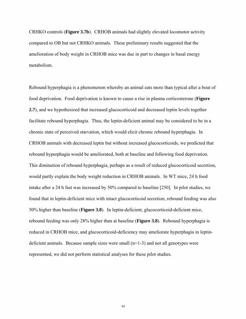
CRHKO controls (Figure 3.7b). CRHOB animals had slightly elevated locomotor activity
compared to OB but not CRHKO animals. These preliminary results suggested that the
amelioration of body weight in CRHOB mice was due in part to changes in basal energy
metabolism.
Rebound hyperphagia is a phenomenon whereby an animal eats more than typical after a bout of
food deprivation. Food deprivation is known to cause a rise in plasma corticosterone (Figure
2.7), and we hypothesized that increased glucocorticoid and decreased leptin levels together
facilitate rebound hyperphagia. Thus, the leptin-deficient animal may be considered to be in a
chronic state of perceived starvation, which would elicit chronic rebound hyperphagia. In
CRHOB animals with decreased leptin but without increased glucocorticoids, we predicted that
rebound hyperphagia would be ameliorated, both at baseline and following food deprivation.
This diminution of rebound hyperphagia, perhaps as a result of reduced glucocorticoid secretion,
would partly explain the body weight reduction in CRHOB animals. In WT mice, 24 h food
intake after a 24 h fast was increased by 50% compared to baseline [250]. In pilot studies, we
found that in leptin-deficient mice with intact glucocorticoid secretion, rebound feeding was also
50% higher than baseline (Figure 3.8). In leptin-deficient, glucocorticoid-deficient mice,
rebound feeding was only 28% higher than at baseline (Figure 3.8). Rebound hyperphagia is
reduced in CRHOB mice, and glucocorticoid-deficiency may ameliorate hyperphagia in leptin-
deficient animals. Because sample sizes were small (n=1-3) and not all genotypes were
represented, we did not perform statistical analyses for these pilot studies.
94
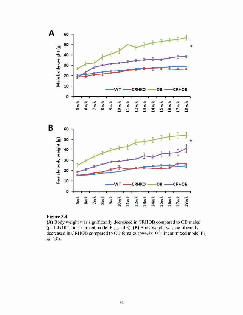
Figure 3.4 (A) Body weight was significantly decreased in CRHOB compared to OB males (p=1.4x10⁻⁵, linear mixed model F13, 94=4.3). (B) Body weight was significantly decreased in CRHOB compared to OB females (p=4.8x10⁻⁴, linear mixed model F5,
80=5.0).
95
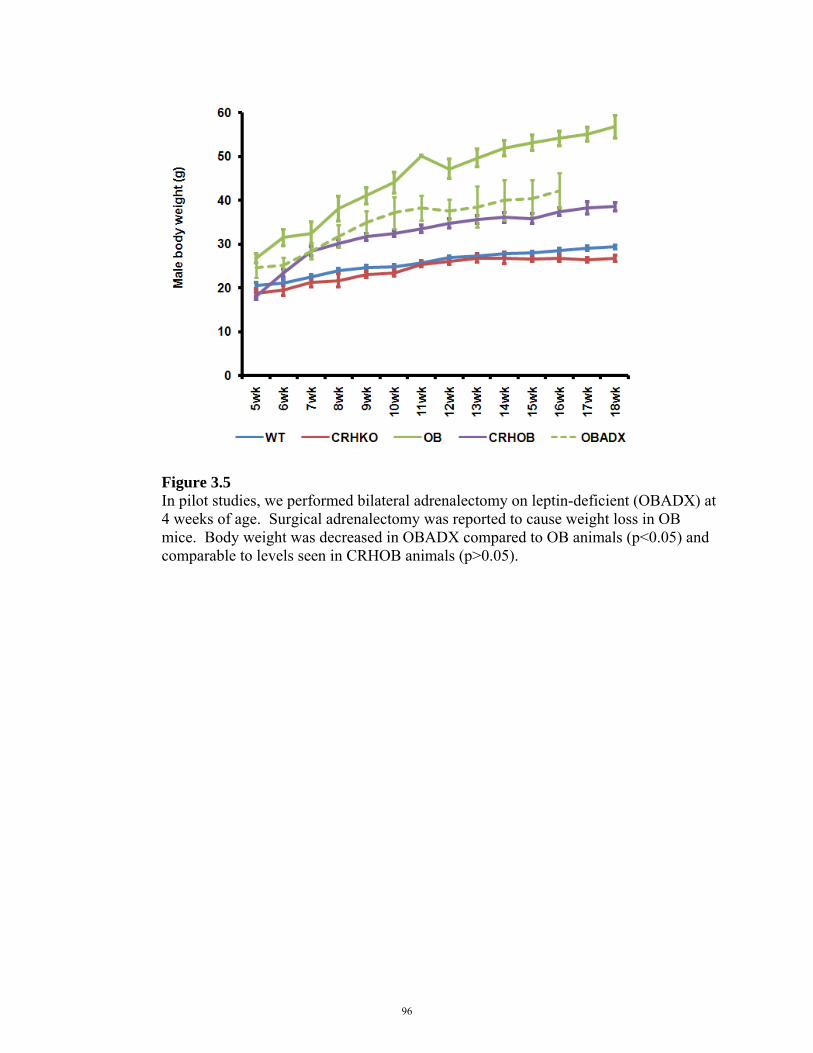
Figure 3.5 In pilot studies, we performed bilateral adrenalectomy on leptin-deficient (OBADX) at 4 weeks of age. Surgical adrenalectomy was reported to cause weight loss in OB mice. Body weight was decreased in OBADX compared to OB animals (p<0.05) and comparable to levels seen in CRHOB animals (p>0.05).
96

Figure 3.6 (A) Hyperphagia was reduced in CRHOB compared to OB males (p=4.9x10⁻⁴, LSD post hoc test). (B) Body fat composition was reduced in CRHOB compared to OB males (p=1.7x10⁻¹¹, LSD post hoc test). (C) Hyperphagia was reduced in CRHOB compared to OB females (p=0.002, LSD post hoc test). (D) Body fat composition was reduced in CRHOB compared to OB females (p=1.7x10⁻¹³, LSD post hoc test). (E) Representative DEXA body scans.
97

Figure 3.7 In pilot studies, CRHOB mice were monitored in metabolic cages. (A) CRHKO control animals oxidized carbohydrates during the night (RER ≈ 1) and fatty acids during the day (RER ≈ 0.7), but OB animals did not exhibit this circadian change in energy utilization. Energy utilization was skewed towards fatty acid oxidation during the nocturnal activity period in OB mice. CRHOB were more similar to CRHKO controls, oxidizing carbohydrates in the activity period and fatty acids in the quiescent period. (B) Locomotor activity at night, assayed as the sum of beam breaks in the x-, y-, and z-axes, was significantly lower in OB mice compared to in CRHKO controls. CRHOB animals had slightly elevated locomotor activity compared to OB but not CRHKO animals. Because sample sizes were small (n=2-3) and not all genotypes were represented, we did not perform statistical analyses for these pilot studies.
98
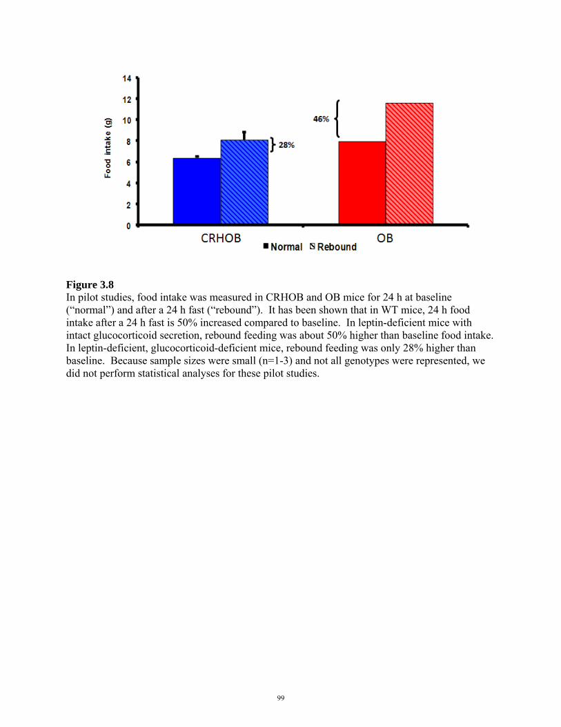
Figure 3.8 In pilot studies, food intake was measured in CRHOB and OB mice for 24 h at baseline (“normal”) and after a 24 h fast (“rebound”). It has been shown that in WT mice, 24 h food intake after a 24 h fast is 50% increased compared to baseline. In leptin-deficient mice with intact glucocorticoid secretion, rebound feeding was about 50% higher than baseline food intake. In leptin-deficient, glucocorticoid-deficient mice, rebound feeding was only 28% higher than baseline. Because sample sizes were small (n=1-3) and not all genotypes were represented, we did not perform statistical analyses for these pilot studies.
99
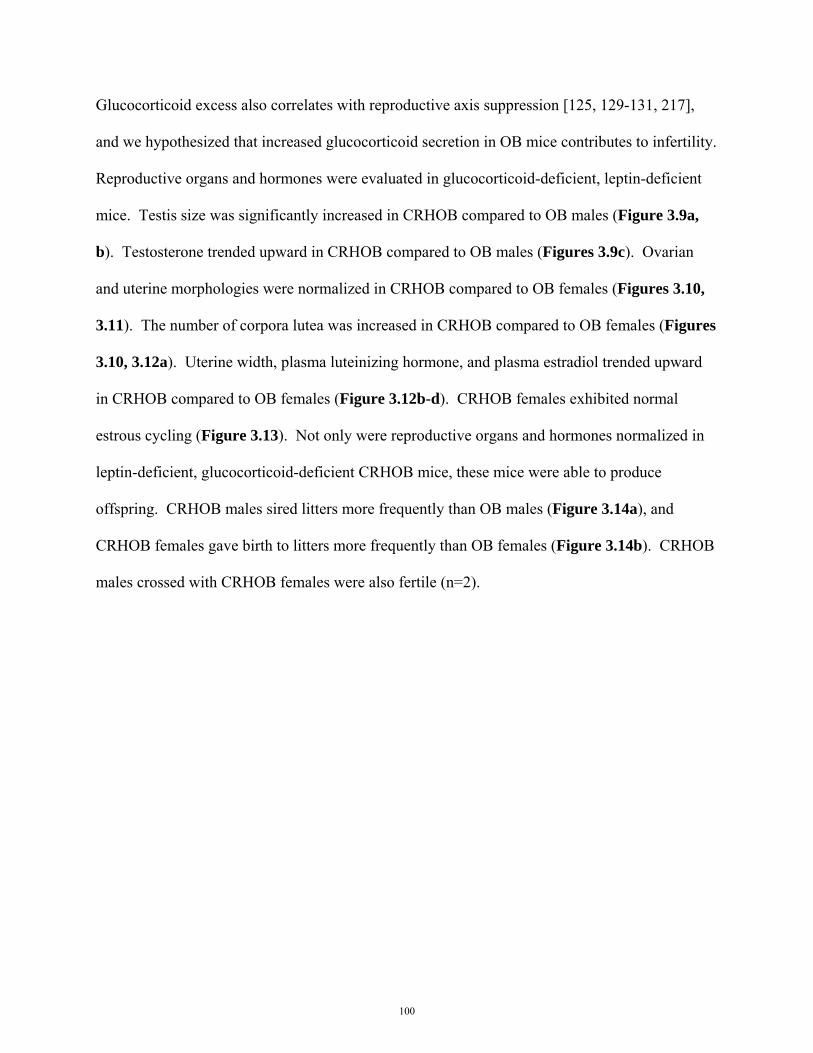
Glucocorticoid excess also correlates with reproductive axis suppression [125, 129-131, 217],
and we hypothesized that increased glucocorticoid secretion in OB mice contributes to infertility.
Reproductive organs and hormones were evaluated in glucocorticoid-deficient, leptin-deficient
mice. Testis size was significantly increased in CRHOB compared to OB males (Figure 3.9a,
b). Testosterone trended upward in CRHOB compared to OB males (Figures 3.9c). Ovarian
and uterine morphologies were normalized in CRHOB compared to OB females (Figures 3.10,
3.11). The number of corpora lutea was increased in CRHOB compared to OB females (Figures
3.10, 3.12a). Uterine width, plasma luteinizing hormone, and plasma estradiol trended upward
in CRHOB compared to OB females (Figure 3.12b-d). CRHOB females exhibited normal
estrous cycling (Figure 3.13). Not only were reproductive organs and hormones normalized in
leptin-deficient, glucocorticoid-deficient CRHOB mice, these mice were able to produce
offspring. CRHOB males sired litters more frequently than OB males (Figure 3.14a), and
CRHOB females gave birth to litters more frequently than OB females (Figure 3.14b). CRHOB
males crossed with CRHOB females were also fertile (n=2).
100
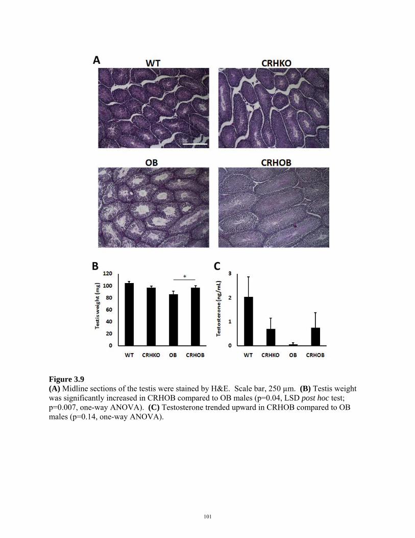
Figure 3.9 (A) Midline sections of the testis were stained by H&E. Scale bar, 250 µm. (B) Testis weight was significantly increased in CRHOB compared to OB males (p=0.04, LSD post hoc test; p=0.007, one-way ANOVA). (C) Testosterone trended upward in CRHOB compared to OB males (p=0.14, one-way ANOVA).
101
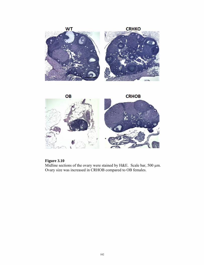
Figure 3.10 Midline sections of the ovary were stained by H&E. Scale bar, 500 µm. Ovary size was increased in CRHOB compared to OB females.
102
Figure 3.11 Midline sections of the uterus were stained by H&E. Scale bar, 500 µm. Uterus width was increased in CRHOB females compared to the thread-like uterus in OB females.
103
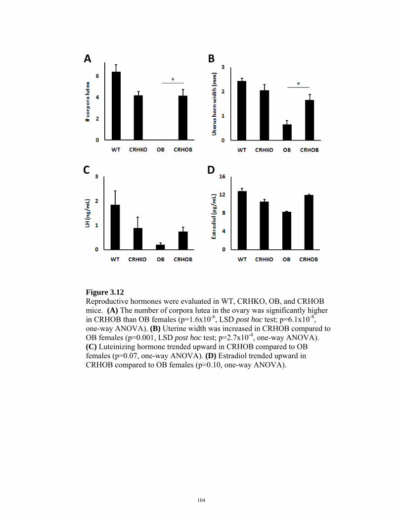
Figure 3.12 Reproductive hormones were evaluated in WT, CRHKO, OB, and CRHOB mice. (A) The number of corpora lutea in the ovary was significantly higher in CRHOB than OB females (p=1.6x10⁻⁶, LSD post hoc test; p=6.1x10⁻⁸, one-way ANOVA). (B) Uterine width was increased in CRHOB compared to OB females (p=0.001, LSD post hoc test; p=2.7x10⁻⁴, one-way ANOVA). (C) Luteinizing hormone trended upward in CRHOB compared to OB females (p=0.07, one-way ANOVA). (D) Estradiol trended upward in CRHOB compared to OB females (p=0.10, one-way ANOVA).
104
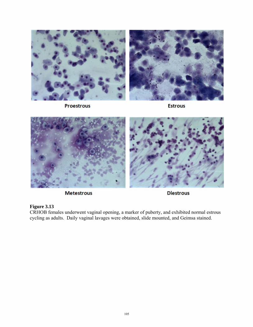
Figure 3.13 CRHOB females underwent vaginal opening, a marker of puberty, and exhibited normal estrous cycling as adults. Daily vaginal lavages were obtained, slide mounted, and Geimsa stained.
105
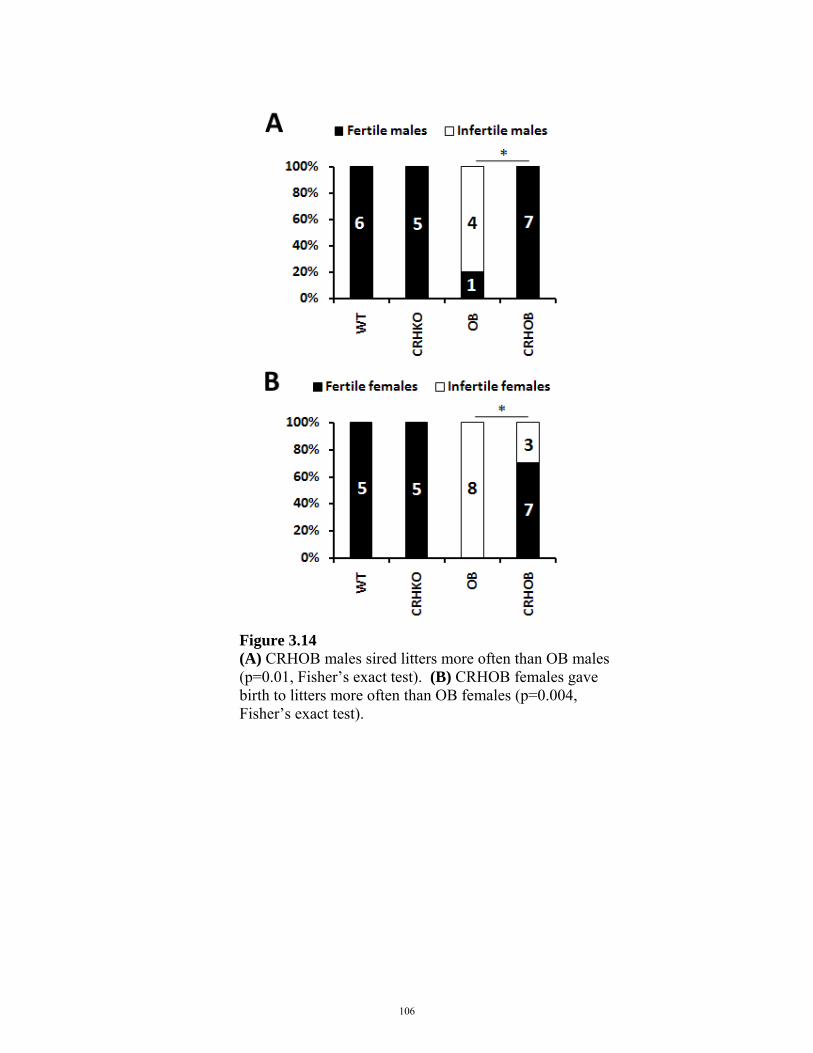
Figure 3.14 (A) CRHOB males sired litters more often than OB males (p=0.01, Fisher’s exact test). (B) CRHOB females gave birth to litters more often than OB females (p=0.004, Fisher’s exact test).
106
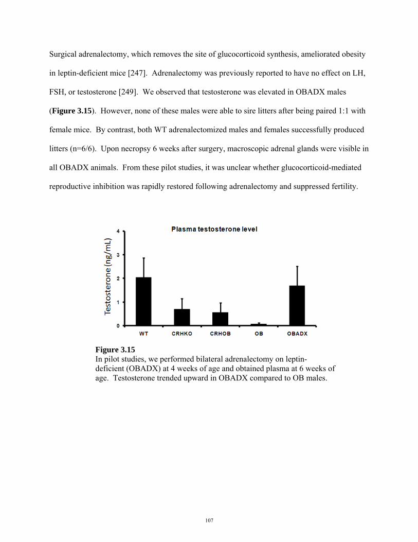
Surgical adrenalectomy, which removes the site of glucocorticoid synthesis, ameliorated obesity
in leptin-deficient mice [247]. Adrenalectomy was previously reported to have no effect on LH,
FSH, or testosterone [249]. We observed that testosterone was elevated in OBADX males
(Figure 3.15). However, none of these males were able to sire litters after being paired 1:1 with
female mice. By contrast, both WT adrenalectomized males and females successfully produced
litters (n=6/6). Upon necropsy 6 weeks after surgery, macroscopic adrenal glands were visible in
all OBADX animals. From these pilot studies, it was unclear whether glucocorticoid-mediated
reproductive inhibition was rapidly restored following adrenalectomy and suppressed fertility.
Figure 3.15 In pilot studies, we performed bilateral adrenalectomy on leptin-deficient (OBADX) at 4 weeks of age and obtained plasma at 6 weeks of age. Testosterone trended upward in OBADX compared to OB males.
107
Because CRHOB mice are CRH- and glucocorticoid-deficient, it is possible that CRH
contributes to infertility [132] in leptin deficiency. However, in situ hybridization for Crh
mRNA expression in the paraventricular nucleus (PVN) of the hypothalamus showed no
differences in Crh expression between WT and OB mice (Figure 3.16a). To examine whether
changes in fertility were glucocorticoid-dependent, we next treated mice with 30 µg/mL of
corticosterone in drinking water, which produced physiologic concentrations of plasma
corticosterone in WT, CRHKO, OB, and CRHOB males (Figure 3.16b). Compared to untreated
controls, plasma corticosterone was elevated only in corticosterone-treated CRHKO and
CRHOB mice (Figure 3.16b). After 1 week of corticosterone supplementation, CRHOB males
were unable to sire litters and became as infertile as OB males (Figure 3.16c). Following
termination of corticosterone treatment, CRHOB males were fertile (Figure 3.16d).
108

Figure 3.16 (A) In situ hybridization for Crh mRNA in the paraventricular nucleus (PVN) showed no differences between WT and OB mice (p>0.05; data courtesy of Satoru Sakihara). Insets, in situ hybridization for Crh mRNA in the PVN by autoradiographic film. (B) Male mice were supplemented with 30 µg/mL of corticosterone in drinking water. Compared to untreated controls, plasma corticosterone was elevated only in corticosterone-treated CRHKO (p=0.04, independent samples t-test) and CRHOB (p=0.008, independent samples t-test) mice. (C) After 1 week of supplementation with corticosterone, CRHOB males were unable to sire litters. Corticosterone-treated CRHOB males were as infertile as OB males (p=0.99, Fisher’s exact test). (D) Following withdrawal of corticosterone, CRHOB males were once again able to sire litters compared to OB males (p=0.008, Fisher’s exact test).
109

Because CRHOB mice are CRH- and glucocorticoid-deficient, it may be possible that CRH
contributes to obesity in leptin deficient states. This is unlikely, however, because CRH is a
known anorectic [248, 253, 254], and thus, CRH deficiency should promote orexigenic effects.
The effect of corticosterone on body weight was evaluated in WT, CRHKO, OB, and CRHOB
mice. Male mice were supplemented with 30 µg/mL of corticosterone in drinking water. Body
weight was unchanged in corticosterone-treated WT, CRHKO, and OB males compared to
untreated controls. Body weight was increased in corticosterone-treated compared to untreated
CRHOB males (Figure 3.17). This increase in body weight persisted even after termination of
corticosterone treatment (Figure 3.17). Plasma triglycerides were unchanged in corticosterone-
treated WT and OB males compared to untreated controls (Figure 3.18). Plasma triglycerides
were slightly but significantly elevated in corticosterone-treated CRHKO males (Figure 3.18).
Plasma triglycerides increased approximately three-fold after corticosterone treatment in
CRHOB males (Figure 3.18).
110
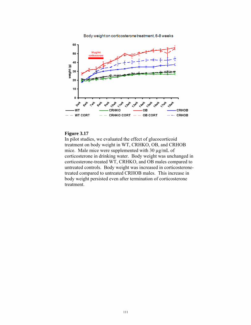
Figure 3.17 In pilot studies, we evaluated the effect of glucocorticoid treatment on body weight in WT, CRHKO, OB, and CRHOB mice. Male mice were supplemented with 30 µg/mL of corticosterone in drinking water. Body weight was unchanged in corticosterone-treated WT, CRHKO, and OB males compared to untreated controls. Body weight was increased in corticosterone-treated compared to untreated CRHOB males. This increase in body weight persisted even after termination of corticosterone treatment.
111

Figure 3.18 The effect of glucocorticoids on plasma triglycerides was evaluated in WT, CRHKO, OB, and CRHOB mice. Male mice were supplemented with 30 µg/mL of corticosterone in drinking water. Plasma triglycerides were unchanged in corticosterone-treated WT and OB males compared to untreated controls. Plasma triglycerides were slightly but significantly elevated in corticosterone-treated CRHKO males. Plasma triglycerides increased approximately three-fold after 1 week of corticosterone treatment in CRHOB males. * p<0.05.
112
To evaluate the role of kisspeptin neurons in the infertile and obese LepOb/Ob mouse, we first
measured Kiss1 mRNA expression in WT, CRHKO, OB, and CRHOB males. Kiss1 mRNA
expression trended downward in OB and CRHOB males compared to WT and CRHKO controls
(Figure 3.19a). We generated leptin-deficient mice in which GR signaling was selective deleted
from kisspeptin neurons. Thread-like uteri were observed in Kiss1CreBAC::GRflox/flox::LepOb/Ob
(Kiss GR OB) females, and neither males nor females were fertile (Figure 3.19b). Body weights
(Figure 3.19c) and food intake (Figure 3.19d) in Kiss1CreBAC::GRflox/flox::LepOb/Ob mice were
comparable to those of LepOb/Ob controls.
113

Figure 3.19 In pilot studies, we examined the role of kisspeptin neurons in mediating fertility in CRHOB animals. (A) Kiss1 mRNA expression trended downward in OB and CRHOB males compared to WT and CRHKO controls (n=5-6 per genotype). (B) We generated Kiss1CreBAC::GRflox/flox::LepOb/Ob (Kiss GR OB) animals, in which GR was excised from kisspeptin neurons in leptin-deficient animals. Thread-like uteri were observed in Kiss GR OB females (n=2), and neither males nor females were fertile. (C) Body weight and (D) food intake in Kiss1CreBAC::GRflox/flox::LepOb/Ob male mice (n=6) were comparable to those of LepOb/Ob controls (n=5).
114
In studies of stress-induced reproductive inhibition in WT mice, we demonstrated that
suppression of kisspeptin expression was not necessary for reproductive inhibition in cold stress
(Chapter 2). Conversely, maintaining Kiss1 mRNA expression at normal levels during stress in
Kiss1CreBAC::GRflox/flox mice did not protect animals from stress-induced suppression of
testosterone and copulatory behaviors immediately after acute stress. GR signaling in kisspeptin
neurons is unlikely to be the sole inhibitory signal to the HPG axis, as there are multiple
glucocorticoid-dependent and -independent stress pathways that inhibit the reproductive axis
acutely. For example, catecholamines can mediate direct testicular inhibition [235, 255], which
is independent of both glucocorticoid and kisspeptin signaling. Such inhibitory inputs may
suppress reproductive function in Kiss1CreBAC::GRflox/flox::LepOb/Ob mice despite the lack of GR-
mediated inhibition of kisspeptin neurons. It may be interesting to assess whether Kiss1 mRNA
expression is affected in Kiss1CreBAC::GRflox/flox::LepOb/Ob mice, as we would predict that Kiss1
mRNA expression remains unsuppressed in this model compared to LepOb/Ob controls.
Kiss1CreBAC::GRflox/flox::LepOb/Ob males and females were infertile and obese, suggesting that
glucocorticoid receptor signaling in kisspeptin neurons did not regulate fertility or body weight
in LepOb/Ob mice. Since deletion of GR in kisspeptin neurons did not restore fertility in Lepob/ob
mice, we examined whether neuronal glucocorticoid receptor signaling affected fertility or
obesity. We did not use NestinCre for Cre-mediated excision of GR in the brain, because
NestinCre::GRflox/flox animals have decreased body weight [24] and would confound the
interpretation of any body weight phenotype in a LepOb/Ob background. Instead, we obtained
transgenic BlbpCreBAC mice [251], in which the brain lipid binding protein promoter drives Cre
expression in neural stem cells. We aimed to determine whether BlbpCreBAC would be a suitable
115
brain-specific driver that did not produce body weight changes in controls. Unlike
NestinCre::GRflox/flox mice, neither BlbpCreBAC nor BlbpCreBAC::GRflox/flox animals had altered body
weight compared to WT littermates. We proceeded to generate BlbpCreBAC::GRflox/flox::LepOb/Ob
animals, but body weight, food intake, and fertility were not normalized in these animals
(Figures 3.20, 3.21). Because not all neurons were targeted by BlbpCreBAC, however, it is unclear
whether this reflects non-central mechanisms of glucocorticoid receptor signaling in fertility and
obesity, or insufficient excision of GR in the brain by BlbpCreBAC.
116

Figure 3.20 In pilot studies, we generated BlbpCreBAC::GRflox/flox::LepOb/Ob females (n=6), in which the brain lipid binding protein promoter drives Cre expression in neural stem cells and induces Cre-mediated GR excision in a leptin-deficient background. In females, (A) body weight at 5 weeks, (B) body weight at 10 weeks, and (C) food intake were comparable to those of LepOb/Ob animals. ★p<0.05.
117
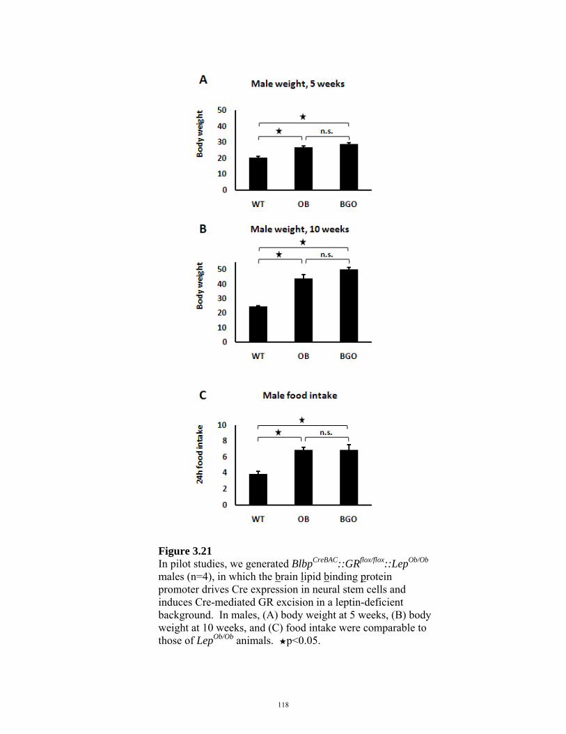
Figure 3.21 In pilot studies, we generated BlbpCreBAC::GRflox/flox::LepOb/Ob males (n=4), in which the brain lipid binding protein promoter drives Cre expression in neural stem cells and induces Cre-mediated GR excision in a leptin-deficient background. In males, (A) body weight at 5 weeks, (B) body weight at 10 weeks, and (C) food intake were comparable to those of LepOb/Ob animals. ★p<0.05.
118
3.5 DISCUSSION
Leptin is a potent satiety hormone, and leptin deficiency creates a chronic state of perceived
starvation. Starvation causes increased glucocorticoid secretion [246], and glucocorticoids can
inhibit reproduction through direct and indirect mechanisms that are leptin-independent [125,
129-131, 256]. Excess glucocorticoids due to glucocorticoid therapy [217] and disease (e.g.,
Cushing’s, anorexia nervosa) correlate with suppressed fertility [132, 189]. Hypercortisolemia in
anorexia nervosa and Cushing’s disease are both associated with reproductive inhibition, despite
dramatic differences in body weight and leptin levels in these disorders.
We hypothesized that elevated glucocorticoid secretion, rather than leptin deficiency per se,
caused infertility in leptin-deficient mice. To assess the role of glucocorticoids in fertility, we
generated leptin-, CRH-, and glucocorticoid-deficient CRHOB mice. Unlike OB mice, CRHOB
mice were fertile and had normalized reproductive organs and hormones. Body weight, fat
composition, and food intake were also decreased in CRHOB compared to OB mice. In pilot
studies, basal metabolic rate, but not locomotor activity, was normalized in CRHOB animals. In
pilot studies, rebound hyperphagia was decreased in CRHOB compared to OB animals. We
hypothesized that low leptin and high corticosterone together signal starvation and stimulate
rebound hyperphagia. Thus, the OB animal is in a chronic state of perceived starvation and
rebound hyperphagia, leading to its morbid obesity. In CRHOB mice that lack excess
glucocorticoids, this hyperphagia is ameliorated, and animals are significantly leaner than OB
littermates. Body weight in CRHOB animals was comparable to that of adrenalectomized OB
animals. In pilot studies, we found that adrenalectomized OB males were unable to sire
offspring. Within two weeks of adrenalectomy, macroscopic adrenal glands could be identified
119

in these animals; it is possible that rapid regeneration of adrenal tissue prevented further
activation of the reproductive axis, and the CRHOB model allowed for more complete and
permanent removal of glucocorticoid secretion.
CRH is normally inhibited by glucocorticoids and disinhibited by adrenalectomy or
glucocorticoid deficiency. In studying the role of glucocorticoids in fertility, we aimed to avoid
confounding the decrease in glucocorticoids with the increase in CRH. However, by eliminating
both CRH and glucocorticoids in CRHOB animals, it was possible that CRH deficiency, rather
than glucocorticoid deficiency, restored fertility in leptin-deficient mice. There is evidence that
CRH can inhibit reproductive axis hormones [132, 133]. However, even significantly increased
CRH, caused by primary adrenal insufficiency, produces only prolonged estrous cycling but not
infertility [257]. Additionally, we demonstrated that Crh mRNA expression was not elevated in
OB compared to WT animals. We demonstrated that corticosterone supplementation was
sufficient to elicit infertility in CRHOB mice that was comparable to that of OB controls. When
corticosterone treatment was terminated, CRHOB mice regained reproductive capacity. This
indicates that glucocorticoids and not CRH, perhaps in conjunction with low leptin, are
responsible for inhibition of fertility in OB mice. Corticosterone supplementation also caused
weight gain and increased plasma triglyceride concentrations only in CRHOB mice.
The cellular sites and mechanisms for leptin and glucocorticoid interaction are still unclear. In
pilot studies, we deleted glucocorticoid receptors from the brain, using a novel brain-specific Cre
targeting strategy, and from kisspeptin neurons in the background of leptin-deficiency.
Abrogating GR signaling in kisspeptin neurons did not restore fertility or decrease body weight
120
in leptin-deficient animals. GR was not sufficiently deleted from all neurons in the brain, and
these animals were infertile and obese. It remains unclear whether the interaction between leptin
and glucocorticoids occurs in the brain, and if so, which populations of neurons mediate this
effect.
Leptin deficiency on a C57BL/6J background produces more severe infertility than leptin
deficiency on a BALB/cJ background [184]. Under normal conditions, it is possible that
physiologic increases in glucocorticoids and CRH and decreases in leptin together contribute to
the suppression of reproductive function. In humans with leptin deficiency, plasma
glucocorticoid concentrations are not strikingly elevated, and it is possible that glucocorticoids
play a less important role in reproductive inhibition in humans. In our model, mice with global
leptin deficiency in conjunction with global glucocorticoid deficiency were fertile. Eliminating
hypercortisolism in leptin-deficient mice was sufficient to restore fertility, and leptin was not
required for fertility. Thus, glucocorticoids suppress fertility and contribute to obesity in the
presence of leptin deficiency. These findings may provide novel molecular targets in the
reproductive and metabolic aspects of disorders characterized by glucocorticoid dysregulation,
including leptin deficiency, Cushing’s disease, anorexia nervosa, and mood disorders.
121

CHAPTER 4
CONCLUSIONS
122
Summary
Glucocorticoids are an important output of the hypothalamic-pituitary-adrenal axis, and classical
actions of glucocorticoids are exerted through glucocorticoid receptors expressed throughout the
body and brain. Stress responses, including increased glucocorticoid secretion, are adaptive and
facilitate the return to homeostasis. Adaptations include the suppression of non-essential
activities, such as reproduction, growth, and immune reactions.
Chronic or hyperactive stress responses, however, are associated with multiple pathological
conditions, including Cushing’s disease, post-traumatic stress disorder, anorexia nervosa, major
depression, and other affective disorders. Reproductive function is negatively affected in these
hypercortisolemic conditions, and glucocorticoid treatment is known to suppress the
hypothalamic-pituitary-gonadal axis as well. The cellular substrates and mechanisms
responsible for inhibiting and reactivating the reproductive axis during and after stress,
respectively, are not well understood.
In Chapter 2, we demonstrated that kisspeptin expression in the arcuate nucleus of the
hypothalamus was sensitive to stress-induced changes in glucocorticoids. We examined changes
in kisspeptin and the reproductive axis in mice subjected to restraint, food deprivation, cold
exposure, and corticosterone administration. In all cases, there was an inverse relationship
between plasma corticosterone concentrations and arcuate nucleus kisspeptin expression.
Restraint and food deprivation caused increases in plasma corticosterone concentrations and
decreases in Kiss1 mRNA expression. Because these stressors likely elicited stress responses in
addition to elevated glucocorticoid secretion, we treated mice with corticosterone to study the
123

isolated effects of glucocorticoids on kisspeptin neurons. Kiss1 mRNA expression was
decreased in mice treated with corticosterone.
Genetic manipulation of kisspeptin neurons
We next generated mice lacking glucocorticoid receptors specifically in kisspeptin-containing
neurons. In these animals, kisspeptin expression was no longer inhibited by restraint stress, and
both testosterone and copulatory behaviors recovered faster following restraint. However, male
mice lacking glucocorticoid receptors in kisspeptin neurons still exhibited suppression of the
reproductive axis immediately after restraint, consistent with the reproductive inhibition
observed in male mice following cold exposure despite unsuppressed levels of corticosterone and
Kiss1 mRNA, indicating that other glucocorticoid-dependent and -independent pathways can
suppress the reproductive axis. Thus, we conclude that glucocorticoid receptor signaling in
kisspeptin neurons regulates the recovery of reproductive function following stress, but not the
inhibition of the reproductive axis during stress.
In the studies described above, we analyzed Kiss1 mRNA expression as a measure of kisspeptin
neuron output. Electrical activity, peptide release, and other important synaptic events cannot be
captured by measurements of mRNA expression. Changes in gene expression also occur on a
slower time scale than changes in electrical activity. Using optogenetic techniques, we could
probe the immediate effects of manipulating kisspeptin neurons. Channelrhodopsin (ChR) and
halorhodopsin (HR) are microbial ion channels [258]. Channelrhodopsin is activated by blue
light and conducts cations, while halorhodopsin is activated by yellow light and conducts
chloride anions. Neurons that express ChR are depolarized upon stimulation with blue light,
124

while neurons that express HR are hyperpolarized and inhibited by yellow light [259]. Delivery
of these opsins can be mediated by lentiviral and adeno-associated viral vectors, in which ChR or
HR is flanked by loxP sites, allowing for temporal and site-specific control of Cre-mediated
recombination and expression [258]. Because these experiments require the implantation of site-
specific fiberoptic cables, ventral sites of the brain were relatively inaccessible for many years.
Recently, however, optogenic targeting of the ventromedial nucleus of the hypothalamus [260]
and orexin neurons of the lateral hypothalamus [261] successfully demonstrated that optogenetic
control of ventral brain structures is possible.
Our current findings indicate that kisspeptin neurons govern the time course of recovery from
stress-induced reproductive inhibition. We hypothesize that kisspeptin neurons must be
reactivated following glucocorticoid-dependent inhibition to turn on the downstream
hypothalamic-pituitary-reproductive axis. In one potential experiment, we would induce Cre-
mediated expression of HR (e.g., eNpHR-eYFP) [262] in kisspeptin neurons in adult Kiss1Cre
male mice. We would subject these animals to restraint stress, and following release, deliver
yellow light to hyperpolarize the kisspeptin neurons. We predict that the inability of kisspeptin
neurons to reactivate would prevent the restoration of testosterone and copulatory behaviors.
With inhibition of kisspeptin neurons, we would expect copulatory behaviors to remain
suppressed for longer than the one week time period described in Chapter 2. Using the same
optogenetic model, we could induce hyperpolarization of kisspeptin neurons in the absence of an
external stressor to determine whether inhibition of kisspeptin neurons alone is sufficient to
suppress reproductive function. Lastly, we would hyperpolarize kisspeptin neurons in
prepubertal Kiss1Cre mice to determine whether pubertal onset can be delayed, or whether
125
redundant, kisspeptin-independent mechanisms can elicit puberty. If inhibition of kisspeptin
neurons is sufficient to delay puberty, we would then induce acute halorhodopsin-mediated
inhibition of these neurons at different prepubertal time points to determine the time course of,
and perhaps a critical period for, kisspeptin neuron-dependent regulation of puberty.
Our current findings also suggest that kisspeptin neurons do not wholly mediate the acute
suppression of reproductive function during stress. Mice subjected to cold exposure did not
exhibit an increase in corticosterone or decrease in Kiss1 mRNA expression, but testosterone was
still suppressed. Male mice in which glucocorticoid receptors were deleted from kisspeptin
neurons no longer exhibited a decrease in Kiss1 mRNA expression after restraint stress, but
testosterone and copulatory behaviors were still suppressed immediately after the restraint. We
predict that preventing the suppression of kisspeptin neurons during acute stress would not
preserve testosterone secretion or copulatory behaviors. Stress levels of glucocorticoids, for
example, can exert direct effects on the testis and induce apoptosis of Leydig cells [129, 130]. If
one generated an animal in which kisspeptin neurons were constitutively active, for example, we
would predict that testosterone and copulatory behaviors would still be suppressed following
stress. As a caveat, constitutive activity is not an ideal model, because kisspeptin is likely
secreted in a pulsatile manner, much like GnRH, and continuous activation is more likely to
suppress than activate the reproductive axis [263]. By using ChR, we could more precisely
control the activity of kisspeptin neurons. We hypothesize that 1) kisspeptin activity does not
wholly mediate the inhibition of downstream reproductive function during acute stress, and 2)
kisspeptin neuron reactivation governs the time course of recovery from stress-induced
reproductive inhibition. In potential experiments, we would induce Cre-mediated expression of
126
ChR (e.g., ChR2-eYFP) [262] in kisspeptin neurons in adult Kiss1Cre male mice. This would
require preliminary studies to determine the appropriate pulse frequency of blue light in order to
mimic physiologic kisspeptin pulsatile release, itself a parameter that has never been
characterized. We would subject these animals to restraint and deliver the appropriate frequency
of blue light to depolarize the kisspeptin neurons throughout the stressor. We predict that
testosterone and copulatory behaviors would still be inhibited following stress, despite the
continued activation of kisspeptin neurons, through inhibitory pathways at sites other than the
kisspeptin neuron (e.g., direct glucocorticoid-mediated inhibition testosterone biosynthesis [129,
130]). Following restraint, however, we would predict that testosterone and copulatory
behaviors would recover faster in mice that express ChR2-eYFP and receive blue light
stimulation than in eYFP controls. The accelerated recovery may be comparable to the one week
time period observed in Kiss1CreBAC::GRflox/flox males. In Kiss1CreBAC::GRflox/flox males, however,
we had no control over the time course of kisspeptin neuron reactivation. By continuing to
induce optogenetic activation of kisspeptin neurons following restraint, we may be able to restore
reproductive function even faster than one week following stress. Using this model, we could
also depolarize kisspeptin neurons in peripubertal mice to determine whether early activation of
these neurons is sufficient to elicit advanced puberty.
These optogenetic experiments would allow us to isolate the time course of activation and
suppression of the reproductive axis, both in juveniles during puberty and in adults during stress-
induced reproductive inhibition.
127

Pharmacologic manipulation of kisspeptin neurons
Our studies may lead to the study of drugs that have not been previously considered for the
treatment of stress- and glucocorticoid-induced reproductive inhibition. We would administer
mifepristone (RU486) to male mice in order to evaluate the efficacy of possible pharmacological
candidates to attenuate the inhibitory reproductive effects of stress on the hypothalamic-
pituitary-gonadal (HPG) axis. Mifespristone is a non-specific glucocorticoid and progesterone
antagonist that, through it action on progesterone receptors, is commonly known as the
“morning-after pill” due to its use as an abortifactant in females [264-269]. Mifespristone may
also be an attractive drug candidate to study the reversal of glucocorticoid-induced suppression
of the reproductive axis in males. We would predict that preventing glucocorticoid-mediated
suppression of kisspeptin neurons would not prevent reproductive axis suppression acutely, but
that the reproductive axis would recover relatively quickly following the termination of stress.
This may also be achieved by manipulating kisspeptin secretion directly. Kisspeptin-10 is the
active decapeptide fragment of endogenous kisspeptin-54 and binds to the kisspeptin receptor
with similar affinity but increased bioactivity [270, 271]. We would administer kisspeptin-10 to
mice subjected to stress-induced reproductive inhibition and determine whether pre- or post-
stress treatment with kisspeptin can prevent prolonged suppression of the reproductive axis.
These drugs may demonstrate novel utility in the treatment of the reproductive aspects of post-
traumatic stress disorder, mood disorders, and anorexia nervosa.
Kisspeptin neurons contribute to reproductive behaviors
We report that kisspeptin neurons can be protected from stress by the removal of glucocorticoid
receptor signaling, and that doing so has the ability to orchestrate a repertoire of complex
128
behaviors leading to more rapid return of reproductive function, possibly by the downstream
regulation of sex steroid secretion. If the kisspeptin neuron-dependent recovery of the
reproductive axis in males is mediated by changes in testosterone, we would expect multiple
testosterone-dependent behaviors to be affected. Aggression, for example, is a sexually
dimorphic behavior widely considered to be mediated by testosterone in mammals [272]. If
glucocorticoid receptor signaling in kisspeptin governs the time course of recovery of
testosterone, which in turn governs the display of copulatory behaviors, then we would expect
aggression to be altered in male mice in which glucocorticoid receptors are selectively deleted
from kisspeptin neurons following stress. To test this hypothesis, we would subject control and
Kiss1CreBAC::GRflox/flox males to restraint stress and assay aggression using the resident-intruder
paradigm [273]. The restrained male would serve as the resident in all cases, and age-matched
males would serve as intruders. Attack frequency, duration, and latency to first attack would be
monitored at baseline, immediately after restraint, and following one week of restraint, consistent
with the time course of our studies on copulatory behaviors. We predict that all resident males
would fail to attack intruders immediately after restraint, consistent with stress-induced
inhibition of testosterone secretion. Following one week of recovery, when
Kiss1CreBAC::GRflox/flox males exhibited accelerated recovery of testosterone and copulatory
behaviors, we predict that these animals would exhibit increased aggression towards intruders
compared to control animals with intact glucocorticoid receptor signaling. These experiments
may demonstrate that kisspeptin neurons, through the action of testosterone, govern the recovery
of multiple complex behaviors following stress.
129

We do not wish to oversimplify the relationship between glucocorticoids and reproductive axis
hormones. Elevated glucocorticoids do not always correlate with suppressed testosterone. In the
wild baboon colonies, for example, alpha males can exhibit high levels of testosterone, but also
higher levels of glucocorticoids compared to beta males [274]. During times of hierarchy
instability, glucocorticoids increase in alpha males, but testosterone is unaffected compared to
during times of hierarchy stability [274]. In certain species, the reproductive system of seasonal
breeders is also insensitive to glucocorticoids [275]. The mechanisms by which testosterone
secretion, and possibly kisspeptin neurons, can become more or less sensitive to glucocorticoids
by season or status within a social hierarchy may provide new insights into the glucocorticoid-
mediated inhibition of reproduction.
Implications of kisspeptin neuron suppression
Loss-of-function studies have demonstrated that kisspeptin signaling is crucial for pubertal
development [83, 84]. Selective deletion of estrogen receptor alpha in kisspeptin neurons
elicited advanced puberty [276]. Diphtheria toxin-mediated ablation of kisspeptin neurons in
adult mice disrupted estrous cyclicity [216]. Our studies are the first to describe the targeted
disruption of a signaling pathway within kisspeptin neurons that alters adult behaviors in mice.
We also provide evidence that kisspeptin neurons are directly and potently regulated by an
inhibitory input. This insight may lead investigators to consider the role of inhibitory regulation
in the characterization of kisspeptin neurons. Much effort is directed towards identifying
mechanisms by which kisspeptin neurons are activated during puberty. It is possible that
kisspeptin neurons receive tonic inhibitory inputs in prepubertal development, and puberty (like
recovery from stress-induced reproductive inhibition) represents a release from inhibition. There
130

is evidence that disinhibition of GABAergic inputs to LHRH neurons in the median eminence
precedes the onset of puberty [277]. Tonic inhibition of kisspeptin neurons may be mediated by
GABAergic neurons, the hypothalamic-pituitary-adrenal axis, or other inhibitory inputs.
GABAergic networks are known to be plastic during embryonic and postnatal development, and
the hypothalamic-pituitary-adrenal axis is especially plastic during the peripubertal period [278-
280]. Taking into consideration the mechanisms by which kisspeptin neurons can be disinhibited
during puberty may prove to be a valuable complement to studies investigating the circuitry that
activate these neurons during puberty.
Model of pathological reproductive inhibition
Our findings may suggest novel therapeutic targets for disorders characterized by dysregulated
glucocorticoid secretion, such as mood disorders, anorexia nervosa, and post-traumatic stress
disorder. However, the weeklong suppression of reproduction following restraint should not
necessarily be interpreted as a model of murine post-traumatic stress disorder. Instead, this
suppression could be an adaptive response by which animals, when faced with severely
unfavorable conditions, avoid expending energy on reproduction for prolonged periods of time.
During a protracted famine, for example, it may not be energetically favorable to reactivate the
reproductive axis after finding and consuming just one meal. Naturally slow recovery of
kisspeptin neurons may facilitate the continued suppression of downstream reproductive axis
signaling or render the axis more sensitive to additional stressors, until such a time when the
animal has experienced a number of low-stress days (e.g., plentiful food, lack of predator
presence). In the case of starvation, signals such as low leptin and high glucocorticoids may
collaborate to turn off the reproductive axis.
131
Hypercortisolemia in leptin deficiency
Genetic leptin deficiency creates a chronic state of perceived starvation, and leptin-deficient
mice exhibit elevated plasma glucocorticoid concentrations, morbid obesity, and infertility. We
hypothesized that glucocorticoid excess contributed to obesity and infertility in leptin-deficient
mice. In Chapter 3, we demonstrated that leptin-deficient, glucocorticoid-deficient mice
exhibited decreased body weight and fat composition, decreased hyperphagia, and normal
fertility. When supplemented with glucocorticoids back to the initial levels present in leptin
deficiency, these mice gained weight and became infertile. Thus, leptin is not directly required
for fertility as previously believed, and glucocorticoids can contribute to obesity and suppress
fertility in leptin-deficient mice.
We decreased systemic glucocorticoid secretion and restored fertility in the background of leptin
deficiency. The site(s) of glucocorticoid action remain unclear, and the mechanisms by which
glucocorticoids and leptin interact are unknown. After the initial discovery of kisspeptin, many
investigators believed that kisspeptin neurons were the ideal candidate for the integration of
metabolic and reproductive signals. Kisspeptin expression was decreased following food
deprivation in mice, rats, and macaques, and in leptin-deficient mice, and kisspeptin expression
was restored following leptin replacement [185, 186, 224]. Kisspeptin neurons were
hypothesized to be a direct target of leptin action. However, very few kisspeptin neurons
expressed leptin receptors, and mice in which leptin receptors were selectively deleted from
kisspeptin neurons exhibited neither metabolic nor reproductive phenotypes (unpublished
observations, Mayer et al., The Endocrine Society Annual Meeting, 2010).
132
Our results suggest that decreased Kiss1 mRNA expression following food deprivation and
restraint correlate with, but is not the sole cause of downstream reproductive axis suppression.
Mice subjected to cold exposure did not exhibit a decrease in Kiss1 mRNA expression, but did
exhibit a decrease in testosterone. Mice in which glucocorticoid receptors were deleted from
kisspeptin neurons no longer exhibited a decrease in Kiss1 mRNA expression after restraint, but
testosterone was still suppressed. Thus, downregulation of kisspeptin expression following food
deprivation or in leptin deficiency is not the only inhibitory input to the hypothalamic-pituitary-
gonadal reproductive axis. We would predict that if one generated mice in which kisspeptin
neurons remained unsuppressed by inducing appropriate pulsatile activation using optogenetic
stimulation, the reproductive axis would still be suppressed by food deprivation and leptin
deficiency through kisspeptin-independent mechanisms. We generated an animal model in
which glucocorticoid receptors were deleted from kisspeptin neurons in the background of leptin
deficiency. These mice were as obese and infertile as leptin-deficient controls. It is possible that
Kiss1 mRNA expression is unsuppressed in these animals, as with Kiss1CreBAC::GRflox/flox males
following restraint stress, but even so, we predict that downstream reproductive axis sites could
still be inhibited by other glucocorticoid-dependent and -independent mechanisms.
Glucocorticoids exert numerous effects on peripheral tissues [227], and it is possible that the
interaction between leptin and glucocorticoids does not occur wholly within the brain. Because
animals with brain-specific deletion of glucocorticoid receptors generated by using NestinCre
were significantly runted compared to littermate controls [24], we do not consider this to be an
ideal model in which to study the effects of body weight in the background of leptin deficiency.
We attempted to generate animals with brain-specific deletion of glucocorticoid receptors in the
background of leptin deficiency by using BlbpCre transgenic mice, but Cre expression in these
133
brains was incomplete, and BlbpCreBAC::GRflox/flox::LepOb/Ob mice were infertile and obese.
Generating an animal in which glucocorticoid receptors are selectively deleted from the brain in
the background of leptin deficiency may be possible if we were able to identify other brain-
specific Cre mice in which Cre expression is widespread, but glucocorticoid receptor deletion in
itself does not produce a body weight phenotype.
Sites of glucocorticoid and leptin interaction
As we and others explore sites of glucocorticoid action in regulating fertility and energy
metabolism, we bear in mind that the site of glucocorticoid and leptin interaction may remain
elusive, because the site of leptin action itself is not clear. Classically, first-order leptin-sensitive
neurons are described to be the POMC/CART and AGRP/NPY neurons of the arcuate nucleus of
the hypothalamus [281]. However, selective deletion of leptin receptors from POMC/CART and
AGRP/NPY neurons produces only mild obesity compared to global leptin receptor knockout
mice [282-284]. Selective deletion of leptin receptors from GABAergic neurons, however,
produces mice with obesity as severe as that of global leptin receptor knockout mice [285]. In an
initial effort to identify the site of interaction between leptin and glucocorticoids, we would
generate Vgat-ires-Cre::GRflox/flox::LepOb/Ob and Vglut2-ires-Cre::GRflox/flox::LepOb/Ob mice in
which glucocorticoid receptors are selectively deleted from GABAergic or glutamatergic
neurons, respectively, in the background of leptin deficiency. Identifying the site of
glucocorticoid action could elucidate an important component of the circuitry of feeding and
fertility. Identifying systemic glucocorticoid action as an important contributor to energy
metabolism and fertility in leptin-deficient mice introduces a novel therapeutic target in the
treatment of obesity and infertility.
134

Our studies indicate a role for glucocorticoids in the regulation of obesity and reproductive
inhibition caused by stress. Together, these findings may provide novel mechanisms and
molecular targets in the reproductive and metabolic aspects of disorders characterized by
glucocorticoid dysregulation.
135
REFERENCES
1. Armario, A., The hypothalamic-pituitary-adrenal axis: what can it tell us about stressors? CNS Neurol Disord Drug Targets, 2006. 5(5): p. 485-501.
2. Wong, D.L., et al., Epinephrine: A Short- and Long-Term Regulator of Stress and Development of Illness : A Potential New Role for Epinephrine in Stress. Cell Mol Neurobiol, 2011.
3. Vale, W., et al., Characterization of a 41-residue ovine hypothalamic peptide that stimulates secretion of corticotropin and beta-endorphin. Science, 1981. 213(4514): p. 1394-7.
4. Aguilera, G., et al., Corticotropin releasing hormone receptors: two decades later. Peptides, 2004. 25(3): p. 319-29.
5. Weninger, S.C., et al., Stress-induced behaviors require the corticotropin-releasing hormone (CRH) receptor, but not CRH. Proc Natl Acad Sci U S A, 1999. 96(14): p. 8283-8.
6. Kolber, B.J., et al., Transient early-life forebrain corticotropin-releasing hormone elevation causes long-lasting anxiogenic and despair-like changes in mice. J Neurosci, 2010. 30(7): p. 2571-81.
7. Smith, R., et al., Corticotropin-releasing hormone directly and preferentially stimulates dehydroepiandrosterone sulfate secretion by human fetal adrenal cortical cells. J Clin Endocrinol Metab, 1998. 83(8): p. 2916-20.
8. Muglia, L., et al., Corticotropin-releasing hormone deficiency reveals major fetal but not adult glucocorticoid need. Nature, 1995. 373(6513): p. 427-32.
9. Schulz, D.W., et al., CP-154,526: a potent and selective nonpeptide antagonist of corticotropin releasing factor receptors. Proc Natl Acad Sci U S A, 1996. 93(19): p. 10477-82.
10. Zoumakis, E., et al., Potential uses of corticotropin-releasing hormone antagonists. Ann N Y Acad Sci, 2006. 1083: p. 239-51.
11. Bonfiglio, J.J., et al., The corticotropin-releasing hormone network and the hypothalamic-pituitary-adrenal axis: molecular and cellular mechanisms involved. Neuroendocrinology, 2011. 94(1): p. 12-20.
12. Aguilera, G., C. Rabadan-Diehl, and M. Nikodemova, Regulation of pituitary corticotropin releasing hormone receptors. Peptides, 2001. 22(5): p. 769-74.
13. Bicknell, A.B., The tissue-specific processing of pro-opiomelanocortin. J Neuroendocrinol, 2008. 20(6): p. 692-9.
14. Jenks, B.G., Regulation of proopiomelanocortin gene expression: an overview of the signaling cascades, transcription factors, and responsive elements involved. Ann N Y Acad Sci, 2009. 1163: p. 17-30.
15. Revollo, J.R. and J.A. Cidlowski, Mechanisms generating diversity in glucocorticoid receptor signaling. Ann N Y Acad Sci, 2009. 1179: p. 167-78.
16. Turner, J.D., et al., Transcriptional control of the glucocorticoid receptor: CpG islands, epigenetics and more. Biochem Pharmacol, 2010. 80(12): p. 1860-8.
17. Nicolaides, N.C., et al., The human glucocorticoid receptor: molecular basis of biologic function. Steroids, 2010. 75(1): p. 1-12.
136
18. Dostert, A. and T. Heinzel, Negative glucocorticoid receptor response elements and their role in glucocorticoid action. Curr Pharm Des, 2004. 10(23): p. 2807-16.
19. Surjit, M., et al., Widespread negative response elements mediate direct repression by agonist-liganded glucocorticoid receptor. Cell, 2011. 145(2): p. 224-41.
20. Seckl, J.R., Glucocorticoids, developmental 'programming' and the risk of affective dysfunction. Prog Brain Res, 2008. 167: p. 17-34.
21. Weaver, I.C., et al., Epigenetic programming by maternal behavior. Nat Neurosci, 2004. 7(8): p. 847-54.
22. Karst, H. and M. Joels, Brief RU 38486 treatment normalizes the effects of chronic stress on calcium currents in rat hippocampal CA1 neurons. Neuropsychopharmacology, 2007. 32(8): p. 1830-9.
23. Karst, H., et al., Corticosteroid actions in hippocampus require DNA binding of glucocorticoid receptor homodimers. Nat Neurosci, 2000. 3(10): p. 977-8.
24. Tronche, F., et al., Disruption of the glucocorticoid receptor gene in the nervous system results in reduced anxiety. Nat Genet, 1999. 23(1): p. 99-103.
25. Ambroggi, F., et al., Stress and addiction: glucocorticoid receptor in dopaminoceptive neurons facilitates cocaine seeking. Nat Neurosci, 2009. 12(3): p. 247-9.
26. Kolber, B.J., et al., Central amygdala glucocorticoid receptor action promotes fear-associated CRH activation and conditioning. Proc Natl Acad Sci U S A, 2008. 105(33): p. 12004-9.
27. Boyle, M.P., et al., Forebrain glucocorticoid receptors modulate anxiety-associated locomotor activation and adrenal responsiveness. J Neurosci, 2006. 26(7): p. 1971-8.
28. Tuckermann, J.P., et al., Macrophages and neutrophils are the targets for immune suppression by glucocorticoids in contact allergy. J Clin Invest, 2007. 117(5): p. 1381-90.
29. Tronche, F., et al., Glucocorticoid receptor function in hepatocytes is essential to promote postnatal body growth. Genes Dev, 2004. 18(5): p. 492-7.
30. Brewer, J.A., et al., T-cell glucocorticoid receptor is required to suppress COX-2-mediated lethal immune activation. Nat Med, 2003. 9(10): p. 1318-22.
31. Manwani, N., et al., Reduced viability of mice with lung epithelial-specific knockout of glucocorticoid receptor. Am J Respir Cell Mol Biol, 2010. 43(5): p. 599-606.
32. Kim, H.J., et al., Glucocorticoids suppress bone formation via the osteoclast. J Clin Invest, 2006. 116(8): p. 2152-60.
33. Cole, T.J., et al., Targeted disruption of the glucocorticoid receptor gene blocks adrenergic chromaffin cell development and severely retards lung maturation. Genes Dev, 1995. 9(13): p. 1608-21.
34. Ulrich-Lai, Y.M. and J.P. Herman, Neural regulation of endocrine and autonomic stress responses. Nat Rev Neurosci, 2009. 10(6): p. 397-409.
35. Herman, J.P., et al., Central mechanisms of stress integration: hierarchical circuitry controlling hypothalamo-pituitary-adrenocortical responsiveness. Front Neuroendocrinol, 2003. 24(3): p. 151-80.
36. Hokfelt, T., et al., Coexistence of classical transmitters and peptides with special reference to the arcuate nucleus--median eminence complex. Adv Biochem Psychopharmacol, 1987. 43: p. 21-34.
137
37. Larsen, P.J., et al., Distribution of glucagon-like peptide-1 and other preproglucagon-derived peptides in the rat hypothalamus and brainstem. Neuroscience, 1997. 77(1): p. 257-70.
38. Sawchenko, P.E., C. Arias, and J.C. Bittencourt, Inhibin beta, somatostatin, and enkephalin immunoreactivities coexist in caudal medullary neurons that project to the paraventricular nucleus of the hypothalamus. J Comp Neurol, 1990. 291(2): p. 269-80.
39. Sawchenko, P.E., et al., The distribution and cells of origin of serotonergic inputs to the paraventricular and supraoptic nuclei of the rat. Brain Res, 1983. 277(2): p. 355-60.
40. Jorgensen, H., et al., Serotonergic involvement in stress-induced ACTH release. Brain Res, 1998. 811(1-2): p. 10-20.
41. Berk, M.L. and J.A. Finkelstein, Afferent projections to the preoptic area and hypothalamic regions in the rat brain. Neuroscience, 1981. 6(8): p. 1601-24.
42. Sawchenko, P.E. and L.W. Swanson, The organization of forebrain afferents to the paraventricular and supraoptic nuclei of the rat. J Comp Neurol, 1983. 218(2): p. 121-44.
43. Plotsky, P.M., et al., Analysis of the role of angiotensin II in mediation of adrenocorticotropin secretion. Endocrinology, 1988. 122(2): p. 538-45.
44. Cullinan, W.E., J.P. Herman, and S.J. Watson, Ventral subicular interaction with the hypothalamic paraventricular nucleus: evidence for a relay in the bed nucleus of the stria terminalis. J Comp Neurol, 1993. 332(1): p. 1-20.
45. Choi, D.C., et al., Bed nucleus of the stria terminalis subregions differentially regulate hypothalamic-pituitary-adrenal axis activity: implications for the integration of limbic inputs. J Neurosci, 2007. 27(8): p. 2025-34.
46. Gray, T.S., et al., Ibotenic acid lesions in the bed nucleus of the stria terminalis attenuate conditioned stress-induced increases in prolactin, ACTH and corticosterone. Neuroendocrinology, 1993. 57(3): p. 517-24.
47. Herman, J.P., W.E. Cullinan, and S.J. Watson, Involvement of the bed nucleus of the stria terminalis in tonic regulation of paraventricular hypothalamic CRH and AVP mRNA expression. J Neuroendocrinol, 1994. 6(4): p. 433-42.
48. Meloni, E.G., et al., Behavioral and anatomical interactions between dopamine and corticotropin-releasing factor in the rat. J Neurosci, 2006. 26(14): p. 3855-63.
49. Campeau, S., H. Akil, and S.J. Watson, Lesions of the medial geniculate nuclei specifically block corticosterone release and induction of c-fos mRNA in the forebrain associated with audiogenic stress in rats. J Neurosci, 1997. 17(15): p. 5979-92.
50. Campeau, S. and S.J. Watson, Neuroendocrine and behavioral responses and brain pattern of c-fos induction associated with audiogenic stress. J Neuroendocrinol, 1997. 9(8): p. 577-88.
51. Wagner, C.K., et al., Efferent projections from the region of the medial zona incerta containing A13 dopaminergic neurons: a PHA-L anterograde tract-tracing study in the rat. Brain Res, 1995. 677(2): p. 229-37.
52. Cheung, S., et al., Contribution of dopamine neurons in the medial zona incerta to the innervation of the central nucleus of the amygdala, horizontal diagonal band of Broca and hypothalamic paraventricular nucleus. Brain Res, 1998. 808(2): p. 174-81.
53. Herman, J.P. and W.E. Cullinan, Neurocircuitry of stress: central control of the hypothalamo-pituitary-adrenocortical axis. Trends Neurosci, 1997. 20(2): p. 78-84.
138

54. Boudaba, C., K. Szabo, and J.G. Tasker, Physiological mapping of local inhibitory inputs to the hypothalamic paraventricular nucleus. J Neurosci, 1996. 16(22): p. 7151-60.
55. Roland, B.L. and P.E. Sawchenko, Local origins of some GABAergic projections to the paraventricular and supraoptic nuclei of the hypothalamus in the rat. J Comp Neurol, 1993. 332(1): p. 123-43.
56. Jankord, R. and J.P. Herman, Limbic regulation of hypothalamo-pituitary-adrenocortical function during acute and chronic stress. Ann N Y Acad Sci, 2008. 1148: p. 64-73.
57. Fendler, K., G. Karmos, and G. Telegdy, The effect of hippocampal lesion on pituitary-adrenal function. Acta Physiol Acad Sci Hung, 1961. 20: p. 293-7.
58. Knigge, K.M., Adrenocortical response to stress in rats with lesions in hippocampus and amygdala. Proc Soc Exp Biol Med, 1961. 108: p. 18-21.
59. Rubin, R.T., A.J. Mandell, and P.H. Crandall, Corticosteroid responses to limbic stimulation in man: localization of stimulus sites. Science, 1966. 153(3737): p. 767-8.
60. Dunn, J.D. and S.E. Orr, Differential plasma corticosterone responses to hippocampal stimulation. Exp Brain Res, 1984. 54(1): p. 1-6.
61. Cullinan, W.E., et al., Pattern and time course of immediate early gene expression in rat brain following acute stress. Neuroscience, 1995. 64(2): p. 477-505.
62. Duncan, G.E., K.B. Johnson, and G.R. Breese, Topographic patterns of brain activity in response to swim stress: assessment by 2-deoxyglucose uptake and expression of Fos-like immunoreactivity. J Neurosci, 1993. 13(9): p. 3932-43.
63. Kollack-Walker, S., et al., Differential expression of c-fos mRNA within neurocircuits of male hamsters exposed to acute or chronic defeat. J Neuroendocrinol, 1999. 11(7): p. 547-59.
64. Martinez, M., P.J. Phillips, and J. Herbert, Adaptation in patterns of c-fos expression in the brain associated with exposure to either single or repeated social stress in male rats. Eur J Neurosci, 1998. 10(1): p. 20-33.
65. Brake, W.G., et al., Enhanced nucleus accumbens dopamine and plasma corticosterone stress responses in adult rats with neonatal excitotoxic lesions to the medial prefrontal cortex. Neuroscience, 2000. 96(4): p. 687-95.
66. Diorio, D., V. Viau, and M.J. Meaney, The role of the medial prefrontal cortex (cingulate gyrus) in the regulation of hypothalamic-pituitary-adrenal responses to stress. J Neurosci, 1993. 13(9): p. 3839-47.
67. Figueiredo, H.F., et al., The medial prefrontal cortex differentially regulates stress-induced c-fos expression in the forebrain depending on type of stressor. Eur J Neurosci, 2003. 18(8): p. 2357-64.
68. Meloni, E.G., et al., Activation of raphe efferents to the medial prefrontal cortex by corticotropin-releasing factor: correlation with anxiety-like behavior. Biol Psychiatry, 2008. 63(9): p. 832-9.
69. Cunningham, M.G., et al., Amygdalar GABAergic-rich neural grafts attenuate anxiety-like behavior in rats. Behav Brain Res, 2009. 205(1): p. 146-53.
70. Redgate, E.S. and E.E. Fahringer, A comparison of the pituitary adrenal activity elicited by electrical stimulation of preoptic, amygdaloid and hypothalamic sites in the rat brain. Neuroendocrinology, 1973. 12(6): p. 334-43.
71. Mason, J.W., Plasma 17-hydroxycorticosteroid levels during electrical stimulation of the amygdaloid complex in conscious monkeys. Am J Physiol, 1959. 196(1): p. 44-8.
139
72. Gallagher, B.B., et al., The effect of electrical stimulation of medial temporal lobe structures in epileptic patients upon ACTH, prolactin, and growth hormone. Neurology, 1987. 37(2): p. 299-303.
73. Feldman, S. and N. Conforti, Amygdalectomy Inhibits adrenocortical responses to somatosensory and olfactory stimulation. Neuroendocrinology, 1981. 32(6): p. 330-4.
74. Borowsky, B. and C.M. Kuhn, D1 and D2 dopamine receptors stimulate hypothalamo-pituitary-adrenal activity in rats. Neuropharmacology, 1992. 31(7): p. 671-8.
75. Boudaba, C., L.A. Schrader, and J.G. Tasker, Physiological evidence for local excitatory synaptic circuits in the rat hypothalamus. J Neurophysiol, 1997. 77(6): p. 3396-400.
76. Rissman, E.F., Behavioral regulation of gonadotropin-releasing hormone. Biol Reprod, 1996. 54(2): p. 413-9.
77. Lee, J.H., et al., KiSS-1, a novel human malignant melanoma metastasis-suppressor gene. J Natl Cancer Inst, 1996. 88(23): p. 1731-7.
78. West, A., et al., Chromosome localization and genomic structure of the KiSS-1 metastasis suppressor gene (KISS1). Genomics, 1998. 54(1): p. 145-8.
79. Lee, D.K., et al., Discovery of a receptor related to the galanin receptors. FEBS Lett, 1999. 446(1): p. 103-7.
80. Kotani, M., et al., The metastasis suppressor gene KiSS-1 encodes kisspeptins, the natural ligands of the orphan G protein-coupled receptor GPR54. J Biol Chem, 2001. 276(37): p. 34631-6.
81. Ohtaki, T., et al., Metastasis suppressor gene KiSS-1 encodes peptide ligand of a G-protein-coupled receptor. Nature, 2001. 411(6837): p. 613-7.
82. Muir, A.I., et al., AXOR12, a novel human G protein-coupled receptor, activated by the peptide KiSS-1. J Biol Chem, 2001. 276(31): p. 28969-75.
83. Seminara, S.B., et al., The GPR54 gene as a regulator of puberty. N Engl J Med, 2003. 349(17): p. 1614-27.
84. de Roux, N., et al., Hypogonadotropic hypogonadism due to loss of function of the KiSS1-derived peptide receptor GPR54. Proc Natl Acad Sci U S A, 2003. 100(19): p. 10972-6.
85. Topaloglu, A.K., et al., Inactivating KISS1 mutation and hypogonadotropic hypogonadism. N Engl J Med, 2012. 366(7): p. 629-35.
86. Teles, M.G., et al., A GPR54-activating mutation in a patient with central precocious puberty. N Engl J Med, 2008. 358(7): p. 709-15.
87. Navarro, V.M., et al., Developmental and hormonally regulated messenger ribonucleic acid expression of KiSS-1 and its putative receptor, GPR54, in rat hypothalamus and potent luteinizing hormone-releasing activity of KiSS-1 peptide. Endocrinology, 2004. 145(10): p. 4565-74.
88. Shahab, M., et al., Increased hypothalamic GPR54 signaling: a potential mechanism for initiation of puberty in primates. Proc Natl Acad Sci U S A, 2005. 102(6): p. 2129-34.
89. Parhar, I.S., S. Ogawa, and Y. Sakuma, Laser-captured single digoxigenin-labeled neurons of gonadotropin-releasing hormone types reveal a novel G protein-coupled receptor (Gpr54) during maturation in cichlid fish. Endocrinology, 2004. 145(8): p. 3613-8.
90. Greives, T.J., et al., Environmental control of kisspeptin: implications for seasonal reproduction. Endocrinology, 2007. 148(3): p. 1158-66.
140
91. Pompolo, S., et al., Colocalization of kisspeptin and gonadotropin-releasing hormone in the ovine brain. Endocrinology, 2006. 147(2): p. 804-10.
92. Suzuki, S., H. Kadokawa, and T. Hashizume, Direct kisspeptin-10 stimulation on luteinizing hormone secretion from bovine and porcine anterior pituitary cells. Anim Reprod Sci, 2008. 103(3-4): p. 360-5.
93. Funes, S., et al., The KiSS-1 receptor GPR54 is essential for the development of the murine reproductive system. Biochem Biophys Res Commun, 2003. 312(4): p. 1357-63.
94. Lapatto, R., et al., Kiss1-/- mice exhibit more variable hypogonadism than Gpr54-/- mice. Endocrinology, 2007. 148(10): p. 4927-36.
95. d'Anglemont de Tassigny, X., et al., Hypogonadotropic hypogonadism in mice lacking a functional Kiss1 gene. Proc Natl Acad Sci U S A, 2007. 104(25): p. 10714-9.
96. Clarkson, J. and A.E. Herbison, Postnatal development of kisspeptin neurons in mouse hypothalamus; sexual dimorphism and projections to gonadotropin-releasing hormone neurons. Endocrinology, 2006. 147(12): p. 5817-25.
97. Dhillo, W.S., et al., Kisspeptin-54 stimulates the hypothalamic-pituitary gonadal axis in human males. J Clin Endocrinol Metab, 2005. 90(12): p. 6609-15.
98. Dhillo, W.S., et al., Kisspeptin-54 stimulates gonadotropin release most potently during the preovulatory phase of the menstrual cycle in women. J Clin Endocrinol Metab, 2007. 92(10): p. 3958-66.
99. Roa, J., et al., Hypothalamic expression of KiSS-1 system and gonadotropin-releasing effects of kisspeptin in different reproductive states of the female Rat. Endocrinology, 2006. 147(6): p. 2864-78.
100. Gottsch, M.L., et al., A role for kisspeptins in the regulation of gonadotropin secretion in the mouse. Endocrinology, 2004. 145(9): p. 4073-7.
101. Irwig, M.S., et al., Kisspeptin activation of gonadotropin releasing hormone neurons and regulation of KiSS-1 mRNA in the male rat. Neuroendocrinology, 2004. 80(4): p. 264-72.
102. Kinsey-Jones, J.S., et al., Effects of kisspeptin-10 on the electrophysiological manifestation of gonadotropin-releasing hormone pulse generator activity in the female rat. Endocrinology, 2008. 149(3): p. 1004-8.
103. Navarro, V.M., et al., Characterization of the potent luteinizing hormone-releasing activity of KiSS-1 peptide, the natural ligand of GPR54. Endocrinology, 2005. 146(1): p. 156-63.
104. Messager, S., et al., Kisspeptin directly stimulates gonadotropin-releasing hormone release via G protein-coupled receptor 54. Proc Natl Acad Sci U S A, 2005. 102(5): p. 1761-6.
105. Kinoshita, M., et al., Involvement of central metastin in the regulation of preovulatory luteinizing hormone surge and estrous cyclicity in female rats. Endocrinology, 2005. 146(10): p. 4431-6.
106. Han, S.K., et al., Activation of gonadotropin-releasing hormone neurons by kisspeptin as a neuroendocrine switch for the onset of puberty. J Neurosci, 2005. 25(49): p. 11349-56.
107. Quaynor, S., et al., Expression of a functional g protein-coupled receptor 54-kisspeptin autoregulatory system in hypothalamic gonadotropin-releasing hormone neurons. Mol Endocrinol, 2007. 21(12): p. 3062-70.
108. Richard, N., et al., KiSS-1 and GPR54 genes are co-expressed in rat gonadotrophs and differentially regulated in vivo by oestradiol and gonadotrophin-releasing hormone. J Neuroendocrinol, 2008. 20(3): p. 381-93.
141

109. Ramaswamy, S., et al., Effect of continuous intravenous administration of human metastin 45-54 on the neuroendocrine activity of the hypothalamic-pituitary-testicular axis in the adult male rhesus monkey (Macaca mulatta). Endocrinology, 2007. 148(7): p. 3364-70.
110. Franceschini, I., et al., Kisspeptin immunoreactive cells of the ovine preoptic area and arcuate nucleus co-express estrogen receptor alpha. Neurosci Lett, 2006. 401(3): p. 225-30.
111. Smith, J.T., et al., Regulation of Kiss1 gene expression in the brain of the female mouse. Endocrinology, 2005. 146(9): p. 3686-92.
112. Smith, J.T., et al., Differential regulation of KiSS-1 mRNA expression by sex steroids in the brain of the male mouse. Endocrinology, 2005. 146(7): p. 2976-84.
113. Adachi, S., et al., Involvement of anteroventral periventricular metastin/kisspeptin neurons in estrogen positive feedback action on luteinizing hormone release in female rats. J Reprod Dev, 2007. 53(2): p. 367-78.
114. Smith, J.T., D.K. Clifton, and R.A. Steiner, Regulation of the neuroendocrine reproductive axis by kisspeptin-GPR54 signaling. Reproduction, 2006. 131(4): p. 623-30.
115. Dungan, H.M., et al., The role of kisspeptin-GPR54 signaling in the tonic regulation and surge release of gonadotropin-releasing hormone/luteinizing hormone. J Neurosci, 2007. 27(44): p. 12088-95.
116. Maeda, K., et al., Metastin/kisspeptin and control of estrous cycle in rats. Rev Endocr Metab Disord, 2007. 8(1): p. 21-9.
117. Dulac, C. and T. Kimchi, Neural mechanisms underlying sex-specific behaviors in vertebrates. Curr Opin Neurobiol, 2007. 17(6): p. 675-83.
118. MacConnie, S.E., et al., Decreased hypothalamic gonadotropin-releasing hormone secretion in male marathon runners. N Engl J Med, 1986. 315(7): p. 411-7.
119. Luger, A., et al., Acute hypothalamic-pituitary-adrenal responses to the stress of treadmill exercise. Physiologic adaptations to physical training. N Engl J Med, 1987. 316(21): p. 1309-15.
120. Warren, M.P., The effects of exercise on pubertal progression and reproductive function in girls. J Clin Endocrinol Metab, 1980. 51(5): p. 1150-7.
121. Weimann, E., et al., Hypoleptinemia in female and male elite gymnasts. Eur J Clin Invest, 1999. 29(10): p. 853-60.
122. Munoz, M.T. and J. Argente, Anorexia nervosa in female adolescents: endocrine and bone mineral density disturbances. Eur J Endocrinol, 2002. 147(3): p. 275-86.
123. Shi, L., et al., Prepubertal glucocorticoid status and pubertal timing. J Clin Endocrinol Metab, 2011. 96(6): p. E891-8.
124. Elloumi, M., et al., Behaviour of saliva cortisol [C], testosterone [T] and the T/C ratio during a rugby match and during the post-competition recovery days. Eur J Appl Physiol, 2003. 90(1-2): p. 23-8.
125. Politch, J.A. and L.R. Herrenkohl, Postnatal ACTH and corticosterone: effects on reproduction in mice. Physiol Behav, 1984. 32(3): p. 447-52.
126. Politch, J.A. and L.R. Herrenkohl, Effects of prenatal stress on reproduction in male and female mice. Physiol Behav, 1984. 32(1): p. 95-9.
127. Briski, K.P., K.L. Vogel, and A.R. McIntyre, The antiglucocorticoid, RU486, attenuates stress-induced decreases in plasma-luteinizing hormone concentrations in male rats. Neuroendocrinology, 1995. 61(6): p. 638-45.
142
128. McGivern, R.F. and E. Redei, Adrenalectomy reverses stress-induced suppression of luteinizing hormone secretion in long-term ovariectomized rats. Physiol Behav, 1994. 55(6): p. 1147-50.
129. Dong, Q., et al., Rapid glucocorticoid mediation of suppressed testosterone biosynthesis in male mice subjected to immobilization stress. J Androl, 2004. 25(6): p. 973-81.
130. Gao, H.B., et al., Glucocorticoid induces apoptosis in rat leydig cells. Endocrinology, 2002. 143(1): p. 130-8.
131. Orr, T.E. and D.R. Mann, Role of glucocorticoids in the stress-induced suppression of testicular steroidogenesis in adult male rats. Horm Behav, 1992. 26(3): p. 350-63.
132. Young, E. and A. Korszun, Psychoneuroendocrinology of depression. Hypothalamic-pituitary-gonadal axis. Psychiatr Clin North Am, 1998. 21(2): p. 309-23.
133. Viau, V., Functional cross-talk between the hypothalamic-pituitary-gonadal and -adrenal axes. J Neuroendocrinol, 2002. 14(6): p. 506-13.
134. Vulliemoz, N.R., et al., Astressin B, a nonselective corticotropin-releasing hormone receptor antagonist, prevents the inhibitory effect of ghrelin on luteinizing hormone pulse frequency in the ovariectomized rhesus monkey. Endocrinology, 2008. 149(3): p. 869-74.
135. Berga, S.L., Stress and reprodution: a tale of false dichotomy? Endocrinology, 2008. 149(3): p. 867-8.
136. Lin, Y., et al., The role of the medial and central amygdala in stress-induced suppression of pulsatile LH secretion in female rats. Endocrinology, 2011. 152(2): p. 545-55.
137. Quadros, P.S., et al., Progesterone receptors and the sexual differentiation of the medial preoptic nucleus. J Neurobiol, 2002. 51(1): p. 24-32.
138. Quadros, P.S., et al., Sex differences in progesterone receptor expression: a potential mechanism for estradiol-mediated sexual differentiation. Endocrinology, 2002. 143(10): p. 3727-39.
139. Han, T.M. and G.J. De Vries, Organizational effects of testosterone, estradiol, and dihydrotestosterone on vasopressin mRNA expression in the bed nucleus of the stria terminalis. J Neurobiol, 2003. 54(3): p. 502-10.
140. Wagner, C.K., et al., Neonatal mice possessing an Sry transgene show a masculinized pattern of progesterone receptor expression in the brain independent of sex chromosome status. Endocrinology, 2004. 145(3): p. 1046-9.
141. Forger, N.G., et al., Deletion of Bax eliminates sex differences in the mouse forebrain. Proc Natl Acad Sci U S A, 2004. 101(37): p. 13666-71.
142. Phoenix, C.H., Organizing action of prenatally administered testosterone propionate on the tissues mediating mating behavior in the female guinea pig. Horm Behav, 2009. 55(5): p. 566.
143. Scordalakes, E.M., D.B. Imwalle, and E.F. Rissman, Oestrogen's masculine side: mediation of mating in male mice. Reproduction, 2002. 124(3): p. 331-8.
144. Lovell-Badge, R., Sex determining gene expression during embryogenesis. Philos Trans R Soc Lond B Biol Sci, 1993. 339(1288): p. 159-64.
145. Jost, A., Hormonal factors in the sex differentiation of the mammalian foetus. Philos Trans R Soc Lond B Biol Sci, 1970. 259(828): p. 119-30.
146. De Vries, G.J., et al., A model system for study of sex chromosome effects on sexually dimorphic neural and behavioral traits. J Neurosci, 2002. 22(20): p. 9005-14.
147. Beach, F.A., A review of physiological and psychological studies of sexual behavior in mammals. Physiol Rev, 1947. 27(2): p. 240-307.
143
148. Naftolin, F., Brain aromatization of androgens. J Reprod Med, 1994. 39(4): p. 257-61. 149. Lephart, E.D., et al., Brain aromatase cytochrome P-450 messenger RNA levels and
enzyme activity during prenatal and perinatal development in the rat. Brain Res Mol Brain Res, 1992. 16(3-4): p. 187-92.
150. Roselli, C.E., S.E. Abdelgadir, and J.A. Resko, Regulation of aromatase gene expression in the adult rat brain. Brain Res Bull, 1997. 44(4): p. 351-7.
151. Rissman, E.F., et al., Estrogen receptors are essential for female sexual receptivity. Endocrinology, 1997. 138(1): p. 507-10.
152. Baum, M.J. and J.T. Vreeburg, Copulation in castrated male rats following combined treatment with estradiol and dihydrotestosterone. Science, 1973. 182(4109): p. 283-5.
153. Vagell, M.E. and M.Y. McGinnis, The role of gonadal steroid receptor activation in the restoration of sociosexual behavior in adult male rats. Horm Behav, 1998. 33(3): p. 163-79.
154. Merkx, J., Effect of castration and subsequent substitution with testosterone, dihydrotestosterone and oestradiol on sexual preference behaviour in the male rat. Behav Brain Res, 1984. 11(1): p. 59-65.
155. Vale, J.R., D. Ray, and C.A. Vale, The interaction of genotype and exogenous neonatal androgen and estrogen: sex behavior in female mice. Dev Psychobiol, 1973. 6(4): p. 319-27.
156. Semaan, S.J., et al., BAX-dependent and BAX-independent regulation of Kiss1 neuron development in mice. Endocrinology, 2010. 151(12): p. 5807-17.
157. Holmes, M.M., et al., Effects of Bax gene deletion on social behaviors and neural response to olfactory cues in mice. Eur J Neurosci, 2011. 34(9): p. 1492-9.
158. Dardeno, T.A., et al., Leptin in human physiology and therapeutics. Front Neuroendocrinol, 2010. 31(3): p. 377-93.
159. Considine, R.V., et al., Serum immunoreactive-leptin concentrations in normal-weight and obese humans. N Engl J Med, 1996. 334(5): p. 292-5.
160. Chan, J.L., et al., The role of falling leptin levels in the neuroendocrine and metabolic adaptation to short-term starvation in healthy men. J Clin Invest, 2003. 111(9): p. 1409-21.
161. Boden, G., et al., Effect of fasting on serum leptin in normal human subjects. J Clin Endocrinol Metab, 1996. 81(9): p. 3419-23.
162. Lee, G.H., et al., Abnormal splicing of the leptin receptor in diabetic mice. Nature, 1996. 379(6566): p. 632-5.
163. Bjorbaek, C., et al., Expression of leptin receptor isoforms in rat brain microvessels. Endocrinology, 1998. 139(8): p. 3485-91.
164. Bates, S.H., et al., STAT3 signalling is required for leptin regulation of energy balance but not reproduction. Nature, 2003. 421(6925): p. 856-9.
165. Saper, C.B., T.C. Chou, and J.K. Elmquist, The need to feed: homeostatic and hedonic control of eating. Neuron, 2002. 36(2): p. 199-211.
166. Bagnol, D., et al., Anatomy of an endogenous antagonist: relationship between Agouti-related protein and proopiomelanocortin in brain. J Neurosci, 1999. 19(18): p. RC26.
167. Cowley, M.A., et al., Integration of NPY, AGRP, and melanocortin signals in the hypothalamic paraventricular nucleus: evidence of a cellular basis for the adipostat. Neuron, 1999. 24(1): p. 155-63.
144
168. Mountjoy, K.G., et al., Localization of the melanocortin-4 receptor (MC4-R) in neuroendocrine and autonomic control circuits in the brain. Mol Endocrinol, 1994. 8(10): p. 1298-308.
169. Huszar, D., et al., Targeted disruption of the melanocortin-4 receptor results in obesity in mice. Cell, 1997. 88(1): p. 131-41.
170. Balthasar, N., et al., Divergence of melanocortin pathways in the control of food intake and energy expenditure. Cell, 2005. 123(3): p. 493-505.
171. Zhang, Y., et al., Positional cloning of the mouse obese gene and its human homologue. Nature, 1994. 372(6505): p. 425-32.
172. Tartaglia, L.A., et al., Identification and expression cloning of a leptin receptor, OB-R. Cell, 1995. 83(7): p. 1263-71.
173. Coleman, D.L., Effects of parabiosis of obese with diabetes and normal mice. Diabetologia, 1973. 9(4): p. 294-8.
174. Coleman, D.L., A historical perspective on leptin. Nat Med, 2010. 16(10): p. 1097-9. 175. Coleman, D.L. and K.P. Hummel, Effects of parabiosis of normal with genetically
diabetic mice. Am J Physiol, 1969. 217(5): p. 1298-304. 176. Montague, C.T., et al., Congenital leptin deficiency is associated with severe early-onset
obesity in humans. Nature, 1997. 387(6636): p. 903-8. 177. Farooqi, I.S., et al., Effects of recombinant leptin therapy in a child with congenital leptin
deficiency. N Engl J Med, 1999. 341(12): p. 879-84. 178. Chehab, F.F., et al., Early onset of reproductive function in normal female mice treated
with leptin. Science, 1997. 275(5296): p. 88-90. 179. Finn, P.D., et al., The stimulatory effect of leptin on the neuroendocrine reproductive axis
of the monkey. Endocrinology, 1998. 139(11): p. 4652-62. 180. Hakansson, M.L., et al., Leptin receptor immunoreactivity in chemically defined target
neurons of the hypothalamus. J Neurosci, 1998. 18(1): p. 559-72. 181. Quennell, J.H., et al., Leptin indirectly regulates gonadotropin-releasing hormone
neuronal function. Endocrinology, 2009. 150(6): p. 2805-12. 182. Yu, W.H., et al., Role of leptin in hypothalamic-pituitary function. Proc Natl Acad Sci U
S A, 1997. 94(3): p. 1023-8. 183. Hill, J.W., J.K. Elmquist, and C.F. Elias, Hypothalamic pathways linking energy balance
and reproduction. Am J Physiol Endocrinol Metab, 2008. 294(5): p. E827-32. 184. Ewart-Toland, A., et al., Effect of the genetic background on the reproduction of leptin-
deficient obese mice. Endocrinology, 1999. 140(2): p. 732-8. 185. Castellano, J.M., et al., Changes in hypothalamic KiSS-1 system and restoration of
pubertal activation of the reproductive axis by kisspeptin in undernutrition. Endocrinology, 2005. 146(9): p. 3917-25.
186. Smith, J.T., et al., KiSS-1 neurones are direct targets for leptin in the ob/ob mouse. J Neuroendocrinol, 2006. 18(4): p. 298-303.
187. Luque, R.M., R.D. Kineman, and M. Tena-Sempere, Regulation of hypothalamic expression of KiSS-1 and GPR54 genes by metabolic factors: analyses using mouse models and a cell line. Endocrinology, 2007. 148(10): p. 4601-11.
188. Brown, R.E., et al., KiSS-1 mRNA in adipose tissue is regulated by sex hormones and food intake. Mol Cell Endocrinol, 2008. 281(1-2): p. 64-72.
189. Smals, A.G., P.W. Kloppenborg, and T.J. Benraad, Plasma testosterone profiles in Cushing's syndrome. J Clin Endocrinol Metab, 1977. 45(2): p. 240-5.
145

190. Vierhapper, H., P. Nowotny, and W. Waldhausl, Production rates of testosterone in patients with Cushing's syndrome. Metabolism, 2000. 49(2): p. 229-31.
191. Sonino, N. and G.A. Fava, Psychiatric disorders associated with Cushing's syndrome. Epidemiology, pathophysiology and treatment. CNS Drugs, 2001. 15(5): p. 361-73.
192. Sonino, N., et al., Clinical correlates of major depression in Cushing's disease. Psychopathology, 1998. 31(6): p. 302-6.
193. Widjaja, A., et al., Determinants of serum leptin levels in Cushing's syndrome. J Clin Endocrinol Metab, 1998. 83(2): p. 600-3.
194. Chiodini, I., et al., Cortisol secretion in patients with type 2 diabetes: relationship with chronic complications. Diabetes Care, 2007. 30(1): p. 83-8.
195. Reynolds, R.M., et al., Morning cortisol levels and cognitive abilities in people with type 2 diabetes: the Edinburgh type 2 diabetes study. Diabetes Care, 2010. 33(4): p. 714-20.
196. Tomar, R., et al., Contrasting testosterone concentrations in type 1 and type 2 diabetes. Diabetes Care, 2006. 29(5): p. 1120-2.
197. Praveen, E.P., et al., Plasma testosterone in adult normoglycaemic men: impact of hyperinsulinaemia. Andrologia, 2012.
198. McNeely, M.J., et al., Association between baseline plasma leptin levels and subsequent development of diabetes in Japanese Americans. Diabetes Care, 1999. 22(1): p. 65-70.
199. Welsh, P., et al., Leptin predicts diabetes but not cardiovascular disease: results from a large prospective study in an elderly population. Diabetes Care, 2009. 32(2): p. 308-10.
200. Usdan, L.S., L. Khaodhiar, and C.M. Apovian, The endocrinopathies of anorexia nervosa. Endocr Pract, 2008. 14(8): p. 1055-63.
201. Loucks, A.B., Energy availability and infertility. Curr Opin Endocrinol Diabetes Obes, 2007. 14(6): p. 470-4.
202. Glass, A.R., et al., Amenorrhea in Olympic marathon runners. Fertil Steril, 1987. 48(5): p. 740-5.
203. Marinelli, M., et al., Cortisol, testosterone, and free testosterone in athletes performing a marathon at 4,000 m altitude. Horm Res, 1994. 41(5-6): p. 225-9.
204. Karkoulias, K., et al., Hormonal responses to marathon running in non-elite athletes. Eur J Intern Med, 2008. 19(8): p. 598-601.
205. Fournier, P.E., et al., Effects of a 110 kilometers ultra-marathon race on plasma hormone levels. Int J Sports Med, 1997. 18(4): p. 252-6.
206. Franca, S.C., et al., Divergent responses of serum testosterone and cortisol in athlete men after a marathon race. Arq Bras Endocrinol Metabol, 2006. 50(6): p. 1082-7.
207. Bobbert, T., et al., Leptin and endocrine parameters in marathon runners. Int J Sports Med, 2012. 33(3): p. 244-8.
208. Green, B., Post-traumatic stress disorder: symptom profiles in men and women. Curr Med Res Opin, 2003. 19(3): p. 200-4.
209. Hirsch, K.A., Sexual dysfunction in male Operation Enduring Freedom/Operation Iraqi Freedom patients with severe post-traumatic stress disorder. Mil Med, 2009. 174(5): p. 520-2.
210. Morgan, C.A., 3rd, et al., Hormone profiles in humans experiencing military survival training. Biol Psychiatry, 2000. 47(10): p. 891-901.
211. Kobeissi, L., et al., Civil war and male infertility in Lebanon. Fertil Steril, 2008. 90(2): p. 340-5.
146
212. Mulchahey, J.J., et al., Cerebrospinal fluid and plasma testosterone levels in post-traumatic stress disorder and tobacco dependence. Psychoneuroendocrinology, 2001. 26(3): p. 273-85.
213. de Kloet, E.R., M. Joels, and F. Holsboer, Stress and the brain: from adaptation to disease. Nat Rev Neurosci, 2005. 6(6): p. 463-75.
214. Navarro, V.M., et al., Advanced vaginal opening and precocious activation of the reproductive axis by KiSS-1 peptide, the endogenous ligand of GPR54. J Physiol, 2004. 561(Pt 2): p. 379-86.
215. Brailoiu, G.C., et al., KiSS-1 expression and metastin-like immunoreactivity in the rat brain. J Comp Neurol, 2005. 481(3): p. 314-29.
216. Mayer, C. and U. Boehm, Female reproductive maturation in the absence of kisspeptin/GPR54 signaling. Nat Neurosci, 2011. 14(6): p. 704-10.
217. MacAdams, M.R., R.H. White, and B.E. Chipps, Reduction of serum testosterone levels during chronic glucocorticoid therapy. Ann Intern Med, 1986. 104(5): p. 648-51.
218. Yang, X.W., P. Model, and N. Heintz, Homologous recombination based modification in Escherichia coli and germline transmission in transgenic mice of a bacterial artificial chromosome. Nat Biotechnol, 1997. 15(9): p. 859-65.
219. Buynitsky, T. and D.I. Mostofsky, Restraint stress in biobehavioral research: Recent developments. Neurosci Biobehav Rev, 2009. 33(7): p. 1089-98.
220. Fitzsimons, C.P., et al., The microtubule-associated protein doublecortin-like regulates the transport of the glucocorticoid receptor in neuronal progenitor cells. Mol Endocrinol, 2008. 22(2): p. 248-62.
221. Lanjuin, A. and C. Dulac, Time of Puberty Onset Is Regulated by Estrogen Signaling in Kisspeptin Neurons. The Annual Meeting of the Endocrine Society, 2011. P3-146.
222. Madisen, L., et al., A robust and high-throughput Cre reporting and characterization system for the whole mouse brain. Nat Neurosci, 2010. 13(1): p. 133-40.
223. Muzumdar, M.D., et al., A global double-fluorescent Cre reporter mouse. Genesis, 2007. 45(9): p. 593-605.
224. Fernandez-Fernandez, R., et al., Novel signals for the integration of energy balance and reproduction. Mol Cell Endocrinol, 2006. 254-255: p. 127-32.
225. Pacak, K. and M. Palkovits, Stressor specificity of central neuroendocrine responses: implications for stress-related disorders. Endocr Rev, 2001. 22(4): p. 502-48.
226. Wong, E.Y. and J. Herbert, Raised circulating corticosterone inhibits neuronal differentiation of progenitor cells in the adult hippocampus. Neuroscience, 2006. 137(1): p. 83-92.
227. Brewer, J.A., et al., Dissecting adrenal and behavioral responses to stress by targeted gene inactivation in mice. Stress, 2003. 6(2): p. 121-5.
228. Prut, L. and C. Belzung, The open field as a paradigm to measure the effects of drugs on anxiety-like behaviors: a review. Eur J Pharmacol, 2003. 463(1-3): p. 3-33.
229. Wahab, F., et al., Decrease in hypothalamic Kiss1 and Kiss1r expression: a potential mechanism for fasting-induced suppression of the HPG axis in the adult male rhesus monkey (Macaca mulatta). Horm Metab Res, 2011. 43(2): p. 81-5.
230. Iwasa, T., et al., Sensitivities of mRNA expression levels of Kiss1 and its receptor, Kiss1r, to nutritional status are changed during the developmental period in female rats. J Endocrinol, 2010. 207(2): p. 195-202.
147
231. Kinsey-Jones, J.S., et al., Down-regulation of hypothalamic kisspeptin and its receptor, Kiss1r, mRNA expression is associated with stress-induced suppression of luteinising hormone secretion in the female rat. J Neuroendocrinol, 2009. 21(1): p. 20-9.
232. Smith, J.T. and S.J. Spencer, Pre-Weaning Over- and Under-Feeding Alters Onset of Puberty in the Rat Without Affecting Kisspeptin. Biol Reprod, 2012.
233. Joels, M. and T.Z. Baram, The neuro-symphony of stress. Nat Rev Neurosci, 2009. 10(6): p. 459-66.
234. Wagenmaker, E.R., et al., Psychosocial stress inhibits amplitude of gonadotropin-releasing hormone pulses independent of cortisol action on the type II glucocorticoid receptor. Endocrinology, 2009. 150(2): p. 762-9.
235. Selvage, D.J., L. Parsons, and C. Rivier, Role played by brainstem neurons in regulating testosterone secretion via a direct neural pathway between the hypothalamus and the testes. Endocrinology, 2006. 147(6): p. 3070-5.
236. Navarro, V.M., et al., Regulation of gonadotropin-releasing hormone secretion by kisspeptin/dynorphin/neurokinin B neurons in the arcuate nucleus of the mouse. J Neurosci, 2009. 29(38): p. 11859-66.
237. Goodman, R.L., et al., Kisspeptin neurons in the arcuate nucleus of the ewe express both dynorphin A and neurokinin B. Endocrinology, 2007. 148(12): p. 5752-60.
238. Land, B.B., et al., The dysphoric component of stress is encoded by activation of the dynorphin kappa-opioid system. J Neurosci, 2008. 28(2): p. 407-14.
239. Reyes, B.A., G. Drolet, and E.J. Van Bockstaele, Dynorphin and stress-related peptides in rat locus coeruleus: contribution of amygdalar efferents. J Comp Neurol, 2008. 508(4): p. 663-75.
240. Knoll, A.T. and W.A. Carlezon, Jr., Dynorphin, stress, and depression. Brain Res, 2010. 1314: p. 56-73.
241. Knoll, A.T., et al., Anxiolytic-like effects of kappa-opioid receptor antagonists in models of unlearned and learned fear in rats. J Pharmacol Exp Ther, 2007. 323(3): p. 838-45.
242. Knoll, A.T., et al., Kappa opioid receptor signaling in the basolateral amygdala regulates conditioned fear and anxiety in rats. Biol Psychiatry, 2011. 70(5): p. 425-33.
243. Kaouane, N., et al., Glucocorticoids Can Induce PTSD-Like Memory Impairments in Mice. Science, 2012.
244. Mounzih, K., R. Lu, and F.F. Chehab, Leptin treatment rescues the sterility of genetically obese ob/ob males. Endocrinology, 1997. 138(3): p. 1190-3.
245. Naeser, P., Structure of the adrenal glands in mice with the obese-hyperglycaemic syndrome (gene symbol ob). Acta Pathol Microbiol Scand A, 1975. 83(1): p. 120-6.
246. Sims, E.A. and E.S. Horton, Endocrine and metabolic adaptation to obesity and starvation. Am J Clin Nutr, 1968. 21(12): p. 1455-70.
247. Saito, M. and G.A. Bray, Adrenalectomy and food restriction in the genetically obese (ob/ob) mouse. Am J Physiol, 1984. 246(1 Pt 2): p. R20-5.
248. Glowa, J.R. and P.W. Gold, Corticotropin releasing hormone produces profound anorexigenic effects in the rhesus monkey. Neuropeptides, 1991. 18(1): p. 55-61.
249. Shimizu, H., et al., Adrenalectomy and castration in the genetically obese (ob/ob) mouse. Obes Res, 1993. 1(5): p. 377-83.
250. Marks, D.L., et al., Differential role of melanocortin receptor subtypes in cachexia. Endocrinology, 2003. 144(4): p. 1513-23.
148
251. Hegedus, B., et al., Neurofibromatosis-1 regulates neuronal and glial cell differentiation from neuroglial progenitors in vivo by both cAMP- and Ras-dependent mechanisms. Cell Stem Cell, 2007. 1(4): p. 443-57.
252. Osborn, O., et al., Metabolic characterization of a mouse deficient in all known leptin receptor isoforms. Cell Mol Neurobiol, 2010. 30(1): p. 23-33.
253. Glowa, J.R., et al., Selective anorexigenic effects of corticotropin releasing hormone in the rhesus monkey. Prog Neuropsychopharmacol Biol Psychiatry, 1991. 15(3): p. 379-91.
254. Krahn, D.D., B.A. Gosnell, and M.J. Majchrzak, The anorectic effects of CRH and restraint stress decrease with repeated exposures. Biol Psychiatry, 1990. 27(10): p. 1094-102.
255. Selvage, D.J., et al., A hypothalamic-testicular neural pathway is influenced by brain catecholamines, but not testicular blood flow. Endocrinology, 2004. 145(4): p. 1750-9.
256. Orr, T.E., et al., Acute immobilization stress disrupts testicular steroidogenesis in adult male rats by inhibiting the activities of 17 alpha-hydroxylase and 17,20-lyase without affecting the binding of LH/hCG receptors. J Androl, 1994. 15(4): p. 302-8.
257. Matsuwaki, T., et al., Functional hypothalamic amenorrhea due to increased CRH tone in melanocortin receptor 2-deficient mice. Endocrinology, 2010. 151(11): p. 5489-96.
258. Fenno, L., O. Yizhar, and K. Deisseroth, The development and application of optogenetics. Annu Rev Neurosci, 2011. 34: p. 389-412.
259. Boyden, E.S., et al., Millisecond-timescale, genetically targeted optical control of neural activity. Nat Neurosci, 2005. 8(9): p. 1263-8.
260. Lin, D., et al., Functional identification of an aggression locus in the mouse hypothalamus. Nature, 2011. 470(7333): p. 221-6.
261. Adamantidis, A.R., et al., Neural substrates of awakening probed with optogenetic control of hypocretin neurons. Nature, 2007. 450(7168): p. 420-4.
262. Sohal, V.S., et al., Parvalbumin neurons and gamma rhythms enhance cortical circuit performance. Nature, 2009. 459(7247): p. 698-702.
263. Conn, P.M. and W.F. Crowley, Jr., Gonadotropin-releasing hormone and its analogs. Annu Rev Med, 1994. 45: p. 391-405.
264. Baulieu, E.E., RU 486 (mifepristone). A short overview of its mechanisms of action and clinical uses at the end of 1996. Ann N Y Acad Sci, 1997. 828: p. 47-58.
265. Baulieu, E.E., RU486 and the early nineties. Adv Contracept, 1991. 7(4): p. 345-51. 266. Baulieu, E.E., RU486 (an anti-steroid hormone) receptor structure and heat shock
protein mol. wt 90,000 (hsp 90). Hum Reprod, 1988. 3(4): p. 541-7. 267. Baulieu, E.E., A. Ulmann, and D. Philibert, Contragestion by antiprogestin RU 486: a
review. Arch Gynecol Obstet, 1987. 241(2): p. 73-85. 268. Agarwal, M.K., et al., Glucocorticoid antagonists. FEBS Lett, 1987. 217(2): p. 221-6. 269. Lazar, G., et al., The influence of antiglucocorticoids on stress and shock. Ann N Y Acad
Sci, 1995. 761: p. 276-95. 270. Curtis, A.E., et al., A kisspeptin-10 analog with greater in vivo bioactivity than
kisspeptin-10. Am J Physiol Endocrinol Metab, 2010. 298(2): p. E296-303. 271. Mikkelsen, J.D., et al., Comparison of the effects of peripherally administered
kisspeptins. Regul Pept, 2009. 152(1-3): p. 95-100. 272. Montoya, E.R., et al., Testosterone, cortisol, and serotonin as key regulators of social
aggression: A review and theoretical perspective. Motiv Emot, 2012. 36(1): p. 65-73.
149

273. Takayanagi, Y., et al., Pervasive social deficits, but normal parturition, in oxytocin receptor-deficient mice. Proc Natl Acad Sci U S A, 2005. 102(44): p. 16096-101.
274. Gesquiere, L.R., et al., Life at the top: rank and stress in wild male baboons. Science, 2011. 333(6040): p. 357-60.
275. Wingfield, J.C. and R.M. Sapolsky, Reproduction and resistance to stress: when and how. J Neuroendocrinol, 2003. 15(8): p. 711-24.
276. Mayer, C., et al., Timing and completion of puberty in female mice depend on estrogen receptor alpha-signaling in kisspeptin neurons. Proc Natl Acad Sci U S A, 2010. 107(52): p. 22693-8.
277. Terasawa, E., et al., An increase in glutamate release follows a decrease in gamma aminobutyric acid and the pubertal increase in luteinizing hormone releasing hormone release in the female rhesus monkeys. J Neuroendocrinol, 1999. 11(4): p. 275-82.
278. Evuarherhe, O., et al., Organizational role for pubertal androgens on adult hypothalamic-pituitary-adrenal sensitivity to testosterone in the male rat. J Physiol, 2009. 587(Pt 12): p. 2977-85.
279. Gunnar, M.R., et al., Developmental changes in hypothalamus-pituitary-adrenal activity over the transition to adolescence: normative changes and associations with puberty. Dev Psychopathol, 2009. 21(1): p. 69-85.
280. Romeo, R.D., Pubertal maturation and programming of hypothalamic-pituitary-adrenal reactivity. Front Neuroendocrinol, 2010. 31(2): p. 232-40.
281. Friedman, J.M., Leptin at 14 y of age: an ongoing story. Am J Clin Nutr, 2009. 89(3): p. 973S-979S.
282. Hill, J.W., et al., Direct insulin and leptin action on pro-opiomelanocortin neurons is required for normal glucose homeostasis and fertility. Cell Metab, 2010. 11(4): p. 286-97.
283. Balthasar, N., et al., Leptin receptor signaling in POMC neurons is required for normal body weight homeostasis. Neuron, 2004. 42(6): p. 983-91.
284. van de Wall, E., et al., Collective and individual functions of leptin receptor modulated neurons controlling metabolism and ingestion. Endocrinology, 2008. 149(4): p. 1773-85.
285. Vong, L., et al., Leptin action on GABAergic neurons prevents obesity and reduces inhibitory tone to POMC neurons. Neuron, 2011. 71(1): p. 142-54.
150