Embed Size (px)
Citation preview
Molecular Vision 2007; 13:1746-57 <http://www.molvis.org/molvis/v13/a195/>Received 12 June 2007 | Accepted 14 August 2007 | Published 19 September 2007
Intravitreal triamcinolone acetonide (TA; Kenalog® orKenacort® Retard, Bristol-Meyers Squibb, Princeton, NJ) iswidely used for the treatment of macular oedema [1-4], ocu-lar inflammation [5-7] and ocular neovascularization [8-10].This specific corticosteroid formulation has been selected forintravitreal injection because of its sustained effect [11]. Theobserved long lasting effect of TA is due to its very low in-traocular solubility. Long term visual acuity improvement isobserved in about 50% of patients treated for macular edemaafter repeated injections [1].
In the rabbit eye, after one single intravitreal injection ofTA, no influence on the global ERG responses were detected[12-14]. In vitro, TA induces a clear toxic effect on retinalpigment epithelial cells (RPE), retinal glial Müller cells (RMG)and retinal neurosensory cells [15-17]. It has been reportedthat the glucocorticoid retinal cells cytotoxicity in vitro ismediated through alterations of mitochondrial activity [15,17].
More recently, it was shown that the crystalline form and thesoluble form of TA have different toxic effects while the ve-hicle (benzyl alcohol), present in the injected preparation, maycontribute to enhance the cellular toxicity [18,19]. Becausethey are widely and increasingly used in various ocular dis-eases, more efficient and specifically targeted effects of corti-costeroids are needed. To achieve these aims, a better under-standing and characterization of the glucocorticoids influenceon eye tissues is essential. The aim of the present study is tothoroughly analyze the potential deleterious effects of gluco-corticoids on ocular cell and tissues.
During pathological processes cell death is essentiallycarried out either through passive pathways as cell necrosis orthrough active and programmed cell death [20]. The caspase-dependent apoptosis is the most widely studied and character-ized pathway of programmed cell death [21]. More recently,other forms of active cell death are being recognized [22] andtheir role in tissue homeostasis and pathology unveiled [23].Autophagic cell death, for instance, is an evolutionary con-served mechanism allowing the cells to eliminate unneces-sary organelles and recycle their own proteins. In neural cells,autophagy is essential for cell survival but when over acti-
©2007 Molecular Vision
Glucocorticoids induce retinal toxicity through mechanisms mainlyassociated with paraptosis
Fatemeh Valamanesh,1,2 Alicia Torriglia, 1 Michéle Savoldelli,1,3 Christelle Gandolphe,1 Jean-Claude Jeanny,1
David BenEzra,1,4 Francine Behar-Cohen1,2,3
1INSERM, Physiopathology of ocular diseases: Therapeutic innovations, Institut des Cordeliers, 15 Rue de L’Ecole de Médecine,Université René Descartes; 2Laboratoire d’Innovations Thérapeutiques, Fondation Rothschild; 3Université René Descartes, HotelDieu University hospital, Paris, France; 4Hadassah Hebrew University Hospital, Jerusalem, Israel
Purpose: Corticosteroids have recorded beneficial clinical effects and are widely used in medicine. In ophthalmology,besides their treatment benefits, side effects, including ocular toxicity have been observed especially when intraoculardelivery is used. The mechanism of these toxic events remains, however, poorly understood. In our present study, weinvestigated the mechanisms and potential pathways of corticosteroid-induced retinal cell death.Methods: Rats were sacrificed 24 h and 8 days after an intravitreous injection of 1 µl (40 µg) of Kenacort Retard®. Theeyes were processed for ultra structure analysis and detection of activated caspase-3, cytochrome-C, apoptosis-inducingfactor (AIF), LEI-L-Dnase II, terminal transferase dUTP nick end labeling (TUNEL), and microtubule-associated protein1-light chain 3 (MAP-LC3). In vitro, rat retinal pigment epithelial cells (RPE), retinal Müller glial cells (RMG) andhuman ARPE-19 cells were treated with triamcinolone acetonide (TA) or other glucocorticoids. Cell viability was quan-tified by 3-(4,5-dimethylthiazol-2-yl)-2,5 phenyltetrazolium bromide test (MTT) assay and cell counts. Nuclei staining,TUNEL assay, annexin-V binding, activated caspase-3 and lactate dehydrogenase (LDH) production characterized celldeath. Localization of cytochrome-C, AIF, LEI-and L-Dnase II, and staining with MAP-LC3 or monodansylcadaverinewere also carried out. Finally, ARPE-19 cells transfected with AIP-1/Alix were exposed to TA.Results: In vitro incubation of retinal cell in the presence of corticosteroids induced a specific and dose-dependent reduc-tion of cell viability. These toxic events were not associated with the anti-inflammatory activity of these compounds butdepended on the hydro solubility of their formulation. Before cell death, extensive cytoplasmic vacuolization was ob-served in the retinal pigment epithelial (RPE) cells in vivo and in vitro. The cells however, did not show known caspase-dependent or caspase-independent apoptotic reactions. These intracellular vacuoles were negative for MAP-LC3 butsome stained positive for monodansylcadaverine. Furthermore, over expression of AIP-1/Alix inhibited RPE cell death.Conclusions: These observations suggest that corticosteroid-induced retinal cell death may be carried out mainly througha paraptosis pathway.
Correspondence to: Prof. Francine Behar-Cohen, INSERM UMRS872 eq. 17, Institut des Cordeliers, 15, rue de l’Ecole de Médecine,75006, Paris, Phone: 33 1 40 46 78 46; FAX: 33 1 40 46 78 55;email: [email protected]
1746
vated may also lead to cell death [24,25]. Recently, an addi-tional cytoplasmic programmed cell death mechanism coinedparaptosis has been described [26-29].
In this study, we have investigated the involvement ofvarious potential cell death mechanisms induced by corticos-teroids in vitro and in vivo.
METHODSReagents: All culture reagents were obtained from (Gibco,New York, NY). Corticosteroids were purchased from Sigma(Saint-Quentin Fallavier, France) except for Kenacort Retard®purchased from Bristol-Myers Squibb, (Paris, France).
Intravitreous injection of Kenacort retard in rats: Theuse of animals adhered to the ARVO statement for Ophthalmicand Vision Research and protocols were approved by the ethi-cal committee of René Descartes University of Paris. TwelveLewis rats (6-8 weeks-old) received 1 µl of Kenacort Retard®(40 mg/ml) in the vitreous of one eye, using disposable microfine syringes and 30G needle (Beckton Dickinson, Spain) and1 µl sterile BSS (Alcon, Rueil Malmaison, France) in the othereye. Twenty-four h (n=4) and 8 days after injection (n=8), ratswere sacrificed using a lethal dose of pentobarbital. Out of the8 rats sacrificed at 8 days after injection, the eyes of 4 ratswere used for semi-thin and transmission electron microscopy(TEM) analysis and the 4 other rat eyes were used forcryosections.
Structure analysis: After sacrifice, the eyes were enucle-ated and fixed in 2.5% glutaraldehyde cacodylate buffer (Na0.1 M, pH 7.4) for 1 h, then dissected and the posterior seg-ments fixed for 3 h. Specimens were fixed in 1% osmiumtetroxyde in cacodylate buffer (Na 0.1 M, pH 7.4) and pro-gressively dehydrated in graduated ethanol solution (50, 70,95, and 100%). Each posterior segment was separated in 2samples, included in epoxy resin and oriented. Semi-thin sec-tions (1 µm) were obtained with an ultra microtome ReichertUltracut (Leica, Switzerland) and stained with toluidin blue.Ultra-thin sections (80 nm) were contrasted by uranyl acetateand lead citrate, and analysed with a Philips CM10 electronmicroscope.
Immunochemical analysis: For immunohistochemistry,rat eyes were fixed in 4% paraformaldehyde (Merck Eurolab,Fontenay Sous-Bois, France) for 2 h, then rinsed with phos-phate buffered saline (PBS), and cryo-protected with sucrose5, 10, and 15% (1 h, 1 h, and overnight, respectively). Eyeswere included in optimal cutting temperature (OCT) compound(Tissue-Tek, Sakura, Zoeterwoude, Netherlands), snap frozenand cryo-sectioned (7 µm thick). Sections were fixed in 4%paraformaldehyde, rinsed with PBS and incubated in 0.1%Triton X-100 (Sigma-Aldrich)-PBS for 30 min. Non-specificfixation sites were saturated with 5% skimmed milk in PBSfor 1 h, and then incubated 1 h with one of the following anti-bodies:
Anti-active caspase-3 polyclonal rabbit antibody (BD.Pharmingen, San Diego, CA) diluted (1/50) in PBS contain-ing 1% bovine serum albumin (BSA).
Anti-L-DNase II polyclonal rabbit antibody [30,31] wasdiluted (1/100) in PBS containing 1% skimmed milk.
Anti-MAP LC3 polyclonal goat antibody (Santa Cruz bio-technology, Heidelberg, Germany) was diluted (1/50) in PBScontaining 1% BSA [32,33].
After incubation with the specific primary antibodies, thesections were rinsed (3 times 5 min each) with PBS, incu-bated for 1 h at room temperature with rabbit anti-goat IgG(Jackson Immuno Research laboratories INC, Suffolk, UK)washed and incubated with goat anti-rabbit IgG (MolecularProbes) diluted (1/250) in PBS containing 1% BSA, washed,counter-stained for 5 min with 0.1 mg/ml 4',6'-diamidino-2-phenyindole (DAPI) at room temperature and rinsed (5 times3 min) with PBS. The sections were mounted in Gel Mount(Biomeda, Burlingame, CA). PBS-BSA 1% instead of the pri-mary antibody was used as a negative control. Slides wereexamined with a fluorescent microscope Olympus IX70coupled to a digital camera.
Terminal transferase dUTP nick end labeling: Tunel as-say was carried out as previously described (Roche, Basel,Switzerland) [34]. Positive controls were obtained by induc-ing apoptosis with 1 µM staurosporine.
Cultures of rat retinal Müller glial cells and pigment epi-thelial cells: RMG and RPE cells were isolated under sterileconditions from Long Evans rats at postnatal (PN) day 8 to 12as previously described [35]. Sub-confluent rat RPE and RMGcells were treated for 24 h with 1 or 0.1 mg/ml TA, dexam-ethasone (fluoromethylprednisolone; Dex), dexamethasonesodium phosphate (Dex-p), or hydrocortisone (Hyd-c). Thesecompound were added directly to the culture medium. Theeffect of polysorbate 80 (FlukaSigma Saint-Quentin, Fallavier,France; 0.02, 0.2, and 2 mg/ml), the excipient in KenacortRetard® preparation was also evaluated. Untreated cell cul-tures were used as controls.
Human ARPE-19 cells: Confluent ARPE-19 cultured cellswere treated for 24 h with 0.1 and 1 mg/ml TA, dissolved ornot in medium containing 1% ethanol (TA-eth) and KenacortRetard® (Ken). Cells were also treated with Dexamethasone(Dex), Dexamethasone phosphate (Dex-p), or Hydrocortisone(Hyd-c) in medium containing 1% ethanol, or with polysor-bate 80 (0.02, 0.2, and 2 mg/ml). Control cells were eitheruntreated or treated with 1% ethanol.
Analysis of glucocorticoids effects on cultured cells: Cellviability: Viability was assessed by the tetrazolium 3-(4,5-dimethylthiazol-2-yl)-2,5 phenyltetrazolium bromide test(MTT) (Sigma Chemical, Saint Louis, CO) using 100.000 cellsper well in 24 wells plates. Briefly, culture medium was re-moved and 250 µl of MTT (1 mg/ml in PBS) were added toeach well, incubated for 1 h at 37 °C, lysed with 250 µl ofisopropanol and assessed by measuring absorption at 570 nmversus 630 nm using a microplate reader (BioRad, San Diego,CA). Using a standard curve for each experiment, the colorintensity observed in each well was correlated to the numberof viable cells assessed by the trypan blue exclusion assay.
Evaluation of TA-induced cell death mechanism: ARPE-19 cells were seeded on round cover slides introduced at thebottom of 24 wells plates (2x10⁴ cells/well) and grown at 37°C in a humidified atmosphere containing 5% CO
2 and 95%
air. Sub confluent cells were treated with 0.1 mg/ml TA or TA
©2007 Molecular VisionMolecular Vision 2007; 13:1746-57 <http://www.molvis.org/molvis/v13/a195/>
1747

in 1% ethanol. Control cells were run using the culture me-dium alone or the culture medium containing 1% ethanol. Allexperiments were performed in triplicates and repeated twice.
Immunocytochemistry: Cell staining using the followingantibodies was carried out as specifically described [30-33,36-38]:
Polyclonal L-DNase II antibody [30,31] diluted (1/100)in PBS containing 1% skimmed milk.
Anti-Apoptosis-inducing factor (AIF) [36,37] (Sigma,Saint-Quentin Fallavier, France) diluted (1/100) in PBS con-taining 1% BSA.
Anti-Cytochrome C [38] (Sigma) diluted (1/250) in PBScontaining 1% BSA.
Anti microtubules-associated protein and light chain 3(MAP-LC3) [32,33] diluted (1/50) in PBS containing 1% fattyfree milk.
After incubation with the specific primary antibodies, thecells were rinsed (3 times, 5 min each) with PBS, and thenincubated for 1 h with goat Alexa Fluor anti-rabbit IgG (Mo-lecular Probe Invitrogen, NY) diluted (1/250) in PBS contain-ing 1% BSA, rabbit anti-goat texas red dye-conjugated (Jack-son Immuno research laboratories INC) and chicken IGY-FITCconjugate (Promega) for 1 h, then washed in PBS (5 times, 5min each). Positive controls for L-DNase II and AIF wereobtained by inducing apoptosis with HMA (Hexa-methyleneamiloride; Sigma) at 40 µM for 24 h. Positive controls forLC3 were obtained by culture in an amino acid depleted me-dium for LC3. Positive controls for apoptosis were obtainedby inducing cell death with 1 µM staurosporine.
Western-blot analysis: Total ARPE-19 cell extracts wereobtained by collecting the cells at the end of the treatmentperiod and lysed in Laemmli sample buffer. Extracted pro-teins were separated by SDS-PAGE, immobilized on nitrocel-lulose membrane (Millipore, Billerica, Billerica, MA) and blot-ted with:
Rabbit polyclonal anti-caspase-3 antibody (Santa CruzBiotechnology, Santa Cruz, CA) in a 1:800 dilution
Affinity purified rabbit anti-active Caspase-3 polyclonalantibody in a 1:500 dilution (Calbiochem, San Diego, CA)
Rabbit polyclonal anti-JNK1 (Santa Cruz Biotechnology,Santa Cruz, CA) in a 1:500 dilution
Rabbit polyclonal anti-active JNK pAb (Santa Cruz Bio-technology) in a 1:1000 dilution
The secondary goat-anti-rabbit IgG and rabbit anti goatIgG antibodies (Vector Laboratories, Burlingame, CA) wereused in a 1/5000 and 1/10000 dilution. The amount of totalprotein extracts analysed was 30 µg/lane. Positive controlsfor caspase 3 activation were run on HL-60 cells treated for24 h with 50 µM etoposide [39].
Lactate dehydrogenase cytotoxicity assay: ARPE-19 cellswere seeded in 24 wells cell culture plates. Sub confluent cellswere treated with TA (1 and 0.1 mg/ml), TA/ethanol (1 and0.1 mg/ml, 1% ethanol/ml), Polysorbate 80 (2, 0.2, and 0.02mg/ml) or Triton (0.2%) for 24 h. Released LDH in culturesupernatants was measured using Cytotoxicity Detection Kit(Roche Applied Science). Dye absorbance was measured at490 nm using a standard ELISA plate reader.
Vacuoles staining using monodansylcadaverine: Detec-tion of monodansylcadaverine, a marker for autophagic vacu-oles was performed as described [40]. Positive controls wererun by incubating cells with an amino acid depleted medium(AAdep).
Evaluation of AIP-1/Alix transfection on triamcinoloneacetonide-reduced cell viability: ARPE-19 cells werenucleofected using the cell line nucleofector kit V (AmaxaBio systems, Cologne, Germany), with 2 µg of pCI mamma-lian expression vectors, empty or containing Alix-Wild typeor its C-terminal moiety (a gift of Professor Remy Sadoul).Twenty-four h post nucleofection, the cells were treated by0.1 mg/ml TA-1%ethanol. Cell viability was evaluated after24 h of incubation using the MTT test as described above.
Transmission electron microscopy of cultured cells: Fortransmission electron microscopy (TEM), treated and controlcells were washed in PBS, fixed with 2% glutaraldehyde for30 min at room temperature, washed in 0.1 M cacodylate bufferpH 7.4 and post-fixed with 1% osmium tetroxyde for 15 minat room temperature. Cells were dehydrated in a graded seriesof ethanol (70-100 °C) and flat-embedded in Epon. Ultra-thinsections (70-80 nm) were obtained using an ultra-microtome(Reichert OM, UZ), counterstained with uranyl acetate andlead citrate and examined using an electron microscope (PhilipsCM10).
Statistical analyses: Statistical analyses were performedby computer (GraphPad Software Inc., San Diego, CA). Nor-mality was tested with the Kolmogorov-Smirnov test. Differ-ences between groups were compared by using the nonpara-metric Mann-Whitney test. Data are expressed as themean±SD, and the differences was considered statistically sig-nificant at p<0.05.
RESULTSAnalysis of the rat retina after intra vitreous injection ofKenacort: In order to investigate the effects Kenacort® onrat retina, 1 µl (40 mg/ml) was injected into the vitreous. Anequal volume of PBS was injected in control eyes and micro-scopic analysis was performed on semi-thin sections. AfterPBS injection, no structural changes were observed at 24 hand 8 days (Figure 1A). In eyes receiving Kenacort®, whileno detectable anatomical changes were detected after 24 h (notshown), marked morphological changes in RPE cells and pho-toreceptors outer and inner segments were detected at 8 days(Figure 1B,C). At this time point, the photoreceptors outersegments were disorganized but the cell bodies remained rela-tively preserved. No cell gaps were detected in the RPE layer.However, the individual RPE cells were enlarged, demon-strated various sized cytoplasm vacuoles and increased mi-crovilli (Figure 1B,C, short arrows). This morphological as-pect suggested that replacement of lost RPE cells by enlarge-ment of neighboring cells is taking place.
TEM observations correlated well with the changes ob-served on semi-thin sections. Figure 1D represents normal RPEcells from a PBS injected eye. In Kenacort ® treated eyes, theRPE cells cytoplasm was filled with vacuoles which weremembrane-limited and contained cellular debris (Figure 1E,
©2007 Molecular VisionMolecular Vision 2007; 13:1746-57 <http://www.molvis.org/molvis/v13/a195/>
1748

©2007 Molecular VisionMolecular Vision 2007; 13:1746-57 <http://www.molvis.org/molvis/v13/a195/>
Figure 2. Effect of different glucocorticoids and polysorbate 80 on rat retinal pigment epithelium and retinal Müller glial and on human ARPE-19 cell survival. A, B: Primary culture of rat retinal Müller glial (RMG; white bars) or retinal pigment epithelium (RPE; grey bars) cells. C,D: Subconfluent ARPE-19 cells. The rate of survival was measured with the 3-(4,5-dimethylthiazol-2-yl)-2,5 phenyltetrazolium bromide testassay. TA represents triamcinolone, ken represents kenacort®, Dex represents dexamethasone, Dex-p represents dexamethasone sodiumphosphate. Asterisk (*) indicates a p<0.05, double asterisk (**) a p<0.01, triple asterisk (***) a p<0.001.
Figure 1. Structural alterations of the rat retina 8 days after intravitreous injection of Kenacort. A: Retina from a PBS-treated rat (control). B,C: Retina from Kenacort®-injected eyes. Thin black arrows show vacuoles and enlarged retinal pigment epithelium (RPE) cells. Thick arrowsindicate increased RPE microvilli length. Scale bar represents 10 µm. Lower panels are ultrathin sections. D: Retina from a PBS-treated rat(control). E, F, G: Retina from Kenacort®-injected eyes. E: RPE cells show cytosolic vacuoles and dilated mitochondria (black arrows), aswell as undigested debris (white arrow). A higher magnification of degraded mitochondria is shown on F (arrows). G: Vacuoles (arrows) arealso observed in retina glial Müller cells prolongations. Note preservation of photoreceptor nuclei. ROS indicates rod outer segments, ONLindicates outer nuclear layer.
1749

arrows). These vacuoles appeared to be formed by confluentmembranes of “empty” mitochondria (Figure 1F). The pres-ence of large phagosomes was also seen (Figure 1E, whitearrow). In some areas, cytoplasm elongated digitations wereobserved. Interestingly, vacuolization within the cytoplasm wasalso observed in RMG. In these cells, the vacuoles were mostlylocated within their prolongations in the outer nuclear layer(Figure 1G arrowheads). The photoreceptor nuclei, exhibiteda normal morphology. These morphological changes indicateda cellular stress but they were not in agreement with anapoptotic form of cell death.
In order to verify if classical executors of apoptosis wereactivated, we immunostained cryosections from rats treatedas before with anti-activated caspase 3. The control andKenacort® treated eyes demonstrate similar patterns of stain-ing for activated caspase 3, suggesting that caspase-depen-dent pathway is not activated (not shown). Same results wereobtained when an anti-LEI/L-DNase II antibody was used (notshown) suggesting that this caspase independent apoptoticpathway, was not activated either. Finally, as the presence ofmultiple vacuoles may suggest activation of autophagy, weperformed the same experiments using anti-LC3, a protein ofthe autophagic vesicles. Here again control and Kenacort®retinas presented similar immunostaining patterns.
Effect of corticosteroids on the viability of rat retinal mac-roglia Müller cells and retinal pigment epithelium cells in cul-ture: In vivo studies suggested that RPE and Müller glial cellswere affected by the intravitreous injection of Kenacort®.Viability of RPE and RMG cells was therefore evaluated invitro in the presence of triamcinolone acetonide and other glu-cocorticoid chemical forms. As shown in Figure 2A, all testedcorticosteroid formulations: Dexamethasone (Dex), dexam-
ethasone sodium phosphate (Dex-p), TA, and hydrocortisone(Hyd-c) altered at various degrees the cell viability. RMG cellswere more sensitive than RPE cells to the presence of all glu-cocorticoids in the culture medium. TA induced a dose-de-pendent reduction in cell viability with the loss of about 50%of living cells at 0.1 mg/ml. The reduced viability measuredby the MTT assay was correlated to a reduced number of liv-ing cells as counted using the trypan blue assay. Dex and TAinduced a higher toxic effect while Hydro-c and Dex-p wereless toxic, particularly on RPE cells. This differential toxicitywas less striking on RMG cells. The reduction in cell viabilitywas associated with the chemical properties of the compounds:the more hydrosoluble formulation was correlated to a weakertoxicity. This was mostly evident when the different dexam-
©2007 Molecular VisionMolecular Vision 2007; 13:1746-57 <http://www.molvis.org/molvis/v13/a195/>
Figure 4. Absence of activation of caspase 3 in ARPE-19 cells.Subconfluent ARPE 19 cells were incubated for 72 h in the presence0.1 mg/ml triamcinolone (in 1% ethanol). Protein extracts were sepa-rated in 4-10% PAGE, transferred to nitrocellulose and revealed withanti-caspase 3. No activation of caspases 3 is seen in TA-treated cells.Apoptosis induced in HL-60 with etoposide (50 µM for 24 h) wasused as positive control.
Figure 3. Absence of apoptosis markers in ARPE-19 cells treated with triamcinolone acetonide. Subconfluent ARPE 19 cells were incubatedfor 72 h in the presence triamcinolone (TA). Cells were then stained with TUNEL assay (A) or with annexin V (B). ARPE cells induced to dieby treatment with 1 µM staurosporin (stp) for 24 h were used as positive controls (stp). Scale bar represents 50 µm.
1750

ethasone preparations were used. Dex-p is less toxic than Dex.Polysorbate 80, one of the Kenacort excipient significantly
reduced both RPE and RMG cell viability, but only at the high-est concentration tested (2 mg/ml). No significant toxicity wasobserved when 0.02 mg/ml (concentration in the Kenacort®preparation) and 0.2 mg/ml were used (Figure 2B).
We also observed that hydrophobic glucocorticoids addedto the cell cultures without previous dispersion in ethanolformed dense and adherent aggregates that could not be re-moved by successive rinsing. These deposits on the cell sur-face interfered with histochemistry interpretation. All furtheranalyses were therefore carried out with the glucocorticoidsdissolved in medium containing 1% ethanol.
Effect of different triamcinolone acetonide formulationsand polysorbate 80 on human ARPE-19 cell viability: Previ-ous experiments indicated that glucocorticoids affect the vi-ability of rat retinal cells. In order to further investigate thetoxic mechanisms, we used ARPE19 cells, an established cellline derived from human RPE, which keeps differentiationmarkers in culture similar to those of primary human RPEcells.
First ARPE-19 viability was evaluated after treatment withTA. As expected, the solubilization of TA in ethanol reducedits toxicity (Figure 2C). Control cells treated with 1% ethanol,did not show any reduced viability. At a concentration of 0.1mg/ml, 80% of the cells remained viable when the drug dis-solved in ethanol was used while only 20% of the cells areviable when the insoluble TA powder is used. At the concen-tration of 1 mg/ml, only part of the TA is dissolved by thealcohol. Kenacort® toxicity is intermediate between TA inethanol and TA in solution inducing a 50% cell death at 0.1mg/ml. Toxicity of 1 mg/ml dexamethasone dissolved in etha-nol was also dependent on the chemical form used with a muchhigher toxicity induced by the fluoromethyl than by the so-dium phosphate form. These results showed again that thelower solubility and higher hydrophobic glucocorticoid formsare more toxic for the ARPE-19 cells. Figure 2C shows thatpolysorbate 80 at a concentration of 0.02 mg/ml (similar tothat of the Kenacort® preparation) or even at a higher con-centration of 0.2 mg/ml, did not influence ARPE-19 cell vi-ability (Figure 2D).
©2007 Molecular VisionMolecular Vision 2007; 13:1746-57 <http://www.molvis.org/molvis/v13/a195/>
Figure 5. No release of cytochrome C from mitochondria in triamcinolone-treated-ARPE-19 cells. ARPE 19 cells cultured in the presence 0.1mg/ml triamcinolone (in 1% ethanol) were immunostained with anti cytochrome C. Dotted disposition of staining in both control and TAtreated cells indicate that this molecule remains in the mitochondria. Apoptosis induced in ARPE cells with staurosporine was used as positivecontrol. Scale bar represents 10 µm.
1751

Apoptosis in triamcinolone acetonide associated toxic-ity: Caspase-dependent apoptosis was often associated with apositive TUNEL staining. No TUNEL positive ARPE-19 cellscould be detected after treatment with TA/ethanol (Figure 3A,TA) or in control ethanol treated cultures (Figure 3A, Con-trol) In addition, annexin-V did not bind to ARPE-19 cellsexposed to TA (Figure 3B) indicating that no exposition ofphosphatidylserine is induced. On the contrary, Cells treatedwith staurosporine (Stp), a well know apoptosis inducer wereTUNEL and annexin positive (Figure 3A,B, Stp). Other bio-chemical markers of apoptosis were also negative. No acti-vated caspase-3 was detected (Figure 4) in any of the TA-treated or control cultures suggesting that caspase-3 cleavagedid not occur. In addition, cytochome C cell localization wassimilar in cell cultures exposed to TA and in negative controls(Figure 5). Thus, release of cytochrome C from mitochondria,a major event during apoptosis, did not occur in TA-treatedcells but occurred in Stp treated cells (Figure 5, Stp). Thesedata excluded caspase-dependent apoptosis as a potentialmechanism for the observed cell death induced by the corti-costeroids.
Two markers of caspase-independent apoptosis were alsoinvestigated: LE/L-DNase II and AIF (Apoptosis inducing fac-tor). LEI/L-DNase II antibody recognizes both the leucocyteelastase inhibitor (LEI) and the L-DNase II forms of this pro-tein. Nuclearization of the signal detects the cleavage of LEIinto L-DNase II [30,31] and the activation of this non-caspaseapoptotic pathway in cell death. In the same way, when trans-located into cell nuclei, AIF is a marker of caspase-indepen-dent. apoptosis AIF and LEI were observed within the cyto-
plasm of the cells but did not undergo nuclear translocationafter TA treatment (Figure 6A). In cells treated with HMA, aknown inducer of non-caspase apoptosis, both LEI and AIFdid translocate into the nucleus of ARPE-19 cells (Figure 6B).
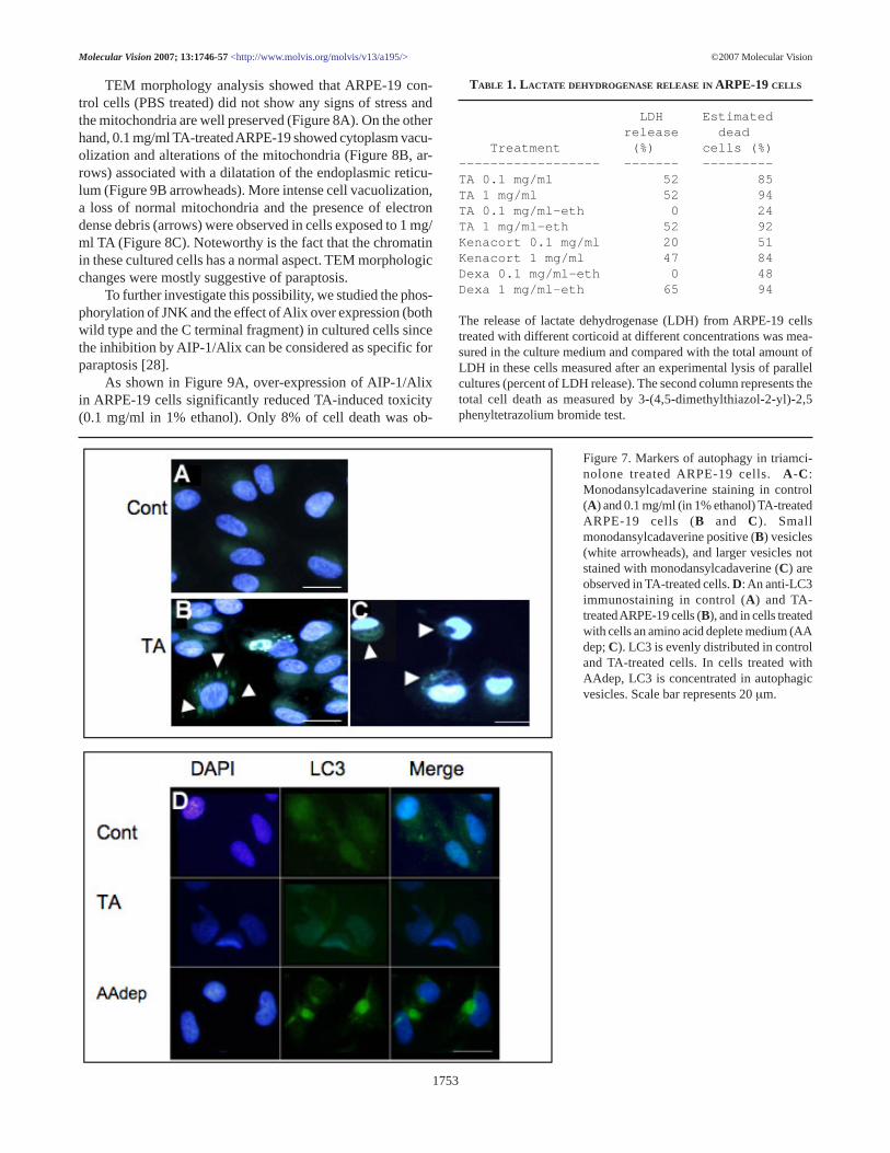
Necrosis: TA-induced cell death lacked the specific mark-ers and morphology for apoptotic cell death. To investigatethe possibility of a passive cell death, e.g. necrosis, we mea-sured the amount of free LDH in the various types of cell cul-tures. Table 1 shows the absence of necrosis with 0.1 mg/mlTA in 0.1%/ethanol. Nonetheless, higher TA concentrationsas well as addition of the more hydrophobic glucocoticoidcompounds induced a measurable LDH release. These resultscould indicate that at high concentrations, a fraction of thecorticosteroid-induced toxicity may be driven by necrosis.However they also indicated the presence of another cell deathmechanism, as the percentage of dead cells is always higherthan the percentage of necrotic cells.
Autophagy and paraptosis: Autophagic activity was in-vestigated by vacuoles labeling using bothmonodansylcadaverine and anti-LC3, an antibody stainingmature autophagic vacuoles [33].
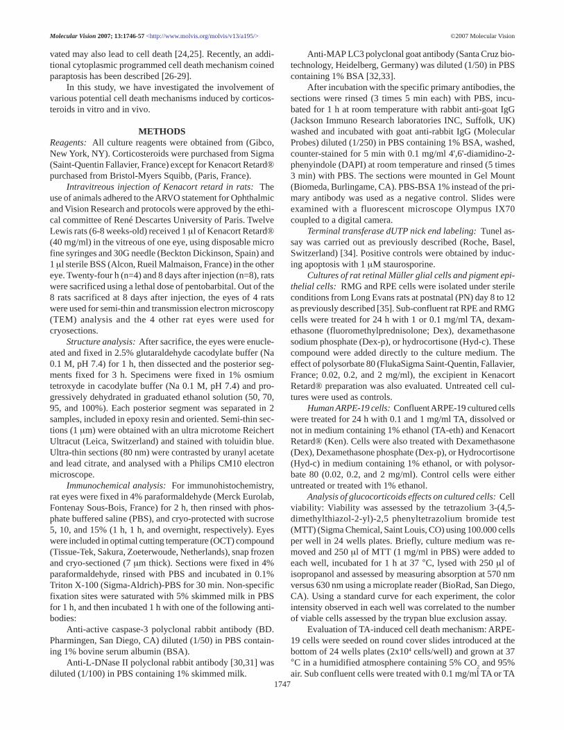
Only some of the vacuoles were stained withmonodansylcadaverine in TA-treated RPE cells (Figure 7A,arrowheads), while larger vacuoles inducing a modificationof the cell nucleus shape (Figure 7A,D arrowheads) were notstained. MAP-LC3 labeling did not significantly differ in TAtreated and control cells, while MAP-LC3 positively stainedvacuoles of cells incubated in amino acid depleted medium(AAdep, Figure 7).
©2007 Molecular VisionMolecular Vision 2007; 13:1746-57 <http://www.molvis.org/molvis/v13/a195/>
Figure 6. No caspase-independent markers of apoptosis in triamcinolone treated ARPE-19 cells. A- G: Triamcinolone-treated (0.1 mg/ml in1% ethanol) or control ARPE 19 cells were stained with DAPI or immunostained with anti-AIF or anti LEI/L-DNase II. No nuclear transloca-tion of these apoptosis markers was seen, neither in cells with normal morphology, nor in cells with an altered nuclear morphology (A, whitearrows). H-M : Induction of apoptosis with HMA was used as positive control and shows nuclear translocation of both LEI and AIF, bothmarkers of caspase independent apoptosis. Sacle bar represents 20 µm.
1752
TEM morphology analysis showed that ARPE-19 con-trol cells (PBS treated) did not show any signs of stress andthe mitochondria are well preserved (Figure 8A). On the otherhand, 0.1 mg/ml TA-treated ARPE-19 showed cytoplasm vacu-olization and alterations of the mitochondria (Figure 8B, ar-rows) associated with a dilatation of the endoplasmic reticu-lum (Figure 9B arrowheads). More intense cell vacuolization,a loss of normal mitochondria and the presence of electrondense debris (arrows) were observed in cells exposed to 1 mg/ml TA (Figure 8C). Noteworthy is the fact that the chromatinin these cultured cells has a normal aspect. TEM morphologicchanges were mostly suggestive of paraptosis.
To further investigate this possibility, we studied the phos-phorylation of JNK and the effect of Alix over expression (bothwild type and the C terminal fragment) in cultured cells sincethe inhibition by AIP-1/Alix can be considered as specific forparaptosis [28].
As shown in Figure 9A, over-expression of AIP-1/Alixin ARPE-19 cells significantly reduced TA-induced toxicity(0.1 mg/ml in 1% ethanol). Only 8% of cell death was ob-
©2007 Molecular VisionMolecular Vision 2007; 13:1746-57 <http://www.molvis.org/molvis/v13/a195/>
Figure 7. Markers of autophagy in triamci-nolone treated ARPE-19 cells. A -C:Monodansylcadaverine staining in control(A) and 0.1 mg/ml (in 1% ethanol) TA-treatedARPE-19 cells (B and C). Smallmonodansylcadaverine positive (B) vesicles(white arrowheads), and larger vesicles notstained with monodansylcadaverine (C) areobserved in TA-treated cells. D: An anti-LC3immunostaining in control (A) and TA-treated ARPE-19 cells (B), and in cells treatedwith cells an amino acid deplete medium (AAdep; C). LC3 is evenly distributed in controland TA-treated cells. In cells treated withAAdep, LC3 is concentrated in autophagicvesicles. Scale bar represents 20 µm.
TABLE 1. LACTATE DEHYDROGENASE RELEASE IN ARPE-19 CELLS
LDH Estimated release dead Treatment (%) cells (%)------------------ ------- ---------TA 0.1 mg/ml 52 85TA 1 mg/ml 52 94TA 0.1 mg/ml-eth 0 24TA 1 mg/ml-eth 52 92Kenacort 0.1 mg/ml 20 51Kenacort 1 mg/ml 47 84Dexa 0.1 mg/ml-eth 0 48Dexa 1 mg/ml-eth 65 94
The release of lactate dehydrogenase (LDH) from ARPE-19 cellstreated with different corticoid at different concentrations was mea-sured in the culture medium and compared with the total amount ofLDH in these cells measured after an experimental lysis of parallelcultures (percent of LDH release). The second column represents thetotal cell death as measured by 3-(4,5-dimethylthiazol-2-yl)-2,5phenyltetrazolium bromide test.
1753

served in AIP-1/Alix-WT transfected cells as compared to 23%in non-transfected cells or in cells transfected with wild typeAIP-1/Alix-CT (Figure 9A). Thus, the over-expression of Alix-WT was associated with a significant protection against TA-induced cell death. Activation of JNK-1 and JNK-2 was in-vestigated using anti-phospho JNK antibodies. TA inducedphosphorylation of a 57 kDa protein that may correspond toJNK-2 but did not demonstrate an increase in JNK-1 phos-phorylation (Figure 9B).
DISCUSSION Contradictory observations and discrepancies regarding theclinical safety of intravitreous TA and the in vitro TA-inducedtoxicity have been published [12,17], depending mostly onthe techniques used to detect toxicity. In the rat eye, usingsemi-thin histology and TEM 8 days after the intravitreousinjection of Kenacort®, we can detect clear lesions in RPEand macroglial cells. These morphological abnormalities arenot associated with infiltration of inflammatory cells, or with
glial activation (no GFAP labeling is found, not shown). Re-cently, TEM analysis of the rabbit retina after the intravitre-ous injection of Kenalog®, showed the same lesions [41]. Thefact that all currently used immunohistochemical markers forapoptosis or autophagy (activated caspase-3, TUNEL assay,and MAP-LCA) are negative may explain why other authorspreviously failed to detect any toxic effects in the rabbit eye[12,14].
Our in vitro studies confirm that glucocorticoids have atoxic effect on rat RPE and RMG cells and on human ARPE-19 cells. A significant and dose dependent toxicity was in-duced by different glucocorticoid formulations. The chemicalproperties of the glucocorticoids determine their toxicity: morehydrophobic compounds induce a more toxic effect. Dex andTA demonstrate the highest toxic effect while Hydro-c andDex-p are less toxic. This phenomenon may be associated withthe lesser ability of the cells to metabolize the hydrophobiccompounds allowing for their accumulation and higher con-centration in the cell and intracellular organ membranes.
©2007 Molecular VisionMolecular Vision 2007; 13:1746-57 <http://www.molvis.org/molvis/v13/a195/>
Figure 8. Transmission electron microscopyobservations of TA-treated ARPE 19 cells. A: Control ARPE 19 cells. B and C: ARPE-19 cellsTA-treated with 0.1 mg/ml (B) and 1 mg/ml TA (C), showing membrane limited (arrows) or unlimited vesicles (stars) and dilated endoplasmicreticulum (arrowhead). In picture C, a more advanced stage of degeneration is represented.
1754

Kenacort®, commonly used for clinical use contains twophases: crystals of TA in suspension and a low fraction of TAsolubilized in polysorbate 80 (0.02 mg/ml) and benzyl alco-hol. Polysorbate 80 is an anionic detergent of low toxicity [42]which can however, increase susceptibility to oxidative stress[43]. When cells are exposed in vitro to polysorbate 80 at con-centrations of 0.02 mg/ml no toxicity is observed. These re-sults indicate that polysorbate 80 per se is not toxic. Othersauthors have reported that Kenalog® vehicle enhance the tox-icity of TA on retinal cells in culture [17]. Indeed, in contrastto polysorbate 80, benzyl alcohol (contained in Kenalog®)induce ARPE-19 apoptosis, even at low concentrations [18].Therefore, it was recommended to remove the solubilizingcompounds within the Kenalog® preparation before its injec-tion in the human eye [19,44]. However, the potential toxiceffect of the TA crystals was somewhat overlooked.
From our present study, it can be concluded that an im-portant part of the TA toxicity is associated with the low solu-bility of the compound. The cell toxicity of 0.1 mg/ml TA in1% ethanol was significantly reduced when compared to the
same concentration of TA in crystal form. At the concentra-tion of 1 mg/ml TA, the use of 1% ethanol did not influencethe extent of cell toxicity because a large proportion of the TAcrystals still remain insoluble. Similarly, it was recently shownthat direct contact of insoluble TA with ARPE-19 cells inducedcell death through an apopto-necrotic mechanism [18]. Previ-ous in vitro experiments have also shown that direct incuba-tion of cells with crystalline TA induce activation of caspasesand stress proteins [15]. On the other hand, when TA crystalsare separated from the ARPE-19 cultured cells by amicroporous membrane, no apoptotic cell death occurred [18].However, in these experiments, other potential mechanismsof cell death, different from apoptosis were not explored.
In the brain, it was reported that the glucocorticoid II re-ceptor mediates cell toxicity [45] and in hyppocampal neu-rons, corticosterone neurotoxicity has been correlated to al-teration of the mitochondrial membrane potential. Interestingly,in these studies, no apoptosis markers could be detected andthe exact mechanism of cell death remains elusive [46].
Although corticosteroids significantly reduce the num-ber of living cells, we failed to observe any significant activa-tion of the known apoptotic pathways in retinal cells, which isin line with previous reports indicating the failure to detectevident apoptosis during RPE cell death induced by corticos-teroids [15,17]. Our further exploration of other types of pro-grammed cell death sheds light on the possible mechanismsof retinal cell death pathways activated by the corticosteroids.
The extensive vacuolization within the cytoplasm and de-formation of the nucleus shape and contour in TA treated cellscould suggest potential an autophagic mechanism, which ischaracterized by sequestration of bulk cytoplasm and the for-mation of organelles in double or multimembrane acidic au-tophagic vesicles [23,25]. During autophagy, a specific formof the microtubule -associated protein light chain 3 (LC3), theLC3-2 form has been described to increase in association withthe autophagosome membrane [31,32]. This reaction seemshowever not to be specific for autophagy [32,39]. In our study,using both immunostaining and western blotting (not shown),we did not find any evidence for LC3 association with theobserved vacuoles and could not detect any increase of LC3-2 in TA treated cells. This did not favor the hypothesis of au-tophagy. Interestingly, in neural cells, the suppression of basalautophagy may rather cause neurodegeneration [47,48].
However, the observation that in some TA treated cells,vesicles positive for monodansylsacaderine were detected doesnot allow to totally ruled out the possibility that autophagycan be part of the mechanisms involved in TA induced celldeath.
TEM observations of retina and cells treated with TAshowed that the large irregular cytoplasmic vacuoles are as-sociated with swelling of the mitochondria and enlargementof the endoplasmic reticulum along with preservation of thenuclear chromatin, which may suggest paraptotic cell death[27,29]. Paraptosis takes place during cell differentiation inthe development of the nervous system as well as in somecases of neurodegeneration [22]. It is mediated by mitogen-activated protein kinases and can be triggered by the TNF re-
©2007 Molecular VisionMolecular Vision 2007; 13:1746-57 <http://www.molvis.org/molvis/v13/a195/>
Figure 9. Paraptosis in triamcinolone treated-ARPE-19 cells. A: Ef-fect of Alix overexpression on TA induced cell death. ARPE 19 cellswere nucleofected with Alix-WT or with its C terminal moiety. Con-trols were run with untransfected cells or in cells transfected with thePCI empty vector. 48 h after transfection cells were treated with TAand cell survival was measured with the 3-(4,5-dimethylthiazol-2-yl)-2,5 phenyltetrazolium bromide test method. Alix-CT has no ef-fect on cell survival but Alix-WT protects cells form death. * indi-cates a p<0.05. B: Western-blot using anti-PJNK (upper panel) oranti-JNK-1 (lower panel) was assayed in 0.1 mg/ml (in 1% ethanol)TA treated (TA) or control (C, 1% ethanol) ARPE-19 cells. An in-crease of P-JNK-2 is seen in TA treated cells, while no JNK-1 overactivation is observed in TA-treated cells compared to control cells.
1755
ceptor family member TAJ/TROY [29]. The fact that ARPE-19 cells TA-induced toxicity can be blocked by the paraptosisinhibitor AIP-1/Alix, but not with its anti-apoptotic C-termi-nal fragment (Alix-CT), favors further the hypothesis ofparaptosis [28]. It was recently shown that in insulin-likegrowth factor I receptor (IGFIR)-induced paraptosis the MEK/JNK-1 pathway was involved [28]. In TA-induced paraptosisof ARPE-19 cells, we found that phosphorylation of JNK-2occurs but no phosphorylation of JNK-1. This situation hasalready been described for other cells [49].
Taken together, our observations suggest that non classi-cal mechanisms of cell death are induced by the corticoster-oids. Apparently, the activated specific mechanism dependson the formulation of the used compounds and involves dif-ferent mechanisms of cell death. The presence of insolubleparticles induces mostly a necrotic response. In the presenceof soluble formulations of TA, which is studied in this work,the major cell death mechanism is related to paraptosis.
In conclusion, we demonstrate that glucocorticoids inducenon-apoptotic cell death through a mechanism related mainlyto paraptosis. Whether glucocorticoids can induce similar toxiceffects in other cell types remain to be investigated, but thefailure to detect apoptotic markers in other neural cells favorthis hypothesis. These observations enhance our understand-ing of the glucocorticoid mechanism of action on ocular tis-sues and open the way for the investigations of more efficientand safer future therapeutic avenues.
ACKNOWLEDGEMENTS We are greatly indebted to Prof. R. Sadoul and Dr. C.Chatellard-Cause (EMI 0108, LAPSEN, Grenoble, France) forthe generous gift of Alix and Alix-CT plasmids.
REFERENCES 1. Jonas JB, Hayler JK, Sofker A, Panda-Jonas S. Intravitreal injec-
tion of crystalline cortisone as adjunctive treatment of prolif-erative diabetic retinopathy. Am J Ophthalmol 2001; 131:468-71.
2. Jonas JB, Kamppeter BA, Harder B, Vossmerbaeumer U, SauderG, Spandau UH. Intravitreal triamcinolone acetonide for dia-betic macular edema: a prospective, randomized study. J OculPharmacol Ther 2006; 22:200-7.
3. Martidis A, Duker JS, Greenberg PB, Rogers AH, Puliafito CA,Reichel E, Baumal C. Intravitreal triamcinolone for refractorydiabetic macular edema. Ophthalmology 2002; 109:920-7.
4. Greenberg PB, Martidis A, Rogers AH, Duker JS, Reichel E.Intravitreal triamcinolone acetonide for macular oedema due tocentral retinal vein occlusion. Br J Ophthalmol 2002; 86:247-8.
5. Antcliff RJ, Spalton DJ, Stanford MR, Graham EM, ffytche TJ,Marshall J. Intravitreal triamcinolone for uveitic cystoid macu-lar edema: an optical coherence tomography study. Ophthal-mology 2001; 108:765-72.
6. Munir WM, Pulido JS, Sharma MC, Buerk BM. Intravitreal triam-cinolone for treatment of complicated proliferative diabetic re-tinopathy and proliferative vitreoretinopathy. Can J Ophthalmol2005; 40:598-604.
7. Young S, Larkin G, Branley M, Lightman S. Safety and efficacyof intravitreal triamcinolone for cystoid macular oedema in uvei-tis. Clin Experiment Ophthalmol 2001; 29:2-6.
8. Challa JK, Gillies MC, Penfold PL, Gyory JF, Hunyor AB, BillsonFA. Exudative macular degeneration and intravitreal triamci-nolone: 18 month follow up. Aust N Z J Ophthalmol 1998;26:277-81.
9. Danis RP, Ciulla TA, Pratt LM, Anliker W. Intravitreal triamcino-lone acetonide in exudative age-related macular degeneration.Retina 2000; 20:244-50.
10. Ciulla TA, Criswell MH, Danis RP, Hill TE. Intravitreal triamci-nolone acetonide inhibits choroidal neovascularization in a la-ser-treated rat model. Arch Ophthalmol 2001; 119:399-404.
11. Beer PM, Bakri SJ, Singh RJ, Liu W, Peters GB 3rd, Miller M.Intraocular concentration and pharmacokinetics of triamcino-lone acetonide after a single intravitreal injection. Ophthalmol-ogy 2003; 110:681-6.
12. McCuen BW 2nd, Bessler M, Tano Y, Chandler D, Machemer R.The lack of toxicity of intravitreally administered triamcino-lone acetonide. Am J Ophthalmol 1981; 91:785-8.
13. Kivilcim M, Peyman GA, El-Dessouky ES, Kazi AA, CheemaR, Hegazy H. Retinal toxicity of triamcinolone acetonide in sili-cone-filled eyes. Ophthalmic Surg Lasers 2000; 31:474-8.
14. Dierks D, Lei B, Zhang K, Hainsworth DP. Electroretinographiceffects of an intravitreal injection of triamcinolone in rabbitretina. Arch Ophthalmol 2005; 123:1563-9.
15. Yeung CK, Chan KP, Chiang SW, Pang CP, Lam DS. The toxicand stress responses of cultured human retinal pigment epithe-lium (ARPE19) and human glial cells (SVG) in the presence oftriamcinolone. Invest Ophthalmol Vis Sci 2003; 44:5293-300.
16. Yeung CK, Chan KP, Chan CK, Pang CP, Lam DS. Cytotoxicityof triamcinolone on cultured human retinal pigment epithelialcells: comparison with dexamethasone and hydrocortisone. JpnJ Ophthalmol 2004; 48:236-42.
17. Narayanan R, Mungcal JK, Kenney MC, Seigel GM, KuppermannBD. Toxicity of triamcinolone acetonide on retinal neurosen-sory and pigment epithelial cells. Invest Ophthalmol Vis Sci2006; 47:722-8.
18. Szurman P, Kaczmarek R, Spitzer MS, Jaissle GB, Decker P,Grisanti S, Henke-Fahle S, Aisenbrey S, Bartz-Schmidt KU.Differential toxic effect of dissolved triamcinolone and its crys-talline deposits on cultured human retinal pigment epithelium(ARPE19) cells. Exp Eye Res 2006; 83:584-92.
19. Kai W, Yanrong J, Xiaoxin L. Vehicle of triamcinolone acetonideis associated with retinal toxicity and transient increase of lensdensity. Graefes Arch Clin Exp Ophthalmol 2006; 244:1152-9.
20. Lockshin RA, Zakeri Z. Apoptosis, autophagy, and more. Int JBiochem Cell Biol 2004; 36:2405-19.
21. Siegel RM. Caspases at the crossroads of immune-cell life anddeath. Nat Rev Immunol 2006; 6:308-17.
22. Broker LE, Kruyt FA, Giaccone G. Cell death independent ofcaspases: a review. Clin Cancer Res 2005; 11:3155-62.
23. Heymann D. Autophagy: A protective mechanism in response tostress and inflammation. Curr Opin Investig Drugs 2006; 7:443-50.
24. Yu L, Wan F, Dutta S, Welsh S, Liu Z, Freundt E, Baehrecke EH,Lenardo M. Autophagic programmed cell death by selectivecatalase degradation. Proc Natl Acad Sci U S A 2006; 103:4952-7.
25. Kelekar A. Autophagy. Ann N Y Acad Sci 2005; 1066:259-71.26. Fietta P. Many ways to die: passive and active cell death styles.
Riv Biol 2006; 99:69-83.27. Fombonne J, Padron L, Enjalbert A, Krantic S, Torriglia A. A
novel paraptosis pathway involving LEI/L-DNaseII for EGF-induced cell death in somato-lactotrope pituitary cells. Apoptosis2006; 11:367-75.
©2007 Molecular VisionMolecular Vision 2007; 13:1746-57 <http://www.molvis.org/molvis/v13/a195/>
1756

28. Sperandio S, Poksay K, de Belle I, Lafuente MJ, Liu B, Nasir J,Bredesen DE. Paraptosis: mediation by MAP kinases and inhi-bition by AIP-1/Alix. Cell Death Differ 2004; 11:1066-75.
29. Wang Y, Li X, Wang L, Ding P, Zhang Y, Han W, Ma D. Analternative form of paraptosis-like cell death, triggered by TAJ/TROY and enhanced by PDCD5 overexpression. J Cell Sci 2004;117:1525-32.
30. Torriglia A, Chaudun E, Chany-Fournier F, Jeanny JC, CourtoisY, Counis MF. Involvement of DNase II in nuclear degenera-tion during lens cell differentiation. J Biol Chem 1995;270:28579-85.
31. Altairac S, Zeggai S, Perani P, Courtois Y, Torriglia A. Apoptosisinduced by Na+/H+ antiport inhibition activates the LEI/L-DNase II pathway. Cell Death Differ 2003; 10:548-57.
32. Tanida I, Tanida-Miyake E, Ueno T, Kominami E. The humanhomolog of Saccharomyces cerevisiae Apg7p is a Protein-acti-vating enzyme for multiple substrates including human Apg12p,GATE-16, GABARAP, and MAP-LC3. J Biol Chem 2001;276:1701-6.
33. Asanuma K, Tanida I, Shirato I, Ueno T, Takahara H, Nishitani T,Kominami E, Tomino Y. MAP-LC3, a promisingautophagosomal marker, is processed during the differentiationand recovery of podocytes from PAN nephrosis. FASEB J 2003;17:1165-7.
34. Bourges JL, Valamanesh F, Torriglia A, Jeanny JC, Savoldelli M,Renard G, BenEzra D, de Kozak Y, Behar-Cohen F. Cornea graftendothelial cells undergo apoptosis by way of an alternate(caspase-independent) pathway. Transplantation 2004; 78:316-23. Erratum in: Transplantation. 2005; 79:502.
35. Behar-Cohen FF, Thillaye-Goldenberg B, de Bizemont T,Savoldelli M, Chauvaud D, de Kozak Y. EIU in the rat pro-motes the potential of syngeneic retinal cells injected into thevitreous cavity to induce PVR. Invest Ophthalmol Vis Sci 2000;41:3915-24.
36. Yasugi E, Kumagai T, Nishikawa Y, Okuma E, Saeki K, OshimaM, Susin SA, Kroemer G, Yuo A. Involvement of apoptosis-inducing factor during dolichyl monophosphate-inducedapoptosis in U937 cells. FEBS Lett 2000; 480:197-200.
37. Daugas E, Susin SA, Zamzami N, Ferri KF, Irinopoulou T,Larochette N, Prevost MC, Leber B, Andrews D, Penninger J,Kroemer G. Mitochondrio-nuclear translocation of AIF inapoptosis and necrosis. FASEB J 2000; 14:729-39.
38. Nur-E-Kamal A, Gross SR, Pan Z, Balklava Z, Ma J, Liu LF.
Nuclear translocation of cytochrome c during apoptosis. J BiolChem 2004; 279:24911-4.
39. Gokhale P, Patel T, Morrison MJ, Vissers MC. The effect of in-tracellular ascorbate on the susceptibility of HL60 and Jurkatcells to chemotherapy agents. Apoptosis 2006; 11:1737-46.
40. Biederbick A, Kern HF, Elsasser HP. Monodansylcadaverine(MDC) is a specific in vivo marker for autophagic vacuoles.Eur J Cell Biol 1995; 66:3-14.
41. Yu SY, Damico FM, Viola F, D’Amico DJ, Young LH. Retinaltoxicity of intravitreal triamcinolone acetonide: a morphologi-cal study. Retina 2006; 26:531-6.
42. Sellers RS, Antman M, Phillips J, Khan KN, Furst SM. Effects ofmiglyol 812 on rats after 4 weeks of gavage as compared withmethylcellulose/tween 80. Drug Chem Toxicol 2005; 28:423-32.
43. Tatsuishi T, Oyama Y, Iwase K, Yamaguchi JY, Kobayashi M,Nishimura Y, Kanada A, Hirama S. Polysorbate 80 increasesthe susceptibility to oxidative stress in rat thymocytes. Toxicol-ogy 2005; 207:7-14.
44. Hernaez-Ortega MC, Soto-Pedre E. A simple and rapid methodfor purification of triamcinolone acetonide suspension forintravitreal injection. Ophthalmic Surg Lasers Imaging 2004;35:350-1.
45. Liu Y, Imai H, Sadamatsu M, Tsunashima K, Kato N. Cytokinesparticipate in neuronal death induced by trimethyltin in the rathippocampus via type II glucocorticoid receptors. Neurosci Res2005; 51:319-27.
46. Roy M, Sapolsky RM. The exacerbation of hippocampalexcitotoxicity by glucocorticoids is not mediated by apoptosis.Neuroendocrinology 2003; 77:24-31.
47. Komatsu M, Waguri S, Chiba T, Murata S, Iwata J, Tanida I,Ueno T, Koike M, Uchiyama Y, Kominami E, Tanaka K. Lossof autophagy in the central nervous system causesneurodegeneration in mice. Nature 2006; 441:880-4.
48. Hara T, Nakamura K, Matsui M, Yamamoto A, Nakahara Y,Suzuki-Migishima R, Yokoyama M, Mishima K, Saito I, OkanoH, Mizushima N. Suppression of basal autophagy in neural cellscauses neurodegenerative disease in mice. Nature 2006; 441:885-9.
49. Coffey ET, Smiciene G, Hongisto V, Cao J, Brecht S, HerdegenT, Courtney MJ. c-Jun N-terminal protein kinase (JNK) 2/3 isspecifically activated by stress, mediating c-Jun activation, inthe presence of constitutive JNK1 activity in cerebellar neu-rons. J Neurosci 2002; 22:4335-45.
©2007 Molecular VisionMolecular Vision 2007; 13:1746-57 <http://www.molvis.org/molvis/v13/a195/>
1757
The print version of this article was created on 19 Sep 2007. This reflects all typographical corrections and errata to the article through thatdate. Details of any changes may be found in the online version of the article. α