Embed Size (px)
Citation preview
Global Role of Cyclic AMP Signaling inpH-Dependent Responses in Candidaalbicans
Jeffrey M. Hollomon, Nora Grahl, Sven D. Willger, Katja Koeppen,Deborah A. HoganDepartment of Microbiology and Immunology, Geisel School of Medicine at Dartmouth, Hanover, NewHampshire, USA
ABSTRACT Candida albicans behaviors are affected by pH, an important environ-mental variable. Filamentous growth is a pH-responsive behavior, where alkalineconditions favor hyphal growth and acid conditions favor growth as yeast. We em-ployed filamentous growth as a tool to study the impact of pH on the hyphalgrowth regulator Cyr1, and we report that downregulation of cyclic AMP (cAMP) sig-naling by acidic pH contributes to the inhibition of hyphal growth in minimal me-dium with GlcNAc. Ras1 and Cyr1 are generally required for efficient hyphal growth,and the effects of low pH on Ras1 proteolysis and GTP binding are consistent withdiminished cAMP output. Active alleles of ras1 do not suppress the hyphal growthdefect at low pH, while dibutyryl cAMP partially rescues filamentous growth at lowpH in a cyr1 mutant. These observations are consistent with Ras1-independentdownregulation of Cyr1 by low pH. We also report that extracellular pH leads torapid and prolonged decreases in intracellular pH, and these changes may contrib-ute to reduced cAMP signaling by reducing intracellular bicarbonate pools. Tran-scriptomics analyses found that the loss of Cyr1 at either acidic or neutral pH leadsto increases in transcripts involved in carbohydrate catabolism and protein transla-tion and glycosylation and decreases in transcripts involved in oxidative metabolism,fluconazole transport, metal transport, and biofilm formation. Other pathways weremodulated in pH-dependent ways. Our findings indicate that cAMP has a global rolein pH-dependent responses, and this effect is mediated, at least in part, throughCyr1 in a Ras1-independent fashion.
IMPORTANCE Candida albicans is a human commensal and the causative agent ofcandidiasis, a potentially invasive and life-threatening infection. C. albicans experi-ences wide changes in pH during both benign commensalism (a common condition)and pathogenesis, and its morphology changes in response to this stimulus. NeutralpH is considered an activator of hyphal growth through Rim101, but the effect oflow pH on other morphology-related pathways has not been extensively studied.We sought to determine the role of cyclic AMP signaling, a central regulator ofmorphology, in the sensing of pH. In addition, we asked broadly what cellular pro-cesses were altered by pH in both the presence and absence of this important sig-nal integration system. We concluded that cAMP signaling is impacted by pH andthat cAMP broadly impacts C. albicans physiology in both pH-dependent and-independent ways.
KEYWORDS: Candida albicans, Cyr1, Ras1, Rim101, adenylate cyclase, morphology, pH
Local pH is one facet of the environment that varies widely in the human host-associated niches occupied by Candida albicans, a commensal fungus and oppor-
tunistic pathogen. Benign chronic carriage of C. albicans in the gastrointestinal tractoccurs frequently, and over the length of this organ system, the pH ranges from 2 in the
Received 19 September 2016 Accepted 20October 2016 Published 30 November 2016
Citation Hollomon JM, Grahl N, Willger SD,Koeppen K, Hogan DA. 2016. Global role ofcyclic AMP signaling in pH-dependentresponses in Candida albicans. mSphere1(6):e00283-16. doi:10.1128/mSphere.00283-16.
Editor Aaron P. Mitchell, Carnegie MellonUniversity
Copyright © 2016 Hollomon et al. This is anopen-access article distributed under the termsof the Creative Commons Attribution 4.0International license.
Address correspondence to Deborah A. Hogan,[email protected].
RESEARCH ARTICLEMolecular Biology and Physiology
crossmark
Volume 1 Issue 6 e00283-16 msphere.asm.org 1
on April 27, 2019 by guest
http://msphere.asm
.org/D
ownloaded from

stomach to 8 in the large intestine and can vary widely from subject to subject (1).Additionally, the vagina is acidic during benign C. albicans carriage and Candidavaginitis (2). We and others have shown that endogenous fermentative metabolism cansubstantially reduce the extracellular pH in sugar-rich environments, particularly insituations where respiratory metabolism is limited (3, 4). Filamentous growth, apathogenesis-related trait, is pH sensitive in vitro, with the number of hyphae in thepopulation increasing over the pH range from 5 to 7 (5).
C. albicans pH sensing and its morphological response to pH are in part controlledby the PacC ortholog Rim101, a transcription factor that is posttranslationally activatedin response to elevated pH (6). Rim101 is a major contributor to in vitro hyphal growthin rich, high-pH tissue culture medium (M199 at pH 8) (7), and disruption of thepH-sensing regulator Rim101 results in striking virulence defects in murine models ofdisseminated and oropharyngeal candidiasis and Candida keratomycosis (8–10). How-ever, Rim101 is not absolutely required for hyphal growth; it is dispensable for theformation of hyphae in response to serum, which is thought to activate hyphal growththrough the cyclic AMP (cAMP) pathway.
Ras1 and adenylate cyclase (Cyr1) are central components of cAMP signaling. Theywork together to govern filamentous growth, responses to stress, coordination ofmulticellular behaviors, and white-opaque switching in C. albicans (reviewed in refer-ence 11), and both Ras1 and Cyr1 contribute to virulence in a murine model ofdisseminated candidiasis (12, 13). Ras1 is a soluble small GTPase which, in its GTP-bound conformation, activates the adenylate cyclase Cyr1 (also referred to as Cdc35 inthe literature) (14). The resultant increase in intracellular cAMP relieves repression ofprotein kinase A by its regulatory subunit Bcy1, resulting in the initiation of the hyphalgrowth program (15–17). This pathway integrates a multitude of environmental signals,and induction of hyphal growth in response to a number of stimuli has been shown toinvolve the Ras1/cAMP pathway, notably serum, GlcNAc, muramyl dipeptides, andelevated temperature (18–21). Findings from our group, however, have called intoquestion the canonical unidirectional and linear relationship where stimuli act on Ras1to activate Cyr1. We have shown Cyr1 to act upon Ras1 by negatively regulating boththe as-yet-unidentified protease that alters Ras1 localization and the Ras1 GTPase-activating protein Ira2 (22, 23). Ras1-independent activation of Cyr1 leads, throughrepression of Ras1 cleavage, to reinforcement of Cyr1 activation. Conversely, Cyr1inhibition of Ira2 negatively regulates the activation of Cyr1 by Ras1 in response tocascade output. Synthesis of these observations yields a model where the relationshipbetween Ras1 and Cyr1 is complex and nonlinear, in which Ras1 can activate Cyr1 inresponse to some stimuli but Ras1 activation is in turn tightly regulated by Cyr1.
Here, using a citrate-buffered defined medium that can be poised at either pH 4 or7, we confirmed that more cells had hyphal morphology at neutral pH, whereaspseudohyphae and yeast predominated at pH 4. As these hypha-inducing conditions(37°C with GlcNAc) activate Ras1 and Cyr1 (21, 24) and true hyphal growth is sup-pressed in this medium by low pH, we asked if Ras1 or Cyr1 is downregulated inresponse to low pH, and if so, whether the reduction in activity of this pathwaycontributes to the observed repression of hyphal growth at low pH. We found that thefraction of Ras1 in the proteolyzed form and the fraction of Ras1 in its active GTP-boundconformation increase at low pH. These changes in Ras1 require Cyr1, which we haveshown previously to negatively regulate both Ras1 cleavage and Ras1-GTP levels. Thus,we propose that low pH downregulates Cyr1 activity, and this is consistent with theobservation that dibutyryl cyclic AMP restores hyphal growth in a cyr1Δ/Δ mutant strainat both pH 4 and pH 7 (22, 23). A transcriptional analysis revealed pH-dependent and-independent effects of Cyr1 on gene expression.
RESULTSLow pH represses growth morphology of hyphae and promotes growth as pseu-dohyphae in inducing medium with GlcNAc. In order to study the effects of pH onC. albicans, we sought to implement a buffer system for use in a defined, hypha-
Hollomon et al.
Volume 1 Issue 6 e00283-16 msphere.asm.org 2
on April 27, 2019 by guest
http://msphere.asm
.org/D
ownloaded from
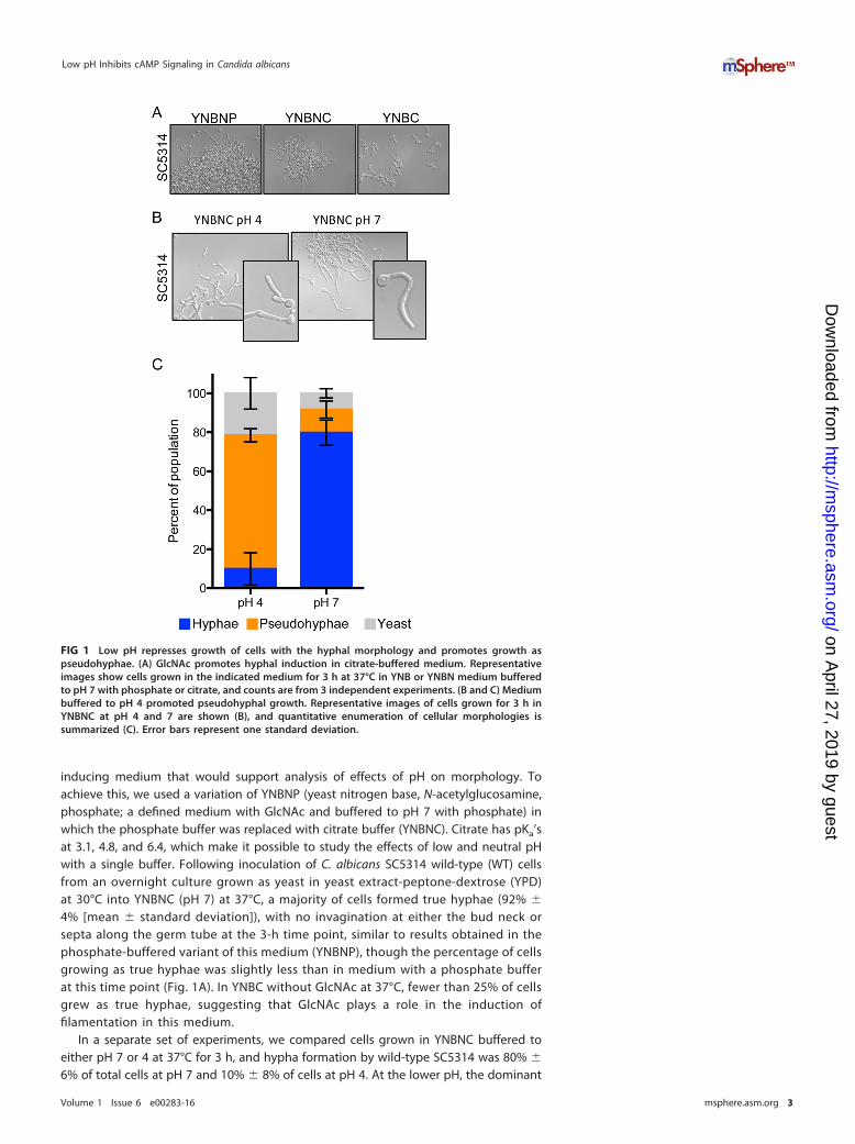
inducing medium that would support analysis of effects of pH on morphology. Toachieve this, we used a variation of YNBNP (yeast nitrogen base, N-acetylglucosamine,phosphate; a defined medium with GlcNAc and buffered to pH 7 with phosphate) inwhich the phosphate buffer was replaced with citrate buffer (YNBNC). Citrate has pKa’sat 3.1, 4.8, and 6.4, which make it possible to study the effects of low and neutral pHwith a single buffer. Following inoculation of C. albicans SC5314 wild-type (WT) cellsfrom an overnight culture grown as yeast in yeast extract-peptone-dextrose (YPD)at 30°C into YNBNC (pH 7) at 37°C, a majority of cells formed true hyphae (92% �
4% [mean � standard deviation]), with no invagination at either the bud neck orsepta along the germ tube at the 3-h time point, similar to results obtained in thephosphate-buffered variant of this medium (YNBNP), though the percentage of cellsgrowing as true hyphae was slightly less than in medium with a phosphate bufferat this time point (Fig. 1A). In YNBC without GlcNAc at 37°C, fewer than 25% of cellsgrew as true hyphae, suggesting that GlcNAc plays a role in the induction offilamentation in this medium.
In a separate set of experiments, we compared cells grown in YNBNC buffered toeither pH 7 or 4 at 37°C for 3 h, and hypha formation by wild-type SC5314 was 80% �
6% of total cells at pH 7 and 10% � 8% of cells at pH 4. At the lower pH, the dominant
FIG 1 Low pH represses growth of cells with the hyphal morphology and promotes growth aspseudohyphae. (A) GlcNAc promotes hyphal induction in citrate-buffered medium. Representativeimages show cells grown in the indicated medium for 3 h at 37°C in YNB or YNBN medium bufferedto pH 7 with phosphate or citrate, and counts are from 3 independent experiments. (B and C) Mediumbuffered to pH 4 promoted pseudohyphal growth. Representative images of cells grown for 3 h inYNBNC at pH 4 and 7 are shown (B), and quantitative enumeration of cellular morphologies issummarized (C). Error bars represent one standard deviation.
Low pH Inhibits cAMP Signaling in Candida albicans
Volume 1 Issue 6 e00283-16 msphere.asm.org 3
on April 27, 2019 by guest
http://msphere.asm
.org/D
ownloaded from
population was pseudohyphae (68% � 3% of total cells) (Fig. 1B and C). This finding isconsistent with previously published observations in which filamentous growth isantagonized by medium pH values below ~6 (5, 7, 25). To determine if the lack offilamentation under the pH 4 culture conditions was due to inhibition of growth, wecompared growth rates at the two different pHs and assessed growth by using theyeast-locked tetO-NRG1 strain (bearing a tetracycline-repressible copy of the NRG1-encoded hyphal repressor [26]) that enabled growth analysis by measurement of lightabsorbance. We found that growth was not lower but in fact slightly higher at pH 4than at pH 7. In YNBNC at 37°C, this strain had a doubling time of 102 � 1 min at pH 4and 112 � 3 min at pH 7. To rule out the possibility that NRG1 overexpression wasaffecting growth, we also examined growth of WT SC5314 at pH 4 and pH 7 at 30°C inYNBC, which is identical to YNBNC except it lacks the hyphal growth inducer GlcNAc.Again, growth was slightly but significantly faster at pH 4 than at pH 7 (113 � 1 minand 120 � 6 min, respectively). In both cases, the slightly faster growth at pH 4 wasstatistically significant (P � 0.05).
Disruption of Cyr1 and Ras1 results in an inability to grow as hyphae atpH 4 and 7. Cyr1 and Ras1 work together to positively regulate filamentous growth inresponse to GlcNAc and elevated temperatures in liquid medium, and mutants lackingthe genes encoding either of these proteins are impaired in hyphal growth (12, 19, 21).The adenylate cyclase Cyr1 has been described as essential for filamentation in liquidmedium, and when morphology was assessed over the course of 24 h, cyr1�/� mutantsgrew exclusively as yeast in YNBNC at either pH 7 or pH 4, whereas the WT and thecyr1�/�::CYR1 mutant strain formed predominantly pseudohyphae at pH 4 and hyphaeat pH 7 (Fig. 2) (13). Consistent with previous studies that have shown that strainslacking RAS1 have a less severe defect than do cyr1 null mutants, a ras1Δ/Δ mutantstrain grew as a combination of pseudohyphae and yeast (Fig. 2). Reconstitution of awild-type allele of RAS1 at the native locus restored growth similar to that of the wildtype as true hyphae at pH 7, but cells still had pseudohyphal morphology at pH 4(Fig. 2).
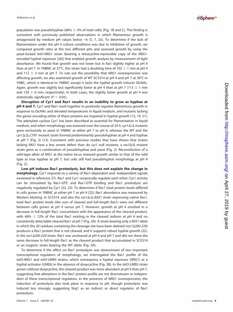
Low pH induces Ras1 proteolysis, but this does not explain the change inmorphology. Cyr1 responds to a variety of Ras1-dependent and -independent signals(reviewed in reference 27). Ras1 and Cyr1 reciprocally regulate each other; Cyr1 activitycan be stimulated by Ras1-GTP, and Ras1-GTP binding and Ras1 proteolysis arenegatively regulated by Cyr1 (22, 23). To determine if Ras1 total protein levels differedin cells grown in YNBNC at either pH 7 or pH 4 (22), Ras1 abundance was measured byWestern blotting. In SC5314, and also the ras1�/�::RAS1 strain expressing native Ras1,total Ras1 protein levels (the sum of cleaved and full-length Ras1) were not differentbetween cells grown at pH 4 versus pH 7. However, growth at pH 4 resulted in adecrease in full-length Ras1 concomitant with the appearance of the cleaved product,with 48% � 12% of the total Ras1 existing in the cleaved isoform at pH 4 and noconsistently detectable cleaved Ras1 at pH 7 (Fig. 3A). A strain bearing only a RAS1 allelein which the 20 residues containing the cleavage site have been deleted (ras1�200-220)produces a Ras1 protein that is not cleaved, and it supports robust hyphal growth (22).In the ras1�200-220 strain, Ras1 was uncleaved at pH 4 and pH 7 and did not show thesame decrease in full-length Ras1 as the cleaved product that accumulated in SC5314or an isogenic strain bearing the WT allele (Fig. 3A).
To determine if the effect on Ras1 proteolysis was downstream of two importanttranscriptional regulators of morphology, we interrogated the Ras1 profile of thetetO-NRG1 and tetO-UME6 strains, which overexpress a hyphal repressor (NRG1) or ahyphal activator (UME6) in the absence of doxycycline (Fig. 3B). In the tetO-UME6 straingrown without doxycycline, the cleaved product was more abundant at pH 4 than pH 7,suggesting that alterations in the Ras1 protein profile are not downstream or indepen-dent of these transcriptional regulators. In the presence of NRG1 overexpression, theinduction of proteolysis also took place in response to pH, though proteolysis wasinduced less strongly, suggesting Nrg1 as an indirect or direct regulator of Ras1proteolysis.
Hollomon et al.
Volume 1 Issue 6 e00283-16 msphere.asm.org 4
on April 27, 2019 by guest
http://msphere.asm
.org/D
ownloaded from

Ras1-GTP binding increases following exposure to low pH. As the effect oflow pH on morphology is consistent with downregulation of Ras signaling, we soughtto determine the effect of pH on Ras1-GTP binding. The fraction of Ras1 in its activeGTP-bound state was assayed by precipitation with recombinant Ras binding domain(RBD) followed by elution and detection by Western blotting with an anti-Ras antibody.The proportion of the total Ras1 pool that was GTP bound was markedly higher in cellsgrown at pH 4 than in those at pH 7, with both the full-length and cleaved isoformsshowing increased GTP binding (Fig. 3A). Ras1 cleavage is not responsible for theincrease in GTP binding, as increased Ras1-GTP binding at low pH was also observed ina strain with the ras1�/�::ras1�200-220 genotype.
The effects of low pH on Ras1 cleavage and GTP binding are rapid. Theeffects of the pH shift on Ras1 were rapid as well as sustained. When cells were grownat neutral pH, we observed the appearance of the Ras1 cleavage product within 15 minafter cells were shifted to pH 4 with citrate buffer, but not when cells were transferredto a medium buffered to pH 7 with citrate. Furthermore, this difference in Ras1 profilesbetween pHs persisted over the course of growth and was stable for an hour followingthe shift to pH 4 from pH 7, suggesting that the alteration of Ras1 cleavage by pH is fastas well as persistent (Fig. 3C). The increase in Ras1-GTP binding was also rapid and wasevident within 30 min of a shift to low pH (Fig. 3D).
Ras1 proteolysis and GTP binding are not responsible for pseudohyphalmorphology at pH 4. Above, we reported that low pH increases Ras1 cleavage and
FIG 2 Disruption of Cyr1 and Ras1 results in an inability to grow as hyphae at pH 4 and 7. Ras/cAMPsignaling is required for efficient induction of filamentation in YNBNC. Strains were photographedfollowing 3 h of growth at 37°C in YNBNC at pH 4 or 7.
Low pH Inhibits cAMP Signaling in Candida albicans
Volume 1 Issue 6 e00283-16 msphere.asm.org 5
on April 27, 2019 by guest
http://msphere.asm
.org/D
ownloaded from

Ras1-GTP binding. Ras1 proteolysis delocalizes the Ras1 N-terminal catalytic domainfrom the plasma membrane and decreases Ras1- and Cyr1-dependent hyphal growth(22). To determine if pH-induced cleavage of Ras1, and the resultant decrease in theamount of Ras1 at the membrane, is responsible for the decreased growth as hyphaeat pH 4, we assessed morphology of the WT RAS1 strain and the ras1�200-220 mutantstrain in cultures grown at pH 4 and 7. The ability to cleave Ras1 did not affect theproportion of the population growing as pseudohyphae at low pH; the ras1�/�::RAS1and ras1�/�::ras1�200-220 mutant strains similarly grew as hyphae at pH 7 andpseudohyphae at pH 4, with no statistically significant difference in the ratio of hyphaeto pseudohyphae between the two strains at pH 4 or pH 7 (Fig. 3E).
Although hyperactivation of cAMP signaling frequently results in hyphal growth, ithas also been reported to result in pseudohyphae in place of hyphae underfilamentation-inducing conditions (12, 28). To determine if the increase in GTP bindingwas responsible for pseudohyphal growth at pH 4, we compared the morphology ofthe ras1Δ/Δ::ras1G13V mutant (bearing an allele that encodes a constitutively GTP-
FIG 3 Low pH alters Ras1 localization and its activation state, but these changes are not responsible for theeffect of pH on morphology. (A) Total Ras levels (top) were comparable at pH 4 and 7, although cleavageprevailed at pH 4. (Top) Western blot results for the GTP-bound fraction of the lysates. (Middle) Anti-Ras1Western blot results with whole-cell lysates. (Bottom) Gelcode Blue stain of the membrane blotted in the toppanel, to indicate loading. Cells were grown for 3 h in YNBNC at the indicated pH. (B) Western blot of the Ras1profile and microscopy of tetO-UME6 and tetO-NRG1 strains with and without 50 �g per ml doxycycline. (C)Ras1 profile of cells of the indicated genotype grown for 3 h in YNB–GlcNAc– 0.2% glucose (YNBNG) adjustedto pH 7 and subsequently buffered to the indicated pH with citrate for the indicated time. (D) Ras1-GTP bindingof RAS1 cells grown for 3 h in YNBNG adjusted to pH 7 and subsequently buffered to the indicated pH withcitrate for 30 min. (E) Quantitative morphology of the indicated strains grown for 3 h in YNBNC at 37°C at pH 4or pH 7. Error bars represent one standard deviation.
Hollomon et al.
Volume 1 Issue 6 e00283-16 msphere.asm.org 6
on April 27, 2019 by guest
http://msphere.asm
.org/D
ownloaded from

bound Ras1) to that of the ras1Δ/Δ::RAS1 mutant. The ras1�/�::ras1G13V mutant and itsisogenic ras1�/�::RAS1 comparator mutant strain behaved like the wild type and weresimilarly pseudohyphal at pH 4 and hyphal at pH 7 (Fig. 3E). These data indicate thatchanges in Ras1 cleavage and GTP binding are not responsible for the decrease inhyphal growth at pH 4.
Effects of pH on Ras1 require Cyr1, and dibutyrl cAMP (dbcAMP) can rescuehyphal growth at low pH. Work from our group has previously shown that Cyr1-synthesized cAMP represses Ras1 proteolysis in hyphae (22). Additionally, we havefound that diminution of Cyr1 activity results in increased Ras1-GTP binding throughinactivation of Ira2 (23). In an ira2-deficient strain, which lacked the gene encoding theRas1 GTPase-activating protein, Ras1 was proteolyzed in response to low pH but hadhigh and pH-insensitive Ras1-GTP binding, indicating that the increase in GTP bindingin response to pH is mediated through Ira2 (Fig. 3A). Based on this observation, weconcluded that the increase in proteolysis at low pH was not solely due to the increasein Ras1-GTP levels. Moreover, as with Ras1 proteolysis, the increase in GTP bindingoccurred following exposure of preformed hyphae grown at neutral pH to low pH(Fig. 3D).
Based on the known Cyr1 repression of levels of proteolyzed Ras1 and Ras1-GTP,and the observed increase in both of these Ras1 forms in cells at low pH, wehypothesized that low pH inhibits Cyr1 in a Ras1-independent fashion. To test thishypothesis, we examined the effect of pH on Ras1 proteolysis and GTP binding in thepresence and absence of Cyr1. Consistent with this model, we found that the inductionof Ras1 proteolysis and GTP binding in response to low pH was substantially diminishedin the cyr1 null strain (Fig. 3A and 4A). Our data indicated that the absence of thecleaved product from the input blot in the GTP binding assay in the cyr1 mutant wasdue to postlysis degradation in the Ras1-GTP pulldown buffers; the cleaved productwas present when lysates were prepared in homogenization buffer (defined in Mate-rials and Methods) (see Fig. S1 in the supplemental material). The basis of the differencein the stability of the Ras1 cleaved product between the cyr1 null strain and itscomplement is not yet understood. To test the hypothesis that this event took place asa result of a change in the output of the Rim101 pathway, we examined the Ras1cleavage profile of cells shifted to pH 4 or maintained at pH 7, with and without RIM101.The induction of cleavage was observed in rim101�/� strains, indicating that regulationof this proteolytic event by pH is independent or upstream of the Rim101 cascade (seeFig. S2).
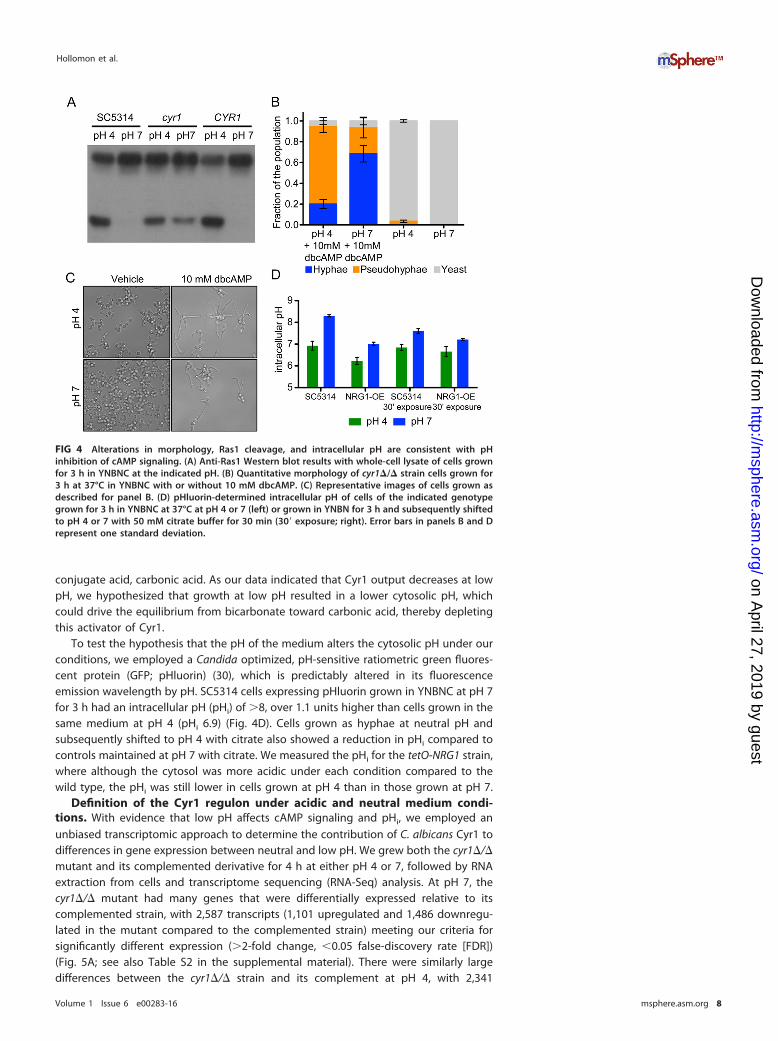
We and others have observed that the nonhydrolyzable, cell-permeable cAMPanalog dbcAMP is able to rescue the phenotypic effects of reduced cyclic AMPsignaling, and we hypothesized that addition of dbcAMP would suppress the effect ofpH on morphology under our conditions (12, 13, 22, 24). Addition of dbcAMP was ableto induce filamentous growth at both pH 4 and pH 7 in a cyr1 null strain (Fig. 4B andC). At pH 4 with 10 mM dbcAMP, 20% � 4% of the population grew as true hyphae,74% � 6% of the population grew as pseudohyphae, and the remainder grew as yeast,whereas in the absence of dbcAMP all cells were in the yeast form. This indicated to usthat addition of dbcAMP was able to partially overcome the inhibitory effect of low pHon filamentous growth. The addition of dbcAMP also stimulated hyphal growth at pH 7,whereas in the presence of dbcAMP, 68% � 8% of the population grew as true hyphae,25% � 10% of the population grew as pseudohyphae, and the rest grew as yeast; atpH 7 without dbcAMP, cells similarly grew as yeast. The observation that at pH 4 a cyr1null strain is capable of both hyphal and pseudohyphal growth in the presence ofdbcAMP suggests that the effect of pH on filamentous growth, at least in part, requiresCyr1. Taken together, these data are consistent with the model that low pH inhibitscAMP synthesis by Cyr1 independently of Ras1 to negatively regulate hyphal growth inresponse to acidic pH.
pH of the medium affects intracellular pH in YNBNC. Work in the Mühlschle-gel lab identified bicarbonate as an important activator of Cyr1 in Candida albicans (29).At physiological pH, bicarbonate acts as a buffer and exists in equilibrium with its
Low pH Inhibits cAMP Signaling in Candida albicans
Volume 1 Issue 6 e00283-16 msphere.asm.org 7
on April 27, 2019 by guest
http://msphere.asm
.org/D
ownloaded from
conjugate acid, carbonic acid. As our data indicated that Cyr1 output decreases at lowpH, we hypothesized that growth at low pH resulted in a lower cytosolic pH, whichcould drive the equilibrium from bicarbonate toward carbonic acid, thereby depletingthis activator of Cyr1.
To test the hypothesis that the pH of the medium alters the cytosolic pH under ourconditions, we employed a Candida optimized, pH-sensitive ratiometric green fluores-cent protein (GFP; pHluorin) (30), which is predictably altered in its fluorescenceemission wavelength by pH. SC5314 cells expressing pHluorin grown in YNBNC at pH 7for 3 h had an intracellular pH (pHi) of �8, over 1.1 units higher than cells grown in thesame medium at pH 4 (pHi 6.9) (Fig. 4D). Cells grown as hyphae at neutral pH andsubsequently shifted to pH 4 with citrate also showed a reduction in pHi compared tocontrols maintained at pH 7 with citrate. We measured the pHi for the tetO-NRG1 strain,where although the cytosol was more acidic under each condition compared to thewild type, the pHi was still lower in cells grown at pH 4 than in those grown at pH 7.
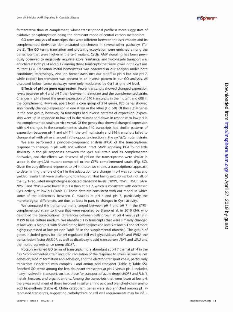
Definition of the Cyr1 regulon under acidic and neutral medium condi-tions. With evidence that low pH affects cAMP signaling and pHi, we employed anunbiased transcriptomic approach to determine the contribution of C. albicans Cyr1 todifferences in gene expression between neutral and low pH. We grew both the cyr1�/�mutant and its complemented derivative for 4 h at either pH 4 or 7, followed by RNAextraction from cells and transcriptome sequencing (RNA-Seq) analysis. At pH 7, thecyr1�/� mutant had many genes that were differentially expressed relative to itscomplemented strain, with 2,587 transcripts (1,101 upregulated and 1,486 downregu-lated in the mutant compared to the complemented strain) meeting our criteria forsignificantly different expression (�2-fold change, �0.05 false-discovery rate [FDR])(Fig. 5A; see also Table S2 in the supplemental material). There were similarly largedifferences between the cyr1�/� strain and its complement at pH 4, with 2,341
FIG 4 Alterations in morphology, Ras1 cleavage, and intracellular pH are consistent with pHinhibition of cAMP signaling. (A) Anti-Ras1 Western blot results with whole-cell lysate of cells grownfor 3 h in YNBNC at the indicated pH. (B) Quantitative morphology of cyr1�/� strain cells grown for3 h at 37°C in YNBNC with or without 10 mM dbcAMP. (C) Representative images of cells grown asdescribed for panel B. (D) pHluorin-determined intracellular pH of cells of the indicated genotypegrown for 3 h in YNBNC at 37°C at pH 4 or 7 (left) or grown in YNBN for 3 h and subsequently shiftedto pH 4 or 7 with 50 mM citrate buffer for 30 min (30= exposure; right). Error bars in panels B and Drepresent one standard deviation.
Hollomon et al.
Volume 1 Issue 6 e00283-16 msphere.asm.org 8
on April 27, 2019 by guest
http://msphere.asm
.org/D
ownloaded from

significantly different transcripts (1,019 increased and 1,322 decreased) (Fig. 5A). Acrossboth pH 4 and pH 7, 1,808 transcripts were significantly changed in the absence of Cyr1(771 increased and 1,037 decreased at pH 7, 769 increased and 1,039 decreased at pH 4,and 16 transcripts lower at one pH and higher at the other). Comparison of thedifferentially expressed genes between the cyr1 mutant and the CYR1 complementedstrain to the Cyr1 regulon defined by Harcus et al. in 2004 (31) found 196 transcripts (88positively regulated and 108 negatively regulated by Cyr1) that were differentiallyexpressed in both data sets (Table S3), and they included transcripts associated withhyphal growth, including ECE1, HWP1, HYR1, SAP4, and SAP6. In our data, the yeast-locked cyr1 mutant also had lower levels of other hypha-associated transcripts UME6,RBT1, ALS3, SAP5, IHD1, and HGC1 and higher levels of the yeast-related transcriptsYWP1, ALS4, and NRG1 (Table 1). Analysis by gene ontology (GO) term enrichment of theCyr1 regulon at pH 7 and pH 4 revealed an enrichment in genes annotated as havinga role in biofilm formation, which includes many of the morphology-associated genes(HWP1, HYR1, ECE1, and HGC1) (Table 2; Table S5).
Alongside morphology, many transcripts related to metabolism were differentiallyexpressed between the cyr1Δ/Δ mutant and its complemented derivative at both pH 4and 7 (Table 2; Tables S2 and S4 in the supplemental material). Consistent withobservations made by Harcus and colleagues (31), transcripts encoding elements of thetricarboxylic acid (TCA) cycle were lower in the cyr1Δ/Δ mutant. In addition, we foundsignificantly lower levels of transcripts corresponding to the glyoxylate shunt enzymesICL1 and MLS1 and the pyruvate dehydrogenase complex (Table S4). Among thepathways that were significantly enriched among the transcripts that were lower in thecyr1Δ/Δ mutant were carboxylic acid catabolism, lipid catabolism, and amino acid andpeptide transport (Table 2). For example, the mutant had lower levels of transcriptsencoding JEN1 and JEN2, which encode carboxylic acid transporters known to take upTCA cycle intermediates, and the latter of which was shown to be Cyr1 regulated byHarcus and colleagues (31, 32). GO terms associated with carbohydrate catabolism,N-acetylglucosamine catabolism, gluconeogenesis, and the pentose-phosphate shunt
FIG 5 RNA-Seq analysis of CYR1 cells in the adaptation to acid pH. (A) Black shows the sum oftranscripts that were >2-fold different and had an FDR of <0.05; blue shows transcripts that wereupregulated at pH 7 and complemented; red shows transcripts that were downregulated at pH 7 andcomplemented. (B) Overlap of genes differentially regulated by pH in the presence and absence ofstrain CYR1. (C) Principal component analysis of the cyr1�/� mutant and CYR1 strain samples at pH 4and pH 7.
Low pH Inhibits cAMP Signaling in Candida albicans
Volume 1 Issue 6 e00283-16 msphere.asm.org 9
on April 27, 2019 by guest
http://msphere.asm
.org/D
ownloaded from
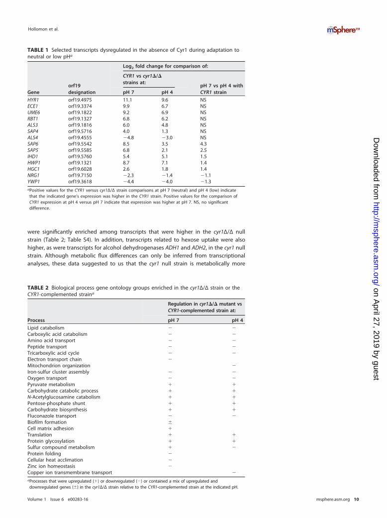
were significantly enriched among transcripts that were higher in the cyr1Δ/Δ nullstrain (Table 2; Table S4). In addition, transcripts related to hexose uptake were alsohigher, as were transcripts for alcohol dehydrogenases ADH1 and ADH2, in the cyr1 nullstrain. Although metabolic flux differences can only be inferred from transcriptionalanalyses, these data suggested to us that the cyr1 null strain is metabolically more
TABLE 1 Selected transcripts dysregulated in the absence of Cyr1 during adaptation toneutral or low pHa
Geneorf19designation
Log2 fold change for comparison of:
CYR1 vs cyr1�/�strains at:
pH 7 vs pH 4 withCYR1 strainpH 7 pH 4
HYR1 orf19.4975 11.1 9.6 NSECE1 orf19.3374 9.9 6.7 NSUME6 orf19.1822 9.2 6.9 NSRBT1 orf19.1327 6.8 6.2 NSALS3 orf19.1816 6.0 4.8 NSSAP4 orf19.5716 4.0 1.3 NSALS4 orf19.4555 �4.8 �3.0 NSSAP6 orf19.5542 8.5 3.5 4.3SAP5 orf19.5585 6.8 2.1 2.5IHD1 orf19.5760 5.4 5.1 1.5HWP1 orf19.1321 8.7 7.1 1.4HGC1 orf19.6028 2.6 1.8 1.4NRG1 orf19.7150 �2.3 �1.4 �1.1YWP1 orf19.3618 �4.4 �4.0 �1.3aPositive values for the CYR1 versus cyr1Δ/Δ strain comparisons at pH 7 (neutral) and pH 4 (low) indicatethat the indicated gene’s expression was higher in the CYR1 strain. Positive values for the comparison ofCYR1 expression at pH 4 versus pH 7 indicate that expression was higher at pH 7. NS, no significantdifference.
TABLE 2 Biological process gene ontology groups enriched in the cyr1Δ/Δ strain or theCYR1-complemented straina
Process
Regulation in cyr1�/� mutant vsCYR1-complemented strain at:
pH 7 pH 4
Lipid catabolism � �Carboxylic acid catabolism � �Amino acid transport � �Peptide transport � �Tricarboxylic acid cycle � �Electron transport chain �Mitochondrion organization �Iron-sulfur cluster assembly � �Oxygen transport � �Pyruvate metabolism � �Carbohydrate catabolic process � �N-Acetylglucosamine catabolism � �Pentose-phosphate shunt � �Carbohydrate biosynthesis � �Fluconazole transport � �Biofilm formation �Cell matrix adhesion �Translation � �Protein glycosylation � �Sulfur compound metabolism � �Protein folding �Cellular heat acclimation �Zinc ion homeostasis �Copper ion transmembrane transport �
aProcesses that were upregulated (�) or downregulated (�) or contained a mix of upregulated anddownregulated genes (�) in the cyr1Δ/Δ strain relative to the CYR1-complemented strain at the indicated pH.
Hollomon et al.
Volume 1 Issue 6 e00283-16 msphere.asm.org 10
on April 27, 2019 by guest
http://msphere.asm
.org/D
ownloaded from
fermentative than its complement, whose transcriptional profile is more suggestive ofoxidative phosphorylation being the dominant mode of central carbon metabolism.
GO term analysis of transcripts that were different between the cyr1 mutant and itscomplemented derivative demonstrated enrichment in several other pathways (Ta-ble 2). The GO terms translation and protein glycosylation were enriched among thetranscripts that were higher in the cyr1 mutant. Cyclic AMP signaling has been previ-ously observed to negatively regulate azole resistance, and fluconazole transport wasenriched at both pH 4 and pH 7 among those transcripts that were lower in the cyr1 nullmutant (33). Transition metal homeostasis was observed in our analysis under bothconditions; interestingly, zinc ion homeostasis met our cutoff at pH 4 but not pH 7,while copper ion transport was present in an inverse pattern in our GO analysis. Asdiscussed below, some pathways were only modulated by Cyr1 at one pH level.
Effects of pH on gene expression. Fewer transcripts showed changed expressionlevels between pH 4 and pH 7 than between the mutant and the complemented strain.Changes in pH altered the gene expression of 640 transcripts in the mutant and 608 inthe complement. However, apart from a core group of 214 genes, 820 genes showedsignificantly changed expression in one strain or the other (Fig. 5B). Of those 214 genesin the core group, however, 74 transcripts had inverse patterns of expression (expres-sion went up in response to low pH in the mutant and down in response to low pH inthe complemented strain, or vice versa). Of the genes that showed changed expressionwith pH changes in the complemented strain, 140 transcripts had similar patterns ofexpression between pH 4 and pH 7 in the cyr1 null strain and 896 transcripts failed tochange at all with pH or changed in the opposite direction in the cyr1�/� mutant strain.
We also performed a principal-component analysis (PCA) of the transcriptionalresponse to changes in pH with and without intact cAMP signaling. PCA found littlesimilarity in the pH responses between the cyr1 null strain and its complementedderivative, and the effects we observed of pH on the transcriptome were similar inscope in the cyr1Δ/Δ mutant compared to the CYR1 complemented strain (Fig. 5C).Given the very different responses to pH in these two strains, a transcriptional approachto determining the role of Cyr1 in the adaptation to a change in pH was complex andyielded results that were challenging to interpret. That being said, some, but not all, ofthe Cyr1-regulated morphology-associated transcript levels (HWP1, YWP1, HGC1, SAP6,NRG1, and YWP1) were lower at pH 4 than at pH 7, which is consistent with decreasedCyr1 activity at low pH (Table 1). These data are consistent with our model in whichsome of the differences between C. albicans at pH 4 and pH 7, particularly themorphological differences, are due, at least in part, to changes in Cyr1 activity.
We compared the transcripts that changed between pH 4 and pH 7 in the CYR1-complemented strain to those that were reported by Bruno et al. in 2010 (34), whodescribed the transcriptional differences between cells grown at pH 4 versus pH 8 inM199 tissue culture medium. We identified 115 transcripts that were similarly changedat low versus high pH, with 66 exhibiting lower expression levels at low pH and 59 morehighly expressed at low pH (see Table S6 in the supplemental material). This group ofgenes included genes for the pH-regulated cell wall glycosidases PHR1 and PHR2, thetranscription factor RIM101, as well as dicarboxylic acid transporters JEN1 and JEN2 andthe multidrug resistance pump MDR1.
Notably enriched GO terms of transcripts more abundant at pH 7 than at pH 4 in theCYR1-complemented strain included regulation of the response to stress, as well as celladhesion, biofilm formation and adhesion, and the electron transport chain, particularlytranscripts associated with complex I and amino acid transport (Table 3; Table S5).Enriched GO terms among the less abundant transcripts at pH 7 versus pH 4 includedmany involved in transport, such as those for transport of azole drugs (MDR1 and FLU1),metals, hexoses, and organic anions. Among the transcripts that were lower at low pH,there was enrichment of those involved in sulfur amino acid and branched-chain aminoacid biosynthesis (Table 4). Chitin catabolism genes were also enriched among pH 7-repressed transcripts, suggesting carbohydrate or cell wall requirements may be influ-
Low pH Inhibits cAMP Signaling in Candida albicans
Volume 1 Issue 6 e00283-16 msphere.asm.org 11
on April 27, 2019 by guest
http://msphere.asm
.org/D
ownloaded from

enced by pH. Consistent with the model that pH reduces Cyr1 activity, some of thesame GO term categories (amino acid transport, azole transport, and biofilm formation)were enriched among transcripts that were lower in cells grown at pH and in thecyr1Δ/Δ mutant cells.
Some pathways changed significantly in opposing directions with pH changes whenthe cyr1 null and complemented strains were compared. Notable among these weregenes involved in sulfate assimilation (MET3, MET14, MET16, and MET10, which encodeenzymes involved in the conversion of sulfate to hydrogen sulfide), which increased atpH 4 in the mutant and decreased in its complement (Table 2). A number of transcriptsencoded on the mitochondrial genome also showed Cyr1-dependent changes inexpression with pH changes; transcripts encoding components of the electron trans-port chain significantly decreased in abundance in the complemented strain at low pH,but in the mutant many of these components showed inverse patterns of expression(see Table S4).
We examined the data for transcripts of genes known to be involved in the pHresponse. PHR1 and PHR2 are two pH-responsive transcripts that encoded cell wallglycosidases that are reciprocally expressed at low and neutral pH, with PHR1 showinghigher expression at pH 7, and they have previously been observed to be dysregulatedin the absence of Cyr1 (31). ATO1 is an ammonium exporter that participates in mediumalkalinization and autoinduction of hyphal growth (35, 36). Of the transcripts thatchanged significantly in similar directions with pH in both the mutant and comple-mented strain were PHR1 and PHR2, as well as ATO1; however, the magnitude of thesechanges was lower in the absence of Cyr1. ATO1 was increased 71-fold in the comple-mented strain at pH 7 versus that at pH 4 but only 18-fold in the mutant, and PHR1 wassimilarly induced 11-fold in the mutant and 21-fold in the complemented strain underneutral pH conditions compared to acidic pH. PHR2 decreased 2.9- and 6.8-fold in themutant and complemented strain, respectively, at pH 7 versus pH 4.
RIM101 was more highly expressed at pH 7 than pH 4 in the complemented strain,but it was not significantly changed in the cyr1 null strain with pH changes. A numberof genes whose expression changed with pH have previously been described to beregulated by Rim101. In total, 41 transcripts annotated as Rim101 regulated underwentstatistically significant changes in either the presence or absence of Cyr1 (Table 5).Thirty-one transcripts annotated as Rim101 regulated by CGD were altered in expres-
TABLE 3 Biological process gene ontology groups enriched among the subset oftranscripts that were higher at pH 7 than at pH 4 in the CYR1-complemented strain
Process Genes in GO group
Regulation of response to stress SAP6, ALS1, SAP5, RIM101, PRA1, ACE2, HGT1, BCR1,CPH1, SRR1, AHR1
Biofilm formation PHR1, ALS1, IPT1, TRY6, RIM101, TRY4, SUC1, ACE2, HWP1,HGC1, HSP104, WOR1, QDR1, BCR1, CPH1, AHR1, ADH5
Cell adhesion PHR1, ALS1, TRY6, RIM101, PRA1, AAH1, TRY4, SUC1, ACE2,HWP1, WOR1, BCR1, AHR1
Electron transport chain NAD5, NAD3, NAD4, COX2, NAD2, NAD6Amino acid transport AGP2, GAP6, CAN2, GAP2, PUT4, DIP5, GNP3, GAP4, HNM4
TABLE 4 Biological process gene ontology groups enriched among the subset oftranscripts that were lower at pH 7 than at pH 4 in the CYR1-complemented strain
Process Genes in GO group
Azole transport FLU1, MDR1Metal ion transport ZRT2, TRK1, FTR1, FET3, CTR1, MAC1, PHO87,
FET99, HAK1, FRE10Hexose transport HGT8, HGT10, NAG3, NAG4, HGT19, HGT7, HGT17Organic anion transport JEN2, TPO5, GIT4, GNP1, ALP1, JEN1Chitin catabolic process CHT2, CHT1Branched-chain amino acid biosynthesis ILV5, BAT21, BAT22, PDC11Sulfur amino acid biosynthesis MET16, ECM17, MET2, MET1, MET15, MET3
Hollomon et al.
Volume 1 Issue 6 e00283-16 msphere.asm.org 12
on April 27, 2019 by guest
http://msphere.asm
.org/D
ownloaded from

sion in the CYR1 complemented strain (including RIM101), and 25 were altered in themutant strain (37). Of these, however, only 16 changed in both strains.
Using NanoString nCounter technology, we used a code set that included morphol-ogy genes and metabolism and used it to interrogate RNA from cells grown in aseparate experiment on a separate day than the RNA-Seq analysis. The impact of Cyr1was strongest in the regulation of the morphological genes, but there was a pH effecton hypha- and yeast-associated transcripts in the complemented strain, with theexception of HYR1, which increased in abundance at low pH. In the strain with intactCyr1 signaling, we observed the induction of PHR1 and PHR2 at neutral and low pH,respectively, and the expression pattern of these transcripts was notably muted in theabsence of Cyr1 (Table 6; see also Table S7 in the supplemental material).
DISCUSSION
In this work, we set out to determine if changes in cAMP signaling participate in therepression of filamentous growth induced by a low-pH environment in C. albicans. We
TABLE 5 Transcripts significantly different based on RNA-seq data that are annotated asRim101-regulated in the Candida Genome Databasea
Presence of CYR1 and gene nameorf19designation
Log2 fold change in expressionat pH 7 vs pH 4 in:
CYR1-complementedstrain
cyr1�/�strain
Expressed only when CYR1 is absentMUP1 orf19.5280 NS 1.9PHO113 orf19.2619 NS �1.8RBR3 orf19.5124 NS �1.2RBT1 orf19.1327 NS �1.1GIT1 orf19.34 NS �3.7PGA10 orf19.5674 NS �2.2PGA23 orf19.3740 NS �1.4PHO112 orf19.3727 NS �1.8
Expressed only when CYR1 is presentRIM101 orf19.7247 2.1 NSHOL4 orf19.4546 1.8 NSCDR11 orf19.918 1.8 NSFRP2 orf19.7112 1.5 NS
orf19.7566 1.3 NSDIP5 orf19.2942 1.3 NSECM21 orf19.4887 1.3 NSPHO8 orf19.984 1 NSPGA7 orf19.5635 �1.1 NSKRE6 orf19.7363 �1.1 NSCRH12 orf19.3966 �1.2 NSPTR22 orf19.6937 �1.7 NSGNP1 orf19.1193 �2.1 NSFRE10 orf19.1415 �3 NSRBE1 orf19.7218 �3.2 NS
Expressed in both strainsb
CFL2 orf19.1264 2.3 3.8PUT1 orf19.4274 1 �1.1
orf19.851 �1.1 �3.3CTR1 orf19.3646 �1.3 �1.6RBR2 orf19.532 �1.5 �1.5
orf19.7077 �1.7 �2.4OPT3 orf19.3749 �2.3 �2.7FET99 orf19.4212 �2.7 �9.5JEN1 orf19.7447 �2.9 �1.3CRZ2 orf19.2356 �3.7 �6RBR1 orf19.535 �6.8 �7.1
aPositive values indicate expression was higher at pH 7. NS, no significant difference.bThe following Rim101-regulated transcripts were differentially expressed in response to pH 7 versus pH 4,but expression was not different between the CYR1-complemented and cyr1Δ/Δ strains at pH 7: FRP1,HMX1, SKN1, ENA2, PHO89, PHO87, and PHR2.
Low pH Inhibits cAMP Signaling in Candida albicans
Volume 1 Issue 6 e00283-16 msphere.asm.org 13
on April 27, 2019 by guest
http://msphere.asm
.org/D
ownloaded from

found that hyphal growth was antagonized by low pH under medium conditions thatstimulated hyphal growth in a Ras1- and Cyr1-dependent manner. Low extracellular pHresulted in Cyr1-dependent increases in Ras1 proteolysis and GTP binding, which wereboth rapid and sustained, but neither increased GTP binding nor cleavage was respon-sible for the effect of low pH on morphology. Filamentous growth could be rescued ina cyr1 null mutant by dbcAMP at both low and neutral pHs, which supports a model inwhich low pH acts at least partially upstream of Cyr1. Under our medium conditions,intracellular pH was reduced by extracellular pH, and we hypothesize that this reducesthe availability of bicarbonate, a Cyr1-stimulatory factor. Analysis of the transcriptomein the presence and absence of intact cAMP signaling at acid and neutral pH painteda complicated picture, with Cyr1-dependent and -independent changes in response topH and differences in pH response that were dependent on whether cAMP signalingwas intact. Consistent with Cyr1 playing a role in the adaptation to low and neutral pH,Cyr-regulated transcripts related to morphology were altered in expression by changesin pH, and the expression levels of the pH-specific genes PHR1 and PHR2 weresubstantially greater in the presence of Cyr1.
Cytosolic pH is the cumulative result of buffering by a large number of differentmolecules (charged amino acid side chains, free amino acids, glycolytic intermedi-ates, and others), and the activity of plasma membrane and vacuolar ATPases(V-ATPases) and proton pumps (38). In Saccharomyces, V-ATPases are regulated byRas and phosphoinositides, and V-ATPases themselves reciprocally regulate Ras(39–41). Intracellular pH in Saccharomyces cerevisiae has been shown to be reducedin the presence of citric acid/phosphate buffer at low pH, and in C. albicans, growthin medium favoring yeast growth due to low-pH medium has been described toresult in cytosolic acidification compared to an otherwise identical neutral hypha-inducing medium, and the dynamics of changes in intracellular pH are differentbetween cells growing as hyphae and those growing as yeast (42, 43). Despitemechanisms to modulate intracellular pH, our findings using pHluorin-expressingcells showed that extracellular pH has rapid and prolonged effects on intracellularpH. These findings mirror observations by Kaur and colleagues (43), who saw alower pHi in cells in pH 4.5 medium than in cells at pH 6.5, using [14C]propionatedistribution as an indicator of pHi. In the tetO-NRG1 strain, the external pH stillimpacted cytosolic pH to a similar extent, suggesting to us that extracellular pH, aswell as the hyphal growth transcriptional network itself, make independent butadditive contributions to intracellular pH.
In C. albicans, bicarbonate is a Ras1-independent activator of Cyr1, and itsavailability is determined by the carbonic anhydrase Nce103, its substrate CO2, andpH (29). As bicarbonate exists in equilibrium with its conjugate acid, carbonic acid,a reduction in pH would drive that equilibrium toward the conjugate acid, deplet-ing a Cyr1 activator. CO2 has been demonstrated to control hyphal morphologythrough Cyr1, and mutation of the specific residues of Cyr1 responsible for bicar-bonate sensing resulted in a diminished ability to form filaments under high CO2
TABLE 6 Selected morphology- and pH-related transcription changes
Gene
Log2 fold change in transcription for:
CYR1 vs cyr1�/� strain at:pH 7 vs pH 4 inCYR1 strainpH 7 pH 4
HWP1 9.0 9.6 0.4HYR1 9.0 10.6 �1.5ECE1 8.3 8.6 1.0SAP4 3.8 2.4 1.2YWP1 �4.3 �2.6 �1.3ALS4 �6.3 �2.5 �0.8NRG1 �1.8 �0.7 �1.2PHR1 2.6 3.0 3.5PHR2 �0.2 1.4 �4.5
Hollomon et al.
Volume 1 Issue 6 e00283-16 msphere.asm.org 14
on April 27, 2019 by guest
http://msphere.asm
.org/D
ownloaded from

(which is Ras independent) but a normal ability to do so in response to serum(which is Ras dependent) (44). White-opaque switching has also been shown to beregulated by CO2 in a Cyr1-dependent, but also partially Ras1-dependent, fashion(45). Taken together, these observations suggest that Cyr1 integrates Ras1-dependent and -independent inputs, with different phenotypes requiring differentdegrees of contribution from each source.
pH has been postulated to regulate bicarbonate-sensitive adenylate cyclases in anumber of systems (development of mammalian spermatozoa during capacitationbeing the classic example), but to our knowledge, the extent of perturbation of pHi
necessary to alter adenylate cyclase output has not been empirically determined in vivo(46–48). We observed a shift in strain SC5314 from pHi 8.0 to pHi 6.6 between neutraland acidic culture conditions, and it is plausible that this represents a significant changein the bicarbonate/carbonic acid equilibrium and Cyr1 activation. This equilibrium hasa theoretical pKa of 3.6 in the absence of carbon dioxide, but likely a much highereffective pKa in vivo, as CO2 is generated through oxidative metabolism. Using thecarbonic acid/bicarbonate pKa for mammalian blood chemistry (6.4), one can calculatethe fraction of bicarbonate predicted to exist in the unprotonated form at our mea-sured pHi levels. In cells grown in YNBNC at pH 7, where the pHi was 8.0, 98% of thebicarbonate would be in its unprotonated from, whereas in cells grown at pH 4, the pHi
was 6.9, where 80% of the bicarbonate would be in its unprotonated form. Alterna-tively, it is possible that the change in pH alone is necessary to alter cyclase activity, asreports have suggested that soluble adenylate cyclases favor neutral to basic pHs forcatalysis, although these experiments were conducted with crude cellular extracts thatlikely contained bicarbonate, and it may not be possible to extricate the role of pH perse from the availability of bicarbonate (49, 50).
From the massive transcriptional changes induced by pH for genes associated withdiverse cellular processes such as hyphal growth, cell wall architecture, and metabo-lism, it is clear that ambient pH is an important regulator of C. albicans physiology (34,51). C. albicans has been shown to actively modulate the pH of its environment; workfrom the Lorenz group has illuminated a mechanism by which C. albicans inducesfilamentation by manipulating the environmental pH through secretion of basic prod-ucts of metabolism (35). C. albicans Rim101 was initially characterized as an activator ofhyphal growth in response to high pH as a stimulus (7, 25, 52, 53). Low pH can bethought of as the absence of high pH, or a stimulus unto itself, and analysis of our dataalongside published work is consistent with a model that integrates both of theseelements. Consistent with the model that there exist multiple pH-sensitive regulators,in our dbcAMP rescue assays more cells grew as hyphae at pH 7 than pH 4 in thepresence of dbcAMP, suggesting that Cyr1 is only one layer of morphological regula-tion by pH.
Growth at low pH under our conditions resulted in a population that was predom-inantly pseudohyphal, which is thought to represent an intermediate state betweenyeast and hyphal growth (54). Thus, we argue that our data suggest that low pH resultsin an intermediate level of cAMP output, where the hypha-activating cues are able toturn on the pathway sufficiently to support pseudohyphal growth, but low pH preventsit from being turned on to a sufficient extent to permit true hyphal growth. Thisintermediate state is phenotypically very different from the total absence of cAMPsignaling that we saw in the cyr1 null mutant, and it had a remarkably differenttranscriptional profile; in this state, we are able to see the effects of other regulators,such as Rim101, in the transcriptional data that are washed out in the completeabsence of Cyr1. A previous transcriptional analysis of the role of Rim101 in theadaptation to acidic and alkaline pH found that Rim101 regulated some, but not all,genes that changed with pH and that Rim101 regulated a number of genes indepen-dently of pH (51). Via synthesis of this finding with the fact that the effect of pH wasdifficult to discern through the overwhelming effect of Cyr1 in our data, we are able toinfer that the genetic basis of the response to pH is complicated, and we suggest
Low pH Inhibits cAMP Signaling in Candida albicans
Volume 1 Issue 6 e00283-16 msphere.asm.org 15
on April 27, 2019 by guest
http://msphere.asm
.org/D
ownloaded from
deletion of single regulators cannot fully explain changes in the transcriptome inducedby changes in pH.
With respect to the role of pH in the course of human disease, acidic pH is anenvironmental condition confronted by C. albicans in the phagolysosome, wherealkalinization by C. albicans metabolism is thought to facilitate survival of the fungusthrough the autoinduction of hyphal growth (35, 36, 55). There is precedent forinappropriate activation of cAMP signaling preventing the adaptation to a low-pHenvironment; Wilson and colleagues (56) previously described cAMP hyperactivation,due to the loss of the major cAMP phosphodiesterase, rendered C. albicans sensitive toacidic pH, with a pde2 null mutant unable to grow at pH 2.5. This suggests thatdownregulation of cAMP signaling participates in the adaptation to low-pH environ-ments. Cyr1 has been demonstrated to be necessary for pathogenesis in immunocom-petent mice in a model of disseminated candidiasis, and under our experimentalconditions, the ATO1-encoded ammonium exporter through which C. albicans alkalin-izes its environment, in addition to being regulated by pH itself was altered in itsregulation in the absence of Cyr1 (35, 36). Notably, two transcripts that we sawprominently induced by low pH, JEN1 and JEN2, have been shown to be induced uponphagocytosis by neutrophils (32). Based on these observations, the regulation of Cyr1by pH may play a role in the adaptation to the phagocytic vacuole. Additionally,Candida adapts to extremely low pH values in the stomach in the course of intestinalcommensalism, which can be the source from which C. albicans disseminates ininvasive disease.
Vulvovaginal candidiasis (VVC) is one of the most common fungal infections inhumans, affecting most women at some point over the course of their lives, andrepresents a context in which acidic pH is a prominent feature of the environment.Vaginal pH in the context of health is low (�4.5); however, unlike other forms ofvaginitis, VVC is not associated with alkalization of the vagina, and diagnostically, anacidic vaginal pH is consistent with VVC (2). Histological evidence indicates filamentousforms are present in VVC, and a yeast-locked strain is defective in a rat model of VVC(57). Notably, a cyr1 null strain is also defective in the ability to persist in the murinevaginal mucosa compared to its complemented derivative, signifying the importance ofthis signaling component in this niche (13). Growth of wild-type Candida in associationwith vaginal epithelial cells produced transcriptional changes in many of the samepathways we observed in our transcriptional analysis (glucose and GlcNAc metabolism,Efg1), suggesting that cAMP plays a role in this environment (58). Taken together, theseobservations strongly imply that there are other factors apart from pH that governhyphal growth in the host, including the existence of other stimuli that supersede thepH signal or alternate pathways capable of inducing hyphal growth in a cAMP- andRim101-independent fashion. We suggest that the integration of pH into the complexdecision-making circuits governing Candida morphology merits further study.
MATERIALS AND METHODSStrains and growth conditions. All C. albicans strains were streaked from frozen stocks maintained at�80°C onto YPD (1% yeast extract, 2% peptone, 2% glucose) plates, incubated at 30°C for 24 to 48 h,then stored at room temperature. All strains used in this study can be found in Table S1 in thesupplemental material.
Strain construction. The BWP17 rim101�/� strain and BWP17 rim101�/� strain were constructed aspreviously described, using deletion amplicons amplified from pGEM-HIS1 and pRS-ARG4 with flankinghomology to the RIM101 open reading frame (52, 59). Cells were transformed via electroporation,selected on YNB medium lacking the appropriate amino acid(s), and confirmed using PCR with primersflanking the RIM101 locus. The SC5314 rim101�/� mutant was generated with the transient CRISPR-Cas9system (60). Briefly, strain SC5314 was cotransformed with the RIM101-NAT deletion construct (3 �g), theCaCAS9 cassette (1 �g), and the sgRNA cassette (1 �g) by using the lithium acetate transformationmethod (61). We used the following sgRNA RIM101 guide RNA sequence, published by Vyas et al. (2015)to generate the deletion mutant: AGCAAAAGCTGCTGGCTTGG (62).
Morphological assessment. For morphological assessment, overnight cultures were grown in YPDand used to inoculate YNBNP medium (as described by Piispanen et al. [22]), or YNBNC (0.67% yeastnitrogen base–5 mM N-acetylglucosamine– 0.2% glucose–50 mM citrate at pH 4 or pH 7) to a final densityof 106 cells per ml. YNBNC was prepared by addition of YNB salts without amino acids (RPI Corp.), GlcNAc(Alfa Aesar) from a 1 M stock solution prepared in water, and glucose from a 20% (wt/vol) solution in
Hollomon et al.
Volume 1 Issue 6 e00283-16 msphere.asm.org 16
on April 27, 2019 by guest
http://msphere.asm
.org/D
ownloaded from

water and adjustment to final volume with distilled water. This medium was adjusted to pH 7 with strongbase (NaOH) to make YNBN (pH 7-adjusted) medium and filter sterilized. YNBNC was made by additionof 50 mM citrate at the indicated pH from a sterile 1 M citrate buffer stock to YNBN medium. Five-millilitercultures were subsequently incubated at 37°C in tubes on a roller drum for 3 h, an aliquot of culture wastransferred to a slide, and morphology was assessed via differential interference contrast (DIC) micros-copy.
Growth rate experiments. Growth was assayed by dilution of overnight cultures to an opticaldensity at 600 nm (OD600) of 0.05, and optical density was measured over time using a Spectronic 20 Dspectrophotometer.
dbcAMP experiments. Cells were inoculated into the indicated medium at a density of 5 � 105 cellsper ml from a YPD overnight culture. Medium amended with 10 mM dbcAMP from a 100 mM stocksolution of dbcAMP (D0627; Sigma) in water. Cultures were incubated at 37°C for 3 h in glass-bottom12-well dishes and then fixed with 0.37% formaldehyde, and morphological assessments by DICmicroscopy were performed on an inverted microscope.
Western blot analysis of Ras1. Overnight cultures in YPD were washed once in target medium andinoculated into YNBNC at 5 � 107 cells per ml, and cultures were incubated as described above. After3 h, cells were pelleted by centrifugation at 4,500 � g for 5 min and snap-frozen in liquid nitrogen. Pelletswere thawed on ice, washed once in homogenization buffer (10 mM Tris [pH 7.4], 150 mM NaCl, 5 mMEDTA, 2� Halt protease inhibitor cocktail [Fisher], and 10% [wt/vol] sucrose), resuspended in homoge-nization buffer, and lysed via bead beating. Protein concentrations of whole-cell lysates were assessedwith the Bradford assay (Bio-Rad Quick Start with Bradford dye reagent). SDS-PAGE and Western blottingwere conducted as previously described, using anti-RAS clone 10 mouse monoclonal antibody (EMDMillipore).
Analysis of Ras1-GTP binding state. The Pierce Active Ras pulldown kit was used for analysis ofRas1-GTP binding, and whole-cell lysates were prepared as for the Western blotting assays, with thereplacement of homogenization buffer (HB) or with lysis-binding-wash (LBW) buffer (25 mM Tris-HCl[pH 7.2], 150 mM NaCl, 5 mM MgCl2, 1% NP-40, and 5% glycerol) supplemented with 2� Halt proteaseinhibitor cocktail. Two hundred micrograms of total protein was incubated with RBD-conjugated agaroseand eluted by boiling in Laemmli sample buffer. The eluate as well as the input lysate were subjectedto Western blotting as described above.
pHluorin analysis of intracellular pH. Cells expressing the Candida-optimized pHluorin allele wereanalyzed as described in reference 30. Briefly, cells were grown as described above (in YNBN or YNBNCat pH 37°C) and with YNB LoFlo (catalog number CYN6201; Formedium), a low-fluorescence variant ofYNB salts used to replace YNB salts), and the cytosolic pH was determined by measuring florescence atan emission wavelength at 518 nm and excitation wavelengths of 405 nm and 485 nm. Permeabilizedcontrol cells were transferred to calibration buffers to generate a standard curve, and the intracellular pHfor experimental cells was extrapolated from that curve.
nCounter transcriptional analysis. YNBNC was inoculated with 5 � 107 cells per ml as describedabove, and cultures were grown for 4 h at 37°C. Samples were prepared in duplicate, and cells werepelleted by centrifugation at 4,500 � g for 5 min and snap-frozen in liquid nitrogen. RNA was extractedwith the Epicentre MasterPure yeast RNA purification kit (MPY03010), and 70 ng total RNA was hybridizedto the NanoString probe set and quantified using the nCounter platform. Counts were normalized tototal signal for each given sample, and replicates were averaged.
RNA-Seq. For RNA-Seq, YNBNC medium was inoculated with 5 � 107 cells per ml as described above,and cultures were grown for 4 h at 37°C. Cells were pelleted by centrifugation at 4,500 � g for 5 min andsnap-frozen in liquid nitrogen. RNA was extracted with the Epicentre MasterPure yeast RNA purificationkit (MPY03010). RNA quality and quantity were assessed by using a fragment analyzer (AdvancedAnalytical, Ankeny, IA) and Qubit (Invitrogen, Carlsbad, CA), respectively. mRNA was enriched byhybridization to oligo(dT) beads. Directional RNA-Seq libraries were prepared with TruSeq strandedmRNA library prep chemistry with unique TruSeq indices, using an automated liquid-handling system.Libraries were pooled and sequenced on a NextSeq500 instrument, using 2� 75-bp paired-end sequenc-ing (a high-output flow cell). Raw reads were processed using the CLC Genomics Workbench platform (v.8.5.1) and the default parameter settings installed by the manufacturer. All sequences were trimmed andmapped to the SC5314 reference genome (version A21-s02-m09-r04; http://www.candidagenome.org)and with the use of the RNA-Seq analysis tool, and mapped reads were normalized to control for anydifferences in library size by using the commands “calcNormFactors,” “estimateCommonDisp,” and“estimateTagwiseDisp” with default settings in the edgeR package (v. 3.14.0).
The full Gene Ontology annotation was used for GO term analysis. The gene association file, created19 September 2016, was downloaded from the CGD website (http://www.candidagenome.org), and onlyannotations assigned to C. albicans (taxon 5476) were used. In total, 6,313 unique genes had at least oneassociated GO term and served as the background distribution of observed gene ontologies for theC. albicans genome in this study. GO enrichment analysis of the comparisons between the WT and thecyr1 mutant at pH 4 and pH 7 was evaluated using an R script (GOstats.R, within bioconductor), in whichthe GSEAGOHyperGParams function was used for calculating a Bonferroni-corrected P value with a cutoffof 0.05 to determine significant GO term enrichment in the categories Biological Process, CellularComponent, and Molecular Function. The 100 most significantly enriched terms were retained foranalysis, followed by removal of similar terms.
Accession number(s). The raw and processed RNA-Seq data have been deposited into NCBI GeneExpression Omnibus under GenBank accession number GSE86540.
Low pH Inhibits cAMP Signaling in Candida albicans
Volume 1 Issue 6 e00283-16 msphere.asm.org 17
on April 27, 2019 by guest
http://msphere.asm
.org/D
ownloaded from
SUPPLEMENTAL MATERIALSupplemental material for this article may be found at http://dx.doi.org/10.1128/mSphere.00283-16.
Figure S1, JPG file, 0.3 MB.Figure S2, JPG file, 0.2 MB.Table S1, DOCX file, 0.1 MB.Table S2, XLSX file, 0.7 MB.Table S3, XLSX file, 0.1 MB.Table S4, XLSX file, 0.03 MB.Table S5, XLSX file, 0.1 MB.Table S6, XLSX file, 0.1 MB.Table S7, XLSX file, 0.1 MB.
ACKNOWLEDGMENTSThis work was funded by NIH NIGMS award R01GM108492 to D.A.H., and we graciouslyacknowledge support to J.M.H. from the Molecular Microbiology and PathogenesisProgram (M2P2) training grant (NIH T32AI007519), to N.G. from the HHMI through a LifeSciences Research Foundation postdoctoral fellowship, and to K.K. from NIHR01HL074175 and Cystic Fibrosis Foundation awards STANTO19R0 and STANTO16GO.
We also thank Julia Köhler for generously sharing a C. albicans codon-optimizedpHluorin allele ahead of its publication and for technical assistance in its use.
FUNDING INFORMATIONThis work, including the efforts of Jeffrey M. Hollomon, was funded by HHS | NIH |National Institute of Allergy and Infectious Diseases (NIAID) (T32AI007519). This work,including the efforts of Deborah A. Hogan, was funded by HHS | NIH | National Instituteof General Medical Sciences (NIGMS) (R01GM108492). This work, including the efforts ofNora Grahl, was funded by Howard Hughes Medical Institute (HHMI). This work,including the efforts of Katja Koeppen, was funded by HHS | NIH | National Heart, Lung,and Blood Institute (NHLBI) (R01HL074175). This work, including the efforts of KatjaKoeppen, was funded by Cystic Fibrosis Foundation (CF Foundation) (STANTO19R0).This work, including the efforts of Katja Koeppen, was funded by Cystic FibrosisFoundation (CF Foundation) (STANTO15R0).
REFERENCES1. Koziolek M, Grimm M, Becker D, Iordanov V, Zou H, Shimizu J,
Wanke C, Garbacz G, Weitschies W. 2015. Investigation of pH andtemperature profiles in the GI tract of fasted human subjects using theIntellicap(R) system. J Pharm Sci 104:2855–2863. http://dx.doi.org/10.1002/jps.24274.
2. Sobel JD. 2007. Vulvovaginal candidosis. Lancet 369:1961–1971. http://dx.doi.org/10.1016/S0140-6736(07)60917-9.
3. Lindsay AK, Morales DK, Liu Z, Grahl N, Zhang A, Willger SD, MyersLC, Hogan DA. 2014. Analysis of Candida albicans mutants defective inthe Cdk8 module of mediator reveal links between metabolism andbiofilm formation. PLoS Genet 10:e1004567. http://dx.doi.org/10.1371/journal.pgen.1004567.
4. Morales DK, Grahl N, Okegbe C, Dietrich LE, Jacobs NJ, Hogan DA.2013. Control of Candida albicans metabolism and biofilm formation byPseudomonas aeruginosa phenazines. mBio 4:e00526-12. http://dx.doi.org/10.1128/mBio.00526-12.
5. Buffo J, Herman MA, Soll DR. 1984. A characterization of pH-regulateddimorphism in Candida albicans. Mycopathologia 85:21–30. http://dx.doi.org/10.1007/BF00436698.
6. Dorn G. 1965. Phosphatase mutants in Aspergillus nidulans. Science150:1183–1184. http://dx.doi.org/10.1126/science.150.3700.1183.
7. Davis D, Wilson RB, Mitchell AP. 2000. RIM101-dependent and-independent pathways govern pH responses in Candida albicans. MolCell Biol 20:971–978. http://dx.doi.org/10.1128/MCB.20.3.971-978.2000.
8. Nobile CJ, Solis N, Myers CL, Fay AJ, Deneault JS, Nantel A, MitchellAP, Filler SG. 2008. Candida albicans transcription factor Rim101 medi-ates pathogenic interactions through cell wall functions. Cell Microbiol10:2180 –2196. http://dx.doi.org/10.1111/j.1462-5822.2008.01198.x.
9. Yuan X, Mitchell BM, Hua X, Davis DA, Wilhelmus KR. 2010. TheRIM101 signal transduction pathway regulates Candida albicans viru-lence during experimental keratomycosis. Invest Ophthalmol Vis Sci51:4668 – 4676. http://dx.doi.org/10.1167/iovs.09-4726.
10. Davis D, Edwards JE, Jr., Mitchell AP, Ibrahim AS. 2000. Candidaalbicans RIM101 pH response pathway is required for host-pathogeninteractions. Infect Immun 68:5953–5959. http://dx.doi.org/10.1128/IAI.68.10.5953-5959.2000.
11. Inglis DO, Sherlock G. 2013. Ras signaling gets fine-tuned: regulation ofmultiple pathogenic traits of Candida albicans. Eukaryot Cell 12:1316 –1325. http://dx.doi.org/10.1128/EC.00094-13.
12. Leberer E, Harcus D, Dignard D, Johnson L, Ushinsky S, Thomas DY,Schröppel K. 2001. Ras links cellular morphogenesis to virulence byregulation of the MAP kinase and cAMP signalling pathways in thepathogenic fungus Candida albicans. Mol Microbiol 42:673– 687. http://dx.doi.org/10.1046/j.1365-2958.2001.02672.x.
13. Rocha CR, Schröppel K, Harcus D, Marcil A, Dignard D, Taylor BN,Thomas DY, Whiteway M, Leberer E. 2001. Signaling through adenylylcyclase is essential for hyphal growth and virulence in the pathogenicfungus Candida albicans. Mol Biol Cell 12:3631–3643. http://dx.doi.org/10.1091/mbc.12.11.3631.
14. Fang HM, Wang Y. 2006. RA domain-mediated interaction of Cdc35with Ras1 is essential for increasing cellular cAMP level for Candidaalbicans hyphal development. Mol Microbiol 61:484 – 496. http://dx.doi.org/10.1111/j.1365-2958.2006.05248.x.
15. Cassola A, Parrot M, Silberstein S, Magee BB, Passeron S, Giasson L,Cantore ML. 2004. Candida albicans lacking the gene encoding theregulatory subunit of protein kinase A displays a defect in hyphal
Hollomon et al.
Volume 1 Issue 6 e00283-16 msphere.asm.org 18
on April 27, 2019 by guest
http://msphere.asm
.org/D
ownloaded from

formation and an altered localization of the catalytic subunit. EukaryotCell 3:190 –199. http://dx.doi.org/10.1128/EC.3.1.190-199.2004.
16. Cloutier M, Castilla R, Bolduc N, Zelada A, Martineau P, Bouillon M,Magee BB, Passeron S, Giasson L, Cantore ML. 2003. The two isoformsof the cAMP-dependent protein kinase catalytic subunit are involved inthe control of dimorphism in the human fungal pathogen Candidaalbicans. Fungal Genet Biol 38:133–141. http://dx.doi.org/10.1016/S1087-1845(02)00520-0.
17. Bockmühl DP, Krishnamurthy S, Gerads M, Sonneborn A, Ernst JF.2001. Distinct and redundant roles of the two protein kinase A isoformsTpk1p and Tpk2p in morphogenesis and growth of Candida albicans.Mol Microbiol 42:1243–1257. http://dx.doi.org/10.1046/j.1365-2958.2001.02688.x.
18. Xu XL, Lee RT, Fang HM, Wang YM, Li R, Zou H, Zhu Y, Wang Y. 2008.Bacterial peptidoglycan triggers Candida albicans hyphal growth bydirectly activating the adenylyl cyclase Cyr1p. Cell Host Microbe4:28 –39. http://dx.doi.org/10.1016/j.chom.2008.05.014.
19. Castilla R, Passeron S, Cantore ML. 1998. N-Acetyl-D-glucosamineinduces germination in Candida albicans through a mechanism sensitiveto inhibitors of cAMP-dependent protein kinase. Cell Signal 10:713–719.http://dx.doi.org/10.1016/S0898-6568(98)00015-1.
20. Feng Q, Summers E, Guo B, Fink G. 1999. Ras signaling is required forserum-induced hyphal differentiation in Candida albicans. J Bacteriol181:6339 – 6346.
21. Shapiro RS, Uppuluri P, Zaas AK, Collins C, Senn H, Perfect JR,Heitman J, Cowen LE. 2009. Hsp90 orchestrates temperature-dependent Candida albicans morphogenesis via Ras1-PKA signaling.Curr Biol 19:621– 629. http://dx.doi.org/10.1016/j.cub.2009.03.017.
22. Piispanen AE, Grahl N, Hollomon JM, Hogan DA. 2013. Regulatedproteolysis of Candida albicans Ras1 is involved in morphogenesis andquorum sensing regulation. Mol Microbiol 89:166 –178. http://dx.doi.org/10.1111/mmi.12268.
23. Grahl N, Demers EG, Lindsay AK, Harty CE, Willger SD, Piispanen AE,Hogan DA. 2015. Mitochondrial activity and Cyr1 are key regulators ofRas1 activation of C. albicans virulence pathways. PLoS Pathog 11:e1005133. http://dx.doi.org/10.1371/journal.ppat.1005133.
24. Davis-Hanna A, Piispanen AE, Stateva LI, Hogan DA. 2008. Farnesoland dodecanol effects on the Candida albicans Ras1-cAMP signallingpathway and the regulation of morphogenesis. Mol Microbiol 67:47– 62.http://dx.doi.org/10.1111/j.1365-2958.2007.06013.x.
25. El Barkani A, Kurzai O, Fonzi WA, Ramon A, Porta A, Frosch M,Mühlschlegel FA. 2000. Dominant active alleles of RIM101 (PRR2) by-pass the pH restriction on filamentation of Candida albicans. Mol CellBiol 20:4635– 4647. http://dx.doi.org/10.1128/MCB.20.13.4635-4647.2000.
26. Peters BM, Palmer GE, Nash AK, Lilly EA, Fidel PL, Jr., Noverr MC.2014. Fungal morphogenetic pathways are required for the hallmarkinflammatory response during Candida albicans vaginitis. Infect Immun82:532–543. http://dx.doi.org/10.1128/IAI.01417-13.
27. Wang Y. 2013. Fungal adenylyl cyclase acts as a signal sensor andintegrator and plays a central role in interaction with bacteria. PLoSPathog 9:e1003612. http://dx.doi.org/10.1371/journal.ppat.1003612.
28. Jung WH, Stateva LI. 2003. The cAMP phosphodiesterase encoded byCaPDE2 is required for hyphal development in Candida albicans. Micro-biology 149:2961–2976. http://dx.doi.org/10.1099/mic.0.26517-0.
29. Klengel T, Liang WJ, Chaloupka J, Ruoff C, Schröppel K, Naglik JR,Eckert SE, Mogensen EG, Haynes K, Tuite MF, Levin LR, Buck J,Mühlschlegel FA. 2005. Fungal adenylyl cyclase integrates CO2 sensingwith cAMP signaling and virulence. Curr Biol 15:2021–2026. http://dx.doi.org/10.1016/j.cub.2005.10.040.
30. Liu NN, Köhler JR. 2016. Antagonism of fluconazole and a proton pumpinhibitor against Candida albicans. Antimicrob Agents Chemother 60:1145–1147. http://dx.doi.org/10.1128/AAC.02043-15.
31. Harcus D, Nantel A, Marcil A, Rigby T, Whiteway M. 2004. Transcrip-tion profiling of cyclic AMP signaling in Candida albicans. Mol Biol Cell15:4490 – 4499. http://dx.doi.org/10.1091/mbc.E04-02-0144.
32. Vieira N, Casal M, Johansson B, MacCallum DM, Brown AJ, Paiva S.2010. Functional specialization and differential regulation of short-chaincarboxylic acid transporters in the pathogen Candida albicans. MolMicrobiol 75:1337–1354. http://dx.doi.org/10.1111/j.1365-2958.2009.07003.x.
33. Jain P, Akula I, Edlind T. 2003. Cyclic AMP signaling pathway modu-lates susceptibility of Candida species and Saccharomyces cerevisiae toantifungal azoles and other sterol biosynthesis inhibitors. Antimicrob
Agents Chemother 47:3195–3201. http://dx.doi.org/10.1128/AAC.47.10.3195-3201.2003.
34. Bruno VM, Wang Z, Marjani SL, Euskirchen GM, Martin J, Sherlock G,Snyder M. 2010. Comprehensive annotation of the transcriptome of thehuman fungal pathogen Candida albicans using RNA-Seq. Genome Res20:1451–1458. http://dx.doi.org/10.1101/gr.109553.110.
35. Vylkova S, Carman AJ, Danhof HA, Collette JR, Zhou H, Lorenz MC.2011. The fungal pathogen Candida albicans autoinduces hyphal mor-phogenesis by raising extracellular pH. mBio 2:e00055-11. http://dx.doi.org/10.1128/mBio.00055-11.
36. Danhof HA, Lorenz MC. 2015. The Candida albicans ATO gene familypromotes neutralization of the macrophage phagolysosome. Infect Im-mun 83:4416 – 4426. http://dx.doi.org/10.1128/IAI.00984-15.
37. Binkley J, Arnaud MB, Inglis DO, Skrzypek MS, Shah P, Wymore F,Binkley G, Miyasato SR, Simison M, Sherlock G. 2014. The CandidaGenome Database: the new homology information page highlightsprotein similarity and phylogeny. Nucleic Acids Res 42:D711–D716.http://dx.doi.org/10.1093/nar/gkt1046.
38. Orij R, Brul S, Smits GJ. 2011. Intracellular pH is a tightly controlledsignal in yeast. Biochim Biophys Acta 1810:933–944. http://dx.doi.org/10.1016/j.bbagen.2011.03.011.
39. Bond S, Forgac M. 2008. The Ras/cAMP/protein kinase A pathwayregulates glucose-dependent assembly of the vacuolar H�-ATPase inyeast. J Biol Chem 283:36513–36521. http://dx.doi.org/10.1074/jbc.M805232200.
40. Parra KJ, Chan CY, Chen J. 2014. Saccharomyces cerevisiae vacuolarH�-ATPase regulation by disassembly and reassembly: one structureand multiple signals. Eukaryot Cell 13:706 –714. http://dx.doi.org/10.1128/EC.00050-14.
41. Dechant R, Binda M, Lee SS, Pelet S, Winderickx J, Peter M. 2010.Cytosolic pH is a second messenger for glucose and regulates the PKApathway through V-ATPase. EMBO J 29:2515–2526. http://dx.doi.org/10.1038/emboj.2010.138.
42. Valli M, Sauer M, Branduardi P, Borth N, Porro D, Mattanovich D.2005. Intracellular pH distribution in Saccharomyces cerevisiae cell pop-ulations, analyzed by flow cytometry. Appl Environ Microbiol 71:1515–1521. http://dx.doi.org/10.1128/AEM.71.3.1515-1521.2005.
43. Kaur S, Mishra P, Prasad R. 1988. Dimorphism-associated changes inintracellular pH of Candida albicans. Biochim Biophys Acta 972:277–282.
44. Hall RA, De Sordi L, Maccallum DM, Topal H, Eaton R, Bloor JW,Robinson GK, Levin LR, Buck J, Wang Y, Gow NA, Steegborn C,Mühlschlegel FA. 2010. CO2 acts as a signalling molecule in populationsof the fungal pathogen Candida albicans. PLoS Pathog 6:e1001193.http://dx.doi.org/10.1371/journal.ppat.1001193.
45. Huang G, Srikantha T, Sahni N, Yi S, Soll DR. 2009. CO2 regulateswhite-to-opaque switching in Candida albicans. Curr Biol 19:330 –334.http://dx.doi.org/10.1016/j.cub.2009.01.018.
46. Wang D, Hu J, Bobulescu IA, Quill TA, McLeroy P, Moe OW, GarbersDL. 2007. A sperm-specific Na�/H� exchanger (sNHE) is critical forexpression and in vivo bicarbonate regulation of the soluble adenylylcyclase (sAC). Proc Natl Acad Sci U S A 104:9325–9330. http://dx.doi.org/10.1073/pnas.0611296104.
47. Chang JC, Oude-Elferink RP. 2014. Role of the bicarbonate-responsivesoluble adenylyl cyclase in pH sensing and metabolic regulation. FrontPhysiol 5:42. http://dx.doi.org/10.3389/fphys.2014.00042.
48. Xie F, Garcia MA, Carlson AE, Schuh SM, Babcock DF, Jaiswal BS,Gossen JA, Esposito G, van Duin M, Conti M. 2006. Soluble adenylylcyclase (sAC) is indispensable for sperm function and fertilization. DevBiol 296:353–362. http://dx.doi.org/10.1016/j.ydbio.2006.05.038.
49. Nieuw Amerongen AV, Roukema PA, Vreugdenhil AP. 1978. Compar-ison of adenylate cyclase activity and in vitro secretion in the parotidand sublingual glands of the mouse. J Physiol 283:211–221. http://dx.doi.org/10.1113/jphysiol.1978.sp012497.
50. Klein C. 1976. Adenylate cyclase activity in Dictyostelium discoideumamoebae and its changes during differentiation. FEBS Lett 68:125–128.http://dx.doi.org/10.1016/0014-5793(76)80419-X.
51. Bensen ES, Martin SJ, Li M, Berman J, Davis DA. 2004. Transcriptionalprofiling in Candida albicans reveals new adaptive responses to extra-cellular pH and functions for Rim101p. Mol Microbiol 54:1335–1351.http://dx.doi.org/10.1111/j.1365-2958.2004.04350.x.
52. Wilson RB, Davis D, Mitchell AP. 1999. Rapid hypothesis testing withCandida albicans through gene disruption with short homology regions.J Bacteriol 181:1868 –1874.
53. Ramon AM, Porta A, Fonzi WA. 1999. Effect of environmental pH on
Low pH Inhibits cAMP Signaling in Candida albicans
Volume 1 Issue 6 e00283-16 msphere.asm.org 19
on April 27, 2019 by guest
http://msphere.asm
.org/D
ownloaded from

morphological development of Candida albicans is mediated via thePacC-related transcription factor encoded by PRR2. J Bacteriol 181:7524 –7530.
54. Carlisle PL, Kadosh D. 2013. A genome-wide transcriptional analysis ofmorphology determination in Candida albicans. Mol Biol Cell 24:246 –260. http://dx.doi.org/10.1091/mbc.E12-01-0065.
55. Vylkova S, Lorenz MC. 2014. Modulation of phagosomal pH by Candidaalbicans promotes hyphal morphogenesis and requires Stp2p, a regula-tor of amino acid transport. PLoS Pathog 10:e1003995. http://dx.doi.org/10.1371/journal.ppat.1003995.
56. Wilson D, Tutulan-Cunita A, Jung W, Hauser NC, Hernandez R,Williamson T, Piekarska K, Rupp S, Young T, Stateva L. 2007. Deletionof the high-affinity cAMP phosphodiesterase encoded by PDE2 affectsstress responses and virulence in Candida albicans. Mol Microbiol 65:841– 856. http://dx.doi.org/10.1111/j.1365-2958.2007.05788.x.
57. Sobel JD, Muller G, Buckley HR. 1984. Critical role of germ tubeformation in the pathogenesis of candidal vaginitis. Infect Immun 44:576 –580.
58. Manczinger M, Bocsik A, Kocsis GF, Vörös A, Hegedus Z, Ördögh L,Kondorosi É, Marton A, Vízler C, Tubak V, Deli M, Kemény L, NagyI, Lakatos L. 2015. The absence of N-acetyl-D-glucosamine causes at-tenuation of virulence of Candida albicans upon interaction with vaginalepithelial cells in vitro. BioMed Res Int 2015:398045. http://dx.doi.org/10.1155/2015/398045.
59. Enloe B, Diamond A, Mitchell AP. 2000. A single-transformation genefunction test in diploid Candida albicans. J Bacteriol 182:5730 –5736.http://dx.doi.org/10.1128/JB.182.20.5730-5736.2000.
60. Min K, Ichikawa Y, Woolford CA, Mitchell AP. 2016. Candida albicansgene deletion with a transient CRISPR-Cas9 system. mSphere 1:e00130-16.http://dx.doi.org/10.1128/mSphere.00130-16.
61. Walther A, Wendland J. 2003. An improved transformation protocol forthe human fungal pathogen Candida albicans. Curr Genet 42:339 –343.http://dx.doi.org/10.1007/s00294-002-0349-0.
62. Vyas VK, Barrasa MI, Fink GR. 2015. A Candida albicans CRISPR systempermits genetic engineering of essential genes and gene families. SciAdv 1:e1500248. http://dx.doi.org/10.1126/sciadv.1500248.
Hollomon et al.
Volume 1 Issue 6 e00283-16 msphere.asm.org 20
on April 27, 2019 by guest
http://msphere.asm
.org/D
ownloaded from