Embed Size (px)
Citation preview

CELLULAR & MOLECULAR BIOLOGY LETTERS http://www.cmbl.org.pl
Received: 30 August 2009 Volume 15 (2010) pp 260-271 Final form accepted: 17 February 2010 DOI: 10.2478/s11658-010-0003-7 Published online: 19 March 2010 © 2010 by the University of Wrocław, Poland
* Author for correspondence. e-mail: [email protected], tel: +86 755 26036017, fax: +86 755 26032094
Abbreviations used: ChIP – chromatin immunoprecipitation; FASN – synthase gene; GO – gene ontology; KEGG – Kyoto Encyclopedia of Genes and Genomes; PMSF – phenylmethylsulfonyl fluoride
Research article
GLOBAL MAPPING OF ZBTB7A TRANSCRIPTION FACTOR BINDING SITES IN HepG2 CELLS
XUYU ZU1, LINGLING YU1, YIMING SUN3, JING TIAN1, FENG LIU1,
QINSHENG SUN1, SHENGNAN HE1, GUANG SUN1, WEISHI LUO1 and YUYANG JIANG1,2*
1The Key Laboratory of Chemical Biology, Guangdong Province, Graduate School at Shenzhen, Tsinghua University, Shenzhen, Guangdong 518055,
People’s Republic of China, 2School of Medicine, Tsinghua University, Beijing 100084, People’s Republic of China, 3Medical Systems Biology Research Center, Tsinghua University, Beijing 100084, People’s Republic of China,
National Engineering Research Center for Beijing Biochip Technology (NERCBBT), 18 Life Science Parkway, Beijing 102206,
People's Republic of China Abstract: ZBTB7A is a known proto-oncogene that is implicated in carcinogenesis and cell differentiation and development. Fully understanding the function of ZBTB7A in cellular processes could provide useful strategies for cancer treatment and development-associated disease therapy. Here, global mapping of ZBTB7A transcription factor binding sites was developed by utilizing microarray technology in HepG2 cells. The data obtained from the microarrays was further validated via chromatin immunoprecipitation-PCR (ChIP-PCR) and real time-PCR, and it was revealed that ZBTB7A may be one of the regulators of neural development. ZBTB7A target signal pathways were identified in signal pathway and GO (Gene Ontology) analyses. This is the first report on the global mapping of ZBTB7A downstream direct targets, and these findings will be useful in understanding the roles of ZBTB7A in cellular processes. Key words: ZBTB7A, Proto-oncogene, Transcription factor, ChIP-on-chip analysis, Signal pathway

CELLULAR & MOLECULAR BIOLOGY LETTERS
261
INTRODUCTION ZBTB7A, also known as Pokemon, FBI [1] or LRF [2], is a transcriptional factor that belongs to the POK protein family. The ZBTB7A gene has been shown to play roles in adipogenesis [3], T-lymphoid lineage differentiation, terminal preadipocyte differentiation promotion [2] and carcinogenesis [4, 5]. Homozygous deletion of the ZBTB7A gene resulted in embryonic lethality in a mouse model, indicating that the ZBTB7A gene has an important physiological function. ZBTB7A can self-associate via both the POZ and zinc finger domains [6], and it can also interact with BCL-6, another member of the POK protein family [7]. Considerable effort has been made to identify ZBTB7A target genes in order to elucidate the roles of ZBTB7A in carcinogenesis and cell differentiation and development. It has been shown that FBI-1 represses the transcription of some extracellular matrix genes [8] and suppresses the activity of the ADH5/FDH promoter [9]. Laudes et al. reported that ZBTB7A represses the expression level of E2F4 in a direct mechanism via ZBTB7A regulatory elements within the E2F4 promoter, and that the cyclin A expression level is reduced by ZBTB7A expression through an indirect mechanism without ZBTB7A binding to the promoter of the cyclin A gene [10]. The downregulation of the tumor suppressor Rb gene expression induced by ZBTB7A was found to be associated with the inhibition of C2C12 myoblast cell differentiation [11]. ZBTB7A was proved to be a critical factor in carcinogenesis when it was found to specifically repress the transcription of the tumor suppressor gene ARF through direct binding with the ARF promoter [4]. ZBTB7A was also found to activate the transcription of the fatty acid synthase gene (FASN) together with SREBP-1 [12] . To date, no high-throughput expression profiling had been applied to identify the ZBTB7A target genes. Chip is a powerful tool that identifies direct target genes by isolating DNA bound by proteins. When it was coupled with the microassay detection method (ChIP-chip), direct p53 and c-Myc binding loci on the genomes could be identified [13, 14]. Here, we performed a Zbtb7 ChIP-on-chip study on HepG2 cells, a cell line with a high endogenous ZBTB7A gene expression level, with the aim to delineate the ZBTB7A regulatory network. MATERIALS AND METHODS Cell culture Cells of the human hepatocellular carcinoma cell line HepG2 were purchased from the American Type Culture Collection and were maintained in Dulbecco’s modified Eagle’s medium (DMEM, Gibco BRL) supplemented with 10% heated fetal bovine serum (FBS, Gibco, Grand Island, NY), 2 mM glutamine, 100 U/ml penicillin, and 100 μg/ml streptomycin at 37ºC, 5% CO2. Cells at 85-90% confluence were passaged by trypsinization.
Vol. 15. No. 2. 2010 CELL. MOL. BIOL. LETT.
262
Chromatin immunoprecipitation and genome-wide ChIP-chip Chromatin immunoprecipitation and genome-wide location analysis were performed as described previously [15]. Briefly, HepG2 cells cultured to a confluence of 80% were treated with formaldehyde (1%) for 30 min, and the cells were collected by centrifugation, washed with ice-cold TBS, and disrupted by vortexing in a lysis buffer. The chromatin was sonicated to yield DNA fragments with an average length of 500 bp. The DNA fragments crosslinked to the proteins were enriched via immunoprecipitation with the ZBTB7A antibody (Abcam, ab36606, Cambridge, MA) and IgG (Abcam, ab37373, Cambridge, MA). After purification, the immunoprecipitated and input DNA were respectively labeled with Cy5 and Cy3 fluorescent dyes via ligation-mediated PCR. Both pools of labeled DNA were hybridized to Affymetrix GeneChip® Tiling Arrays. Images of the Cy5 and Cy3 fluorescence intensities were scanned using a LuxScan-10KA laser confocal scanner (CapitalBio). The signal intensities for each spot were calculated by subtracting the local background using LuxScan 3.0 software (CapitalBio). For each microarray, linear normalization using a per-channel 50th percentile method was adopted to get the ratio for each spot, and any fluorescence ratio over 2-fold higher than the norm was denoted as significant. The data from three independent experiments was combined for a statistical analysis using Student’s t test. Chromatin immunoprecipitation and PCR An independent Chromatin Immunoprecipitation was performed on HepG2 cells, using the same methods as described above. The PCR primers for ZBTB7A target validation are listed in Tab. 1. Tab. 1. PCR primers for ChIP PCR and real time-PCR.
Gene Sense (5’-3’) Antisense (5’-3’) Primers for ChIP-PCR CFL1 DPYSL2 LRRC4C RGS3 SEMA4B
TTTGCTAGTAGAGTAGGGTG TATTCGTGGGAGTTTACC TACTTCAGAGAAGTGTTGTAAG GCGAACATGAGCAAGAGG CAAACCAACGCCACTAGCAG
AGAGCCTCAGAAGACACC AACAGGTGCTAAGATATGTG GGAATTCAAACAGTCTGC CAAATCCAGAGTCGGGAGAG GAGAGCAGGAGCGACGGTGT
Primers for real time-PCR CFL1 DPYSL2 RGS3 SEMA4B GAPDH
CTGCCTGAGTGAGGACAAG CCTCGTGTACATGGCTTTC CATCTCGGATGAAAGTGCTTTG GAGGTGAACCGTGAGACAC AGCCTCAAGATCATCAGCAATG
TTGATGGCGTCCTTGGAG TCCCAGATGACGGACATCC TCCTTGCATGCCTGGATC TGAGGCTGGTAGAGGCTGAC TGTGGTCATGAGTCCTTCCACG
Transient transfection of ZBTB7A targeting SiRNA into HePG2 cells Pokemon targeting siRNA (5’-GCUGGACCUUGUAGAUCAAtt-3’, 5’-UUGAU CUACAAGGUCCAGCtt-3’) was synthesized (GeneParma Co, Shanghai, China), and a scramble RNA was used as a control. For the siRNA
CELLULAR & MOLECULAR BIOLOGY LETTERS
263
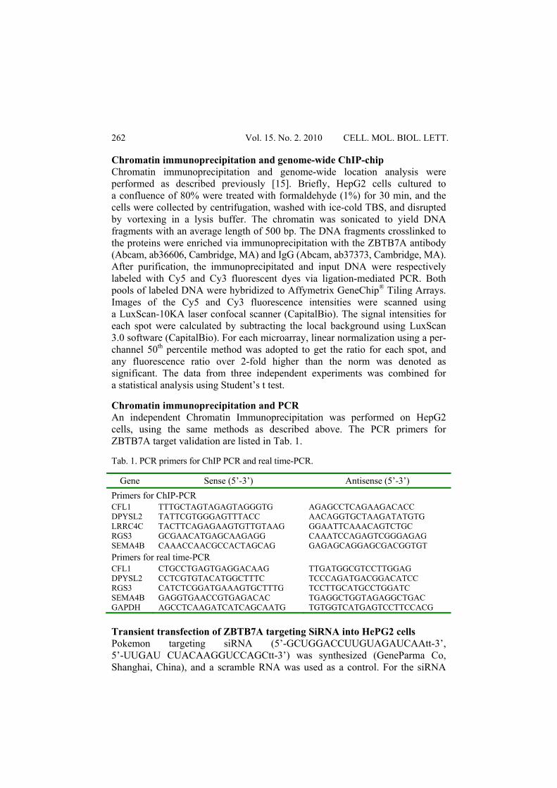
delivery, HepG2 cells were mixed gently with siRNA and OligofectAMINE (Invitrogen, CA) in a volume of 0.5 ml according to the manufacturer’s instructions, and incubated at 37ºC, 5% CO2 for 4 h, followed by the addition of an equal volume of fresh medium containing 20% FBS. The cells were continuously incubated until harvest. Real time-PCR analysis Total RNA was prepared from cells using TRIzol reagent (Invitrogen, CA) according to the manufacturer’s instructions. After being isolated, the RNA was reverse transcribed to cDNA using random primers (Promega, CA). The reaction mixture containing SYBR Green PCR Master Mix (TaKaRa, Dalian, China) was run in a 7500 real-time PCR System (Applied Biosystems, CA). The PCR primers used for quantitative RT-PCR are listed in Tab. 2. The real-time PCR parameters used were as follows: 95ºC for 10 s; 95ºC for 5 s, 60ºC for 34 s for 40 cycles; 95ºC for 15 s, 60ºC for 1 min and 95ºC for 15 s. Tab. 2. Prospective genes identified to be part of the ZBTB7A target signal pathway.
Pathway name
Gene bank ID Gene name Gene
symbol
Avg. fold
change Aminosugar metabolism
NM_003828 myotubularin related protein 1 MTMR1 2.98
NM_000188 hexokinase 1 HK1 2.46
NM_004388 chitobiase CTBS 2.7
NM_003115 UDP-N-acteylglucosamine pyrophosphorylase 1 UAP1 2.03
Epithelial cell signaling in Helicobacter pylori infection
NM_020529 nuclear factor of kappa light polypeptide gene enhancer in B-cell inhibitor, alpha NFKBIA 2.27
NM_002751 mitogen-activated protein kinase 11 MAPK11 2.17
NM_006092 caspase recruitment domain family, member 4 CARD4 2.5
NM_002834 protein tyrosine phosphatase, non-receptor type 11 PTPN11 3.06
MAPK signaling pathway
NM_018398 calcium channel, voltage-dependent, alpha 2/delta 3 subunit
CACNA2D3 2.6
NM_002011 fibroblast growth factor receptor 4 FGFR4 2.14
NM_001394 dual specificity phosphatase 4 DUSP4 3.03
NM_001540 heat shock 27 kDa protein 1 HSPB1 2.09
NM_004134 heat shock 70 kDa protein 9B HSPA9B 2.29
NM_005204 mitogen-activated protein kinase kinase kinase 8 MAP3K8 3.15
NM_002751 mitogen-activated protein kinase 11 MAPK11 2.17
NM_002706 protein phosphatase 1B PPM1B 2.62
NM_007181 mitogen-activated protein kinase 1 MAP4K1 2.03
NM_000267 neurofibromin 1 NF1 2.07

Vol. 15. No. 2. 2010 CELL. MOL. BIOL. LETT.
264
Pathway name
Gene bank ID Gene name Gene
symbol
Avg. fold
change Maturity onset diabetes of the young
NM_005524 hairy and enhancer of split 1 HES1 3.01
NM_004394 islet amyloid polypeptide DAP 2.39
NM_000457 hepatocyte nuclear factor 4, alpha HNF4A 3.59
Arachidonic acid metabolism
NM_001757 carbonyl reductase 1 CBR1 2.12
NM_000954 prostaglandin D2 synthase 21 kDa (brain) PTGDS 2.3
NM_001061 thromboxane A synthase 1 TBXAS1 2.12
NM_000895 leukotriene A4 hydrolase LTA4H 2.2
Tryptophan metabolism
NM_000462 ubiquitin protein ligase E3An UBE3A 32.01
NM_000790 dopa decarboxylase DDC 2.04
NM_003679 kynurenine 3-monooxygenase KMO 2.18
NM_145214 tripartite motif-containing 11 TRIM11 2.68
Axon guidance
NM_020210 sema domain, immunoglobulin domain SEMA4B 2.06
NM_017790 regulator of G-protein signalling 3 RGS3 2.17
XM_045271 leucine rich repeat containing 4C NGL-1 2.28
NM_001386 dihydropyrimidinase-like 2 DPYSL2 2.28
NM_005507 cofilin 1 CFL1 2.39
Glycosylphosphatidylinositol-anchor (GPI-anchor) biosynthesis
NM_002641 phosphatidylinositol glycan, class A PIGA 2.09
NM_145167 phosphatidylinositol glycan, class M PIGM 2.21
Pyrimidine metabolism
NM_138338 polymerase (RNA) III POLR3H 2.53
NM_001161 nudix (nucleoside diphosphate linked moiety X)-type motif 2 NUDT2 4.52
NM_007055 polymerase (RNA) III (DNA directed) polypeptide A RPC155 2.1
NM_175859 CTP synthase II CTPS2 2.18
Regulation of actin cytoskeleton
NM_025104 diaphanous homolog 1 DRF1 2.32
NM_000177 gelsolin GSN 2.49
NM_000740 cholinergic receptor, muscarinic 3 CHRM3 3.32
NM_005507 cofilin 1 CFL1 2.39
NM_006990 WAS protein family, member 2 WASF2 2.63
NM_002011 fibroblast growth factor receptor 4 FGFR4 2.14

CELLULAR & MOLECULAR BIOLOGY LETTERS
265
Pathway name
Gene bank ID Gene name Gene
symbol
Avg. fold
change Cell communication
NM_153368 connexin40.1 CX40.1 8.89
NM_181703 gap junction protein, alpha 5, 40 kDa GJA5 2.47
NM_000421 keratin 10 KRT10 2.44
NM_000426 laminin, alpha 2 LAMA2 2.39
Oxidative phosphorylation
NM_004376 COX15 homolog, cytochrome c oxidase assembly protein COX15 2.56
NM_005003 NADH dehydrogenase 1 NDUFAB1 2.23
NM_001696 ATPase, H+ transporting, lysosomal 31 kDa, V1 subunit E1
ATP6V1E1 2.28
NM_004074 cytochrome c oxidase subunit 8A COX8A 2.32
Purine metabolism
NM_138338 polymerase (RNA) III polypeptide H (22.9kD) POLR3H 2.53
NM_001161 nudix (nucleoside diphosphate linked moiety X)-type motif 2 NUDT2 4.52
NM_007055 polymerase (RNA) III polypeptide A, 155 kDa RPC155 2.1
NM_000026 adenylosuccinate lyase ADSL 2.17
Western blot analysis HepG2 cells transfected with pokemon targeting siRNA and scramble RNA were lysed on ice for 30 min with a lysis buffer consisting of 10 mM N-2-hydro-xyethylpiperazine-N’-2-ethanesulfonic acid, 10 mM KCl, 1 mM ethylenediaminetetra acetic acid (EDTA; pH 8.0), 0.1% NP-40, 1 mM DTT, 1 mM PMSF, and 0.5 mM Na3VO4. Soluble protein (30 μg) was separated on a 12% sodium dodecyl sulfate-polyacrylamide electrophoresis gel and blotted onto a pure nitrocellulose membrane (Bio-Rad, Hercules, CA) at 180 mA for 2 h. After blockage with 5% skim milk in phosphate-buffered saline at room temperature for 45 min, the membranes were incubated with ZBTB7A antibody (1:500, Abcam) in the same buffer for 2 h, followed by incubation with goat anti-rabbit IgG (1:2000, Abcam) for 1 h. Antibody binding was detected using an enhanced chemiluminescence system (Pierce, Rockford, IL). To correct the protein loading amounts, the membranes were reprobed with β-actin monoclonal antibody (1:40,000; Sigma, St. Louis, MO). RESULTS Global mapping of ZBTB7A target genes in HepG2 cells To identify ZBTB7A target genes in the human genome, cells of the human hepato-carcinoma cancer cell line HepG2 were used to perform ChIP-on-chip analysis. This cell line is an ideal cell model for global mapping of ZBTB7A target genes, because it has a high expression of ZBTB7A. The ZBTB7A-bound

Vol. 15. No. 2. 2010 CELL. MOL. BIOL. LETT.
266
DNA fragments enriched by ChIP were subjected to microarray analysis, and 556 genes were identified to be potential direct targets of ZBTB7A in the HepG2 cells. To obtain more detailed information, the set of 556 genes was further analyzed for their roles in pathways and biological processes with the Kyoto Encyclopedia of Genes and Genomes (KEGG) and Gene Ontology (GO) databases using the Molecular Annotation System (MAS2.0) from CapitalBio. Of these genes, 423 had functional annotation and the rest did not have any annotation in GO with respect to biological processes. The potential ZBTB7A target genes were classified according to their physiological function (Fig. 1). The results showed that the potential target genes of ZBTB7A mainly cluster to metabolism, transcription regulation and cell signal transduction. Of the 556 genes, 74 encoded for metabolic pathways, as shown in the KEGG database. Forty four metabolic pathways were compiled from the KEGG database, in which at least one gene was shown to be a potential direct target of ZBTB7A. The most ZBTB7A-targeted cellular metabolic pathways include the aminosugar, arachidonic acid, tryptophan, pyrimidine and purine metabolic pathways. In addition, starch and sucrose metabolism, vitamin B6 metabolism, valine, leucine and isoleucine biosynthesis and degradation, folate biosynthesis, fatty acid metabolism and glycerolipid metabolism were shown to be potential regulatory targets of ZBTB7A.
Fig. 1. A functional classification of 556 genes identified as potential direct targets of ZBTB7A. The data obtained from a ChIP-on-chip assay of HepG2 cells was analyzed with the Kyoto Encyclopedia of Genes and Genomes (KEGG) and Gene Ontology (GO) databases using Molecular Annotation System (MAS2.0) from CapitalBio.
CELLULAR & MOLECULAR BIOLOGY LETTERS
267
The ZBTB7A targeting pathways were then separated and classified based on their names as published in the KEGG to obtain a global view of the pathways regulated by ZBTB7A. The ZBTB7A-targeted main signaling pathways are presented in Tab. 2, and the results showed that 10 genes involved in the MAPK signaling pathway are potential targets of ZBTB7A, which suggests that ZBTB7A might be an important regulatory factor in the MAPK signaling pathway. Axon guidance, a key stage in the formation of the neuronal network, was also found to be partly regulated by ZBTB7A: five genes in this biological process were identified to be potential targets of ZBTB7A. Those results strongly suggest an important role of ZBTB7A in the regulation of cell proliferation and differentiation. Validation of the ZBTB7A direct target genes involved in the axon guidance process To further confirm the results obtained from the ChIP-on-chip assay, five axon guidance process-associated genes, CFL1, RGS3, DPYSL2, LRRC4C and SEMA4B, were chosen for verification by ChIP-PCR in HepG2 cells. It was revealed that the consensus sequence G(A/G)GGG(T/C)(C/T)(T/C)(C/T) and the single guanine-rich sites are the preferable binding sites of ZBTB7A [16]. The PCR primers were designed according to the above criteria for the amplification
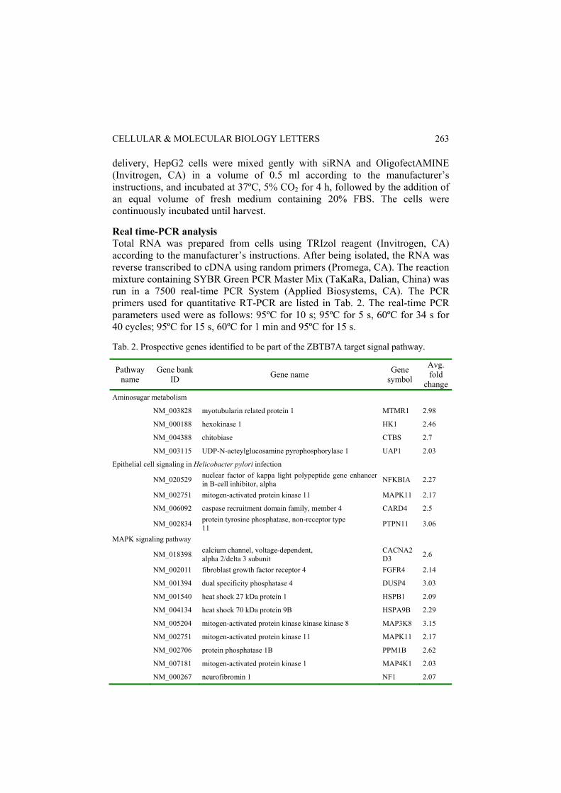
Fig. 2. Validation of ZBTB7A direct target genes involved in the axon guidance process. A – The binding between ZBTB7A protein and the promoters of genes involved in axon guidance were verified by ChIP-PCR of HepG2 cells. B – Total RNA was extracted from HepG2 cells transfected with ZBTB7A-targeting siRNA and scramble RNA for 72 h and quantitative real time-PCR was performed to confirm the effect of ZBTB7A expression on the transcription of the CFL1, SEMA4B, RGS3 and DPYSL2 genes. The fold change was determined relative to the mRNA level in HepG2 cells transfected with scramble RNA, and the values are the means ± SD from three independent experiments. *p < 0.05, ** p < 0.01. C – The expression level of ZBTB7A protein in siRNA-transfected HepG2 cells was detected by western blot analysis.

Vol. 15. No. 2. 2010 CELL. MOL. BIOL. LETT.
268
of ZBTB7A antibody-enriched promoter fragments. As shown in Fig. 2A, ZBTB7A was shown to directly bind with the promoters of the CFL1, SEMA4B, RGS3 and DPYSL2 genes, while no binding was observed between ZBTB7A and the promoter of the LRRC4C gene. To further understand the role of ZBTB7A in the transcription regulation of the above genes, real time-PCR was performed in HepG2 cells with ZBTB7A knockdown induced by ZBTB7A targeting siRNA. As shown in Fig. 2B, the knockdown of ZBTB7A resulted in a decrease in the level of CFL1 and RGS3 gene transcription and an increase in the transcription activity of the DPYSL2 and SEMA4B genes in HepG2 cells. Those results provide evidence for the effect of ZBTB7A on the transcription regulation of genes involved in the axon guidance process. DISCUSSION In this study, by using a high-throughout microarray, we obtained a global map of the binding sites of ZBTB7A in cells of HepG2, a human hepato-carcinoma cell line with a high level of ZBTB7A expression. More than 500 genes were identified to be potential direct targets of ZBTB7A, and those genes were denoted and classified by utilizing pathway study and GO analysis software. Of the ZBTB7A-targeted genes, 74 genes clustered to metabolic pathways, and 44 of those metabolic pathways (including nucleic acid synthesis, fatty acid metabolism and sugar degradation) were shown to be associated with ZBTB7A, indicating that ZBTB7A is a critical regulator in cellular physiological processes. Currently, ZBTB7A is known as a proto-oncogene, as serial studies have revealed a correlation between ZBTB7A and carcinogenesis [4, 5, 11]. Maeda et al. reported that ZBTB7A shows a high expression level in T-cell and B-cell lymphoma cases, and that ZBTB7A overexpression leads to oncogenic transformation in mouse embryonic fibroblasts both in vitro and in vivo in transgenic mice [4]. ZBTB7A was also found to have become overexpressed in non-small cell lung cancer due to the gene amplification event [5]. Although the role of ZBTB7A in carcinogenesis has been confirmed by various research groups, other functions of ZBTB7A in human B versus T lymphocytes and human and murine preadipocyte differentiation was also proposed [2]. The different roles of ZBTB7A in the regulation of cellular behavior suggest that ZBTB7A is a multi-functional protein, and our data from the global microarray screening of ZBTB7A direct target genes also provides important evidence that ZBTB7A is not only a carcinogenesis-associated gene but also a necessary gene for cellular metabolism regulation, cell differentiation and embryonic development. This notion is supported by the fact that homozygous deletion of ZBTB7A resulted in embryonic lethality due to severe anemia in a mouse model [2]. To validate the results obtained from the microarray, five genes involved in axon guidance were chosen for ChIP-PCR and real time-PCR. ZBTB7A was shown to have a direct binding with the promoters of the CFL1, SEMA4B, RGS3 and DPYSL2 genes, and no binding was observed between the ZBTB7A and

CELLULAR & MOLECULAR BIOLOGY LETTERS
269
LRRC4C promoter in HepG2 cells. The Q-PCR confirmed that the transcription level of the CFL1 and RGS3 genes was reduced and the mRNA level of the SEMA4B and LRRC4C genes was elevated by siRNA-induced ZBTB7A silence in HepG2 cells. Those results verified that the CFL1, SEMA4B, RGS3 and DPYSL2 genes, which are implicated in neural development, are directly regulated by the expression of ZBTB7A in HepG2 cells. ZBTB7A may function as a regulator in neural development, and those molecular events associated with the cellular processes remain to be elucidated. Many genes, including Egr-1, c-Krox, CyclinA, E2F4, ADH5, Rb, FANS, p107 and ARF, were identified to be ZBTB7A downstream targets in recent years [4, 10-12, 16, 17]. Of these, E2F4, FANS, ARF and ADH5 were also detected in our global mapping of ZBTB7A binding sites, further supporting the data we obtained from the ChIP-on-chip assay. Increasing evidence suggests that the ZBTB7A gene is involved in many different physiological processes. In this study, we provide the first overview of the direct targets of the transcription factor ZBTB7A in HepG2 cells by utilizing ChIP-on-chip technology. The global mapping of ZBTB7A binding sites will yield useful information for completely understanding the function of the ZBTB7A gene in cellular processes. Acknowledgements. This study was supported by a grant from National 863 Project of the Ministry of Science and Technology (China, 2007AA02Z160), and by a grant from the China Postdoctoral Foundation (200880440390). REFERENCES 1. Pessler, F., Pendergrast, P.S. and Hernandez, N. Purification and
characterization of FBI-1, a cellular factor that binds to the human immunodeficiency virus type 1 inducer of short transcripts. Mol. Cell. Biol. 17 (1997) 3786-3798.
2. Maeda, T., Hobbs, R.M., Dong, L., Maeda, M., Zakrzewski, J., Zelent, A., Shigematsu, H., Akashi, K., Cattoretti, G. and Pandolfi, P.P. Regulation of B versus T lymphoid lineage fate decision by the proto-oncogene LRF. Science 316 (2007) 860-866.
3. Laudes, M., Christodoulides, C., Sewter, C., Rochford, J.J., Considine, R.V., Sethi, J.K., Vidal-Puig, A. and O'Rahilly, S. Role of the POZ zinc finger transcription factor FBI-1 in human and murine adipogenesis. J. Biol. Chem. 279 (2004) 11711-11718.
4. Maeda, T., Hobbs, R.M., Merghoub, T., Guernah, I., Zelent, A., Cordon-Cardo, C., Teruya-Feldstein, J. and Pandolfi, P.P. Role of the proto-oncogene Pokemon in cellular transformation and ARF repression. Nature 433 (2005) 278-285.
5. Apostolopoulou, K., Evangelou, K., Tsantoulis, P.K., Liontos. M., Kittas, C., Tiniakos, D.G., Kotsinas, A., Cordon-Cardo C. and Gorgoulis, V.G. Gene
Vol. 15. No. 2. 2010 CELL. MOL. BIOL. LETT.
270
amplification is a relatively frequent event leading to ZBTB7A (Pokemon) overexpression in non-small cell lung cancer. J. Pathol. 213 (2007) 294-302.
6. Morrison, D.J., Stavropoulos, P., Colmenares, S.U., Kobayashi, R. and Hernandez, N. FBI-1, a factor that binds to the HIV-1 inducer of short transcripts (IST), is a POZ domain protein. Nucleic Acids Res. 27 (1999) 1251-1262.
7. Davies, J.M., Hawe, N., Kabarowski, J., Huang, Q.H., Zhu, J., Brand, N.J., Leprince, D., Dhordain. P., Cook, M., Morriss-Kay, G. and Zelent, A. Novel BTB/POZ domain zinc-finger protein, LRF, is a potential target of the LAZ-3/BCL-6 oncogene. Oncogene 18 (1999) 365-375.
8. Widom, R.L., Lee, J.Y., Joseph, C., Gordon-Froome, I. and Korn, J.H. The hcKrox gene family regulates multiple extracellular matrix genes. Matrix Biol. 20 (2001) 451-462.
9. Lee, D.K., Suh, D., Edenberg, H.J. and Hur, M.W. POZ Domain transcription factor, FBI-1, represses transcription of ADH5/FDH by interacting with the zinc finger and interfering with DNA binding activity of Sp1. J. Biol. Chem. 277 (2002) 26761-26768.
10. Laudes, M., Bilkovski, R., Oberhauser, F., Droste, A., Gomolka, M., Leeser, U., Udelhoven, M. and Krone, W. Transcription factor FBI-1 acts as a dual regulator in adipogenesis by coordinated regulation of cyclin-A and E2F-4. J. Mol. Med. 86 (2008) 597-608.
11. Jeon, B.N., Yoo, J.Y., Choi, W.I., Lee, C.E., Yoon, H.G and Hur, M.W. Proto-oncogene FBI-1 (Pokemon/ZBTB7A) represses transcription of the tumor suppressor Rb gene via binding competition with Sp1 and recruitment of co-repressors. J. Biol. Chem. 283 (2008) 33199-33210
12. Choi, W.I., Jeon, B.N., Park, H., Yoo, J.Y., Kim, Y.S., Koh, D.I., Kim, M.H., Kim, Y.R., Lee, C.E., Kim, K.S., Osborne, T.F and Hur, M.W. Proto-oncogene FBI-1 (Pokemon) and SREBP-1 synergistically activate transcription of fatty-acid synthase gene (FASN). J. Biol. Chem. 283 (2008) 29341-29354.
13. Chia, Q.W., Wei, L., Vega, V.B., Chiu, K.P., Ng, P., Zhang, T., Shahab, A., Yong, H.C., Fu, Y.T., Weng, Z.P., Liu, J.J., Zhao, X.D., Chew, J.L., Lee, Y.L., Kuznetsov, V.A., Sung, W.K., Miller, L.D., Lim, B., Liu, E.T., Yu, Q., Ng, H.H and Ruan, Y.J. A global map of p53 transcription-factor binding sites in the human genome. Cell 124 (2006) 207-219.
14. Zeller, K.I., Zhao, X., Lee, C.W., Chiu, K.P., Yao, F., Yustein, J.T., Ooi, H.S., Orlov, Y.L., Shahab, A., Yong, H.C., Fu, Y., Weng, Z., Kuznetsov, V.A., Sung, W.K., Ruan, Y., Dang, C.V. and Wei, C.L. Global mapping of c-Myc binding sites and target gene networks in human B cells. Proc. Natl. Acad. Sci. U.S.A. 103 (2006) 17834-17839.
15. Testa, A., Donati, G., Yan, P., Romani, F., Huang, T.H, Vigano, M.A. and Montovani, R. Chromatin immunoprecipitation (ChIP) on chip experiments uncover a widespread distribution of NF-Y binding CCAAT sites outside of core promoters. J. Biol. Chem. 280 (2005) 13606-13615.

CELLULAR & MOLECULAR BIOLOGY LETTERS
271
16. Pessler, F. and Hernandez, N. Flexible DNA binding of the BTB/POZ-domain protein FBI-1. J. Biol. Chem. 278 (2003) 29327-29335.
17. Leet, D.K., Suh, D., Edenberg, H.J. and Hur, M.W. POZ domain transcription factor, FBI-1, represses transcription of ADH5/FDH by interacting with the zinc finger and interfering with DNA binding activity of Sp1. J. Biol. Chem. 277 (2002) 26761-26768.